repression of hybrid dysgenesis in drosophila melanogaster
TRANSCRIPT
Copyright 0 1993 by the Genetics Society of America
Repression of Hybrid Dysgenesis in Drosophila melanogaster by Individual Naturally Occurring P Elements
Korise E. Rasmusson, John D. Raymond and Michael J. Simmons’
Department of Genetics and Cell Biology, University of Minnesota, St. Paul, Minnesota 55108-1095 Manuscript received September 9, 1992
Accepted for publication November 17, 1992
ABSTRACT Individual P elements that were genetically isolated from wild-type strains were tested for their
abilities to repress two aspects of hybrid dysgenesis: gonadal dysgenesis and mutability of a double? element-insertion allele of the singed locus (snw). These elements were also characterized by Southern blotting, polymerase chain reaction amplification and DNA sequencing. Three of the elements were 1.1-kb KP elements, one was a 1.2-kb element called 050, and one was a 0.5-kb element called SP. These three types of elements could encode polypeptides of 207,204, and 14 amino acids, respectively. Gonadal dysgenesis was repressed by two of the KP elements (denoted KP(1) and KP(6)) and by SP, but not by the third KP element (KP(D)), nor by 050. Repression of gonadal dysgenesis was mediated by a maternal effect, or by a combination of zygotic and maternal effects generated by the P elements themselves. The mutability of snw was repressed by the KP(1) and KP(6) elements, by 050 and by SP, but not by KP(D); however, the SP element repressed snw mutability only when the transposase came from complete P elements and the D50 element repressed it only when the transposase came from the modified P element known as A2-3. In all cases, repression of snw mutability appeared to be mediated by a zygotic effect of the isolated P element. Each of the isolated elements was also tested for its ability to suppress the phenotype of a P-insertion mutation of the vestigial locus (vg””). D50 was a moderate suppressor whereas SP and the three KP elements had little or no effect. These results indicate that each isolated P element had its own profile of repression and suppression abilities. It is
by P-encoded polypeptides or by antisense P RNAs suggested that these abilities may be mediated initiated from external genomic promoters.
T HERE are more than 40 families of transposable elements in the Drosophila melanogaster genome.
Of these, the best understood is the P family, which consists of complete elements 2,907 base pairs (bp) long and a wide assortment of smaller elements de- rived from the complete ones by different internal deletions (O’HARE and RUBIN 1983; ENGELS 1989; O’HARE et al. 1992). T h e complete elements encode a trans-acting transposase, which is the catalytic agent for mobilizing the members of the P family (SPRA- DLING and RUBIN 1982; RUBIN and SPRADLING 1982; ENCELS 1984; KARESS and RUBIN 1984). Detailed molecular and biochemical studies have elucidated the primary structure of this polypeptide as well as the DNA sequences with which it interacts (O’HARE and RUBIN 1983; RIO, LASKI and RUBIN 1986; LASKI, RIO and RUBIN 1986; KAUFMAN, DOLL and RIO 1989; MULLINS, RIO and RUBIN 1989). The transposase is encoded by four exons, denoted 0-3, spanning the length of the P sequence. It contains 75 1 amino acids, has a mass of 87 kilodaltons (kD), and binds to regions near the 31-bp inverted terminal repeats of P ele- ments. T h e synthesis of the transposase is restricted to the germ line because proteins in the somatic tissues
I Author to whom correspondence should be addressed
Genetics 133: 605-622 (March, 1993)
prevent the removal of the last (2-3) intron in the pre- m R N A (RIo, LASKI and RUBIN 1986; LASKI, RIO and RUBIN 1986; LASKI and RUBIN 1989; SIEBEL and RIO 1990). Translation of this incompletely processed R N A results in a 66-kD protein that is identical with the transposase for much of its length; a stop codon in the 2-3 intron accounts for the smaller size. When the 2-3 intron is artificially removed from a complete P element, the 87-kD transposase is made in the somatic cells. Such modified elements, called A2-3 elements, have been introduced into Drosophila stocks, where they engender somatic P transposition (LASKI, RIO and RUBIN 1986; ROBERTSON et al. 1988).
T h e induction of P activity in the germ line leads to a syndrome of genetic abnormalities called hybrid dysgenesis (KIDWELL, KIDWELL and SVED 1977). These abnormalities include mutations, chromosome breakage, male recombination, chromosome nondis- junction, chromosome segregation distortion and ste- rility. Typically they occur in the progeny of a cross between a male that carries P elements in its genome (from a P strain) and a female that does not (from an M strain). T h e progeny of the reciprocal cross, P female X M male, are usually not dysgenic, indicating that P activity is repressed by factors contributed

606 K. E. Rasmusson, J. D. Raymond and M. J. Simmons
maternally from a P strain. Genetic analyses have suggested that these factors are products of the P elements themselves (ENGELS 1979a; KIDWELL 198 1,
Early studies established a nomenclature to describe P element regulation. Strains with a strong ability to repress P activity were said to have the P cytotype and strains without this ability were said to have the M cytotype (ENGELS 1979a). Subsequent studies have shown that regulatory ability actually follows a contin- uous distribution in Drosophila populations, with the M and P cytotypes lying at the extremes (KIDWELL 1983, 1985; SIMMONS et al. 1987, 1990; HEATH and SIMMONS 199 1). Strains with intermediate regulatory ability have been called M' or pseudo", and molec- ular analyses have shown that, like P cytotype strains, they carry P elements in their genomes; true M cyto- type strains, however, do not (BINGHAM, KIDWELL and RUBIN 1982). This variability suggests that P element regulation is determined by a complex array of genetic factors, presumably the P elements them- selves.
Several investigators have proposed that regulation of the P family is mediated by P-encoded polypeptides that repress transposase activity [reviewed in RIO (1 990)]. However, because the coding capacity of P elements is limited, these repressor polypeptides would have to share some of the same amino acid sequences as the transposase. These shared sequences might specify features such as DNA binding domains or oligomerization motifs that are critical for repres- sor function.
The most vigorously championed candidate for a repressor is the 66-kD polypeptide made from the incompletely processed transcripts of complete P ele- ments (ROBERTSON and ENCELS 1989; MISRA and RIO 1990). Biochemical data suggest that this polypeptide is produced in the germ line and that it may act to repress the transcription of complete P elements (MISRA and RIO 1990; KAUFMAN and RIO 199 1). Studies with modified P elements designed to produce this polypeptide have shown that it confers repression potential. However, the 66-kD polypeptide cannot be the only P element repressor since strains without it have been found to repress transposition (SIMMONS et al. 1987, 1990). Consequently, researchers have pro- posed that polypeptides from various incomplete P elements also function as repressors.
One candidate is the 207-amino acid polypeptide that could be made by a 1.15-kb element called KP. K P elements are present in Drosophila populations throughout the world, but they are especially abun- dant in the M' populations of Eurasia (BLACK et al. 1987; C. PRESTON and W. ENGELS, personal commu- nication). O n the basis of indirect genetic analyses, it has been suggested that K P elements are responsible
1985).
for intermediate repression potential (BLACK et al. 1987; JACKSON, BLACK and DOVER 1988). However, the existence of the K P polypeptide has not yet been demonstrated, nor has there been a published report of an individual KP element repressing hybrid dysge- nesis. Other less well characterized incomplete P ele- ments have also been proposed as possible sources of repressor polypeptides [e .g . , NITASAKA, MUKAI and YAMAZAKI (1987)l. Further research is needed to identify their potential polypeptide products and to test their repressor functions.
In this paper we analyze the repression of hybrid dysgenesis by three distinct types of incomplete P elements isolated from a strain called Sexi, which was derived from a natural population in Spain (KIDWELL 1985). Our results unambiguously demonstrate that individual, naturally occurring P elements possess repression potential and suggest that in some cases, repression may be mediated by an antisense P RNA rather than by a P-encoded polypeptide.
MATERIALS AND METHODS
Stocks: All stocks and experimental cultures were raised in vials or half-pint milk bottles on a standard cornmeal- molasses-agar medium. Unless otherwise stated, the cultur- ing temperature was 25 O . Additional information about the genetic markers and chromosome rearrangements can be found in LINDSLEY and ZIMM (1992) and SIMMONS et al. (1990).
Strains without P elements: Canton S (CS), a wild-type strain. bw; st , homozygous for the recessive eye color markers
brown (bw) and scarlet (st), which are located on the second and third chromosomes, respectively.
C(I)DX, y f /Y /y cin w f suO3u6'g. The females have attached- X chromosomes and the males have an X-linked tempera- ture-sensitive lethal mutation (su03""7B) which aids in the collection of virgin females.
C(I)DX, y f / Y / y shi"; bw; st, another attached-X stock in which the males have a different X-linked temperature- sensitive lethal mutation (shi"). Both males and females are homozygous for the autosomal markers bw and st.
C(I)DX, yf /Y/FM6, an attached-X stock in which the X chromosome in the male is the FM6 balancer.
y sn3 v cur, a stock homozygous for four recessive X-linked markers, yellow ( y ) body, extreme singed (sn') bristles, ver- milion ( v ) eyes and carnation (car) eyes.
M5-CS, homozygous for the M5 (Muller-5) balancer X chromosome. The genetic background is from a Canton S (CS) wild-type stock.
M5; bw; s t , homozygous for the M5 balancer X chromo- some and the autosomal markers bw and st.
TM?, Sb Ser eITM6, Tb e, abbreviated TM3ITM6, heter- ozygous for two third chromosome balancers, one carrying the dominant mutations Stubble (Sb) bristles and Serrate (Ser) wings and the other carrying the dominant mutation Tubby (Tb) body. Both chromosomes are homozygous lethal.
FM7, y ? I d sc8 snTZ B/lns(l) sc7 + AM, sc7 w' ptg4 b~~~~ l(l)78h, abbreviated FM7/sc7 1, heterozygous for the X chro- mosome balancer FM7, which carries the dominant muta- tion Bar ( R ) eyes and the female sterilizing mutation snxz, and a lethal X chromosome.
Repression of P Element Activity 607
Strains with P elements: cn v$'-' bw, homozygous for the P element-insertion
mutation v8'-' of the vestigial wing locus on the second chromosome (WILLIAMS, PAPPU and BELL 1988). There are no other P elements present. This stock is also homozygous for the recessive eye color markers cinnabar (cn) and bw which flank the ug locus.
C(I)DX, y f/Y; cn v8'-' bw, an attached-X stock homozygous for the cn v8'-' bw second chromosome.
rySo6 P[ry+ SalI](89D), a transformed stock homozygous for a single, modified P element at cytological position 89D on chromosome 111 (ROBERTSON and ENGELS 1989). This P element, abbreviated P[SalI], has a frameshift mutation in the Sal1 restriction site of exon 3 and therefore does not produce any active transposase (KARESS and RUBIN 1984). It is also marked with a wild-type allele of the rosy (ry) gene.
C(I)DX, y f/Y; 9 P[ry+ SalI](89D), an attached-X stock homozygous for the P[SalI] third chromosome.
ry5" P[ry+ A2-3](99B), a transformed stock homozygous for a double P element insertion at 99B on chromosome ZZI (H. ROBERTSON, personal communication). This insertion, abbreviated A2-3, consists of two modified P elements that lack the intron between exons 2 and 3 (LASKI, RIO and RUBIN 1986). It is marked with ry+ and produces a high level of transposase activity in the soma as well as in the germ line. However, the insertion itself is essentially immo- bile (ROBERTSON et al. 1988). There are no other P elements present in the A2-3 stock.
y snw/y+ Y; bw; st, homozygous for the double P ele- ment-insertion mutation sn" [weak-singed bristles; ENGELS ( 1 979b)l. In the presence of the P transposase, this X-linked mutation becomes unstable, mutating to a more extreme allele (sn') or to an essentially wild-type allele (sn(+)). These changes are due to the excision of one or the other of the inserted P elements (ROIHA, RUBIN and O'HARE 1988) and therefore can be used as indicators of transposase action. The only other P element present in this stock is an approx- imately 0.6-kb element tightly linked to the singed locus. The stock is also homozy ous for bw and st.
C(I)DX, yf/Y/y sn"; ry5 f P[ry+ SalI](89D), an attached-X stock in which the X chromosome in the males carries snw and the third chromosome is homozygous for the P[SalI] element.
C(I)DX, y f/Y/T-5, y sn"; bw; st, an attached-X stock con- sisting of several sublines in which the male's X chromosome, called T-5, carries sn" and at least one transposase-producing P element (SIMMONS et al. 1987). Due to the activity of the transposase in these sublines, the sn" allele is unstable and mutates at a high rate. In order to maintain this stock, phenotypically weak-singed males from each subline are selected and mated individually at 21 O to C(I)DX, yf/Y; bw; st females from a true M strain. P elements are therefore restricted mainly to the T-5 X chromosome since the Y chromosome and half the autosomes are replaced by M chromosomes every generation. The T-5 sublines K5, p3" and P3" were used to induce hybrid dysgenesis in these experiments.
C(I)DX, y f/Y/sn"; 7r2, an attached-X stock with autosomes derived from the wild-type P strain 7r2 (ENGELS and PRESTON 1979). Because this stock has the P cytotype, the sn" muta- tion is essentially stable.
D2.2, a wild-type stock containing a single P element in the genome. The stock was established from flies that were being used in germ line transformation experiments but its single P element was not derived from a transformation event. Rather, it was apparently ac uired from one of the parental strains (TM3/TM6 and rJo6 P[ry+ A2-3]), which must have been segregating this element at low frequency.
Inbred Sexi lines: These lines were derived from a wild- type strain called Sexi by many generations of full-sib mating (RASMUSSON et al. 1990). The lines are denoted Sexi. 1, Sexi.2, etc.
Genetic isolation of individual P elements: Individual X-linked P elements were isolated from three of the inbred Sexi lines (Sexi. 1, Sexi.5 and Sexi.6) by recombination with multiply marked X chromosomes that were devoid of P elements. The autosomes in these recombination experi- ments were all from an M strain. Southern blotting and in situ hybridization were used to monitor the number and position of the X-linked P elements during the isolation process. The final recombinant chromosomes were made homozygous using the M5-CS balancer or kept in stock with attached-X females. Full details of the isolation process are found in RASMUSSON (1 99 1).
Tests for the suppression of Males with an isolated P element on the X chromosome were crossed to C(I)DX, y f/Y; cn v8'-' bw females. Their sons were then backcrossed to produce males hemizygous for the isolated P element and homozygous for u p - ' . Suppression of the v$'-' phenotype was assessed in these males.
Tests for the repression of gonadal dysgenesis (GD sterility): Our experimental design utilized "reciprocal" crosses that permitted the identification of separate mater- nal and zygotic contributions to the repression of gonadal dysgenesis (Figure 1). In the GI, flies bearing a sterility- inducing T-5 X chromosome were crossed to males and females carrying the chomosome to be tested for repression potential. Because in some cases this chromosome carried an isolated P element, it is denoted as (P) in the Figure. Only a single pair of flies was used in each GI cross; all these crosses were initiated at 21 ', but after 2 days, the mated flies were transferred to fresh cultures, which were incu- bated at 29". The progeny that emerged by day 11 were transferred to fresh vials and aged at 21 O for 2-3 days. Then from each vial, as many as 12 females of particular genotypes were examined for egg production by squashing them in colored water between two glass plates. Those females without any eggs were considered to have gonadal dysgenesis. A low frequency of dysgenesis indicated repres- sion, presumably by factors associated with the isolated P elements. Repression by zygotic factors was ascertained in the T-5/(P) daughters of cross 1, repression by maternal factors was ascertained in the FM7/T-5 daughters of cross 2, and repression by a combination of zygotic and maternal factors was ascertained in the (P)/T-5 daughters of cross 2. As a positive control in these experiments, the P[SalI] ele- ment was also tested for its effect on T-5-induced GD sterility. In the G I , T-5/M5 females were crossed to males homozygous for the P[SalI] element (cross 1) and T-5 males were crossed to P[SalI]/TM3 females (cross 2). Daughters from these crosses were then examined for gonadal dysge- nesis.
Tests for the repression of snw mutability: X chromo- somes carrying an isolated P element and the P element- insertion mutation sn" were constructed by recombination. The presence of the isolated P element was ascertained by the polymerase chain reaction (PCR) with P-specific primers. Recombinant sn" chromosomes that lacked the isolated P element were also identified so they could be used as con- trols in the repression tests. Two types of tests were con- ducted. In the first, sn" mutability was induced in the germ line by a T-5 X chromosome that carried a complete P element and a snc allele. The sn" allele that was destabilized was located on the homologous X chromosome. In the second test, sn" mutability was induced in the soma and in the germ line by the A2-3 P element.

K . E. Rasmusson, J. D. Raymond and M. J. Simmons 608
Go
c,
cz A A
FIGURE 1.-Mating scheme to test for the repression of gonadal dysgenesis by individual P elements. The recessive au- tosomal markers bw and st , which were present in the Go flies, are not shown. Many replicate cultures of the Go double matings were established at 21 O to pro- duce the flies used in the GI matings.
ZYGOTIC MATERNAL EFFECT
ZYGOTIC
MATERNAL EFFECTS
EFFECT AND
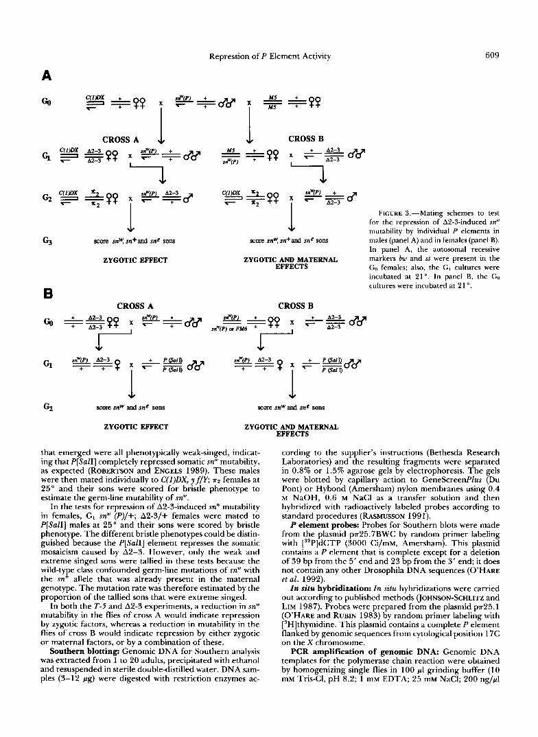
The T-5 experiments for repression of sn" mutability (Figure 2) commenced with males from a T-5 subline in which sn" had already mutated to sn'. The sn" chromosome to be tested for repression potential is denoted snw (P). In the GP, phenotypically weak-singed T-5, sne/snw (P) females from each of thet two types of GI crosses were individually mated to y sn' v car males at 25" and the progeny were scored for bristle phenotype through day 17 (snw is dominant to sn' and sn', and sn+ is dominant to sn", sn' and sn'). The germ-line mutability of sn'" was estimated by calculating the proportion of wild-type flies among the wild-type and weak- singed progeny. This calculation ignores mutations to sn' because they could not be distinguished from the sn' allele that was already present on the T-5 chromosome; it there- fore yields a partial mutation rate. As a positive control, the P[SalI] element was also tested for its effect on T-5-induced sn" mutability; snw; P[SalI] males were mated at 21 O to T-5, snc/M5 females and their T-5, sn"/sn"; P[SalI] /+ daughters were crossed individually toy sn3 v cur males. The progeny of these crosses were then scored for bristle phenotype.
The A2-3 experiments for repression of sn" mutability were performed in two ways, one testing for repression in males (Figure 3A) and the other testing for repression in
females (Figure 3B). In the tests for repression in males, G2 snw (P) males that were heterozygous for A2-3 were exam- ined for bristle mosaicism to determine if the isolated P element had any effect on the somatic mutability of snw. These males were then mated individually at 25 " to C(I)DX, yf/Y; HZ females, which had the P cytotype, and their sons were scored for bristle phenotype to determine if the iso- lated P element had any effect on the germ-line mutability of sn". The extreme-singed, weak-singed and wild-type phe- notypes in these sons could be distinguished because the P cytotype, which they had inherited maternally, suppressed the somatic mosaicism that ordinarily occurs when A2-3 is combined with sn" (ROBERTSON et al. 1988). The proportion of extreme-singed and wild-type sons was used to estimate the germ-line mutability of sn". As a positive control, the P[SalI] element was also tested for its effect on A2-3-induced sn" mutability. These tests began by mating sn"; P[SalI] males separately to C(l)DX, y fly; P[SalI] and P[SalI] females at 25". sn"; P[SalI] sons from the first mating were crossed to C(l)DX, y f/Y; A2-3 females (cross A) and sn"/+; P[SalI] daughters from the second mating were crossed to A2-3 males (cross B); the cultures from both sets of crosses were reared at 21 '. In both cases, the sn"; P[SalI]/A2-3 males
Go
c,
G2
MS T-5. sn e
MS
score snw and sn+ progeny score snw and sn+ progeny
FIGURE 2.-Mating scheme to test for the repression of T-5-induced sn" mutability by individual P elements. The recessive au- tosomal markers bw and st, which were pres- ent in the GO flies, are not shown. The GO and GI matings were replicated many times and the cultures were incubated at 21 '.
ZYGOTIC EFFECT
ZYGOTIC AND MATERNAL EFFECTS
Repression of P Element Activity 609
A
co
CROSS A & CROSS B
63 1
ZYGOTIC EFFECT
B CROSS A
FIGURE 3,"Mating schemes to test for the repression of A2-3-induced sn" mutability by individual P elements in
In panel A, the autosomal recessive ZYGOTIC AND MATERNAL
EFFECTS markers bw and st were present in the Go females; also, the GI cultures were incubated at 21 '. In panel B, the GO cultures were incubated at 21 '.
score snw, sn+and sfle sons males (panel A) and in females (panel B).
CROSS B
G2
ZYGOTIC EFFECT ZYGOTIC AND MATERNAL EFFECTS
that emerged were all phenotypically weak-singed, indicat- ing that P[SalI] completely repressed somatic snw mutability, as expected (ROBERTSON and ENGELS 1989). These males were then mated individually to C(I)DX, yf/Y; 7r2 females at 25" and their sons were scored for bristle phenotype to estimate the germ-line mutability of snw.
In the tests for repression of A2-3-induced snw mutability in females, GI snW (P)/+; A2-3/+ females were mated to P[SalI] males at 25" and their sons were scored by bristle phenotype. The different bristle phenotypes could be distin- guished because the P[SalI] element represses the somatic mosaicism caused by A2-3. However, only the weak and extreme singed sons were tallied in these tests because the wild-type class confounded germ-line mutations of snw with the sn+ allele that was already present in the maternal genotype. The mutation rate was therefore estimated by the proportion of the tallied sons that were extreme singed.
In both the T-5 and A2-3 experiments, a reduction in snw mutability in the flies of cross A would indicate repression by zygotic factors, whereas a reduction in mutability in the flies of cross B would indicate repression by either zygotic or maternal factors, or by a combination of these.
Southern blotting: Genomic DNA for Southern analysis was extracted from 1 to 20 adults, precipitated with ethanol and resuspended in sterile double-distilled water. DNA sam- ples (3-12 Pg) were digested with restriction enzymes ac-
cording to the supplier's instructions (Bethesda Research Laboratories) and the resulting fragments were separated in 0.8% or 1.5% agarose gels by electrophoresis. The gels were blotted by capillary action to GeneScreenPlus (Du Pont) or Hybond (Amersham) nylon membranes using 0.4 M NaOH, 0.6 M NaCl as a transfer solution and then hybridized with radioactively labeled probes according to standard procedures (RASMUSSON 1991). P element probes: Probes for Southern blots were made
from the plasmid p~25.7BWC by random primer labeling with [32P]dCTP (3000 Ci/mM, Amersham). This plasmid contains a P element that is complete except for a deletion of 39 bp from the 5' end and 23 bp from the 3' end; it does not contain any other Drosophila DNA sequences (O'HARE et al. 1992). In situ hybridization: In situ hybridizations were carried
out according to published methods (JOHNSON-SCHLITZ and LIM 1987). Probes were prepared from the plasmid ~ ~ 2 5 . 1 (O'HARE and RUBIN 1983) by random primer labeling with [3H]thymidine. This plasmid contains a complete P element flanked by genomic sequences from cytological position 17C on the X chromosome.
PCR amplification of genomic DNA: Genomic DNA templates for the polymerase chain reaction were obtained by homogenizing single flies in 100 PI grinding buffer (1 0 mM Tris-CI, pH 8.2; 1 mM EDTA; 25 mM NaCI; 200 ng/Pl

610 K . E. Rasmusson, J. D. Raymond and M. J. Simmons
Proteinase K) and incubating at 37" for 20 min. (GLOOR et al. 199 1). The proteinase was then inactivated by treatment at 95" for 2 min. The resulting solution contained approx- imately 30 ng/pI single-stranded DNA (G. GLOOR, personal communication) and 1-2 pI were used for each amplification reaction. Amplifications were carried out in 20-50-pl vol- umes using Taq DNA polymerase (Promega) and a Coy Company Tempcycler. Each reaction contained l x Pro- mega reaction buffer (1 0 mM Tris, pH 9.0; 50 mM KCI; 1.5 mM MgCI,; 0.01% gelatin (w/v); 0.1% Triton X-loo), 0.2 mM each dATP, dCTP, dGTP, dTTP, 1.5 ng/pl each of two oligonucleotide primers (or 3.0 ng/pl inverted repeat primer, see below), approximately 1.5 ng/pl genomic DNA and 0.025 units/pl Taq DNA polymerase. The reaction mixtures were covered with a minimal volume of mineral or paraffin oil to prevent evaporation. The temperature profile for each reaction cycle was 0.5-1 .O min at 94" to denature the DNA, 1 min at 60" to anneal the primers and 1.5 min at 72" to synthesize new DNA strands; 25-30 cycles were performed. Tracking dye was added to the PCR prod- ucts and samples were fractionated in 1.0% agarose gels.
DNA sequencing: DNA for sequencing was prepared using asymmetric PCR (GYLLENSTEN and EHRLICH 1988). Each reaction was carried out in 50 pl with 0.04 unit of Taq DNA polymerase and 1.5 ng/pl of one primer and 0.015 ng/pl of the other. Buffer, deoxyribonucleotides and ge- nomic DNA were included as in standard PCR. The tem- perature profile of each reaction was 45 sec at 92", 2 min at 55" and 2 min at 72". There were 40 cycles. The amplified DNA was extracted with 2-4 volumes of chloro- form-isoamylalcohol (24: l), precipitated with ethanol and resuspended in 7 p1 double-distilled water. DNA sequencing was performed using the Sequenase Version 2.0 kit (U.S. Biochemical Gorp.) with slight modifications of the sup- plier's instructions. Sequencing reaction products were frac- tionated in 0.4 mm gels of 6% polyacrylamide, 7 M urea prepared in 100 mM Tris-borate electrophoresis buffer.
DNA primers: Oligonucleotides for PCR and sequencing of P element DNA were provided by WILLIAM ENGELS (University of Wisconsin, Madison) or were purchased from Oligos, Etc. All oligonucleotide sequences are written 5' to 3' and their positions in the sequence of a complete P element are shown in parentheses.
Inverted repeat (IR) primer, (12/2896) AACA- TAAGGTGGTCCCGTCG (31/2877). This primer is de- rived from a sequence within the 31-bp inverted terminal repeats of P and primes DNA synthesis inward. It was used to amplify P elements with intact termini.
5'-subterminal primer, (35) GCCGAAGCTTACCGAA GTAT (54). This primer is derived from a sequence just inside the 5' inverted terminal repeat of P and primes DNA synthesis toward the 3' end.
KP-specific primer, (2577) ATCAACATCGACGTTTC ;cdCAC (805). This primer, designed by G. GLOOR and W. ENGELS, spans the deletion breakpoint (A) in KP elements and primes DNA synthesis toward the 5' end. It was used with the 5"subterminal primer in PCR amplifications to identify K P elements. Extensive tests have established that it amplifies only KP elements (C. PRESTON and W. ENGELS, personal communication).
Oligonucleotides that were used strictly for DNA se- quencing are listed in RASMUSSON (1 99 1).
RESULTS
Molecular characterization of individual natu- rally occurring P elements: Naturally occurring P
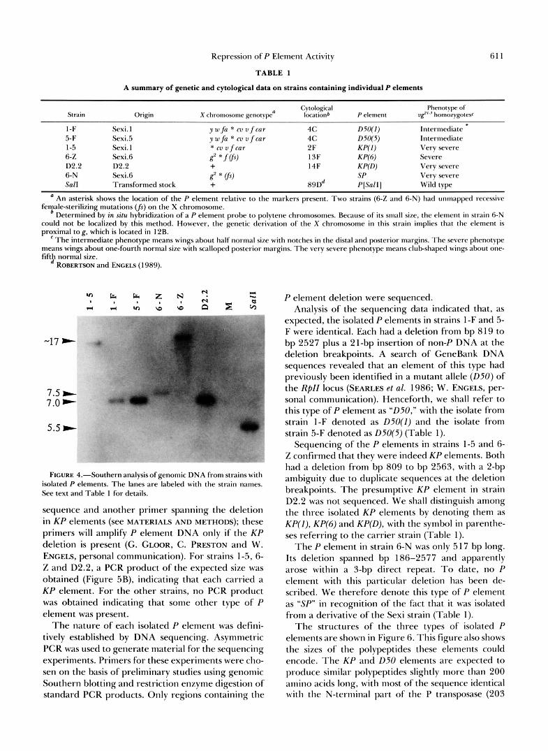
elements were isolated by meiotic recombination from the X chromosomes of the inbred lines Sexi. 1, Sexi.5 and Sexi.6 and from the X chromosome of the wild- type stock D2.2. Each of the strains carrying these isolated elements is listed in Table 1, along with its origin and X chromosome genotype. Southern blot- ting was used to show that only a single P element was present in the genome and in situ hybridization and PCR were used to determine the cytological location and size of this element.
T h e results of the Southern analysis are shown in Figure 4. Genomic DNA from each strain was di- gested with BamHI, a restriction enzyme which does not cleave within the P sequence. Except for the M strain control, each lane of the autoradiogram shows a single band of hybridization, indicating that only one P-containing fragment was present in the digested DNA. For strains 1-F and 5-F, the hybridizing bands were apparently the same size, suggesting that these two strains might carry the same isolated P element.
T h e cytological locations of the isolated P elements were established by in situ hybridization with a P element probe made from the plasmid pa25.1. Be- cause this plasmid contains DNA from cytological location 17C on the X chromosome, all the hybridi- zations showed label a t this site. Five of the six strains that were examined also showed label at one other X chromosome site (Table l ) , revealing the location of the isolated P element. T h e sixth strain, 6-N, did not show this additional label. Subsequent PCR amplifi- cation of the P element in strain 6-N demonstrated that it was too small to be detected by our in situ hybridization procedure (see below). Two of the strains (1-F and 5-F) showed label in cytological region 4C. These were the same strains that had given ap- parently identical results in the Southern analysis, reinforcing the suggestion that they carried the same isolated P element.
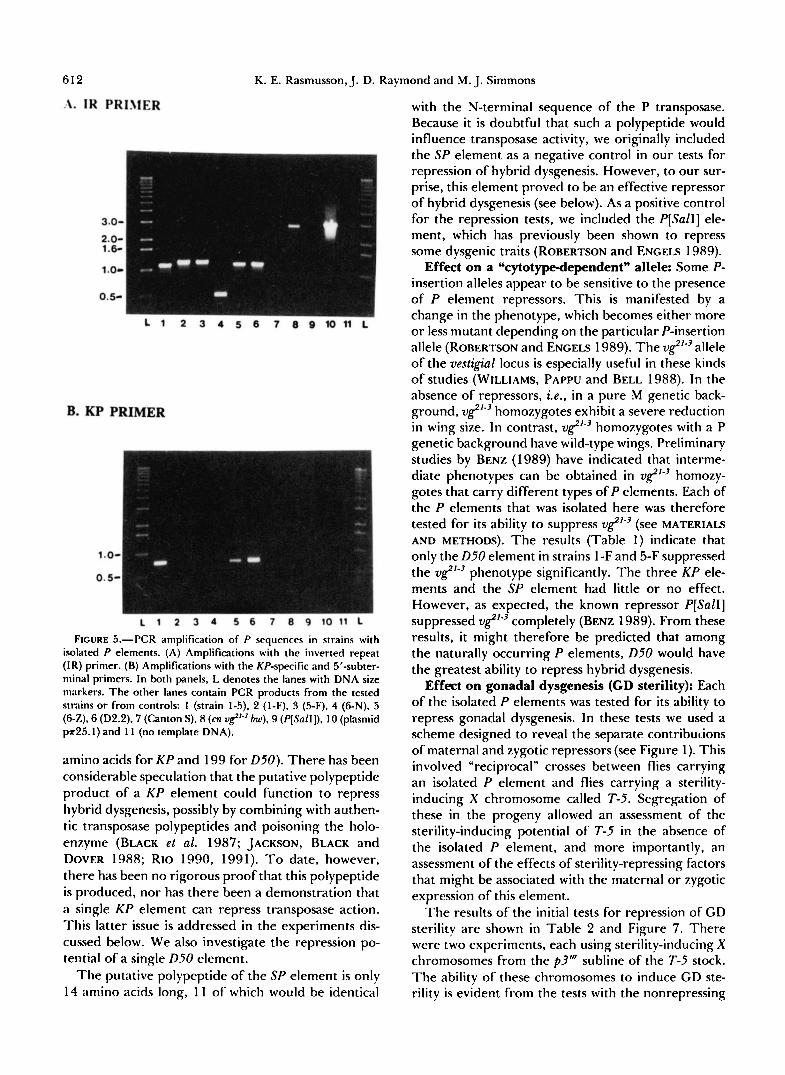
T h e sizes of the isolated P elements were deter- mined by PCR amplification using a primer comple- mentary to a sequence in the inverted terminal re- peats. Figure 5A shows the results of amplifications with this IR primer. As expected, the apparently identical P elements in strains 1-F and 5-F were the same size, about 1.2 kb long. T h e P elements in strains 1-5, 6-Z and D2.2 were also the same size (1.1 kb) even though they were at different cytological posi- tions. Strain 6-N carried the smallest of the isolated P elements, about 500 bp long, and was therefore too small to be detected by our in situ hybridization tech- nique.
Because of their size and origin, it seemed possible that the 1.1 kb P elements in strains 1-5,6-Z and D2.2 might be KP elements (BLACK et al. 1987). This POS- sibility was investigated by PCR amplification of ge- nomic DNA using a primer near the 5' end of the P
Repression of P Element Activity 61 1
TABLE 1
A summary of genetic and cytological data on strains containing individual P elements
Strain Cytological Phenotvpe of
Origin X chromosome genotype’ locationb P element I&‘.’ homozygotesc
1 -F Sexi. I y w f a * m v f c a r 4 c D50(1) Intermediate 5-F Sexi.5 y w f a * c v v f c a r 4 c D50(5) Intermediate 1-5 Sexi. 1 * c v v f c a r 2F KP(I ) Very severe 6-Z Sexi.6 8 * f vj, 1 SF KP(6) Severe D2.2 D2.2 + 14F K P W ) Very severe
Sal1 Transformed stock + 89Dd PISall] Wild type 6-N Sexi.6 8 * vs, Sf Very severe
’ An asterisk shows the location of the P element relative to the markers present. T w o strains (6-7, and 6-N) had unmapped recessive female-sterilizing mutations v j ) on the X chromosome.
Determined by in situ hybridbation of a P element probe to polytene chromosomes. Because of its small size, the element in strain 6-N could not be localized by this method. However, the genetic derivation of the X chromosome in this strain implies that the element is proximal to g, which is located in 12B.
The intermediate phenotype means wings about half normal size with notches in the distal and posterior margins. The severe phenotype means wings about one-fourth normal size with scalloped posterior margins. The very severe phenotype means clubshaped wings about one- fifth normal size.
ROBERTSON and ENCELS (1989).
-17
7.5 b 7.0
5.5 m
FIGURE 4.”Southern analysisof genomic DNA from strains with isolated P elements. The lanes are labeled with the strain names. See text and Table 1 for details.
sequence and another primer spanning the deletion in K P elements (see MATERIALS AND METHODS); these primers will amplify P element DNA only if the K P deletion is present (G. GLOOR, C. PRESTON and W. ENGELS, personal communication). For strains 1-5, 6- Z and D2.2, a PCR product of the expected size was obtained (Figure 5B), indicating that each carried a K P element. For the other strains, no PCR product was obtained indicating that some other type of P element was present.
The nature of each isolated P element was defini- tively established by DNA sequencing. Asymmetric PCR was used to generate material for the sequencing experiments. Primers for these experiments were cho- sen on the basis of preliminary studies using genomic Southern blotting and restriction enzyme digestion of standard PCR products. Only regions containing the
P element deletion were sequenced. Analysis of the sequencing data indicated that, as
expected, the isolated P elements in strains 1-F and 5- F were identical. Each had a deletion from bp 8 19 to bp 2527 plus a 21-bp insertion of non-P DNA at the deletion breakpoints. A search of GeneBank DNA sequences revealed that an element of this type had previously been identified in a mutant allele ( D 5 0 ) of the Rpll locus (SEARLES et al. 1986; W. ENGELS, per- sonal communication). Henceforth, we shall refer to this type of P element as “D50,” with the isolate from strain 1-F denoted as D50(1) and the isolate from strain 5-F denoted as D50(5) (Table 1).
Sequencing of the P elements in strains 1-5 and 6- Z confirmed that they were indeed KP elements. Both had a deletion from bp 809 to bp 2563, with a 2-bp ambiguity due to duplicate sequences at the deletion breakpoints. The presumptive KP element in strain D2.2 was not sequenced. We shall distinguish among the three isolated K P elements by denoting them as KP(I), KP(6) and KP(D), with the symbol in parenthe- ses referring to the carrier strain (Table 1).
The P element in strain 6-N was only 5 17 bp long. Its deletion spanned bp 186-2577 and apparently arose within a 3-bp direct repeat. T o date, no P element with this particular deletion has been de- scribed. We therefore denote this type of P element as “SP in recognition of the fact that it was isolated from a derivative of the Sexi strain (Table 1).
The structures of the three types of isolated P elements are shown in Figure 6. This figure also shows the sizes of the polypeptides these elements could encode. The K P and D50 elements are expected to produce similar polypeptides slightly more than 200 amino acids long, with most of the sequence identical with the N-ternlinal part of the P transposase (203
612 K. E. Rasmusson, J. D. Raymond and M. J. Simmons
F~CURE 5.--PCR amplification of P sequences in strains with isolated P elements. (A) Amplifications with the inverted repeat (IR) primer. (B) Amplifications with the KP-specific and 5”subter- minal primers. In both panels, L denotes the lanes with DNA size rnarkers. The other lanes contain PCR products from the tested strains or from controls: 1 (strain 1-5). 2 (l-F), S (5-F), 4 (6-N), 5 (6-Z),6 (D2.2), 7 (Canton S), 8 (cn u&”~ h), 9 (P[Sall]), 10 (plasmid ~ ~ 2 5 . 1 ) and 1 1 (no template DNA).
amino acids for KP and 199 for 050). There has been considerable speculation that the putative polypeptide product of a K P element could function to repress hybrid dysgenesis, possibly by combining with authen- tic transposase polypeptides and poisoning the holo- enzyme (BLACK et al. 1987; JACKSON, BLACK and DOVER 1988; RIO 1990, 1991). T o date, however, there has been no rigorous proof that this polypeptide is produced, nor has there been a demonstration that
with the N-terminal sequence of the P transposase. Because it is doubtful that such a polypeptide would influence transposase activity, we originally included the SP element as a negative control in our tests for repression of hybrid dysgenesis. However, to our sur- prise, this element proved to be an effective repressor of hybrid dysgenesis (see below). As a positive control for the repression tests, we included the P[SalI] ele- ment, which has previously been shown to repress some dysgenic traits (ROBERTSON and ENCELS 1989).
Effect on a “cytotype-dependent” allele: Some P- insertion alleles appear to be sensitive to the presence of P element repressors. This is manifested by a change in the phenotype, which becomes either more or less mutant depending on the particular P-insertion allele (ROBERTSON and ENGELS 1989). The v$’-’ allele of the vestigial locus is especially useful in these kinds of studies (WILLIAMS, PAPPU and BELL 1988). In the absence of repressors, i.e., in a pure M genetic back- ground, v8”’ homozygotes exhibit a severe reduction in wing size. In contrast, v$”’ homozygotes with a P genetic background have wild-type wings. Preliminary studies by BENZ (1 989) have indicated that interme- diate phenotypes can be obtained in v8”’ homozy- gotes that carry different types of P elements. Each of the P elements that was isolated here was therefore tested for its ability to suppress ,g’-’ (see MATERIALS AND METHODS). The results (Table 1) indicate that only the 050 element in strains l -F and 5-F suppressed the v8”’ phenotype significantly. The three KP ele- ments and the SP element had little or no effect. However, as expected, the known repressor P[SalI] suppressed v8’-’ completely (BENZ 1989). From these results, it might therefore be predicted that among the naturally occurring P elements, 050 would have the greatest ability to repress hybrid dysgenesis.
Effect on gonadal dysgenesis (CD sterility): Each of the isolated P elements was tested for its ability to repress gonadal dysgenesis. In these tests we used a scheme designed to reveal the separate contribuLions of maternal and zygotic repressors (see Figure 1). This involved “reciprocal” crosses between flies carrying an isolated P element and flies carrying a sterility- inducing X chromosome called T-5. Segregation of these in the progeny allowed an assessment of the sterility-inducing potential of T-5 in the absence of the isolated P element, and more importantly, an assessment of the effects of sterility-repressing factors that might be associated with the maternal or zygotic exmession of this element.
a single K P element can repress transposase action. The results of the initial tests for repression of GD This latter issue is addressed in the experiments dis- sterility are shown in Table 2 and Figure 7. There cussed below. We also investigate the repression PO- were two experiments, each using sterility-inducing X tential of a single D50 element. chromosomes from the p3” subline of the T-5 stock.
The putative polypeptide of the SP element is only The ability of these chromosomes to induce CD ste- 14 amino acids long, 1 1 of which would be identical rility is evident from the tests with the nonrepressing
I

Repression of P Element Activity 613
Complete P
- - - - 751 ..a.
STRAIN
D50 819 I
TAAAAAGAGGATGTITGGAT 1-F, 5-F TAAAAAGA-AATAATGT
” 204 an. TTTGAAATTACAAATAATGT
KP
-207
I 2521
I 2563
SP 186
- - 14 ..a. 6-N C A A A G C T G ~ C T G G A G T A A
CAAAGCTGTGBTAAACAAG GTCGATGTWTAAACAAG
I
FIGURE 6.-The 0 5 0 , KP and SP elements compared to the complete P element. The open regions indicate deletions and the triangle indicates an insertion of n o n 8 DNA. The coding regions are shown beneath each element, with the number of amino acids (a.a.) in the expected polypeptide indicated at the right. At the far right, the DNA sequences spanning the deletion breakpoints are shown in between sequences from the complete P element; the upper and lower sequences demarcate the left (5’) and right (3‘) breakpoints, respectively, with boldface denoting the portions shared by the complete and incomplete elements. The non-P DNA in the 050 element is underlined. Regions spanning the deletions were sequenced in all the elements except the KP element in strain D2.2, which is listed in parentheses.
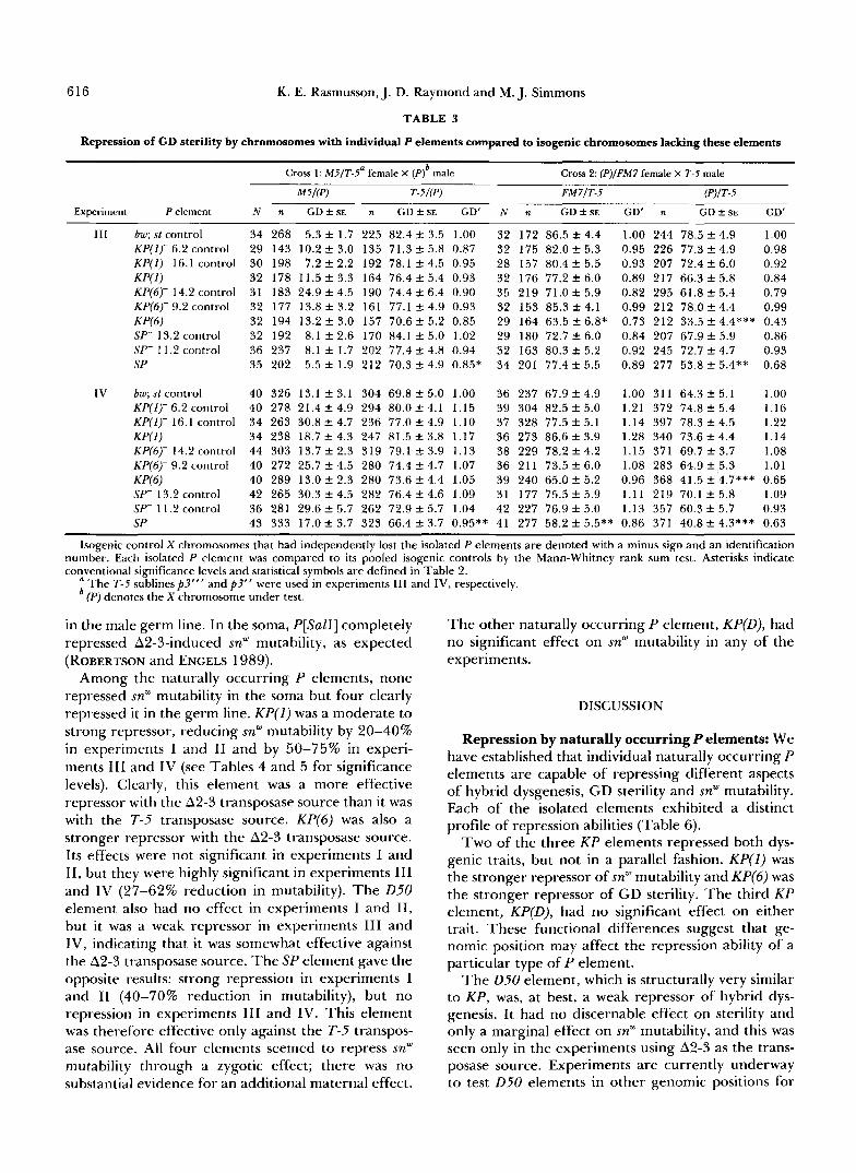
TABLE 2
Repression of CD sterility by chromosomes with individual P elements
Cross 1: M5/T-5a female X ( P f male Cross 2: (P)/FM7‘ female X T-5 male
M5I(P) T-5/(P) FM7/T-5 W I T - 5
Experiment P element N d ne G D ~ + s E n GD + S E G D ‘ ~ N n GD ~ S E GD‘ n GD + S E GD’
1 bw; st control 60 641 7.0 f 1.5 626 74.2 f 3.7 1.00 59 497 65.3 f 3.7 1.00 589 53.6 f 3.9 1.00 D50h 45 451 8.9 f 2.3 435 76.1 f 4.1 1.02 60 588 54.5 f 3.6* 0.83 663 58.1 f 3.6 1.08
~ ~ ~~
KP(1) 30 309 4.7 f 1.4 324 66.7 f 5.3 0.90 30 300 41.4 f 4.5*** 0.63 326 22.4 f 4.2*** 0.41 W 6 ) 30 309 14.5 f 2.8 307 75.5 f 5.1 1.01 30 228 21 .6f 4.2*** 0.33 270 20.7 f 4.2*** 0.39 KP(D) 30 319 4.8 f 1.8 314 83.4 f 3.7 1.12 30 320 44.7 f 6.0*** 0.68 339 50.3 f 5.8 0.93 SP 30 301 3 . 9 f 1.8 308 68.1 f 4.0* 0.91 30 270 15.5 f 2.7*** 0.24 315 4.8 f 1.3*** 0.09 P[SalI] 30 327 1.8 f 0.8 321 39 .4f 5.7*** 0.53 30 177 34.2 f4 .1*** 0.52 283 0.6 f 0.6*** 0.01
I1 bw; st control 108 1097 8.3 f 1.3 1121 44.1 f 3.6 1.00 100 952 50.5 f 4.0 1.00 1090 45.1 f 3.7 1.00 D50h 64 595 12.2 f 1.9 583 54.0 f 4.8 1.22 60 576 56.4 f 4.8 1.12 692 55.2 f 4.6 1.22 W 1 ) 30 299 9.0 f 2.1 293 50.5 f 6.8 1.15 30 358 45.7 f 6.3 0.90 360 20 .0f 3.7*** 0.44 KP(4 30 331 5.7 f 1.6 336 50.8 f 6.9 1.15 30 307 13.7 f 3.1*** 0.27 350 8.7 f 2.6*** 0.19 K V ) 30 321 11.6 f 2.9 344 52.9 f 6.8 1.20 30 266 40.1 f 6.4 0.79 344 36.1 f 5.9 0.80 SP 30 285 16.5 f 4.7 261 62.8 f 6.8 1.42 29 300 27.0 f 4.9*** 0.53 348 14.4 f 3.3*** 0.31 P[SalI] 30 342 4.1 f 1.1 332 25.8 f 5.3** 0.58 33 297 16.2 f 4.1*** 0.32 381 3.9 f 1.3*** 0.09
The Mann-Whitney rank sum test was used to determine if the percentage of GD sterility was significantly less than that of the bw; st control, with each culture treated as an independent unit. Asterisks indicate conventional levels of significance.
The T-5 subline p3”’ was used in these experiments. (P) denotes the X chromosome under test. A third chromosome carrying the P[SalI] element was also tested. The genotypes of the
In the tests involving the P[SalI] element, P[SalI]/TM3 females were used in this cross. Their daughters had the genotypes T-5/+;
Number of cultures. Number of females examined.
Average percentage of females with GD sterility normalized to the b w ; st control. Although the D50(1) and D50(5) elements were tested separately, the data have been pooled in this analysis.
dayghters in these tests were M5/+; P[SalI]/+ (left column) and T-5/+; P[SalI]/f (right column).
TM//+ (left column) and T-5/+; P[SalI]/+ (right column).
funweighted average percentage of females with GD sterility f standard error, which was calculated empirically.
M strain, bw; st . In cross 1 of these tests, moderate to and 44.1% in experiment 11) were observed in the high frequencies of sterility (74.2% in experiment I daughters that carried a T-5 X chromosome, but little

614 K. E. Rasmusson, J. D. Raymond and M. J. Simmons
*\
I I I 1 - 0 0.5 1.0
I I I 0 0.5 1.0
I I I 0 0.5 1 .o
Frequency of gonadal dysgenesis normalized to bw; st controls
FIGURE 7.-Repression of gonadal dysgenesis by individual P elements in experiments I and 11. Cross-hatching is used for the bw; st controls; the results that were significantly less than these are indicated by a solid bar. See text and Table 2 for details.
sterility (7.0% in experiment I and 8.3% in experi- ment 11) was seen in the daughters that lacked this chromosome.
In these experiments, repression of T-5-induced sterility was ascertained by comparing the results of the tests with the isolated P elements to those with the bw; st controls. Accordingly, all the data were normal- ized to these controls. The P[SalI] element was in- cluded as a positive control and, as expected, it signif- icantly reduced the frequency of sterility in both ex- periments. Furthermore, the repressing effect of P[SalI] seemed to be due to a combination of mater- nally and zygotically expressed factors. The zygotic factors were evident in the data from cross 1, in which P[SalI] was transmitted paternally. Among the T-5/+; P[SalI] daughters from this cross, the frequency of sterility was only about half that seen among the daughters from the bw; st controls-a highly significant
difference. The P[SaZI] element also repressed sterility through a purely maternal effect. This was evident in the T-5/+; TM3/+ daughters from cross 2 which inherited cytoplasm from their P[SalI]/TM3 mothers even though they did not inherit P[SalI] itself. These daughters showed 50-70% less sterility than the daughters from the bw; st controls-a highly significant difference, implying that factors transmitted mater- nally but independently of the P[SaZI] element were influential in repressing gonadal dysgenesis in the offspring. When these maternally transmitted factors were combined with the P[SaZI] element itself (see the T-5/+; P[SaZI]/+ daughters of cross 2), gonadal dys- genesis was almost completely repressed.
None of the naturally occurring P elements showed much ability to repress dysgenesis through a purely zygotic effect. For each of the elements, the frequency of sterility among the T-5/(P) daughters of cross 1 was

Repression of P Element Activity 615
approximately equal to or greater than the corre- sponding frequency for the bw; st controls. In contrast, two of the elements consistently showed an ability to repress dysgenesis through a purely maternal effect. This was seen in the FM7/T-5 daughters of cross 2, where KP(6) and, surprisingly, SP caused significant reductions (46-76%) in the frequency of sterility. The D50, KP(1) and KP(D) elements also appeared to re- duce this sterility somewhat (17-37%), but their ef- fects were significant only in experiment I. The com- bined maternal and zygotic effects of the isolated P elements were assessed in the (P)/T-5 daughters of cross 2. Among these, the KP(l), KP(6) and SP ele- ments caused large and highly significant reductions in sterility (56-91 %) in both experiments but the D50 and KP(D) elements did not.
Additional experiments using more rigorous con- trols were performed to determine if the repression of sterility by KP(I), KP(6) and SP was due specifically to these elements or to some extraneous factor. X chromosomes that were completely isogenic with the chromosomes that carried these isolated elements were made by excising the elements through trans- posase action. This was accomplished by passing the X chromosomes that carried the elements through females that were heterozygous for the A2-3 trans- posase source. In these females, the X chromosomes were balanced by FM7. Non-FM7, non-A2-3 sons from these females were mated to C(I)DX, y f/Y; bw; st females to establish lines and then screened by PCR with the IR P element primer to determine which had lost the isolated P element. Lines so identified were then backcrossed to C(I)DX, y f /Y ; bw; st females to fix the recessive autosomal markers. X chromosomes car- rying the isolated P elements were also put into this genetic background. All the X chromosomes were then tested for their abilities to repress T-5-induced gonadal dysgenesis using the scheme in Figure 1. For each P-containing chromosome, there were two in- dependently generated isogenic controls, each having been double-checked for the loss of its P element. A bw; st control was also included in these experiments.
The results of two experiments that utilized differ- ent T-5 sublines are shown in Table 3 and Figure 8. The effect of each P element was ascertained by comparing it with its two isogenic controls, which were pooled. This pooling was justified since only one sig- nificant difference at the 5% level was detected in 18 independent comparisons between the isogenic con- trols. Only SP repressed dysgenesis through a purely zygotic effect (see the T-5/(P) daughters from cross 1); although small, this effect was significant in both experiments. SP also repressed dysgenesis through a purely maternal effect, as did KP(6) (see the FM7/T-5 daughters from cross 2); however, for SP this effect was significant only in experiment 111 and for KP(6) it
was significant only in experiment IV. Both SP and KP(6) repressed dysgenesis very significantly when their zygotic and maternal effects were combined (see the (P)/T-5 daughters from cross 2) and they did so in both experiments. KP(I), by contrast, had no signifi- cant repression potential in either experiment.
These results demonstrated that the KP(6) and SP elements were directly responsible for the repression of gonadal dysgenesis, but they did not confirm the repression that was previously found to be associated with KP(1). This may mean that KP(1) is not a bona f ide repressor of gonadal dysgenesis, or that its ability to repress was obscured by other factors. In this context, it should be noted that neither KP(6) nor SP was as effective a repressor in experiments 111 and IV as it was in experiments I and 11. One possible expla- nation is the genetic background, which may have weakened the repression ability of all three P ele- ments, especially KP(I), in Experiments 111 and IV. Another is the T-5 chromosomes, which were clearly stronger inducers of sterility in experiments 111 and IV than they were in experiments I and I1 (compare the results from the bw; st controls). By presenting a greater challenge, these chromosomes may have made it more difficult to detect repression by the isolated P elements.
Effect on snw mutability: Each of the isolated P elements was tested for its ability to repress the mut- ability of the double-P element-insertion mutation snw. In experiments I and 11, the snw allele was destabilized by different T-5 X chromosomes (Table 4 and Figure 9) and in experiments 111 and IV it was destabilized by A2-3 (Table 5 and Figure IO). “Reciprocal” crosses (A and B) were used in all four experiments (see the schemes in Figures 2 and 3) to identify separate zyg- otic and maternal components of repression. The P[SalI] element was included as a positive control, and there were also several independent negative controls (see MATERIALS AND METHODS).
As expected, P[SalI] was a strong repressor of snw mutability (ROBERTSON and ENCELS 1989). In Exper- iments I and 11, it reduced the germ-line mutation rate to a small fraction of that seen with the bw; st controls. This happened even though the element was inherited paternally, indicating that repression was caused by a purely zygotic effect. (There was no separate test for a maternal effect of P[SalI] in exper- iments I and 11.) In experiment 111, P[SalI] reduced the germ-line mutability of snw to about 20% of the bw; st controls, regardless of whether it was inherited paternally or maternally. This indicated that repres- sion by P[SalI] did not involve a maternal effect, confirming a finding of ROBERTSON and ENCELS (1 989). In experiment IV, the reduction in snw muta- bility was even greater, suggesting that P[SalI] was a stronger repressor in the female germ line than it was
616 K. E. Rasmusson, J. D. Raymond and M. J. Simmons
TABLE 3
Repression of GD sterility by chromosomes with individual P elements compared to isogenic chromosomes lacking these elements
Cross 1 : M5/T-5a female X ( P ) male ~~
b ~ ~ ~~~~~~ ____
Cross 2: (P)/FM7 female X T-5 male
M5I(P) T-5/(P) FM7/T-5 @‘)IT-5
Experiment P element N n GD CSE n GDCSE GD‘ N n GD CSE GD‘ n GD C S E GD‘
111 bw; st control 34 268 5.3 f 1.7 225 82.4 -1- 3.5 1.00 32 172 86.5 f 4.4 1.00 244 7 8 . 5 f 4.9 1.00 KP(I)-6.2 control 29 143 10.2 k 3.0 135 71.3 k 5.8 0.87 32 175 82.0 f 5.3 0.95 226 77.3 f 4.9 0.98 KP(I)- 16.1 control 30 198 7.2 f 2.2 192 78.1 k 4.5 0.95 28 157 80.4 f 5.5 0.93 207 72.4 f 6.0 0.92
KP(6)- 14.2control 31 183 2 4 . 9 f 4.5 190 74.4 k 6.4 0.90 35 219 71.0 f 5.9 0.82 295 61.8+ 5.4 0.79 KP(6)-9.2control 32 177 13.8 f 3.2 161 77.1 k 4.9 0.93 32 153 85.3 f 4.1 0.99 212 7 8 . 0 f 4.4 0.99
SP- 13.2 control 32 192 8.1 f 2.6 170 84.1 k 5.0 1.02 29 180 72.7 f 6.0 0.84 207 67 .9f 5.9 0.86 SP- 11.2 control 36 237 8.1 k 1.7 202 77.4 +- 4.8 0.94 32 163 80.3 f 5.2 0.92 245 72.7 k 4.7 0.93 SP 35 202 5.5 f 1.9 212 70.3 k 4.9 0.85* 34 201 77.4 f 5.5 0.89 277 53.8 f 5.4** 0.68
IV bw; st control 40 326 1 3 . 1 f 3 . 1 304 69.8k5.0 1.00 36 237 6 7 . 9 f 4 . 9 1.00 311 6 4 . 3 f 5 . 1 1.00 KP(lr6.2 control 40 278 21.4 f 4.9 294 80.0 k 4.1 1.15 39 304 82.5 f 5.0 1.21 372 74.8 f 5.4 1.16 KP(1)- 16.1 control 34 263 30.8 f 4.7 236 77.0 k 4.9 1.10 37 328 77.5 f 5.1 1.14 397 78.3 f 4.5 1.22
KP(6)- 14.2 control 44 303 13.7 f 2.3 319 79.1 k 3.9 1.13 38 229 78.2 f 4.2 1.15 371 69.7 f 3.7 1.08 KP(6)-9.2 control 40 272 25.7 k 4.5 280 74.4 k 4.7 1.07 36 211 73.5 f 6.0 1.08 283 64.9 f 5.3 1.01
SP- 13.2control 42 265 30.3 f 4 . 5 282 76.4k4.6 1.09 31 177 7 5 . 5 f 5 . 9 1.11 219 70.1 f 5 . 8 1.09 SP- 11.2 control 36 281 29.6 k 5.7 262 72.9 k 5.7 1.04 42 227 76.9 f 5.0 1.13 357 60.3 k 5.7 0.93 SP 43 333 17.0 f 3.7 323 66.4 k 3.7 0.95** 41 277 58.2 f 5.5** 0.86 371 40.8 k 4.3*** 0.63
KP(1) 32 178 11.5 f 3.3 164 76.4 k 5.4 0.93 32 176 77.2f 6.0 0.89 217 66.3 f 5.8 0.84
KP(6) 32 194 13.2 f 3.0 157 70.6 f 5.2 0.85 29 164 63.5 f 6.8* 0.73 212 33.5 & 4.4*** 0.43
KP(1) 34 238 1 8 . 7 f 4.3 247 81.5 f 3.8 1.17 36 273 8 6 . 6 f 3.9 1.28 340 7 3 . 6 f 4 . 4 1.14
KP(6) 40 289 13.0 f 2.3 280 73.6 k 4.4 1.05 39 240 65.0 f 5.2 0.96 368 41.5 f 4.7*** 0.65
Isogenic control X chromosomes that had independently lost the isolated P elements are denoted with a minus sign and an identification number. Each isolated P element was compared to its pooled isogenic controls by the Mann-Whitney rank sum test. Asterisks indicate conventional significance levels and statistical symbols are defined in Table 2.
The T-5 sublines p3”’ and p3” were used in experiments I11 and IV, respectively. ’ (P) denotes the Xchromosome under test.
in the male germ line. In the soma, P[SaZI] completely repressed A2-3-induced snw mutability, as expected (ROBERTSON and ENGELS 1989).
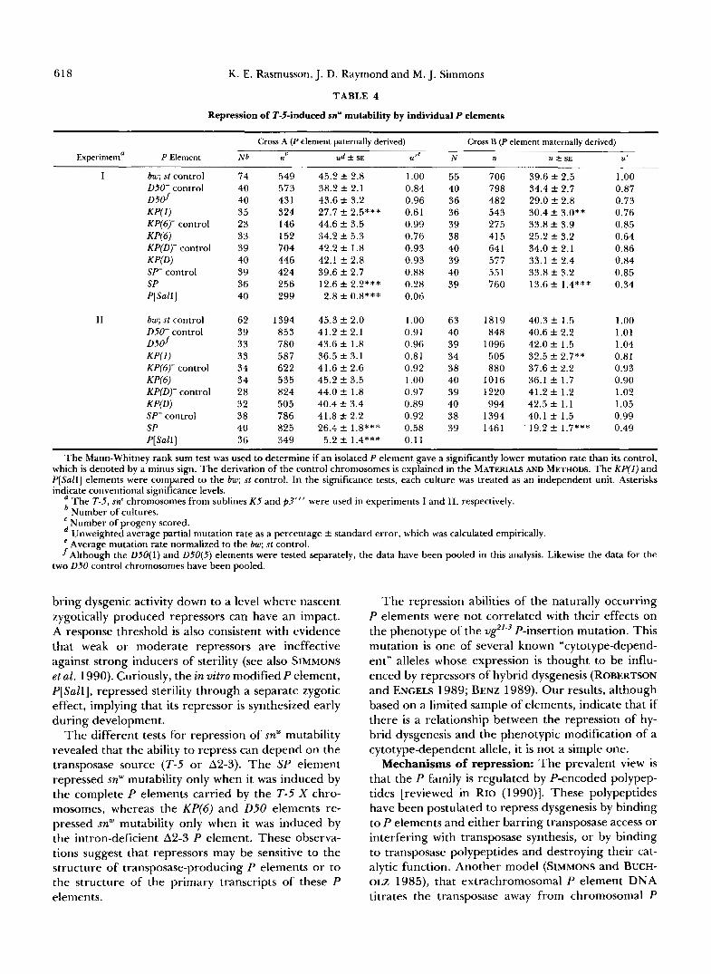
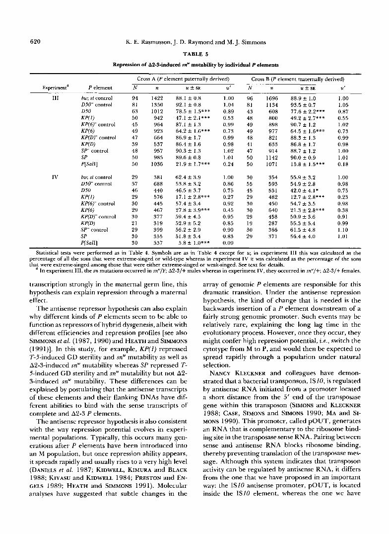
Among the naturally occurring P elements, none repressed snw mutability in the soma but four clearly repressed it in the germ line. KP(1) was a moderate to strong repressor, reducing snw mutability by 20-40% in experiments I and I1 and by 50-75% in experi- ments 111 and IV (see Tables 4 and 5 for significance levels). Clearly, this element was a more effective repressor with the A2-3 transposase source than it was with the T-5 transposase source. KP(6) was also a stronger repressor with the A2-3 transposase source. Its effects were not significant in experiments I and 11, but they were highly significant in experiments I11 and IV (27-62% reduction in mutability). The 050 element also had no effect in experiments I and 11, but it was a weak repressor in experiments 111 and IV, indicating that it was somewhat effective against the A2-3 transposase source. The SP element gave the opposite results: strong repression in experiments I and I1 (40-70% reduction in mutability), but no repression in experiments I11 and IV. This element was therefore effective only against the T-5 transpos- ase source. All four elements seemed to repress snw mutability through a zygotic effect; there was no substantial evidence for an additional maternal effect.
The other naturally occurring P element, KP(D), had no significant effect on snw mutability in any of the experiments.
DISCUSSION
Repression by naturally occurring P elements: We have established that individual naturally occurring P elements are capable of repressing different aspects of hybrid dysgenesis, GD sterility and snw mutability. Each of the isolated elements exhibited a distinct profile of repression abilities (Table 6).
Two of the three K P elements repressed both dys- genic traits, but not in a parallel fashion. KP(1) was the stronger repressor of snw mutability and KP(6) was the stronger repressor of GD sterility. The third KP element, KP(D), had no significant effect on either trait. These functional differences suggest that ge- nomic position may affect the repression ability of a particular type of P element.
The D50 element, which is structurally very similar to KP, was, at best, a weak repressor of hybrid dys- genesis. It had no discernable effect on sterility and only a marginal effect on snw mutability, and this was seen only in the experiments using A2-3 as the trans- posase source. Experiments are currently underway to test D 5 0 elements in other genomic positions for

Repression of P Element Activity 617
CROSS 1
T - W P ) '$0 Experiment I11
bw; sr control -1 K P ( 1 ) - 6.2 control
KP(I) -16 .1 control
K P ( 1 ) - KP(6)-14.2 control 1- KP(6)-9 .2 control ~- K P ( 6 ) - Sf - 13.2 control
SP- 11.2 control k-1 Sf -
Experiment IV
bw; sr control
K P ( 1 ) - 6.2 control -1 K P ( 1 ) - 16.1 control 7 K P ( 1 ) - K P ( 6 ) - 14.2 control
KP(6)-9 .2 control
K P ( 6 ) - Sf- 13.2 control
SP- 11.2 control -j
Sf -
CROSS 2
-(P)IT-S tt
I I I I I I I I I
0 0.5 1.0 0 0.5 1.0 0 0.5 1.0
Frequency of gonadal dysgenesis normalized to bw; st controls FIGURE 8.-Repression of gonadal dysgenesis by individual P elements in exeriments I11 and IV. Cross-hatching denotes the bw; st and
various isogenic controls and solid color denotes the cases in which the frequency of sterility was significantly less than the controls. See text and Table 3 for details.
repression of dysgenic traits (C. PRESTON and W. ENGELS, personal communication).
The most surprising finding was that the SP ele- ment, only 5 17 bp long, was a strong repressor of GD sterility and T-5-induced snw mutability. This element was originally included in the experiments because we thought it was too small to encode a polypeptide repressor and could therefore serve as a negative control. Instead, it turned out to be the strongest repressor among the naturally occurring elements that were tested. The positive control element P[SaZI] also repressed GD sterility and snw mutability, as ex- pected (ROBERTSON and ENCELS 1989).
Zygotic and maternal effects, results with differ- ent transposase sources and phenotypic suppression of v$'-': In all cases, the naturally occurring P ele-
ments seemed to repress GD sterility through a ma- ternal effect, or through a combination of maternal and zygotic effects. In contrast, they seemed to repress snw mutability through a zygotic effect alone. One possible explanation is that a reduction in snw muta- bility can result from zygotically produced repressors acting in the developed, functioning germ line, whereas a reduction in GD sterility requires repressor action early during germ line development when the events leading to sterility occur (ENGELS and PRESTON 1979). These early acting repressors would most likely be provided by the maternal cytoplasm, explaining the maternal effect. However, there was also clear evidence for a zygotic effect working in concert with the maternal effect. This suggests a threshold phe- nomenon in which maternally produced repressors
618 K. E. Rasmusson, J. D. Raymond and M. J. Simmons
TABLE 4
Repression of T-5-induced sn’” mutability by individual P elements
Cross A (P element paternally derived) Cross B (P element maternally derived)
Experiment’ P Element N b n ud k SE Ute N n 21 k SE Ut C
I bw; st control D50- control D 5 d KP(1) KP(6)- control KP(6) KP(D)- control KPfD) SI“ control SP P[SalI]
I I bw; st control 050- control D 5 d KP(1) KP(6)- control W 6 ) KP(D)- control K P P ) SP- control SP P[SalI]
74 40 40 35 23 33 39 40 39 36 40
62 39 33 33 34 34 28 32 38 40 36
549 573 43 1 324 146 152 704 446 424 256 299
1394 853 780 587 622 535 824 505 786 825 349
45.2 f 2.8 38.2 f 2.1 43.6 +- 3.2 27.7 f 2.5*** 44.6 f 3.5 34.2 f 5.3 42.2 f 1.8 42.1 f 2.8 39.6 f 2.7 12.6 f 2.2*** 2.8 f 0.8***
45.3 f 2.0 41.2 k 2.1 43.6 f 1.8 36.5 f 3.1 41.6 ? 2.6 45.2 f 3.5 44.0 f 1.8 40.4 f 3.4 41.8 k 2.2 26.4 f 1.8***
5.2 k 1.4***
1 .oo 0.84 0.96 0.61 0.99 0.76 0.93 0.93 0.88 0.28 0.06
1 .oo 0.91 0.96 0.81 0.92 1 .oo 0.97 0.89 0.92 0.58 0.1 1
55 40 36 36 39 38 40 39 40 39
63 40 39 34 38 40 39 40 38 39
706 798 482 543 275 415 64 1 577 55 1 760
1819 848
1096 505 880
1016 1220 994
1394 1461
39.6 f 2.5 34.4 f 2.7 29.0 f 2.8 30.4 f 3.0** 33.8 k 3.9 25.2 f 3.2 34.0 -?r 2.1 33.1 f 2.4 33.8 f 3.2 13.6 f 1.4***
40.3 f 1.5 40.6 k 2.2 42.0 f 1.5 32.5 f 2.7** 37.6 f 2.2 36.1 f 1.7 41.2 f 1.2 42.5 f 1.1 40.1 f 1.5 19.2 k 1.7***
1 .oo 0.87 0.73 0.76 0.85 0.64 0.86 0.84 0.85 0.34
1 .oo 1.01 1.04 0.81 0.93 0.90 1.02 1.05 0.99 0.49
The Mann-Whitney rank sum test was used to determine if an isolated P element gave a significantly lower mutation rate than its control, which is denoted by a minus sign. The derivation of the control chromosomes is explained in the MATERIALS AND METHODS. The KP(1) and P[SalI] elements were compared to the bw; st control. In the significance tests, each culture was treated as an independent unit. Asterisks indicate conventional significance levels. ‘ The T-5, sn‘ chromosomes from sublines K 5 and p3”‘ were used in experiments I and 11, respectively.
Number of cultures. Number of progeny scored. Unweighted average partial mutation rate as a percentage f standard error, which was calculated empirically. Average mutation rate normalized to the bw; st control.
fAlthough the D50(1) and D50(5) elements were tested separately, the data have been pooled in this analysis. Likewise the data for the two 050 control chromosomes have been pooled.
bring dysgenic activity down to a level where nascent zygotically produced repressors can have an impact. A response threshold is also consistent with evidence that weak or moderate repressors are ineffective against strong inducers of sterility (see also SIMMONS et al. 1990). Curiously, the in vitro modified P element, P[SalI], repressed sterility through a separate zygotic effect, implying that its repressor is synthesized early during development.
The different tests for repression of snw mutability revealed that the ability to repress can depend on the transposase source (T-5 or An-3). The SP element repressed snw mutability only when it was induced by the complete P elements carried by the T-5 X chro- mosomes, whereas the KP(6) and D50 elements re- pressed snw mutability only when it was induced by the intron-deficient A2-3 P element. These observa- tions suggest that repressors may be sensitive to the structure of transposase-producing P elements or to the structure of the primary transcripts of these P elements.
The repression abilities of the naturally occurring P elements were not correlated with their effects on the phenotype of the vg”’ P-insertion mutation. This mutation is one of several known “cytotype-depend- ent” alleles whose expression is thought to be influ- enced by repressors of hybrid dysgenesis (ROBERTSON and ENGELS 1989; BENZ 1989). Our results, although based on a limited sample of elements, indicate that if there is a relationship between the repression of hy- brid dysgenesis and the phenotypic modification of a cytotype-dependent allele, it is not a simple one.
Mechanisms of repression: The prevalent view is that the P family is regulated by P-encoded polypep- tides [reviewed in RIO (1990)l. These polypeptides have been postulated to repress dysgenesis by binding to P elements and either barring transposase access or interfering with transposase synthesis, or by binding to transposase polypeptides and destroying their cat- alytic function. Another model (SIMMONS and BUCH- OLZ 1985), that extrachromosomal P element DNA titrates the transposase away from chromosomal P

Repression of P Element Activity 619
CROSS A (Pekmnt patemnlly derived) Gp element matenully derived)
CROSS B
EXPERIMENT I
bw; st control 1- - DSO- control
Mutation rate normalized to bw; st controls FIGURE 9.-Repression of T-5-induced snw mutability by individ-
ual P elements. Cross-hatching denotes the various controls and solid color denotes the cases in which snw mutability was significantly less than that of the relevant control. See text and Table 4 for details.
elements, thereby making them less active, has re- ceived little support.
The polypeptide repressor hypothesis is supported by evidence from tests with experimentally modified P elements that have been introduced into Drosophila strains by germ-line transformation. ROBERTSON and ENGELS (1989) showed that P[SaZl] was an effective repressor of A2-3-induced GD sterility, sa"' mutability and pupal lethality. Our results have confirmed and extended their findings. This modified P element has a frameshift mutation in the last exon of the transpos- ase gene, making it incapable of encoding a catalyti- cally active product. It could, however, produce a polypeptide that would be isosequential with the trans- posase for much of its length. Because such a polypep- tide could possess some of the binding and oligomer- ization properties of the transposase, it might be able to function in a competitive or antagonistic way as a repressor of transposase activity. Other modified P elements with frameshift mutations in exons 0, 1 or 2 did not repress transposase activity, suggesting that these exons are critical for repressor function (ROB- ERTSON and ENGELS 1989). Taken together, these results are consistent with the idea that the 66-kD polypeptide made from the first three exons of incom- pletely processed complete P element transcripts is a
natural repressor of transposase activity (LASKI, RIO and RUBIN 1986; ROBERTSON and ENGELS 1989; MISRA and RIO 1990). Molecular constructs designed to produce this polypeptide under the control of a heat shock-inducible promoter have provided some supporting evidence (MISRA and RIO 1990).
The polypeptide repressor hypothesis could also be used to explain repression by small P elements, such as KP, as long as their polypeptide products retain some of the binding or oligomerization properties of the transposase. The predicted KP polypeptide would contain 207 amino acids, including an oligomerization motif that might allow it to bind to transposase poly- peptides and disrupt their catalytic function (RIO 1990). However, there is currently no evidence for this polypeptide, much less for a disrupting interaction with the transposase. Consequently, although repres- sion by individual KP elements has been established in fact, its underlying mechanism is still a matter of speculation. However, assuming the polypeptide re- pressor hypothesis to be correct, the inability of some KP elements such as KP(D) to repress dysgenesis could be attributed to transcriptional inactivity caused by a nearby silencer sequence (O'KANE and GEHRING 1987) or to a nonsense or missense mutation in the coding sequence of the element itself.
Our results with the SP element are not very easily explained by the polypeptide repressor hypothesis. The predicted SP polypeptide would contain only 14 amino acids-probably too small to be of any signifi- cance. However, SP was a potent repressor of both GD sterility and T-5-induced sa"' mutability. One pos- sible explanation is that repression by SP is mediated by an antisense RNA that interferes with the process- ing or translation of complete P element transcripts. This antisense RNA could be produced by transcrip- tion backward through the SP element from an exter- nal promoter. Once formed, the antisense RNA mol- ecules could bind to sense RNA molecules produced by complete P elements, blocking their processing or translation, or even causing their degradation by RNases. Any of these events would impair the synthe- sis of transposase polypeptides and ultimately reduce the incidence and severity of hybrid dysgenesis. Of course any kind of P element could insert backwards and downstream of a genomic promoter, leading to the synthesis of antisense P RNA. P elements appar- ently do have a tendency to insert in promoter regions (KELLEY et al. 1987). This mechanism of repression could therefore be quite general, and could even apply to complete P elements. In addition, the requirement for an inverted orientation downstream of a genomic promoter would explain why a particular type of P element, such as KP, might repress dysgenesis in one genomic location but not in another. Furthermore, because some promoters would be expected to initiate
620 K. E. Rasmusson, J. D. Raymond and M. J. Simmons
TABLE 5
Repression of A2-3-induced sn'" mutability by individual P elements
Experimenta P element N n U ? SE
Cross A ( P element paternally derived) ~
111 bw; st control D50- control D50 KP(1) KP(6)- control KP(6) KP(D)- control K P O Sf- control Sf P[SalI]
IV bw; st control D50- control D50 W 1 ) KP(6)- control KP(6) KP(D)- control K P P ) Sf- control Sf P[SalI]
94 81 63 50 45 49 47 39 48 50 50
29 57 46 29 30 29 30 21 29 30 30
1422 1350 1012 942 964 923 664 537 957 985
1036
38 1 688 440 576 445 467 377 319 399 355 537
88.1 f 0.8 92.1 f 0.8 78.5 f 1.5*** 47.1 f 2.1*** 87.1 f 1.3 64.2 f 1.6*** 86.9 f 1.7 86.4 f 1.6 90.3 f 1.3 89.6 f 0.8 21.9 f 1.7***
62.4 f 3.9 53.8 f 3.2 46.5 f 3.7 17.1 f 2.8*** 57.4 f 3.4 27.8 f 3.9*** 59.4 f 4.5 52.9 f 5.2 56.2 f 2.9 51.8 f 3.4
5.8 f 1.0***
~
U'
1 .oo 1.04 0.89 0.53 0.99 0.73 0.99 0.98 1.02 1.01 0.24
1 .oo 0.86 0.75 0.27 0.92 0.45 0.95 0.85 0.90 0.83 0.09
~
Cross B ( P element maternally derived) N n U f SE U'
96 81 43 48 49 49 48 41 47 50 50
30 55 45 29 30 30 29 19 30 29
1696 1134 608 800 898 977 82 1 633 914
1142 1071
354 593 531 482 450 640 458 287 366 371
88.9 f 1.0 93.5 f 0.7 77.6 f 2.2*** 49.2 f 2.7*** 90.7 f 1.2 64.5 f 1.6*** 88.3 f 1.3 86.8 f 1.7 88.7 f 1.2 90.0 f 0.9 15.8 f 1.5***
55.9 f 3.2 54.9 f 2.8 42.0 f 4.1 * 12.7 f 2.8*** 54.7 f 3.5 21.3 f 2.8*** 50.9 f 3.6 55.3 f 5.4 61.5 f 4.8 56.4 f 4.0
1 .oo 1.05 0.87 0.55 1.02 0.73 0.99 0.98 1 .oo 1.01 0.18
1 .oo 0.98 0.75 0.23 0.98 0.38 0.91 0.99 1.10 1.01
Statistical tests were performed as in Table 4. Symbols are as in Table 4 except for u; in experiment 111 this was calculated as the percentage of all the sons that were extreme-singed or wild-type whereas in experiment IV it was calculated as the percentage of the sons that were extreme-singed among those that were either extreme-singed or weak-singed. See text for details.
In experiment 111, the sn mutations occurred in sn'"/Y; A2-3/+ males whereas in experiment IV, they occurred in snW/+; A2-3/+ females.
transcription strongly in the maternal germ line, this hypothesis can explain repression through a maternal effect.
The antisense repressor hypothesis can also explain why different kinds of P elements seem to be able to function as repressors of hybrid dysgenesis, albeit with different efficiencies and repression profiles [see also SIMMONS et al. (1 987,1990) and HEATH and SIMMONS (1991)l. In this study, for example, KP(1) repressed T-5-induced GD sterility and snw mutability as well as A2-3-induced snw mutability whereas SP repressed T- 5-induced GD sterility and snw mutability but not A2- 3-induced snw mutability. These differences can be explained by postulating that the antisense transcripts of these elements and their flanking DNAs have dif- ferent abilities to bind with the sense transcripts of complete and A2-3 P elements.
The antisense repressor hypothesis is also consistent with the way repression potential evolves in experi- mental populations. Typically, this occurs many gen- erations after P elements have been introduced into an M population, but once repression ability appears, it spreads rapidly and usually rises to a very high level (DANIELS et al. 1987; KIDWELL, KIMURA and BLACK 1988; KIYASU and KIDWELL 1984; PRESTON and EN- GELS 1989; HEATH and SIMMONS 1991). Molecular analyses have suggested that subtle changes in the
array of genomic P elements are responsible for this dramatic transition. Under the antisense repression hypothesis, the kind of change that is needed is the backwards insertion of a P element downstream of a fairly strong genomic promoter. Such events may be relatively rare, explaining the long lag time in the evolutionary process. However, once they occur, they might confer high repression potential, i .e. , switch the cytotype from M to P, and would then be expected to spread rapidly through a population under natural selection.
NANCY KLECKNER and colleagues have demon- strated that a bacterial transponson, I S I O , is regulated by antisense RNA initiated from a promoter located a short distance from the 5' end of the transposase gene within this transposon (SIMONS and KLECKNER 1988; CASE, SIMONS and SIMONS 1990; MA and SI- MONS 1990). This promoter, called POUT, generates an RNA that is complementary to the ribosome bind- ing site in the transposase sense RNA. Pairing between sense and antisense RNA blocks ribosome binding, thereby preventing translation of the transposase mes- sage. Although this system indicates that transposon activity can be regulated by antisense RNA, it differs from the one that we have proposed in an important way: the IS10 antisense promoter, POUT, is located inside the IS10 element, whereas the one we have

Repression of P Element Activity 62 1
CROSS A ( P element patem.lly derived) (P element maternally derived)
CROSS B
EXPERIMENT III
bw; st centrol - 050- control !-! - DSO - - KP(1) - - KP(6)- control - KP(6) D - KP(D)- control - KPfD) - - SP- control - SP - - P Bar I) m I
EXPERIMENT IV
bw; st control - DSO- control - 0 5 0 0 - KP(1) m = KPf6)- control - KPf6) - - KP(D)- control 1- - KPfD) - - SP- control - S P 0 - P6al I) I -
0 0.5 1.0 0 0.5 1.0 -
Mutation rate normalized to bw; st controls FIGURE 10.-Repression of A2-3-induced sn" mutability by indi-
vidual P elements. Cross-hatching denotes the various controls and solid color denotes the cases in which sn'" mutability was significantly less than that of the relevant control. See text and Table 5 for details.
TABLE 6
Summary of the properties of the isolated P elements
Repression of
Element GDad snwa,d snw b,c snw b d ug.21-3
KP(I) ++ ++ +++ +++ KP(6) +++ - ++ ++ KP(D) -
* 050 SP +++ +++ - P[SalI] +++ +++ +++ +++ +++ The strength of the effect is indicated by the number of plus
a Induced by the T-5 X chromosomes.
Suppression of
-
- - - - - - + + ++
- -
signs; a minus sign means no significant effect.
Induced by the A2-3 P element. Assessed in males. Assessed in females.
proposed to initiate the synthesis of antisense P RNA is located outside the P element.
The hypothesis enunciated here may apply to other families of transposable elements. In Drosophila mela- nogaster, the copy number of each transposon family appears to be regulated, at least approximately (CHAR- LESWORTH and LANGLEY 1989). We suggest that in many cases antisense RNA may be the regulatory agent. This hypothesis obviates the need for regula-
tory proteins that are specific for each transposon family and capitalizes on the most outstanding prop- erty of transposons, namely, that they transpose. Oc- casionally, a transposon should insert backwards and downstream of a strong genomic promoter, leading to the production of antisense transposon RNA. This RNA could then interfere with the expression of sense RNA from other copies of the transposon, thereby blocking the synthesis of the proteins needed for transposition. Under this hypothesis, the evolution of the regulatory state depends on the ability of genomic promoters to initiate the synthesis of antisense tran- sposon RNA. Such promoters would therefore pro- vide the genome with general and effective instru- ments to combat the accumulation of a multitude of parasitic transposable elements.
We thank WILLIAM ENGELS for providing some of the DNA primers and GREGORY GLOOR for helping with the DNA sequenc- ing. WILLIAM ENGELS, GREGORY GLOOR, MARGARET KIDWELL, KEVIN O'HARE and CHRISTINE PRESTON read a draft of this report and suggested improvements. Financial support came from Na- tional Institutes of Health grant GM40263.
LITERATURE CITED
BENZ, W. K., 1989 Transposition and excision of P elements in the genome of Drosophila melanogaster and the localization of repressor-producing elements. Ph. D. Thesis, University of Wisconsin-Madison.
BINGHAM, P. M., M. G. KIDWELL and G. M. RUBIN, 1982 The molecular basis of P-M hybrid dysgenesis: the role of the P element, a P strain-specific transposon family. Cell 29: 995- 1004.
BLACK, D. M., M. S. JACKSON, M. G. KIDWELL and G. A. DOVER, 1987 KP elements repress P-induced hybrid dysgenesis in D. melanogaster. EMBO J. 6: 4 125-4 135.
CASE, C. C., E. L. SIMONS and R. W. SIMONS, 1990 The IS10 transposase mRNA is destabilized during antisense RNA con- trol. EMBO J. 9: 1259-1266.
CHARLESWORTH, B., and C. H. LANGLEY, 1989 The population genetics of Drosophila transposable elements. Annu. Rev. Ge- net. 23: 251-287.
DANIELS, S. B., S. H. CLARK, M. G. KIDWELL and A. CHOVNICK, 1987 Genetic transformation of Drosophila melanogaster with an autonomous P element: phenotypic and molecular analyses of long-established transformed lines. Genetics 115: 71 1-723.
ENGELS, W. R., 1979a Hybrid dysgenesis in Drosophila melano- gaster: rules of inheritance of female sterility. Genet. Res. 33:
ENGELS, W. R., 1979b Extrachromosomal control of mutability in Drosophila melanogaster. Proc. Natl. Acad. Sci. USA 76: 401 1-4015.
ENGELS, W. R., 1984 A trans-acting product needed for P factor transposition in Drosophila. Science 226 1194-1 196.
ENGELS, W. R., 1989 P elements in Drosophila melanogaster, pp. 437-484 in Mobile DNA, edited by D. E. BERG and M. M. HOWE. American Society for Microbiology, Washington, D.C.
ENGELS, W. R., and C. R. PRESTON, 1979 Hybrid dysgenesis in Drosophila melanogaster: the biology of male and female steril- ity. Genetics 92: 161-175.
GLOOR, G . B., N. A. NASSIF, D. M. JOHNSON-SCHLITZ, C. R. PRES- TON and W. R. ENGELS, 1991 Targeted gene replacement in Drosophila via P element-induced gap repair. Science 253: 1110-1117.
219-236.

622 K. E. Rasmusson, J. D. Raymond and M. J. Simmons
GYLLENSTEN, U . , and H. ERLICH, 1988 Generation of single- stranded DNA by the polymerase chain reaction and its appli- cation to direct sequencing of the HLA-DQA locus. Proc. Natl. Acad. Sci. USA 85: 7652-7656.
HEATH, E. M., and M. J. SIMMONS, 1991 Genetic and molecular analysis of repression in the P-M system of hybrid dysgenesis in Drosophila melanogaster. Genet. Res. 57: 213-226.
JACKSON, M. S., D, M. BLACK and G. A. DOVER, 1988 Amplification of K P elements associated with the repression of hybrid dysgenesis in Drosophila melanogaster. Ge- netics 120: 1003-1013.
JOHNSON-SCHLITZ, D., and J. K. LIM, 1987 Cytogenetics of Notch mutations arising in the unstable X chromosome Uc of Drosoph- ila melanogaster. Genetics 115: 70 1-709.
KARESS, R. E., and G. M. RUBIN, 1984 Analysis of P transposable element functions in Drosophila. Cell 38: 135-146.
KAUFMAN, P. K., R. F. DOLL and D. C. RIO, 1989 Drosophila P element transposase recognizes internal P element DNA se- quences. Cell 59: 359-371.
KAUFMAN, P. K., and D. C. RIO, 1991 Drosophila P element transposase acts as a transcriptional repressor in vitro. Proc. Natl. Acad. Sci. USA 88: 2613-2617.
KELLEY, M. R., S. KIDD, R. L. BERG and M. W. YOUNG, 1987 Restriction of P-element insertions at the Notch locus of Drosophila melanogaster. Mol. Cell. Biol. 7: 1545-1548.
KIDWELL, M. G., 1981 Hybrid dysgenesis in Drosophila melano- gaster: the genetics of cytotype determination in a neutral strain. Genetics 98: 275-290.
KIDWELL, M. G., 1983 Evolution of hybrid dysgenesis determi- nants in Drosophila melanogaster. Proc. Natl. Acad. Sci. USA
KIDWELL, M. G., 1985 Hybrid dysgenesis in Drosophila melano- gaster: nature and inheritance o f P element regulation. Genetics
KIDWELL, M. G., J. F. KIDWELL and J. A. SVED, 1977 Hybrid dysgenesis in Drosophila melanogaster: a syndrome of aberrant traits including mutation, sterility, and male recombination. Genetics 86: 813-833.
KIDWELL, M. G., K. KIMURA and D. M. BLACK, 1988 Evolution of hybrid dysgenesis potential following P element contamina- tion in Drosophila melanogaster. Genetics 1 1 9 81 5-828.
KIYASU, P. K., and M. G. KIDWELL, 1984 Hybrid dysgenesis in Drosophila melanogaster: the evolution of mixed P and M pop- ulations maintained at high temperature. Genet. Res. 4 4 251- 259.
LASKI, F. A,, D. C. Rro and G. M. RUBIN, 1986 Tissue specificity of Drosophila P element transposition is regulated at the level of mRNA splicing. Cell 4 4 7-19.
LASKI, F. A,, and G. M. RUBIN, 1989 Anaylsisofcis-actingrequire- ments for germline-specific splicing of the P element ORF2- ORF3 intron. Genes Dev. 3: 720-728.
LINDSLEY, D., and G. ZIMM, 1992 The genome of Drosophila melanogaster. Academic Press, New York.
MA, C., and R. W. SIMONS, 1990 The IS10 antisense RNA blocks ribosome binding at the transposase translation initiation site.
MISRA, S., and D. C. RIO, 1990 Cytotype control of Drosophila P element transposition: the 66 kD protein is a repressor of transposase activity. Cell 62: 269-284.
MULLINS, M. C., D. C. RIO and G. M. RUBIN, 1989 Cis-acting DNA sequence requirements for P-element transposition. Genes Dev. 3: 729-738.
NITASAKA, E., T. MUKAI and T. YAMAZAKI, 1987 Repressor of P elements in Drosophila melanogaster: cytotype determination by a defective P element with only open reading frames 0 through 2. Proc. Natl. Acad. Sci. USA 84: 7605-7608.
O'HARE, K., and G . M. RUBIN, 1983 Structure of P transposable
8 0 1655-1659.
111: 337-350.
EMBO J. 9 1267-1274.
elements and their sites of insertion and excision in the Dro- sophila melanogaster genome. Cell 34: 25-35.
O'HARE, K., A. DRIVER, S. M C G R A T H ~ ~ ~ D. M. JOHNSON-SCHLITZ, 1992 Distribution and structure of cloned P elements from the Drosophila melanogaster P strain 9 2 . Genet. Res. 6 0 33-41.
O'KANE, C. J. and W. J. GEHRING, 1987 Detection in situ of genomic regulatory elements in Drosophila. Proc. Natl. Acad. Sci. USA 8 4 9123-9127.
PRESTON, C. R., and W. R. ENCELS, 1989 Spread of P transposable elements in inbred lines of Drosophila melanogaster, pp. 71-85 in Progress in Nucleic Acid Research and Molecular Biology: Hol- laender Symposium Proceedings, edited by W. COHN and K. MOLDAVE. Academic Press, New York.
RASMUSSON, K. E., 1991 Genetic and molecular analyis of P ele- ment-mediated hybrid dysgenesis in Drosophila melanogaster. Ph.D. Thesis, University of Minnesota.
RASMUSSON, K. E., M. J. SIMMONS, J. D. RAYMOND and C. F. MCLARNON, 1990 Quantitative effects of P elements on hy- brid dysgenesis in Drosophila melanogaster. Genetics 124: 647- 662.
RIO, D. C., 1990 Molecular mechanisms regulating Drosophila P element transposition. Annu. Rev. Genet. 24: 543-578.
RIO, D. C., 1991 Regulation of Drosophila P element transposi- tion. Trends Genet. 7: 282-287.
Rro, D. C., F. A. LASKI and G . M. RUBIN, 1986 Identification and immunochemical analysis of biologically active Drosophila P element transposase. Cell 44: 21-32.
ROBERTSON, H. M., and W. R. ENGELS, 1989 Modified P elements that mimic the P cytotype in Drosophila melanogaster. Genetics 123: 815-824.
ROBERTSON, H . M., C. R. PRESTON, R. W. PHILLIS, D. JOHNSON- SCHLITZ, W. K. BENZ and W. R. ENGELS. 1988 A stable genomic source of P element transposase in Drosophila mela- nogaster. Genetics 118: 461-470.
ROIHA, H., G. M. RUBIN and K. O'HARE, 1988 P element inser- tions and rearrangements at the singed locus of Drosophila melanogaster. Genetics 1 1 9 75-83.
RUBIN, G. M., and A. C. SPRADLINC, 1982 Genetic transformation of Drosophila with transposable element vectors. Science 218: 348-353.
SEARLES, L. L., A. L. GREENLEAF, W. E. KEMP and R. A. VOELKER, 1986 Sites of P element insertion and structures of P element deletions in the 5' region of Drosophila melanogaster RpII215. Mol. Cell. Biol. 6 3312-3319.
SIEBEL, C. W., and D. C. Rro, 1990 Regulated splicing of the Drosophila P transposabele element third intron in vitro: somatic repression. Science 248: 1200-1208.
SIMONS, R. W., and N. KLECKNER, 1988 Biological regulation by antisense RNA in prokaryotes. Ann. Rev. Genet. 22: 567-600.
SIMMONS, M. J., and L. M. BUCHOLZ, 1985 Transposase titration in Drosophila melanogaster: a model of cytotype in the P-M system of hybrid dysgenesis. Proc. Natl. Acad. Sci. USA 82:
SIMMONS, M. J., J. D. RAYMOND, M. J. BOEDICHEIMER and J. R. ZUNT, 1987 The influence of nonautonomous P elements on hybrid dysgenesis in Drosophila melanogaster. Genetics 117: 671-685.
SIMMONS, M. J., J. D. RAYMOND, K. E. RASMUSSON, L. M. MILLER, C. F. MCLARNON and J. R. ZUNT, 1990 Repression of P element-mediated hybrid dysgenesis in Drosophila melanogaster. Genetics 124: 663-676.
SPRADLING, A. C., and G. M. RUBIN, 1982 Transposition of cloned P elements into Drosophila germ line chromosomes. Science 218: 341-347.
WILLIAMS, J. A,, S. S. PAPPU and J. B. BELL, 1988 Suppressible P element alleles of the vestigtal locus in Drosophila melanogaster. Mol. Gen. Genet. 212: 370-374.
8119-8123.
Communicating editor: C. C. LAURIE