reprinted from cou.oms and - columbia universityps24/pdfs/electrokinetic properties of... ·...
TRANSCRIPT
Reprinted from
cou.omsANDSURFACESAN ~ .K:UIIIAl
B: BlOiNTERFACES
Colloids and Sudaces B: Biointerfaces 4 (1995) 87-95
Electrokinetic properties of Streptococcus sanguis andActinomyces naeslundii
M.K. Yelloji Rao., P. Somasundaran .", K.M. Schilling b, R. Carson b,K.P. Ananthapadmanabhan b
. Langmuir Centrefor Colloids &: Interfaces. Columbia University. New York. NYl0027. USA.b Unilever Research. 45 Rioer Road. Edgewater, N J 07020. USA.
ELSEVIER
COLLOIDS AND SURFACESB: BIOINTERFACES
AN INTERNATIONAL JOURNAL DEVOTED TO FUNDAMENTAL AND APPLIED RESEARCH ONCOLLOID AND INTERFACIAL PHENOMENA IN RELATION TO SYSTEMS OF BIOLOGICAL ORIGIN
Editors
J.l. Brash, McMaster University, Chemical Engineering Dept., 1280 Main West, Hamilton, Ontol85 4l7, Canada (Tel: (905) 525-9140; Fax: (905) 521-1350)
H.J. Busscher, University of Groningen, laboratory for Materia Technica, Bloemsingel 10,9712KZ Groningen, The Netherlands
H. Ohshima, Faculty of Pharmaceutical Sciences, Science University of Tokyo, 12 IchigayaFunagawara-machi, Shinjuku-ku, Tokyo 162, Japan (Tel: (81) 3-3260-6725 ext. 5060; Fax: (81)3-3268-3045)
D.W. Osborne, ViroTex Corporation, 4200 Research Forest Drive, Suite 350, The Woodlands,TX 77381, U.S.A. (Tel: (713) 264-0442; Fax: (713) 364-0414)
J. Wietfeldt, The Procter & Gamble Company, Miami Valley laboratory, P.O. Box 538707,Cincinnati, OH 45253-8707, U.S.A. (Tel: (513) 627-1257; Fax: (513) 627-1045)
Editorial BoardZ. Adamczyk (Krakow, Poland) M.N. Jones (Manchester, U.K.)A. Baszkin (Chatenay-Malabry, France) C.J. Kirkpatrick (Mainz, Germany)K.D. Caldwell (Salt Lake City, UT, U.S.A) T. Kondo (Chiba, Japan)J.P. Cazenave (Strasbourg, France) B. Lindman (Lund, Sweden)K.E. Cooksey (Boseman, MT, U.S.A.) L. Melo (Braga, Portugal)D.G. Dalgleish (Guelph, Ont., Canada) A.W. Neumann (Toronto, Ont., Canada)P. Dejardin (Strasbourg, France) W. Norde (Wageningen, The Netherlands)EA. Dennis (La Jolla, CA, U.S.A.) R.O. Potts (Redwood City, CA, U.SA)E. Dickinson (Leeds, U.K.) H. Quiquampoix (Montpellier, France)H. Elwing (linkoping, Sweden) B.D. Ratner (Seattle. WA. U.S.A)E. Evans (Vancouver, B.C., Canada) G. Reid (London, Ont.. Canada)J. Gardella (Buffalo, NY, U.S.A.) J.M. Schakenraad (Groningen, The Netherlands)J.T. Holah (Chipping Campden, U.K.) C.J. Van Oss (Buffalo, NY, U.S.A.)T.A. Horbett (Seattle, WA, U.S.A.)Scope of the JournalColloids and Surfaces B: Biointerlaces is an international joumal devoted to fundamental and applied research on colloidand interfacial phenomena in relation to systems of biological origin, having particular relevance to the medical,pharmaceutical, biotechnological. food and cosmetic fields. . .. ...Examples of targeted topics are as follows: surface properties of materials of both synthetic and ~Iologlcal origin. Inrelation to biological interactions; adsorption of surfactants. proteins and other biopolymers, bac~ena a~d mammaliancells; cell-<:ell interactions and membrane fusion; diffusion and transport in membranes and vesicles; biopolymer andcell immobilization; biofouling of surfaces; biosurfactants and biopolymers - synthesis, surface activity, aggregation,solubilization and phase behaviour; colloidal drug carriers. including nanoparticles, nanocapsules, liposomes. micro.spheres and microcapsules - drug diffusion, drug release kinetics, interactions of drugs and carrier materials.Papers on the use of experimental methods such as X-ray diffraction. scanning probe microscopies (STM, AFM). electronmicroscopy. light.scattering techniques, FT-IR, NMR, and other methods for the study of rapid reactions (temperatu~ejump, stopped flow, time-resolved fluorescence) and their application to biological problems of an interfacial nature willalso be included.
Publication Schedule and Subscription Information . .Colloids and Surfaces B: Biointerlaces (ISSN 0927-7765). For 1995 volumes 3-4 are scheduled for publication.Subscription prices are available upon request from the publisher. Subscriptions are accepted ?n a prepa.id basis onlyand are entered on a calendar year basis. Issues are sent by surface mall except to the followIng countries. where. airdelivery via SAL mail is ensured: Argentina, Australia, Brazil, Canada, Hong Kong. India. Israel, Japan, Malaysia. Mexl?o,New Zealand, Pakistan, PR China. Singapore, South Africa. South Korea, Taiwan, Thailand, U.S.A. For all other countrIesairmail rates are available upon request.Claims for missing issues must be made within six months of our publication (mailing) date.Please address all your requests regarding orders and subscription queries to: Elsevier Science. Journal Department.P.O. Box 211,1000 AE Amsterdam, The Netherlands. Tel: 31-20-5803642, Fax: 31-20-5803598.

COLLOIDSANDSURFACFB B
El.SEVIER Colloids and Surfaces B: Biointerfaces 4 (1995) 87-95
Electrokinetic properties of Streptococcus sanguis andActinomyces naeslundii
M.K. Yelloji Rao a, P. Somasundaran a.*, K.M. Schilling b, R. Carson b,K.P. Ananthapadmanabhan b. Langmuir Centrefor Colloids &. Interfaces, Columbia University, New York, NYlOO27, USA
b Unilever Research, 45 River Road. Edgewater, NJ 07020, USA
Received 1(i November 1993; accepted 29 June 1994
~
The surface charge of the bacteria is an important property that can be expected to playa major role in bacterialbehavior such as adhesion, coaggregation and flocculation. When the bacteria are bathed in a medium containingpotential-determining ionic species, the surface charge can get altered markedly. In the present study, the zeta potentialsof the oral bacteria Streptococcus sanguis (G9B) and Actinomyces naeslundii (PK29) are shown to be sensitive to changesin the electrolytes of the medium. Various inorganic species present in saliva impart individually different effects on thezeta potential of the bacteria. Interestingly, these effects are significantly different from their collective influence. Theeffect of individual species is attributed to specific adsorption prOCess while the collective effect could be due tothe formation of complexes in solution as well as at the interface. Different effects observed for these two bacteria in thepresence of KCI are discussed in terms of possible configurational changes of the fimbrial surface structure.
Keywords: Actinomyces naeslundii; Electrokinetic properties; Streptococcus sanguis
I. Introduction provides both organic and inorganic potential-determining species [4]. Organic species are pre-dominantly salivary proteins and giycoproteins.Inorganic species include PO~-, Ca2+, Mg2+, K +
and CI- [4] and all these species are known fortheir role in determining the zeta potential ofminerals such as calcite and hydroxyapatite [5].Similarly, these ions and the salivary proteins canbe expected to have an effect on bacterial surfaces.The concentrations of inorganic species in salivacan change depending on the time of day, themeans of stimulation of flow, and other faf:tors[6]. It is important to understand the influence ofsalivary electrolytes in altering the surface chargeof the bacteria and the processes governed byelectrostatic mechanisms.
Changes in the surface properties of bacteriaaccompany changes in their environment. Bacterialsurfaces, like many solid-liquid and gas-liquidinterfaces, possess an electrical charge, generallynegative under physiological pH conditions [1-3].Bacteria which are present in an environmentcontaining various potential-determining speciesare subject to changes in the surface charge whichcould significantly alter the related processes suchas adhesion, flocculation, aggregation, cell-to-cellinteractions and binding to macromolecules. Theoral cavity represents an environment which
Corresponding author.
0927-7765/95/$09.50 C 1995 Elsevier Science B. V. AU rights reservedSSDIO927-776S(94)OI157-Z

88 M.K YelJoji Roo el aI/Colloids Surfaces B: Biointerfaces 4 ( 1995) 87-95
timing their rate of movement in a d.c. voltagefield The suspension to be tested was placed in azeta meter cell, and electrodes are placed in posi-tion and connected to a power unit. The ~ll wasthen placed on the cell holder. After properlyfocusing and positioning of the cell, the particleswere tracked by applying an appropriate voltageand timing the particles as they traversed one griddivision. Cell concentrations of 106 cells ml-1 ofS. sanguis and 105 cells ml-1 of A. naeslundii wereused and monitored using a UV -visible spectro-photometer at 540 nm. Bacteria were conditionedfor 1/2 h in solutions adjusted to the desired pHvalues. Dilute (0.01 and 0.1 N) hydrochloric acidand sodium hydroxide were used for adjusting tothe desired pH. At least 10 individual measure-ments were made for each bacterial suspension.For each zeta potential-pH curve, measurementsat the three pH values were duplicated withthe same suspension. Also measurements at thethree pH values were repeated using differentsuspensions.
In this paper, the influence of the relevantinorganic species present in the saliva on thezeta potential of two of the initial colonizers,Streptococcus sanguis and Actinomyces naeslundii,has been examined. Measurements have also beenmade in "adherence buffer" (synthetic saliva)medium in order to understand the collective effectof these species. For the purpose of comparison,measurements in triply distilled water were alsomade. The results are discussed in terms of specificadsorption and/or interaction in the solution.
2. Experimental
Oral bacterial strains Streptococcus sanguis(G9B) and Actinomyces naeslundii (PK29) wereobtained from the culture collection at UnileverResearch. U.S. Inc. These bacteria were chosensince both of them are not only identified as initialcolonizers but are also found in larger numbers indental plaque [7]. Additionally, these two bacterialspecies are fimbriated and participate in bacterialcell-to-cell cohesion (coaggregation) [8]. All themeasurements were carried out using freshly growncultures. Bacteria were routinely grown in tryp-tone-yeast extract (TYE; 2.5% tryptone, 1.5% yeastextract, 0.5% K2HPO4, 0.1% MgSO4 plus 1%fructose) broth. The strains were tested to confirmthe gram positive behavior of the cultures. Further,the presence of fimbriae was verified by subjectingthem to coaggregation tests. Cells were grown at37°C under anaerobic conditions for 20 h (steadystate), centrifuged at 10000 rev min -1 for 10 miD,
washed three times in adherence buffer solution(10-3 M KPO4, 5.0 X 10⁻² M KCI, 10-3 M CaC12and 10-4 M MgC12) and used. The KPO4 solutionused is a mixture of equimolar mono- and dipotas-sium phosphates mixed to obtain a final pH of 6.8.This adherence buffer solution, around pH 6.8,approximates the inorganic species present in oralsaliva [4] and is referred to as "synthetic saliva".For surface charge measurements in the absenceof inorganic species, the cells were washed in triplydistilled water instead of adherence buffer solution.
The zeta potential of the bacteria was measuredusing a model 0 zeta meter system. The zeta metermeasures the mobility of the charged particles by
3. Results and discussion
The zeta potential measurements involve thedetermination of the mobility of the bacteria inthe medium of interest under an applied electricfield. In a typical test, the mobility of severalbatches of bacteria are monitored and an averagevalue is obtained. Such measurements can be donewithout experimental difficulties under low ionicstrength (below 10-3 M) conditions. Measure-ments under high ionic strength conditions(10-2 M) were, however, difficult because of ther-mal overturn and evolution of gases at theelectrode surface. In the present study, the ionicstrength of the adherence buffer solution wasaround 5.2 x 10⁻² M. In order to avoid the aboveproblems under such high ionic strength condi-tions, the following procedure was adopted. Thepotential was measured (referred to as the 1st)within a short period of about 5 s (before theabove difficulties occur) and the field 'was thenturned off. This was followed by three more succes-sive measurements (referred to as the 2nd, 3rd,4th). The first measurement of all the trials is a

MK Yelloji Rao et aI.ICo/low Surfaces B: Bioinlerfaces 4 ( 1995) 87-95 89
new experiment. Since, prior to making the meas-urements, electrodes were washed in triply distilledwater and rinsed in the medium in which themeasurements are to be made, there will be no gaspresent at the electrode surfaces. Also, the purposewas to check how many measurements could bemade before gas evolution creates difficulties, andtherefore no special precaution was taken withrespect to the washing of electrodes in between themeasurements. The results given in Table 1 showthat the potential increases with each successivemeasurement, indicating the influence of the above-mentioned effects on the potential. It can, however,be seen that in aU the trials, the zeta potentialvalue corresponding to the 1st measurementappears to be reproducible. Since the 2nd, 3rd and4th measurements in each trial did show differentvalues, there is a possibility that this was due tothe evolution of gas at the electrode surfaces. Notethat such successive measurements under lowerionic strength conditions (below 10-3 M) yield thesame value of the zeta potential, clearly indicatingthat the observed effect is due to the high concen-tration of ions and the consequent gas evolution.
Studies were also carried out to determine thetime required for the system to return to normal(relaxation time) after exposure to a higher voltageunder high ionic strength conditions. After the firstzeta potential measurement, the electric field wasapplied for 20 s (sufficient to make four successivemeasurements) and then allowed to undergo anyrestoration of the charge. During the first measure-ments, there was no gas at the electrode surfaces,at least in the beginning. After applying the poten-tial for the 1st measurement, the suspension was
left in the zeta meter cell and the electrodes werenot cleaned in between the measurements. Further,measurements were made as a function of agingtime and the values obtained were compared withthose of the first measurements. Results given inTable 2 show that the zeta potential valuesobtained after applying the voltage and aging forless than 15 min are different from the valueobtained before the application of the potential.This could have been due to the evolution of gasat the electrode surfaces. However, the zeta poten-tial after the application of the voltage and agingfor 15 min or more is the same as it was beforethe potential was applied, suggesting that gas wasnot present at the electrode surfaces. Therefore,unlike the situation in triply distilled watermedium, only first measurements are consideredin the case of adherence buffer solutions.
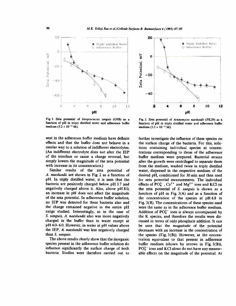
Zeta potentials of S. sanguis in triply distilledwater and adherence buffer media are shown inFig. 1 as a function of pH. Zeta potential valueswere observed to spread over a range, this isattributed to the distribution of bacteria at variousstages of growth. In all the figures this range isalso shown. In triply distilled water, it can be seenthat S. sanguis is positively charged below pH 3.2and negatively charged above it. In adherencebuffer solution. no isoelectric point (IEP) isobtained and the bacteria remained negativelycharged in the entire pH range studied Also, exceptfor pH 6-4, the magnitude of the potential inadherence buffer solution is higher than in triplydistilled water. This suggests that the species pre-
Table 2Effect of aging on the zeta potential of Streptococcus sanguisafter applying a potential of 67 V for 20 s following the firstmeasurement
Table 1Successive measurements of the zeta potential of Streptococcussanguis in adherence buffer solution"
Zeta potential,1st measurement(mY)
Zeta potentialafter aging (mY)
Aging time(min)
Trialno.
Zeta potential (mY)
1st 2nd 3rd 4th lit 2nd
-47 5 -59-;:80-43.5 10 -47.5 -55-45 15 -48 -53-43 20 -46 -53-46 30 -45-48
12.34
-42-45-47.5-45
-45
-53-59-53
-49-59-SO-63
-so-108
-102. Concentration, 5.2 x 10⁻² M; pH 6.8. Potential applied, 67 V.
90 MK Yellofi Rao et ai/Colloids Surfaces B: Biointerfaces 4 (1995) 87-95
20
~ 0-;:;jdIJ)
"0 -20Q.tU
-'IJ)
N
-40
a~ & 8 10 12.
PH pHFig. I. Zeta potential of Streptococcus sanguis (G98) as afunction of pH in triply distilled water and adherence buffermedium (5.2 x 10⁻¹ M).
Fig. 2. Zeta potential of Acti1Wmyces naeslundii (PK29) as afunction of pH in triply distilled water and adherence buffermedium (5.2 x 10⁻² M).
further investigate the influence of these species onthe surface charge of the bacteria. For this, solu-tions containing individual species at concen-trations corresponding to those of the adherencebuffer medium were prepared. Bacterial strainsafter the growth were centrifuged to separate themfrom the medium, washed twice in triply distilledwater, dispersed in the respective medium of thedesired pH, conditioned for 30 min and then usedfor zeta potential measurements. The individualeffects of PO~-, Ca2+ and Mg2+ ions and KCI onthe zeta potential of S. sanguis is shown as afunction of pH in Fig. 3(A) and as a function ofthe concentration of the species at pH 6.8 inFig. 3(B). The concentrations of these species usedwere the same as in the adherence buffer medium.Addition of PO~- ions is always accompanied bythe K species, and therefore the results were dis-cussed in terms of only phosphate addition. It canbe seen that the magnitude of the potentialdecreases with an increase in the concentration ofthe species (Fig. 3(B». However, at the concen-tration equivalent to that present in adherencebuffer medium (shown by arrows in Fig. 3(B»,PO~ - ions and KCI alone do not have any measur-
able effects on the magnitude of the potential. At
sent in the adherence buffer medium have definiteeffects and that the buffer does not behave in asimilar way to a solution of indifferent electrolytes.(An indifferent electrolyte does not alter the IEPof the interface or cause a charge reversal, butsimply lowers the magnitude of the zeta potentialwith increase in its concentration.)
Similar results of the zeta potential ofA. naeslundii are shown in Fig. 2 as a function ofpH. In triply distilled water, it is seen that thebacteria are positively charged below pH 3.7 andnegatively charged above it. Also, above pH 8.0,an increase in pH does not affect the magnitudeof the zeta potential. In adherence buffer solution,no IEP was detected for these bacteria also andthe charge remained negative in the entire pHrange studied Interestingly, as in the case ofS. sanguis, A. naeslundii also was more negativelycharged in the buffer than in water except atpH 4.0-6.0. However, in water at pH values abovethe IEP, A. naeslundii was less negatively chargedthan S. sanguis.
The above results clearly show that the inorganicspecies present in the adherence buffer solution doinfluence significantly the surface charge of bothbacteria. Studies were therefore carried out to

M.K YelJoji Roo et ai/Colloids Surfaces B: Biointerfaces 4 ( 1995) 87-95 91
~~:- ~ti
E-e~~Q>
l.,
A
~
~~
,
i~
40
~+0 Ca -~+ -
6 WI.
~.".~
pH
potential were presumably due to the specificadsorption of these ions on the bacterial surface.
The effect of the addition of two or more speciesat the concentrations used in the adherence bufferon the zeta potential of S. sanguis is shown inFig. 4 as a function of pH. MgZ + ions alone wereshown to make the bacteria less negatively chargedIt can be seen that subsequent addition of Ca2+ions reduced the potential even more. Furtheraddition of POi3 ions makes the bacteria morenegatively charged but not as much as it was whenMg2+ ions alone were present.
The effect of these ions on the zeta potential ofA. naeslundii is shown in Fig. 5(A) as a function ofpH and in Fig. 5(B) as a function of their concen-tration. The influence of Ca2+, MgZ+ and PO~-ions appears to be similar to that observed in thecase of S. sanguis. KCI, on the other hand, lowersthe zeta potential below and above the IEP.Evidently, KCI behaved in an expected manner,compressing the double layer in this case.
A comparison of the zeta potential resultsobtained in the presence of the whole adherencebuffer with those obtained in the presence ofindividual components clearly shows that the sumtotal effect is markedly different from the individual
20
>e.-.--
..~
,
£G
....
~higher concentrations, the observed decrease in themagnitude of the potential is due to the compres-sion of the electric double layer. Also, the lack ofany change in the IEP suggests that there is nospecific adsorption of PO~- ions (Fig. 3(A». Onthe other hand, both Ca2+ and Mg2+ ions inducesignificant changes in the surface charge of thebacteria, shifting the IEP from 3.2 to about 4. Thisshift in the IEP and the decrease in the zeta
-60
pJlFig. 4. Effect of PO~- (10-3 M), Ca2+ (10-3 M) and Mg2+(10-4 M) when present together on the zeta potential ofStreptococcus sanguis (G98) at pH 6.8.
0 PO ~-
on .. .~.., . h I . Tr:p\y Distilled Water.. K~\ T6 8 10 12
0
-~
-40

92 M.K Yelloji Roo et aL/ColJoidr Surfaces B: Biointerfaces 4 (1995) 87-95
20
\~iO ,. 2+ .
a Ca 2+0 Mg. PO 3-
46 Triply Distilled Water
~
> --
8
... 0-
...c""'0 -10a-ISt -20N
(A)30 'ti~}-1-1.-1-40 . ~..:_';~j;..,;
6 8 10~12
pJJ~
O;r~?"""'"1 IS}
5
a+0 Ca
2+0 y~
3. PO. ~ Jrt1
t
>e -10
G:;]~ -15"
...
0
C1.
., -20...
tI
N...
,J/'30 -!-
-b10
behavior. The lack of any KCI effect on S. sanguisand the observed increase in the negative potentialin the whole adherence buffer are, however, unex-pected. One possible reason for the absence ofeffects of Ca2+ and Mg2+ ions in the adheren~buffer may be due to the relative concentration ofthe latter two components (10-3 and 10-4 Mrespectively) in comparison with that of KCI(5 x 10⁻² M). Thus, KCI can compete with thecalcium and magnesium ions and reduce theirbinding markedly, or can mask their effects. Infact, using a Boltzmann-type expressionC. = Cbe( - zef,jkT)
the distribution of bivalent and monovalent cationsnear the charged surface at a concentration Cb of10-3 M of the bivalent cation and a concentrationC. of 2 x 10⁻² M of the monovalent cations andat a zeta potential' of 50 m V is significantly infavor of the monovalent ion (C.(K+)jC.(Ca2+)=7). Thus, in the absence of any specific interactionsbetween calcium and the bacteria, it can be saidthat K + ions can mask all the bivalent cation
effects. (Note that phosphate addition is in theform of a potassium salt but the above argumentis invalid here because of the low concentration ofthe latter.) In addition, K + ions may even displace
some of the multivalent cations associated withthe bacteria even in the absence of the addedbuffer. Such a removal would obviously increasethe negative potential of the bacteria. Yet anotherfactor which can reduce the binding of bivalentcations onto the surface is the phosphate specieswhich, by complexing with the bivalent cations,reduces their solution activity. Thus, it is possibleto account for the lack of reduction in the potentialand the observed increase in the negative potentialin adherence buffer solutions.
The lack of double layer compression effects isfurther illustrated in Fig. 6 which shows that thezeta potential of S. sanguis as a function of KCIconcentration remains constant up to salt levels ashigh as 5 x 10⁻² M. However, A. naeslundii doesshow decreasing zeta potential with increasingconcentrations of KCI (Fig. 7). It is interesting thatKCI by itself does not alter the potential ofS. sanguis. In this case, the double layer compres-sion effects are possibly compensated by the
component effects. Importantly, the sum total effectthat would be predicted from combining theobserved single component effects is opposite tothat of the experimentally observed increase in thenegative potential in the adherence buffer. Possiblereasons for this interesting effect are given below.
The absence of any effects of phosphate, andCa2+ and Mg2+ ions on the zeta potential of boththe bacteria are in accordance with the expected
--f--,"""-- 0/ -'--- ~ 10~5 10~4 10'-3 1~-2 1~-1
Concentration, W
Fig. 5. Effect or PO~- (10-3 M), Ca1+ (10-3 M), Mr+(10-4 M) and KG (5.0 x 10⁻¹ M) on the zeta potential orActilWmyces llaeslundii (PK29) (A) as a runction or pH and(B) as a runction or concentration.

M.K Ye/loji Rao et ai/Colloids Surfaces B: Biointerfaces4 (/995) 87-95 93
7,/, 1 1
!--~--.,~~ --~ - ~ ~PH 3.0 *=:::::: ::::+-1
-"'10", ,.
~20
~
~:;::.c=Q)
...0~'"
...Q)
N
~~~
.pH 40
ii, " .-~3r, ~~;:=j==~t=~
~
pH 8.8-40 /,/1-6 ":. I :i"::t'~"-i
0 10 10 10 10 10Concentration. M
Fig. 6. Effect of KO concentration on tbe ~ta potential of
Streptococcus SaIInis (09B\.
,
0 10 -
.J...;a ~1'
to to
Concentration. W
Fig. 8. Effect of Nfi.CO] concentration on the zeta potentialof Streptococcus sanguis (098).
~
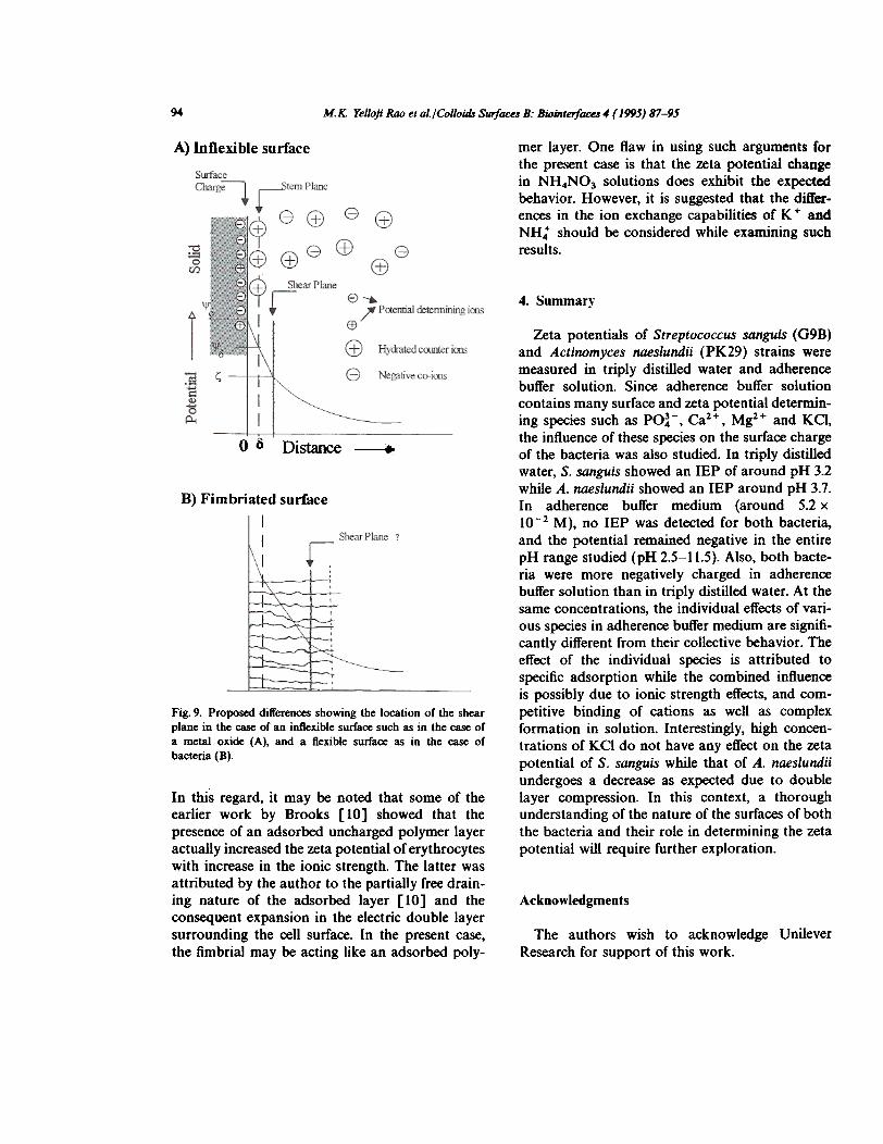
In all the above analyses, the nature of thebacterial surface and its effect on the measuredpotential have not been considered. It is importantto recognize that the zeta potential is a measureof the potential at the shear plane between thesolid (bacteria) and the liquid. The location of theshear plane in the case of an inflexible surface suchas that of a metal oxide is some distance awayfrom the physical surface bearing the electriccharge, and the measured potential is the potentialat that plane (Fig. 9(A». The surface of a bacteriumis complex with a saccharide envelope and fimbriaeextending into the solution. For example, the sur-face of S. sanguis is known to have a thick fuzzylayer with short fimbriae while A. naeslundii hasless densely populated, longer fimbriae [9]. Incontrast to the case for the inflexible surface, theshear plane for the fimbriated surface may be eitherwithin the fimbriated region or just at the fimbrialsurface (Fig. 9(B». The possibility that the shearplane is farther away from the fimbrial surface canbe ruled out, since the measured potential is inde-pendent of the KCI concentration. Dependingupon the actual location of the shear plane, thedistribution of the electrolytes in the fimbriatedregion and its dependence on concentration willgovern the changes in the measured zeta potential.
~~~~
> 10e
] 0-Ct)
~ -10.~N -20
~~~
~
~~~
increase in the negative charge because of thedisplacement of the multivalent ions associatedwith the bacteria. Note, however, that NH4NO3 atpH 6.8 does lower the zeta potential of S. sanguisat high concentrations (see Fig. 8). The differencesin the ion exchange capabilities of K + and NH:should be considered while examining such results.
~ .~~'-.." c
:c
"-80;G '"
"'tI -20N
-40
. ~ ~ -,
20
-30
0 10 10 10 10 10-1
Concentration. M
Fig. 7. Effect or Ka concentration on the zeta potential orActirwmyces naeslundii (PK29).
>e:! 0...~Q)
...0
~
94 M.K Yelloft Roo et aI/Colloids surfaces B: Biointerfaces 4 (1995) 87-95
mer layer. One flaw in using such arguments forthe present case is that the zeta potential changein NH4NO3 solutions does exhibit the expectedbehavior. However, it is suggested that the differ-ences in the ion exchange capabilities of K + and
NH: should be considered while examining suchresults.
A) Inflexible surface
4. Summary
Zeta potentials of Streptococcus sanguis (G9B)and Actinomyces naeslundii (PK29) strains weremeasured in triply distilled water and adherencebuffer solution. Since adherence buffer solutioncontains many sudace and zeta potential determin-ing species such as PO~-, Ca2+, Mg2+ and KCl,the influence of these species on the sudace chargeof the bacteria was also studied. In triply distilledwater, S. sanguis showed an IEP of around pH 3.2while A. naeslundii showed an IEP around pH 3.7.In adherence buffer medium (around 5.2 x10⁻² M), no IEP was detected for both bacteria,and the potential remained negative in the entirepH range studied (pH 2.5-11.5). Also, both bacte-ria were more negatively charged in adherencebuffer solution than in triply distilled water. At thesame concentrations, the individual effects of vari-ous species in adherence buffer medium are signifi-cantly different from their collective behavior. Theeffect of the individual species is attributed tospecific adsorption while the combined influenceis possibly due to ionic strength effects, and com-petitive binding of cations as well as complexformation in solution. Interestingly, high concen-trations of KCI do not have any effect on the zetapotential of S. sanguis while that of A. naeslundiiundergoes a decrease as expected due to doublelayer compression. In this context, a thoroughunderstanding of the nature of the sudaces of boththe bacteria and their role in determining the zetapotential will require further exploration.
0 ~ Distance ~
B) Fimbriated surface
Fig. 9. Proposed differences showing the location of the shearplane in the case of an intlexible surface such as in the case ofa metal oxide (A), and a flexible surface as in the case ofbacteria (8).
In this regard, it may be noted that some of theearlier work by Brooks [10] showed that thepresence of an adsorbed uncharged polymer layeractually increased the zeta potential of erythrocyteswith increase in the ionic strength. The latter wasattributed by the author to the partially free drain-ing nature of the adsorbed layer [10] and theconsequent expansion in the electric double layersurrounding the cell surface. In the present case,the fimbrial may be acting like an adsorbed poly-
Acknowledgments
The authors wish to acknowledge UnileverResearch for support of this work.

M.K Yelloj; Roo et aL/Co/low Surfaces B: Biointerfaces4 (1995) 87-95 9.1
References Oral Microbial Adhesion, American Society forMicrobiology, Washington, D.C., 1985, pp.77-84.
[5] P. Somasundaran, J. Colloid Interface sa., 27 (1968) 6S9.[6] C. Dawes, Arch. Oral BioL, 14 (1969) 277.[7] S.s. Socransky, A.D. Manganiello, D. Propos, V. Oram
and J. van Houte, J. Peridonl Res., 12 (1977) 90.[8] J.O. Cisar, P.E. Kolenbrander and F.c. Mclntice, Infect.
Immun., 24 (1979) 742.[9] P. Handley, Biofouling, 2 (1990) 239.
[10] D.E. Brooks, J. Colloid Interface sa., 43 (1973) 670.
[1] J. Olsson, P.O. Glantz and B. Krasse, Scand J. Dent.Res., 84 (1976) 240.
[2] M.M. Cowan, H.C. van der Mei, P.G. Rouxhet andHJ. Busscher, AppL Environ. Microbiol., 58 (1992) 1326.
[3] H.C. van der Mei, J.J. de Soet, J. de Graaft", P.G. Rouxbetand HJ. Busscher, Caries Res., 25 (1991) 415.
[4] RJ. Gibbons, I. Etherden and W. Peros, in S.E.Mergenhagen and B. Rosan (Eds.), Molecular Basis of


INSTRUCTIONS TO AUTHORS
Submission of ManuscriptsPlease submit the original and three copies of your manuscript. Instructions for the preparation of compulCripts arepublished in Colloids and Surfaces B: Biointemces. 1 (1993) 65-68. Enclose the original illustrations and three s~.of copies.Papers should be sent to one of the Editors:
Prof. J.l. Brash. McMaster University. Chemical Engineering Dept.. 1280 Main West, Hamilton, Onto l85 4l7, CanadaDr. H.J. Busscher, University of Groningen. laboratory for Materia Technica, Bloemsingel10, 9712 KZ Groningen, TheNetherlandsProf. H. Ohshima. Faculty of Pharmaceutical Sciences, Science University of Tokyo. 12 Ichigaya Funagawara-machi.Shinjuku-ku, Tokyo 162. JapanDr. D.W. Osborne, ViroTex Corporation. 4200 Research Forest Drive, Suite 350, The Woodlands. TX 77381. U.S.A.Dr. J. Wietfeldt, The Procter & Gamble Company, Miami Valley laboratory, P.O. Box 538707. Cincinnati, OH45253-8707. U.S.A.
Submission of an article is understood to imply that the article is original and unpublished and is not being consideredfor publication elsewhere. Upon acceptance of an article by the journal. the author(s) will be asked to transfer thecopyright of the article to the publisher. This transfer will ensure the widest possible dissemination of information.
Manuscript PreparationThe official language is English. The manuscript should be typewritten with double spacing and wide margins, and startwith the title followed by the name(s) of the author(s). their affiliations and an abstract of less than 500 words. Anabstract is not requested for brief notes. IUPAC guidelines should be followed for terminology and choosing symbolsand units as much as possible.
IUustrationsIllustrations should be numbered consecutively and referred to in the text. Drawings should be in a form suitable forreproduction, drawn in Indian ink on drawing paper or tracing paper. They should be completely lettered, the size ofthe lettering being appropriate to that of the drawings, but taking Into account the possible need for reduction in sizeof 50%. The page format of the journal should be considered in designing the drawings. Photographs must be of goodquality, printed on glossy paper. Figure captions should be supplied on a separate sheet.
Detailed suggestions and instructions to authors are published in Colloids and Surfaces B: Biointerlaces, 1 (1993) 65-68.A free reprint is available from the publisher upon request.
ProofsProofs will be sent to the author, to be checked for printer's errors. In case of two or more authors please indicate towhom the proofs should be sent.
ReprintsFifty reprints of each article will be supplied free of charge. Additional reprints can be ordered by the authors. An orderform containing price quotations will be sent to the authors together with the proofs.
AdvertisementsAdvertisement rates are available upon request.
0 1_. ElSEVIER SCENCE B.V. ALl RIGHTS RESERVED C827.~.IO
No part of this publicetion m.y be reproduced. Itor8d in . rwtriev81 ~ or Ir8n81nitt8d in 8nY fomI or by .ny ~. eiec:lronlc. mech8nal. PIIotoCOIIYina.recording or otherwise. ~ the prior written permission of the publillMr, EIMvier Science B.V~ Copyr~. Permisai- DeP8rtment. P.O. "Box 521; 10t0AM ~rd8m. The Necllerl8nd8.Upon ~ of .n 8r1icI8 by journ8i. !lie MIIhorCsI will be 88ked to ., copyriglC of 8rticIe to pu~. n.. IrM8ter will - me ~~ ~in8tion of -.fom.-jOn.Special ~1MionI for N8d8fs k\ \tie U.s.A. - This joum8l has ~ ~ Copyright a c r. Inc:. Cof'8ent is ~ for ~ ofMticIes for per8On8I or -.m8I -. or for ~ .- of 8IIeCific ~ Thie - is 8i-. on \tie condition ItI8t cop;... pay m-'8" C- -~ fee for copying beyond thM penniII8d by s-ioM 10? or 108 of U.s. Copyrigtc ~. The -~ fee is ~ In die code-line 81 III. bottom of
III. first pag. of 88eh .!tic.., The .ppropri8l. fee,1OgeIher willi . copy of the II.. page- of die .!tic.., should be forw.rded to the ~ht CI..r.nce C.nter,I~,. 222 ~ Drive. Oenwn. MA 01923. U.S.A. If no code-line .ppe8tS. broed - to copy has not ~ given .nd pennissoon to copy must beobt8ined directly fJOm die ~s). The fee Ind~ on the fi.. page of 8n .rtJde k\ lIIis -- will ~ retn)8Ctlwly to .11 .nicles pubiistled in the jown8I.r888nI.a ~ the - of pub/IcMIon. This - does ~ ~ to kinds of copying. IUCtI .. for ~ di8trbJtion. r-'-. 8dWftising 8ftd prOmoIionPUr-. or for cr e .- ~ worD. Speci8I --- ~ - be oIM8ined ~ pu~ for sudI COPYW'O. .No ~-"Iity . ~ by Pub("" for 8ny .rv 8Id/or 8n8ge to ~ or PfOCI81V .. . ..- of products N8bi1ity. negI~ or ottIerwiM.or ~ 8ny .- or 0Per8ti0n of My metIIod8. P'od-' ~ or ide88 ~ned In the rn8t8ri8I herein.AIthOIJgh .. 8dvertj.;ng ~I i. expected to conform to _al Cmedal) 8I8ndards, i~lulion in this publlc8l;on does not constitute. 8u--" or8ftdoraement of die ~Iity Or v.lue of such product or of III. d8lms made of It by ita m.nuf8eturer,
This i- is prlnl8d on 8dd.frM ~.
PNNTED IN THE ~