reproductive isolation among sympatric cryptic species in
TRANSCRIPT
ARTICLE IN PRESS
http://www.elsevier.de/protisAvailable online 7 December 2006
1
Correspondinfax +39 081 76e-mail amato@
& 2006 Elsevdoi:10.1016/j
158, 193—207, April 2007
Protist, Vol.ORIGINAL PAPER
Reproductive Isolation among Sympatric CrypticSpecies in Marine Diatoms
Alberto Amatoa,1, Wiebe H.C.F. Kooistraa, Jung Hee Levialdi Ghirona, David G. Mannb,Thomas Proscholdc, and Marina Montresora
aStazione Zoologica ‘A. Dohrn’, Villa Comunale, 80121 Napoli, ItalybRoyal Botanic Garden, Edinburgh EH3 5LR, Scotland, UKcCulture Collection of Algae and Protozoa, Scottish Association for Marine Science, Dunstaffnage MarineLaboratory, Dunbeg by Oban, Argyll PA37 1QA, Scotland, UK
Submitted August 18, 2006; Accepted October 1, 2006Monitoring Editor: Robert A. Andersen
Pseudo-nitzschia is a marine cosmopolitan genus of chain-forming planktonic diatoms. As for thevast majority of phytoplankton organisms, species identification within this genus mostly relies uponmorphological features. Taxa were initially identified based on cell shape and gross morphology oftheir composite silica cell wall, called the frustule. Yet, observations of the frustule in electronmicroscopy showed many additional characters for species identification and results of molecularstudies have demonstrated that genetically distinct groups might exist within morpho-species.However, these studies have not addressed the biological meaning of these genetic differences. Here,we bridge that gap by comparing ultrastructural features and sequence data (three ribosomal and oneplastid marker) of 95 strains with results of mating experiments among these strains. Experimentswere performed on two morphologically distinct entities: P. delicatissima and P. pseudodelicatissima.Each of the two entities consisted of multiple genetically distinct and reproductively isolated taxa, alloccurring in sympatry: P. delicatissima was composed of three phylogenetic and reproductivelydistinct groups, whereas P. pseudodelicatissima consisted of up to five. Once these taxa had beendefined both genetically and biologically, subtle ultrastructural differences could be detected as well.Our findings not only show that cryptic genetic variants abound in sympatry, but also that they arereproductively isolated and, therefore, biologically distinct units.& 2006 Elsevier GmbH. All rights reserved.
Key words: diatoms; cryptic diversity; Pseudo-nitzschia; reproductive isolation; species concept.
Introduction
Species are fundamental natural units (Coyne andOrr 2004) and their proper circumscription is anessential requirement for both biodiversity assess-ments and a correct understanding of theirecology, biogeography, evolutionary history, and
g author;41355szn.it (A. Amato).
ier GmbH. All rights reserved..protis.2006.10.001
speciation mechanisms. Within the unicellulareukaryote plankton, biodiversity estimates are stillbased mainly on morphological features butresults of recent molecular taxonomic assess-ments suggest that phytoplankton biodiversity isseriously underestimated. For instance, entirelynew lineages are being found (Guillou et al. 2004;Massana et al. 2004) as well as genetically distinctentities within morphologically delineated species

ARTICLE IN PRESS
194 A. Amato et al.
(Montresor et al. 2003; Saez et al. 2003; Sarnoet al. 2005). Cryptic and pseudo-cryptic speciesthus exist in phytoplankton, just as in other plantand animal groups (Knowlton 1993; Saez andLozano 2005). They are difficult or impossible todistinguish based on morphological characters,but hide divergence in habitat, physiology, or lifecycle traits, with obvious ecological implications.
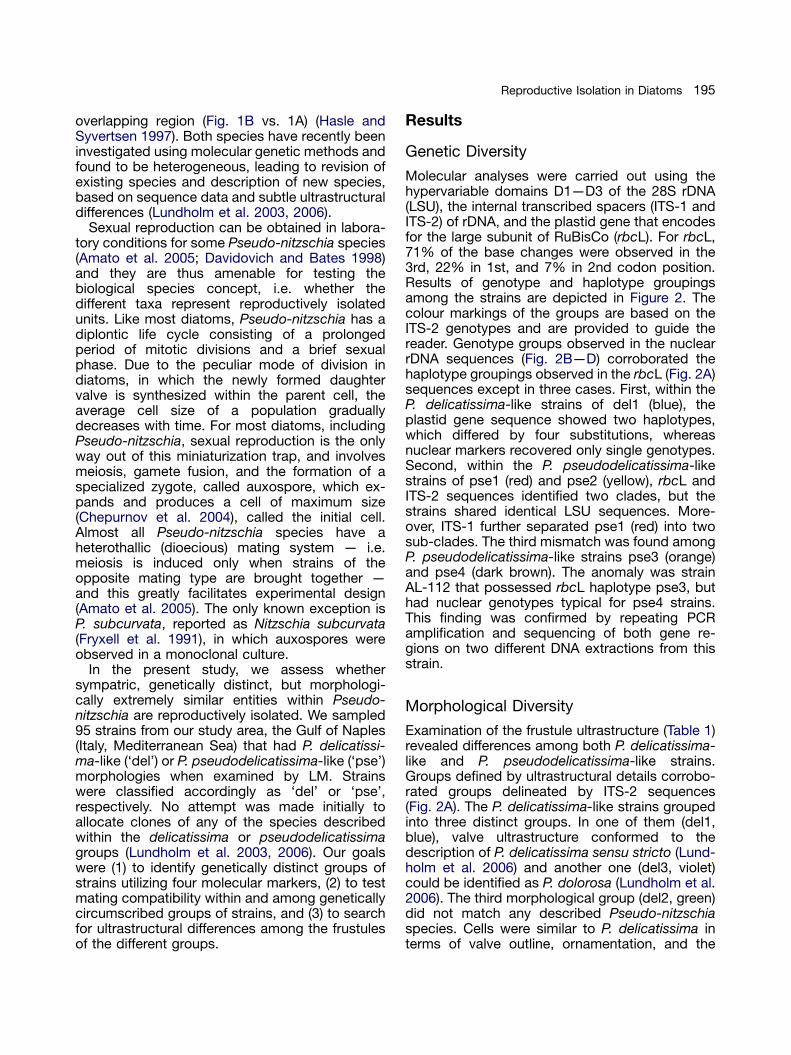
In the present study, we compare morphologicaland genetic species circumscriptions with matingdelineation in sympatric populations of the plank-tonic diatom genus Pseudo-nitzschia. This genusis an important constituent of coastal and oceanicphytoplankton blooms and some of its species arenotorious for producing domoic acid, a neurotoxinresponsible for Amnesic Shellfish Poisoning (ASP;Mos 2001). The needle-shaped cells form steppedchains by cells holding on to one another at theirapices (Fig. 1A,B). Subtle morphological differ-ences in cell shape and size and in the over-lapping patterns of cell apices in the chainrepresent the only characters for identifyingPseudo-nitzschia species by light microscopy(LM). Detailed examination of their compoundsilica cell wall, called the frustule, using electronmicroscopy (Fig. 1C,D), is generally necessary foraccurate species identification (Hasle and Syvert-sen 1997).
The frustule is composed of two valves andaccompanying sets of girdle bands. In Pseudo-
Figure 1. Micrographs from light (A,B) and transmisscalliantha pse4 (A—C) and Pseudo-nitzschia delicatissimregion between two adjacent cells in a chain, dotted arrthe two central fibulae, solid ellipses the striae, dottedScale bars: 20 mm (A,B), 1 mm (C,D).
nitzschia, each valve possesses a slit, called theraphe, which runs along the whole length of thevalve and is reinforced on the interior side withsilica bridges, called fibulae (Fig. 1C,D; dottedarrows). In some species (including those treatedhere), the raphe slit is interrupted centrally by asmall thickened nodule and the fibulae on eitherside of this nodule are more widely spaced(Fig. 1C,D; solid arrows). The valve is ornamentedwith striae (Fig. 1C,D; solid ellipses) — weaklysilicified strips containing small poroids (Fig. 1C,D;arrowheads) — alternating with more heavilysilicified, rib-like strips, called interstriae (Fig. 1C,D;dotted ellipses). The poroids may be subdividedby sets of struts (cribra, Fig. 1C) resembling thetracery of stained glass windows and they are alsooccluded by thin, porous silica sheets (hymens).
Morphological investigations by Hasle (Hasle1965; Hasle and Syvertsen 1997) showed con-siderable diversity in valve structure and orna-mentation and led to the description of around 20species. These included P. delicatissima andP. pseudodelicatissima, called Nitzschia actydro-phila and N. delicatula by Hasle (1965), which,although having similarly short overlaps betweencells in chains, could be separated by cell shapeusing LM: P. delicatissima cells have truncatedends in girdle view, whereas P. pseudodelicatissi-ma cells have pointed ends and their valves aremore linear, with a sharper differentiation of the
ion electron microscopy (C,D) of Pseudo-nitzschiaa del1 (B—D). Solid circles indicate the overlapping
ows the fibulae, solid arrows the wider separation ofellipses the interstriae, and arrowheads the poroids.
ARTICLE IN PRESS
195Reproductive Isolation in Diatoms
overlapping region (Fig. 1B vs. 1A) (Hasle andSyvertsen 1997). Both species have recently beeninvestigated using molecular genetic methods andfound to be heterogeneous, leading to revision ofexisting species and description of new species,based on sequence data and subtle ultrastructuraldifferences (Lundholm et al. 2003, 2006).
Sexual reproduction can be obtained in labora-tory conditions for some Pseudo-nitzschia species(Amato et al. 2005; Davidovich and Bates 1998)and they are thus amenable for testing thebiological species concept, i.e. whether thedifferent taxa represent reproductively isolatedunits. Like most diatoms, Pseudo-nitzschia has adiplontic life cycle consisting of a prolongedperiod of mitotic divisions and a brief sexualphase. Due to the peculiar mode of division indiatoms, in which the newly formed daughtervalve is synthesized within the parent cell, theaverage cell size of a population graduallydecreases with time. For most diatoms, includingPseudo-nitzschia, sexual reproduction is the onlyway out of this miniaturization trap, and involvesmeiosis, gamete fusion, and the formation of aspecialized zygote, called auxospore, which ex-pands and produces a cell of maximum size(Chepurnov et al. 2004), called the initial cell.Almost all Pseudo-nitzschia species have aheterothallic (dioecious) mating system — i.e.meiosis is induced only when strains of theopposite mating type are brought together —and this greatly facilitates experimental design(Amato et al. 2005). The only known exception isP. subcurvata, reported as Nitzschia subcurvata(Fryxell et al. 1991), in which auxospores wereobserved in a monoclonal culture.
In the present study, we assess whethersympatric, genetically distinct, but morphologi-cally extremely similar entities within Pseudo-nitzschia are reproductively isolated. We sampled95 strains from our study area, the Gulf of Naples(Italy, Mediterranean Sea) that had P. delicatissi-ma-like (‘del’) or P. pseudodelicatissima-like (‘pse’)morphologies when examined by LM. Strainswere classified accordingly as ‘del’ or ‘pse’,respectively. No attempt was made initially toallocate clones of any of the species describedwithin the delicatissima or pseudodelicatissimagroups (Lundholm et al. 2003, 2006). Our goalswere (1) to identify genetically distinct groups ofstrains utilizing four molecular markers, (2) to testmating compatibility within and among geneticallycircumscribed groups of strains, and (3) to searchfor ultrastructural differences among the frustulesof the different groups.
Results
Genetic Diversity
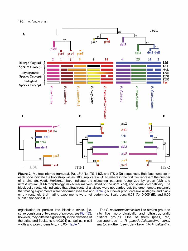
Molecular analyses were carried out using thehypervariable domains D1—D3 of the 28S rDNA(LSU), the internal transcribed spacers (ITS-1 andITS-2) of rDNA, and the plastid gene that encodesfor the large subunit of RuBisCo (rbcL). For rbcL,71% of the base changes were observed in the3rd, 22% in 1st, and 7% in 2nd codon position.Results of genotype and haplotype groupingsamong the strains are depicted in Figure 2. Thecolour markings of the groups are based on theITS-2 genotypes and are provided to guide thereader. Genotype groups observed in the nuclearrDNA sequences (Fig. 2B—D) corroborated thehaplotype groupings observed in the rbcL (Fig. 2A)sequences except in three cases. First, within theP. delicatissima-like strains of del1 (blue), theplastid gene sequence showed two haplotypes,which differed by four substitutions, whereasnuclear markers recovered only single genotypes.Second, within the P. pseudodelicatissima-likestrains of pse1 (red) and pse2 (yellow), rbcL andITS-2 sequences identified two clades, but thestrains shared identical LSU sequences. More-over, ITS-1 further separated pse1 (red) into twosub-clades. The third mismatch was found amongP. pseudodelicatissima-like strains pse3 (orange)and pse4 (dark brown). The anomaly was strainAL-112 that possessed rbcL haplotype pse3, buthad nuclear genotypes typical for pse4 strains.This finding was confirmed by repeating PCRamplification and sequencing of both gene re-gions on two different DNA extractions from thisstrain.
Morphological Diversity
Examination of the frustule ultrastructure (Table 1)revealed differences among both P. delicatissima-like and P. pseudodelicatissima-like strains.Groups defined by ultrastructural details corrobo-rated groups delineated by ITS-2 sequences(Fig. 2A). The P. delicatissima-like strains groupedinto three distinct groups. In one of them (del1,blue), valve ultrastructure conformed to thedescription of P. delicatissima sensu stricto (Lund-holm et al. 2006) and another one (del3, violet)could be identified as P. dolorosa (Lundholm et al.2006). The third morphological group (del2, green)did not match any described Pseudo-nitzschiaspecies. Cells were similar to P. delicatissima interms of valve outline, ornamentation, and the
ARTICLE IN PRESS
Figure 2. ML tree inferred from rbcL (A), LSU (B), ITS-1 (C), and ITS-2 (D) sequences. Boldface numbers ineach node indicate the bootstrap values (1000 replicates). (A) Numbers in the first row represent the numberof strains analysed. Horizontal bars indicate the clustering patterns recognized by gross (LM) andultrastructural (TEM) morphology, molecular markers (listed on the right side), and sexual compatibility. Theblack solid rectangle indicates that ultrastructural analyses were not carried out, the green empty rectanglethat mating experiments were performed (see text and Table 2) but never produced sexual stages, and blackempty rectangle that mating experiments were not performed. Scale bars: 0.01 (A), 0.005 (B), and 0.05substitutions/site (C,D).
196 A. Amato et al.
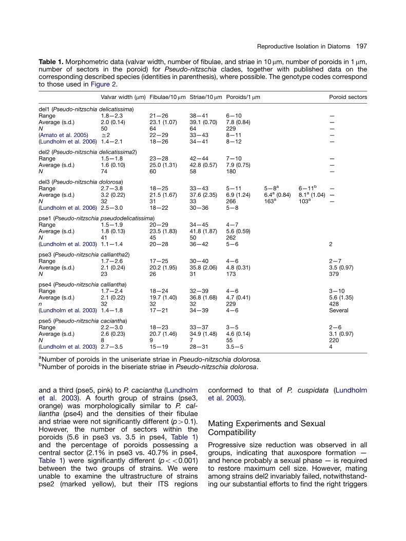
organization of poroids into biseriate striae, (i.e.striae consisting of two rows of poroids; see Fig. 1D);however, they differed significantly in the densities ofthe striae and fibulae (poo0.001) as well as in cellwidth and poroid density (po0.05) (Table 1).
The P. pseudodelicatissima-like strains groupedinto five morphologically and ultrastructurallydistinct groups. One of them (pse1, red)corresponded to P. pseudodelicatissima sensustricto, another (pse4, dark brown) to P. calliantha,
ARTICLE IN PRESS
Table 1. Morphometric data (valvar width, number of fibulae, and striae in 10 mm, number of poroids in 1 mm,number of sectors in the poroid) for Pseudo-nitzschia clades, together with published data on thecorresponding described species (identities in parenthesis), where possible. The genotype codes correspondto those used in Figure 2.
Valvar width (mm) Fibulae/10 mm Striae/10 mm Poroids/1 mm Poroid sectors
del1 (Pseudo-nitzschia delicatissima)Range 1.8—2.3 21—26 38—41 6—10 —Average (s.d.) 2.0 (0.14) 23.1 (1.07) 39.1 (0.70) 7.8 (0.84) —N 50 64 64 229 —(Amato et al. 2005) ffi2 22—29 33—43 8—11 —(Lundholm et al. 2006) 1.4—2.1 18—26 34—41 8—12 —
del2 (Pseudo-nitzschia delicatissima2)Range 1.5—1.8 23—28 42—44 7—10 —Average (s.d.) 1.6 (0.10) 25.0 (1.31) 42.8 (0.57) 7.9 (0.75) —N 74 60 58 180 —
del3 (Pseudo-nitzschia dolorosa)Range 2.7—3.8 18—25 33—43 5—11 5—8a 6—11b —Average (s.d.) 3.2 (0.22) 21.5 (1.67) 37.6 (2.35) 6.9 (1.24) 6.4a (0.84) 8.1a (1.04) —N 32 31 33 266 163a 103a —(Lundholm et al. 2006) 2.5—3.0 18—22 30—36 5—8
pse1 (Pseudo-nitzschia pseudodelicatissima)Range 1.5—1.9 20—29 34—45 4—7Average (s.d.) 1.8 (0.13) 23.5 (1.83) 41.8 (1.87) 5.6 (0.59)N 41 45 50 262(Lundholm et al. 2003) 1.1—1.4 20—28 36—42 5—6 2
pse3 (Pseudo-nitzschia calliantha2)Range 1.7—2.6 17—25 30—40 4—6 2—7Average (s.d.) 2.1 (0.24) 20.2 (1.95) 35.8 (2.06) 4.8 (0.31) 3.5 (0.97)N 23 26 31 173 379
pse4 (Pseudo-nitzschia calliantha)Range 1.7—2.4 18—24 32—39 4—6 3—10Average (s.d.) 2.1 (0.22) 19.7 (1.40) 36.8 (1.68) 4.7 (0.41) 5.6 (1.35)n 32 32 32 229 428(Lundholm et al. 2003) 1.4—1.8 17—21 34—39 4—6 Several
pse5 (Pseudo-nitzschia caciantha)Range 2.2—3.0 18—23 33—37 3—5 2—6Average (s.d.) 2.6 (0.23) 20.7 (1.46) 34.9 (1.48) 4.6 (0.14) 3.1 (0.97)N 8 9 7 55 220(Lundholm et al. 2003) 2.7—3.5 15—19 28—31 3.5—5 4
aNumber of poroids in the uniseriate striae in Pseudo-nitzschia dolorosa.bNumber of poroids in the biseriate striae in Pseudo-nitzschia dolorosa.
197Reproductive Isolation in Diatoms
and a third (pse5, pink) to P. caciantha (Lundholmet al. 2003). A fourth group of strains (pse3,orange) was morphologically similar to P. cal-liantha (pse4) and the densities of their fibulaeand striae were not significantly different (p40:1).However, the number of sectors within theporoids (5.6 in pse3 vs. 3.5 in pse4, Table 1)and the percentage of poroids possessing acentral sector (2.1% in pse3 vs. 40.7% in pse4,Table 1) were significantly different (poo0:001)between the two groups of strains. We wereunable to examine the ultrastructure of strainspse2 (marked yellow), but their ITS regions
conformed to that of P. cuspidata (Lundholmet al. 2003).
Mating Experiments and SexualCompatibility
Progressive size reduction was observed in allgroups, indicating that auxospore formation —and hence probably a sexual phase — is requiredto restore maximum cell size. However, matingamong strains del2 invariably failed, notwithstand-ing our substantial efforts to find the right triggers

ARTICLE IN PRESS
198 A. Amato et al.
(723 attempts, testing temperature and day-lengthsettings matching those recorded in the Gulf ofNaples during different seasons, see Table 2). Itseems unlikely that the 25 strains belonged to thesame mating type because they were isolatedfrom net samples collected on different dates(Table 3). In all other genotypes, the mating-typeratio was close to 1:1, and even among smallnumbers of isolates, opposite mating types werealways recorded. Many diatoms have relativelynarrow size windows for sexualization, but in del2,we failed to obtain any sexual reproduction amongcells between 62 and 15mm in length.
All groups within which sexual reproduction wassuccessful showed a heterothallic mating system;homothally — sexual reproduction within a clonalculture — was never observed. Features of thesexual cycle matched those described for P.delicatissima (del1) (Amato et al. 2005). Matingexperiments were carried out to verify sexualcompatibility within genotypic groups and toassess mating boundaries amongst them(Fig. 2A, Table 2). Successful crosses weredefined as those in which auxospores producedenlarged initial cells capable of dividing, giving risewithin a few days to short chains of F1 cells.Successful crosses were restricted to crosses
Table 2. Matrix of crosses. Number of crosses performebasing on the ITS-2 clades (see Fig. 2). Grey boxesboxes ¼ sexual reproduction not observed; black boxe
made within ITS-2 clades. Interclade crossesbetween strains belonging to pse3 and pse4(including strain AL-112, the one with nucleargenotype pse4 but rbcL haplotype pse3) led tosuccessful pairing of gametangia and conjugationof gametes, but the resulting zygotes neverdeveloped into auxospores.
ITS-2 Secondary Structure
To identify genetic characteristics at the specieslevel and below, we compared the secondarystructure of ITS-2 rDNA sequences for all geno-types and determined the Compensatory BaseChanges (CBC including Hemi-CBC [HCBC],changes on one side only) according to Coleman(2000, 2003). Results of the comparisons areprovided in Figure 3. The secondary structures ofall strains had the same organization with fourhelices (I, II, III, and IV); an additional helix (IIa),which is characteristic for Pseudo-nitzschia andsome other diatoms, was identified betweenhelices II and III. The ITS-2 rDNA sequencesdiffered in length and in the composition of theend-loops in the helices (Fig. 3). The conservedregions of ITS-2 are highlighted in boldface inFigure 3. The closely related P. pseudodelicatissima
d within and among the different genotypes identified¼ successful sexual reproduction observed; whites ¼ combination not tested.
_
_ _ _
_
ARTICLE IN PRESS
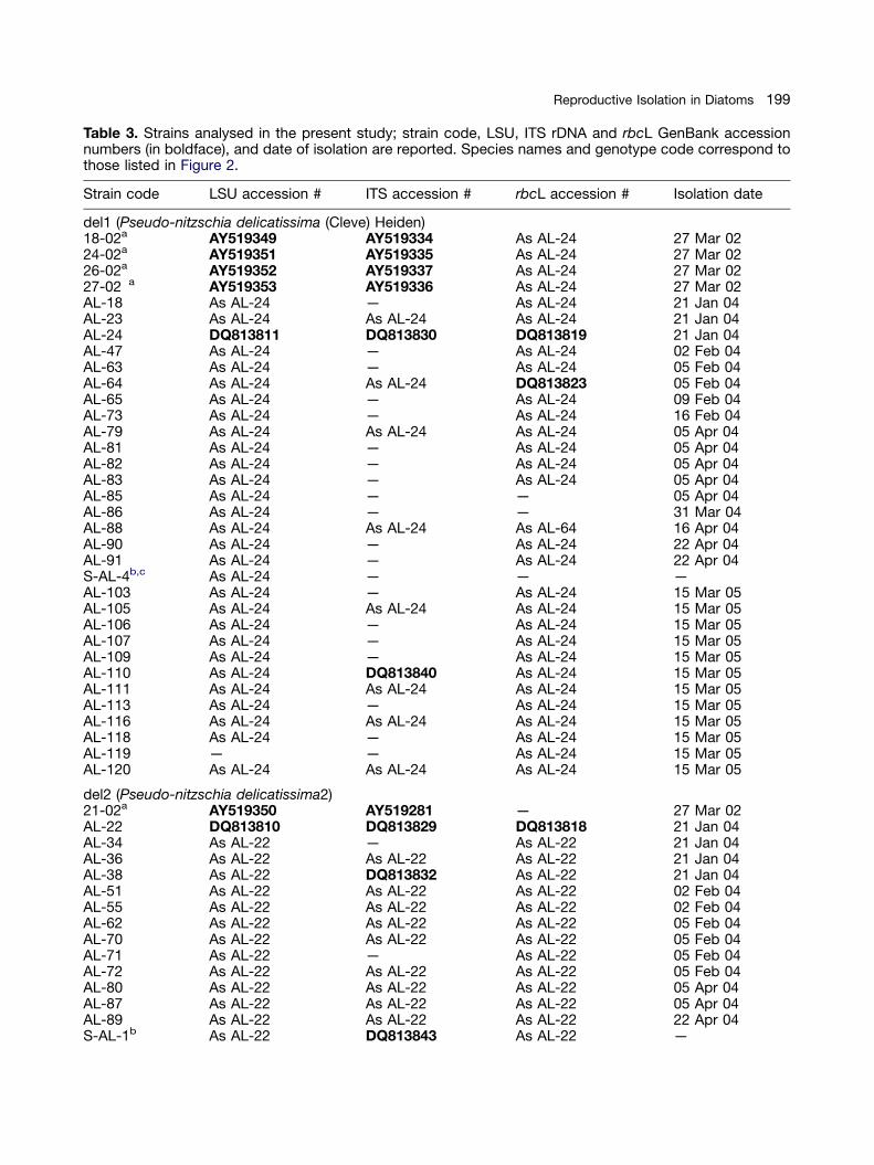
Table 3. Strains analysed in the present study; strain code, LSU, ITS rDNA and rbcL GenBank accessionnumbers (in boldface), and date of isolation are reported. Species names and genotype code correspond tothose listed in Figure 2.
Strain code LSU accession # ITS accession # rbcL accession # Isolation date
del1 (Pseudo-nitzschia delicatissima (Cleve) Heiden)18-02a AY519349 AY519334 As AL-24 27 Mar 0224-02a AY519351 AY519335 As AL-24 27 Mar 0226-02a AY519352 AY519337 As AL-24 27 Mar 0227-02 a AY519353 AY519336 As AL-24 27 Mar 02AL-18 As AL-24 — As AL-24 21 Jan 04AL-23 As AL-24 As AL-24 As AL-24 21 Jan 04AL-24 DQ813811 DQ813830 DQ813819 21 Jan 04AL-47 As AL-24 — As AL-24 02 Feb 04AL-63 As AL-24 — As AL-24 05 Feb 04AL-64 As AL-24 As AL-24 DQ813823 05 Feb 04AL-65 As AL-24 — As AL-24 09 Feb 04AL-73 As AL-24 — As AL-24 16 Feb 04AL-79 As AL-24 As AL-24 As AL-24 05 Apr 04AL-81 As AL-24 — As AL-24 05 Apr 04AL-82 As AL-24 — As AL-24 05 Apr 04AL-83 As AL-24 — As AL-24 05 Apr 04AL-85 As AL-24 — — 05 Apr 04AL-86 As AL-24 — — 31 Mar 04AL-88 As AL-24 As AL-24 As AL-64 16 Apr 04AL-90 As AL-24 — As AL-24 22 Apr 04AL-91 As AL-24 — As AL-24 22 Apr 04S-AL-4b,c As AL-24 — — —AL-103 As AL-24 — As AL-24 15 Mar 05AL-105 As AL-24 As AL-24 As AL-24 15 Mar 05AL-106 As AL-24 — As AL-24 15 Mar 05AL-107 As AL-24 — As AL-24 15 Mar 05AL-109 As AL-24 — As AL-24 15 Mar 05AL-110 As AL-24 DQ813840 As AL-24 15 Mar 05AL-111 As AL-24 As AL-24 As AL-24 15 Mar 05AL-113 As AL-24 — As AL-24 15 Mar 05AL-116 As AL-24 As AL-24 As AL-24 15 Mar 05AL-118 As AL-24 — As AL-24 15 Mar 05AL-119 — — As AL-24 15 Mar 05AL-120 As AL-24 As AL-24 As AL-24 15 Mar 05
del2 (Pseudo-nitzschia delicatissima2)21-02a AY519350 AY519281 — 27 Mar 02AL-22 DQ813810 DQ813829 DQ813818 21 Jan 04AL-34 As AL-22 — As AL-22 21 Jan 04AL-36 As AL-22 As AL-22 As AL-22 21 Jan 04AL-38 As AL-22 DQ813832 As AL-22 21 Jan 04AL-51 As AL-22 As AL-22 As AL-22 02 Feb 04AL-55 As AL-22 As AL-22 As AL-22 02 Feb 04AL-62 As AL-22 As AL-22 As AL-22 05 Feb 04AL-70 As AL-22 As AL-22 As AL-22 05 Feb 04AL-71 As AL-22 — As AL-22 05 Feb 04AL-72 As AL-22 As AL-22 As AL-22 05 Feb 04AL-80 As AL-22 As AL-22 As AL-22 05 Apr 04AL-87 As AL-22 As AL-22 As AL-22 05 Apr 04AL-89 As AL-22 As AL-22 As AL-22 22 Apr 04S-AL-1b As AL-22 DQ813843 As AL-22 —
199Reproductive Isolation in Diatoms

ARTICLE IN PRESS
Table 3. (continued )
Strain code LSU accession # ITS accession # rbcL accession # Isolation date
S-AL-2b As AL-22 — As AL-22 —AL-94 As AL-22 As AL-22 As AL-22 18 Jun 04AL-95 As AL-22 — As AL-22 18 Jun 04AL-95bis As AL-22 As AL-22 As AL-22 18 Jun 04AL-95ter As AL-22 As AL-22 As AL-22 18 Jun 04AL-96 As AL-22 As AL-22 As AL-22 18 Jun 04AL-97 As AL-22 As AL-22 As AL-22 18 Jun 04AL-97bis As AL-22 As AL-22 As AL-22 18 Jun 04AL-97ter As AL-22 As AL-22 As AL-22 18 Jun 04AL-97quater As AL-22 — — 18 Jun 04
del3 (Pseudo-nitzschia dolorosa Lundholm et Moestrup)20-02a AY519348 AY519275 — 27 Mar 02AL-59 DQ813813 DQ813835 DQ813822 02 Feb 04AL-67 As AL-59 DQ813837 As AL-59 05 Feb 04AL-69 — — As AL-59 05 Feb 04AL-74 As AL-59 DQ813838 As AL-59 16 Feb 04C-AL-2c As AL-59 As AL-59 As AL-59 25 Feb 04
pse1 (Pseudo-nitzschia pseudodelicatissima (Hasle) Hasle emend. Lundholm, Hasle et Moestrup)SZN-B109a AY550126 As AL-41 — 04 Jun 02SZN-B111a AY550128 As AL-41 — 04 Jun 02SZN-B112a AY550127 — — 04 Jun 02SZN-B113a AY550129 — — 04 Jun 02AL-15 DQ813808 DQ813826 DQ813817 19 Jan 04AL-19 As AL-15 DQ813828 As AL-15 21 Jan 04AL-20 As AL-15 As AL-19 As AL-15 21 Jan 04AL-21 As AL-15 As AL-41 As AL-15 21 Jan 04AL-27 As AL-15 As AL-19 As AL-15 26 Jan 04AL-29 As AL-15 DQ813831 — 26 Jan 04AL-31 As AL-15 — — 26 Jan 04AL-40 As AL-15 As AL-19 As AL-15 02 Feb 04AL-41 As AL-15 DQ813833 As AL-15 02 Feb 04AL-60 As AL-15 DQ813836 As AL-15 02 Feb 04
pse2 (Pseudo-nitzschia cuspidata (Hasle) Hasle emend. Lundholm, Moestrup et Hasle)AL-17 DQ813809 DQ813827 — 19 Jan 04AL-28 As AL-15 As AL-17 DQ813820 26 Jan 04AL-57 As AL-15 As AL-17 As AL-28 02 Feb 04AL-61 As AL-15 As AL-17 As AL-28 02 Feb 04
pse3 (Pseudo-nitzschia calliantha 2)P2a As AL-101 — — 17 Jan 02P4a DQ813816 — — 17 Jan 02P5a As AL-101 As AL-101 — 17 Jan 02P6a — As AL-101 — 17 Jan 02AL-11 As AL-101 — As AL-101 07 Oct 03AL-13 As AL-101 — As AL-101 07 Oct 03AL-101 DQ813814 DQ813839 DQ813824 14 Oct 04C-AL-1c As AL-101 DQ813842 As AL-101 25 Feb 04
pse4 (Pseudo-nitzschia calliantha Lundholm, Moestrup et Hasle)AL-112 DQ813815 DQ813841 As AL-101 15 Mar 05AL-117 As AL-112 As AL-112 DQ813825 15 Mar 05
200 A. Amato et al.

ARTICLE IN PRESS
Table 3. (continued )
Strain code LSU accession # ITS accession # rbcL accession # Isolation date
pse5 (Pseudo-nitzschia caciantha Lundholm, Moestrup et Hasle)AL-46c As AL-56 — As AL-56 26 Jan 04AL-56c DQ813812 DQ813834 DQ813821 02 Feb 04
aStrain isolated by Luisa Orsini.bStrain isolated by Sarah M. McDonald.cStrain not used in crossing experiments:— ¼ information not available.
Figure 3. ITS-2 rRNA secondary structure models of (A) Pseudo-nitzschia pseudodelicatissima (strain AL-15;pse1) and (B) Pseudo-nitzschia calliantha (strain AL-112; pse4). The base pairs marked in grey indicatecompensatory base changes (CBC or HCBC) compared to strains AL-17 (pse2) in (A) and AL-101 (pse3) in(B). In circles are marked single base changes and in boxes the changes of the ends in the helices betweenthe strains respectively. The conserved regions of ITS-2 are highlighted in bold.
201Reproductive Isolation in Diatoms
(pse1) and P. cuspidata (pse2) (Fig. 3A) differed intwo HCBCs (marked in grey in Fig. 3A) occurring inhelix I (U:A 2 U:G and G:C 2 G:U), three in helix
III (A:U 2 G:U, G:U 2 A:U, and U:G 2 U:A), andone CBC in helix IV (U:A 2 C:G). In P. calliantha(pse4) and P. calliantha2 (pse3) (Fig. 3B), two

ARTICLE IN PRESS
202 A. Amato et al.
HCBCs (marked in grey in Fig. 3B) were recorded inhelix I, G:U 2 G:C, U:U 2 A:U; the latter involvinga non-canonical but proven pair (Gutell 1994). Twobase changes that break two bonds, A:U 2 A Cand A:U 2 C U, were recorded in helix III; the lattercould produce the non-canonical pair Y:Y (Gutell1994).
Discussion
Our data show that groups of Pseudo-nitzschiastrains distinguished on the basis of four mole-cular markers are reproductively isolated. Since allthese genetically distinct groups occur in sympa-try in the Gulf of Naples, they must constitutebiologically distinct species. Genetically and bio-logically distinct species within the P. delicatissi-ma-like group reveal subtle ultrastructuraldifferences, and the same applies for thosewithin the P. pseudodelicatissima-like group.However, identification is extremely difficult andthe ranges of morphometric examined charactersoften overlap. Thus, species within each of thesegroups can be defined as pseudo-cryptic (semi-cryptic sensu Mann 1999) species.
We tested genetic relatedness among thedifferent strains using three nuclear and oneplastid marker that are widely used in assessingrelationships at the species level or below (Lund-holm et al. 2003; Paul et al. 2000; Sarno et al.2005). Of these four markers, ITS-2 appears to bethe only one that discriminates accurately be-tween biological species. Two strains matedsuccessfully only if they had ITS-2 sequenceswith identical helix regions. The LSU region is nota perfect discriminator because in one case, thisregion did not separate two different species(pse1 and pse2 ¼ P. pseudodelicatissima andP. cuspidata) that failed to interbreed and pos-sessed different rbcL and ITS sequences. TherbcL marker is not a perfect discriminator either,because pairs of P. delicatissima (del1) strains withdifferent rbcL genotypes mated successfully, i.e.they generated a perfectly viable and fertile F1generation. These two groups of strains pos-sessed identical LSU and ITS sequences. Appar-ently, different rbcL haplotypes can occur within asingle interbreeding population. ITS-1 is alsounsatisfactory as a biological species discrimina-tor, because compatible P. pseudodelicatissima(pse1) strains generate a viable F1 generation,irrespective of their ITS-1 genotype.
Phylogenetic trees based on ribosomal andchloroplast markers show minor differences in
the ramifications, but they generally corroborateone another in the grouping of sequences inend nodes. There are four deviations fromthis concordance. (1) Single interbreeding speciesP. delicatissima (del1) possesses two rbcL types.The polymorphism may be recent or ancient(Takahashi et al. 2001) and may have arisen beforeor after separation of del1 from other lineages. It ispossible that one of the variants was introducedby hybridization with some other unknown ‘del’lineage. (2) Within P. calliantha, strain AL-112 hasthe rbcL haplotype of pse3, but shares the LSUand ITS genotypes with pse4. It also has theultrastructure of pse4 and interbreeds success-fully with the compatible strain of pse4, but notwith the ones of pse3. Again, it is possible that thispattern arose through introgression, followed byrepeated backcrossing of the hybrid offspring withP. calliantha pse4. (3) The different ITS-1 se-quences found in P. pseudodelicatissima (pse1)could be interpreted as a case of polymorphismnot yet homogenized by concerted evolution,and the region in which it occurs could be afast-evolving one. Anyway, the strains shareidentical sequences at all other markers investi-gated and interbreed freely. (4) The shared LSUgenotype of P. pseudodelicatissima (pse1, red)and P. cuspidata (pse2, yellow) is probablybecause the LSU region generally evolves moreslowly than the ITS regions. The other markersindicate that the two taxa are close relatives,suggesting that the LSU marker has not hadenough time to accumulate differences.
Although ITS-1 and ITS-2 are transcribedregions but are not part of the mature ribosomes,they have complex secondary structures and playa crucial role in the construction of ribosomes(Tschochner and Hurt 2003). Thus, the sequencescorresponding to the helix regions are functionallyconstrained and only those few base changes thatdo not interfere with the proper folding of theprimary transcripts (CBCs or HCBCs) are permis-sible (Gutell 1994). The secondary structures ofthe ITS-2 region in Pseudo-nitzschia speciesanalysed in the present study show the distincteukaryotic hallmarks: (1) four main helices, (2) ofwhich helix III is the longest, (3) the presence of acharacteristic motif at the apex of helix III, and (4)a pyrimidine-pyrimidine mismatch in helix II(Schultz et al. 2005). A correlation between ITS-2sequence evolution and sexual compatibility wasshown for green freshwater microalgae of theorder Volvocales (Chlorophyta) and for the ciliateParamecium aurelia (Coleman 2000, 2005; Fabryet al. 1999). Reproductive isolation occurred if

ARTICLE IN PRESS
203Reproductive Isolation in Diatoms
there were CBCs or HCBCs, but the reverse wasnot necessarily true: some pairs of Parameciumsyngens were effectively reproductively isolated,despite having identical ITS sequences. ThePseudo-nitzschia ITS-2 genotypes analysed inthe present study belong to different CBC-cladesand do not show any sexual interactions witheach other. The only exception concerns pse4(P. calliantha) and pse3, whose ITS-2 differed bythree HCBCs but where the gametangia stillrecognized one another as potential mates andgametes fused into a zygote. However, that iswhere the process stopped; the zygote failed todevelop into an auxospore. The most closelyrelated species grouping in different CBC-cladeswere P. pseudodelicatissima (pse1) and P. cuspi-data (pse2). In fact, they differ by only one CBC(and five HCBCs) and their cells did not evenrecognize one another as potential partners. Thisfinding confirms that strains clustering in differentCBC-clades are sexually incompatible.
ITS-2 secondary structures are available for onlyvery few microalgae; nevertheless, these few resultsalready reveal that the number of CBCs (includingHCBCs) at which reproductive isolation sets indiffers among phylogenetic groups. Mating com-patibility among strains of the chlorophyceanGonium pectorale is compromised if they differ byeven one HCBC in conserved helix regions (Cole-man 2000). Behnke et al. (2004) found variations inITS rDNA sequences between locally interbreedingpopulations of the benthic freshwater diatom Sell-aphora pupula. However, the secondary structure ofITS-2 is unique for Sellaphora and is not compar-able to that of other diatoms and eukaryotes.
Few studies have evaluated the capacity ofmorphological and molecular data to resolverelationships among species of marine planktonicprotists, and even fewer have provided matchinginformation about reproductive isolation. Molecu-lar data revealed genetic differentiation amongmorphologically apparently identical planktonicforaminifera, but subsequent morphological in-vestigations revealed small differences in theporosity of the calcareous test (de Vargas et al.1999; Huber et al. 1997). Also in coccolithophor-ids, different genotypes match differences in celland coccolith size and morphology (Saez et al.2003). All Pseudo-nitzschia strains examined inour study share the same gross morphology,suggesting that their cell shape and colony typeare adaptations for life in the planktonic environ-ment. The fact that reproductively isolated, ge-netically differentiated Pseudo-nitzschia speciesalso show small but consistent ultrastructural
differences in their frustule architecture, whichhas been the principal trait for their taxonomysince 1965 (Hasle 1965; Hasle et al. 1996),suggests that these morphological differencesare ecologically relevant as well. The stria pores,occluded by sieve-like membranes (hymenes)serve as transport channels between the exteriorenvironment and the cytoplasm and it has beenshown that they might control diffusion andadvection of particles and molecules, acting asparticle sorters (Hale and Mitchell 2001; Losic etal. 2006). Kumar (1980) showed that they can alsobe the points of entry for species-specific para-sitoids (although the raphe is another and largerpotential route for infection in Pseudo-nitzschia)and, potentially, for viruses. Many algal species,including Pseudo-nitzschia (Elbrachter andSchnepf 1998; Rosowski et al. 1992), are provingto be affected by parasites and the highly species-specific host—parasite interaction characterizedby continuous reciprocal selection, might result inrapid co-evolution with a corresponding diversifi-cation of the ultrastructure of the host organism(cf. Mann 1999, p. 481).
The finding of reproductive isolation amongPseudo-nitzschia strains that are genetically dis-tinct but morphologically nearly identical raisesquestions about the ecological meaning of thishidden diversity. Classical ecological competitiontheory predicts that species, including crypticspecies, can coexist at equilibrium only if theyshow some level of ecological specialization(Hutchinson 1978; Tilman 2004). Although ourunderstanding of the biological characteristics ofmarine plankton is rather limited, there are datasupporting the idea that different cryptic speciesoccupy distinct ecological niches. For example,among planktonic foraminifera, the large-scalespatial distribution of different genotypes ofOrbulina universa is correlated with hydrographicprovince and surface ocean primary production(de Vargas et al. 1999) and such variation can haveimportant consequences for reconstruction ofpast oceanographic conditions (Kucera and Dar-ling 2002). In a study carried out during a springbloom of P. delicatissima in the Gulf of Naples,three ‘delicatissima’ ITS types (corresponding todel1, del2, and del3 of the present study) wererecorded together in pre-bloom conditions, butonly one of them (del1) contributed substantially tothe spring bloom (Orsini et al. 2004). The partitionof distinct genotypes between different temporalwindows — i.e. blooming at different times whilemaintaining a low concentration all year round —in an annual or multi-annual cycle might represent
ARTICLE IN PRESS
204 A. Amato et al.
a strategy for sharing the same environment. Asan example, different populations of Skeletonemacostatum characterized by distinct isozyme band-ing patterns — possibly pseudo-cryptic species(Sarno et al. 2005) — were recorded during thespring and autumn blooms in Narragansett Bay(Gallagher 1980). An additional clue that crypticspecies behave differently comes from our failureto entice the strains of the del2 genotype to mate.Whatever the explanation for the lack of sexualreproduction in culture, this genotype clearly haspeculiar life cycle traits or requirements for sexualinduction as compared to closely related geno-types. Cryptic diversity may explain the observedstrain-specific differences in the capacity forproducing toxins (Mos 2001) or secondary meta-bolites (Wichard et al. 2005), which may conferselective advantages to particular genotypes.
The finding of cryptic or pseudo-cryptic diver-sity will be ‘annoying’ for ecologists who identifyand enumerate phytoplankton using light micro-scopy. We are in fact confronted with theimpossibility of recognizing the units of interest— species — with standard tools. However, a levelof complexity higher than has previously beenrealized may provide the key for interpretingapparently meaningless patterns in the distribu-tion, biology, and succession of phytoplanktonspecies.
It is remarkable that by sampling 95 strains ofP. delicatissima- and P. pseudodelicatissima-likediatoms at a single site we uncovered all but one(P. decipiens) of the genotypes previously de-scribed for these species complexes on the basisof strains isolated from a series of geographicallydistant localities (Lundholm et al. 2003, 2006).Moreover, we found two novel genotypes, show-ing that genetic diversity within the two speciescomplexes is probably even wider. The existenceof closely related but genetically distinct popula-tions living in sympatry implies the evolution ofeffective mechanisms of reproductive isolationthat prevent interbreeding and we have demon-strated these by direct tests of compatibility.During bloom conditions — in which cell concen-trations increase greatly — encounter ratesamong gametangia will be maximal. Thus, if thetiming of the bloom differs among differentgenotypes, the chances of finding conspecificcells within the bloom will be high and may largelyor completely prevent interbreeding. If, on theother hand, blooms are mixed, then efficientrecognition systems should evolve, allowing dis-crimination between members of the same spe-cies and others. Our data indicate a lack of
prezygotic isolation only between pse3 andpse4, indicating that there has usually beenselection for efficient recognition. The geneticcomposition of blooms awaits more detailed studyand is crucial for elucidating the mode of specia-tion in these Pseudo-nitzschia species, whichrepresent a valuable model system for investigat-ing the evolution of marine phytoplankton.
Methods
Cultures and Mating Experiments: Strains ana-lysed in the present work are listed in Table 3.Selected strains were prepared for electron micro-scopy as described in Orsini et al. (2004) andexamined with a TEM LEO 912AB or a SEM JEOLJSM-6500F (JEOL-USA Inc., Peabody, MA, USA).The following ultrastructural characters of thefrustule used in morpho-taxonomy (Lundholm etal. 2003, 2006) were estimated: (1) valve width, (2)number of fibulae (Fig. 1C,D; dotted arrows) in10mm, (3) number of striae (Fig. 1C,D; solid ellipses)in 10 mm, (4) the number of poroids (Fig. 1C,D;arrowheads) in 1mm, and (5) the number of sectorsin the poroids. The statistical significance ofmorphometric differences among the differentgenotypes was tested with Student’s t-test.
Mating experiments were carried out bymixing pairs of exponentially growing culturesat concentrations of about 4000 cells ml�1 foreach strain and incubating them at 18 1C,100mmol photons m—2 s�1, and 14:10 h L:D photo-cycle. Controls were run with the single strains, anda compatible pair of P. delicatissima (del1) strainswas used as positive control. Before performing theexperiments, cell length was measured using LM.For del2 strains, additional mating experiments wererun at higher (150mmol photons m�2 s�1) and lower(10mmol photons m�2 s�1) irradiances, and with cellconcentrations of 2000 and 8000 cells ml�1. Withthese strains, experiments were also run at differentdaylengths and temperatures, simulating thoserecorded in summer (23 1C and 14:10 h L:D), earlyautumn (23 1C and 12:12 h L:D), and late autumn(18 1C and 9:15 h L:D) in the Gulf of Naples.
DNA Analyses: Genomic DNA was extractedfollowing Kooistra et al. (2003), then amplified bypolymerase chain reaction (PCR) using the pri-mers D1R and D3Ca (Lenaers et al. 1989; Scholinet al. 1994) for the hypervariable domains ‘D1’ and‘D3’of LSU rDNA, and using the universal primersITS1 and ITS4 (White et al. 1990) for the ITS rDNAregions. Forty cycles (35 s at 94 1C, 35 s at 46.2 1C,and 60 s at 72 1C) were performed, with an initial

ARTICLE IN PRESS
205Reproductive Isolation in Diatoms
step of 120 s at 94 1C and a final one of 300 s at72 1C. The rbcL region was amplified using thetwo degenerated primers modified from DPrbcL1and DPrbcL7 (Daugbjerg and Andersen 1997).Thirty-five cycles (60 s at 94 1C, 60 s at 55 1C, and90 s at 72 1C) were performed, with an initial stepof 180 s at 94 1C, and a final one of 300 s at 72 1C.
Ribosomal fragments were directly sequencedusing the amplification primers, while rbcL frag-ments were sequenced using amplification pri-mers as external sequence primers and twointernal primers, rbcL I1F (5’-TTAGAAGA-CATGCGTATT) modified from 16F (Jones et al.2005) and rbcL I1R (5’-CAGTGTAACCCATAAC)modified from NDrbcL11 (Daugbjerg and Ander-sen 1997). Sequence reactions were obtainedwith the BigDye Terminator Cycle Sequencingtechnology (Applied Biosyhelixs, Foster City CA)and purified in automation using the roboticstation Biomek FX (Beckman Coulter, FullertonCA). Products were analysed on an AutomatedCapillary Electrophoresis Sequencer 3730 DNAAnalyzer (Applied Biosyhelixs). Sequences wereassembled using the SeqMan II 3.61 computerprogram (DNASTAR inc.) and then aligned usingClustalW (Thompson et al. 1994) in BioEdit 7.01(Hall 1999). Distance (neighbor-joining; NJ) andmaximum likelihood (ML) analyses were con-ducted using MEGA 3.0 (Kumar et al. 2004) andPAUP* version 4.0b10 (Swofford 2002). ML ana-lysis of rbcL sequences (Fig. 2A) considered 1st,2nd, and 3rd codon positions and was con-strained with GTR+I (Modeltest 3.06, Posadaand Crandall 1998; AIC, Akaike 1974); resultingtree-length: 151 steps; rescaled consistencyindex: 0.758; homoplasy index: 0.159. The MLtree inferred from LSU (Fig. 2B) was obtained withlikelihood settings from best-fit model GTR+I(Modeltest 3.06; AIC). The NJ trees based onITS-1 and ITS-2 (Fig. 2C, D) were inferred frompairwise Kimura-2-parameter distances. Confi-dence of branching was assessed using 1000bootstrap replicates and the same settings as inthe NJ or ML analyses.
The secondary structures of ITS-2 rDNA se-quences were constructed using the mfold com-puter program (version 3.2; Mathews et al. 1999;Zuker 2003 http://www.bioinfo.rpi.edu/applica-tions/mfold/rna/form1.cgi). The structures weresummarized including the CBCs and HCBCshighlighted in Figure 3 using LoopDloop (version2.02a; Gilbert 1992: http://iubio.bio.indiana.edu/soft/molbio/java/apps/loops/loopDloop-doc.html).The helices were then labelled according to Maiand Coleman (1997).
Acknowledgements
This work forms part of the Ph.D. programme ofA.A. (Open University London, UK; StazioneZoologica ‘A. Dohrn’, Napoli, Italy). The authorsthank the Molecular Biology Service of theStazione Zoologica ‘A. Dohrn’ for DNA sequen-cing; the Electron Microscopy service and G.Forlani for assistance in electron microscopypreparations. The project has been carried out inthe frame of the MarBEF Network of Excellence‘Marine Biodiversity and Ecosystem Functioning’(contract no. GOCE-CT-2003-505446) and theMarine Genomics Europe Network of Excellence(contract no. GOCE-CT-2004-505403), which arefunded in the EU’s Sixth Framework Programme.
References
Akaike H (1974) A new look at the statistical modelidentification. IEEE Trans Contr 19: 716—723
Amato A, Orsini L, D’Alelio D, Montresor M (2005) Lifecycle, size reduction patterns, and ultrastructure of thepennate planktonic diatom Pseudo-nitzschia delicatissima(Bacillariophyceae). J Phycol 41: 542—556
Behnke A, Friedl T, Chepurnov VA, Mann DG (2004)Reproductive compatibility and rDNA sequence analyses inthe Sellaphora pupula species complex (Bacillariophyta).J Phycol 40: 193—208
Chepurnov VA, Mann DG, Sabbe K, Vyverman W (2004)Experimental studies on sexual reproduction in diatoms. IntRev Cytol 237: 91—154
Coleman AW (2000) The significance of a coincidencebetween evolutionary landmarks found in mating affinity anda DNA sequence. Protist 151: 1—9
Coleman AW (2003) ITS2 is a double-edged tool foreukaryote evolutionary comparisons. Trends Genet 19:370—375
Coleman AW (2005) Paramecium aurelia revisited. J EukaryotMicrobiol 52: 68—77
Coyne JA, Orr HA (2004) Speciation. Sinauer Associates,Inc., Sunderland
Daugbjerg N, Andersen RA (1997) A molecular phylogeny ofthe heterokont algae based on analyses of chloroplast-encoded rbcL sequence data. J Phycol 33: 1031—1041
Davidovich NA, Bates SS (1998) Sexual reproduction in thepennate diatoms Pseudo-nitzschia multiseries and P. pseu-dodelicatissima (Bacillariophyceae). J Phycol 34: 126—137
de Vargas C, Norris R, Zaninetti L, Gibbs SW, Pawlowski J(1999) Molecular evidence of cryptic speciation in planktonicforaminifers and their relation to oceanic provinces. Proc NatlAcad Sci USA 96: 2864—2868
Elbrachter M, Schnepf E. (1998) Parasites of Harmful Algae.In Anderson DM, Cembella AD, Hallegraeff GM (eds).

ARTICLE IN PRESS
206 A. Amato et al.
Physiological Ecology of Harmful Algae. Springer, Berlin,pp 350—369
Fabry S, Kohler A, Coleman AW (1999) Intraspecies analysis:comparison of ITS sequence data and gene intron sequencedata with breeding data for a worldwide collection of Goniumpectorale. J Mol Evol 48: 94—101
Fryxell GA, Garza SA, Roelke DL (1991) Auxospore forma-tion in an Antarctic clone of Nitzschia subcurvata Hasle.Diatom Res 6: 235—245
Gallagher JC (1980) Population genetics of Skeletonemacostatum (Bacillariophyceae) in Narragansett Bay. J Phycol16: 464—474
Gilbert DG (1992) LoopDloop, a Macintosh program forvisualizing RNA secondary structure, http://iubio.bio.india-na.edu/soft/molbio/java/apps/loops/loopDloop-doc.html
Guillou L, Eikrem W, Chretiennot-Dinet MJ, Le Gall F,Massana R, Romari K, Pedros- Alio C, Valuot D (2004)Diversity of picoplanktonic prasinophytes assessed by directnuclear SSU rDNA sequencing of environmental samples andnovel isolates retrieved from oceanic and coastal marineecosystems. Protist 155: 193—214
Gutell RR (1994) Lessons from an evolving ribosomal-RNA -16s and 23s ribosomal-RNA structures from a comparativeperspective. Microbiol Rev 58: 10—26
Hale MS, Mitchell JG (2001) Functional morphology ofdiatom frustule microstructures: hydrodynamic control ofBrownian particle diffusion and advection. Aquat Microb Ecol24: 287—295
Hal TA (1999). BioEdit: A user-friendly biological sequencealignment editor and analysis program for Windows 95/98/NT.Nucleic Acids Symp Ser 41: 95—98
Hasle GR (1965) Nitzschia and Fragilariopsis species studiedin the light and electron microscopes. II. The group Pseudo-nitzschia. Skr Nor Vidensk-Akad Oslo I Mat-Naturvidensk Kl18: 1—45
Hasle GR, Syvertsen EE (1997) Marine Diatoms. In: TomasCR (eds) Identifying Marine Phytoplankton. Academic Press,San Diego, pp 5—385
Hasle GR, Lange CB, Syvertsen EE (1996) A review ofPseudo-nitzschia, with special reference to the Skagerrak,North Atlantic, and adjacent waters. Helgol Meeresunters 50:131—175
Huber BT, Bijma J, Darling K (1997) Cryptic speciation in theliving planktonic foraminifer Globigerinella siphonifera (d’Or-bigny). Paleobiology 23: 33—62
Hutchinson GE (1978) An Introduction to Population Ecology.Yale University Press, New Haven
Jones HM, Simpson GE, Stickle AJ, Mann DG (2005) Lifehistory and systematics of Petroneis (Bacillariophyta), withspecial reference to British waters. Eur J Phycol 40: 61—87
Knowlton N (1993) Sibling species in the sea. Annu Rev EcolSyst 24: 189—216
Kooistra WHCF, De Stefano M, Medlin LK, Mann DG (2003)The phylogenetic position of Toxarium, a pennate-like lineagewithin centric diatoms (Bacillariophyceae). J Phycol 39:185—197
Kucera M, Darling KF (2002) Cryptic species of planktonicforaminifera: their effect on palaeoceanographic reconstruc-tions. Phil Trans R Soc Lond A 360: 695—718
Kumar CR (1980) An ultrastructural study of the marinediatom Licmophora hyalina and its parasite Ectrogellaperforans. I. Infection of host cells. Can J Bot 58: 1280—1290
Kumar S, Tamura K, Nei M (2004) MEGA3: integratedsoftware for molecular evolutionary genetics analysis andsequence alignment. Brief Bioinform 5: 150—163
Lenaers G, Maroteaux L, Michot B, Herzog M (1989)Dinoflagellates in evolution. A molecular phylogeneticanalysis of large subunit ribosomal RNA. J Mol Evol 29:40—51
Losic D, Rosengarten G, Mitchell JG, Voelcker NH (2006)Pore architecture of diatom frustules: Potential nanostructuredmembranes for molecular and particle separations. J NanosciNanotechnol 6: 982—989
Lundholm N, Moestrup Ø, Hasle GR, Hoef-Emden K (2003)A study of the Pseudo-nitzschia pseudodelicatissima/cuspi-data complex (Bacillariophyceae): what is P. pseudodelica-tissima? J Phycol 39: 797—813
Lundholm N, Moestrup Ø, Kotaki Y, Hoef-Emden K,Scholin C, Miller P (2006) Inter- and intraspecific variationof the Pseudo-nitzschia delicatissima complex (Bacillariophy-ceae) illustrated by rRNA probes, morphological data andphylogenetic analyses. J Phycol 42: 464—481
Mai JC, Coleman AW (1997) The internal transcribed spacer2 exhibits a common secondary structure in green algae andflowering plants. J Mol Evol 44: 258—271
Mann DG (1999) The species concept in diatoms. Phycologia38: 437—495
Massana R, Castresana J, Balague V, Guillou L, Romari K,Groisillier A, Valentin K, Pedros-Alio C (2004) Phylogeneticand ecological analysis of novel marine stramenopiles. ApplEnviron Microbiol 70: 3528—3534
Mathews DH, Sabina J, Zuker M, Turner DH (1999)Expanded sequence dependence of thermodynamic para-meters improves prediction of RNA secondary structure.J Mol Biol 288: 911—940
Montresor M, Sgrosso S, Procaccini G, Kooistra WHCF(2003) Intraspecific diversity in Scrippsiella trochoidea(Dinophyceae): evidence for cryptic species. Phycologia 42:56—70
Mos L (2001) Domoic acid: a fascinating marine toxin. EnvironToxicol Pharmacol 9: 79—85
Orsini L, Procaccini G, Sarno D, Montresor M (2004)Multiple rDNA ITS-types within the diatom Pseudo-nitzschiadelicatissima (Bacillariophyceae) and their relative abun-dances across a spring bloom in the Gulf of Naples. MarEcol Prog Ser 271: 87—98
Paul JH, Alfreider A, Wawrik B (2000) Micro- and macro-diversity in rbcL sequences in ambient phytoplankton popula-tions from the southeastern Gulf of Mexico. Mar Ecol Prog Ser198: 9—18
Posada D, Crandall KA (1998) Modeltest: testing the modelof DNA substitution. Bioinformatics 14: 818
ARTICLE IN PRESS
207Reproductive Isolation in Diatoms
Rosowski JR, Johnson LM, Mann DG (1992) On the reportof gametogenesis, oogamy, and uniflagellated sperm in thepennate diatom Nitzschia pungens. J Phycol 28: 570—574
Saez AG, Lozano E (2005) Body doubles. Nature 433: 111
Saez AG, Probert I, Geisen M, Quinn P, Young JR, MedlinLK (2003) Pseudo-cryptic speciation in coccolithophores.Proc Natl Acad Sci USA 100: 7163—7168
Sarno D, Kooistra WCHF, Medlin LK, Percopo I, Zingone A(2005) Diversity in the genus Skeletonema (Bacillariophyceae).II. An assessment of the taxonomy of S. costatum-likespecies, with the description of four new species. J Phycol41: 151—176
Scholin CA, Villac MC, Buck KR, Krupp JM, Powers DA,Fryxell GA, Chavez FP (1994) Ribosomal DNA sequencesdiscriminate among toxic and non-toxic Pseudonitzschiaspecies. Nat Toxins 2: 152—165
Schultz J, Maisel S, Gerlach D, Muller T, Wolf M (2005) Acommon core of secondary structure of the internal tran-scribed spacer 2 (ITS2) throughout the Eukaryota. RNA-PublRNA Soc 11: 361—364
Swofford DL (2002) PAUP*-Phylogenetic Analysis UsingParsimony (* and other methods). Version 4.0b10. SinauerAssociates Inc, Sunderland, MA
Takahashi K, Terai Y, Nishida M, Okada N (2001) Phyloge-netic relationships and ancient incomplete lineage sorting
among cichlid fishes in Lake Tanganyika as revealed byanalysis of the insertion of retroposons. Mol Biol Evol 18:2057—2066
Thompson JD, Higgins DG, Gibbson TJ (1994) Clustal W:improving the sensitivity of progressive multiple sequencealignment through sequence weighting, position-specific gappenalities and weight matrix choice. Nucleic Acids Res 22:4673—4680
Tilman D (2004) Niche tradeoffs, neutrality, and communitystructure: a stochastic theory of resource competition,invasion, and community assembly. Proc Natl Acad Sci USA101: 10854—10861
Tschochner H, Hurt E (2003) Pre-ribosomes on the road fromthe nucleolus to the cytoplasm. Trends Cell Biol 13: 255—263
White TJ, Bruns T, Lee S, Taylor J (1990) Amplificationand Direct Sequencing of Fungal Ribosomal RNA Genes forPhylogenetics. In Innis MA, Gelfand DH, Sninsky JJ, WhiteTJ (eds) PCR Protocols. Academic Press, New York,pp 315—322
Wichard T, Poulet S, Halsband-Lenk C, Albaina A, HarrisRP, Liu D, Pohnert G (2005) Survey of the chemical defencepotential of diatoms: screening of fifty one species for a, b, c,d-unsaturated aldehydes. J Chem Ecol 31: 949—958
Zuker M (2003) Mfold web server for nucleic acid folding andhybridization prediction. Nucleic Acids Res 31: 3406—3415