repub.eur.nl peters, theodorus hendrikus franciscus.pdfrepub.eur.nl
TRANSCRIPT
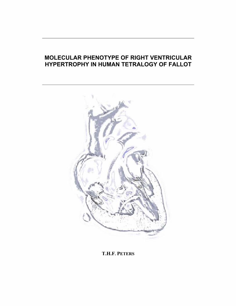
MOLECULAR PHENOTYPE OF RIGHT VENTRICULAR HYPERTROPHY IN HUMAN TETRALOGY OF FALLOT
T.H.F. PETERS
Molecular phenotype of right ventricular hypertrophy in human tetralogy of Fallot.
Thesis, Erasmus Medical Center, Rotterdam.
ISBN 90-6734-038-3
© T.H.F. Peters 2003 All rights reserved. Save exceptions stated by law, no part of this publication may be reproduced, stored in a retrieval system of any nature, or transmitted in any form or by means, electronic, mechanical, photocopying, recording or otherwise, including a complete or partial transcription, without the prior written permission of the author, application for which should be addressed to T.H.F. Peters, Department of Cardiothoracic Surgery, Erasmus Medical Center, Rotterdam, P.O. Box 2040, 3000 CA Rotterdam, The Netherlands. Printed by Optima Grafische Communicatie, Rotterdam
MOLECULAR PHENOTYPE OF RIGHT VENTRICULAR HYPERTROPHY IN HUMAN TETRALOGY OF FALLOT
MOLECULAIR FENOTYPE VAN RECHTER VENTRIKEL
HYPERTROFIE IN TETRALOGIE VAN FALLOT BIJ DE MENS
Proefschrift
ter verkrijging van de graad van doctor
aan de Erasmus Universiteit Rotterdam
op gezag van de Rector Magnificus
Prof. dr. ir. J.H. van Bemmel
en volgens het besluit van het College voor Promoties
De openbare verdediging zal plaatsvinden op
woensdag 26 november om 13.45 uur
door
Theodorus Hendrikus Franciscus Peters
geboren te Sambeek
Promotiecommissie
Promotoren: Prof. dr. A.J.J.C. Bogers
Prof. dr. P.R. Saxena
Overige leden: Prof. dr. J.M.J. Lamers
Prof. dr. R.E. Poelmann
Prof. dr. W.A. Helbing
Co-promotor: Dr. H.S. Sharma
The study described in this thesis was supported by a grant of the Netherlands Heart Foundation (NHS 96.082). Financial support by the Netherlands Heart Foundation for the publication of this thesis is gratefully acknowledged. Further financial support by Stichting Biomedical Engineering, Jacques H. de Jong Stichting and J.E. Jurriaanse Stichting for the publication of this thesis is gratefully acknowledged.

Voor mijn ouders
Table of Contents
Chapter 1. General introduction 9
1.1 Introduction 1.2 Surgery of tetralogy of Fallot 1.3 Myocardial hypertrophy 1.4 Myocardial fibrosis 1.5 Myocardial angiogenesis 1.6 Aims of the thesis - Myocardial phenotype of right ventricular hypertrophy in human tetralogy of Fallot; Proceedings of 2nd World Congress of PCCS. 1998; 746-749 - Tetralogy of Fallot, surgical aspects; Congenital Heart Disease. 1999;59-65 - Molecular phenotype of the developing heart with a congenital anomaly; Cardiac Development 2002; 195-212
Chapter 2. Quantitative analysis of collagens and fibronectin 43 expression in human right ventricular hypertrophy
- Ann N Y Acad Sci 1999; 874: 278-285 Chapter 3. Computerized image analysis in the quantitative 55
assessment of interstitial fibrosis late after correction of tetralogy of Fallot - Cardiovasc Eng 2003; 8: 114-120
Chapter 4. DNA micro-array and myocardial fibrosis in patients 69
with tetralogy of Fallot - Submitted
Chapter 5. Right ventricular fibrosis levels in patients with 91
pulmonary atresia and ventricular septal defect - Mol Cell Biochem 2003; 251: 27-32
Chapter 6. Myocardial angiogenesis and fibrosis in human 111
tetralogy of Fallot - Angiogenesis and fibrosis during right ventricular hypertrophy in human tetralogy of Fallot; The hypertrophied heart. 2000; 227-24
Chapter 7. Right ventricular hypertrophy in human tetralogy of Fallot is 131 associated with enhanced vascularization and expression of vascular endothelial growth factor
- Submitted Chapter 8. General discussion and conclusions 151
8.1 Introduction 8.2 Changes in the right ventricular myocardial phenotype 8.3 Clinical implications in tetralogy of Fallot 8.4 Limitations of the study 8.5 Implications for future research
List of abbreviations 170 Summary 171
Samenvatting 174 Dankwoord 177 Curriculum vitae 180 Publications 181

Chapter 1
8
General introduction
9
Chapter 1
General introduction
Based on: • Bogers AJJC, Peters THF, Hokken RB, Saxena PR and Sharma HS. Myocardial
phenotype of right ventricular hypertrophy in human tetralogy of Fallot. In: Imai Y, Momma K. eds: Proceedings of 2nd World Congress of PCCS 1998; 746-749: Futura, Armonk, New York, USA.
• Bogers AJJC, Peters THF, Jong de PL. Tetralogy of Fallot, surgical aspects. In: Bartelings MM, Wenink ACG eds: Congenital Heart Disease. suppl.: Boerhaave Committee for Postgraduate Medical Education. 1999; 59-65: Leiden, The Netherlands.
• Peters THF, Klompe L, Bogers AJJC and Sharma HS. Molecular phenotype of the developing heart with a congenital anomaly. In: Ostadal B, Nagano M and Dhalla NS eds.: Cardiac Development 2002; 195-212: Kluwer Academic Publishers, Boston, USA.

Chapter 1
10
General introduction
11
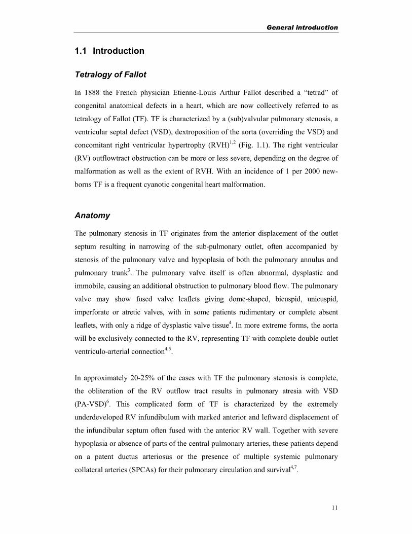
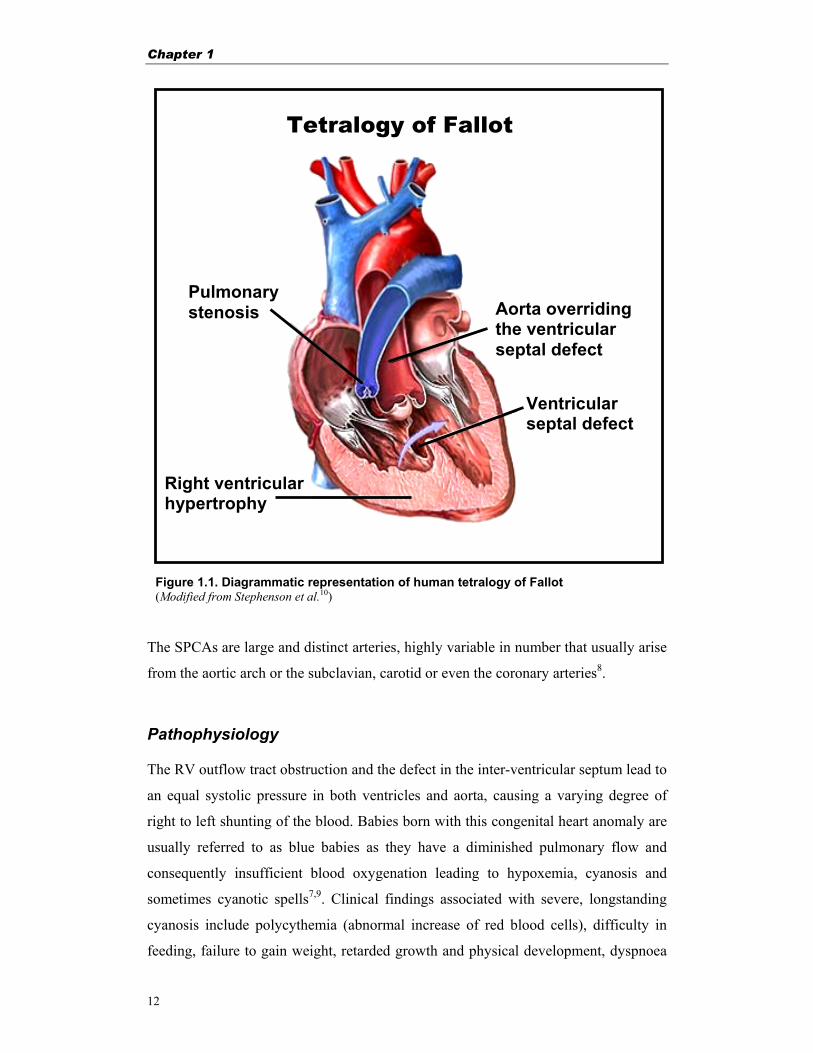
1.1 Introduction Tetralogy of Fallot In 1888 the French physician Etienne-Louis Arthur Fallot described a “tetrad” of
congenital anatomical defects in a heart, which are now collectively referred to as
tetralogy of Fallot (TF). TF is characterized by a (sub)valvular pulmonary stenosis, a
ventricular septal defect (VSD), dextroposition of the aorta (overriding the VSD) and
concomitant right ventricular hypertrophy (RVH)1,2 (Fig. 1.1). The right ventricular
(RV) outflowtract obstruction can be more or less severe, depending on the degree of
malformation as well as the extent of RVH. With an incidence of 1 per 2000 new-
borns TF is a frequent cyanotic congenital heart malformation.
Anatomy The pulmonary stenosis in TF originates from the anterior displacement of the outlet
septum resulting in narrowing of the sub-pulmonary outlet, often accompanied by
stenosis of the pulmonary valve and hypoplasia of both the pulmonary annulus and
pulmonary trunk3. The pulmonary valve itself is often abnormal, dysplastic and
immobile, causing an additional obstruction to pulmonary blood flow. The pulmonary
valve may show fused valve leaflets giving dome-shaped, bicuspid, unicuspid,
imperforate or atretic valves, with in some patients rudimentary or complete absent
leaflets, with only a ridge of dysplastic valve tissue4. In more extreme forms, the aorta
will be exclusively connected to the RV, representing TF with complete double outlet
ventriculo-arterial connection4,5.
In approximately 20-25% of the cases with TF the pulmonary stenosis is complete,
the obliteration of the RV outflow tract results in pulmonary atresia with VSD
(PA-VSD)6. This complicated form of TF is characterized by the extremely
underdeveloped RV infundibulum with marked anterior and leftward displacement of
the infundibular septum often fused with the anterior RV wall. Together with severe
hypoplasia or absence of parts of the central pulmonary arteries, these patients depend
on a patent ductus arteriosus or the presence of multiple systemic pulmonary
collateral arteries (SPCAs) for their pulmonary circulation and survival4,7.
Chapter 1
12
The SPCAs are large and distinct arteries, highly variable in number that usually arise
from the aortic arch or the subclavian, carotid or even the coronary arteries8.
Pathophysiology The RV outflow tract obstruction and the defect in the inter-ventricular septum lead to
an equal systolic pressure in both ventricles and aorta, causing a varying degree of
right to left shunting of the blood. Babies born with this congenital heart anomaly are
usually referred to as blue babies as they have a diminished pulmonary flow and
consequently insufficient blood oxygenation leading to hypoxemia, cyanosis and
sometimes cyanotic spells7,9. Clinical findings associated with severe, longstanding
cyanosis include polycythemia (abnormal increase of red blood cells), difficulty in
feeding, failure to gain weight, retarded growth and physical development, dyspnoea
Figure 1.1. Diagrammatic representation of human tetralogy of Fallot(Modified from Stephenson et al.10)
Tetralogy of Fallot
Right ventricular hypertrophy
Ventricular septal defect
Aorta overriding the ventricular septal defect
Pulmonary stenosis
General introduction
13
on exertion, and some times cerebral thrombosis7,9. Nowadays clubbing of the fingers
and toes, pulmonary haemorrhage, stroke and infections of the brain and severe
hypoxic spells rarely develop10.
In response to the increased pressure overload caused by pulmonary stenosis in
combination with the VSD, the RV undergoes hypertrophic growth. This is an
adaptive process wherein an increase in RV mass caused by expanding myocytes
attempts to normalize the wall stress. This adaptive response can further lead to a
proportionate increase in coronary flow and growth of the coronary vasculature and to
structural changes in the myocardial architecture (myocardial remodeling)11-13.
1.2 Surgery of tetralogy of Fallot Surgical strategies and timing Without surgical treatment, TF ultimately results in cyanotic complications and
cardiac failure. Approximately 30% of neonates with TF die within the first year of
life if untreated, 90 % of the children would die before the age of 20 years and only 3
% will reach the age of 40 years7. Nearly 70% of patients with TF require an
operation during their first year of life because of hypoxic spells or persistent
hypoxemia defined as resting arterial oxygen saturation less than 70%2,14.
Generally the surgical approach involves primary repair in the first year of life by
means of closing the VSD and relieving the RV outflow tract obstruction2,15-17.
Surgical repair thus aims at preventing complications due to long standing RVH such
as ventricular dysfunction and arrhythmia. Surgical treatment can be corrective in
infants at a very young age, provided that the pulmonary arterial system is complete and
well developed. If not, palliative surgery may improve the clinical situation and
prepare for later correction6.
The aim of early corrective surgery is to relieve the hypoxemia, to eliminate the
stimulus for the adaptive RVH as well as preserving RV function and electrical
stability of the myocardium11,12 and to encourage the normal development of the
pulmonary vasculature17. The surgical strategy of primary repair of TF early in life
Chapter 1
14
has given excellent early and late results and compares favorably with two-stage
approaches. There is increasing evidence that early repair of congenital heart
anomalies minimizes secondary damage to vital organs, particularly of the heart itself,
the lungs, and the brain2,14. Furthermore, by establishing early antegrade pulmonary
blood flow, postnatal pulmonary angiogenesis and alveologenesis may also be
enhanced2. Finally, by eliminating cyanosis as early as possible, the adverse effects of
cyanosis on the central nervous system might also be reduced2.
Encouraged by the results of primary repair for symptomatic infants with TF, there is
a tendency to operate electively on asymptomatic young infants with this abnormality.
Although age less than 3 months was in some reports a risk factor for death after
repair, this risk has lately decreased in neonates2,14. In this regard, neonatal hearts
have less ability to adapt to sudden increases in stroke volume and neonates are more
susceptible to the adverse effect of cardiopulmonary bypass. It is likely that results of
early repair of TF in neonates will soon parallel those obtained in infants beyond the
first month of life. As a result of the developments in extracorporeal circulation,
improved anaesthetic management of neonates and infants, and advances in
postoperative care, both mortality and morbidity after early one-stage repair of TF
have decreased dramatically, and such repair is currently favored in many centres2,14.
Despite adequate primary corrective surgery, on long-term follow up some patients
however, suffer from pulmonary regurgitation, causing RV failure due to volume-
overload. For these patients further secondary corrective surgery later in life is
performed by implanting a pulmonary allograft heart valve between the RV and the
pulmonary artery.
The actuarial survival rate, including hospital deaths, after corrective surgery for TF is
reported to be 89-93% at 15 years2,10,14,18,19. Midterm re-operations are reported in 2-
19% of the patients2,14,19,20, mostly because of residual pulmonary stenosis and to a
lesser extent due to residual VSD. Re-operation for pulmonary valve replacement is
reported to be performed in about 9% of the TF patients19. The actuarial freedom from
re-operation was 85-89% at 15 years2,14.

General introduction
15
1.3 Myocardial hypertrophy Growth of the heart From the postnatal period to maturation the human heart undergoes a six-fold increase in
heart mass21. The growth is most rapid in the first postnatal months, when the total
number of myocytes double and remain relatively constant there
after. In this period the myocyte diameter increases from 5 µm during infancy to 14-16
µm in adults7,22. Although the cardiac myocytes comprise one-third of the total cell
number in the adult heart, they are responsible for 70 percent of the cardiac volume21.
The growth of the heart becomes slower with the age, were the RV growth ceases at
approximately 50 years of age while the left ventricle slowly continues to increases in
volume23. The increase in myocardial mass is a result of myocyte hyperplasia (increase
in cell number) and hypertrophy (increase in cell size). Myocyte hypertrophy can be
either more eccentric, with longitudinal growth of the myocytes and normal wall
thickness/dimension ratio, or concentric with increased wall thickness/heart dimension
ratio24. The physiological increase in heart size by eccentric hypertrophy can be a result
of isotonic athletic training like running, whereas isometric exercise such as weight
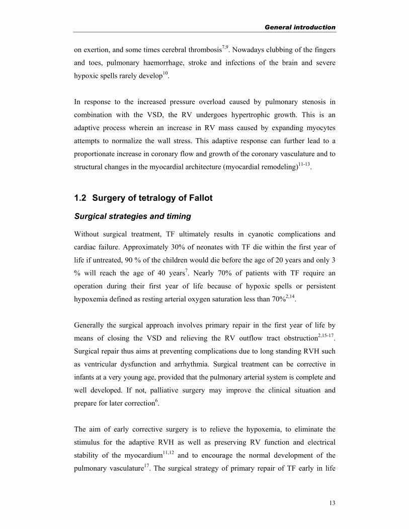
Figure 1.2. Blood flow before and after surgical correction of VSD and alleviation of pulmonary stenosis. (Modified from Losekoot et al.18)
A Before correction After correction B
RV LV
Chapter 1
16
lifting stimulates concentric hypertrophy24. The diameter of long-distance runners can
grow up to a 14% greater maximum diameter and 80% greater mass in the left
ventricle25. The growth of the adult heart can be enhanced by the thyroid hormones,
catecholamines and the renin-angiotensin system (RAS) hormones23. Under normal
physiological conditions the adult human cardiac myocytes are believed to be
terminally differentiated and have lost their ability to proliferate. Although some mild
concentric hypertrophy may develop in the left ventricle as a consequence of age
related decrease in sensibility of the peripheral vasculature26. However, under
pathological conditions, such as pressure and volume overload and heart failure, there
are some indications that cardiomyocytes re-initiate myocyte differentiation23,27.
Under those circumstances the myocyte diameter can increase to more then 40 µm,
depending on the degree of compensatory hypertrophy7.
Pathological myocardial hypertrophy In response to environmental and pathological conditions the heart may remodel to
sustain the cardiac performance. Pathological myocardial cell hypertrophy is a
physiological adaptive compensatory response, to increased haemodynamic overload,
where the increase of cardiac mass serves to normalize wall stress and to permit
normal cardiovascular function. Chronic haemodynamic overload will lead to cardiac
remodeling with an increased risk of decompensated hypertrophy and finally result in
cardiac failure11,13,21,28. The decompensated hypertrophy is a result from a number of
intrinsic and extrinsic mechanisms of the cardiac myocytes, including changes in
cardiomyocytes contractility, in regulatory-calcium-cycling and structural proteins
together with apoptosis, necrosis, inadequate growth secondary to altered signal
transduction pathways and extracellular matrix remodeling29,30. The myocyte dropout,
due to myocardial cell death and hypertrophy, of the remaining viable myocytes has
been proposed to account for impaired function and failure during ageing and
hypertension21,31-33. At the same time apoptosis is also of importance in contributing
to the remodeling of the cardiac chambers and vascular geometry in response to
pathologic stimuli34,35.
Pathological conditions such as ventricular outflow tract obstruction, systemic or
pulmonary arterial hypertension or aortic coarctation, may cause pressure overload
General introduction
17
and cardiac hypertrophy. This will increase the wall stress and result in concentric
hypertrophy, with characteristic wall thickening. Pressure overload hypertrophy will
activate early oncogenes and angiotensin II, mediated by the RAS-system and the
responses of the cardiomyocytes to strech23. Volume overload, occurring during
mitral or aortic regurgitation, however, may cause increased either diastolic wall
stress or both systolic and diastolic wall stress and result in (left) ventricular
hypertrophy21.
Molecular markers of cardiac hypertrophy Alterations in gene expression play an important role in the myocardial adaptation in
response to increased work load36. A battery of genes including immediate early
genes (like proto-oncogenes, c-fos, c-jun and c-myc) and genes from the fetal period
(like ANF, ß-MHC and α-skeletal actin) have been implicated in the cellular
alterations leading to ventricular hypertrophy37-39. The altered and increased cardiac
protein synthesis, resulting in a hypertrophic phenotype, can be triggered by dynamic
or static stretch of neonatal or adult cardiomyocytes40. Increased stretch of myocytes
activates the transcription of the early genes c-fos, c-jun and c-myc stimulating
hyperplasia and (fetal) myocyte growth within minutes41. Angiotensin II, growth
factors and noradrenaline are known to activate these early genes23.
Mechanotransducers (e.g. tyrosine kinase-containing receptors and stretch-activated
sarcolemmal ion channels) activate the process of mechanotransduction that in turn
stimulates the intracellular growth pathways42. Via this cytosolic pathway the
expression of specific “hypertrophic” genes are upregulated43. Insulin like growth
factor (IGF), acidic and basic fibroblast growth factor (FGF-1 and FGF-2), platelet-
derived growth factor (PDGF) and epidermal growth factor (EGF) are all growth
factors that interact with those tyrosine-kinase receptors21. Transforming growth
factor β (TGF-β) however, is one of the factors that has a myocyte proliferation and
growth inhibitory activity and is suppressed during myocyte hypertrophy, allowing
myocytes to grow44. Angiotensin II originated by the RAS-system, activates several
protein kinases involved in cell growth and particularly in structural remodeling in
cardiac hypertrophy and early postnatal development21,45,46. Other hormones known
Chapter 1
18
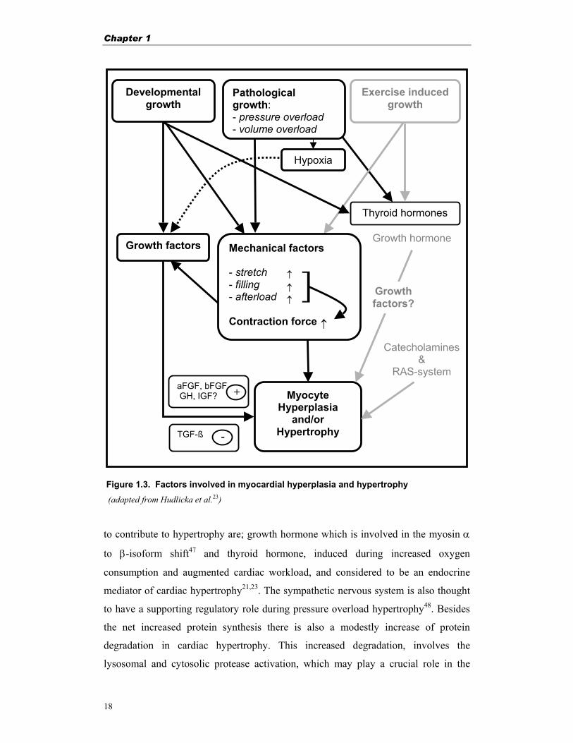
Figure 1.3. Factors involved in myocardial hyperplasia and hypertrophy
(adapted from Hudlicka et al.23) to contribute to hypertrophy are; growth hormone which is involved in the myosin α
to β-isoform shift47 and thyroid hormone, induced during increased oxygen
consumption and augmented cardiac workload, and considered to be an endocrine
mediator of cardiac hypertrophy21,23. The sympathetic nervous system is also thought
to have a supporting regulatory role during pressure overload hypertrophy48. Besides
the net increased protein synthesis there is also a modestly increase of protein
degradation in cardiac hypertrophy. This increased degradation, involves the
lysosomal and cytosolic protease activation, which may play a crucial role in the
Growth hormone
Catecholamines &
RAS-system
Pathological growth: - pressure overload - volume overload
Growth factors
Myocyte Hyperplasia
and/or Hypertrophy TGF-ß
aFGF, bFGF GH, IGF? +
Hypoxia
Growth factors?
Mechanical factors - stretch - filling - afterload Contraction force
↑
↑↑
↑
]
Developmental growth
-
Exercise induced growth
Thyroid hormones
General introduction
19
variably altered geometry of the ventricles in response to pressure overload or volume
overload, hypertrophy, regression, and atrophy49,50. Normally the average half live of
cardiac proteins is 5 days, regenerating the composition of the adult heart
approximately every 3 weeks51,52. During cardiac hypertrophy there is an net increase
in protein synthesis accounting for the hypertrophic myocardial phenotype and
increased mass51,52.
Both myosin and myofibrillar ATPase activity are depressed in the hypertrophied
myocardium. The loss of contractile protein may contribute to the impaired function
as observed in cardiac muscle from failing human hearts53. In small mammals there is
a transcriptionally mediated shift from α- to β-myosin heavy chain (MHC) and from
cardiac to skeletal actin isoform in response to pressure overload54,55. Some studies
report that this MHC shift56,57 is caused by significant loss of α-MHC expression
rather then β-MHC up regulation12,58. Because the α-form has a three to seven fold
greater ATPase activity than β-myosin, it is believed that the abundance in β-MHC
increases the efficiency of force development by producing the same absolute muscle
tension at a slower rate59. In higher mammals and humans, decreased myosin ATPase
activity of the hypertrophied heart is believed to be due to alterations in troponin
isoform composition or post-translational generation of a lower molecular variant of
β-MHC21,60.
Myocardial hypertrophy in tetralogy of Fallot The characteristic pulmonary stenosis with VSD in TF, will lead to increased RV
pressure overload. In an adaptive response to normalize the wall stress, the RV will
hypertrophy with expansion of the cardiomyocytes. This will lead to RV remodeling
and the development of myocardial fibrosis11,12,61. Histopathological studies on TF
myocardium report that the myocardial cell diameter in postoperative patients was
reduced as compared to preoperative patients with TF, but postoperative cellular
diameter was still larger than that in the age matched normal subjects62,63. These
findings suggest that RVH after corrective surgery of TF can regress to some extent
provided that residual pulmonary stenosis can be avoided.
Chapter 1
20
1.4 Myocardial fibrosis Interstitial fibrosis The normal myocardial cellular architecture consists of nicely arrayed cardiac
myocytes comprising cardiac fibroblasts and endothelial cells in the interstitial space.
In-between the myocytes there is a fibrous protein structure, the extracellular matrix,
that contributes to the myocardial organization and architecture, supports transmission
of the contractile force generated by cardiac myocytes, contributes to the passive
stretch and serves to restore the ventricular myocytes to precontraction length. The
extracellular matrix composition exists of collagenous proteins that account for
proteins, for example collagen, fibronectin, glycoproteins, proteoglycans and elastin64.
Throughout normal physiological myocardial growth there is a balance of hyperplasia
of cardiac fibroblasts and connective tissue distribution37,65. The collagen network, in
the extra-cellular space of the myocardium, consists of collagen fibers, which provide
the mechanical strength and stiffness of the myocardium66,67. When the balance is
disturbed, in case of pressure overload, the network gets damaged. As a result to the
increased wall stress, the myocyte support is compromized allowing myocardial tissue
expansion with disarray of myocytes68. This, together with the increased tension on
the fibers leads to a compensating excessive production of connective tissue, the
development of fibrosis. This process may continue until the extracellular matrix
fibers together with the hypertrophied myocytes counterbalance the pressure overload
and prevent (further) dilatation11-13,69. Disproportionate accumulation of collagen,
either reactive or reparative, has been observed during myocardial fibrosis. The
increased accumulation of extracellular matrix proteins during myocardial fibrosis
results in a decrease of compliance and declining contractile performance12,70,71.
Fibronectin and collagen type I and III are major components of the interstitial
fibrillar network. They are predominantly synthesized by cardiac and vascular
fibroblasts with a little expression by myocytes72. The RAS-system, activated during
pressure overload hypertrophy, activates fibroblast cell division enlarging the
synthesis23, where transcriptional upregulation of the encoding collagenous genes
contribute to the myocardial fibrosis12. Experimental73 and clinical data74 show that
General introduction
21
both fibronectin and collagen I and III are up-regulated during RVH75. The TGF-β
expression is also up-regulated during pressure overload cardiac hypertrophy. It is
believed to be involved in the mechanisms that activate fibrosis by the stimulation of
fibronectin and collagen expression in a variety of cell types and their incorporation in
the extracellular matrix12,76. Other factors that are thought to effect the collagenous
synthesis by fibroblasts are PDGF, angiotensin and endothelin, although the exact
mechanisms remains to be elucidated77. Pressure overload hypertrophy, induced by
aortic banding, showed a high increase of collagen I syntheses already after 2 days,
followed by collagen III after one week, suggesting differential expression
regulation77. The higher collagen concentration is a result of a decreased collagen
destruction and a higher collagen synthesis rate. The mRNA levels for collagen as
well as for fibronectin were found to be increased in cardiac pressure-overload
hypertrophy75.
Under normal conditions there is a continuous expression and degradation of
collagens by the adult cardiofibroblast77. There is a rapid collagen turnover in the
heart, were daily 5% of the collagen fibers are renewed. Most mature cross-linked
collagens are degraded extracellularly by members of the family of matrix
metalloproteases (MMPs), whereas already a large fraction (60%) of the newly
synthesized collagens is degraded rapidly within 8 minutes intracellularly, in the
lysosomes and endoplasmic reticulum78,79. MMPs may play an important role in the
tissue remodeling in both normal and pathological conditions. They belong to three
main groups, classified on their substrate specificity. The MMPs involved in the
myocardial remodeling are: the type IV collegenases (MMP-9 and MMP-2), the
stromelysins (MMP-3) and the interstitial collegenases (MMP-1)80. The MMPs are
secreted as a proenzyme (zymogen) by a number of cells including fibroblasts,
endothelial cells, smooth muscle cells and cardiomyocytes and can be activated by
plasmin80,81. The activity can be regulated by different cytokines and tissue inhibitors
of MMPs (TIMPs)81 but the extracellular matrix turn over is mainly regulated by
MMPs and the TIMPs82. TIMPs can bind the active sites of the MMPs thereby
blocking access to the collagen substrate80. An increase of the expression or activation
of the MMPs or an decrease in of TIMPs leads to increased proteolytic activity in the
extracellular space and increased extracellular remodeling including disregulated
Chapter 1
22
fibrillar collagen degradation, myofibril disarray and progressive myocyte loss82.
Increased levels of catecholamines, angiotensin II and endothelin may cause increased
myocardial MMP levels in the remodeling heart whereas tumor necrosis factor-α
(TNF-α) and ischemia are believed to increase the TIMP-1 and TIMP-4 levels
respectively80. From the four TIMPs known, TIMP-4 is the most cardiac specific one,
although all 4 are active in the myocardium83. An second role of TIMP 1 and 2 might
be a fibroblast growth stimulation effect80,84,85.
Peri-vascular fibrosis
During embryonic development, smooth muscle cells, endothelial cells and fibroblasts
secrete extracellular matrix proteins in the peri-vascular area to form a supportive
structure for the vascular wall. Under normal conditions, after birth, the vascular
smooth muscle cells do not proliferate or develop contractile fibers but are kept in an
quiescent state. When the smooth muscle cells are exposed to increased
haemodynamic load, arterial injury, or physiological stress they will get activated,
stimulate vascular remodeling, resulting in smooth muscle cell growth (due to
hypertrophy) or mesenteric medial thickening (due to hyperplasia). Factors such as
FGF, EGF and VEGF are known to stimulate the vascular smooth muscle
hyperplasia21.
Extracellular matrix proteins In the myocardium the myocytes represent only one-third of the number of cells, but
over two-third of their volume, whereas the number of fibroblasts, which produces
most of the collagen fibers represent as much as two-third of the cells86. Four from the
five collagen subtypes in the heart, type I, III, V and VI are produced by cardiac
fibroblasts and type IV by other cell types including cardiac myocytes87-89. Collagen
type I represents 80% of the total cardiac collagen content and is the major
collagenous product of cardiac fibroblasts87,90, which is important for the cardiac
mechanical strength and stiffness66. Collagen type III, important for tissue elasticity,
represents about 12% of total cardiac collagen content91. Collagen type I and III are
widely distributed in-between myocytes and among muscle fibers90,92. Collagen types
IV, V and VI constitute approximately 10-12% of total cardiac collagen content64.
General introduction
23
The collagen fibers have several functions, (i) one is to provide a scaffolding that
supports muscle cells and blood vessels67,93, (ii) to act as a lateral connection between
cells and muscle bundles to control architecture while co-ordinating the delivery of
force from the myocytes to the ventricle67,93,94, (iii) to provide myocardial stiffness by
tensile properties and resilience to resist myocardial deformation, maintain shape and
wall thickness, and to prevent ventricular aneurysm and rupture95,96.
Fibronectin, a glycoprotein located in the extracellular matrix of most tissue, serves as a
bridge between cardiac myocytes and interstitial collagen mesh network64,97. It
influences diverse processes including cell growth, adhesion, migration, and wound
repair91. During the development of cardiac hypertrophy due to pressure overload, fetal
fibronectin has been shown to accumulate in rats75. Fibronectin binds collagen and
modulates the collagen fibrillogenesis, resulting in higher collagen type I
concentration and a lower collagen type III concentration98. The fibronectin mRNA-
splicing variant encoding the EIIIA segment is an isoform, expressed during wound
healing99, embryogenesis100 and pressure-overload hypertrophy101. The structural
properties of the EIIIA isoform of fibronectin are not well understood but it is
postulated that this isoform may be important in the deposition of the extracellular
matrix99-101.
Fibrosis in tetralogy of Fallot Progressive myocardial fibrosis due to cardiac hypertrophy is an important cause of
myocardial dysfunction, clinical deterioration and cardiac death. Morphometrical
studies on TF revealed that the RV is affected with enhanced fibrosis associated with
increased myocytes diameter and myocardial disarray69,102. A post mortem analysis
study of myocardium of TF patients showed normal fibrosis levels in the left ventricle
and abnormal increased levels in the RV of patients older then 1 year with an increase
in outflow tract and subendocardium102. The collagen concentration of the RV is
about 30% higher than the left ventricle, because the RV myocytes are smaller103.
Within the RV the highest concentrations are found in the outflow tract and
subendocardium11. Changes in the cardiac extracellular matrix proteins composition
have also been observed in other congenital heart disease, however in most of the
types they where relatively small37,72,95.
Chapter 1
24
1.5 Myocardial angiogenesis Vasculogenesis and angiogenesis Angiogenesis is generally used to describe the formation of new blood vessels from
pre-existing vessels. This involves not only the formation of capillaries, but also the
formation of small and large blood vessels. The central physiological role of
angiogenesis is to ensure a proper blood flow through the tissue. It is a multi-step
process involving endothelial cell activation, migration, proliferation, tube formation
and maturation. In contrast, vasculogenesis concerns the differentiation of a primitive
network of new vessels during embryogenesis. The tissue glucose and oxygen
concentrations are important environmental factors as pro-angiogenic stimuli. Besides
endothelial cells several other cell types like vascular smooth muscle cells, pericytes
and fibroblasts and non-cellular structures, such as the basal lamina and the
extracellular matrix are involved104,105.
Development of pre-mature vasculature During embryogenesis when the number of cells is increasing within an avascular
tissue, there is an increased demand for oxygen which can not be met by the oxygen
diffusion. The hypoxic state, will trigger vessel formation by a process referred to as
vasculogenesis. Undifferentiated precursor cells (angioblasts) will differentiate to
endothelial cells that assemble into a vascular labyrinth. The endothelial cells will
differentiate and proliferate to form a premature tubular network106. This network
includes some of the major vessels in the embryo, such as the aorta and major veins,
as well as a honey comb-like plexus connecting these major vessels. Vascular
endothelial growth factor (VEGF) is one of the first growth factors to be induced in
this process. It stimulates the formation of an unstable immature primary vasculature.
In the next process, referred to as angiogenic remodeling, the initial primitive network
of premature vessel is modified, through both sprouting and vessel enlargement, to
form the interconnecting branching patterns characteristic for a stable mature complex
vascular network. During this process, vessel walls, premature as well as mature, and
endothelial cells integrate tightly with supporting cells (such as smooth muscle cells
and pericytes) and surrounding matrix, influenced by VEGF, Angiopoietin 1 (Ang-1)
and ephrin B2. The endothelial cells are kept in a quiescence state by Ang-1. Once the
General introduction
25
endothelial cells become quiescent they can survive for years. Expression of Ang-2
(like in pathological conditions) can destabilize the vessels and bring them in
regression. If at the same time VEGF is co-expressed, VEGF can induce angiogenic
sprouting in the destabilized vessel105,107.
During angiogenic sprouting, new vessels (sprouts) from existing vessels are formed
into a previously avascular tissue. In some cases it seems as if mature vessels must
first be destabilized to allow subsequent sprouting. Angiogenic sprouting is
responsible for the vascularization of certain tissues during normal development, such
as the neural tube or the retina, and for most new vessel formation in the adult106,107.
To supply all rapidly growing myocytes of oxygen after birth, the capillary length and
surface increases 23 times faster than the myocardial mass, however the capillary
diameter decreases, limiting the increasing capillary volume23.
Destabilization of the vessel can apparently lead to vascular regression. The survival
of the established vasculature, in the adults, depends on the quiescent state of the
endothelial cells which have prolonged life times (several years). A continuous
quantity of stabilizing factors is needed to keep the endothelial and smooth muscle
cells in a quiescent state. An imbalance of survival and apoptotic factors could
contribute to the regression of terminal myocardial vessels in pathological conditions
of hypertension and diabetes. The formation of new vessels in adults will only occur
by angiogenesis104. Under pathological conditions or during wound healing, when the
quiescent vascular equilibrium is disrupted, autocrine induction or up-regulation of
Ang-2, can reinitiate (angiogenic) vascular remodeling and angiogenic sprouting. In
the presence of VEGF new vessels will be formed, but without VEGF vessels
destabilize and go into regression105. Although sometimes also adult vasculogenesis
occurs, it only leads to immature poorly functional vasculature107.
Angiogenic stimuli and mechanisms Angiogenesis is a complex process regulated and maintained by multiple endogenous
pro-angiogenic and anti-angiogenic factors. In many cases, the activity of factors
depends on their local concentration and/or the microenvironment104,105. The different
angiogenic factors must be expressed in perfect harmony, on a complementary and
Chapter 1
26
co-ordinated manner to form functional vessels and to overcome vascular
regression108. Besides, many other non-vascular endothelium-specific growth factors
like PDGF, TGF-β and many other gene products ranging from transcription factors
to members of the Notch family have been shown to be crucial for proper vessel
formation107,109,110.
Among the angiogenic growth factors acting on the vascular endothelium, are the 5
members of the VEGF family, four members of the angiopoietin family and at least
one family of the large ephrin family105. VEGF is the most critical and specific
vascular growth factor in the vascular formation, required for the initiation of
immature vessels by vasculogenesis or angiogenic sprouting. During hypoxia its half-
life is prolonged from 30 minutes to 8 hours23. Ang-1 and ephrin B2 are needed for
the further remodeling and maturation of the immature vasculature. In this process
epherin B2 is important in the distinction to develop arterial or venous vessels. Ang-1
is important to maintain the quiescence and stable state of the vasculature. Disruption
of the stabilizing Ang-1 signal will reinitiate vascular remodeling in the adult (e.g. in
tumor)105. Autocrine production of Ang-2 (the Ang-1 natural antagonist) by
endothelium cells will destabilize the vessels. VEGFs, angiopoietins and ephrin B2
apparently are not capable to trigger the entire process of vascular remodeling in
adults individually105.
Many of the angiogenic factors involved in the remodeling and establishment of
functional blood vessels are regulated by paracrine signals from the receptor tyrosine
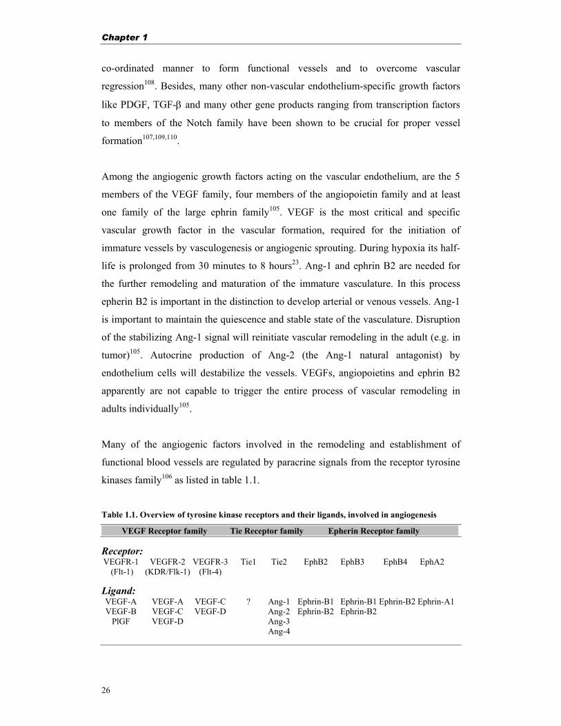
kinases family106 as listed in table 1.1.
Table 1.1. Overview of tyrosine kinase receptors and their ligands, involved in angiogenesis
. VE VEGF Receptor family Tie Receptor family Epherin Receptor family . Receptor: VEGFR-1 VEGFR-2 VEGFR-3 Tie1 Tie2 EphB2 EphB3 EphB4 EphA2 (Flt-1) (KDR/Flk-1) (Flt-4) Ligand: VEGF-A VEGF-A VEGF-C ? Ang-1 Ephrin-B1 Ephrin-B1 Ephrin-B2 Ephrin-A1 VEGF-B VEGF-C VEGF-D Ang-2 Ephrin-B2 Ephrin-B2 PlGF VEGF-D Ang-3 Ang-4
General introduction
27
Other factor involved in angiogenesis are the acidic (FGF-1) and basic (FGF-2)
fibroblast growth factors which stimulate the endothelial cell growth. They are expressed
by fibroblasts in response to hypoxia. FGF-1 stimulates the branching of the myocardial
arteries and stimulates the formation of functional vessels by preventing the
regression23,107. In combination with vasoconstricting growth promoters, FGF-2 is an
important endogenous activator of collateral vessel development in ischemic
myocardium. During ischemia VEGF expression is up-regulated and triggers the release
of stored FGF-2. The vasoconstricting growth promoters, such as angiotensin II,
thrombin, thromboxane, prostaglandins, serotonin and endothelin, are less potent then
peptide growth factors and they are believed to enhance other stimulators of vascular
smooth muscle cell growth. In quiescent vasculature they are found to stimulate FGF-2
expression. TGF-β is one of the regulators that inhibit vascular smooth muscle cell
growth but stimulates the synthesis of the extracellular matrix21. At low concentrations
TGF-β still can stimulate vascular growth, by increasing autocrine PDGF synthesis,
however higher concentrations reduces the PDGF receptor expression, interrupting the
autocrine effect111. Thyroid hormones are involved in the capillary elongation or splitting
of pre-existing vessels, increasing the capillary supply23.
Besides hypoxia, capillary growth in the heart can be stimulated by moderate athletic
training, as found in normal young but not in adult animal hearts, leading to growth of
the arterioles and coronary vessels23. The increased blood flow is stimulating
mechanical factors like shear stress and wall tension, which are stimuli that initiate
vessel growth23. Increased shear stress leads to endothelial release of angiogenic
prostaglandins and nitric oxide, whereas increased wall tension initiate smooth muscle
cell and endothelial cell proliferation with the release of growth factors. Shear stress is
also one of the factors known to be involved in vascular remodeling, where it can
disrupt the peri-vascular matrix and releases plasminogen activator and MMPs
activating a process that enables cell migration and proliferation23. In the same
manner, a lower heart rate is believed to enhance capillary growth. During the
diastole, the capillary diameter is larger than during systole increasing the wall
tension. A lower heart rate will enhance the diastolic period thereby increasing the
capillary wall stress and tension. Besides higher blood pressures and velocity, shear
stress can also be increased by increased haematocrit23,112.
Chapter 1
28
Angiogenic factors VEGF is known to be a critical vascular regulator. Although hypoxia can induce
VEGF, it is not necessarily inducing a useful angiogenic response. A very precise
concentration of other factors is needed to avoid vascular disorganization and
induction of immature and leaky vessels105. Alliterations in VEGF expression during
embryonic development can lead to profound abnormalities or early postnatal
death113,114. The VEGF-family consists of the 4 VEGF homologues (VEGF-A, VEGF-
B, VEGF-C or VEGF-related protein (VRP) and VEGF-D) and placental growth
factor (PlGF)115. The different members of the VEGF family have overlapping
abilities to interact with a set of cell-surface receptors such as VEGFR-1 (also known
as flt-1), VEGFR-2 (also known as Flk-1 or KDR) and VEGFR-3 (also known as Flt-
4). The VEGF receptors are closely related to receptor tyrosine kinase. Angiogenesis
and the initiation of the permeability of blood vessels are regulated by VEGF via its
receptors VEGFR-1 and VEGFR-2105. VEGF-A, one of the most potent and crucial
regulatory factors during myocardial angiogenesis105, is induced in a wide range of
cells under conditions of hypoxia116 and hypo-glycemia117 and in the presence of
some (growth) factors118 like phorbol ester, adenosine, TGF-β, PDGF and TNF-α105.
It is a ligand for the endothelial cell surface receptors, flt-1 and KDR119. VEGF-KDR
binding activates a cascade that promote angiogenesis by promoting vascular
endothelium cell proliferation, inducing vascular leaking and permeability but also by
enhancing the glucose transport120. Although several isoforms (VEGF120, VEGF121
and VEGF165) of this member are known, the exact functions still have to be
elucidated107. It is known that VEGF-A is involved in the formation of the vascular
lumen, when the endothelial cells assemble to solid cords. Intercalation or thinning of
endothelial cells and fusion of pre-existing vessels, influenced by VEGF-A, allow
vessels to increase their diameter and length107. VEGF-B is believed to play a role in
the in the coronary vascularization and growth. Loss of VEGF-B during
embryogenesis reduces the heart size121, whereas VEGF-C, a ligand for VEGFR3, is
involved in embryonic and adult pathological angiogenesis. VEGFR-3 is also
expressed on lymphatic vessels where it regulates, via VEGF-C, the lymphangiogenic
activity122-125. Hardly anything is know about the physiological role of VEGF-D107.
Also from the growth factor PlGF only a little is known, although there are some
indications that it might have a limiting role in adult vascular remodeling107.
General introduction
29
From the VEGF receptors, VEGFR-2 seems to mediate the major growth and
permeability whereas VEGFR-1 has a negative role by acting as a decoy receptor or
by suppressing signaling through VEGFR-2. VEGF affects embryonic, neonatal and
pathological angiogenesis via its receptor VEGFR-2126. The exact involvement of
VEGFR-1 signaling during (pathological) angiogenesis remains undetermined127.
Although VEGF does not seem necessary for the vascular maintenance in adults,
disruption of VEGF expression mimics VEGFR-2 knock out, resulting in only a very
few proliferated endothelial cells and complete absence of vasculature126,128,129.
Angiopoietins are the ligands for the Tie receptors (Tie1 and Tie2) which are like the
VEGF receptors expressed within the vascular endothelium105. The 4 members of the
angiopoietin family (based on their homology with Ang-1) all bind primarily to Tie 2
receptor105,130. Tie2 is widely expressed in cardiovascular tissue131,132. There is some
evidence that the Tie2 cascade regulates the extracellular recruitment of stromal cells
required to encase and thereby stabilize primitive endothelial tubes133,134. Ang-1
binding to endothelial Tie2 receptor is necessary to maintain the quiescence state in
adult vasculature, via a maximum stimulation of the interaction between endothelial
and peri endothelial cells, their surrounding support cells and matrix105. It further
protects the vasculature against vascular permeability and leakage induced by VEGF
or inflammatory agents105. Ang-1 alone fails to induce endothelial
proliferation105,108,135, however in presence of VEGF unprecedented hyper-
vascularization will occur resulting in an increased number and size of the vessels136.
Further Ang-1 is believed to be a chemotactic for endothelial cells. Ang-2 binding to
the Tie2 receptor can either activate or antagonize the Tie2 receptor137. At least in
presence of VEGF, Ang-2 will enhanced the sprouting of capillary vessels by
antagonizing the stabilization effect of Ang-1. Ang-2 itself cannot induce
angiogenesis but is indirectly involved in Angiotensin II-mediated angiogenesis by
enhancing the angiogenic activity of VEGF138. Angiotensin II can stimulate the
remodeling of the heart and vessels via cell growth-promoting effects139. By binding
to Angiotensin II receptors AT1 and AT2 , which are expressed in large amounts on
the vascular endothelial cells in the myocardium140, Angiotensin II can selectively
induce and up-regulate VEGF and Ang-2 expression in cardiac vascular endothelial
cells141.
Chapter 1
30
The ephrin receptors tyrosine kinases belong to the largest known family of growth
factor receptors108. Ephrin-B2 and its EphB4 receptor are believed to play a key role
during vascular development, especially in establishing arterial venous identity and in
fusing arterial and venous vessels at their junctions105. During adult angiogenic
sprouting the expression of ephrin-B2 is found to be strongly up-regulated in the
endothelium of new vessels. Ephrin-B2 is not only required during the earliest stages
of arterial/venous determination, but may also be important during the development of
arteries by regulating interactions between endothelial and smooth muscle cells
involved in the formation of the arterial muscular walls105.
Angiogenesis in tetralogy of Fallot In TF patients the RV myocytes hypertrophy as an adaptive response to the
(sub)pulmonary stenosis in combination with the VSD, to normalize RV wall tension.
This will trigger a response to improve myocardial perfusion by the formation of new
capillaries and by the enlargement of pre-existing collateral vessels to reduce the
increased capillary-myocyte diffusion distance, to prevent hypoxia and decreased
nutrient delivery12,13,142. But also the mechanical factors such as stretch, shear stress,
and increased wall tension, which occur in TF due RV pressure and volume overload
may be stimuli for angiogenesis during cardiac hypertrophy12,13,142. Experimental
animal studies with induced RVH due to pressure overload, revealed increased blood
flow per unit myocardial mass at rest, without an increase in minimum coronary
resistance143. During volume overload RVH however, normal flow values per unit
myocardial mass at rest are measured, with normal or mild increases minimum
coronary resistance and normal or mildly decreased coronary reserve144.
From previous studies on the myocardial vascularization in TF patients it is known
that the overall myocardial capillary density in the infundibulum of the right
ventricular outflow tract of (1-7 years old) TF patients was reported to be similar to
normal capillary density (1648±123 vs. 1782±203 capillaries/mm2)145. Also the
capillary domain (cross-sectional tissue area nearest to that capillary) and the capillary
tissue supply volume are not affected in TF. However the capillary segment length
(distance between two branching points) was found to be increased145 whereas the
capillary radius was decreases, from 2.8 to 2.4 µm146. There are also some indications
General introduction
31
that the capillary supply per myocyte is proportional to the increased myocyte size,
keeping the capillary per myocyte ratio similar147. The elimination of the hypertrophic
stimuli in TF are believed to reverse the RVH and the associated coronary circulatory
abnormalities148.
1.6 Aims of the thesis Different surgical interventions nowadays are applied to TF patients already at an
early age to correct the cardiac malformations. It is, however, not yet established
whether early intervention will result in (complete) regression of RVH and restore
normal cardiac performance.
The underlying molecular mechanisms in the development and regression of RVH at
different stages in the treatment of human TF still remains to be elucidated. The
molecular and cellular characteristics of the RV myocardium at corrective and redo
surgery are thought to correlate with the clinical condition of the patient and with
cardiac prognosis. When experimental data and clinical parameters can be correlated
more information will become available to enhance our knowledge in assessing the
timing for surgery and eventually postoperative cardiac prognosis. Our working
hypothesis is that molecular and cellular characteristics of the RV myocardium at
corrective or redo surgery may provide information, relevant for the assessment of the
clinical condition of the patient and important for cardiac prognosis.
Hence, aims of this thesis were to elucidate the changes in the RV phenotype (at
mRNA and protein level) of patients with TF as compared to the normal myocardial
phenotype by studying:
(i) the myocardial cellular organization and the degree of hypertrophy,
(ii) the degree of fibrosis (e.g. collagens and fibronectin),
(iii) the myocardial vascularization (e.g. VEGF expression and vascular density).
The obtained molecular biological data and clinical parameters (medical history,
echocardiography and cardiac catheterization) of the different forms and stages of
the disease will be analyzed to enhance our knowledge about the myocardial
remodeling and RVH regression after surgery.
Chapter 1
32
In the first part of the thesis our hypothesis was that the pressure overload and
resulting RVH in patients with TF will go along with increased fibrosis represented
by the enhanced expression of collagens and fibronectin. To elucidate the degree of
myocardial fibrosis, we studied the localization of total collagen and fibronectin with
light microscopy, assessed the expression by visual scoring in RV biopsies of TF and
compared the data to autograft root replacement patients (control group), as
described in chapter 2. After computerized video image analysis of the same
sections together with sections of post mortem controls, the autograft control
biopsies appeared to include fibrotic changes as well (chapter 3) and thus could not
be used as a proper control. In order to assess the degree of fibrosis and myocyte
hypertrophy in TF patients and appropriate control groups, we analyzed the
expression patterns of fibronectin and the collagen subtypes I and III at protein and
mRNA level in an extended study. The results are described in chapter 4, together
with mRNA expression data of other factors known to be involved in myocardial
fibrosis achieved by DNA micro-array chip analysis. In a similar fashion the degree
of myocardial fibrosis in the PA-VSD patients has been analyzed in as described in
chapter 5.
In the second part of the thesis the hypothesis was that the remodeling of the heart in
TF patients, apart from RVH, also concerns myocardial angiogenesis, triggered by
the increased capillary-myocyte oxygen diffusion distance. VEGF, a ligand for the
flk-1 receptor, is one of the key players during angiogenesis. In the chapters 6 and 7
the results are described of the assessment of angiogenic factor such as VEGF and
the resulting increase in vascular density.
In chapter 8 the observations of the studies as described in this thesis are discussed
together with the implications for possible treatment and future research.
General introduction
33
References 1. Bogers AJJC, van der Laarse A, Vliegen HW, et al. Assessment of
hypertrophy in myocardial biopsies taken during correction of congenital heart disease. Thorac Cardiovasc Surgeon. 1988;36:137-140.
2. Castaneda AR, Jonas RA, Mayer JE, et al. Cardiac surgery of neonate and infant. Philadelphia, USA: Saunders; 1994.
3. Soto B, Pacifico AD, Ceballos R, et al. Tetralogy of Fallot: an angiographic-pathologic correlative study. Circulation. 1981;64:558-66.
4. Bartelings MM. Morphology of tetralogy of Fallot. In: Bartelings MM, Wenink ACG, eds. Congenital Heart Disease. Leiden: Boerhaave Committee; 1999:49-56.
5. Anderson RH, Tynan M. Tetralogy of Fallot--a centennial review. Int J Cardiol. 1988;21:219-32.
6. Ottenkamp J. Clinical presentation-introduction: What is tetralogy of Fallot? In: Bartelings MM, Wenink ACG, eds. Congenital Heart Disease. Leiden: Boerhaave Committee; 1999:41-46.
7. Schoen F. The Heart. In: Cotran R, Robbins S, Kumar V, eds. Pathologic basis of disease. 5th ed. Philadelphia: W.B. Saunders Company; 1994:560.
8. Jacobs ML. Congenital Heart Surgery Nomenclature and Database Project: tetralogy of Fallot. Ann Thorac Surg. 2000;69:S77-82.
9. Enersen O. Fallot. In: www.whonamedit.com. Oslo; 2001. 10. Stephenson L, Rodengen J. State of the heart. 1 ed. Fort Launderdale: Write
Stuff Enterprises Inc; 1999. 11. van Bilsen M, Chien KR. Growth and hypertrophy of the heart: towards an
understanding of cardiac specific and inducible gene expression. Cardiovasc Res. 1993;27:1140-1149.
12. Boluyt MO, L. O, Meredith AL, et al. Alterations in cardiac gene expression during the transition from stable hypertrophy to heart failure. Marked upregulation of genes encoding extracellular matrix components. Circ Res. 1994;75:23-32.
13. Schwartz K, Carrier L, Mercadier JJ, et al. Molecular phenotype of hypertrophied and failing myocardium. Circulation. 1993;87:VII 5-10.
14. Kirklin JW, Barrat-Boyes BG. Ventricular septal defect and pulmonary stenosis or atresia. 2nd ed. ed. New York: Churchill-Livingstone: Churchill-Livingstone; 1993.
15. Starnes VA, Luciani GB, Latter DA, et al. Current surgical management of tetralogy of Fallot. Ann Thorac Surg. 1994;58:211-215.
16. Van Arsdell GS, Maharaj GS, Tom J, et al. What is the optimal age for repair of tetralogy of Fallot? Circulation. 2000;102:III123-9.
17. Pozzi M, Trivedi DB, Kitchiner D, et al. Tetralogy of Fallot: what operation, at which age. Eur J Cardiothorac Surg. 2000;17:631-636.
18. Losekoot G, Kamphuis R. Kinderen met een hartafwijking. meppel: Boom; 1988.
19. de Ruijter FT, Weenink I, Hitchcock FJ, et al. Right ventricular dysfunction and pulmonary valve replacement after correction of tetralogy of Fallot. Ann Thorac Surg. 2002;73:1794-800; discussion 1800.
20. Pacifico AD. Reoperations after repair of tetralogy of Fallot. In: Stark J, Pacifico AD, eds. Reoperations in cardiac surgery: Springer; 1989:171-86.
Chapter 1
34
21. Walsh R, Dorn G. Growth and hypertrophy of the heart and blood vessels. In: Alexander W, Schlant R, Fuster V, eds. Hurst's The Heart, arteries and veins. 9th edition: The McGraw-Hill Companies; 1998:155-168.
22. Rakusan K. Cardiac growth, maturation and ageing. In: Zak R, ed. Growth of the heart in health and disease. New York: Raven press; 1984:131-164.
23. Hudlicka O, Brown MD. Postnatal growth of the heart and its blood vessels. J Vasc Res. 1996;33:266-87.
24. Ford LE. Heart size. Circ Res. 1976;39:297-303. 25. Schaible T, Scheuer J. Response of heart to exercise training. In: Zak R, ed.
Growth of the heart in health and disease. New York: Raven press; 1984:381-419.
26. Walsh R. Cardiovascular effects of the aging process. Am J Med. 1987;82:34-40.
27. Field LJ. Atrial natriuretic factor-SV40 T antigen transgenes produce tumours and cardiac arrhythmias in mice. Science. 1988;239:1029-33.
28. Grossman W. Cardiac hypertrophy; useful adaptation or pathological process? Am J Med. 1980;69:576-584.
29. Teiger E, Than VD, Richard L, et al. Apoptosis in pressure overload-induced heart hypertrophy in the rat. J Clin Invest. 1996;97:2891-7.
30. Cheng W, Li B, Kajstura J, et al. Stretch-induced programmed myocyte cell death. J Clin Invest. 1995;96:2247-59.
31. Anversa P, Hiler B, Ricci R, et al. Myocyte cell loss and myocyte hypertrophy in the ageing rat heart. J Am Coll Cardiol. 1986;8:1441-1448.
32. Engelmann GL, Vitullo JC, Gerrity RG. Morphometric analysis of cardiac hypertrophy during development, maturation, and senescence in spontaneously hypertensive rats. Circ Res. 1987;60:487-494.
33. Capasso JM, Palackal T, Olivetti G, et al. Left ventricular failure induced by long-term hypertension in rats. Circ Res. 1990;86:1400-1412.
34. Thompson CB. Apoptosis in the pathogenesis and treatment of disease. Science. 1995;267:1456-62.
35. Vaux DL, Strasser A. The molecular biology of apoptosis. Proc Nat Acad Sci U S A. 1996;93:2239-44.
36. Brilla CG, Janicki JS, Weber KT. Impaired diastolic function and coronary reserve in genetic hypertension. Role of interstitial fibrosis and medial thickening of intramyocardial coronary arteries. Circ Res. 1991;69:107-115.
37. Bishop SP. Structural alterations in the hypertrophied and failing myocardium. In: Abel FL, Hewman WH, eds. Functional aspects of the normal hypertrophied and failing heart. The Hague: Martinus Nijhoff Publishing; 1984:278-300.
38. Boheler KR, Dillmann WH. Cardiac response to pressure overload in the rat: the selective alteration of in vitro directed RNA translation products. Circulation Res. 1988;63:448-456.
39. Brand T, Sharma HS, Fleischmann KE, et al. Proto-oncogene expression in porcine myocardium subjected to ischemia and reperfusion. Circ Res. 1992;71:1351-1360.
40. Cooper Gt. Cardiocyte adaptation to chronically altered load. Ann Rev Physiol. 1987;49:501-18.
41. Lompre AM, Mercadier JJ, Schwartz K. Changes in gene expression during cardiac growth. Int Rev Cytol. 1991;124:137-86.
General introduction
35
42. watson P. Mechanical activation of signalling pathways in cardiovascular system. Trends Cardiovasc Med. 1996;6:73-79.
43. Sugden PH, Bogoyevitch MA. Intracellular signalling through protein kinases in the heart. Cardiovasc Res. 1995;30:478-92.
44. Engelmann GL, Boehm KD, Birchenall-Roberts MC, et al. Transforming growth factor-beta 1 in heart development. Mech Dev. 1992;38:85-97.
45. Beinlich CJ, Baker KM, White GJ, et al. Control of growth in the neonatal pig heart. Am J Physiol. 1991;261:3-7.
46. Falkenhahn M, Gohlke P, Paul M, et al. The renin-angiotensin system in the heart and vascular wall: new therapeutic aspects. J Cardiovasc Pharmacol. 1994;24 Suppl 2:S6-13.
47. Sacca L, Cittadini A, Fazio S. Growth hormone and the heart. Endocr Rev. 1994;15:555-73.
48. Strauer BE. [Progression and regression of heart hypertrophy in arterial hypertension: pathophysiology and clinical aspects]. Z Kardiol. 1985;74 Suppl 7:171-8.
49. Parker TG, Packer SE, Schneider MD. Peptide growth factors can provoke "fetal" contractile protein gene expression in rat cardiac myocytes. J Clin Invest. 1990;85:507-14.
50. Samarel AM. Hemodynamic overload and the regulation of myofibrillar protein degradation. Circulation. 1993;87:1418-20.
51. Morgan HE, Gordon EE, Kira Y, et al. Biochemical mechanisms of cardiac hypertrophy. Ann Rev Physiol. 1987;49:533-43.
52. Hannan RD, Luyken J, Rothblum LI. Regulation of ribosomal DNA transcription during contraction-induced hypertrophy of neonatal cardiomyocytes. J Biol Chem. 1996;271:3213-20.
53. Pagani ED, Alousi AA, Grant AM, et al. Changes in myofibrillar content and Mg-ATPase activity in ventricular tissues from patients with heart failure caused by coronary artery. Circ Res. 1998;63(2):380-385.
54. Morkin E. Regulation of myosin heavy chain genes in the heart. Circulation. 1993;87:1451-60.
55. Boheler KR, Chassagne C, Martin X, et al. Cardiac expressions of alpha- and beta-myosin heavy chains and sarcomeric alpha-actins are regulated through transcriptional mechanisms. Results from nuclear run-on assays in isolated rat cardiac nuclei. J Biol Chem. 1992;267:12979-85.
56. Bing OH, W. BW, Conrad CH, et al. Intracellular calcium transients in myocardium from spontaneously hypertensive rats during the transition to heart failure. Circ Res. 1991;68(5):1390-1400.
57. Conrad CH, W. BW, G. RK, et al. Impaired myocardial function in spontaneously hypertensive rats with heart failure. Am J Physiol. 1991;260 (1 Pt 2):H136-145.
58. Lowes BD, Minobe W, Abraham WT, et al. Changes in gene expression in the intact human heart. Down regulation of alpha-myosin heavy chain in hypertrophied, failing ventricular myocardium. J Clin Invest. 1997;100:2315-24.
59. Cooper Gt. Load and length regulation of cardiac energetics. Ann Rev Physiol. 1990;52:505-22.
60. Anderson PA, Greig A, Mark TM, et al. Molecular basis of human cardiac troponin T isoforms expressed in the developing, adult, and failing heart. Circ Res. 1995;76:681-6.
Chapter 1
36
61. Schwartz SM, Gordon D, Mosca RS, et al. Collagen content in normal, pressure, and pressure-volume overloaded developing human hearts. Am J Cardiol. 1996;77:734-738.
62. Mitsuno M, Nakano S, Shimazaki Y, et al. Fate of right ventricular hypertrophy in tetralogy of Fallot after corrective surgery. Am J Cardiol. 1993;72:694-698.
63. Seliem MA, Wu YT, Glenwright K. Relation between age at surgery and regression of right ventricular hypertrophy in tetralogy of Fallot. Ped Cardiol. 1995;16:53-55.
64. Pelouch V, Dixon IM, Golfman L, et al. Role of extracellular matrix proteins in heart function. Mol Cell Biochem. 1994;129:101-120.
65. Medugorac I. Myocardial collagen in different forms of the heart hypeortrophy in the rat. Res Exp Med. 1980;177:201-211.
66. Weber KT, Sun Y, Tyagi SC, et al. Collagen network of the myocardium: function, structural remodelling and regulatory mechanisms. J Mol Cell Cardiol. 1994;26:279-292.
67. Bishop JE, Rhodes S, Laurent GJ, et al. Increased collagen synthesis and decreased collagen degradation in right ventricular hypertrophy induced by pressure overload. Cardiovasc Res. 1994;28:1581-1585.
68. Whittaker P. Collagen and ventricular remodelling after acute myocardial infarction: concepts and hypotheses. Basic Res Cardiol. 1997;92:79-81.
69. Kawai S, Okada R, Kitamura K, et al. A morphometrical study of myocardial disarray associated with right ventricular outflow tract obstruction. Jpn Circ J. 1984;48:445-456.
70. Conrad CH, Brooks WW, Sen S, et al. Increased collagen content may contribute to increased myocardial stiffness and heart failure in spontaneously hypertensive rat. J Am Coll Cardiol. 1990;15:48A.
71. Weber KT, Brilla CG. Pathological hypertrophy and cardiac interstitium. Fibrosis and renin-angiotensin-aldosterone system. Circulation. 1991;83(6):1849-1865.
72. Bishop JE, Greenbaum M, Gibson DG, et al. Enhanced deposition of predominantly type I collagen in myocardial disease. J Mol Cell Cardiol. 1990;22:1157-1165.
73. Jalil JE, Doering CW, Janicki JS, et al. Fibrillar collagen and myocardial stiffness in the intact hypertrophied rat left ventricle. Circ Res 1989;64:1041-1050.
74. McLenachan JM, Dargie HJ. Ventricular arrhythmias in hypertensive left ventricular hypertrophy. Relationship to coronary artery disease, left ventricular dysfunction, and myocardial fibrosis. Am J Hypertens. 1990;3:735-740.
75. Samuel JL, Barrieux A, Dufour S, et al. Accumulation of fetal fibronectin mRNAs during the development of rat cardiac hypertrophy induced by pressure overload. J Clin Invest. 1991;88:1737-1746.
76. Ignotz RA, Massague J. Transforming growth factor-beta stimulates the expression of fibronectin and collagen and their incorporation into the extracellular matrix. J Biol Chem. 1986;261(9):4337-4345.
77. Bishop JE, Rhodes S, Laurent GJ, et al. The regulation of collagen deposition in the hypertrophying heart. Ann N Y Acad Sci. 1995;752:236-9.
General introduction
37
78. Laurent GJ. Dynamic state of collagen: pathways of collagen degradation in vivo and their possible role in regulation of collagen mass. Am J Physiol. 1987;252:C1-9.
79. Bienkowski RS, Cowan MJ, McDonald JA, et al. Degradation of newly synthesized collagen. J Biol Chem. 1978;253:4356-63.
80. Mann DL, Spinale FG. Activation of matrix metalloproteinases in the failing human heart: breaking the tie that binds. Circulation. 1998;98:1699-702.
81. Galis ZS, Muszynski M, Sukhova GK, et al. Cytokine-stimulated human vascular smooth muscle cells synthesize a complement of enzymes required for extracellular matrix digestion. Circ Res. 1994;75:181-9.
82. Li YY, Feldman AM, Sun Y, et al. Differential expression of tissue inhibitors of metalloproteinases in the failing human heart. Circulation. 1998;98:1728-34.
83. Greene J, Wang M, Liu YE, et al. Molecular cloning and characterization of human tissue inhibitor of metalloproteinase 4. J Biol Chem. 1996;271:30375-80.
84. Dollery CM, McEwan JR, Henney AM. Matrix metalloproteinases and cardiovascular disease. Circ Res. 1995;77:863-8.
85. Birkedal-Hansen H, Moore WG, Bodden MK, et al. Matrix metalloproteinases: a review. Crit Rev Oral Biol Med. 1993;4:197-250.
86. Zak R. Cell proliferation during cardiac growth. Am J Cardiol. 1973;31:211-219.
87. Chapman D, Weber KT, Eghbali M. Regulation of fibrillar collagen types I and III and basement membrane type IV collagen gene expression in pressure overloaded rat myocardium. Circ Res. 1990;67:787-794.
88. Weber KT. Cardiac interstitium in health and disease: The fibrillar collagen network. J Am Coll Cardiol. 1989;13:1637-1652.
89. Speiser B, Riess CF, Schaper J. The extracellular matrix in human myocardium: part 1: Collagens I, II, IV and VI. Cardioscience. 1991;2:225-232.
90. Bashey RI, Martinez-Hernandez A, Jiminez SA. Isolation, characterization, and localization of cardiac collagen type VI. Associations with other extracellular matrix components. Circ Res 70. 1992;70:1006-1017.
91. Farhadian F, Contard F, Corbier A, et al. Fibronectin expression during physiological and pathological cardiac growth. J Mol Cell Cardiol. 1995;27:981-990.
92. Weber KT, Pick R, Jalil JE, et al. Patterns of myocardial fibrosis. J Mol Cell Cardiol. 1989;21 (Suppl V):121-131.
93. Borg TK, Caulfield JB. The collagen matrix of the heart. Fed Proc. 1981;20:2037-2041.
94. Weber KT, Janicki JS, Pick R, et al. Collagen in the hypertrophied pressure-overloaded myocardium. Circulation. 1987;75 (Suppl. I):I-40-7.
95. Dawson R, Milne G, Williams RB. Changes in the collagen or rat heart in copper-deficiency-induced cardiac hypertrophy. Cardiovasc Res. 1982;16:559-65.
96. Factor SM, Robinson TF, Dominitz R, et al. Alterations of the myocardial skeletal framework in acute myocardial infarction with and without ventricular rupture. Am J Cardiovasc Pathol. 1986;1:91-97.
Chapter 1
38
97. Wayner E, Carter W, Piotrowitz R, et al. The function of multiple extracellular matrix receptors mediating cell adhesion to extracellular matrix: Preparation of monoclonal antibodies to the fibronectin receptor that specifically inhibit cell adhesion to fibronectin and react with platelet glycoproteins. J Cell Biol. 1988;107:1881-1891.
98. Kucharz E. The collagens: biochemistry and pathophysiology. Berlin: Springer Verlag; 1992.
99. Ffrench-Constant C, van de Water L, Dvorak HF, et al. Reappearance of an embriotic pattern of fibronectin splicing during wound healing in adult heart. J Cell Biol. 1989;109:903-914.
100. Norton PA, Hynes RO. Alternative splicing of chicken fibronectin in embryos and in normal and transformed cells. Mol Cell Biol. 1987;7(12):4297-4307.
101. Villareal FJ, Dillman WH. cardiac hypertrophy induced changes in mRNA levels for TGF-b, fibronectin and collagen. Am J Physiol. 1992;31:H1861-H1866.
102. Hegerty A, Anderson RH, Deanfield JE. Myocardial fibrosis in tetralogy of Fallot: effect of surgery or a part of the natural history? Br. Heart J. 1988;59:123 (abstract).
103. Caspari PG, Gibson K, Harris P. Changes in myocardial collagen in normal development and after b-blockade. Rec Adv Card Struct Metab. 1976;7:99-104.
104. Huminiecki LB, R. Angiogenesis. In: Bikfalvi A, ed. Encyclopedic reference of vascular biology & pathology. Berlin Heidelberg: Springer Verlag; 2000:2-8.
105. Yancopoulos GD, Davis S, Gale NW, et al. Vascular-specific growth factors and blood vessel formation. Nature. 2000;407:242-8.
106. Risau W. Mechanisms of angiogenesis. Nature. 1997;386:671-4. 107. Carmeliet P. Mechanisms of angiogenesis and arteriogenesis. Nat Med.
2000;6:389-395. 108. Gale NW, Yancopoulos GD. Growth factors acting via endothelial cell-
specific receptor tyrosine kinases: VEGFs, angiopoietins, and ephrins in vascular development. Genes Dev. 1999;13:1055-66.
109. Hellstrom M, Kalen M, Lindahl P, et al. Role of PDGF-B and PDGFR-beta in recruitment of vascular smooth muscle cells and pericytes during embryonic blood vessel formation in the mouse. Development. 1999;126:3047-55.
110. Hirschi KK, Rohovsky SA, Beck LH, et al. Endothelial cells modulate the proliferation of mural cell precursors via platelet-derived growth factor-BB and heterotypic cell contact. Circ Res. 1999;84:298-305.
111. Stouffer GA, Owens GK. TGF-beta promotes proliferation of cultured SMC via both PDGF-AA-dependent and PDGF-AA-independent mechanisms. J Clin Invest. 1994;93:2048-55.
112. Hudlicka O, Brown MD, Walter H, et al. Factors involved in capillary growth in the heart. Mol Cell Biochem. 1995;147:57-68.
113. Miquerol L, Langille BL, Nagy A. Embryonic development is disrupted by modest increases in vascular endothelial growth factor gene expression. Development. 2000;127:3941-6.
General introduction
39
114. Carmeliet P, Ng YS, Nuyens D, et al. Impaired myocardial angiogenesis and ischemic cardiomyopathy in mice lacking the vascular endothelial growth factor isoforms VEGF164 and VEGF188. Nat Med. 1999;5:495-502.
115. Eriksson U, Alitalo K. Structure, expression and receptor-binding properties of novel vascular endothelial growth factors. Curr Top Microbiol Immunol. 1999;237:41-57.
116. Shweiki D, Itin A, Soffer D, et al. Vascular endothelial growth factor induced by hypoxia may mediate hypoxia-initiated angiogenesis. Nature. 1992;359:843-845.
117. Shweiki D, Neeman M, Itin A, et al. Induction of vascular endothelial growth factor expression by hypoxia and by glucose deficiency in multicell spheroids: implications for tumour angiogenesis. Proc Nat Acad Sci U S A. 1995;92:768-772.
118. Horiuchi T, Weller PF. Expression of vascular endothelial growth factor by human eosinophils: upregulation by granulocyte macrophage colony-stimulating factor and interleukin-5. Am J Respir Cell Mol Biol. 1997;17:70-77.
119. Detmar M, Brown LF, Berse B, et al. Hypoxia regulates the expression of vascular permeability factor/vascular endothelial growth factor (VPF/VEGF) and its receptors in human skin. J Invest Dermatol. 1997;108:263-268.
120. Waltenberger J, Mayr U, Pentz S, et al. Functional upregulation of the vascular endothelial growth factor receptor KDR by hypoxia. Circulation. 1996;94:1647-1654.
121. Bellomo D, Headrick JP, Silins GU, et al. Mice lacking the vascular endothelial growth factor-B gene (VEGF-B) have smaller hearts, dysfunctional coronary vasculature, and impaired recovery from cardiac ischemia. Circ Res. 2000;86:E29-35.
122. Dumont DJ, Jussila L, Taipale J, et al. Cardiovascular failure in mouse embryos deficient in VEGF receptor-3. Science. 1998;282:946-9.
123. Ferrara N, Alitalo K. Clinical applications of angiogenic growth factors and their inhibitors. Nat Med. 1999;5:1359-64.
124. Taipale J, Makinen T, Arighi E, et al. Vascular endothelial growth factor receptor-3. Curr Top Microbiol Immunol. 1999;237:85-96.
125. Olofsson B, Jeltsch M, Eriksson U, et al. Current biology of VEGF-B and VEGF-C. Curr Opin Biotechnol. 1999;10:528-35.
126. Carmeliet P, Ferreira V, Breier G, et al. Abnormal blood vessel development and lethality in embryos lacking a single VEGF allele. Nature. 1996;380:435-9.
127. Hiratsuka S, Minowa O, Kuno J, et al. Flt-1 lacking the tyrosine kinase domain is sufficient for normal development and angiogenesis in mice. Proc Nat Acad Sci U S A. 1998;95:9349-54.
128. Ferrara N, Chen H, Davis-Smyth T, et al. Vascular endothelial growth factor is essential for corpus luteum angiogenesis. Nat Med. 1998;4:336-40.
129. Shalaby F, Rossant J, Yamaguchi TP, et al. Failure of blood-island formation and vasculogenesis in Flk-1-deficient mice. Nature. 1995;376:62-6.
130. Maisonpierre PC, Suri C, Jones PF, et al. Angiopoietin-2, a natural antagonist for Tie2 that disrupts in vivo angiogenesis. Science. 1997;277:55-60.
131. Wong AL, Haroon ZA, Werner S, et al. Tie2 expression and phosphorylation in angiogenic and quiescent adult tissues. Circ Res. 1997;81:567-74.
Chapter 1
40
132. Witzenbichler B, Maisonpierre PC, Jones P, et al. Chemotactic properties of angiopoietin-1 and -2, ligands for the endothelial-specific receptor tyrosine kinase Tie2. J Biol Chem. 1998;273:18514-21.
133. Dumont DJ, Gradwohl G, Fong GH, et al. Dominant-negative and targeted null mutations in the endothelial receptor tyrosine kinase, tek, reveal a critical role in vasculogenesis of the embryo. Genes Dev. 1994;8:1897-909.
134. Vikkula M, Boon LM, Carraway KL, 3rd, et al. Vascular dysmorphogenesis caused by an activating mutation in the receptor tyrosine kinase Tie2. Cell. 1996;87:1181-90.
135. Suri C, McClain J, Thurston G, et al. Increased vascularization in mice overexpressing angiopoietin-1. Science. 1998;282:468-71.
136. Thurston G, Suri C, Smith K, et al. Leakage-resistant blood vessels in mice transgenically overexpressing angiopoietin-1. Science. 1999;286:2511-4.
137. Carmeliet P. Developmental biology. Controlling the cellular brakes. Nature. 1999;401:657-8.
138. Fujiyama S, Matsubara H, Nozawa Y, et al. Angiotensin AT(1) and AT(2) receptors differentially regulate angiopoietin-2 and vascular endothelial growth factor expression and angiogenesis by modulating heparin binding-epidermal growth factor (EGF)-mediated EGF receptor transactivation. Circ Res. 2001;88:22-9.
139. Baker KM, Booz GW, Dostal DE. Cardiac actions of angiotensin II: Role of an intracardiac renin-angiotensin system. Ann Rev Physiol. 1992;54:227-41.
140. Stoll M, Steckelings UM, Paul M, et al. The angiotensin AT2-receptor mediates inhibition of cell proliferation in coronary endothelial cells. J Clin Invest. 1995;95:651-7.
141. Chua CC, Hamdy RC, Chua BH. Upregulation of vascular endothelial growth factor by H2O2 in rat heart endothelial cells. Free Radic Biol Med. 1998;25:891-7.
142. van Bilsen M, Chien KR. Growth and hypertrophy of the heart: towards an understanding of cardiac specific and inducible gene expression. Cardiovasc Res. 1993;27:1140-1149.
143. Murray PA, Vatner SF. Reduction of maximal coronary vasodilator capacity in conscious dogs with severe right ventricular hypertrophy. Circ Res. 1981;48:25-33.
144. Hultgren PB, Bove AA. Myocardial blood flow and mechanics in volume overload-induced left ventricular hypertrophy in dogs. Cardiovasc Res. 1981;15:522-528.
145. Rakusan K, Cicutti N, Spatenka J, et al. Geometry of the capillary net in human hearts. Int J Microcirc Clin Exp. 1997;17:29-32.
146. Turek Z, Rakusan K. Lognormal distribution of intercapillary distance in normal and hypertrophic rat heart as estimated by the method of concentric circles: its effect on tissue oxygenation. Pflugers Arch. 1981;391:17-21.
147. Rakusan K, Flanagan MF, Geva T, et al. Morphometry of human coronary capillaries during normal growth and the effect of age in left ventricular pressure-overload hypertrophy. Circulation. 1992;86:38-46.
148. Isoyama S, Ito N, Kuroha M, et al. Complete reversibility of physiological coronary vascular abnormalities in hypertrophied hearts produced by pressure overload in the rat. J Clin Invest. 1989;84:288-294.

General introduction
41

Chapter 2
42
Fibrosis in right ventricular hypertrophy
43
Chapter 2
Quantitative analysis of collagens and fibronectin expression in human right ventricular hypertrophy
Based on: T.H.F. Peters, H.S. Sharma, E. Yilmaz and A.J.J.C. Bogers. Quantitative analysis of collagens and fibronectin expression in human right ventricular hypertrophy. Ann N Y Acad Sci 1999; 874:278-285.
Chapter 2
44
Abstract
One of the main features in human Tetralogy of Fallot (TF) is right ventricular
hypertrophy (RVH) due to pressure (sub-pulmonary stenosis) and volume overload
(ventricular septal defect). Currently, primary correction at a young age is the
treatment of choice. To unravel the role of extracellular matrix in RVH, we examined
myocardial expression of collagens and fibronectin in TF patients; primary correction
(TF-1, mean age 0.7 ± 0.2 years), secondary surgery (TF-2, mean age 36.9 ± 4.6
years) and in age matched control patients. Picro-sirius red staining quantified by
video imaging showed significantly increased interstitial staining for collagens in
both TF-1 and TF-2 groups as compared to respective controls. Fibronectin was
expressed in extracellular spaces, peri-vascular regions and in some cardiomyocytes.
Quantitative analysis of fibronectin revealed increased expression in only TF-1 group
as compared to respective control. Our results indicate an increased amount of
myocardial extracellular matrix deposition as a sign of fibrosis during RVH in
patients with TF.

Fibrosis in right ventricular hypertrophy
45
Introduction
Tetralogy of Fallot (TF) is a common form of cyanotic congenital heart disease, with an
incidence rate of 1 out of 2000 new borns. TF is characterized by overriding of the aorta,
a ventricular septal defect, (sub) valvular pulmonary stenosis and right ventricular
hypertrophy (RVH) resulting in hypoxemia due to diminished pulmonary flow1. The
RVH in human TF is an adaptive response of cardiomyocytes to increased pressure as
well as volume overload caused by (sub) pulmonary stenosis and by ventricular septal
defect, respectively. The adaptive response requires proportionate increase of coronary
flow or growth of the coronary vasculature, leading to myocardial remodeling as well as
development of fibrosis and, without treatment, ultimately results in cardiac failure2-5.
In myocardial fibrosis, collagens and fibronectin play a prominent role. Damage of the
collagen structure compromises the myocyte support, decreases myocardial strength and
stiffness and allows expansion of the tissue6. Collagen fibers are organized in the
collagen network, which is found in the extracellular space of the myocardium7-10. In
myocardial fibrosis a disproportionate accumulation, either reactive or reparative, of
collagen has been observed. Experimental11 and clinical data12,13 show that a rise in
collagen content increases myocardial stiffness and promotes abnormalities of cardiac
function during hypertrophy. In a morphometrical study, an increase in fibrosis in TF
was associated with an increased diameter of myocytes and an increase of myocardial
disarray14. In addition, the peri-vascular accumulation of collagen fibers may impair the
vasodilator capacity of intramyocardial coronary arteries and may contribute to the
decrease in coronary reserve15.
Fibronectin is found in the extracellular matrix of most tissue, serving as a bridge
between cells and interstitial collagen mesh network and influencing diverse processes
including cell growth, adhesion, migration, and wound repair16. In rats, fetal fibronectin
was shown to be accumulated during development of cardiac hypertrophy by pressure
overload17. The hypertrophied heart exhibits significant qualitative as well as
quantitative changes in gene expression. The altered expression patterns in hypertrophy,
include transient expression of proto-oncogenes18. It is thought that those changes in
cardiac hypertrophy reflect a change toward an embryonic program of gene expression19.
Chapter 2
46
However, it is still not clarified whether those altered expression patterns are general
markers of cardiac hypertrophy or markers of a specific parallel pathogenic process such
as TF. Animal studies have shown that the levels of mRNA for fibronectin and collagens
were elevated in cardiac hypertrophy17.
Most attention in research on human TF so far is focused on clinical treatment and
surgical methods to limit the myocardial injury. TF patients are usually operated during
their first years of life. The aim of early primary repair is to close the ventricular septal
defect and to remove the right ventricular outflow tract obstruction, thus relieving the
hypoxemia and eliminating the stimulus for the adaptive RVH as well as preserving right
ventricular function1,20. However, after corrective surgery some patients develop right
ventricular failure due to volume-overload caused by pulmonary regurgitation and may
for this reason require further secondary corrective surgery later in life21,22.
Histopathological studies on TF myocardial tissue reported that the myocardial cell
diameter was reduced in patients after corrective surgery, indicating regression of RVH.
Nevertheless, it is not yet known to what extent the RVH in TF patients regresses. Only
little attention has been paid to the molecular component of the disease. In this study, we
examined the degree of fibrosis in human TF by measuring the expression of myocardial
fibrosis markers such as fibronectin and collagen in right ventricular biopsies obtained
during corrective surgery for TF and compared the data with that of age matched
patients with normal right ventricle.
Materials and Methods
Biopsies
The present study was approved by the Medical Ethical Committee of the University
Hospital, Erasmus University, Rotterdam. Myocardial tissue biopsies were obtained
from patients who underwent primary corrective surgery (TF-1, mean age 0.7 ± 0.2
years, n=11), secondary surgery (TF-2, mean age 36.9 ± 4.6 years, n=6) and age
matched control patients. All control patients had clinically normal right ventricle and
underwent autograft aortic root replacement (C-1, mean age 1.5 ± 0.2 years, n=6 and C-
2, mean age 31.7 ± 4.2, n=12). Myocardial tissue was fixed in 4% paraformaldehyde in
Fibrosis in right ventricular hypertrophy
47
PBS for minimal 24 hours and further processed for dehydration and embedding in
paraffin for histological and immunohistochemical studies23.
Picro-sirius red staining
The total collagen in myocardial tissue specimens was stained with Picro-sirius red
F3BA24. Tissue sections of 6 µm thickness, were treated with 0.2% aqueous
phosphomolybdic acid and incubated in 0.1% Picro-sirius red. Before dehydration, the
slides were treated with 0.01N HCl and mounted. The Picro-sirius red stained area was
determined as percentage of total tissue area using a Kontron-KS 400 (Kontron
Electronik/Zeiss, Eching, Germany) computerized image analysis system. Distribution
of collagen fibers in the interstitial space as well as in the peri-vascular region was
measured as Picro-sirius red positive area. 12 different images from each section were
analyzed to calculate the mean of percentage stained tissue area and statistically
significant values were accepted at p≤0.05.
Immunohistochemistry
After fixation and dehydration, the cardiac tissue was embedded in paraffin. Sections of
6 µm thickness were cut and mounted on poly-L-lysin coated microscope slides,
followed by immunohistochemistry using avidin-biotin complex (ABC) method
(BioGenex, San Ramon, CA). The sections were deparaffinized and quenched for
endogenous peroxidase by 2% hydrogen peroxide in methanol. Non-specific binding
sites were blocked by 10% normal goat serum. Human serum pre-absorbed polyclonal
antibodies, against human fibronectin in 1:1000 dilution (Life Technologies, Breda, The
Netherlands), were applied as primary antiserum and the sections were incubated at
room temperature for 30 min. Negative controls were performed by omission of the
primary antibody. After washing in 0.5% tween-20 in PBS solution, the sections were
incubated with mouse biotinylated anti-rabbit IgG (BioGenex, San Ramon, CA) for 30
min. and subsequently, with peroxidase conjugated streptavidin. Color was developed
using 3,3'-diaminobenzidine tetrahydrochloride dehydrate (DAB) and then sections were
counter stained with hematoxylin. Sections were mounted and visualized under the light
microscope and photographed. A semi-quantitative analysis of the sections was done by
2 independent observers using a staining score ranging from 0 (no staining) to 4 (very
Chapter 2
48
strong, dark brown, staining) to asses the level of fibronectin expression in the
myocardial tissue obtained from TF patients and age matched controls.
Results and discussion
Picro-sirius red staining was performed for the total collagens in order to assess the
degree of myocardial fibrosis. The red staining was quantified by video imaging and
showed a significantly increased interstitial and peri-vascular staining for total collagens
in TF-1 (1.4 ± 0.2 fold) (Fig. 2.1B and D) and TF-2 (1.6 ± 0.2 fold) (Fig. 2.1C and D) as
compared to their respective controls (Fig. 2.1A and D). The immunolocalization of
fibronectin showed that fibronectin was expressed in the interstitium, in the peri-vascular
area, as well as in some cardiomyocytes. Semiquantitative analysis using a staining score
ranging from 0-4, revealed that the fibronectin expression was significantly increased in
TF-1 (Fig. 2.2B and D) as compared to control (3.0 ± 0.2 vs. 2.6 ± 0.2)(Fig. 2.2B and D).
There were no differences in fibronectin expression in patients from TF-2 (Fig. 2.2B and
D) as compared with age-matched controls. Our results indicate an increased amount of
myocardial extracellular matrix deposition in RVH in patients with TF, with an apparent
increase of total collagen and a slight increase in fibronectin at young age.
The apparent development of cardiac hypertrophy is induced by changes in cardiac gene
expression that provide the heart means of compensation for increased haemodynamic
load25. In this regard, our data are in agreement with the histopathologic findings from
the right ventricular myocardium in preoperative TF that show interstitial fibrosis,
myofibrillar disorganization, disarray and degenerative changes in addition to
myocardial cell hypertrophy18,26,27. The changes as found in our study for expression of
collagens and fibronectin could be attributed to altered structure or function of the
myocardium. It is believed that changes in gene expression accompanying cardiac
hypertrophy are the result of a multifactorial process and the observed changes may
therefore be concomitant a phenomena. The alteration in ventricular gene expression
might be an indicator of cardiac hypertrophy and may result from a number of different
stimuli. We assume that the changes in the right ventricular fibronectin and collagen
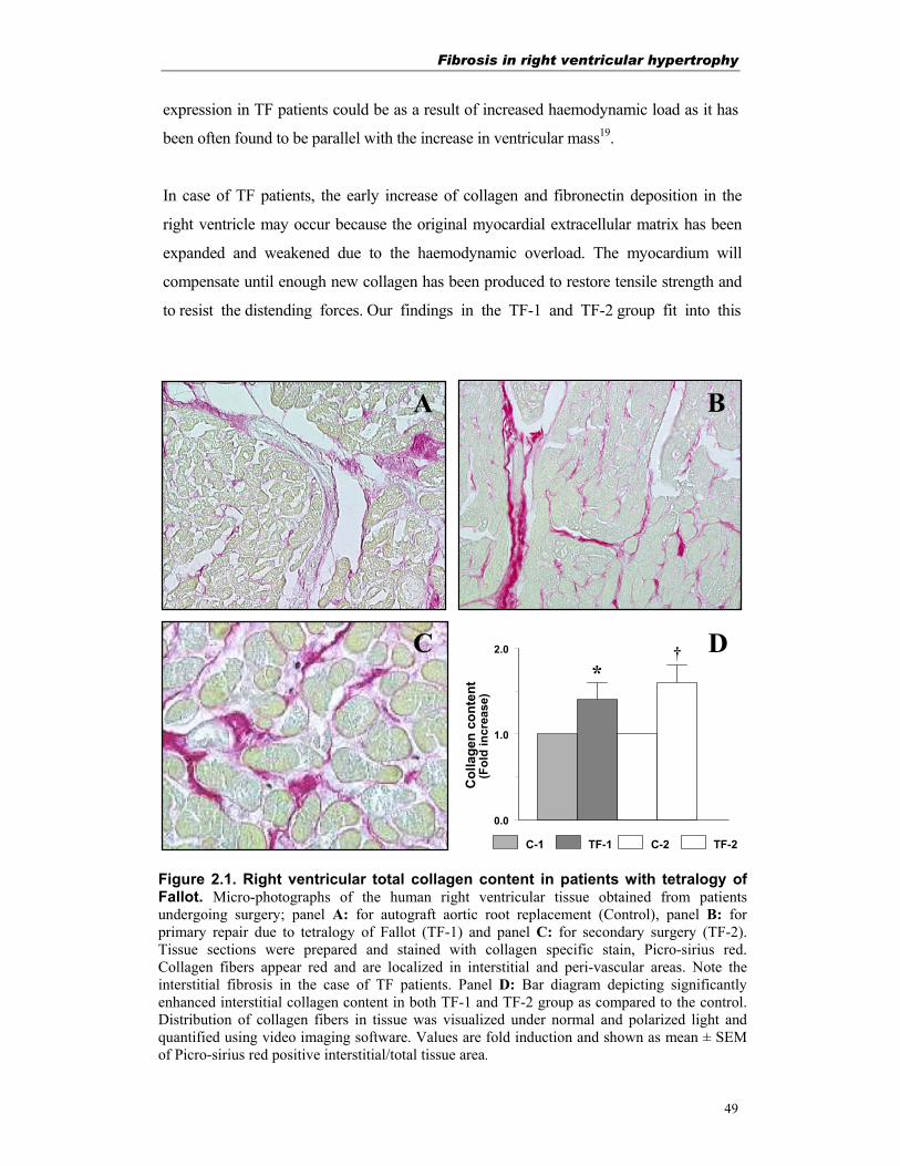
Fibrosis in right ventricular hypertrophy
49
expression in TF patients could be as a result of increased haemodynamic load as it has
been often found to be parallel with the increase in ventricular mass19.
In case of TF patients, the early increase of collagen and fibronectin deposition in the
right ventricle may occur because the original myocardial extracellular matrix has been
expanded and weakened due to the haemodynamic overload. The myocardium will
compensate until enough new collagen has been produced to restore tensile strength and
to resist the distending forces. Our findings in the TF-1 and TF-2 group fit into this
0.0
1.0
2.0
Col
lage
n co
nten
t
C-1 TF-1 C-2 TF-2
(Fol
d in
crea
se)
C
B
D
A
Figure 2.1. Right ventricular total collagen content in patients with tetralogy ofFallot. Micro-photographs of the human right ventricular tissue obtained from patientsundergoing surgery; panel A: for autograft aortic root replacement (Control), panel B: for primary repair due to tetralogy of Fallot (TF-1) and panel C: for secondary surgery (TF-2). Tissue sections were prepared and stained with collagen specific stain, Picro-sirius red. Collagen fibers appear red and are localized in interstitial and peri-vascular areas. Note the interstitial fibrosis in the case of TF patients. Panel D: Bar diagram depicting significantly enhanced interstitial collagen content in both TF-1 and TF-2 group as compared to the control. Distribution of collagen fibers in tissue was visualized under normal and polarized light and quantified using video imaging software. Values are fold induction and shown as mean ± SEMof Picro-sirius red positive interstitial/total tissue area.
† *
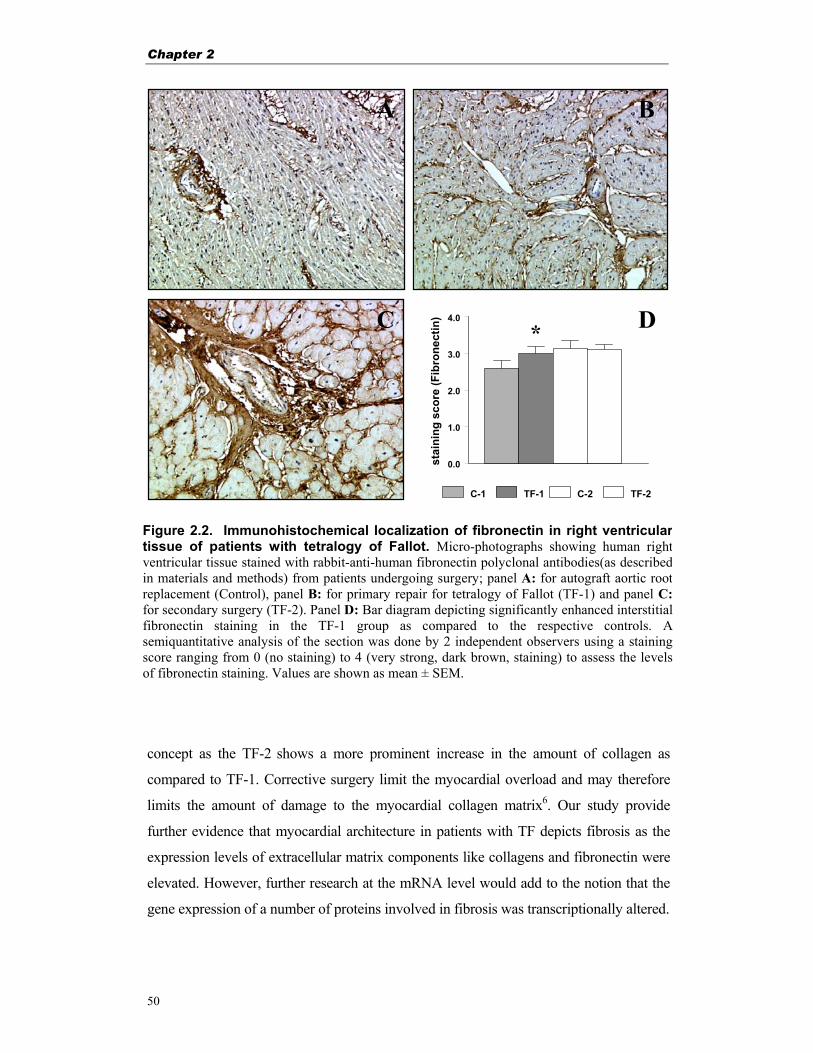
Chapter 2
50
concept as the TF-2 shows a more prominent increase in the amount of collagen as
compared to TF-1. Corrective surgery limit the myocardial overload and may therefore
limits the amount of damage to the myocardial collagen matrix6. Our study provide
further evidence that myocardial architecture in patients with TF depicts fibrosis as the
expression levels of extracellular matrix components like collagens and fibronectin were
elevated. However, further research at the mRNA level would add to the notion that the
gene expression of a number of proteins involved in fibrosis was transcriptionally altered.
0.0
1.0
2.0
3.0
4.0
stai
ning
sco
re (F
ibro
nect
in)
C-1 TF-1 C-2 TF-2
A
DC
B
*
Figure 2.2. Immunohistochemical localization of fibronectin in right ventriculartissue of patients with tetralogy of Fallot. Micro-photographs showing human rightventricular tissue stained with rabbit-anti-human fibronectin polyclonal antibodies(as describedin materials and methods) from patients undergoing surgery; panel A: for autograft aortic rootreplacement (Control), panel B: for primary repair for tetralogy of Fallot (TF-1) and panel C:for secondary surgery (TF-2). Panel D: Bar diagram depicting significantly enhanced interstitialfibronectin staining in the TF-1 group as compared to the respective controls. Asemiquantitative analysis of the section was done by 2 independent observers using a stainingscore ranging from 0 (no staining) to 4 (very strong, dark brown, staining) to assess the levelsof fibronectin staining. Values are shown as mean ± SEM.
Fibrosis in right ventricular hypertrophy
51
Summary and conclusions
In the present study the degree of myocardial fibrosis at protein level in patients with
tetralogy of Fallot was examined by assessing quantitatively the expression of total
collagens and fibronectin. We found a significant increase in staining for interstitial
and total collagens at primary correction as well as at secondary corrective surgery as
compared to the age matched control patients. However, immuno-histochemical
localization of fibronectin revealed significant increase in this protein levels at
primary correction only. Our results clearly demonstrate an increased amount of
myocardial extracellular matrix deposition resulting in tissue fibrosis in TF patients
with right ventricular hypertrophy. This vital information could be of great help in
assessing the timing of surgery as well as post-operative prognosis.
Acknowledgements Financial support from the Netherlands Heart Foundation (NHS 96.082) is gratefully
acknowledged.
References
1. Castaneda AR, Jonas RA, Mayer JE, et al. Cardiac surgery of neonate and infant. Philadelphia, USA: Saunders; 1994.
2. Arai M, Matsui H, Periasamy M. Sarcoplasmic reticulum gene expression in cardiac hypertrophy and heart failure. Circ Res. 1994;74:555-564.
3. Boluyt MO, L. O, Meredith AL, et al. Alterations in cardiac gene expression during the transition from stable hypertrophy to heart failure. Marked upregulation of genes encoding extracellular matrix components. Circ Res. 1994;75:23-32.
4. van Bilsen M, Chien KR. Growth and hypertrophy of the heart: towards an understanding of cardiac specific and inducible gene expression. Cardiovasc Res. 1993;27:1140-1149.
5. Schwartz K, Carrier L, Mercadier JJ, et al. Molecular phenotype of the hypertrophied and failing myocardium. Circulation. 1993;87:VII 5-10.
6. Whittaker P. Collagen and ventricular remodelling after acute myocardial infarction: concepts and hypotheses. Basic Res Cardiol. 1997;92:79-81.
7. Weber KT, Sun Y, Tyagi SC, et al. Collagen network of the myocardium: function, structural remodelling and regulatory mechanisms. J Mol Cell Cardiol. 1994;26:279-292.
Chapter 2
52
8. Bishop JE, Rhodes S, Laurent GJ, et al. Increased collagen synthesis and decreased collagen degradation in right ventricular hypertrophy induced by pressure overload. Cardiovasc Res. 1994;28:1581-1585.
9. Brilla CG, Janicki JS, Weber KT. Impaired diastolic function and coronary reserve in genetic hypertension. Role of interstitial fibrosis and medial thickening of intramyocardial coronary arteries. Circ Res. 1991;69:107-15.
10. Chapman D, Weber KT, Eghbali M. Regulation of fibrillar collagen types I and III and basement membrane type IV collagen gene expression in pressure overloaded rat myocardium. Circ Res. 1990;67:787-794.
11. Jalil JE, Doering CW, Janicki JS, et al. Fibrillar collagen and myocardial stiffness in the intact hypertrophied rat left ventricle. Circ. Res. 1989;64:1041-1050.
12. Hess OM, Schneider J, Koch R, et al. Diastolic function and myocardial structure in patients with myocardial hypertrophy. Special reference to normalized viscoelastic data. Circulation. 1981;63:360-371.
13. McLenachan JM, Dargie HJ. Ventricular arrhythmias in hypertensive left ventricular hypertrophy. Relationship to coronary artery disease, left ventricular dysfunction, and myocardial fibrosis. Am J Hypertens. 1990;3:735-740.
14. Kawai S, Okada R, Kitamura K, et al. A morphometrical study of myocardial disarray associated with right ventricular outflow tract obstruction. Jpn Circ J. 1984;48:445-456.
15. Strauer BE. The significance of coronary reserve in clinical heart disease. J Am Coll Cardiol. 1990;15:775-783.
16. Farhadian F, Contard F, Corbier A, et al. Fibronectin expression during physiological and pathological cardiac growth. J Mol Cell Cardiol. 1995;27:981-990.
17. Samuel JL, Barrieux A, Dufour S, et al. Accumulation of fetal fibronectin mRNAs during the development of rat cardiac hypertrophy induced by pressure overload. J Clin Invest. 1991;88:1737-1746.
18. Brand T, Sharma HS, Fleischmann KE, et al. Proto-oncogene expression in porcine myocardium subjected to ischemia and reperfusion. Circ Res. 1992;71:1351-1360.
19. Vikstrom KL, Bohlmeyer T, Factor SM, et al. Hypertrophy, pathology, and molecular markers of cardiac pathogenesis. Circ Res. 1998;82:773-778.
20. Starnes VA, Luciani GB, Latter DA, et al. Current surgical management of tetralogy of Fallot. Ann Thorac Surg. 1994;58:211-215.
21. Murphy JG, Gersh BJ, Mair DD, et al. Long-term outcome in patients undergoing surgical repair of tetralogy of Fallot. N Engl J Med. 1993;329:593-599.
22. Warner KG, Anderson JE, Fulton DR, et al. Restoration of the pulmonary valve reduces right ventricular volume overload after previous repair of tetralogy of Fallot. Circulation. 1993;88:II189-197.
23. Sharma HS, Tang ZH. Expression and immunohistochemical localization of vascular endothelial growth factor during ischemia induced ventricular dysfunction in pigs. Circulation. 1994;90:I-522.
24. Takahashi T, Schunkert H, Isoyama S, et al. Age-related differences in the expression of proto-oncogene and contractile protein genes in response to pressure overload in the rat myocardium. J Clin Invest. 1992;89:939-946.
25. Katz AM. Cardiomyopathy of overload. A major determinant of prognosis in congestive heart failure. N. Engl. J. Med. 1990;322:100-110.

Fibrosis in right ventricular hypertrophy
53
26. Brand T, Sharma HS, Schaper W. Expression of nuclear proto-oncogenes in isoproterenol-induced cardiac hypertrophy. J Mol Cell Cardiol. 1993;25:1325-1337.
27. Mitsuno M, Nakano S, Shimazaki Y, et al. Fate of right ventricular hypertrophy in tetralogy of Fallot after corrective surgery. Am J Cardiol. 1993;72:694-698.

Chapter 3
54
Computerized image analysis of myocardial fibrosis
55
Chapter 3
Computerized image analysis in the quantitative assessment of interstitial fibrosis
late after correction of tetralogy of Fallot Based on: Peters THF, Sharma HS and Bogers AJJC. Computerized image analysis in the quantitative assessment of interstitial fibrosis late after correction of tetralogy of Fallot. Cardiovasc Eng 2003; 8:114-120.
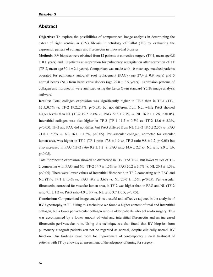
Chapter 3
56
Abstract
Objective: To explore the possibilities of computerized image analysis in determining the
extent of right ventricular (RV) fibrosis in tetralogy of Fallot (TF) by evaluating the
expression pattern of collagen and fibronectin in myocardial biopsies.
Methods: RV biopsies were obtained from 12 patients at corrective surgery (TF-1, mean age 0.8
± 0.1 years) and 10 patients at reoperation for pulmonary regurgitation after correction of TF
(TF-2, mean age 30.1 ± 2.4 years). Comparison was made with 10 mean age matched patients
operated for pulmonary autograft root replacement (PAG) (age 27.4 ± 0.9 years) and 5
normal hearts (NL) from heart valve donors (age 29.8 ± 3.9 years). Expression patterns of
collagen and fibronectin were analyzed using the Leica Qwin standard Y2.2b image analysis
software.
Results: Total collagen expression was significantly higher in TF-2 than in TF-1 (TF-1
12.5±0.7% vs. TF-2 19.2±2.4%, p<0.05), but not different from NL, while PAG showed
higher levels than NL (TF-2 19.2±2.4% vs. PAG 22.5 ± 2.7% vs. NL 16.9 ± 1.7%, p<0.05).
Interstitial collagen was also higher in TF-2 (TF-1 11.2 ± 0.7% vs. TF-2 18.6 ± 2.3%,
p<0.05). TF-2 and PAG did not differ, but PAG differed from NL (TF-2 18.6 ± 2.3% vs. PAG
21.8 ± 2.7% vs. NL 16.1 ± 1.5%, p<0.05). Peri-vascular collagen, corrected for vascular
lumen area, was higher in TF-1 (TF-1 ratio 17.8 ± 1.9 vs. TF-2 ratio 9.8 ± 1.2, p<0.05) but
also increased in PAG (TF-2 ratio 9.8 ± 1.2 vs. PAG ratio 14.6 ± 2.2 vs. NL ratio 8.9 ± 1.6,
p<0.05).
Total fibronectin expression showed no difference in TF-1 and TF-2, but lower values of TF-
2 comparing with PAG and NL (TF-2 14.7 ± 1.5% vs. PAG 20.2 ± 3.6% vs. NL 20.3 ± 1.5%,
p<0.05). There were lower values of interstitial fibronectin in TF-2 comparing with PAG and
NL (TF-2 14.1 ± 1.4% vs. PAG 19.8 ± 3.6% vs. NL 20.0 ± 1.5%, p<0.05). Peri-vascular
fibronectin, corrected for vascular lumen area, in TF-2 was higher than in PAG and NL (TF-2
ratio 7.1 ± 1.2 vs. PAG ratio 4.9 ± 0.9 vs. NL ratio 3.7 ± 0.5, p<0.05).
Conclusion: Computerized image analysis is a useful and effective adjunct in the analysis of
RV hypertrophy in TF. Using this technique we found a higher content of total and interstitial
collagen, but a lower peri-vascular collagen ratio in older patients who got re-do surgery. This
was accompanied by a lower amount of total and interstitial fibronectin and an increased
fibronectin peri-vascular ratio. Using this technique we also found that RV biopsies from
pulmonary autograft patients can not be regarded as normal, despite clinically normal RV
function. Our findings leave room for improvement of contemporary clinical treatment of
patients with TF by allowing an assessment of the adequacy of timing for surgery.
Computerized image analysis of myocardial fibrosis
57
Introduction
Nowadays most patients with tetralogy of Fallot (TF) are operated in their first year of
live by primary repair. The aim of early primary repair is to relieve the hypoxemia and
to eliminate the stimulus for the adaptive right ventricular hypertrophy (RVH) as well as
to preserve right ventricular (RV) function1. On long-term follow up however, a number
of patients suffer from pulmonary regurgitation after primary correction, causing RV
dilatation and failure due to volume-overload. For these patients further secondary
corrective surgery later in life is performed by implanting a pulmonary allograft heart
valve between the RV and the pulmonary artery2,3. At these different stages of disease,
there is a different and evoluating response of the RV myocardium, leading to
remodeling of the myocardium and to the development of fibrosis4-6. Collagen and
fibronectin both play an important part in myocardial fibrosis. The collagen network in
the extracellular space of the myocardium, consists of collagen fibers, which provide the
myocardial strength7-9. Experimental10 and clinical data11 show that a rise in the collagen
content increases the myocardial stiffness and promotes abnormalities of cardiac
function during hypertrophy. An increase in fibrosis in TF was associated with an
increased diameter of myocytes and an increase of myocardial disarray12. In this regard,
fibronectin, located in the extracellular matrix of most tissues, serves as a bridge
between cells and interstitial collagen mesh network. It further influences diverse
processes including cell growth, adhesion, migration, and wound repair13.
Most attention in research on human TF so far is focused on clinical treatment and
surgical methods. In addition, histopathological data on TF myocardial tissue showed a
reduction of the myocardial cell diameter in patients after corrective surgery14. This
would indicate a diminishing degree of RVH. Hardly any research has been done in TF
at molecular level. In this study, we examined the degree of fibrosis at protein level in
human TF by evaluating the expression of collagen and fibronectin in RV biopsies
obtained during redo surgery and compared the data with that of age matched patients
with a normal RV.
Chapter 3
58
Material and Methods
Patients
Our study was approved by the Medical Ethical Committee of the University Hospital
Rotterdam (MEC153.268/1996/119). The group of patients operated upon for primary
correction of TF (TF-1) consisted of 12 patients. The mean age of the TF-1 group at
the time of operation was 0.8 ± 0.1 years. The group of patients operated upon for
pulmonary allograft implantation late after correction of TF (TF-2) consisted of 10
patients, with a mean age of 30.1 ± 2.4 years. They were compared with an age-
matched control group consisting of 10 patients who were operated upon for a
pulmonary autograft root replacement (PAG) with a mean age of 27.4 ± 0.9 years and
with a group of 5 normal hearts (NL) from heart valve donors (mean age 29.8 ± 3.9
years). All patients operated upon for a pulmonary autograft procedure showed
clinically normal RV function.
All operations were done with the use of cardiopulmonary bypass with
moderate hypothermia using bicaval cannulation. In selected cases of both PAG and
TF-2, single right atrial cannulation was used. St.Thomas hospital cardioplegic
solution was applied except in some of the TF-2 patients, in whom the pulmonary
allograft was implanted in the beating heart. In TF-1 pulmonary valve function was
preserved in 7 patients, a transannular patch was necessary in the remaining 5
patients. In patients with preserved pulmonary valve function the biopsies consisted of
removed endomyocardial tissue from the RV outflow tract. In patients with a
transannular patch the biopsies consisted of a transmural sample from the incision in
the anterior RV wall. In TF-2 pulmonary allografts were implanted as previously
described15,16. Biopsies consisted of tissue from the anterior free wall of the right
ventriculotomy. All PAG procedures were done as previously described17,18. In
harvesting the pulmonary autograft, the main pulmonary artery was transsected just
proximal to its bifurcation and excision was done from the right RV outflow tract
leaving a ridge of supporting myocardial cuff. From the area of the free RV wall of
this cuff the PAG biopsies were taken as a transmural sample. Five biopsies were
obtained from normal hearts from the Heart Valve Bank Rotterdam (Heart Valve
Bank Rotterdam, Rotterdam, The Netherlands)
Computerized image analysis of myocardial fibrosis
59
The myocardial biopsies were fixed in 4% paraformaldehyde in PBS for at least 24
hours and further processed for dehydration and embedding in paraffin for
histological and immunohistochemical studies.
There was no operative or other early mortality. There were no complications
that could be attributed to taking the biopsies.
Picro-sirius red staining
The total collagen in myocardial tissue specimens was stained with Picro-sirius red
F3BA19. Tissue sections (5 µm thickness) were treated with 0.2% aqueous
phosphomolybdic acid and incubated in 0.1% Picro-sirius red. Before dehydration, the
slides were treated with 0.01N HCl and mounted. Slides were visualized under light
microscope and eventually collagen contents were quantified.
Immunohistochemistry
Paraffin sections (5 µm thickness) of the myocardial tissues were cut and mounted on
3-amino-propyl-trioxysilane (Sigma, St Louis, MO, USA) coated glass slides.
Immunohistochemistry using a multiple step avidin-biotin complex (ABC) method
was performed essentially following suppliers instructions. In brief, after
deparaffinization in xylene and rehydration through graded alcohol the slides were
rinsed with water and phosphate buffered saline and quenched for endogenous
peroxidase by 2 % hydrogen peroxide in methanol. The slides were placed in a Sequeza
Immunostaining Workstation (Shandon Scientific, Astmoor, UK). Slides were pre-
incubated for 15 minutes with 10% normal goat serum to block non-specific binding.
Specific human-preabsorbed polyclonal antibodies, against human fibronectin in 1:1000
dilution (Life Technologies, Breda, The Netherlands), were applied as primary antiserum
and the sections were incubated at room temperature for 30 min. The supra optimal
dilution was obtained by examining the intensity of staining obtained with a series of
dilutions of the antisera from 1:100 to 1:2500 and selecting the dilution that gave
specific but easily visible signals. Negative controls were performed by omission of the
primary antibody. After washing in 0.5% tween-20 in PBS solution, the sections were
incubated with mouse biotinylated anti-rabbit IgG (BioGenex, San Ramon, CA, USA)
for 30 min and subsequently with peroxidase conjugated streptavidin. Color was
developed using DAB (3,3'-diaminobenzidine tetrahydrochloride dehydrate) and the
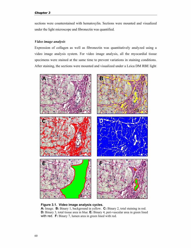
Chapter 3
60
sections were counterstained with hematoxylin. Sections were mounted and visualized
under the light microscope and fibronectin was quantified.
Video image analysis
Expression of collagen as well as fibronectin was quantitatively analyzed using a
video image analysis system. For video image analysis, all the myocardial tissue
specimens were stained at the same time to prevent variations in staining conditions.
After staining, the sections were mounted and visualized under a Leica DM RBE light
Figure 3.1. Video image analysis cycles. A: Image. B: Binary 1, background in yellow. C: Binary 2, total staining in red. D: Binary 3, total tissue area in blue. E: Binary 4, peri-vascular area in green lined with red. F: Binary 7, lumen area in green lined with red.
A B
FE
C D
Computerized image analysis of myocardial fibrosis
61
microscope (Leica, Wetzlar, Germany) using a 20x lens. The microscopic light was
optimized with a standard grey filter, a N16 neutralizing filter and a daylight filter.
From each section a series of 12 digital color images (736x574 pixels) were taken
from a representative area of the myocardium with a Sony DXC-950 3 CCD Power
HAD color video camera (Sony, Tokyo, Japan) after auto white balancing. The
camera contains a 0.5 inch tree-chip powered hole-accumulated (HAD) charge-
coupled device (CCD) light sensitive (2000 lux, F8.5, 3200K) chip with a resolution
of 752x582 effective picture elements (horizontal resolution of 750 TV lines) and a
scanning frequency of 15.625 kHz horizontal and 50 Hz vertical. All sections were
photographed in the same session, using the same settings for the microscope and the
Dell dimension XPS D266 computer with 64 RAM, connected to a 19.6 inch high
resolution Dell D2128-TCO screen with 1024x768 pixel setting, powered by a
Mantrox Millennium II AGP graphics card and software (Dell, Amsterdam, The
Netherlands). The digital images were analyzed for the staining of interest with a
special designed analysis program (macro) for the Leica Qwin standard image
analysis software (Y2.2b) (Leica Imaging Systems, Cambridge, UK) operating under
Microsoft windows 95 (Microsoft, Redmond, WA, USA). After optimalization, the
staining detection levels for hue, saturation and intensity in the computer were set and
used for analysis of all images derived from one staining session. The analysis macro
is designed to reduce the staining background by eroding and dilating each pixel
followed by creating binary files for the detected color intervals. Binary files are
created for white background (Binary1), total stained area (Binary2), tissue area
(Binary3 = total image - Binary1), peri-vascular area (Binary4; drawn manually),
peri-vascular staining (Binary5; overlap of Binary2 and Binary4), interstitial staining
(Binary6 = Binary2 - Binary5) and vessel lumen area (Binary7; drawn manually)(Fig.
3.1). Results are expressed as the ratio of staining area per analyzed total tissue area.
Interstitial staining is expressed as percentage stained tissue area excluding peri-
vascular area per analyzed total tissue area. Peri-vascular staining is expressed as
percentage peri-vascular staining per analyzed total tissue area. Peri-vascular staining
was normalized to its vascular lumen area, to correct for the vessel size, and expressed
as the ratio of peri-vascular stained area divided by vascular lumen area20.
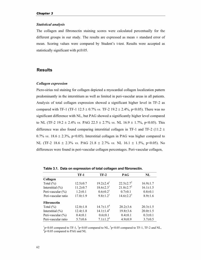
Chapter 3
62
Statistical analysis
The collagen and fibronectin staining scores were calculated percentually for the
different groups in our study. The results are expressed as mean ± standard error of
mean. Scoring values were compared by Student’s t-test. Results were accepted as
statistically significant with p≤0.05.
Results
Collagen expression
Picro-sirius red staining for collagen depicted a myocardial collagen localization pattern
predominantly in the interstitium as well as limited in peri-vascular areas in all patients.
Analysis of total collagen expression showed a significant higher level in TF-2 as
compared with TF-1 (TF-1 12.5 ± 0.7% vs. TF-2 19.2 ± 2.4%, p<0.05). There was no
significant difference with NL, but PAG showed a significantly higher level compared
to NL (TF-2 19.2 ± 2.4% vs. PAG 22.5 ± 2.7% vs. NL 16.9 ± 1.7%, p<0.05). This
difference was also found comparing interstitial collagen in TF-1 and TF-2 (11.2 ±
0.7% vs. 18.6 ± 2.3%, p<0.05). Interstitial collagen in PAG was higher compared to
NL (TF-2 18.6 ± 2.3% vs. PAG 21.8 ± 2.7% vs. NL 16.1 ± 1.5%, p<0.05). No
differences were found in peri-vascular collagen percentages. Peri-vascular collagen,
Table 3.1. Data on expression of total collagen and fibronectin.
TF-1 TF-2 PAG NL Collagen Total (%) 12.5±0.7 19.2±2.41 22.5±2.72 16.9±1.7 Interstitial (%) 11.2±0.7 18.6±2.31 21.8±2.72 16.1±1.5 Peri-vascular (%) 1.2±0.1 0.6±0.21 0.7±0.1 0.8±0.1 Peri-vascular ratio 17.8±1.9 9.8±1.21 14.6±2.23 8.9±1.6
Fibronectin Total (%) 12.8±1.8 14.7±1.54 20.2±3.6 20.3±1.5 Interstitial (%) 12.4±1.8 14.1±1.44 19.8±3.6 20.0±1.5 Peri-vascular (%) 0.4±0.1 0.6±0.1 0.4±0.1 0.3±0.1 Peri-vascular ratio 5.7±0.6 7.1±1.24 4.9±0.9 3.7±0.5
1p<0.05 compared to TF-1, 2p<0.05 compared to NL, 3p<0.05 compared to TF-1, TF-2 and NL, 4p<0.05 compared to PAG and NL
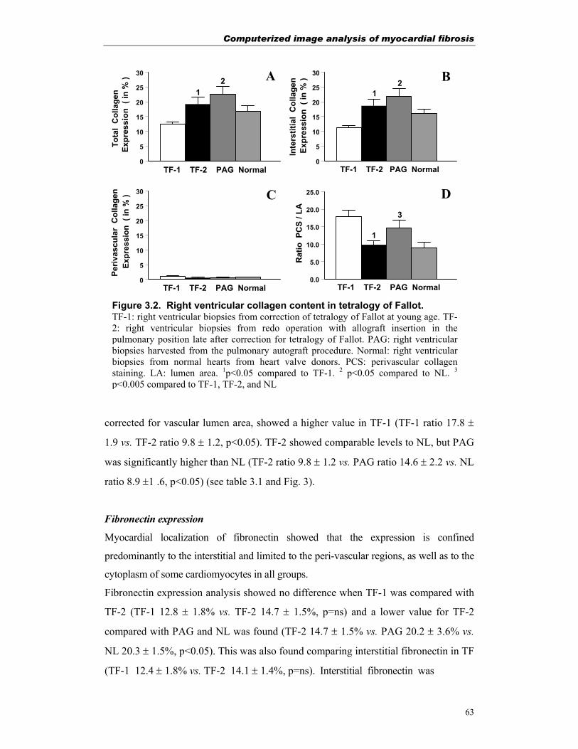
Computerized image analysis of myocardial fibrosis
63
corrected for vascular lumen area, showed a higher value in TF-1 (TF-1 ratio 17.8 ±
1.9 vs. TF-2 ratio 9.8 ± 1.2, p<0.05). TF-2 showed comparable levels to NL, but PAG
was significantly higher than NL (TF-2 ratio 9.8 ± 1.2 vs. PAG ratio 14.6 ± 2.2 vs. NL
ratio 8.9 ±1 .6, p<0.05) (see table 3.1 and Fig. 3).
Fibronectin expression
Myocardial localization of fibronectin showed that the expression is confined
predominantly to the interstitial and limited to the peri-vascular regions, as well as to the
cytoplasm of some cardiomyocytes in all groups.
Fibronectin expression analysis showed no difference when TF-1 was compared with
TF-2 (TF-1 12.8 ± 1.8% vs. TF-2 14.7 ± 1.5%, p=ns) and a lower value for TF-2
compared with PAG and NL was found (TF-2 14.7 ± 1.5% vs. PAG 20.2 ± 3.6% vs.
NL 20.3 ± 1.5%, p<0.05). This was also found comparing interstitial fibronectin in TF
(TF-1 12.4 ± 1.8% vs. TF-2 14.1 ± 1.4%, p=ns). Interstitial fibronectin was
0
5
10
15
20
25
30
Tota
l C
olla
gen
0
5
10
15
20
25
30
0
5
10
15
20
25
30
0.0
5.0
10.0
15.0
20.0
25.0
A B
C
Rat
io P
CS
/ LA
Periv
ascu
lar
Col
lage
n D
1
1
2 21
3
Expr
essi
on (
in %
)Ex
pres
sion
( in
% )
Expr
essi
on (
in %
)In
ters
titia
l C
olla
gen
TF-1 TF-2 PAG Normal TF-1 TF-2 PAG Normal
TF-1 TF-2 PAG Normal TF-1 TF-2 PAG Normal
Figure 3.2. Right ventricular collagen content in tetralogy of Fallot. TF-1: right ventricular biopsies from correction of tetralogy of Fallot at young age. TF-2: right ventricular biopsies from redo operation with allograft insertion in the pulmonary position late after correction for tetralogy of Fallot. PAG: right ventricular biopsies harvested from the pulmonary autograft procedure. Normal: right ventricular biopsies from normal hearts from heart valve donors. PCS: perivascular collagen staining. LA: lumen area. 1p<0.05 compared to TF-1. 2 p<0.05 compared to NL. 3 p<0.005 compared to TF-1, TF-2, and NL
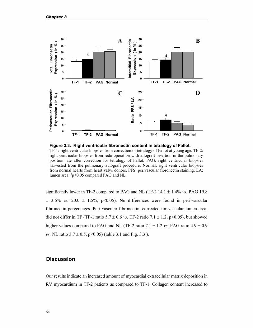
Chapter 3
64
significantly lower in TF-2 compared to PAG and NL (TF-2 14.1 ± 1.4% vs. PAG 19.8
± 3.6% vs. 20.0 ± 1.5%, p<0.05). No differences were found in peri-vascular
fibronectin percentages. Peri-vascular fibronectin, corrected for vascular lumen area,
did not differ in TF (TF-1 ratio 5.7 ± 0.6 vs. TF-2 ratio 7.1 ± 1.2, p<0.05), but showed
higher values compared to PAG and NL (TF-2 ratio 7.1 ± 1.2 vs. PAG ratio 4.9 ± 0.9
vs. NL ratio 3.7 ± 0.5, p<0.05) (table 3.1 and Fig. 3.3 ).
Discussion
Our results indicate an increased amount of myocardial extracellular matrix deposition in
RV myocardium in TF-2 patients as compared to TF-1. Collagen content increased to
Figure 3.3. Right ventricular fibronectin content in tetralogy of Fallot. TF-1: right ventricular biopsies from correction of tetralogy of Fallot at young age. TF-2: right ventricular biopsies from redo operation with allograft insertion in the pulmonary position late after correction for tetralogy of Fallot. PAG: right ventricular biopsies harvested from the pulmonary autograft procedure. Normal: right ventricular biopsies from normal hearts from heart valve donors. PFS: perivascular fibronectin staining. LA: lumen area. 4p<0.05 compared PAG and NL
0
5
10
15
20
25
30
Tota
l Fi
bron
ectin
0
5
10
15
20
25
30
0
5
10
15
20
25
30
0
5
10
15
20
25
A B
C
Rat
io P
FS /
LA
Periv
ascu
lar
Fibr
onec
tin
D
4
4
4Ex
pres
sion
( in
% )
Expr
essi
on (
in %
)
Expr
essi
on (
in %
)In
ters
titia
l Fi
bron
ectin
TF-1 TF-2 PAG Normal TF-1 TF-2 PAG Normal
TF-1 TF-2 PAG Normal TF-1 TF-2 PAG Normal
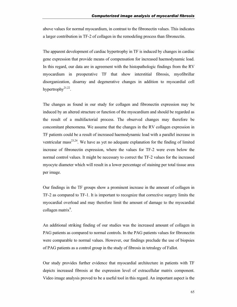
Computerized image analysis of myocardial fibrosis
65
above values for normal myocardium, in contrast to the fibronectin values. This indicates
a larger contribution in TF-2 of collagen in the remodeling process than fibronectin.
The apparent development of cardiac hypertrophy in TF is induced by changes in cardiac
gene expression that provide means of compensation for increased haemodynamic load.
In this regard, our data are in agreement with the histopathologic findings from the RV
myocardium in preoperative TF that show interstitial fibrosis, myofibrillar
disorganization, disarray and degenerative changes in addition to myocardial cell
hypertrophy21,22.
The changes as found in our study for collagen and fibronectin expression may be
induced by an altered structure or function of the myocardium and should be regarded as
the result of a multifactorial process. The observed changes may therefore be
concomitant phenomena. We assume that the changes in the RV collagen expression in
TF patients could be a result of increased haemodynamic load with a parallel increase in
ventricular mass23,24. We have as yet no adequate explanation for the finding of limited
increase of fibronectin expression, where the values for TF-2 were even below the
normal control values. It might be necessary to correct the TF-2 values for the increased
myocyte diameter which will result in a lower percentage of staining per total tissue area
per image.
Our findings in the TF groups show a prominent increase in the amount of collagen in
TF-2 as compared to TF-1. It is important to recognize that corrective surgery limits the
myocardial overload and may therefore limit the amount of damage to the myocardial
collagen matrix9.
An additional striking finding of our studies was the increased amount of collagen in
PAG patients as compared to normal controls. In the PAG patients values for fibronectin
were comparable to normal values. However, our findings preclude the use of biopsies
of PAG patients as a control group in the study of fibrosis in tetralogy of Fallot.
Our study provides further evidence that myocardial architecture in patients with TF
depicts increased fibrosis at the expression level of extracellular matrix component.
Video image analysis proved to be a useful tool in this regard. An important aspect is the
Chapter 3
66
constant and consequent approach to the tissue handling and staining techniques on one
hand and the settings of the software and hardware on the other hand. In this way more
specific and objective measurements can be done. Computerized image analysis
allows to objectivate the small differences in the peri-vascular accumulation of
collagen and fibronectin (panels C and D in Fig. 3.1 and 3.2). In addition, further
research at the mRNA level would add to the notion that the gene expression in fibrosis
was altered.
Nevertheless, studies of this kind provide information that leaves room for improvement
of contemporary clinical treatment of patients with tetralogy of Fallot by allowing an
assessment of the adequacy of timing for surgery and consequently provide data that
may be useful with regard to long-term postoperative prognosis.
Acknowledgements
Financial support from the Netherlands Heart Foundation (NHS 96.082) is gratefully
acknowledged. We thank Mrs Yilmaz for technical support advice and the Heart Valve
Bank Rotterdam for providing the control biopts.
References 1. Starnes VA, Luciani GB, Latter DA, et al. Current surgical management of tetralogy
of Fallot. Ann Thorac Surg. 1994;58:211-215. 2. Warner KG, Anderson JE, Fulton DR, et al. Restoration of the pulmonary valve
reduces right ventricular volume overload after previous repair of tetralogy of Fallot. Circulation. 1993;88:II189-197.
3. Murphy JG, Gersh BJ, Mair DD, et al. Long-term outcome in patients undergoing surgical repair of tetralogy of Fallot. N Engl J Med. 1993;329:593-599.
4. Schwartz K, Carrier L, Mercadier JJ, et al. Molecular phenotype of the hypertrophied and failing myocardium. Circulation. 1993;87:VII 5-10.
5. van Bilsen M, Chien KR. Growth and hypertrophy of the heart: towards an understanding of cardiac specific and inducible gene expression. Cardiovasc Res. 1993;27:1140-1149.
6. Boluyt MO, L. O, Meredith AL, et al. Alterations in cardiac gene expression during the transition from stable hypertrophy to heart failure. Marked upregulation of genes encoding extracellular matrix components. Circ Res. 1994;75:23-32.
Computerized image analysis of myocardial fibrosis
67
7. Bishop JE, Rhodes S, Laurent GJ, et al. Increased collagen synthesis and decreased collagen degradation in right ventricular hypertrophy induced by pressure overload. Cardiovasc Res. 1994;28:1581-1585.
8. Weber KT, Sun Y, Tyagi SC, et al. Collagen network of the myocardium: function, structural remodelling and regulatory mechanisms. J Mol Cell Cardiol. 1994;26:279-292.
9. Whittaker P. Collagen and ventricular remodelling after acute myocardial infarction: concepts and hypotheses. Basic Res Cardiol. 1997;92:79-81.
10. Jalil JE, Doering CW, Janicki JS, et al. Fibrillar collagen and myocardial stiffness in the intact hypertrophied rat left ventricle. Circ. Res. 1989;64:1041-1050.
11. McLenachan JM, Dargie HJ. Ventricular arrhythmias in hypertensive left ventricular hypertrophy. Relationship to coronary artery disease, left ventricular dysfunction, and myocardial fibrosis. Am J Hypertens. 1990;3:735-740.
12. Kawai S, Okada R, Kitamura K, et al. A morphometrical study of myocardial disarray associated with right ventricular outflow tract obstruction. Jpn Circ J. 1984;48:445-456.
13. Farhadian F, Contard F, Corbier A, et al. Fibronectin expression during physiological and pathological cardiac growth. J Mol Cell Cardiol. 1995;27:981-990.
14. Seliem MA, Wu YT, Glenwright K. Relation between age at surgery and regression of right ventricular hypertrophy in tetralogy of Fallot. Pediatr Cardiol. 1995;16:53-55.
15. Hokken RB, Bogers AJJC, Spitaels SEC, et al. Pulmonary homograft insertion after repair of pulmonary stenosis. J Heart Valve Dis. 1995;4:182-186.
16. Bogers AJJC, Roofthooft M, Pisters H, et al. Longterm follow-up of gamma irradiated transannular homograft patch in surgical treatment of tetralogy of Fallot. Thorac Cardiovasc Surgeon. 1994;42:337-339.
17. Schoof PH, Cromme-Dijkhuis AH, Bogers AJJC, et al. Aortic root replacement with pulmonary autograft in children. J Thorac Cardiovasc Surg. 1994;107:367-73.
18. Hokken RB, Bogers AJJC, Taams MA, et al. Aortic root replacement with a pulmonary autograft. Eur J Cardiothor Surg. 1995;9:378-383.
19. Takahashi T, Schunkert H, Isoyama S, et al. Age-related differences in the expression of proto-oncogene and contractile protein genes in response to pressure overload in the rat myocardium. J Clin Invest. 1992;89:939-946.
20. Brilla CG, Janicki JS, Weber KT. Impaired diastolic function and coronary reserve in genetic hypertension. Role of interstitial fibrosis and medial thickening of intramyocardial coronary arteries. Circ Res. 1991;69:107-115.
21. Brand T, Sharma HS, Fleischmann KE, et al. Proto-oncogene expression in porcine myocardium subjected to ischemia and reperfusion. Circ Res. 1992;71:1351-1360.
22. Brand T, Sharma HS, Schaper W. Expression of nuclear proto-oncogenes in isoproterenol-induced cardiac hypertrophy. J Mol Cell Cardiol. 1993;25:1325-1337.
23. Schwartz SM, Gordon D, Mosca RS, et al. Collagen content in normal, pressure, and pressure-volume overloaded developing human hearts. Am J Cardiol. 1996;77:734-738.
24. Vikstrom KL, Bohlmeyer T, Factor SM, et al. Hypertrophy, pathology, and molecular markers of cardiac pathogenesis. Circ Res. 1998;82:773-778.

Chapter 4
68
Myocardial fibrosis in tetralogy of Fallot
69
Chapter 4
DNA micro-array and myocardial fibrosis in patients with tetralogy of Fallot
Based on: Peters THF, Bogers AJJC, Staal FJT, Poelmann RE, Saxena PR and Hari S Sharma. DNA micro-array and quantitative analysis reveal up-regulation of myocardial genes encoding extracellular matrix proteins in patients with tetralogy of Fallot. (submitted).
Chapter 4
70
Abstract
Background: Tetralogy of Fallot (TF) is a congenital heart disease with adaptive right
ventricular hypertrophy where cardiac surgery is performed at a young age to prevent
cardiac failure on the long term. To unravel molecular phenotype, we investigated the
myocardial differential gene expression profile and quantitatively analyzed the
expression of extracellular matrix (ECM) markers in TF patients.
Methods and Results: Differential gene expression profile was assessed using high
density Affymetrix® GeneChip analysis on right ventricular biopsies from TF patients who
underwent primary correction (TF-1, n=12, mean age 0.8 year). Employing quantitative
immunohistochemistry, ECM proteins (collagens and fibronectin) and myocyte cross
sectional area were evaluated in TF-1 and adult patients (TF-2, n=8, mean age 30 years)
who underwent surgery for pulmonary regurgitation and were compared with data of age
matched controls (n=8/11). Among 236 differentially expressed genes in TF-1 patients,
ECM markers were clearly up-regulated (fibronectin, 2.4; collagen Iα, 7.5 and collagen III,
4.4 fold). Most MMPs remained either unchanged or declined (MMP-13 and 17) whereas,
TIMPs were down regulated. TF-1 patients showed myocyte hypertrophy (261±21 vs.
81±7µm2, p<0.01) with linear correlation with age (r=0.92, p<0.01). Interstitial as well as
peri-vascular fibronectin and total collagen contents were increased (p<0.01) in TF patients
as compared to age matched controls.
Conclusions: Myocardial phenotype in TF patients depicts a complex gene expression
profile with elevated tissue distribution of ECM markers, that eventually contribute to the
cardiac fibrosis and failure.
Myocardial fibrosis in tetralogy of Fallot
71
Introduction
Tetralogy of Fallot (TF) is a cyanotic congenital heart malformation where primary
cardiac surgery is inevitable during the first year of life by closing the ventricular septal
defect and removing the right ventricular (RV) outflow tract obstruction1-4. However,
some patients suffer from pulmonary regurgitation after primary correction causing RV
failure due to volume overload and they undergo a second corrective surgery later in life
by implanting a pulmonary allograft heart valve between the RV and the pulmonary
artery5. Information on myocardial phenotype, specially at gene level may help in
developing innovative treatment strategies and perhaps halt the ongoing debate for the
optimal timing of corrective surgery in patients with TF.
Right ventricular hypertrophy (RVH) develops in TF patients as an adaptive response
to the increased pressure overload caused by (sub) pulmonary stenosis in combination
with a ventricular septal defect2,6-8. Experimental animal studies have shown that this
adaptive response leads to myocardial remodeling accompanied with interstitial
fibrosis that give rise to cardiac stiffness and abnormal function9-16. The developing
human heart predominantly contains collagen type I that provides cardiac stiffness,
whereas tissue elasticity is provided by the less abundant collagen III14,16. Expression
levels of extracellular matrix (ECM) protein fibronectin, that serves as a bridge
between cells and interstitial collagen mesh network, has been shown to be up-regulated
during cardiac hypertrophy17. Studies demonstrated that only the RV collagen levels in
TF patients during pressure and volume overload were affected7. Normally, the
collagen turnover rate is high where mature cross-linked collagens fibers are degraded
extracellularly by a family of matrix metalloproteinases (MMPs) that play an
important role in tissue remodeling9,18,19. The activity of MMPs can be regulated by
different cytokines and tissue inhibitors of MMPs (TIMPs)18-20. Increased expression
or activation of the MMPs and/or decreased levels of TIMPs lead to enhanced
proteolytic activity and ECM remodeling with myofibril disarray and progressive
myocyte loss21,22.
Most attention in research on human TF so far has been focused on clinical treatment
and surgical procedures. Reduction in myocardial cell diameter in TF patients after
corrective surgery has been observed6,23 indicating for the diminished degree of RVH.
Chapter 4
72
But to what extent the RVH in TF patients after corrective surgery regresses is still
uncertain. Additionally, information concerning myocardial architecture in TF at a
molecular level is limited. To assess the differential myocardial gene expression in
patients with TF at the time of corrective surgery, we performed DNA micro-array
followed by quantitative expression analysis of genes encoding ECM proteins in relation to
age matched controls.
Material and Methods
Study subjects
The study design was approved by the Medical Ethical Committee of the University
Hospital Rotterdam (MEC153.268/119). Studied groups and their clinical parameters
are described in table 4.1. 12 patients underwent primary correction (TF-1 group) at a
mean age of 0.8 years (range 0.4-1.8 years) whereas, 8 subjects (TF-2 group) with a
mean age of 30.0 years (range 19.3-38.8 years) were operated for pulmonary allograft
implantation late after primary correction of TF. All operations were done with the
use of cardiopulmonary bypass with moderate hypothermia using bicaval cannulation.
In selected cases, single right atrial cannulation was used. St.Thomas hospital
cardioplegic solution was applied except in some of the TF-2 patients, in whom the
pulmonary allograft was implanted in the beating heart24,25. In TF-1 patients,
pulmonary valve function could be preserved in 5 patients, whereas a transannular
patch was necessary in 3 patients. The young controls (C-1 group) comprised of 5
post mortem cases and 3 heart valve donors with a mean age of 0.8 years (range 0.01-
1.9 years), whereas the adult controls (C-2 group) were of 11 heart valve donors with
a mean age of 26.9 years (range 17.1-44.6 years). All heart valve donors and post
mortem cases died due to non-cardiac complications.
Tissue biopsies
Endomyocardial tissue biopsies were removed from the anterior wall of the RV
outflow tract in patients with preserved pulmonary valve function whereas, in patients
with a transannular patch, the biopsies consisted of a transmural sample from the
incision level of anterior RV wall. There was no operative or other early mortality and
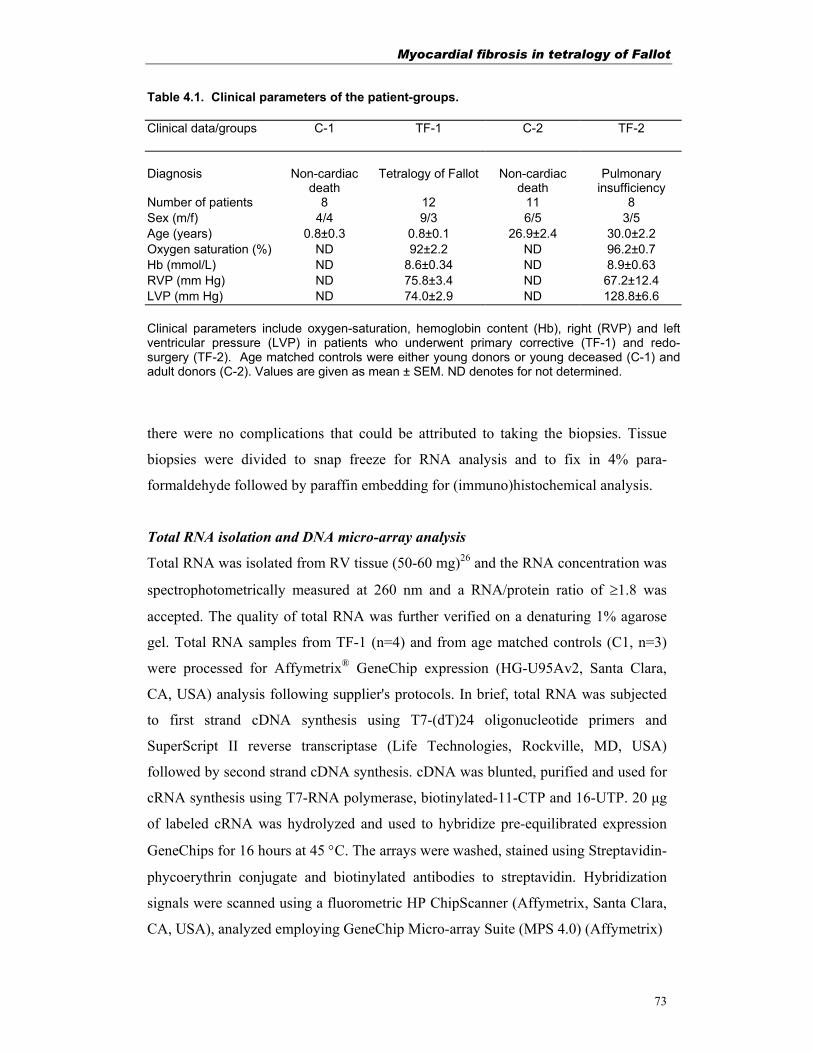
Myocardial fibrosis in tetralogy of Fallot
73
Table 4.1. Clinical parameters of the patient-groups.
Clinical data/groups C-1 TF-1 C-2 TF-2
Diagnosis Non-cardiac death
Tetralogy of Fallot Non-cardiac death
Pulmonary insufficiency
Number of patients 8 12 11 8 Sex (m/f) 4/4 9/3 6/5 3/5 Age (years) 0.8±0.3 0.8±0.1 26.9±2.4 30.0±2.2 Oxygen saturation (%) ND 92±2.2 ND 96.2±0.7 Hb (mmol/L) ND 8.6±0.34 ND 8.9±0.63 RVP (mm Hg) ND 75.8±3.4 ND 67.2±12.4 LVP (mm Hg) ND 74.0±2.9 ND 128.8±6.6
Clinical parameters include oxygen-saturation, hemoglobin content (Hb), right (RVP) and left ventricular pressure (LVP) in patients who underwent primary corrective (TF-1) and redo-surgery (TF-2). Age matched controls were either young donors or young deceased (C-1) and adult donors (C-2). Values are given as mean ± SEM. ND denotes for not determined.
there were no complications that could be attributed to taking the biopsies. Tissue
biopsies were divided to snap freeze for RNA analysis and to fix in 4% para-
formaldehyde followed by paraffin embedding for (immuno)histochemical analysis.
Total RNA isolation and DNA micro-array analysis
Total RNA was isolated from RV tissue (50-60 mg)26 and the RNA concentration was
spectrophotometrically measured at 260 nm and a RNA/protein ratio of ≥1.8 was
accepted. The quality of total RNA was further verified on a denaturing 1% agarose
gel. Total RNA samples from TF-1 (n=4) and from age matched controls (C1, n=3)
were processed for Affymetrix® GeneChip expression (HG-U95Av2, Santa Clara,
CA, USA) analysis following supplier's protocols. In brief, total RNA was subjected
to first strand cDNA synthesis using T7-(dT)24 oligonucleotide primers and
SuperScript II reverse transcriptase (Life Technologies, Rockville, MD, USA)
followed by second strand cDNA synthesis. cDNA was blunted, purified and used for
cRNA synthesis using T7-RNA polymerase, biotinylated-11-CTP and 16-UTP. 20 µg
of labeled cRNA was hydrolyzed and used to hybridize pre-equilibrated expression
GeneChips for 16 hours at 45 °C. The arrays were washed, stained using Streptavidin-
phycoerythrin conjugate and biotinylated antibodies to streptavidin. Hybridization
signals were scanned using a fluorometric HP ChipScanner (Affymetrix, Santa Clara,
CA, USA), analyzed employing GeneChip Micro-array Suite (MPS 4.0) (Affymetrix)
Chapter 4
74
and total gene expression profile was used for normalization27. A comparison of the
two expression profiles was made and genes changing over 2 fold (up or down
regulated) were considered significantly altered in expression28. Among the large
number of genes screened (www.netaffx.com), a group of genes encoding ECM
proteins (e.g. fibronectin and collagens) was further quantitatively analyzed by
immunohistochemistry and video image analysis.
Histochemistry
The total collagen in myocardial tissue specimens was stained with Picro-sirius red
F3BA29. Tissue sections of 5 µm thickness, were treated with aqueous
phosphomolybdic acid and incubated in 0.1% Picro-sirius red. For determining the
myocyte surface size, the cell (membrane) reticulin fibers were stained by Gomori’s
reticulin silver staining technique30. All sections were mounted and visualized under
light microscope.
Immunolocalization of fibronectin, collagen Iα and collagen III
Immunohistochemical staining was performed on 5 µm thick paraffin sections of
myocardial tissue, using a multiple step avidin-biotin complex (ABC) method
(Biogenex, San Ramon, CA, USA). In brief, after deparaffinization in xylene and
rehydration through graded alcohol, the slides were processed for antigen retrieval
using citrate boiling method (collagen Iα, fibronectin) or using 0.1 % pronase
treatment (collagen III). 10% normal goat serum was used to block non-specific
binding. Human specific mouse monoclonal antibodies against fibronectin (Life
Technologies, Breda, The Netherlands), collagen Iα (Sigma, St Louis, MO, USA) or
collagen III (BioGenex, San Ramon, CA, USA) were applied as primary antiserum.
The sections were further incubated with mouse biotinylated anti-rabbit IgG
(BioGenex, San Ramon, CA) followed with alkaline phosphate conjugated
streptavidin. Color was developed using Naphtol AS-MX phosphate and new fuchsin.
For collagen III, streptavidin conjugated to peroxidase (BioGenex, San Ramon, CA)
was applied and subsequently sections were colored using 3,3-diaminobenzidine
(Sigma, St Louis, MO, USA). The supra optimal dilution was obtained by examining
the intensity of staining obtained with a series of dilutions of the antibody and
selecting the dilution that gave specific, weak but easily visible signal. Negative
Myocardial fibrosis in tetralogy of Fallot
75
controls were performed by omission of the primary antibody. All sections were
counter stained with Mayer's hematoxylin, mounted and visualized under the light
microscope.
Video image analysis
Expression of fibronectin, total collagen as well as collagen sub-type Iα and III were
quantitatively analyzed using a Leica Qwin video image analysis system (version 3.0,
Leica, Rijswijk, The Netherlands). From each section, series of 12 rectangular digital
color images (736x574 pixels) were taken using a CCD video camera connected to the
light microscope (Leica DM RBE, Leica GmbH, Wetzlar, Germany) and were
analyzed with an image analysis software (QWin-V 2.8, Cambridge Biotech, UK).
After optimization for hue, saturation and intensity, the staining detection levels for all
images were assessed as pixel counts at the constant settings of the microscope-image
analysis system. Interstitial expression was represented as staining per total tissue
area, excluding vascular areas. For peri-vascular measurements, the entire vessel was
drawn manually and analyzed. Peri-vascular localization was depicted as peri-
vascular collagen/fibronectin area (PVCA/PVFA) normalized to vessel luminal area
(VA)11. In the similar set-up, the myocyte cross sectional area was calculated in µm2
after drawing the reticulin stained cell membrane in 20 transversely cut myocytes with
intact nucleus. Influence of RVH was taken into account and interstitial expression of
ECM markers was normalized with the increase in cell size of the respective patient.
Statistical analysis
Statistical analysis for micro-array results was performed with comparison ranking
within software Data Mining Tool (Affymetrix). Using SPSS software (SPSS Inc.
Chicago, USA), statistical probability was assessed by (non-parametric) Kruskal-
Wallis one way analysis of variance and linear (Pearson's) regression analysis was
performed for assessing RVH. Values are expressed as mean ± SEM and accepted as
statistically significant with p≤0.05.
Chapter 4
76
Results
DNA micro-array analysis
To asses the differential gene-expression profile, RNA samples obtained from TF-1
and age matched controls were processed for Affymetrix® GeneChip analysis. Gene
expression was considered to be up or down regulated if the expression levels were 2
fold higher/lower than the controls as presented in a differential scatter graph in the
figure 4.1. Comparison ranking analysis revealed that in TF-1 patients, 236 genes
showed altered expression pattern. Analysis of a selected group of genes dedicated to
ECM homeostasis in the heart, showed that a number of genes encoding ECM
proteins were up-regulated in TF-1 patients as compared to the age matched controls
(table 4.2). With the same threshold no fibrosis markers were found to be down
regulated. Collagen type I and III drastically (up to 7.5 fold) and collagen type IX and
XV were moderately up-regulated in TF patients whereas, other subtypes remained
unchanged (table 4.2). Expression levels for fibronectin were also enhanced (2-3 fold)
in TF patients. The MMP expression profile remained unchanged or declined (MMP-
13,17) whereas, their physiological inhibitors (TIMP-1, 4) showed decreased (2-4
fold) expression levels in TF group (table 4.3).
Quantitative analysis of collagen expression
Picro-sirius red staining showed myocardial collagen localization in the interstitium as
well as in peri-vascular areas in all patients (Fig. 4.2). TF patients showed altered
distribution of collagen fibers, myocyte organization and shape as compared to age
matched controls. Controls showed an arrayed pattern of uniform shaped long
myocyte bundles surrounded by tight collagen fibers invading between the myocytes,
whereas, TF patients showed less uniformity in shape and array of myocytes with
small dense collagen bundles. In TF-1, collagen fibers were more randomly localized
in the interstitium, whereas in TF-2, collagen layers around the myocytes were thicker
as compared to respective controls. Immunoreactive collagen Iα and III were
confined to the interstitial and peri-vascular areas (Fig. 4.2). Video image analysis of
interstitial as well as peri-vascular total collagen contents revealed significantly
increased (p<0.01) expression in both TF-1 and TF-2 groups as compared to
respective controls (Fig. 4.3). The data presented for interstitial expression was
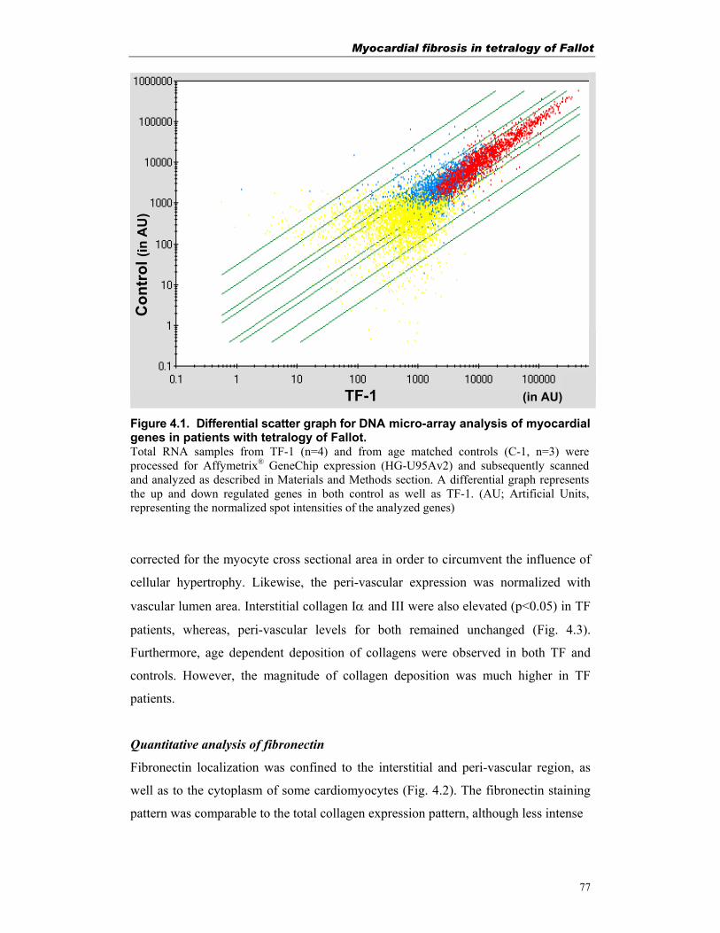
Myocardial fibrosis in tetralogy of Fallot
77
TF-1 (in AU)
Con
trol
(in
AU
)
Figure 4.1. Differential scatter graph for DNA micro-array analysis of myocardial genes in patients with tetralogy of Fallot. Total RNA samples from TF-1 (n=4) and from age matched controls (C-1, n=3) were processed for Affymetrix® GeneChip expression (HG-U95Av2) and subsequently scanned and analyzed as described in Materials and Methods section. A differential graph represents the up and down regulated genes in both control as well as TF-1. (AU; Artificial Units, representing the normalized spot intensities of the analyzed genes)
corrected for the myocyte cross sectional area in order to circumvent the influence of
cellular hypertrophy. Likewise, the peri-vascular expression was normalized with
vascular lumen area. Interstitial collagen Iα and III were also elevated (p<0.05) in TF
patients, whereas, peri-vascular levels for both remained unchanged (Fig. 4.3).
Furthermore, age dependent deposition of collagens were observed in both TF and
controls. However, the magnitude of collagen deposition was much higher in TF
patients.
Quantitative analysis of fibronectin
Fibronectin localization was confined to the interstitial and peri-vascular region, as
well as to the cytoplasm of some cardiomyocytes (Fig. 4.2). The fibronectin staining
pattern was comparable to the total collagen expression pattern, although less intense
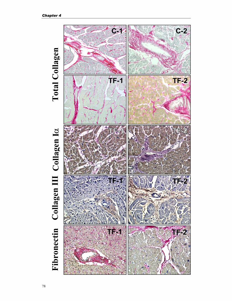
Chapter 4
78
C-1 C-2
TF-1 TF-2
TF-1
TF-1
TF-2
TF-2
Tot
al C
olla
gen
Col
lage
n I α
C
olla
gen
III
Fibr
onec
tin
TF-1 TF-2
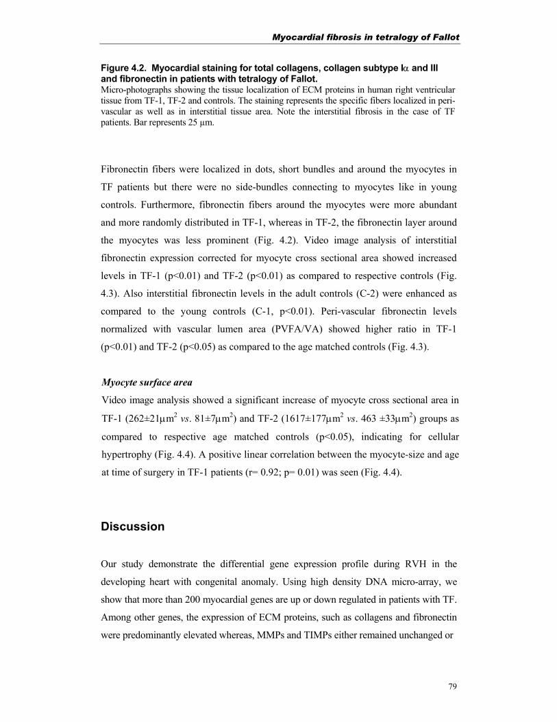
Myocardial fibrosis in tetralogy of Fallot
79
Figure 4.2. Myocardial staining for total collagens, collagen subtype Iα and III and fibronectin in patients with tetralogy of Fallot. Micro-photographs showing the tissue localization of ECM proteins in human right ventricular tissue from TF-1, TF-2 and controls. The staining represents the specific fibers localized in peri-vascular as well as in interstitial tissue area. Note the interstitial fibrosis in the case of TF patients. Bar represents 25 µm.
Fibronectin fibers were localized in dots, short bundles and around the myocytes in
TF patients but there were no side-bundles connecting to myocytes like in young
controls. Furthermore, fibronectin fibers around the myocytes were more abundant
and more randomly distributed in TF-1, whereas in TF-2, the fibronectin layer around
the myocytes was less prominent (Fig. 4.2). Video image analysis of interstitial
fibronectin expression corrected for myocyte cross sectional area showed increased
levels in TF-1 (p<0.01) and TF-2 (p<0.01) as compared to respective controls (Fig.
4.3). Also interstitial fibronectin levels in the adult controls (C-2) were enhanced as
compared to the young controls (C-1, p<0.01). Peri-vascular fibronectin levels
normalized with vascular lumen area (PVFA/VA) showed higher ratio in TF-1
(p<0.01) and TF-2 (p<0.05) as compared to the age matched controls (Fig. 4.3).
Myocyte surface area
Video image analysis showed a significant increase of myocyte cross sectional area in
TF-1 (262±21µm2 vs. 81±7µm2) and TF-2 (1617±177µm2 vs. 463 ±33µm2) groups as
compared to respective age matched controls (p<0.05), indicating for cellular
hypertrophy (Fig. 4.4). A positive linear correlation between the myocyte-size and age
at time of surgery in TF-1 patients (r= 0.92; p= 0.01) was seen (Fig. 4.4).
Discussion
Our study demonstrate the differential gene expression profile during RVH in the
developing heart with congenital anomaly. Using high density DNA micro-array, we
show that more than 200 myocardial genes are up or down regulated in patients with TF.
Among other genes, the expression of ECM proteins, such as collagens and fibronectin
were predominantly elevated whereas, MMPs and TIMPs either remained unchanged or
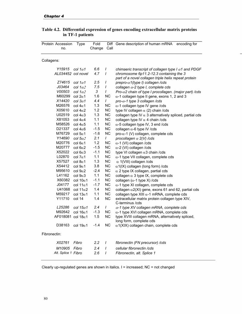
Chapter 4
80
Table 4.2. Differential expression of genes encoding extracellular matrix proteins in TF-1 patients
Protein Accession
no. Type Fold
ChangeDiff Call
Gene description of human mRNA encoding for
Collagens:
Y15915 col 1α1 6.6 I chimaeric transcript of collagen type I α1 and PDGF AL034452 col novel 4.7 I chromosome 6p11.2-12.3 containing the 3
part of a novel collagen triple helix repeat protein Z74615 col 1α1 2.5 I prepro-α1(type I) collagen /cds J03464 col 1α2 7.5 I collagen α-2 type I, complete cds V00503 col 1α2 3 I Pro-α2 chain of type I procollagen. (major part) /cds M60299 col 2α1 1.6 NC α-1 collagen type II gene, exons 1, 2 and 3 X14420 col 3α1 4.4 I pro-α-1 type 3 collagen /cds M26576 col 4α1 1.3 NC α-1 collagen type IV gene /cds X05610 col 4α2 1.2 NC type IV collagen α (2) chain /cds U02519 col 4α3 1.3 NC collagen type IV α 3 alternatively spliced, partial cds X81053 col 4α4 1.1 NC collagen type IV α 4 chain /cds M58526 col 4α5 1.1 NC α-5 collagen type IV, 3 end /cds D21337 col 4α6 -1.5 NC collagen α-6 type IV /cds M76729 col 5α1 -1.6 NC pro-α-1 (V) collagen, complete cds Y14690 col 5α2 2.1 I procollagen α 2(V) /cds M20776 col 6α1 1.2 NC α-1 (VI) collagen /cds M20777 col 6α2 -1.5 NC α-2 (VI) collagen /cds X52022 col 6α3 -1.1 NC type VI collagen α3 chain /cds L02870 col 7α1 1.1 NC α-1 type VII collagen, complete cds X57527 col 8α1 1.3 NC α 1(VIII) collagen /cds X54412 col 9α1 3.8 NC α1(IX) collagen (long form) /cds M95610 col 9α2 -2.4 NC α 2 type IX collagen, partial cds L41162 col 9α3 1.1 NC collagen α 3 type IX, complete cds X60382 col 10α1 -1.1 NC collagen (α-1 type X) /cds J04177 col 11α1 -1.7 NC α-1 type XI collagen, complete cds U41068 col 11α2 1.4 NC collagen α2(XI) gene, exons 61 and 62, partial cds M59217 col 13α1 1.1 NC collagen type XIII α-1 mRNA, complete cds Y11710 col 14 1.4 NC extracellular matrix protein collagen type XIV,
C-terminus /cds L25286 col 15α1 2.4 I α-1 type XV collagen mRNA, complete cds M92642 col 16α1 -1.3 NC α-1 type XVI collagen mRNA, complete cds AF018081 col 18α1 1.5 NC type XVIII collagen mRNA, alternatively spliced,
long form, complete cds D38163 col 19α1 -1.4 NC α1(XIX) collagen chain, complete cds
Fibronectin:
X02761 Fibro 2.2 I fibronectin (FN precursor) /cds M10905 Fibro 2.4 I cellular fibronectin /cds Alt. Splice 1 Fibro 2.6 I Fibronectin, alt. Splice 1
Clearly up-regulated genes are shown in italics. I = increased; NC = not changed
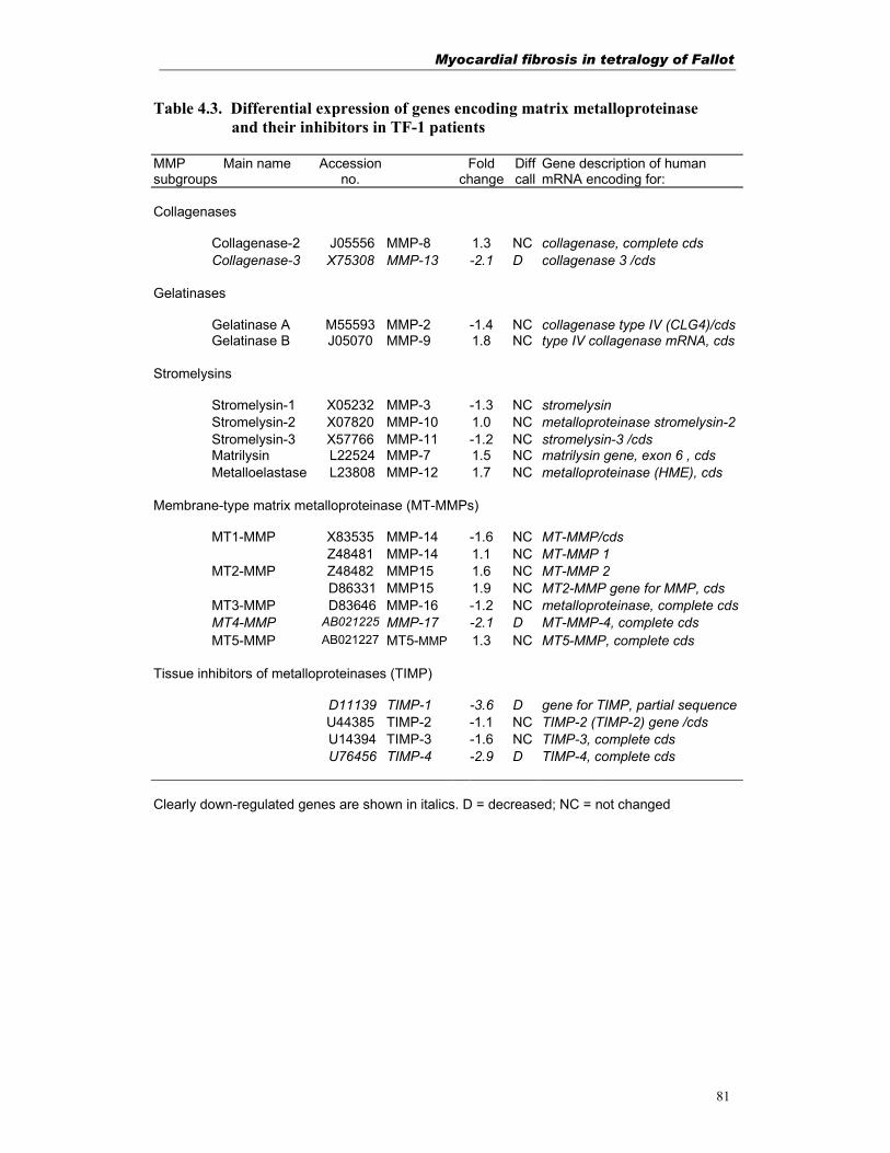
Myocardial fibrosis in tetralogy of Fallot
81
Table 4.3. Differential expression of genes encoding matrix metalloproteinase and their inhibitors in TF-1 patients
MMP subgroups
Main name Accession no.
Fold change
Diff call
Gene description of human mRNA encoding for:
Collagenases
Collagenase-2 J05556 MMP-8 1.3 NC collagenase, complete cds Collagenase-3 X75308 MMP-13 -2.1 D collagenase 3 /cds
Gelatinases
Gelatinase A M55593 MMP-2 -1.4 NC collagenase type IV (CLG4)/cds Gelatinase B J05070 MMP-9 1.8 NC type IV collagenase mRNA, cds
Stromelysins
Stromelysin-1 X05232 MMP-3 -1.3 NC stromelysin Stromelysin-2 X07820 MMP-10 1.0 NC metalloproteinase stromelysin-2 Stromelysin-3 X57766 MMP-11 -1.2 NC stromelysin-3 /cds Matrilysin L22524 MMP-7 1.5 NC matrilysin gene, exon 6 , cds Metalloelastase L23808 MMP-12 1.7 NC metalloproteinase (HME), cds
Membrane-type matrix metalloproteinase (MT-MMPs)
MT1-MMP X83535 MMP-14 -1.6 NC MT-MMP/cds Z48481 MMP-14 1.1 NC MT-MMP 1 MT2-MMP Z48482 MMP15 1.6 NC MT-MMP 2 D86331 MMP15 1.9 NC MT2-MMP gene for MMP, cds
MT3-MMP D83646 MMP-16 -1.2 NC metalloproteinase, complete cds MT4-MMP AB021225 MMP-17 -2.1 D MT-MMP-4, complete cds MT5-MMP AB021227 MT5-MMP 1.3 NC MT5-MMP, complete cds
Tissue inhibitors of metalloproteinases (TIMP)
D11139 TIMP-1 -3.6 D gene for TIMP, partial sequence U44385 TIMP-2 -1.1 NC TIMP-2 (TIMP-2) gene /cds U14394 TIMP-3 -1.6 NC TIMP-3, complete cds U76456 TIMP-4 -2.9 D TIMP-4, complete cds
Clearly down-regulated genes are shown in italics. D = decreased; NC = not changed
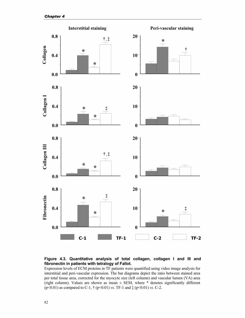
Chapter 4
82
Figure 4.3. Quantitative analysis of total collagen, collagen I and III and fibronectin in patients with tetralogy of Fallot. Expression levels of ECM proteins in TF patients were quantified using video image analysis for interstitial and peri-vascular expression. The bar diagrams depict the ratio between stained area per total tissue area, corrected for the myocyte size (left column) and vascular lumen (VA) area (right column). Values are shown as mean ± SEM, where * denotes significantly different (p<0.01) as compared to C-1, † (p<0.01) vs. TF-1 and ‡ (p<0.01) vs. C-2.
0.0
0.4
0.8C
olla
gen
C-1 TF-1 C-2 TF-2
0
10
20
0.0
0.4
0.8
Col
lage
n I
0
10
20
0.0
0.4
0.8
Col
lage
n II
I
0
10
20
0.0
0.4
0.8
Fibr
onec
tin
0
10
20
Interstitial staining Peri-vascular staining
*
‡
† , ‡
*
*
*
‡
‡*
*
*
† , ‡
*
*
*
†
Myocardial fibrosis in tetralogy of Fallot
83
decreased in TF patients. The global mRNA expression profile of ECM proteins was
further verified by quantitative immunohistochemistry where the distribution of both
collagens and fibronectin was clearly enhanced in the myocardium of patients with TF.
Our results indicate for the myocardial fibrosis that may account for diminished function
in patients with TF.
In the heart fibroblasts, which produces most of the collagen fibers, represent as much
as two-third of the cardiac cells9,15,16,19,31. Collagens are the major ECM proteins in the
heart that surround and interconnect myocytes and muscle fibers to provide mechanical
support to the myocardium and govern tissue stiffness16,19,31. A balance between
synthesis and degradation of ECM has been shown to be important in the process of
ventricular hypertrophy and remodeling during heart failure15,16,18,19. The apparent
development of cardiac hypertrophy in TF could be attributed to the induced gene
expression that provide means of compensation for increased haemodynamic load10,32. In
this regard, our data on histopathological findings in TF patients show interstitial
fibrosis, myofibrillar disorganization and disarray in addition to myocardial cell
hypertrophy. The degree of myocardial hypertrophy (cell radius of 22 µm) in TF-2 group
is in accordance with previously reported myocyte radius of more than 20 µm23.
Whether or not the myocytes of TF patients were already hypertrophied during
pregnancy and at birth is unclear, because no biopsies before the time of primary
corrective surgery are available. But it is assumed that the RV haemodynamic overload
in TF begins after birth when the ductus arteriosus closes and readily results in RVH.
Hypertrophy of the cardiomyocytes in TF patients will increase the total (tissue)
volume. It is likely that the increase in myocyte cross sectional area during hypertrophy
would influence the staining levels of ECM markers as measured by video image
analysis (expressed as a ratio between stained area and total tissue area). Therefore,
the interstitial staining data in this study is corrected for the morphological changes by
multiplying the tissue staining data with the increase in myocyte size of the respective
patients.
Altered expression levels of ECM proteins found in our study could be attributed to the
altered structure or function of the myocardium as an concomitant phenomena. The
Chapter 4
84
alteration in ventricular gene expression as a suggested indicator of cardiac hypertrophy
may result from a number of different stimuli. The changes in the RV collagen and
fibronectin contents in TF patients could be as a result of increased haemodynamic load
with a parallel increase in ventricular mass7,31. Nevertheless, from our findings for the
peri-vascular collagen and fibronectin expression it is clear that the levels are already
high in patients with TF at a very young age (TF-1) and remains elevated later in life
(TF-2). This indicates that surgical correction at young age could be an adequate
treatment to stop peri-vascular fibrosis. Furthermore, our findings on the developing
myocardial hypertrophy in TF patients (Fig. 4.4) support the policy of corrective surgery
at a young age. The positive linear correlation between myocyte size and age in the
TF-1 group suggest that unless hypertrophic stimuli will be removed by early
corrective surgery, the myocyte hypertrophy and accompanied myocardial fibrosis
will persist, although it is still not clear if or to what extend myocytes really regress in
size after corrective surgery.
The early increase of collagen deposition in the RV as found at primary correction may
occur due to the fact that the original myocardial ECM has been expanded and weakened
due to the haemodynamic overload. Additionally, pulmonary regurgitation and volume
overload as in case of TF-2 may account for even higher myocardial collagen deposition.
Adequate corrective surgery limits the myocardial overload and may therefore limit the
amount of damage to the myocardial collagen matrix33.
Higher collagen contents as an adaptive response to hypertrophy is thought to be a
result of a decreased collagen destruction rate and/or a higher synthesis rate9. DNA
micro-array data on MMPs and TIMPs support the notion that in patients with TF,
degradation rate of ECM protein either remains unchanged or is slightly stimulated to
allow RVH. At the same time collagen synthesis appears to be accelerated, resulting
in a net increase in myocardial fibrosis in patients with TF. The increased levels of
mRNA for collagens as well as fibronectin found during cardiac hypertrophy by
pressure-overload10,12,17,34 support our findings in patients with TF. Affymetrix® DNA
micro-array hybridization verified that mRNA levels for collagen Iα2 (3-4 times),
fibronectin (2.4 times), collagen III α1 (4.4 times) and for a novel collagen triple helix
repeat containing protein (4.7 times) were up-regulated.
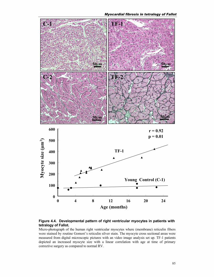
Myocardial fibrosis in tetralogy of Fallot
85
Figure 4.4. Developmental pattern of right ventricular myocytes in patients with tetralogy of Fallot. Micro-photograph of the human right ventricular myocytes where (membrane) reticulin fibers were stained by routine Gomori’s reticulin silver stain. The myocyte cross sectional areas were measured from digital microscopic pictures with an video image analysis set up. TF-1 patients depicted an increased myocyte size with a linear correlation with age at time of primary corrective surgery as compared to normal RV.
50µm 20µm
TF-2 C-2
50µm 50µm
C-1 TF-1
50µm
0 4 8 12 16 20 240
100
200
300
400
500
600
Myo
cyte
size
(µm
2 )
Age (months)
Young Control (C-1)
TF-1
p = 0.01r = 0.92
Chapter 4
86
Taken together, our study provides further evidence that myocardial architecture in
patients with TF depicts a complex and differential gene expression pattern with
drastically increased expression level of ECM proteins (collagen Iα and III and
fibronectin) at mRNA (DNA micro-array) and protein levels (quantitative
immunohistochemistry). We conclude that this up-regulation of genes involved in
ECM homeostasis is associated with RVH and diminished cardiac function in TF
patients. This information leaves room for improvement of contemporary clinical
treatment of patients with tetralogy of Fallot by allowing an assessment of timing for
surgery and possibly support long term postoperative prognosis.
Acknowledgements
Financial support from the Netherlands Heart Foundation (NHS 96.082) is gratefully
acknowledged. Authors thank Mrs. Emine Yilmaz and Mrs. Caroline van den Burg for
their technical assistance.
References
1. Starnes VA, Luciani GB, Latter DA, et al. Current surgical management of tetralogy of Fallot. Ann Thorac Surg. 1994;58:211-215.
2. Pigula FA, Khalil PN, Mayer JE, et al. Repair of tetralogy of Fallot in neonates and young infants. Circulation. 1999;100:II157-61.
3. Reddy VM, Liddicoat JR, McElhinney DB, et al. Routine primary repair of tetralogy of Fallot in neonates and infants less than three months of age. Ann Thorac Surg. 1995;60:S592-6.
4. Van Arsdell GS, Maharaj GS, Tom J, et al. What is the optimal age for repair of tetralogy of Fallot? Circulation. 2000;102:III123-9.
5. Warner KG, Anderson JE, Fulton DR, et al. Restoration of the pulmonary valve reduces right ventricular volume overload after previous repair of tetralogy of Fallot. Circulation. 1993;88:II189-97.
6. Seliem MA, Wu YT, Glenwright K. Relation between age at surgery and regression of right ventricular hypertrophy in tetralogy of Fallot. Ped Cardiol. 1995;16:53-55.
7. Schwartz SM, Gordon D, Mosca RS, et al. Collagen content in normal, pressure, and pressure-volume overloaded developing human hearts. Am J Cardiol. 1996;77:734-8.
8. Castaneda AR, Jonas RA, Mayer JE, et al. Cardiac surgery of neonate and infant. Philadelphia, USA: Saunders; 1994.
Myocardial fibrosis in tetralogy of Fallot
87
9. Bishop JE, Rhodes S, Laurent GJ, et al. Increased collagen synthesis and decreased collagen degradation in right ventricular hypertrophy induced by pressure overload. Cardiovasc Res. 1994;28:1581-1585.
10. Boluyt MO, L. O, Meredith AL, et al. Alterations in cardiac gene expression during the transition from stable hypertrophy to heart failure. Marked upregulation of genes encoding extracellular matrix components. Circ Res. 1994;75:23-32.
11. Brilla CG, Janicki JS, Weber KT. Impaired diastolic function and coronary reserve in genetic hypertension. Role of interstitial fibrosis and medial thickening of intramyocardial coronary arteries. Circ Res. 1991;69:107-115.
12. Jalil JE, Doering CW, Janicki JS, et al. Fibrillar collagen and myocardial stiffness in the intact hypertrophied rat left ventricle. Circ Res. 1989;64:1041-1050.
13. Lamparter S, Sun Y, Weber KT. Angiotensin II receptor blockade during gestation attenuates collagen formation in the developing rat heart. Cardiovasc Res. 1999;43:165-72.
14. Marijianowski MM, van der Loos CM, Mohrschladt MF, et al. The neonatal heart has a relatively high content of total collagen and type I collagen, a condition that may explain the less compliant state. J Am Coll Cardiol. 1994;23:1204-8.
15. Tyagi SC. Extracellular matrix dynamics in heart failure: a prospect for gene therapy. J Cell Biochem. 1998;68:403-10.
16. Weber KT, Sun Y, Tyagi SC, et al. Collagen network of the myocardium: function, structural remodelling and regulatory mechanisms. J Mol Cell Cardiol. 1994;26:279-92.
17. Farhadian F, Contard F, Corbier A, et al. Fibronectin expression during physiological and pathological cardiac growth. J Mol Cell Cardiol. 1995;27:981-990.
18. Mann DL, Spinale FG. Activation of matrix metalloproteinases in the failing human heart: breaking the tie that binds. Circulation. 1998;98:1699-702.
19. D'Armiento J. Matrix metalloproteinase disruption of the extracellular matrix and cardiac dysfunction. Trends Cardiovasc Med. 2002;12:97-101.
20. Galis ZS, Muszynski M, Sukhova GK, et al. Cytokine-stimulated human vascular smooth muscle cells synthesize a complement of enzymes required for extracellular matrix digestion. Circ Res. 1994;75:181-9.
21. Li YY, Feldman AM, Sun Y, et al. Differential expression of tissue inhibitors of metalloproteinases in the failing human heart. Circulation. 1998;98:1728-34.
22. Spinale FG. Matrix metalloproteinases: regulation and dysregulation in the failing heart. Circ Res. 2002;90:520-30.
23. Mitsuno M, Nakano S, Shimazaki Y, et al. Fate of right ventricular hypertrophy in tetralogy of Fallot after corrective surgery. Am J Cardiol. 1993;72:694-8.
24. Hokken RB, Bogers AJJC, Spitaels SEC, et al. Pulmonary homograft insertion after repair of pulmonary stenosis. J Heart Valve Dis. 1995;4:182-186.
25. Bogers AJJC, Roofthooft M, Pisters H, et al. Longterm follow-up of gamma irradiated transannular homograft patch in surgical treatment of tetralogy of Fallot. Thorac Cardiovasc Surgeon. 1994;42:337-339.
26. Sharma HS, van Heugten HA, Goedbloed MA, et al. Angiotensin II induced expression of transcription factors precedes increase in Transforming Growth
Chapter 4
88
Factor-b1 mRNA in neonatal cardiac fibroblasts. Biochem Biophys Res Comm. 1994;205:105-112.
27. Tan FL, Moravec CS, Li J, et al. The gene expression fingerprint of human heart failure. Proc Nat Acad Sci U S A. 2002;99:11387-92.
28. Yajima N, Masuda M, Miyazaki M, et al. Oxidative stress is involved in the development of experimental abdominal aortic aneurysm: a study of the transcription profile with complementary DNA microarray. J Vasc Surg. 2002;36:379-85.
29. Takahashi T, Schunkert H, Isoyama S, et al. Age-related differences in the expression of proto-oncogene and contractile protein genes in response to pressure overload in the rat myocardium. J Clin Invest. 1992;89:939-946.
30. Bancroff JD, Cook HC. Manual of Histological Techniques and Their Diagnostic Aplication. London: Churchill Livingstone; 1994.
31. Vikstrom KL, Bohlmeyer T, Factor SM, et al. Hypertrophy, pathology, and molecular markers of cardiac pathogenesis. Circ Res. 1998;82:773-778.
32. Brand T, Sharma HS, Fleischmann KE, et al. Proto-oncogene expression in porcine myocardium subjected to ischemia and reperfusion. Circ Res. 1992;71:1351-1360.
33. Whittaker P. Collagen and ventricular remodelling after acute myocardial infarction: concepts and hypotheses. Basic Res Cardiol. 1997;92:79-81.
34. Heling A, Zimmermann R, Kostin S, et al. Increased expression of cytoskeletal, linkage, and extracellular proteins in failing human myocardium. Circ Res. 2000;86:846-53.

Myocardial fibrosis in tetralogy of Fallot
89

Chapter 5
90
Right ventricular fibrosis in PA-VSD
91
Chapter 5
Right ventricular fibrosis levels in patients with pulmonary atresia and ventricular septal defect
Based on: Peters THF, de Jong PL, Klompe L, Saxena PR, Sharma HS and Bogers AJJC. Right ventricular collagen and fibronectin levels in patients with pulmonary atresia and ventricular septum defect. Mol Cell Biochem 2003; 251:27-32.
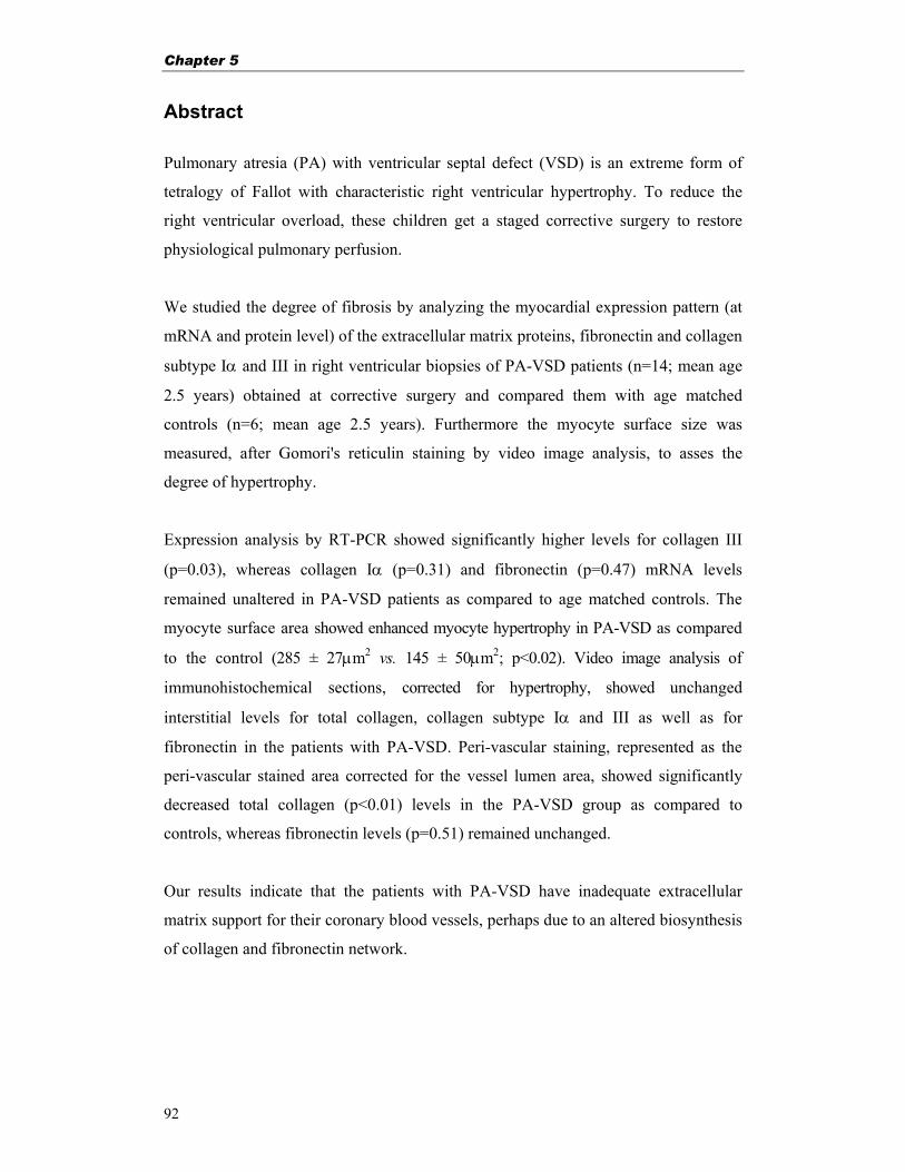
Chapter 5
92
Abstract Pulmonary atresia (PA) with ventricular septal defect (VSD) is an extreme form of
tetralogy of Fallot with characteristic right ventricular hypertrophy. To reduce the
right ventricular overload, these children get a staged corrective surgery to restore
physiological pulmonary perfusion.
We studied the degree of fibrosis by analyzing the myocardial expression pattern (at
mRNA and protein level) of the extracellular matrix proteins, fibronectin and collagen
subtype Iα and III in right ventricular biopsies of PA-VSD patients (n=14; mean age
2.5 years) obtained at corrective surgery and compared them with age matched
controls (n=6; mean age 2.5 years). Furthermore the myocyte surface size was
measured, after Gomori's reticulin staining by video image analysis, to asses the
degree of hypertrophy.
Expression analysis by RT-PCR showed significantly higher levels for collagen III
(p=0.03), whereas collagen Iα (p=0.31) and fibronectin (p=0.47) mRNA levels
remained unaltered in PA-VSD patients as compared to age matched controls. The
myocyte surface area showed enhanced myocyte hypertrophy in PA-VSD as compared
to the control (285 ± 27µm2 vs. 145 ± 50µm2; p<0.02). Video image analysis of
immunohistochemical sections, corrected for hypertrophy, showed unchanged
interstitial levels for total collagen, collagen subtype Iα and III as well as for
fibronectin in the patients with PA-VSD. Peri-vascular staining, represented as the
peri-vascular stained area corrected for the vessel lumen area, showed significantly
decreased total collagen (p<0.01) levels in the PA-VSD group as compared to
controls, whereas fibronectin levels (p=0.51) remained unchanged.
Our results indicate that the patients with PA-VSD have inadequate extracellular
matrix support for their coronary blood vessels, perhaps due to an altered biosynthesis
of collagen and fibronectin network.
Right ventricular fibrosis in PA-VSD
93
Introduction
Pulmonary atresia with ventricular septal defect (PA-VSD) is an extreme form of
tetralogy of Fallot where antegrade blood flow to the pulmonary artery is completely
obstructed1,2. Some PA-VSD patients have a normal central pulmonary arterial system
with a duct-dependent pulmonary circulation, whereas others have either partial or
complete absence of the native pulmonary arteries. Their pulmonary circulation
depends on systemic-pulmonary collateral arteries (SPCAs)1,2. SPCAs are highly
variable in number and usually arise from the aortic arch, descending aorta or from
the side branches.
At the Erasmus Medical Centre Rotterdam, the correction for PA-VSD with or
without SPCAs, is performed in stages. The patients with a duct-dependent
pulmonary circulation are initially palliated with a modified Blalock-Taussig shunt.
The patients with SPCA-dependent pulmonary circulation are initially palliated by
unifocalization of the pulmonary vasculature in either lung, combined with a modified
Blalock-Taussig shunt. In both groups corrective surgery consists of closure of the
modified Blalock-Taussig shunts, closure of the VSD and installing a pulmonary
homograft between the right ventricle and the (unifocalized) pulmonary arterial
system. At correction, the right ventricle in patients with PA-VSD shows without
exception hypertrophy as an adaptive response to the increased pressure overload.
This response could lead to remodeling of the myocardium and to the development of
interstitial fibrosis3-5.
In experimental models cardiac hypertrophy by pressure overload has been shown to
accompany myocardial collagen deposition contributing to the increased stiffness of
the cardiac muscle6,7. Most collagen subtypes in the heart are exclusively produced by
cardiac fibroblasts, with the exception of type IV, which also is produced by other cell
types including cardiac myocytes8. The collagen Iα and III subtypes are the most
abundant forms in the heart. Collagen type Iα represents 80% of the total cardiac
collagen content and is important for the cardiac stiffness8,9. Collagen type III
represents about 12% of the total cardiac collagen content and is important for tissue
elasticity8,9. Collagen type Iα and III are widely distributed between myocytes and
Chapter 5
94
among muscle fibers and morphologic studies have defined the spatial distribution of
these collagen types in normal and hypertrophic hearts7,9-11.
Myocardial fibronectin is homogeneously localized throughout the extracellular space
and is closely associated with collagen fibers12. It is a glycoprotein and serves as a
bridge between cardiac myocytes to the interstitial collagen network5. Fibronectin
binds collagen and modulates the collagen fibrillogenesis, consequently resulting in
higher contents of collagen type Iα and a lower collagen type III concentration13.
Furthermore, fibronectin influences several processes, including cell growth,
adhesion, migration and wound repair14.
The present study was undertaken to describe the degree of myocardial fibrosis in
patients with PA-VSD by investigating the expression of collagens and fibronectin
(both at mRNA and protein level) in right ventricular biopsies obtained from patients
undergoing surgery for PA-VSD. Secondly, we attempted to correlate the expression
data with clinical data.
Materials and Methods
Patients
Present study was approved by the Medical Ethical Committee of the University
Hospital Rotterdam (MEC153. 268/1996/119). Biopsies from the right ventricular
outflow tract were taken at corrective surgery for PA-VSD in 14 patients. Mean age
of the patients at the time of surgery was 2.54 ± 0.32 years (range 0.76-5.51 years).
Age-matched controls (C-1) consists of 2 post mortem biopsies and biopsies from 4
heart valve donors, who died due to non-cardiac cause (mean age 2.52 ± 0.75 years
(range 0.01-5.70 years)). Part of the myocardial biopsy was immediately frozen in
liquid nitrogen for mRNA expression studies, and the other part was fixed in 4%
paraformaldehyde in phosphate buffered saline (PBS) for at least 24 hours and further
processed for histological and immunohistochemical studies.
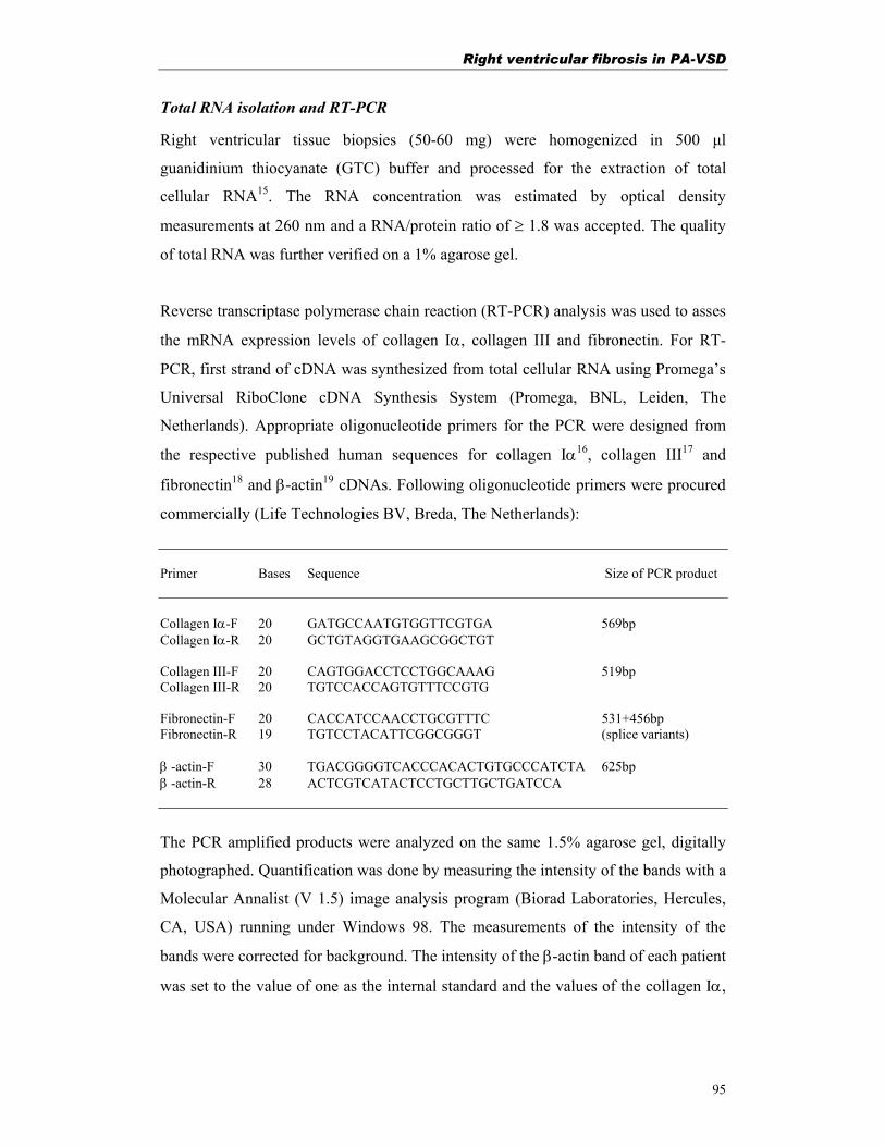
Right ventricular fibrosis in PA-VSD
95
Total RNA isolation and RT-PCR
Right ventricular tissue biopsies (50-60 mg) were homogenized in 500 µl
guanidinium thiocyanate (GTC) buffer and processed for the extraction of total
cellular RNA15. The RNA concentration was estimated by optical density
measurements at 260 nm and a RNA/protein ratio of ≥ 1.8 was accepted. The quality
of total RNA was further verified on a 1% agarose gel.
Reverse transcriptase polymerase chain reaction (RT-PCR) analysis was used to asses
the mRNA expression levels of collagen Iα, collagen III and fibronectin. For RT-
PCR, first strand of cDNA was synthesized from total cellular RNA using Promega’s
Universal RiboClone cDNA Synthesis System (Promega, BNL, Leiden, The
Netherlands). Appropriate oligonucleotide primers for the PCR were designed from
the respective published human sequences for collagen Iα16, collagen III17 and
fibronectin18 and β-actin19 cDNAs. Following oligonucleotide primers were procured
commercially (Life Technologies BV, Breda, The Netherlands): Primer Bases Sequence Size of PCR product Collagen Iα-F 20 GATGCCAATGTGGTTCGTGA 569bp Collagen Iα-R 20 GCTGTAGGTGAAGCGGCTGT Collagen III-F 20 CAGTGGACCTCCTGGCAAAG 519bp Collagen III-R 20 TGTCCACCAGTGTTTCCGTG Fibronectin-F 20 CACCATCCAACCTGCGTTTC 531+456bp Fibronectin-R 19 TGTCCTACATTCGGCGGGT (splice variants) β -actin-F 30 TGACGGGGTCACCCACACTGTGCCCATCTA 625bp β -actin-R 28 ACTCGTCATACTCCTGCTTGCTGATCCA
The PCR amplified products were analyzed on the same 1.5% agarose gel, digitally
photographed. Quantification was done by measuring the intensity of the bands with a
Molecular Annalist (V 1.5) image analysis program (Biorad Laboratories, Hercules,
CA, USA) running under Windows 98. The measurements of the intensity of the
bands were corrected for background. The intensity of the β-actin band of each patient
was set to the value of one as the internal standard and the values of the collagen Iα,
Chapter 5
96
collagen III and fibronectin bands were divided by the β-Actin band values of the
respective patient.
Gomori reticulin staining
For determining the myocyte surface size, the cell (membrane) reticulin fibers were
stained by routine Gomori’s reticulin silver staining technique20 on 5 µm thick
sections. After the silverstaining was developed, slides were mounted and visualized
under light microscope.
Collagen staining
The total collagen in myocardial tissue specimens was stained with Picro-sirius red
F3BA11. Tissue sections of 5 µm thickness, were treated with 0.2% aqueous
phosphomolybdic acid and incubated in 0.1% Picro-sirius red. Before dehydration, the
slides were treated with 0.01N HCl and mounted. Slides were visualized under light
microscope and eventually collagen contents were quantified.
Immunolocalization of fibronectin, collagen Iα and collagen III
Paraffin sections (5 µm thickness) of the myocardial tissues were cut and mounted on
3-amino-propyl-trioxysilane (Sigma, St Louis, MO, USA) coated glass slides.
Immunohistochemical staining was performed using the avidin-biotin complex (ABC)
method following supplier's instructions (BioGenex, San Ramon, CA, USA). Optimal
dilutions for all antibodies were identified by examining the intensity of staining
obtained with a series of dilutions, which gave specific signal on paraffin sections
derived from the same control tissue prior to perform the staining protocol on all
section.
In brief, after deparaffinization in xylene and rehydration through graded
alcohol the slides were rinsed with water. Slides to be stained for collagen Iα were
pretreated in a boiling citrate solution in a microwave for 20 min. and slides to be
stained for collagen III were pretreated with 0.1 % pronase for 10 min. After rinsing in
PBS, slides were pre-incubated with 10% normal goat serum diluted in 5% bovine
serum albumin in phosphate buffered saline (5% BSA/PBS, pH=7.4) for 15 minutes
to block non-specific binding. Specific anti-human antibodies were added in different
Right ventricular fibrosis in PA-VSD
97
solutions for the specific positive staining whereas negative controls were performed by
omission of the primary antibody. For fibronectin staining specific antibodies against
human fibronectin (Clone Ab-3, Neomarkers, Fremont, CA, USA) in 1:150 dilution
were applied as primary antiserum, whereas for collagen Iα, specific human antibodies
against collagen Iα (mouse monoclonal anti-collagen type 1 (Sigma, St Louis, MO,
USA)) and for collagen III mouse monoclonal antibodies to collagen III (BioGenex, San
Ramon, CA, USA), were used in a optimal dilutions of 1: 100. Anti-human mouse
monoclonal antibodies against smooth muscle actin (α-SMA, Clone 1A4,
NeoMarkers Fremont, CA, USA) in a 1: 1000 dilution were used as a positive control.
After incubation over night at 4°C sections were washed in 0.5% tween-20 in
PBS solution. Sections stained for fibronectin and collagen Iα, were incubated with
mouse biotinylated anti-rabbit IgG (BioGenex, San Ramon, CA, USA) for 30 min. and
subsequently, with streptavidin conjugated alkaline phosphatase (label 1:50 dilution,
Biogenex, San Ramon, CA, USA) for 30 min. Color was developed using a substrate of
18 mg Naphtol AS-MX phosphate (N5000), 200 µl new fuchsin, 200 µl sodium
nitrite, 60 ml Tris HCl (pH 8,0) ), while endogenous alkaline phosphatase activity was
inhibited by 15 mg of levamisol. Sections stained for collagen III and α-SMA were
incubated with multilink for 30 min., rinsed followed by 30 min. incubation with
peroxidase conjugated streptavidin at a dilution of 1:50 (Biogenex, San Ramon, MO,
USA). Subsequently, sections were colored, for 7 min. using, 0.025% of 3,3-
diaminobenzidine (DAB) (Sigma, St Louis, MO, USA) in 0.01 mol/L PBS, containing
0.03% H2O2, as a substrate. All sections were counter stained with hematoxylin,
mounted and visualized under the light microscope and photographed.
Video image analysis
Video image analysis was used to quantify the expression levels of collagen and
fibronectin. After staining, the sections were mounted and visualized under light
microscope (Leica DM RBE, Leica GmbH, Wetzlar, Germany) using 20x lens and
grey-filters. From each section, series of 12 consecutive digital color images (736x574
pixels) for staining analysis were taken form a representative area. All sections from
one staining were photographed at the same time using same microscopic and computer
settings. A mm-scale was photographed which was used as a reference to control the
Chapter 5
98
measured values. The digital images were analyzed using a specialized computer
program for the Leica Qwin standard (Y2.2b) image analysis software package (Leica
Imaging Systems Ltd., Cambrige, England).
Distribution of the stained fibers in the interstitial space as well as in the peri-
vascular region was measured as a positive area. To analyze the positive staining
areas of the extra cellular matrix the staining detection levels for hue, saturation and
intensity in the computer program were optimized and set. Those settings were used for
analysis of the all images derived from one staining. Interstitial expression was
represented as ratio staining per total tissue area, excluding vascular areas, whereas
peri-vascular localization was depicted as staining per peri-vascular area. Since the
Peri-Vascular Collagen Area (PVCA) and vessel Luminal Area (LA) are positively
correlated, the stained PVCA was normalized to the LA and represented as PVCA/LA
ratio21. The Peri-Vascular Fibronectin Area (PVFA) was normalized in the same way
for the LA and represented as PVFA/LA ratio.
To asses the degree of myocardial hypertrophy myocyte cell sizes were
measured. From each section 3-4 digital images were photographed with a 20x (adult)
or 40x (young) lens and stored in the computer. With the image analysis software the
myocyte outline of at least 20 transfers cut myocytes, containing a nucleolus, per
section were drawn manually where after the program calculated the myocyte surface
in µm2. Interstitial staining values were corrected for the increase in myocyte size by
multiplying the interstitial staining with the increase in cell surface divided by the
average control cell surface.
Statistical analysis
Results were expressed as mean ± standard error of mean (SEM). Statistical
probability was assessed by (non-parametric) Kruskal-Wallis one way analysis of
variance. Results were considered statistically significant with p<0.05. Regression
analysis of clinical data with collagen and fibronectin expression data was performed
using SPSS software. The correlation coefficient and significance level were
calculated using linear (Pearson’s) regression.
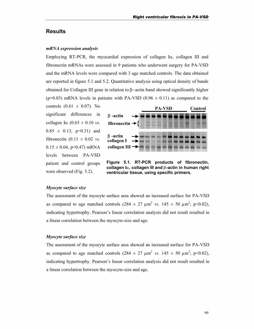
Right ventricular fibrosis in PA-VSD
99
Results
mRNA expression analysis
Employing RT-PCR, the myocardial expression of collagen Iα, collagen III and
fibronectin mRNAs were assessed in 9 patients who underwent surgery for PA-VSD
and the mRNA levels were compared with 3 age matched controls. The data obtained
are reported in figure 5.1 and 5.2. Quantitative analysis using optical density of bands
obtained for Collagen III gene in relation to β−actin band showed significantly higher
(p=0.03) mRNA levels in patients with PA-VSD (0.96 ± 0.11) as compared to the
controls (0.61 ± 0.07). No
significant differences in
collagen Iα (0.65 ± 0.10 vs.
0.85 ± 0.13, p=0.31) and
fibronectin (0.11 ± 0.02 vs.
0.15 ± 0.04, p=0.47) mRNA
levels between PA-VSD
patient and control groups
were observed (Fig. 5.2).
Myocyte surface size
The assessment of the myocyte surface area showed an increased surface for PA-VSD
as compared to age matched controls (284 ± 27 µm2 vs. 145 ± 50 µm2; p<0.02),
indicating hypertrophy. Pearson’s linear correlation analysis did not result resulted in
a linear correlation between the myocyte-size and age.
β -actin
fibronectin
β -actin collagen Icollagen III
{
PA-VSD Control
Figure 5.1. RT-PCR products of fibronectin,collagen Iα, collagen III and β-actin in human rightventricular tissue, using specific primers.
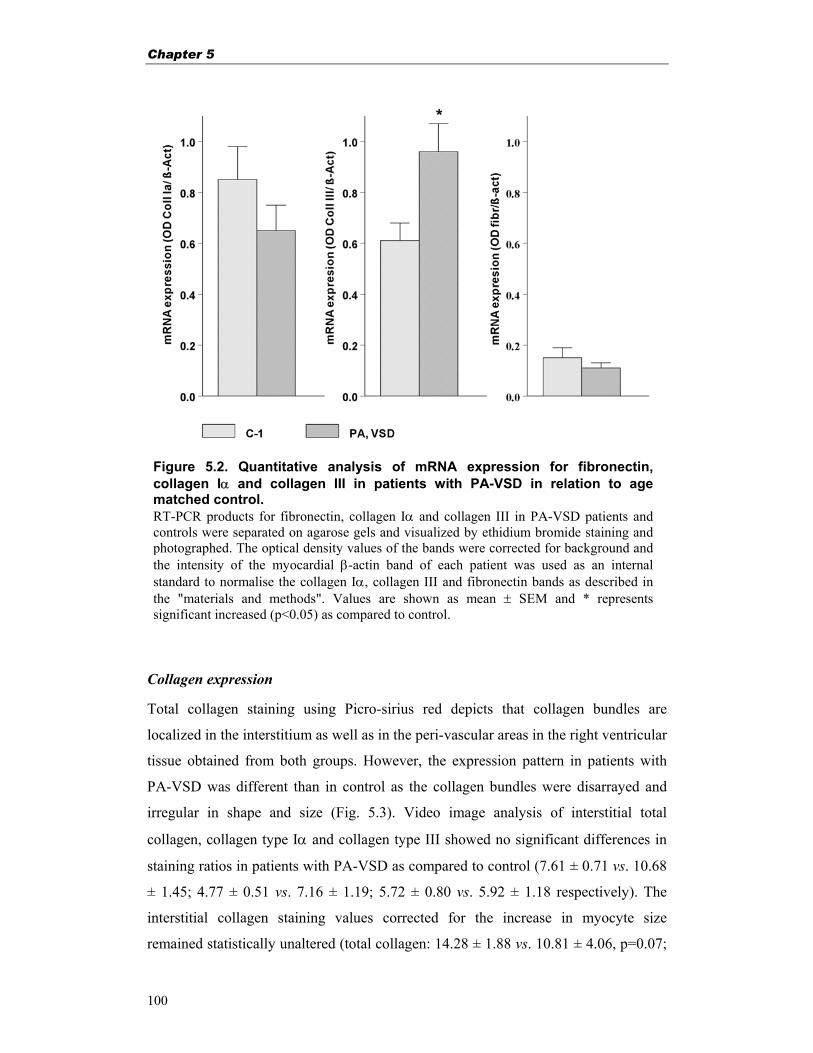
Chapter 5
100
Collagen expression
Total collagen staining using Picro-sirius red depicts that collagen bundles are
localized in the interstitium as well as in the peri-vascular areas in the right ventricular
tissue obtained from both groups. However, the expression pattern in patients with
PA-VSD was different than in control as the collagen bundles were disarrayed and
irregular in shape and size (Fig. 5.3). Video image analysis of interstitial total
collagen, collagen type Iα and collagen type III showed no significant differences in
staining ratios in patients with PA-VSD as compared to control (7.61 ± 0.71 vs. 10.68
± 1.45; 4.77 ± 0.51 vs. 7.16 ± 1.19; 5.72 ± 0.80 vs. 5.92 ± 1.18 respectively). The
interstitial collagen staining values corrected for the increase in myocyte size
remained statistically unaltered (total collagen: 14.28 ± 1.88 vs. 10.81 ± 4.06, p=0.07;
Figure 5.2. Quantitative analysis of mRNA expression for fibronectin,collagen Iα and collagen III in patients with PA-VSD in relation to age matched control. RT-PCR products for fibronectin, collagen Iα and collagen III in PA-VSD patients and controls were separated on agarose gels and visualized by ethidium bromide staining and photographed. The optical density values of the bands were corrected for background andthe intensity of the myocardial β-actin band of each patient was used as an internal standard to normalise the collagen Iα, collagen III and fibronectin bands as described in the "materials and methods". Values are shown as mean ± SEM and * representssignificant increased (p<0.05) as compared to control.
Right ventricular fibrosis in PA-VSD
101
collagen Iα: 9.23 ± 1.49 vs. 6.77 ± 2.07, p=0.11; collagen III: 5.72 ± 0.80 vs. 5.92 ±
1.18, p=0.08) in the PA-VSD group as compared to control (Fig. 5.4). However, for
the normalized peri-vascular collagen area, the PVCA/LA ratio was significantly
lower in the PA-VSD group as compared to control (3.22 ± 0.34 vs. 7.23 ± 1.13,
p<0.01) (Fig. 5.4).
Fibronectin expression The pattern of fibronectin localization was similar to that of collagens as it was
localized in the interstitial spaces and around the blood vessels (Fig. 5.3). Video
image analysis of interstitial fibronectin showed that the expression levels in the PA-
VSD group were not significantly changed as compared to control (11.03 ± 1.49 vs.
12.22 ± 1.43). The expression pattern remained unaltered when the interstitial
fibronectin ratios were corrected for the increase in myocyte size (7.11 ± 1.08 vs. 14.
18 ± 4.10, p=0.10) when patients with PA-VSD were compared to respective controls
(Fig. 5.4). Also the normalized peri-vascular fibronectin area the PVFA/LA ratio
showed no significantly changed levels in the PA-VSD group as compared to control
(2.65 ± 0.48 vs. 2.62 ± 0.47, p=0.52) (Fig. 5.4).
Correlation analysis
Pearson’s correlation and regression analysis was performed on several clinical
parameters, obtained from medical records of the Thoraxcentre, Dijkzigt Hospital and
Sophia Childrens Hospital Rotterdam (Table 5.1), such as, right ventricular pressure
and transcutaneous O2-saturation with the expression levels of collagens and
fibronectin in both interstitium and peri-vascular area. The data on correlation with
right ventricular pressure as well as with O2-saturation showed no significant
regression values. Correlation coefficient (r) and significance level (p value) using
linear regression (Pearson’s) analysis is shown in table 5.2. The data on correlation
with right ventricular pressure as well as with aortic O2 saturation showed no
significant regression values (Table 5.2).
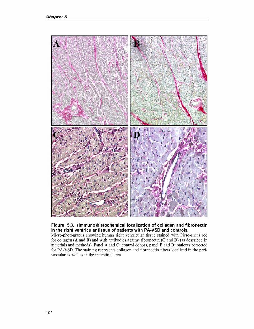
Chapter 5
102
Figure 5.3. (Immuno)histochemical localization of collagen and fibronectinin the right ventricular tissue of patients with PA-VSD and controls. Micro-photographs showing human right ventricular tissue stained with Picro-sirius redfor collagen (A and B) and with antibodies against fibronectin (C and D) (as described inmaterials and methods). Panel A and C: control donors, panel B and D: patients correctedfor PA-VSD. The staining represents collagen and fibronectin fibers localized in the peri-vascular as well as in the interstitial area.
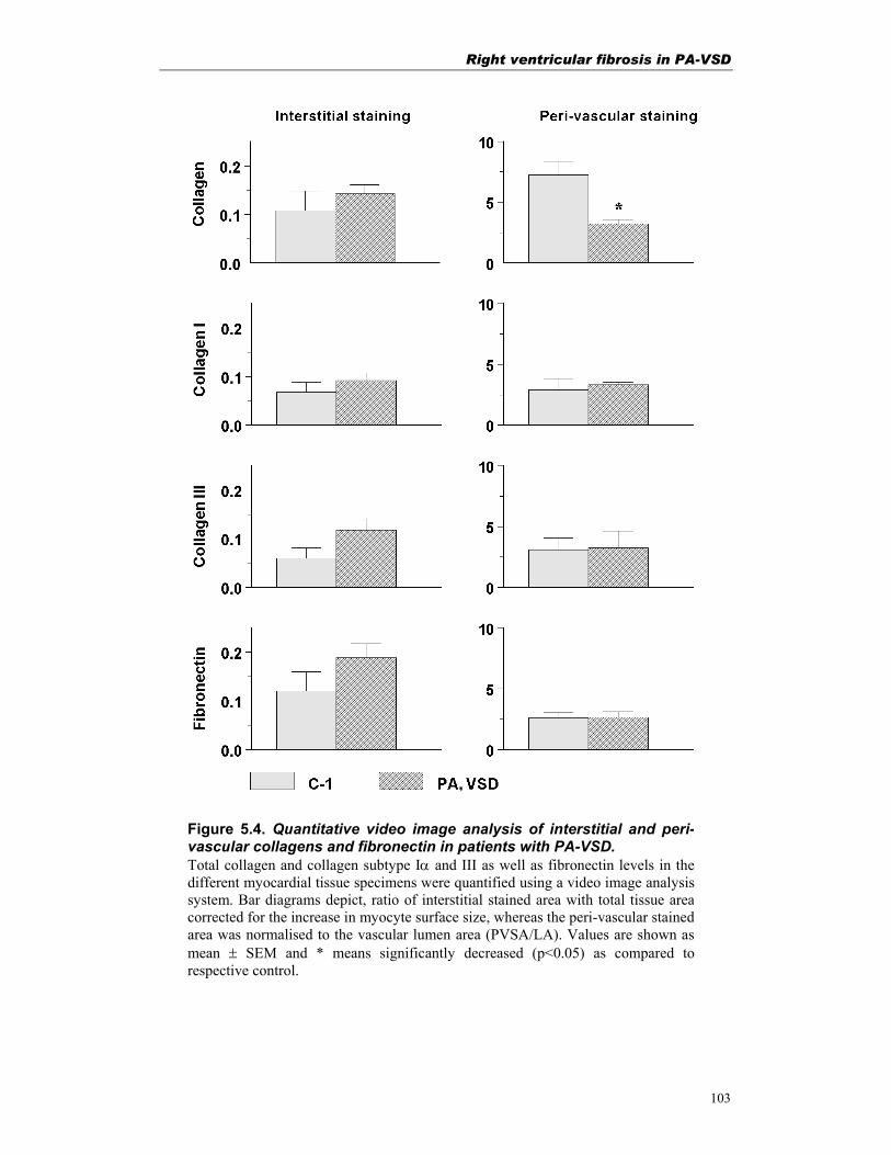
Right ventricular fibrosis in PA-VSD
103
Figure 5.4. Quantitative video image analysis of interstitial and peri-vascular collagens and fibronectin in patients with PA-VSD. Total collagen and collagen subtype Iα and III as well as fibronectin levels in thedifferent myocardial tissue specimens were quantified using a video image analysissystem. Bar diagrams depict, ratio of interstitial stained area with total tissue areacorrected for the increase in myocyte surface size, whereas the peri-vascular stainedarea was normalised to the vascular lumen area (PVSA/LA). Values are shown asmean ± SEM and * means significantly decreased (p<0.05) as compared torespective control.
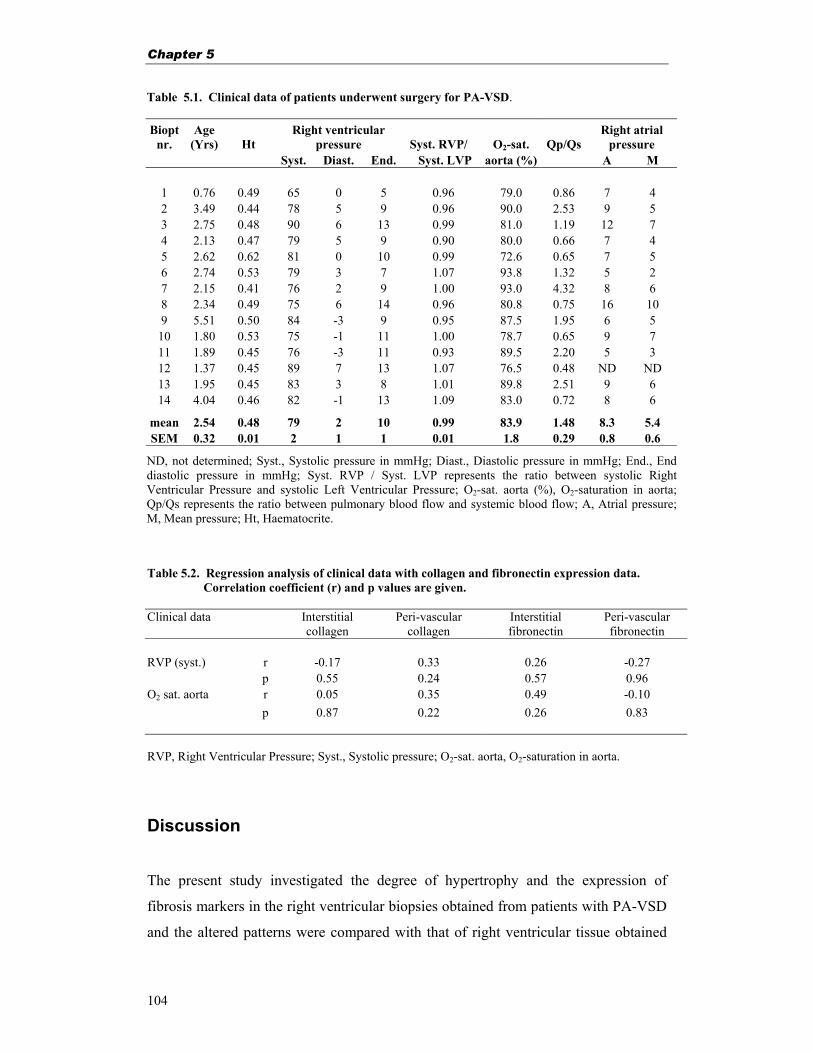
Chapter 5
104
Table 5.1. Clinical data of patients underwent surgery for PA-VSD. Biopt
nr. Age
(Yrs) Ht Right ventricular
pressure Syst. RVP/ O2-sat. Qp/Qs Right atrial
pressure Syst. Diast. End. Syst. LVP aorta (%) A M
1 0.76 0.49 65 0 5 0.96 79.0 0.86 7 4 2 3.49 0.44 78 5 9 0.96 90.0 2.53 9 5 3 2.75 0.48 90 6 13 0.99 81.0 1.19 12 7 4 2.13 0.47 79 5 9 0.90 80.0 0.66 7 4 5 2.62 0.62 81 0 10 0.99 72.6 0.65 7 5 6 2.74 0.53 79 3 7 1.07 93.8 1.32 5 2 7 2.15 0.41 76 2 9 1.00 93.0 4.32 8 6 8 2.34 0.49 75 6 14 0.96 80.8 0.75 16 10 9 5.51 0.50 84 -3 9 0.95 87.5 1.95 6 5 10 1.80 0.53 75 -1 11 1.00 78.7 0.65 9 7 11 1.89 0.45 76 -3 11 0.93 89.5 2.20 5 3 12 1.37 0.45 89 7 13 1.07 76.5 0.48 ND ND 13 1.95 0.45 83 3 8 1.01 89.8 2.51 9 6 14 4.04 0.46 82 -1 13 1.09 83.0 0.72 8 6
mean 2.54 0.48 79 2 10 0.99 83.9 1.48 8.3 5.4 SEM 0.32 0.01 2 1 1 0.01 1.8 0.29 0.8 0.6
ND, not determined; Syst., Systolic pressure in mmHg; Diast., Diastolic pressure in mmHg; End., End diastolic pressure in mmHg; Syst. RVP / Syst. LVP represents the ratio between systolic Right Ventricular Pressure and systolic Left Ventricular Pressure; O2-sat. aorta (%), O2-saturation in aorta; Qp/Qs represents the ratio between pulmonary blood flow and systemic blood flow; A, Atrial pressure; M, Mean pressure; Ht, Haematocrite.
Table 5.2. Regression analysis of clinical data with collagen and fibronectin expression data. Correlation coefficient (r) and p values are given. Clinical data Interstitial
collagen Peri-vascular
collagen Interstitial fibronectin
Peri-vascular fibronectin
RVP (syst.) r -0.17 0.33 0.26 -0.27
p 0.55 0.24 0.57 0.96 O2 sat. aorta r 0.05 0.35 0.49 -0.10
p 0.87 0.22 0.26 0.83
RVP, Right Ventricular Pressure; Syst., Systolic pressure; O2-sat. aorta, O2-saturation in aorta.
Discussion
The present study investigated the degree of hypertrophy and the expression of
fibrosis markers in the right ventricular biopsies obtained from patients with PA-VSD
and the altered patterns were compared with that of right ventricular tissue obtained
Right ventricular fibrosis in PA-VSD
105
from age matched control individuals. The presence of myocyte hypertrophy in
patients with PA-VSD, as a response to the right ventricular pressure overload, is
obvious. This is thought to be accompanied by myocardial fibrosis to increase the
myocardial stiffness and prevent further dilatation6,7. Analyzing the expression pattern
of two fibrosis markers in the myocardium, we found a tendency for limited increase
collagen and fibronectin contents without statistically significant differences when the
study group of PA-VSD patients was compared to the control group. However, in the
peri-vascular stained area, corrected for lumen area, total collagen levels were
significantly lower in the patient group, suggesting a lower amount of collagen
accumulation around the vessels. Additionally, it can be emphasized that the collagen
and fibronectin deposition in the interstitial areas showed an altered pattern, depicting
irregularities in the network of collagen and fibronectin fibers. Our results indicate
that the patients with PA-VSD may have an impaired extracellular matrix support for
their coronary blood vessels.
The expression of collagen and fibronectin was also evaluated at mRNA level by RT-
PCR analysis, where we found significantly increased levels for collagen type III and
unchanged levels for collagen Iα and fibronectin in patients with PA-VSD. Enhanced
levels for collagen III could be attributed to the altered, more compliant, myocardial
architecture due to hypoxemia as well as hypertrophy. Furthermore, the enhanced
levels of collagen III mRNA in the patients with PA-VSD may be attributed to the
decreased degradation of this mRNA. Interestingly, decreased levels of collagen Iα
mRNA did not result in a decreased biosynthesis and accumulation. Perhaps an
altered biosynthesis and an inadequate collagen fibers network around the coronary
blood vessels could contribute to the existing cardiac pathology.
Limited research has been done so far on PA-VSD patients. This study contributes in
the understanding of myocardial architecture in patients with PA-VSD. In a study on
collagen and fibronectin content in patients with tetralogy of Fallot, also no
significant changes in the interstitial collagen or fibronectin expression were found22,
whereas only the peri-vascular collagen area corrected for lumen area was
significantly altered, resulting in a lower expression of total collagen in the peri-
vascular area in adult patients with tetralogy of Fallot. In the present study we found
Chapter 5
106
an increased expression of collagen type III in the myocardium, whereas the collagen
type Iα expression was not changed. Other studies also show that cardiac collagen
expression in normal and hypertrophied myocardium differs qualitatively23.
The PCR amplification with specific primers for fibronectin resulted in two splice
variant products of fibronectin (531 bp and a 456 bp) as shown in figure 5.1. Those two
products are the result of alternative splicing of a common precursor24. When the semi-
quantitative PCR analysis was done, the bands of both splice variants were taken into
account for optical density measurement.
We realize the limitations posed in our human study based on myocardial biopsies
obtained during surgical correction of the congenital anomaly. As the myocytes are
hypertrophied the ratio of cells per digital image will decrease, with consequently
decreasing staining ratios. This will influence the staining data. Therefore the staining
data was corrected for the increase in myocyte size. Hence the interpretation of the
obtained results should be cautiously taken. Yet not much is known about patients
with PA-VSD at a cellular level, so any available new data for this group is taken
along in the understanding of this disease. As mentioned earlier, we studied limited
numbers of patients and controls. Therefore the conclusions can only be drawn with
caution. It may be added here that the limited clinical data on normal myocardium in
young healthy children is a limitation of our study as well. We are currently
accumulating appropriate tissue biopsies to increase the number of controls.
In conclusion, our data suggest that in patients with PA-VSD have an inappropriate
state of the extracellular matrix support for their coronary blood vessels. Speculations
can be made whether or not this is due to an altered biosynthesis of collagen and
fibronectin in the extracellular network. As yet, no correlation with clinical
parameters or outcome could be established.
Acknowledgement
This study was financially supported by the Netherlands Heart Foundation, grant
number NHS 96.082.
Right ventricular fibrosis in PA-VSD
107
References 1. Kirklin JW, Barrat-Boyes BG. Ventricular septal defect and pulmonary stenosis or
atresia. 2nd ed. ed. New York: Churchill-Livingstone: Churchill-Livingstone; 1993. 2. Tchervenkov CI, Roy N. Congenital Heart Surgery Nomenclature and Database
Project: pulmonary atresia-ventricular septal defect. Ann Thorac Surg. 2000;69:S97-105.
3. van Bilsen M, Chien KR. Growth and hypertrophy of the heart: towards an understanding of cardiac specific and inducible gene expression. Cardiovasc Res. 1993;27:1140-1149.
4. Schwartz K, Carrier L, Mercadier JJ, et al. Molecular phenotype of hypertrophied and failing myocardium. Circulation. 1993;87:VII 5-10.
5. Pelouch V, Dixon IM, Golfman L, et al. Role of extracellular matrix proteins in heart function. Mol Cell Biochem. 1994;129:101-120.
6. Samuel JL, Barrieux A, Dufour S, et al. Accumulation of fetal fibronectin mRNAs during the development of rat cardiac hypertrophy induced by pressure overload. J Clin Invest. 1991;88:1737-1746.
7. Bishop JE, Rhodes S, Laurent GJ, et al. Increased collagen synthesis and decreased collagen degradation in right ventricular hypertrophy induced by pressure overload. Cardiovasc Res. 1994;28:1581-1585.
8. Chapman D, Weber KT, Eghbali M. Regulation of fibrillar collagen types I and III and basement membrane type IV collagen gene expression in pressure overloaded rat myocardium. Circ Res. 1990;67:787-794.
9. Bashey RI, Martinez-Hernandez A, Jiminez SA. Isolation, characterization, and localization of cardiac collagen type VI. Associations with other extracellular matrix components. Circ Res 70. 1992;70:1006-1017.
10. Factor SM, Robinson TF, Dominitz R, et al. Alterations of the myocardial skeletal framework in acute myocardial infarction with and without ventricular rupture. Am J Cardiovasc Pathol. 1986;1:91-97.
11. Takahashi T, Schunkert H, Isoyama S, et al. Age-related differences in the expression of proto-oncogene and contractile protein genes in response to pressure overload in the rat myocardium. J Clin Invest. 1992;89:939-946.
12. Speiser B, Weihrauch D, Reiss CF, et al. The extracellular matrix in human cardiac tissue part II: Vimentin, laminin, and fibronectin. Cardioscience. 1992;3:41-49.
13. Speranza ML, Valentini G, Calligaro A. Influence of fibronectin on the fibrillogenesis of type I and type III collagen. Coll Relat Res. 1987;7:115-123.
14. Farhadian F, Contard F, Corbier A, et al. Fibronectin expression during physiological and pathological cardiac growth. J Mol Cell Cardiol. 1995;27:981-990.
15. Chomczynski P, Sacchi N. Single-step method of RNA isolation by acid guanidinium thiocyanate-phenol-chloroform extraction. Anal Biochem. 1987;162:156-159.
16. Bernard MP, Chu ML, Myers JC, et al. Nucleotide sequence of complementary deoxyribonucleic acids for the pro alpha 1 chain of human type 1 procollagen. Statistical evaluation of structures that are conserved during evolution. Biochem. 1983;22:5213-5223.
17. Ala-Kokko L, Kontusaari S, Baldwin CT, et al. Structure of cDNA clones coding for the entire prepro alpha 1 chain of human type III procollagen. Differences in protein structure from type I procollagen and conservation of codon preferences. Biochem J. 1989;260:509-516.
18. Kornblihtt AR, Vibe-Petersen K, Baralle FE. Isolation and characterization of cDNA clones for human and bovine fibronectins. Proc Nat Acad Sci USA. 1983;80(11):3218-3222.
Chapter 5
108
19. Yamaguchi T, Iwano M, Kubo A, et al. IL-6 mRNA synthesis by peripheral blood mononuclear cells (PBMC) in patients with chronic renal failure. Clin Exp Immunol. 1996;103:279-328.
20. Bancroff JD, Cook HC. Manual of Histological Techniques and Their Diagnostic Aplication. London: Churchill Livingstone; 1994.
21. Brilla CG, Janicki JS, Weber KT. Impaired diastolic function and coronary reserve in genetic hypertension. Role of interstitial fibrosis and medial thickening of intramyocardial coronary arteries. Circ Res. 1991;69:107-115.
22. Peters THF, Sharma HS, Yilmaz E, et al. Quantitative analysis of collagens and fibronectin expression in human right ventricular hypertrophy. Ann N Y Acad Sci. 1999;874:278-285.
23. Iimoto DS, Covell JW, Harper E. Increase in crosslinking of type I and type III collagens associated with volume-overloaded hypertrophy. Circ Res. 1988;63:399-408.
24. Kornblihtt AR, Umezawa K, Vibe-Pedersen K, et al. Primary structure of human fibronectin: differential splicing may generate at least 10 polypeptides from a single gene. EMBO J. 1985;4(7):1755-1759.

Right ventricular fibrosis in PA-VSD
109

Chapter 6
110
Angiogenesis and fibrosis in tetralogy of Fallot
111
Chapter 6
Myocardial angiogenesis and fibrosis in human tetralogy of Fallot
Based on: Sharma HS, Peters THF and Bogers AJJC. Angiogenesis and fibrosis during right ventricular hypertrophy in human tetralogy of Fallot. In: N. Takeda, M. Nagano and NS. Dhalla (eds). The Hypertrophied Heart. Kluwer Academic Publishers, Boston 2000; 227-241.
Chapter 6
112
Abstract Right ventricular hypertrophy (RVH) due to pressure as well as volume overload,
caused by (sub) pulmonary stenosis and ventricular septal defect, is one of the main
features in human tetralogy of Fallot (TF). Currently, primary correction at a very
young age is the treatment of choice. However, it is not yet known to what extent
RVH in TF patients regresses after corrective surgery. The aim of our study was to
evaluate the changes in magnitude of the myocardial fibrosis and angiogenesis during
RVH in patients undergoing corrective surgery For this purpose, we examined
endomyocardial biopsies for the expression of extracellular matrix proteins such as
collagens, fibronectin, and an angiogenic growth factor, vascular endothelial growth
factor (VEGF), in TF patients with RVH and compared the expression with patients
without RVH. Myocardial tissue biopsies were obtained from patients undergoing
primary correction (TF-1 group, mean age 0.4 ± 0.1 years, n=8), from those
undergoing secondary surgery (TF-2 group, mean age 38.1 ± 3.7 years, n=6), and
from control patients with normal right ventricle (control group, mean age 36.4 ± 1.8
years, n=12). Picro-sirius red staining, depicting total collagens, and fibronectin
staining were semi-quantitatively analyzed. Interstitial levels for total collagens were
significantly increased in the TF-2 group as compared to the TF-1 group (p<0.04) and
control (p<0.05) groups. Immunolocalization of fibronectin showed the expression in
the interstitium and peri-vascular areas. Semi-quantitative analysis of fibronectin
staining revealed that the expression levels were not significantly different in the TF-2
group as compared to the TF-1 and control groups. Angiogenesis, assessed by the
increased number of small (p<0.05) vessels, was observed in both TF groups as
compared to the control group. Densitometric analysis of mRNAs encoding VEGF
showed significant enhanced expression in TF-1 (p<0.02) and TF-2 (p<0.05) groups
as compared to control. Immunoreactive VEGF was localized mainly in the myocytes
and smooth muscle cells and not in fibrotic areas in the case of TF groups. Our results
show an increased degree of myocardial fibrosis and angiogenesis during RVH in TF
patients. This information leaves room for the improvement in contemporary clinical
treatment of patients with TF by allowing an assessment of timing for surgery and
possibly supports long-term postoperative prognosis.
Angiogenesis and fibrosis in tetralogy of Fallot
113
Introduction
With an incidence of 5 per 10,000 live births, tetralogy of Fallot (TF) is a fairly
common congenital cardiac anomaly1. TF is characterized by overriding of the aorta,
a ventricular septal defect (VSD), (sub)valvular pulmonary stenosis, and right
ventricular hypertrophy (RVH) resulting in hypoxemia due to diminished pulmonary
flow1,2. Without appropriate treatment, TF ultimately results in cyanotic
complications and cardiac failure. For this reason, nearly 70% of patients with TF
require an operation during the first year of life. Due to improved peri-operative care,
the results of primary repair have increased dramatically in the past decade, and such
repair is now the surgical treatment of choice3,4. Nowadays, most TF patients usually
are operated during their first year of life by primary repair. The aim of early primary
repair, by closing the VSD and removing the right ventricular outflow tract
obstruction, is to relieve the hypoxemia and to eliminate the stimulus for the adaptive
RVH as well as to preserve right ventricular function2-4. However, some patients
suffer from pulmonary regurgitation after primary correction, causing right ventricular
failure due to volume overload. For these patients, further secondary corrective
surgery later in life is performed by implanting a pulmonary allograft heart valve
between the right ventricle and the pulmonary artery5,6.
Most attention in research on human TF so far has been focused on clinical treatment
and surgical methods. In addition, histopathological data on TF myocardial tissue
showed a reduction of the myocardial cell diameter in patients after corrective
surgery7. This would indicate a diminishing degree of RVH. But to what extent the
RVH in TF patients finally regresses is still uncertain. Only very limited information
is available concerning the complex molecular phenotype of (right ventricular)
myocardium in the different stages of TF. In this regard, we have developed strategies
to evaluate whether different forms and stages of disease could be correlated with
histopathological and molecular biological data. To elucidate the myocardial
phenotype and fate of RVH after corrective surgery in patients with TF, we examined
the degree of fibrosis and angiogenesis in endomyocardial biopsies obtained during
corrective surgery. We investigated the total collagens and fibronectin contents and
the expression pattern of an angiogenic glycopeptide, vascular endothelial growth
Chapter 6
114
factor (VEGF), and compared the data with those of age matched patients with a
normal right ventricle.
Myocardial hypertrophy
Cellular hypertrophy is an adaptive mechanism in response to chronic haemodynamic
overload, leading to remodeling of the myocardium with an increased risk of
ultimately developing cardiac failure8,9. Histopathological alterations in TF
myocardium in patients under 3 years of age have been reported, but data on
regression of RVH in the later postoperative period are scarce10,11. Recently, a
histopathological study on TF myocardial tissue biopsies reported that the myocardial
cell diameter was reduced in postoperative patients as compared to preoperative
patients with TF, but postoperative cellular diameter was still larger than that in the
age-matched normal subjects 7,12. These findings suggest that RVH after corrective
surgery of TF can regress to some extent, provided that residual pulmonary stenosis
can be avoided. Until now, no data on molecular markers or their dynamics in RVH in
human TF have been available. Several animal models of ventricular
hypertrophy8,13,14 have been described, but none can approximate the TF situation in
humans. Experimental animal studies have mainly focused on the morphological and
cellular changes in the hypertrophic myocardium in response to volume or pressure
overload. A battery of genes, including immediate early genes (such as proto-
oncogenes, c-fos, c-jun and c-myc) and genes from the foetal period (such as ANF, β-
MHC, α-skeletal actin) have been implicated in the cellular alterations leading to
ventricular hypertrophy8,13-15. It is believed that alterations in gene expression play an
important role in myocardial adaptation in response to increased work load13.
Employing molecular biological methods, we have shown in experimental animal
work that a number of genes get induced and reprogrammed during the adaptive
phase of myocardium after an ischemic or a hypertrophic stimulus14,16-18. Exploring
molecular mechanisms contributing to cardiac hypertrophy, we have recently shown
that angiotensin II, a potent vasoconstricting octapeptide, induces expression of
transcription factors that precedes an increase in TGF-β1, in cultured rat cardiac
fibroblasts19. The information on the complex cascade of molecular mechanisms
contributing to RVH in humans is very limited and warrants further investigation.
Angiogenesis and fibrosis in tetralogy of Fallot
115
Myocardial fibrosis in TF
Progressive myocardial fibrosis due to cardiac hypertrophy is an important cause of
myocardial dysfunction, clinical deterioration, and cardiac death. One of the major
contributing factors to myocardial fibrosis is a disproportionate accumulation of,
either reactive or reparative, collagens20-23. Collagen fibers are organized in the
collagen network found in the extracellular space of the myocardium21,23. In a
morphometrical study, increased fibrosis in TF was associated with an increased
diameter of myocytes and an increase in myocardial disarray11. In rats, type Iα and III
collagen mRNA were associated with fibroblasts and type IV collagen mRNA with
interstitium as well as myocytes23. The levels of mRNA encoding for collagen and
fibronectin were found to be increased in experimental models of pressure-overloaded
cardiac hypertrophy24. Studying the expression and localization of genes encoding for
fibronectin, collagens, and collagenases at all stages of RVH in TF may further
elucidate the dynamics of the collagen network and myocardial fibrosis. RVH with
expansion of cardiomyocytes in human TF is an adaptive response to the increased
pressure overload caused by (sub) pulmonary stenosis in combination with the
ventricular septal defect. This response requires a proportionate increase of coronary
flow or growth of the coronary vasculature and leads to remodeling of the
myocardium and to development of fibrosis8,9,25. Collagens and fibronectin play an
important role in myocardial fibrosis. The collagen network, in the extracellular space
of the myocardium, consists of collagen fibers, which support the myocardial strength
and stiffness21,22. When this network is damaged, the myocyte support is
compromised and allows tissue expansion26. Disproportionate accumulation of
collagen, either reactive or reparative, has been observed during myocardial fibrosis.
Experimental27 and clinical data28 show that a rise in the collagen content increases
the myocardial stiffness and promotes abnormalities of cardiac function during
hypertrophy.
Fibronectin located in the extracellular matrix of most tissue serves as a bridge
between cells and the interstitial collagen mesh network. If further influences diverse
processes, including cell growth, adhesion, migration, and wound repair29. During the
development of cardiac hypertrophy due to pressure overload, fetal fibronectin has
been shown to accumulate in rats24. In a hypertrophied heart, a significant change in
Chapter 6
116
the expression pattern, qualitative as well as quantitative, for several genes has been
observed. Focused on fibronectin and collagen, animal studies show an increased
level of mRNA during cardiac hypertrophy.
Myocardial angiogenesis
Cardiac hypertrophy, being an adaptive response of cardiomyocytes to increased work
load, requires proportionate increase of coronary flow or growth of the coronary
vasculature (angiogenesis)15,30. It has been suggested that mechanical factors such as
stretch, shear stress, and wall tension may provide an initial stimulus for capillary
angiogenesis during cardiac hypertrophy15. A number of peptide growth factors,
which are widely attributed to angiogenesis in vivo and in vitro, could play a role in
modulating the angiogenesis during cardiac hypertrophy. An increase in mRNA for
TGF-β1, has been reported in cardiac hypertrophy15. Virtually nothing is known
regarding the molecular mechanisms and role of specific endothelial cell mitogens in
cardiac angiogenesis particularly during right ventricular hypertrophy in TF.
Material and Methods
Patient selection and biopsies
The present study was approved by the Medical Ethical Committee of the University
Hospital, Erasmus University, Rotterdam (MEC153. 268/1996/119). The group
operated upon for primary correction of TF (TF-1) consisted of eight patients. The
mean age of the TF-1 group at the time of operation was 0.4 ± 0.1 years. The group of
patients operated upon for pulmonary allograft implantation late after correction of TF
(TF2) consisted of six patients, with a mean age of 38.1 ± 3.7 years. Their age-
matched control group (Control) consisted of 12 patients who were operated upon for
a pulmonary autograft replacement and who had a mean age of 36.4 ± 1.8 years. All
patients operated upon for a pulmonary autograft procedure showed clinically normal
right ventricular function.
All operations were done with the use of cardiopulmonary bypass with moderate
hypothermia using bicaval cannulation. In selected cases of both Control and TF-2,
single right atrial cannulation was used. The St. Thomas hospital cardioplegic solution
Angiogenesis and fibrosis in tetralogy of Fallot
117
was applied except in some of the TF-2 patients, in whom the pulmonary allograft
was implanted in the beating heart. In primary correction of TF (TF-1), the aim of
preserving pulmonary valve function was met in five patients; a transannular patch
was necessary in the remaining three patients. In those with preserved pulmonary
valve function, the biopsies consisted of removed endomyocardial tissue from the
right ventricular outflow tract. In those patients with a transannular patch, the biopsies
consisted of a transmural sample from the incision level of anterior right ventricular
wall. In late secondary surgery after correction of TF (TF-2), pulmonary allografts
were implanted as previously described31,32. Biopsies consisted of tissue from the
anterior free wall of the right ventriculotomy. All autograft procedures were done as
previously described33,34. In harvesting the pulmonary autograft, the main pulmonary
artery was transacted just proximal to its bifurcation and through the right ventricular
outflow tract, leaving a ridge of supporting myocardial cuff. From the area of the free
right ventricular wall of this cuff, the control biopsies were taken as a transmural
sample. There was no operative or other early mortality. There were no complications
that could be attributed to taking the biopsies.
Part of the myocardial tissue biopsy was immediately frozen in liquid nitrogen for
mRNA expression studies, and the other part was fixed in 4% paraformaldehyde in
PBS for at least 24 hours and further processed for dehydration and embedding in
paraffin for histological and immunohistochemical studies.
Total RNA isolation and Northern blot analysis
Right ventricular tissue biopsies (50-100 mg) were homogenized in guanidinium
isothiocyanate buffer and processed for the extraction of total cellular RNA according
to the methods described elsewhere16,17. The RNA concentration was measured by
spectrophotometry, and the quality of RNA was tested on a denatured formaldehyde
agarose gel. For Northern hybridization, 15 µg of total RNA was electrophoresed on
1% agarose gel containing 2.2 M formaldehyde, and subsequently, RNA was
transferred to hybond-N membrane (Amersham Nederland, Den Bosch, The
Netherlands). Thereafter, filters were air dried and UV cross-linked in a gene linker
(Bio-Rad Laboratories, Veenendaal, The Netherlands), and ribosomal RNA bands
were marked under UV light. Blots were hybridized with a radioactively labeled
cDNA probe encoding human VEGF35 at 42°C in a buffer containing 50% deionized

Chapter 6
118
formamide, 1.0 M sodium chloride, 1% sodium dodecylsulfate (SDS), 0.2% polyvinyl
pyrrolidone, 0.2% ficoll, 0.2% bovine serum albumin, 50 mM TrisHCl (pH 7.5), 0.1%
sodium pyrophosphate, 10% dextran sulfate and denatured salmon sperm DNA (100
µg/ml). Filters were washed at room temperature for 5 min. in 2xSSC (1xSSC; 150
mM NaCl, 15 mM trisodium citrate) containing 0.1% SDS and at 55°C in 0.1xSSC
containing 0.1% SDS for 20 min. Subsequently, filters were wrapped in household
plastic wrap and exposed to Kodak X-OMAT AR films (Kodak Nederland, Odijk,
The Netherlands) at -80°C for 1-3 days. A glyceraldehyde-3-phosphate
dehydrogenase (GAPDH) cDNA probe encoding human enzyme (ATCC, Rockville,
MD, USA) was used to rehybridize membranes for references purposes.
Hybridization signals on autoradiographs were quantified by video scanning in optical
density mode using a densitometer (LKB-Pharmacia Biotech, Roosendaal, The
Netherlands). For normalization, the optical density (OD) of the hybridization signal
for VEGF was divided by the OD of the corresponding GAPDH signal, and relative
percentages of mRNA levels were calculated in each group relative to control.
Picro-sirius red staining
The total collagen in myocardial tissue specimens was stained with Picro-sirius red
F3BA21,36. Tissue sections of 6 µm thickness were treated with 0.2% aqueous
phosphomolybdic acid and incubated in 0.1% Picro-sirius red. Before dehydration,
the slides were treated with 0.01 N HCl and mounted. Slides were visualized under a
light microscope using a polarized filter, and eventually collagen contents were
quantified.
Immunohistochemistry Expression of fibronectin
Paraffin sections (6 µm thickness) of the myocardial tissues were cut and mounted on
3-amino-propyl-trioxysilane (Sigma, St. Louis, MO, USA) coated glass slides.
Immunohistochemistry using a multiple-step avidin-biotin complex (ABC) method
was performed, essentially following the supplier's instructions. In brief, after
deparaffinization in xylene and rehydration through graded alcohol, the slides were
rinsed with water and phosphate buffered saline and were quenched for endogenous
peroxidase by 2% hydrogen peroxide in methanol. The slides were placed in Sequeza
Angiogenesis and fibrosis in tetralogy of Fallot
119
Immunostaining Workstation (Shandon Scientific Ltd, Astmoor, Runcorn). Slides
were preincubated for 15 min. with 10% normal goat serum to block non-specific
binding. Specific human-preabsorbed polyclonal antibodies, against human
fibronectin in 1: 1000 dilution (Life Technologies, Breda, The Netherlands), were
applied as primary antiserum, and the sections were incubated at room temperature
for 30 min. The supraoptimal dilution was obtained by examining the intensity of
staining obtained with a series of dilutions of the antisera from 1:100 to 1:2500 and
selecting the dilution that gave specific, weak, but easily visible signals36. Negative
controls were performed by omission of the primary antibody. After washing in 0.5%
tween-20 in PBS solution, the sections were incubated with mouse biotinylated anti-
rabbit IgG (BioGenex, San Ramon, CA, USA) for 30 min. and, subsequently, with
peroxidase conjugated streptavidin. Color was developed using DAB (3,3-
diaminobenzidine tetrahydrochloride dehydrate), and sections were counterstained
with Mayer’s hematoxylin. Sections were mounted and visualized under the light
microscope and photographed.
Expression of VEGF
Immunohistochemistry was performed using a standard avidin-biotin complex (ABC)
method, as described earlier37,38. In brief, after deparaffinization in xylene and
rehydration through graded alcohol, the slides were rinsed with water and phosphate
buffered saline (PBS) and placed in a Sequeza Immunostaining Workstation (Shandon
Scientific Ltd, Astmoor, Runcorn). Slides were pre-incubated for 15 min. with normal
goat serum to block non-specific binding, then incubated for 30 min. at room
temperature with affinity-purified rabbit polyclonal antibodies in a dilution of 1:200.
The anti-VEGF antiserum used was raised against a 20-amino-acid synthetic peptide
corresponding to residues 1-20 of the amino terminus of human VEGF39 (Santa Cruz
Biotechnology, Inc., Santa Cruz, CA, USA). The optimal dilution was identified by
examining the intensity of staining obtained with a series of dilutions of the antiserum
from 1:50 to 1:500. The dilution (1:200) resulted in specific and easily visible signals
in paraffin sections of a capillary hemangioma. The hemangioma sections served as a
positive control in the study. After washing with PBS, the test and control slides were
incubated for 30 min. with biotinylated secondary antibody (Multilink, 1:75 dilution,
Biogenex, San Ramon, MO, USA). After two washes in PBS, slides were incubated
for 30 min. with alkaline phosphatase-conjugated streptavidin (Biogenex) in a dilution
Chapter 6
120
of 1:50. Finally, the slides were rinsed with 0.2 M TRIS-HCl pH 8.0, Levamizole
(Sigma) was used to block the endogenous alkaline phosphatase activity, and then the
slides were stained for 30 min. with 0.3% New Fuchsin/TRIS-HCl (Sigma) as a color
enhancement system. Negative controls were prepared by omission of the primary
antiserum. Slides were lightly counterstained with Mayer's hematoxylin for 10
seconds.
Expression of CD31 and α-SMA
Immunolocalization of VEGF in endothelial cells as well as in vascular smooth
muscle cells was verified by staining these cells with specific markers. Endothelial
cells were identified by CD31 immunostaining37,38. Staining was done by the ABC
method, but using 0.025% 3,3-diaminobenzidine (DAB) as chromogen. Slides were
incubated for 20 min. in methanol with 0.3% H2O2 to block the endogenous
peroxidase activity. Slides were incubated with the primary anti-human CD31
monoclonal antibody in dilution of 1:80 (Dako Corporation, Glostrup, Denmark) at
room temperature for 30 min. and subsequently visualized after developing the color
using 0.025% DAB. Employing the DAB-based color development method,
consecutive tissue sections were stained with anti-human mouse monoclonal alpha-
smooth muscle actin (α-SMA) antibody (clone 1A4: Biogenex) in a dilution of
1:1000.
Semi-quantitative analysis
Prior to screening, sections were coded, so the observers were unaware of the clinical
details of the case under study. The expression VEGF, fibronectin, and collagens was
analyzed semi-quantitatively, using a visual scale that ranged from 0-4: grade 0=no
staining, grade 1; focal staining, grade 2; diffuse faint staining, grade 3; diffuse
moderate staining, and grade 4; diffuse strong staining37. The entire surface area of
the tissue section on the slides was visualized and scored at the same magnification by
three independent observers. The interstitial staining for collagens and fibronectin was
quantified. The average of the three scores was used for subsequent analysis. This
scoring method has previously been shown to allow the detection of differences in
expression level as small as 1.5 times37,40.
Angiogenesis and fibrosis in tetralogy of Fallot
121
Statistical analysis
The expression of collagens and the fibronectin staining score were calculated for the
three different groups, and the results are expressed as mean ± standard error of mean
(SEM). Statistical analysis was performed after ranking using a non-parametric test-
either the Mann-Whitney test or Fisher's exact test, according to what was appropriate
to the compared groups. Interobserver variability was analyzed by linear regression
and analysis of agreement. Densitometric values for VEGF mRNA expression were
statistically tested. Results were accepted as statistically significant with p<0.05.
Results and discussion
Collagens and fibronectin expression
Myocardial Picro-sirius red staining patterns from both the TF groups showed
extensive staining for collagens in the interstitium as well as around the blood vessels
in all patients. Semi-quantitative analysis revealed an increased interstitial staining for
total collagens in the TF-2 group as compared to the TF-1 group (scoring: 3.1 ± 0.2
vs. 2.4 ± 0.2, p<0.04; Fig. 6.1). Furthermore, collagen contents in the TF-2 group
were also significantly higher than those in the age-matched controls (scoring: 3.1 ±
0.2 vs. 2.6 ± 0. 1, p<0.05; Fig. 6.1). Statistically, interobserver agreement was high
(r=0.9, p<0.05).
Myocardial localization of fibronectin showed that the expression is confined to the
interstitial peri-vascular region, as well as to the cytoplasm of some cardiomyocytes
in all groups. Semi-quantitative analysis revealed that fibronectin expression was not
increased in the TF-1 group (Fig. 6.2) as compared to controls (3.2 ± 0.4 vs. 3.2 ± 0.6,
p=ns). There were no significant differences in fibronectin expression in patients from
the TF-2 group when compared either with the TF-1 group (3.1 ± 0.4 vs. 3.2 ± 0.4,
p=ns) or with age-matched controls (3.1 ± 0.4 vs. 3.2 ± 0.6, p=ns). Interobserver
agreement was high (r=0.9, p<0.05).
Despite the fact that data are lacking on normal myocardium regarding the amount of
collagens and fibronectin in children, our results indicate an increased amount of
Chapter 6
122
myocardial extracellular matrix deposition in RVH in patients with TF, with an
apparent increase of total collagen at a young age. The extracellular matrix deposition
is a sign of fibrosis during RVH in patients with TF. The apparent development of
cardiac hypertrophy is induced by changes in cardiac gene expression that provide a
means of compensation for increased haemodynamic load. In this regard, our data are
in agreement with the histopathologic findings from the right ventricular myocardium
in preoperative TF that show interstitial fibrosis, myofibrillar disorganization,
disarray, and degenerative changes in addition to myocardial cell hypertrophy14,21.
The changes as found in our study for collagen, along with an altered structure or
function of the myocardium, may induce the findings of fibronectin expression. It is
believed that changes in gene expression accompanying cardiac hypertrophy are the
result of a multifactorial process. The observed changes may therefore be concomitant
phenomena. The alteration in right ventricular gene expression might be an indicator
of cardiac hypertrophy and may result from a number of different stimuli. We assume
that the changes in the right ventricular collagen expression in TF patients could be a
result of increased haemodynamic load, with a parallel increase in right ventricular
mass41,42. We have as yet no adequate explanation for the finding of fibronectin
expression, the need for young control biopsies being obvious. Nevertheless, it is
clear from our results that fibronectin levels are already high in patients with TF at a
very young age (TF-1) and remain elevated later in life (TF-2).
The early increase of collagen deposition in the right ventricle as found at primary
correction may occur because the original myocardial extracellular matrix has been
expanded and weakened due to the haemodynamic overload. The myocardium will
compensate until enough new collagen has been produced to restore tensile strength
and to resist the distending forces. Our findings in the TF-1 and TF-2 groups fit into
this concept, since the TF-2 group shows a more prominent increase in the amount of
collagen as compared to the TF-1 group. Corrective surgery limits myocardial
overload and may therefore limit the amount of damage to the myocardial collagen
matrix26.
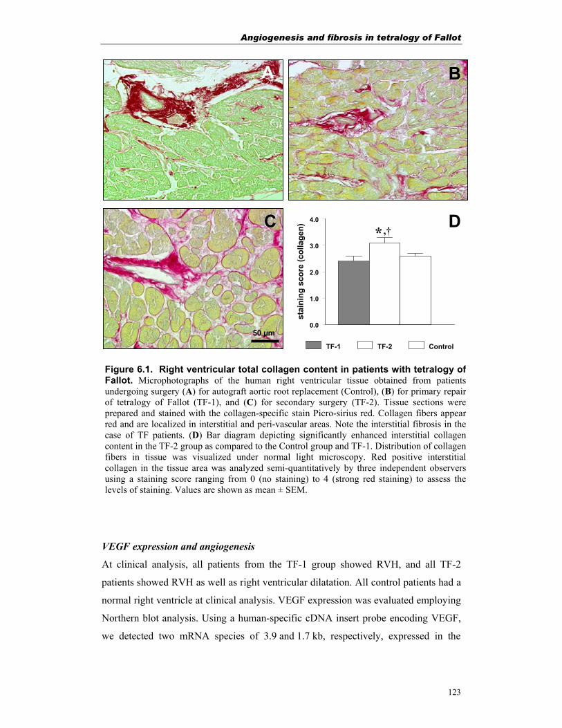
Angiogenesis and fibrosis in tetralogy of Fallot
123
VEGF expression and angiogenesis
At clinical analysis, all patients from the TF-1 group showed RVH, and all TF-2
patients showed RVH as well as right ventricular dilatation. All control patients had a
normal right ventricle at clinical analysis. VEGF expression was evaluated employing
Northern blot analysis. Using a human-specific cDNA insert probe encoding VEGF,
we detected two mRNA species of 3.9 and 1.7 kb, respectively, expressed in the
0.0
1.0
2.0
3.0
4.0
stai
ning
sco
re (c
olla
gen)
TF-1 TF-2 Control
50 µm
DC
A B
Figure 6.1. Right ventricular total collagen content in patients with tetralogy ofFallot. Microphotographs of the human right ventricular tissue obtained from patientsundergoing surgery (A) for autograft aortic root replacement (Control), (B) for primary repairof tetralogy of Fallot (TF-1), and (C) for secondary surgery (TF-2). Tissue sections wereprepared and stained with the collagen-specific stain Picro-sirius red. Collagen fibers appearred and are localized in interstitial and peri-vascular areas. Note the interstitial fibrosis in thecase of TF patients. (D) Bar diagram depicting significantly enhanced interstitial collagencontent in the TF-2 group as compared to the Control group and TF-1. Distribution of collagenfibers in tissue was visualized under normal light microscopy. Red positive interstitialcollagen in the tissue area was analyzed semi-quantitatively by three independent observersusing a staining score ranging from 0 (no staining) to 4 (strong red staining) to assess thelevels of staining. Values are shown as mean ± SEM.
*,†
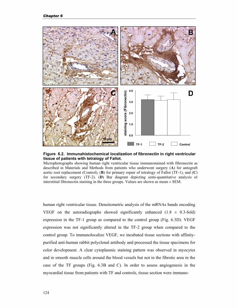
Chapter 6
124
human right ventricular tissue. Densitometric analysis of the mRNAs bands encoding
VEGF on the autoradiographs showed significantly enhanced (1.8 ± 0.3-fold)
expression in the TF-1 group as compared to the control group (Fig. 6.3D). VEGF
expression was not significantly altered in the TF-2 group when compared to the
control group. To immunolocalize VEGF, we incubated tissue sections with affinity-
purified anti-human rabbit polyclonal antibody and processed the tissue specimens for
color development. A clear cytoplasmic staining pattern was observed in myocytes
and in smooth muscle cells around the blood vessels but not in the fibrotic area in the
case of the TF groups (Fig. 6.3B and C). In order to assess angiogenesis in the
myocardial tissue from patients with TF and controls, tissue section were immuno-
0.0
1.0
2.0
3.0
4.0
stai
ning
sco
re (F
ibro
nect
in)
TF-1 TF-2 Control
50 µm
D
A B
C
Figure 6.2. Immunahistochemical localization of fibronectin in right ventricular tissue of patients with tetralogy of Fallot. Microphotographs showing human right ventricular tissue immunostained with fibronectin asdescribed in Materials and Methods from patients who underwent surgery (A) for antograftaortic root replacement (Control), (B) for primary repair of tetralogy of Fallot (TF-1), and (C) for secondary surgery (TF-2). (D) Bar diagram depicting semi-quantitative analysis ofinterstitial fibronectin staining in the three groups. Values are shown as mean ± SEM.
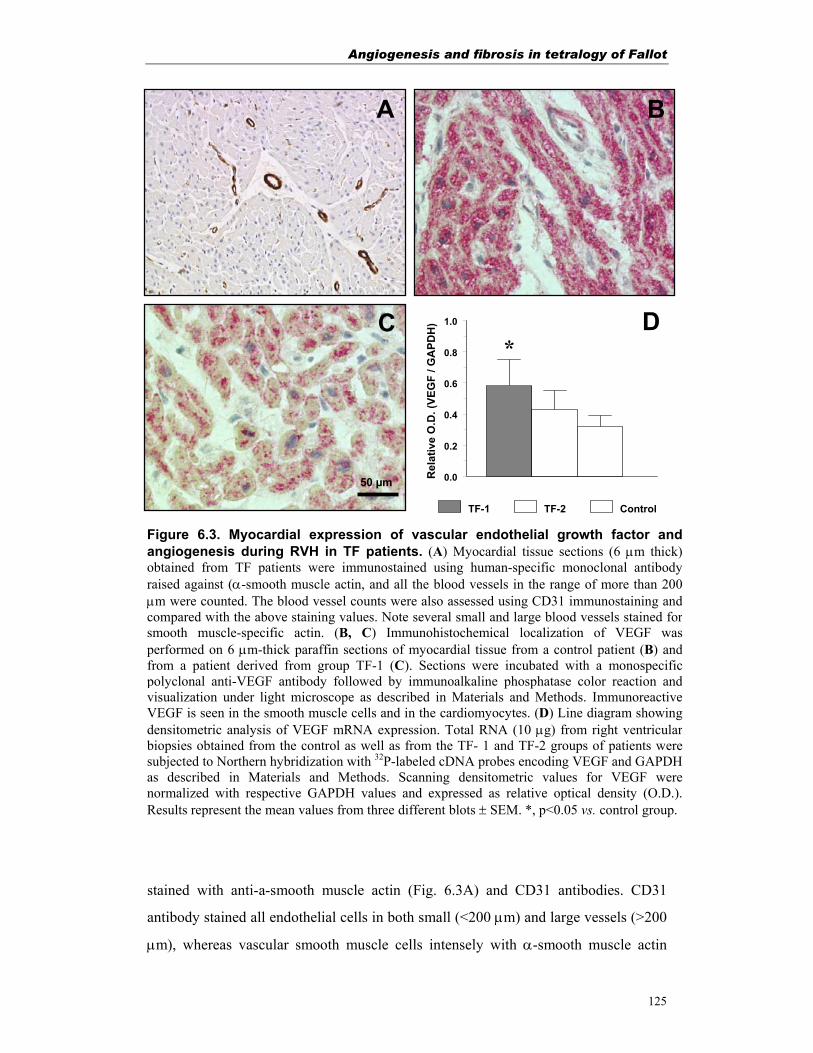
Angiogenesis and fibrosis in tetralogy of Fallot
125
stained with anti-a-smooth muscle actin (Fig. 6.3A) and CD31 antibodies. CD31
antibody stained all endothelial cells in both small (<200 µm) and large vessels (>200
µm), whereas vascular smooth muscle cells intensely with α-smooth muscle actin
0.0
0.2
0.4
0.6
0.8
1.0
Rel
ativ
e O
.D. (
VEG
F / G
APD
H)
TF-1 TF-2 ControlC
A
50 µm
DC
B
Figure 6.3. Myocardial expression of vascular endothelial growth factor andangiogenesis during RVH in TF patients. (A) Myocardial tissue sections (6 µm thick)obtained from TF patients were immunostained using human-specific monoclonal antibodyraised against (α-smooth muscle actin, and all the blood vessels in the range of more than 200µm were counted. The blood vessel counts were also assessed using CD31 immunostaining andcompared with the above staining values. Note several small and large blood vessels stained forsmooth muscle-specific actin. (B, C) Immunohistochemical localization of VEGF wasperformed on 6 µm-thick paraffin sections of myocardial tissue from a control patient (B) andfrom a patient derived from group TF-1 (C). Sections were incubated with a monospecificpolyclonal anti-VEGF antibody followed by immunoalkaline phosphatase color reaction andvisualization under light microscope as described in Materials and Methods. ImmunoreactiveVEGF is seen in the smooth muscle cells and in the cardiomyocytes. (D) Line diagram showingdensitometric analysis of VEGF mRNA expression. Total RNA (10 µg) from right ventricularbiopsies obtained from the control as well as from the TF- 1 and TF-2 groups of patients weresubjected to Northern hybridization with 32P-labeled cDNA probes encoding VEGF and GAPDHas described in Materials and Methods. Scanning densitometric values for VEGF werenormalized with respective GAPDH values and expressed as relative optical density (O.D.).Results represent the mean values from three different blots ± SEM. *, p<0.05 vs. control group.
*
Chapter 6
126
antibodies. Qualitatively, the tissue obtained from TF patients showed an enhanced
number of vessels as compared to the controls35.
In order to assess myocardial angiogenesis in the tissue specimens obtained from TF
patients, the VEGF expression, was examined at the mRNA and protein levels. At the
mRNA level, VEGF showed significantly enhanced expression in the TF-1 group as
compared to the control group. Immunohistochemical localization of VEGF indicates
that cardiac myocytes and vascular smooth muscle cells produce this mitogen, which
could be secreted from these cells to act on endothelial cells in a paracrine manner,
resulting in cardiac angiogenesis. Experimental animal studies from our research
group as well as from others have shown that myocardial ischemia is a strong inducer
of VEGF gene expression17,35,43. It is very likely in the TF situation that right
ventricular tissue experiences hypoxia, which in term could be responsible for
inducing the expression of VEGF. At this juncture, we think that the enhanced
expression of VEGF mRNA could contribute to the angiogenesis in the right ventricle
of TF patients. The increased number of blood vessels as observed by light
microscopy could explain this finding of enhanced VEGF expression in the RV
myocardium from patients with TF. Furthermore, a detailed morphometric analysis of
existing blood vessels would allow us to validate this point.
Conclusion
In conclusion, we have shown that RVH in patients with TF was accompanied with
enhanced myocardial collagen deposition in the interstitium and around the blood
vessels, giving rise to abnormal myocardial architecture. Though we did not measure
the cell size, our data correspond with the finding that after early primary corrective
surgery, RVH appears to regress and eventually leads to less collagen contents, as
observed in the TF-2 group. Our study provides further evidence that myocardial
architecture in patients with TF depicts increased fibrosis at the expression level of
extracellular matrix components such as collagens. However, the lack of data on
normal myocardium in young patients limits the interpretation of data. Additionally,
the hypertrophic state of the myocardium seems to be correlated with increased
angiogenesis. This is evident by enhanced levels of VEGF mRNAs in the children
Angiogenesis and fibrosis in tetralogy of Fallot
127
with TF at the time of primary surgery. Furthermore, histochemical examination of
tissue biopsies showed increased number of blood vessels in patients with TF as
compared to the control group. Thus, our results as presented here clearly indicate an
increased degree of fibrosis and ongoing angiogenesis during RVH in patients with
TF. This information leaves room for the improvement of contemporary clinical
treatment of patients with TF by allowing an assessment of timing for surgery and
possibly supports long-term postoperative prognosis.
Acknowledgements
Financial support from the Netherlands Heart Foundation (NHS 96.082) is gratefully
acknowledged. The authors thank Mrs. E. Yilmaz for her technical assistance.
References
1. Jordan SC, Scott O. Heart disease in pediatrics. Oxford: Butterworth Heinneman; 1989.
2. Bogers AJ, van der Laarse A, Vliegen HW, et al. Assessment of hypertrophy in myocardial biopsies taken during correction of congenital heart disease. Thorac Cardiovasc Surg. 1988;36:137-40.
3. Castaneda AR, Jonas RA, Mayer JE, et al. Cardiac surgery of neonate and infant. Philadelphia, USA: Saunders; 1994.
4. Starnes VA, Luciani GB, Latter DA, et al. Current surgical management of tetralogy of Fallot. Ann Thorac Surg. 1994;58:211-215.
5. Murphy JG, Gersh BJ, Mair DD, et al. Long-term outcome in patients undergoing surgical repair of tetralogy of Fallot. N Engl J Med. 1993;329:593-599.
6. Warner KG, Anderson JE, Fulton DR, et al. Restoration of the pulmonary valve reduces right ventricular volume overload after previous repair of tetralogy of Fallot. Circulation. 1993;88:II189-197.
7. Seliem MA, Wu YT, Glenwright K. Relation between age at surgery and regression of right ventricular hypertrophy in tetralogy of Fallot. Pediatr Cardiol. 1995;16:53-55.
8. Schwartz K, Carrier L, Mercadier JJ, et al. Molecular phenotype of hypertrophied and failing myocardium. Circulation. 1993;87:VII 5-10.
9. van Bilsen M, Chien KR. Growth and hypertrophy of the heart: towards an understanding of cardiac specific and inducible gene expression. Cardiovasc Res. 1993;27:1140-1149.
10. Kato M. Right ventricular hypertrophy in tetralogy of Fallot, a pathohistologic study. Jpn Ass Thorac Surg. 1976;24:1436-1445.
11. Kawai S, Okada R, Kitamura K, et al. A morphometrical study of myocardial disarray associated with right ventricular outflow tract obstruction. Jpn Circ J. 1984;48:445-456.
Chapter 6
128
12. Mitsuno M, Nakano S, Shimazaki Y, et al. Fate of right ventricular hypertrophy in tetralogy of Fallot after corrective surgery. Am J Cardiol. 1993;72:694-698.
13. Boheler KR, Chassagne C, Martin X, et al. Cardiac expressions of alpha- and beta-myosin heavy chains and sarcomeric alpha-actins are regulated through transcriptional mechanisms. Results from nuclear run-on assays in isolated rat cardiac nuclei. J Biol Chem. 1992;267:12979-85.
14. Brand T, Sharma HS, Schaper W. Expression of nuclear proto-oncogenes in isoproterenol-induced cardiac hypertrophy. J Mol Cell Cardiol. 1993;25:1325-1337.
15. Tomanek RJ, Torry RJ. Growth of the coronary vasculature in hypertrophy: mechanisms and model dependence. Cell Mol Biol Res. 1994;40:129-136.
16. Brand T, Sharma HS, Fleischmann KE, et al. Proto-oncogene expression in porcine myocardium subjected to ischemia and reperfusion. Circ Res. 1992;71:1351-1360.
17. Sharma HS, Wunsch M, Brand T, et al. Molecular biology of the coronary vascular and myocardial responses to ischemia. J Cardiovasc Pharmacol. 1992;20:S23-31.
18. Sharma HS, Verdouw PD, Lamers JMJ. Involvement of sarcoplasmic reticulum calcium pump in myocardial contractile dysfunction: Comparison between chronic pressure-overload and stunning. Cardiovasc Drug Ther. 1994;8:468-468.
19. Sharma HS, van Heugten HA, Goedbloed MA, et al. Angiotensin II induced expression of transcription factors precedes increase in Transforming Growth Factor-b1 mRNA in neonatal cardiac fibroblasts. Biochem Biophys Res Comm. 1994;205:105-112.
20. Volders PG, Willems IE, Cleutjens JP, et al. Interstitial collagen is increased in the non-infarcted human myocardium after myocardial infarction. J Mol Cell Cardiol. 1993;25:1317-1323.
21. Weber KT, Sun Y, Tyagi SC, et al. Collagen network of the myocardium: function, structural remodelling and regulatory mechanisms. J Mol Cell Cardiol. 1994;26:279-292.
22. Bishop JE, Rhodes S, Laurent GJ, et al. Increased collagen synthesis and decreased collagen degradation in right ventricular hypertrophy induced by pressure overload. Cardiovasc Res. 1994;28:1581-1585.
23. Chapman D, Weber KT, Eghbali M. Regulation of fibrillar collagen types I and III and basement membrane type IV collagen gene expression in pressure overloaded rat myocardium. Circ Res. 1990;67:787-794.
24. Samuel JL, Barrieux A, Dufour S, et al. Accumulation of fetal fibronectin mRNAs during the development of rat cardiac hypertrophy induced by pressure overload. J Clin Invest. 1991;88:1737-1746.
25. Boluyt MO, L. O, Meredith AL, et al. Alterations in cardiac gene expression during the transition from stable hypertrophy to heart failure. Marked upregulation of genes encoding extracellular matrix components. Circ Res. 1994;75:23-32.
26. Whittaker P. Collagen and ventricular remodelling after acute myocardial infarction: concepts and hypotheses. Basic Res Cardiol. 1997;92:79-81.
27. Jalil JE, Doering CW, Janicki JS, et al. Fibrillar collagen and myocardial stiffness in the intact hypertrophied rat left ventricle. Circ. Res. 1989;64:1041-1050.
28. McLenachan JM, Dargie HJ. Ventricular arrhythmias in hypertensive left ventricular hypertrophy. Relationship to coronary artery disease, left ventricular dysfunction, and myocardial fibrosis. Am J Hypertens. 1990;3:735-740.
29. Farhadian F, Contard F, Corbier A, et al. Fibronectin expression during physiological and pathological cardiac growth. J Mol Cell Cardiol. 1995;27:981-990.
30. Villarreal FJ, Dillmann WH. Cardiac hypertrophy-induced changes in mRNA levels for TGF-ß1, fibronectin and collagen. Am J Physiol. 1992;262:H1861-H1866.
31. Bogers AJJC, Roofthooft M, Pisters H, et al. Long-term follow-up of gamma irradiated transannular homograft patch in surgical treatment of tetralogy of Fallot. Thorac Cardiovasc Surgeon. 1994;42:337-339.
32. Hokken RB, Bogers AJJC, Spitaels SEC, et al. Pulmonary homograft insertion after repair of pulmonary stenosis. J Heart Valve Dis. 1995;4:182-186.
Angiogenesis and fibrosis in tetralogy of Fallot
129
33. Schoof PH, Cromme-Dijkhuis AH, Bogers AJJC, et al. Aortic root replacement with pulmonary autograft in children. J Thorac Cardiovasc Surg. 1994;107:367-73.
34. Hokken RB, Bogers AJJC, Taams MA, et al. Aortic root replacement with a pulmonary autograft. Eur J Cardiothor Surg. 1995;9:378-383.
35. Sharma HS, Tang ZH, Gho BC, et al. Nucleotide sequence and expression of the porcine vascular endothelial growth factor. Biochem Biophys Acta. 1995;1260:235-238.
36. Peters THF, Sharma HS, Yilmaz E, et al. Quantitative analysis of collagens and fibronectin expression in human right ventricular hypertrophy. Ann N Y Acad Sci. 1999;874:278-285.
37. Shehata SM, Mooi WJ, Okazaki T, et al. Enhanced expression of vascular endothelial growth factor in lungs of newborn infants with congenital diaphragmatic hernia and pulmonary hypertension. Thorax. 1999;54:427-31.
38. Sharma HS, Hokken RB, Bosman FT, et al. Myocardial expression and immunohistochemical localization of Vascular Endothelial Growth Factor in human tetralogy of Fallot. Eur Heart J. 1995;16:353.
39. Ferrara N, Houck KA, Jakeman LB, et al. The vascular endothelial growth factor family of polypeptides. J Cell Biochem. 1991;47:211-8.
40. Giaid A, Michel RP, Stewart DJ, et al. Expression of endothelin-1 in lungs of patients with cryptogenic fibrosing alveolitis. Lancet. 1993;341:1550-1554.
41. Vikstrom KL, Bohlmeyer T, Factor SM, et al. Hypertrophy, pathology, and molecular markers of cardiac pathogenesis. Circ Res. 1998;82:773-778.
42. Schwartz SM, Gordon D, Mosca RS, et al. Collagen content in normal, pressure, and pressure-volume overloaded developing human hearts. Am J Cardiol. 1996;77:734-738.
43. Sharma HS, Peters THF, Yilmaz E, et al. Right ventricular hypertrophy in human tetralogy of Fallot is associated with increased myocardial fibrosis and vascularization. circulation. 1997;96:I 17-18.

Chapter 7
130
Angiogenesis in human tetralogy of Fallot
131
Chapter 7 Right ventricular hypertrophy in human tetralogy of
Fallot is associated with enhanced vascularization and
expression of vascular endothelial growth factor
Based on: Peters THF, Bogers AJJC, Saxena PR and Sharma HS. Right ventricular hypertrophy in human tetralogy of Fallot is associated with enhanced vascularization and expression of vascular endothelial growth factor. (Submitted).
Chapter 7
132
Abstract Aims: Tetralogy of Fallot (TF) is a cyanotic congenital heart disease with prominent right
ventricular (RV) hypertrophy where the impaired oxygen and nutrient supply may lead
to an altered myocardial phenotype. The aim of the present study was to investigate
the myocardial differential gene expression profile and to assess myocardial
vascularization in patients with TF.
Methods and Results: DNA micro-array analysis on right ventricular biopsies from
TF patients operated for primary corrective surgery (TF-1, n=7, mean age 0.5 year)
and age matched controls was validated by Northern hybridization and RT-PCR.
Employing immunohistochemistry and video image analysis of vascular endothelial
growth factor (VEGF) expression, vascular density (by α-SMA and CD31 staining)
and myocyte cross sectional area (Gomori's reticulin staining) were assessed in TF-1
and adult patients (TF-2, n=5, mean age 30 years) who underwent surgery for pulmonary
regurgitation and compared the data with age matched controls (n=8/11). DNA micro-
array analysis revealed altered expression pattern for 236 genes including 1.5-2.2 fold
increased expression of angiogenic factors, VEGF, flt-1, flk-1 angiopoietin-2, FGF-2,
FGF-R1, PDGF-A, whereas, flt-4, Tie, TGF-β, TGF-β3R showed decreased (1.6-3.4
fold) expression in TF patients. Northern blot analysis verified VEGF expression
whereas, flk-1 expression remained unaltered in both TF-1 and TF-2 patients. VEGF
staining in cardiomyocytes was increased in TF-1 (1.5 fold, p<0.05) as compared to
TF-2. Video image analysis revealed enhanced vascular density (p<0.01) with
unchanged wall thickness and enlarged myocyte cross sectional area (p<0.01) in both
TF-1 and TF-2 as compared to respective age matched controls.
Conclusions: Right ventricular hypertrophy in TF is associated with an altered gene
profile for a number of genes where the VEGF/VEGF-R system plays an important
role in enhanced myocardial angiogenesis with limited vascular remodeling.
Angiogenesis in human tetralogy of Fallot
133
Introduction In tetralogy of Fallot (TF) the (sub)valvular pulmonary stenosis in combination with
the ventricular septal defect (VSD) causes hypoxemia, diminished pulmonary flow,
right ventricular pressure overload and adaptive right ventricular hypertrophy (RVH)
with expansion of cardiomyocytes. The treatment strategy consists of primary surgical
repair during the first year of life by closing the VSD and removing the RV outflow tract
(RVOT) obstruction in order to relieve the hypoxemia and to eliminate the hypertrophic
stimulus as well as preserving RV function. Some patients however, suffer from
pulmonary regurgitation later in life, causing RV failure due to volume-overload. Those
patients get further secondary corrective surgery by implanting a pulmonary allograft
heart valve between the RV and the pulmonary artery1.
The adaptive RVH will go along with remodeling of the myocardium accompanied with
fibrosis2 and will require a proportionate increase of coronary flow or growth of the
coronary vasculature (angiogenesis) to reduce the increased capillary-myocyte
diffusion distance, to prevent hypoxia and increase nutrient delivery3-5. Angiogenesis,
the formation of new blood vessels from pre-existing vessels, not only concerns the
formation of capillary, but also includes the formation of small and large blood
vessels. In adults, angiogenesis is the only way by which new blood vessels are
formed6. It is a multi-step process involving endothelial cell activation, migration,
proliferation, and tube formation and maturation. Beside endothelial cells several
other cell types like vascular smooth muscle cells, pericytes and fibroblasts and non-
cellular structures, such as the basal lamina and the extracellular matrix are
involved6,7.
Under pathological conditions, autocrine induction or upregulation of angiopoietin 2,
can reinitiate (angiogenic) vascular remodeling and angiogenic sprouting. In the
presence of vascular endothelial growth factor (VEGF) new vessels will be formed,
however without VEGF destabilized vessels will go into regression7. VEGF-A is the
most potent and crucial regulatory factor during myocardial angiogenesis,7 induced in
a wide range of cells under conditions of hypoxia8 and hypo-glycemia9 and in the
presence of some (growth) factors10 like phorbol ester, adenosine, TGF-β, PDGF and
TNF-α7. It binds to the endothelial cell surface receptors, flt-1 and flk-111, where
Chapter 7
134
VEGF-flk-1 binding activates a cascade which promotes angiogenesis,
hyperpermeability and enhances glucose transport12.
Not much is known about the degree of angiogenesis in human RVH. Especially
information about angiogenesis in TF patients, is rare. Most research on human TF is
focussed on clinical treatment. In the present study, we studied the degree of
angiogenesis in the RV of human TF by determining the expression pattern of VEGF
and its receptor flk-1. Furthermore we examined the vessel distribution and assessed the
degree of hypertrophy to elucidate the myocardial phenotype and fate of RVH at time
of corrective surgery.
Material and Methods Patients
The present study was approved by the Medical Ethical Committee of the University
Hospital Rotterdam (MEC153.268/1996/119). The group of patients operated upon
for primary correction of TF (TF-1), consisted of 7 patients with a mean age of
0.5±0.1 years. The group of patients operated upon for pulmonary allograft
implantation late after primary correction of TF13(TF-2), consisted of 5 patients with a
mean age of 33.6±3.5 years. The young control group (C-1) consisted of 4 post
mortem biopsies and 3 heart donors with a mean age of 0.7±0.3 years. The adult
control group (C-2) consisted of 6 donors with a mean age of 23.8±2.3 years. All
controls died on non-cardiac causes.
Tissue biopsies
Endomyocardial tissue biopsies were taken from the anterior free wall of the RVOT.
Part of the myocardial tissue biopsy was immediately frozen in liquid nitrogen for
mRNA expression studies, and the other part was fixed in 4% paraformaldehyde in
PBS for at least 24 hours and further processed for dehydration and embedding in
paraffin for (immuno)histochemical studies.
Total RNA isolation
Tissue biopsies (50-60 mg) were homogenized in guanidinium thiocyanate and
processed for the total cellular RNA extraction as described previously14. The RNA
Angiogenesis in human tetralogy of Fallot
135
concentration was spectrophotometrically measured at 260 nm and a RNA/protein
ratio of ≥1.8 was accepted. The quality of total RNA was further verified on a 1%
agarose gel and considered of sufficient quality when ribosomal RNA bands were
intact.
DNA micro-array hybridization
Total RNA samples from TF-1 (n=4; mean age 0.66±0.12) and controls (n=3; mean
age 3.76±1.11) of respective groups, were pooled and processed for Affymetrix® HG-
U95Av2 GeneChips expression analysis according to suppliers protocols (Santa
Clara, CA, USA). For practical purposes only micro-arrays of TF-1 patients were
analyzed. In brief, 5 µg total RNA was subjected to first strand cDNA synthesis using
T7-(dT)24 primers and SuperScript II reverse transcriptase (Life Technologies,
Rockville, MD, USA) followed by second strand cDNA synthesis. cDNA was
blunted, purified and used for cRNA synthesis using T7-RNA polymerase and
biotinylated-11-CTP and 16-UTP. After spectrophotometrical analysis, 11 µg of
labeled and hydrolyzed cRNA (0.05µg/µl) was hybridized with a pre-equilibrated
GeneChip U95Av2 for 16 hours at 45 °C. The arrays were washed, stained with
streptavidin-phycoerythrin and biotinylated antibodies to streptavidin and
hybridization signals were scanned using a fluormetric HP ChipScanner (Affymetrix,
Santa Clara, CA, USA). Hybridization signals were analyzed employing GeneChip
Micro-array Suite (MPS 4.0) (Affymetrix) and total gene expression profile was used
for normalisation15. A comparison of the two expression profiles was made and genes
changing over 1.5 fold (up or down regulated) were considered significantly changed
in expression16. Among the large number of genes screened (www.netaffx.com), a
group of genes involved in angiogenesis was further analyzed.
Northern blot analysis
From each patients 10 µg of total RNA was used for Northern-blot analysis as
described previously14. Blots were hybridized with 32P-labeled cDNA probes against
human VEGF (3.9 and 1.7kb) and GAPDH (1.2 kb) mRNA. Hybridization signals
quantified with Molecular Annalist (V 1.5) software (Biorad Laboratories, Hercules,
CA, USA) were corrected for background and normalized with respect to GAPDH
values.
Chapter 7
136
Reverse transcriptase polymerase chain reaction (RT-PCR) analysis
Flk-1 mRNA expression levels were semi-quantitatively assessed by RT-PCR
analysis. First strand cDNA was synthesized from total RNA using Universal
RiboClone cDNA Synthesis System (Promega, Leiden, The Netherlands).
Appropriate oligonucleotide primers for PCR were designed for human flk-1
(accession number: AF035121; Forward TCCCTGTGGATCTGAAACGG, Reverse
CAGCAGTCCAGCATGGTCTG, product size 560bp), and β-actin (accession
number: BC001301; Forward TGACGGGGTCACCCACACTG TGCCCATCTA,
Reverse ACTCGTCATACTCCTGCTTGCTGATCCA, product size 625bp). After
PCR amplification (30 cycles) the ethidium-bromide stained products were analyzed
on the same 1.5% agarose gel, digitally photographed and quantified using Molecular
Annalist (V 1.5) software. Measured flk-1 intensity were corrected for background
and normalized with β-actin values of the respective patients.
Gomori reticulin staining
Myocyte cross sectional area was determined after cell (membrane) reticulin fiber
staining by routine Gomori’s reticulin silver staining technique17 in 5 µm thick
sections. After the silver staining was developed, slides were mounted and visualized
under light microscope.
Immunolocalization of VEGF, CD31 and α-smooth muscle actin (α-SMA)
Immunohistochemical detection was performed on 5 µm thick paraffin-embedded
myocardial tissue sections, using a multiple step avidin-biotin complex (ABC) method
(Biogenex, San Ramon, CA, USA). Affinity purified rabbit anti-human-VEGF
antibodies (Santa Cruz Biothechnology, Santa Cruz, USA) were applied as primary
antibody followed by biotinylated anti-immunoglobulins and tertiary streptavidin
conjugated alkaline phosphatase. Color was developed using Naphtol AS-MX
phosphate and new fuchsin. Mouse monoclonal antibodies against human vascular α-
SMA (Clone 1A4, NeoMarkers Fremont, CA, USA) and mAb against endothelial
specific CD31 marker (Ab-3; NeoMarker), to immunulocalize capillaries, were applied
as primary antibodies. Streptavidin conjugated to peroxidase (BioGenex, San Ramon,
CA) was applied and subsequently sections were colored using 3,3-diaminobenzidine
Angiogenesis in human tetralogy of Fallot
137
(Sigma, St Louis, MO, USA). All sections were counter stained with Mayer's
hematoxylin, mounted and visualized under the light microscope.
Visual-scoring
VEGF staining, in the myocyte and vascular area, was semi-quantitative analyzed by
visually scoring, in a blinded fashion in random order, by two independent observers,
using arbitrary visual grading scores of 0, 1, 2, 3 and 4 representing no, weak,
moderate and intense staining, respectively.
Video image analysis
α-SMA stained vessels were analyzed morphometrically using a Leica Qwin video
image analysis system (version 3.0, Leica, Rijswijk, The Netherlands). In 20
rectangular digital photographed images of each section, vessels with an external
diameter (ED) larger then 5µm, in a set tissue area of 12.6 million µm2 were analyzed
using the Leica Qwin standard (Y2.2b) image analysis software package (Leica
Imaging Systems, Cambrige, UK). The myocardial vascular density was assessed by
the number of vessels per unit area. Internal vessel diameters (ID) were calculated
from the average of the largest and smallest ID of a vessel. To exclude measurements
on longitudinal cut vessels, only ratios largest to smallest ID ≤3 were accepted.
External vessel diameters (ED) were assessed similarly. The vessel wall thickness was
assessed from the smallest ED and ID. Vascular wall thickness was also indexed as
ratio of vessel wall area to lumen area and plotted to the ID of the individual vessel to
investigated the possibility of vessel wall remodeling18. Based on the ID the
myocardial vessels were also analyzed in ID-interval (see Fig. 7.4). Myocyte cell size
was measured with the same set-up. In series of 3-4 digital photographs from reticulin
stained sections, the myocyte outline was drawn of at least 20 transversally cut
myocytes showing a nucleolus, where after the program calculated the myocyte cross
sectional area in µm2.
Statistical analysis
Statistical analysis for micro-array results was performed with comparison ranking
with software Data Mining Tool (Affymetrix). Using SPSS software (SPSS, Chicago,
USA), statistical probability was assessed by (non-parametric) Kruskal-Wallis one
Chapter 7
138
way analysis of variance and linear (Pearson’s) regression analysis was performed for
developing cell size. Values are expressed as mean±SEM and accepted as statistically
significant with p≤0.05.
Results
DNA micro-array analysis
To assess the differential gene-expression profile from TF patients, RNA samples
obtained from TF-1 and age matched controls were processed for Affymetrix®
GeneChip expression hybridization followed by scanning and computer analysis.
Hybridization analysis data is presented as a differential scatter graph in figure 7.1.
Comparison ranking analysis revealed that 236 genes showed altered expression
pattern. We analyzed a group of genes involved in myocardial angiogenesis (VEGF-
A, VEGF-B, VEGF-C, VEGF-D, PlGF, flk-1, flt-1, angiopoietin 1 and 2, TNF-α,
TGF-β, PDGF and it’s receptors) (Table 7.1).
VEGF and flk-1 mRNA expression analysis by Northern-blot and RT-PCR
Densitometric analysis of representative Northern-blots, normalized with respect to
GAPDH values, revealed elevated VEGF ratios in TF-1 (0.68±0.13; p<0.05) and TF-2
(0.85±0.18) as compared to C-1 (0.50±0.05) and C-2 (0.76±0.05) (Fig. 7.2).
Flk-1 expression, semi-quantitatively assessed by RT-PCR, revealed unchanged levels
in TF-1 (0.67±0.06) and TF-2 (0.59±0.04) as compared to controls (C-1: 0.60±0.06;
C-2: 0.53±0.03)(Fig. 7.2)
VEGF localization and visual scoring
Immunoreactive VEGF was localized mainly in the cytoplasm of cardiomyocytes and
vascular smooth muscle cells and not in fibrotic areas (Fig. 7.3). The VEGF staining
score in cardiomyocytes, showed unchanged levels in TF patients compared to
respective controls (TF-1: 2.5±0.2 vs. C-1: 2.2±0.4 and TF-2: 1.7±0.2 vs. C-2:
1.5±0.2) whereas the staining score was increased (p<0.05) in young patients and
controls as compared to adult subjects. Although the VEGF staining in the vascular
smooth muscle cells in the young groups was more intense, the staining score
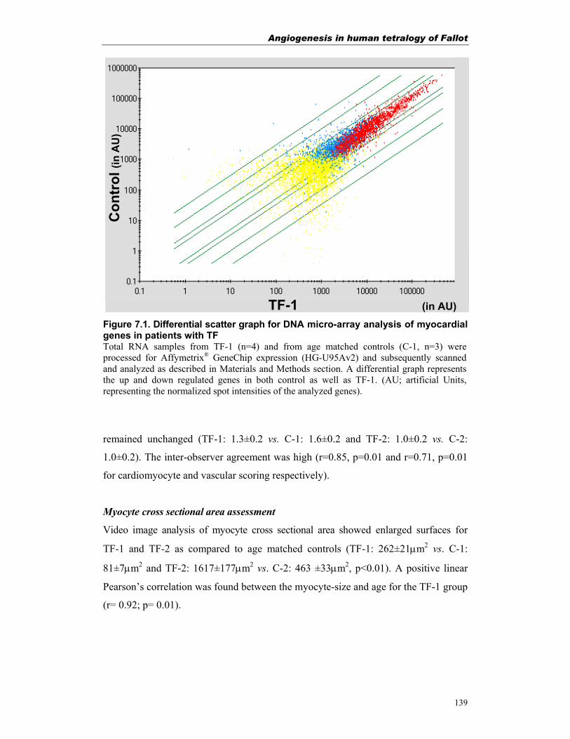
Angiogenesis in human tetralogy of Fallot
139
Figure 7.1. Differential scatter graph for DNA micro-array analysis of myocardial genes in patients with TF Total RNA samples from TF-1 (n=4) and from age matched controls (C-1, n=3) were processed for Affymetrix® GeneChip expression (HG-U95Av2) and subsequently scanned and analyzed as described in Materials and Methods section. A differential graph represents the up and down regulated genes in both control as well as TF-1. (AU; artificial Units, representing the normalized spot intensities of the analyzed genes).
remained unchanged (TF-1: 1.3±0.2 vs. C-1: 1.6±0.2 and TF-2: 1.0±0.2 vs. C-2:
1.0±0.2). The inter-observer agreement was high (r=0.85, p=0.01 and r=0.71, p=0.01
for cardiomyocyte and vascular scoring respectively).
Myocyte cross sectional area assessment
Video image analysis of myocyte cross sectional area showed enlarged surfaces for
TF-1 and TF-2 as compared to age matched controls (TF-1: 262±21µm2 vs. C-1:
81±7µm2 and TF-2: 1617±177µm2 vs. C-2: 463 ±33µm2, p<0.01). A positive linear
Pearson’s correlation was found between the myocyte-size and age for the TF-1 group
(r= 0.92; p= 0.01).
Average differential gene expression scatter
Con
trol
(in
AU
)
TF-1 (in AU)
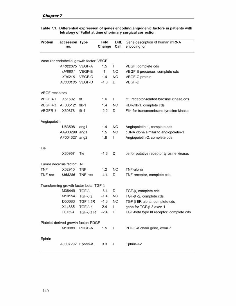
Chapter 7
140
Table 7.1. Differential expression of genes encoding angiogenic factors in patients with tetralogy of Fallot at time of primary surgical correction Protein accession
no. Type Fold
ChangeDiff. Call.
Gene description of human mRNA encoding for
Vascular endothelial growth factor: VEGF AF022375 VEGF-A 1.5 I VEGF, complete cds U48801 VEGF-B 1 NC VEGF B precursor, complete cds X94216 VEGF-C 1.4 NC VEGF-C protein AJ000185 VEGF-D -1.8 D VEGF-D VEGF receptors:
VEGFR-1 X51602 flt 1.6 I flt ; receptor-related tyrosine kinase,cds
VEGFR-2 AF035121 flk-1 1.4 NC KDR/flk-1, complete cds VEGFR-3 X69878 flt-4 -2.2 D Flt4 for transmembrane tyrosine kinase Angiopoietin U83508 ang1 1.4 NC Angiopoietin-1, complete cds AA903299 ang1 1.5 NC cDNA clone similar to angiopoietin-1 AF004327 ang2 1.6 I Angiopoietin-2, complete cds Tie X60957 Tie -1.6 D tie for putative receptor tyrosine kinase, Tumor necrosis factor: TNF TNF X02910 TNF 1.2 NC TNF-alpha TNF-rec M58286 TNF-rec -4.4 D TNF receptor, complete cds Transforming growth factor-beta: TGF-β M38449 TGF-β -3.4 D TGF-β, complete cds M19154 TGF-β 2 -1.4 NC TGF-β -2, complete cds D50683 TGF-β 2R -1.3 NC TGF-β IIR alpha, complete cds X14885 TGF-β 3 2.4 I gene for TGF-β 3 exon 1 L07594 TGF-β 3 R -2.4 D TGF-beta type III receptor, complete cds Platelet-derived growth factor: PDGF M19989 PDGF-A 1.5 I PDGF-A chain gene, exon 7 Ephrin AJ007292 Ephrin-A 3.3 I Ephrin-A2
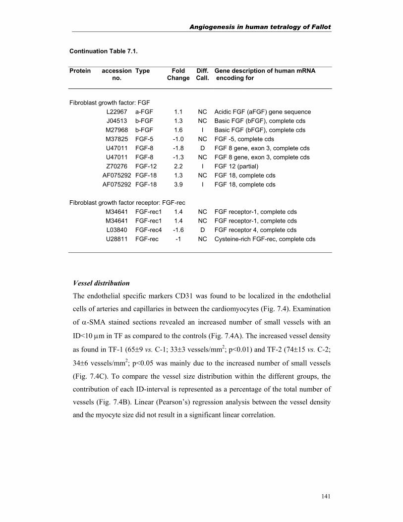
Angiogenesis in human tetralogy of Fallot
141
Continuation Table 7.1. Protein accession
no. Type Fold
ChangeDiff. Call.
Gene description of human mRNA encoding for
Fibroblast growth factor: FGF L22967 a-FGF 1.1 NC Acidic FGF (aFGF) gene sequence J04513 b-FGF 1.3 NC Basic FGF (bFGF), complete cds M27968 b-FGF 1.6 I Basic FGF (bFGF), complete cds M37825 FGF-5 -1.0 NC FGF -5, complete cds U47011 FGF-8 -1.8 D FGF 8 gene, exon 3, complete cds U47011 FGF-8 -1.3 NC FGF 8 gene, exon 3, complete cds Z70276 FGF-12 2.2 I FGF 12 (partial) AF075292 FGF-18 1.3 NC FGF 18, complete cds AF075292 FGF-18 3.9 I FGF 18, complete cds Fibroblast growth factor receptor: FGF-rec M34641 FGF-rec1 1.4 NC FGF receptor-1, complete cds M34641 FGF-rec1 1.4 NC FGF receptor-1, complete cds L03840 FGF-rec4 -1.6 D FGF receptor 4, complete cds U28811 FGF-rec -1 NC Cysteine-rich FGF-rec, complete cds
Vessel distribution
The endothelial specific markers CD31 was found to be localized in the endothelial
cells of arteries and capillaries in between the cardiomyocytes (Fig. 7.4). Examination
of α-SMA stained sections revealed an increased number of small vessels with an
ID<10 µm in TF as compared to the controls (Fig. 7.4A). The increased vessel density
as found in TF-1 (65±9 vs. C-1; 33±3 vessels/mm2; p<0.01) and TF-2 (74±15 vs. C-2;
34±6 vessels/mm2; p<0.05 was mainly due to the increased number of small vessels
(Fig. 7.4C). To compare the vessel size distribution within the different groups, the
contribution of each ID-interval is represented as a percentage of the total number of
vessels (Fig. 7.4B). Linear (Pearson’s) regression analysis between the vessel density
and the myocyte size did not result in a significant linear correlation.
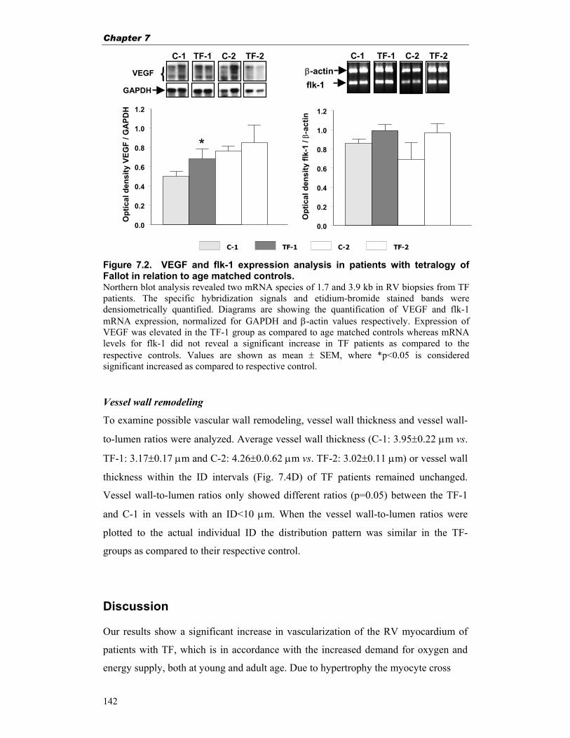
Chapter 7
142
Figure 7.2. VEGF and flk-1 expression analysis in patients with tetralogy of Fallot in relation to age matched controls. Northern blot analysis revealed two mRNA species of 1.7 and 3.9 kb in RV biopsies from TF patients. The specific hybridization signals and etidium-bromide stained bands were densiometrically quantified. Diagrams are showing the quantification of VEGF and flk-1 mRNA expression, normalized for GAPDH and β-actin values respectively. Expression of VEGF was elevated in the TF-1 group as compared to age matched controls whereas mRNA levels for flk-1 did not reveal a significant increase in TF patients as compared to the respective controls. Values are shown as mean ± SEM, where *p<0.05 is considered significant increased as compared to respective control.
Vessel wall remodeling
To examine possible vascular wall remodeling, vessel wall thickness and vessel wall-
to-lumen ratios were analyzed. Average vessel wall thickness (C-1: 3.95±0.22 µm vs.
TF-1: 3.17±0.17 µm and C-2: 4.26±0.0.62 µm vs. TF-2: 3.02±0.11 µm) or vessel wall
thickness within the ID intervals (Fig. 7.4D) of TF patients remained unchanged.
Vessel wall-to-lumen ratios only showed different ratios (p=0.05) between the TF-1
and C-1 in vessels with an ID<10 µm. When the vessel wall-to-lumen ratios were
plotted to the actual individual ID the distribution pattern was similar in the TF-
groups as compared to their respective control.
Discussion Our results show a significant increase in vascularization of the RV myocardium of
patients with TF, which is in accordance with the increased demand for oxygen and
energy supply, both at young and adult age. Due to hypertrophy the myocyte cross
0.0
0.2
0.4
0.6
0.8
1.0
1.2
0.0
0.2
0.4
0.6
0.8
1.0
1.2
β-actinVEGF
GAPDH flk-1
C-1 TF-1 C-2 TF-2
Opt
ical
den
sity
flk-
1 / β
-act
in
Opt
ical
den
sity
VEG
F / G
APD
H
{C-1 TF-1 C-2 TF-2 C-1 TF-1 C-2 TF-2
*
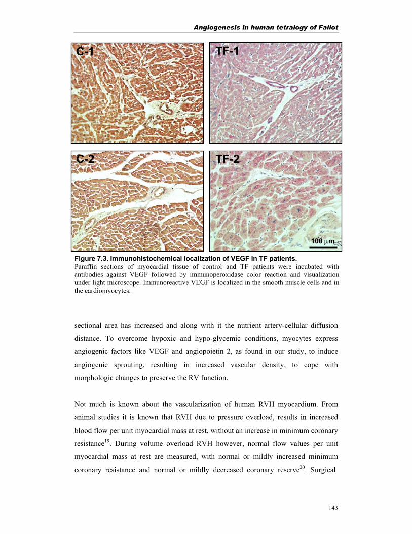
Angiogenesis in human tetralogy of Fallot
143
Figure 7.3. Immunohistochemical localization of VEGF in TF patients. Paraffin sections of myocardial tissue of control and TF patients were incubated with antibodies against VEGF followed by immunoperoxidase color reaction and visualization under light microscope. Immunoreactive VEGF is localized in the smooth muscle cells and in the cardiomyocytes.
sectional area has increased and along with it the nutrient artery-cellular diffusion
distance. To overcome hypoxic and hypo-glycemic conditions, myocytes express
angiogenic factors like VEGF and angiopoietin 2, as found in our study, to induce
angiogenic sprouting, resulting in increased vascular density, to cope with
morphologic changes to preserve the RV function.
Not much is known about the vascularization of human RVH myocardium. From
animal studies it is known that RVH due to pressure overload, results in increased
blood flow per unit myocardial mass at rest, without an increase in minimum coronary
resistance19. During volume overload RVH however, normal flow values per unit
myocardial mass at rest are measured, with normal or mildly increased minimum
coronary resistance and normal or mildly decreased coronary reserve20. Surgical
C-1
TF-2C-2
TF-1
100 µm
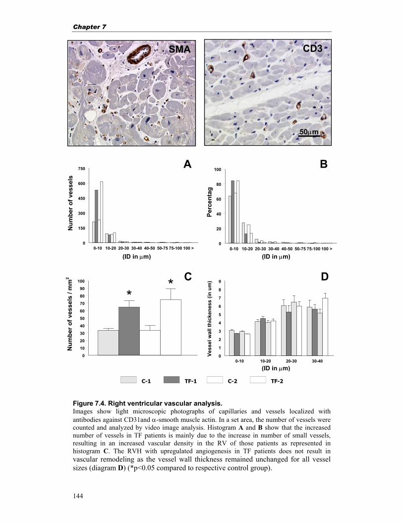
Chapter 7
144
Figure 7.4. Right ventricular vascular analysis. Images show light microscopic photographs of capillaries and vessels localized with antibodies against CD31and α-smooth muscle actin. In a set area, the number of vessels were counted and analyzed by video image analysis. Histogram A and B show that the increased number of vessels in TF patients is mainly due to the increase in number of small vessels, resulting in an increased vascular density in the RV of those patients as represented in histogram C. The RVH with upregulated angiogenesis in TF patients does not result in vascular remodeling as the vessel wall thickness remained unchanged for all vessel sizes (diagram D) (*p<0.05 compared to respective control group).
50µm
0-10 10-20 20-30 30-400
1
2
3
4
5
6
7
8
9
Vess
el w
all t
hick
enes
s (in
um
)
0-10 10-20 20-30 30-40 40-50 50-75 75-100 100 >0
20
40
60
80
100
0
10
20
30
40
50
60
70
80
90
100
0-10 10-20 20-30 30-40 40-50 50-75 75-100 100 >0
150
300
450
600
750
C-1 TF-1 C-2 TF-2
(ID in µm)
(ID in µm)
(ID in µm)
A
C
B
D
* *
CD3
Perc
enta
g
Num
ber o
f ves
sels
N
umbe
r of v
esse
ls /
mm
2
SMA
Angiogenesis in human tetralogy of Fallot
145
interference can partly take away the hypertropic stimuli in TF patients and might
decrease hypertrophy and reverse associated coronary vascular abnormalities21
In addition to normal growth, RVH stimuli will initiate enhanced coronary vascular
growth by angiogenic sprouting triggered by VEGF-A in the presence of angiopoietin
2. The binding of the VEGF-ligand to flk-1 will activate a cascade of angiogenic
factors. This is in accordance with our GeneChip, Northern-blot and semi-quantitative
RT-PCR data where the slightly elevated VEGF levels and equal flk-1 levels in TF-1
and TF-2 reflect enhanced angiogenesis via the activated cascade of angiogenic
factors. The small difference in VEGF expression, as observed by visual scoring, in
TF-1 compared to the age matched control group can be explained by the lower
sensitivity of this analysis method. Both control- and TF-biopsies are from hearts
from young children were the need of new vessels is obvious, resulting in VEGF
upregulation. The young heart has to meet with the increased oxygen and energy
demand due to growth of the heart and myocytes and the increased pulmonary flow
which has to be effectuated by the RV for a sufficient cardiac output7. In addition TF
patients continuously are exposed to stimuli of pressure overload, shear stress and, in
some, hypoxic spells until corrective surgery. This might result in higher expression
levels or slower turnover of angiogenic factors, subsequently resulting in enhanced
RV angiogenesis. Moreover the initiated angiogenesis in TF patients is a long lasting
process compared to the induction of angiogenic factors in acute pathological processes.
Small differences in the expression pattern as detected at time of surgery might therefore
have a bigger impact in the long term than other pathological conditions.
Morphological studies in experimental animals and in patients with pressure overload
hypertrophy demonstrated that the capillaries-to-myocyte ratio remains unchanged22.
Studies on patients with pressure overload left ventricular hypertrophy with normal
capillary density, showed an increased capillary supply per myocyte, proportional to the
increased myocyte size, keeping the capillary–to-myocyte ratio similar23. One study on
TF patients (age 1-7 years) reported that the overall myocardial capillary density, in
the RVOT, was similar to normal myocardial capillary density (1648±123 vs.
1782±203 capillaries/mm2)24 when the increase in cross sectional myocyte area was
not taken into account. Furthermore the capillary domain (cross-sectional tissue area

Chapter 7
146
nearest to that capillary) and the capillary tissue supply volume were found to be the
same in TF- as well as in control-group24. However the capillary segment length
(distance between two branching points) was found to be increased24. The capillaries
and vessels as detected in this study, localized respectively with specific antibodies
against CD31 and vascular SMA, had similar distribution patterns (Fig. 7.4).
Although the overall myocardial capillary density in TF patients was reported to be
unchanged24 the capillary radius was found to be decreased from 2.8 to 2.4 µm, in the
sub-endocardial region of hypertrophied rat hearts25. Based on those criteria we analyzed
vessels with an ED≥5 µm and found enhanced vascular density in TF patients, mainly
due to increased numbers of small vessels (Fig. 7.4). This might indicate an ongoing
process of angiogenic sprouting in order to preserve the aerobic metabolism in those
patients. The enhanced myocyte cross sectional area, indicating myocyte hypertrophy,
consequently results in a decrease of the number of cardiomyocytes per area and an
increase in the number of vessels per myocyte, compensating the increased diffusion
distance. In this respect, our data on the vascular density of larger blood vessels has to be
interpreted with some caution as the biopsies were taken from locations of the free wall
of the RVOT, deliberately excluding areas with epicardial coronary arteries, thereby
decreasing the number of large vessels in our study.
Vascular wall remodeling has been reported by several studies on ischemic
hypertrophied hearts. The arterial morphometry, vessel wall thickness and vessel
wall-to-lumen ratios in RVH myocardium of TF patients however, appeared to be
similar to the arteries in healthy RV. This is supported by the unchanged vessel wall
thickness between the different ID-intervals and the unchanged vessel wall-to-lumen
ratios, between the respective groups, were the decreased vessel wall-to-lumen ratio
as found for the smallest TF-1 vessels (ID: 0-10µm) would rather indicate an ongoing
angiogenic sprouting and development of immature vessels than vascular wall
remodeling. The interpretation of the average wall thickness should be taken with
some caution as the contribution of small vessels in TF patients is much higher than in
the control (Fig. 7.4).

Angiogenesis in human tetralogy of Fallot
147
To prevent vessel analysis on longitudinally cut vessels, only vessels with ratio largest
to smallest ID≤3 were accepted. To check if this arbitrary value would influence the
result also vessel with ID-ratios≤2 were analyzed. Although less vessels met with this
criterium, the distribution pattern between the ID intervals remained the same (data
not shown).
Our study provides further evidence that RVH in TF patients depicts increased RV
vascularization besides the earlier reported increased (peri-vascular) fibrosis2. However,
the lack of data on normal myocardium in young patients limits the interpretation of
data. Due to obvious reasons, it is difficult to obtain any age matched young control
myocardial tissue biopsies or biopsies after the corrective surgery to control whether
pathological stimuli have stopped or the RVH has regressed. In addition, further research
at the mRNA level of other angiogenic factors would provide a better understanding of
the altered gene expression during those pathological conditions.
In conclusion, our data suggest that RVH in TF is associated with an altered gene
expression profile for a number of genes, where the VEGF/VEGF-R system plays an
important role in the enhanced myocardial angiogenesis that could be stunted due to
limited vascular wall remodeling. This information leaves room for improvement of
contemporary clinical treatment of TF patients by allowing an assessment of myocardial
condition at the time of surgery and providing information to be correlated with long
term postoperative prognosis.
Acknowledgements We thank Frank Staal, PhD, for his help in with the GeneChip experiments. Financial
support from the Netherlands Heart Foundation (NHS 96.082) is gratefully
acknowledged.
References 1. Warner KG, Anderson JE, Fulton DR, et al. Restoration of the pulmonary valve
reduces right ventricular volume overload after previous repair of tetralogy of Fallot. Circulation. 1993;88:II189-197.
Chapter 7
148
2. Peters THF, Sharma HS, Yilmaz E, et al. Quantitative analysis of collagens and fibronectin expression in human right ventricular hypertrophy. Ann N Y Acad Sci. 1999;874:278-285.
3. van Bilsen M, Chien KR. Growth and hypertrophy of the heart: towards an understanding of cardiac specific and inducible gene expression. Cardiovasc Res. 1993;27:1140-1149.
4. Boluyt MO, L. O, Meredith AL, et al. Alterations in cardiac gene expression during the transition from stable hypertrophy to heart failure. Marked upregulation of genes encoding extracellular matrix components. Circ Res. 1994;75:23-32.
5. Schwartz K, Carrier L, Mercadier JJ, et al. Molecular phenotype of the hypertrofied and failing myocardium. Circulation. 1993;87:VII 5-10.
6. Huminiecki LB, R. Angiogenesis. In: Bikfalvi A, ed. Encyclopedic reference of vascular biology & pathology. Berlin Heidelberg: Springer Verlag; 2000:2-8.
7. Yancopoulos GD, Davis S, Gale NW, et al. Vascular-specific growth factors and blood vessel formation. Nature. 2000;407:242-8.
8. Shweiki D, Itin A, Soffer D, et al. Vascular endothelial growth factor induced by hypoxia may mediate hypoxia-initiated angiogenesis. Nature. 1992;359:843-845.
9. Shweiki D, Neeman M, Itin A, et al. Induction of vascular endothelial growth factor expression by hypoxia and by glucose deficiency in multicell spheroids: implications for tumor angiogenesis. Proc Nat Acad Sci U S A. 1995;92:768-772.
10. Horiuchi T, Weller PF. Expression of vascular endothelial growth factor by human eosinophils: upregulation by granulocyte macrophage colony-stimulating factor and interleukin-5. Am J Respir Cell Mol Biol. 1997;17:70-77.
11. Detmar M, Brown LF, Berse B, et al. Hypoxia regulates the expression of vascular permeability factor/vascular endothelial growth factor (VPF/VEGF) and its receptors in human skin. J Invest Dermatol. 1997;108:263-268.
12. Waltenberger J, Mayr U, Pentz S, et al. Functional upregulation of the vascular endothelial growth factor receptor KDR by hypoxia. Circulation. 1996;94:1647-1654.
13. Hokken RB, Bogers AJJC, Spitaels SEC, et al. Pulmonary homograft insertion after repair of pulmonary stenosis. J Heart Valve Dis. 1995;4:182-186.
14. McKay S, de Jongste JC, Saxena PR, et al. Angiotensin II induces hypertrophy of human airway smooth muscle cells: expression of transcription factors and transforming growth factor-beta1. Am J Respir Cell Mol Biol. 1998;18:823-833.
15. Tan FL, Moravec CS, Li J, et al. The gene expression fingerprint of human heart failure. Proc Nat Acad Sci U S A. 2002;99:11387-92.
16. Yajima N, Masuda M, Miyazaki M, et al. Oxidative stress is involved in the development of experimental abdominal aortic aneurysm: a study of the transcription profile with complementary DNA microarray. J Vasc Surg. 2002;36:379-85.
17. Bancroff JD, Cook HC. Manual of Histological Techniques and Their Diagnostic Aplication. London: Churchill Livingstone; 1994.
18. Simionescu N, Simionescu M. The cardiovascular system. In: Weis L, ed. Cell and Tissue Biology. A Tekstbook of Histology. Baltimore: Urban and Schwarzenberg; 1988:353-400.
19. Murray PA, Vatner SF. Reduction of maximal coronary vasodilator capacity in conscious dogs with severe right ventricular hypertrophy. Circ Res. 1981;48:25-33.
20. Hultgren PB, Bove AA. Myocardial blood flow and mechanics in volume overload-induced left ventricular hypertrophy in dogs. Cardiovasc Res. 1981;15:522-528.
21. Isoyama S, Ito N, Kuroha M, et al. Complete reversibility of physiological coronary vascular abnormalities in hypertrophied hearts produced by pressure overload in the rat. J Clin Invest. 1989;84:288-294.
22. Bache RJ. Effects of hypertrophy on the coronary circulation. Prog Cardiovasc Dis. 1988;30:403-440.
23. Rakusan K, Flanagan MF, Geva T, et al. Morphometry of human coronary capillaries during normal growth and the effect of age in left ventricular pressure-overload hypertrophy. Circulation. 1992;86:38-46.

Angiogenesis in human tetralogy of Fallot
149
24. Rakusan K, Cicutti N, Spatenka J, et al. Geometry of the capillary net in human hearts. Int J Microcirc Clin Exp. 1997;17:29-32.
25. Turek Z, Rakusan K. Lognormal distribution of intercapillary distance in normal and hypertrophic rat heart as estimated by the method of concentric circles: its effect on tissue oxygenation. Pflugers Arch. 1981;391:17-21.

Chapter 8
150

General discussion and conclusions
151
Chapter 8
General discussion and conclusions
Chapter 8
152
8.1 Introduction Tetralogy of Fallot (TF) is a frequent congenital cyanotic heart malformation. The
cyanosis is a consequence of the reduced pulmonary flow due to the right ventricular
(RV) outflow tract obstruction and intracardiac shunting through the ventricular septal
defect (VSD). This results in right ventricular hypertrophy (RVH). Compensatory
myocardial remodeling, triggered by the haemodynamic overload, shear stress and
myocyte stretch, is necessary to sustain the cardiac performance. Data presented in
this thesis support the observation that the RV phenotypic changes stimulated by the
forementioned stress-factors initiate the up- or down- regulation and release of a
cascade of molecules that co-ordinate the compensatory response. An altered gene
expression pattern will finally result in increased synthesis or reduced degradation of
structural proteins, changing the structural phenotype of the RV in TF patients.
Three mechanisms known to be involved in myocardial remodeling, are myocyte
hypertrophy, myocardial fibrosis and myocardial vascularization. They are assumed to
be active in all stages of TF. We were able to sample tissue biopsies at the time of
primary corrective surgery for TF at a young age (TF-1) and from TF patients who were
operated at an adult age because of pulmonary regurgitation (TF-2). We also collected
biopsies at correction of pulmonary atresia with VSD (PA-VSD). The biopsies were
studied and compared to normal age matched RV tissue (C-1; young, C-2; adult age) for
changes in the RV phenotype (at mRNA and protein level) with regard to myocardial
cellular organisation and degree of hypertrophy, with further analysis of the degree of
fibrosis (e.g. collagens and fibronectin) and myocardial angiogenesis (e.g. VEGF
expression and vascular density) in different stages of human TF.
8.2 Changes in the right ventricular myocardial phenotype Right ventricular myocardial hypertrophy During normal cardiac development the increase in myocardial mass is a result of
myocyte hyperplasia and hypertrophy. After birth, however, myocardial growth is
mainly due to hypertrophy1. In this regard, no biopsies are available to study
cardiomyocytes in TF directly after birth, as we were only able to sample biopsies at
corrective cardiac surgery. In TF the RV remodeling is a combination of myocardial
General discussion and conclusions
153
hypertrophy and fibrosis. Dilatation may occur later in life when (volume) overload is
sustained. Remodeling continues until there is a balance between the RV pressure and
the mechanical stretch and strength. TF patients without corrective cardiac surgery
develop pressure overload where the RV pressure is equal to the left ventricular
pressure (table 4.1). This will increase the myocardial wall stress and result in
pathological concentric hypertrophy of the myocytes, with characteristic myocardial
wall thickening. The haemodynamic stress is generally considered to be the major
cause of hypertrophy with myocyte disarray and myofibrillar disorganization2.
The RV myocyte hypertrophy is believed to be a result of increased mechanical forces
like pressure overload and shear stress1. Jones et al. reported that RVH was correlated
with the systolic outflow gradient and a high RV end systolic pressure3. In our study
we could not find such a correlation between the RV pressure and myocyte cross
sectional area within the respective groups. The myocyte cross sectional area however,
was found to have a linear correlation with age in TF-1 (Fig. 4.4). This is in agreement
with others who found that the RVH in TF has its onset immediately after birth, with
increasing myocyte diameters correlating with age but not with the oxygen saturation
of the arterial blood or cyanotic spells2,4. This implies that until hypertrophic stimuli
are removed, at least in the first year, the pathological hypertrophy is an ongoing
process. Our further findings of a positive correlation between the myocyte cross
sectional area with the age (r=0.90, p<0.05; age range of 0-7 year) in normal hearts
confirm the data of Kawai et al. who found a tendency of increased myocyte size with
the age2.
Rakusan et al. reported a myocyte radius of 2.5 µm in neonates, in both normal and TF-
myocardium5. In our study the radius of TF myocytes increased form 3.7 µm at the age
of 5 months to 5.8 µm at the age of 1.8 years. The increase in myocyte hypertrophy is
believed to be irreversible when exceeding a radius of 7-8 micron at an age of four
years or more. The results clearly show that there is myocyte hypertrophy in all stages
of TF, with an increase in average myocyte cross sectional area of: 3.2, 2.0 and 3.5
fold for TF-1, PA-VSD and TF-2 respectively, as compared to the age matched
controls. Based on these data, it is considered that the optimal time for corrective
Chapter 8
154
surgery for TF, to prevent an irreversible alteration of the RV muscle fibers, should be
at least before the age of three years4.
The largest increase in myocyte cross sectional area was found in TF-2 patients. Those
patients faced haemodynamic overload, first at a young age as a result of the TF
anomalies and secondly due to pulmonary regurgitation after primary correction for
TF, later in life. The pulmonary regurgitation will cause volume overload with
consequently pathological hypertrophy. Furthermore, myocyte disarray is considered
to require more energy for contraction and may act as a generator of cardiac
hypertrophy2. Our histological observations showed that TF-2 patients already faced
myocyte disarray as well as 3.2 fold increase in the myocyte cross sectional area at
young age as compared to normal myocytes. Pulmonary regurgitation later in life
results in an additional increase of cardiomyocyte hypertrophy, that sustains the RV
performance but is assumed to further deteriorate the RV compliance.
Myocardial fibrosis Initially, myocardial fibrosis is an adaptive response to the haemodynamic overload. The
RV pressure overload in TF patients remodels the extracellular matrix scaffold, finally
allowing (pathological) RV expansion. To restore the tensile strength and to resist the
distending forces the myocardial cells, mainly fibroblasts, enhance the collagenous fiber
production until enough new fibers have been produced to prevent further dilatation6,7.
In this regard, our data (chapters 2, 3, 4 and 5) is in agreement with the histopathologic
findings from the RV myocardium in pre-operative TF that show interstitial fibrosis,
myofibrillar disorganization, disarray and degenerative changes in addition to
myocardial cell hypertrophy8,9. Significantly increased interstitial total collagen, collagen
subtypes and fibronectin levels, were found in almost all TF-groups indicating an
ongoing fibrosis to restore the mechanical myocardial strength. The collagen levels in
the peri-vascular area of most TF patients however, were not increased suggesting that
no extra support to the vessels is provided.
The apparent development of cardiac fibrosis is induced by changes in cardiac gene
expression that provide means of compensation for increased haemodynamic load. The
neonatal myocardium will increase the collagen mRNA expression and synthesis6.
General discussion and conclusions
155
Upregulation of fibronectin and collagen expression has also been shown during
congestive heart failure10. Our DNA micro-array data (table 4.2) from TF-1 patients
show similar patterns of up- or down-regulation of collagen subtype Iα and III and
fibronectin. At the same time the extracellular matrix was further remodeled by the
down regulation of TIMPs (tissue inhibitors of metalloproteinases) and up regulation
of MMP-9 gelatinase (matrix metalloproteinases), which are known to favor
extracellular matrix degradation and turnover. Our findings are in agreement with
others11 that the extracellular matrix degradation with collagen fiber disruption may
result in myocardial dilatation, myocyte disarray and myocardial remodeling11,12.
At protein level the interstitial staining levels for total collagen and fibronectin in TF-1
patients were increased 1.5 fold. Doubled fibrosis levels have been reported for 8 year
old TF patients compared to normal hearts2, which suggest an ongoing fibrosis in
young TF patient with the age. A correlation was found between the myocyte diameter
in older TF-1 patients with the increase in fibrosis in areas of disarray and with age2.
In our study however, fibrosis only increased with age but no correlation was found
between fibrosis, RV pressure, myocyte size or age.
The degree of fibrosis in our study was higher in the adult TF as compared to the adult
normal heart, whereas TF-1 values were already higher as compared to the normal
adult myocardial levels. This information suggests that the pressure overload and
stress in the young heart are more severe and therefore needs more extracellular matrix
support to prevent myocardial dilatation. In adult TF-2 patients however, excessive
collagen (4.4 fold) and fibronectin (2.6 fold) contents are far above normal staining
values. At a young age TF-2 patients already got primary corrective surgery which is
believed to regress the myocyte hypertrophy, but most likely will not restore the
myocyte alignment and myofibrillar organization. When later in life, pulmonary
regurgitation with accompanied volume overload is becoming a problem, the myocyte
disarrayed RV compensates again by additional hypertrophy and expression of
collagenous fibers to restore the mechanical myocardial strength, with excessive
fibrosis as a result.
Chapter 8
156
The myocardial fibrosis mainly consists of increased accumulation of collagen
subtype Iα and III fibers, where collagen type Iα provides myocardial rigidity and
type III elasticity13. The collagen type Iα/type III ratio appeared to be very high in the
young hearts, whereas the ratio was decreasing and stabilizing with age. These
findings suggest a rigid, less compliant heart in neonates13. Higher collagen type
Iα/type III ratios were also found in hearts with dilated cardiomyopathy14. Our
additional RT-PCR data indeed showed a slightly higher collagen type Iα/type III
ratio for the normal young myocardium with a decreasing ratio with age. However,
the ratios remained the same in TF patients (chapter 4). The RT-PCR data was
confirmed by the DNA micro-array hybridization signals, although the collagen type
Iα/type III ratio was slightly higher in TF-1. The staining data showed similar patterns
for the young controls, but collagen type Iα/type III ratios were lower in all TF
patients, with already very high collagen Iα levels in TF-1 and TF-2 as compared to
normal hearts. These changes in the collagen subtype composition and
rearrangements of the collagen matrix may have a negative impact on the diastolic
function14. The growth of a collagen type Iα diameter is limited by type III, as the
latter is laid down onto the fiber core of type Iα, thus inhibiting further growth. A
change in the collagen type Iα/type III ratio therefore can regulate collagen growth
and thickness of the collagen fibers14. The observation of thicker uncoordinated
collagen fibers in TF may represent a imbalance of the collagen type Iα/type III ratio,
in order to support RV mechanical strength together with the preservation of the
myocardial compliance without further dilatation. Because the cardiac compliance is
already reduced due to the increased myocardial fibrosis in TF, preservation of
contractility is achieved by reducing the collagen type III degradation and increasing
the collagen III transcription. This might indicate that the hypertrophic non-dilating
heart preserves its contractility by increasing the amount of collagen type III fibers as
compared to normal or dilating hearts. Patients with PA-VSD (chapter 5) showed
increased fibrosis levels mainly by a significant increased collagen III and fibronectin
expression, with lower collagen type Iα/type III ratios both at mRNA and protein
level.
An unexpected finding of our studies was the increased amount of collagen in the RV
of patients operated for a pulmonary autograft root replacement (PAG; chapter 3) as
General discussion and conclusions
157
compared to control biopsies obtained from heart valve donors. However, the
fibronectin levels remained unchanged in PAG as compared to normal values. These
findings lead us to the conclusion that the PAG group of patients could not serve as
controls for our studies. Most probably the haemodynamic overload in the left
ventricle of those patients leads to remodeling of the RV as well, with consequently
an increase in collagen content and myocyte disarray.
Myocardial angiogenesis Our data on the myocardial angiogenesis (chapter 7) revealed increased vascularization
of the RV myocardium at all stages of TF. The activated myocardial angiogenesis in
patients with TF might represent the hypoxic state of the pathologically hypertrophied
heart. In the normal young growing heart, there is an increased demand for oxygen and
energy supply. In the young TF hearts the demands are even higher due to myocyte
hypertrophy. At the same time the mechanical myocardial strength of the RV is restored
by the accumulation of more collagenous fibers. This remodeling process increases the
myocyte disarray and myocardial stiffens and reduces the RV compliance. The
remodeled heart is considered to require more energy for each contraction2. This and the
increasing capillary-myocyte diffusion distance, due to the increasing myocyte cross
sectional area could stimulate the myocardial angiogenesis. The extracellular matrix
degradation is not only important for the cardiac remodeling but may also allow the
formation of new vessels in-between the myocytes15. Our findings described in this
thesis support this observation.
To overcome a hypoxic and hypo-glycemic condition during the RV remodeling,
myocytes express angiogenic factors like VEGF (chapter 7), required for the initiation
of neoangiogenesis. RV haemodynamic overload induced by pulmonary banding
(without effect on blood oxygenation) in experimental animals result in rapid increase
of VEGF expression (2 fold) in the heart. After one day, however, the mRNA levels
declined to normal values16. For the VEGF receptor, flk-1, the mRNA expression
increases till 24 hours after banding, decreases after day 3 and returns to baseline
levels after one week16. In our study, the ongoing angiogenesis in the growing hearts
becomes apparent in both TF-1 as well as in the controls with detection of
immunoreactive VEGF. The VEGF expression, at mRNA level, was increased in TF
Chapter 8
158
patients, at the time of operation, as compared to the normal hearts (chapter 7). It is
known that during hypoxia the half-life of VEGF can be prolonged from 30 minutes to
8 hours16. It might be that the long lasting RV outflow tract obstruction and cyanosis
in TF from birth onward reduce the mRNA degradation for VEGF, rather than the
increase of VEGF expression during an acute ventricular overload as observed during
pulmonary banding. In this way, the decreased turnover results in VEGF accumulation
at a protein level and will stimulate the angiogenic processes16. Similarly the
upregulated trend of flk-1 expression in TF, as found in our micro-array and RT-PCR
data (chapter 7), might be a result of prolonged flk-1 turnover during longstanding
stress, like in VEGF. Lower VEGF mRNA expression profiles in this regard may be
responsible for higher degrees of angiogenesis in TF. This might explain the increased
vascular density in TF-2, with lower VEGF staining values as compared to TF-1
(Fig. 7.3). The vascular density in TF patients was already high at a very young age
and remains high later in life (TF-2).
Increased vascularization and capillary density after long-term pressure overload was
also found after pulmonary artery banding in animal studies17,18, although other studies
report an unchanged capillary to myocyte ratio19. In patients with left ventricular
hypertrophy the capillary density was normal, with an increased capillary supply per
myocyte, proportional to the increased myocyte size20. Although capillary density in TF
patients remained unchanged, the capillary segment length (distance between two
branching points), however, was found to be increased21. In the hypertrophied heart this
will increase the capillary to myocyte ratio with an increased surface for oxygen and
nutrient diffusion. The enhanced vascular density in TF patients, mainly due to the
increased number of small vessels (Fig. 7.4), indicates for an improved nutrient supply to
serve the hypertrophied myocytes in their metabolic and oxygen need, avoiding
anaerobic metabolism. As yet, it is not clear if the increase in vascular density is
representative for the whole of the RV, as RV outflow tract is thought to have an
increased workload compared to other parts of the heart22. This might lead to excessive
shear stress during pressure and volume overload with higher vascular densities as
compared to the rest of the RV.
General discussion and conclusions
159
Right ventricular remodeling in tetralogy of Fallot Data presented in this thesis showed that the RV remodeling in TF was associated
with myocyte hypertrophy as well as with increased myocardial fibrosis, myocyte
disarray and myofibrillar disorganization. The increased amount of collagen fibers in
the interstitium during cardiac hypertrophy by pressure-overload is thought to be a
result of a decreased collagen destruction rate and a higher collagen synthesis rate by
the fibroblast, to increase the myocardial stiffness and to stop further RV
expansion23,24. This is in accordance with the findings in our TF-study. Although RVH
is initially a compensating mechanism to pressure overload, over time it tends to
become more complicated by an increase of accumulated interstitial fibrous tissue
reducing the cardiac compliance and eventually it might result in RV dysfunction.
Both pressure overload and myocardial ischemia associated with cyanosis contribute
to RVH. Depending on the importance and duration of overload and concomitant
pathology, the RV compliance and the ejection fraction may be decreased25. Also the
left ventricular contractile reserve and compliance can be depressed in TF patients,
when undergoing repair older than 2 years26. A problem that most likely will remain
after the surgical correction is the disarray of the myocytes. As a consequence, the
increased interstitial space, after myocyte regression, might accumulate more
extracellular matrix proteins. Furthermore the disarray of the myocytes, among other
factors, will decrease the efficiency of the myocardial contractility and cardiac
conductance.
8.3 Clinical implications in tetralogy of Fallot Correlation with molecular data Our working hypothesis was that molecular and cellular characteristics of the RV
myocardium at corrective and redo surgery may provide information, relevant for the
assessment of the clinical condition of the patient and important for cardiac prognosis.
Although we have compared the molecular and cellular data of this study with the age
at operation, right and left ventricular pressure, oxygen saturation, haemoglobin
content and gender, we did not find any significant correlation.
Chapter 8
160
We only found a linear correlation between the time of corrective surgery and the
myocardial cross sectional area that is referred as cellular hypertrophy. The
correlation of the myocyte diameter with age, in areas with increased fibrosis and
myocyte disarray, in young TF patients is confirmed by others2,4. Furthermore, the
myocyte diameter was not affected by the presence of cyanotic spells2 and there was
no correlation between the myocyte diameter and the oxygen saturation of the arterial
blood or the pulmonary trunk/aorta diameter (PA/Ao) ratio 4. Kato et al. however
found a correlation between myocyte diameter and the haemoglobin level4. We found
a trend of increasing fibrosis with the age and hypertrophy in most TF patients. This
finding is in accordance with the studies of others2,6,14. Furthermore, Jones et al. found
that RVH was correlated with the systolic outflow gradient and a high RV end
systolic pressure but not with the age3. Our results however did not show any
correlation with the right or left ventricular pressure. The pulmonary regurgitation in
TF-2 is often responsible for RV dysfunction with increased fibrosis but also
complete disarray of the myocytes. Earlier studies showed a linear relationship
between the degree of pulmonary regurgitation and RV volume27.
Clinical implications Our studies demonstrate that TF patients compensate for haemodynamic overload by
myocyte hypertrophy and accumulation of collagen and fibronectin in the
extracellular matrix. This is supposed to increase the myocardial stiffness and
mechanical strength, thereby preventing further myocardial dilatation6,7. As a
consequence the cardiac compliance may decline and result in a decrease of tensile
contractility represented by decreased diastolic performance. The RVH with increased
myocardial fibrosis and myocyte disarray is assumed to lead to arrhythmias later in
life28,29.
Several studies, including ours, report a correlation of the myocyte cross sectional
area with the age for young patients with TF4 (chapter 4). At the same time, we found
myocardial fibrosis and myocyte disarray. It is believed that the RVH in TF has its
onset immediately after birth and that hypertrophy becomes irreversible when the
diameter of the myocytes exceeds 15 µm around the age of 4 years4. Therefore, Kato
et al. proposed that corrective surgery should be performed before the age of three
General discussion and conclusions
161
years4. Although there are some indications that the myocyte hypertrophy will regress
after primary surgery, most likely the myocyte disarray is irreversible. The disarray
will require more energy for each contraction with decreased myocardial performance
as compared to normal hearts. Therefore, from a molecular point of view, it is not
only important to remove the stimuli but also to do this at an early stage in life to limit
the irreversible effects. This, together with good survival after primary correction,
support the present policy of surgical correction in infancy to prevent or limit the
irreversible alteration and to reduce the risk for cardiac complications later in life.
Studies by Rabinovitch et al. demonstrated that early correction for TF restores the
normal haemodynamics, with normal blood pressures, which may be desirable for
normal structural development of the lungs30.
The PA-VSD patients in our study already have an average myocyte diameter of 19
µm, at the average surgical age of 2.5 years (range 0.8-5.5 years). It might be
advisable, at least from an cellular point of view, to correct those patients at an earlier
age before the myocyte hypertrophy becomes irreversible.
8.4 Limitations of the study Patients Although our study provides further information about the compensatory mechanisms
involved in the remodeling of the RV myocardium in patients with TF, the
conclusions cannot be drawn for individual patients.
The biopsies examined were obtained from different patients with TF. We classified
them in 3 main groups: TF-1, TF-2 and PA-VSD, according to the time point and
procedure of the surgical intervention. Those patients as such form heterogeneous
groups with a variable degree of hypertrophy, aortic overriding, size of VSD or
pulmonary stenosis and oxygen saturation. But also indirectly related factors such as
further medical history, ethnical identity, genetical background and other (congenital)
complications might influence our findings.
Chapter 8
162
Although our classification of patients was simple, variations in our results were
limited as can be observed by the linear correlation of the myocyte cross sectional
area with the age, at time of primary corrective surgery in TF-1 patients. However, the
variation in cell size was wider and less predictable in the other groups. The limited
number of biopsies prohibits a more detailed analysis of patient subgroups. Within the
TF-1 group for example, there are patients with and without palliative Blalock-
Taussig shunt before corrective surgery. Although the clinical situation might be
different at the time of birth, we could not find any significant difference in the levels
of fibrosis or hypertrophy at time of corrective operation in those sub-groups. The
same holds for the subdivision of PA-VSD patients with or without SPCAs. No
differences in the level of fibrosis could be observed.
In addition, the control group, especially young donors, remains a point of concern.
As the biopsies of the patients who got a pulmonary autograft root replacement
appeared to have higher collagen levels and altered myocardial architecture as
compared to normal hearts, we started to collect RV biopsies from heart valve donors
and of autopsies. It may be added here that due to obvious reasons, it is very difficult to
obtain any young age matched myocardial control biopsies. Furthermore, the lack of
data on normal myocardium in young patients and adult patients limits the interpretation
of our findings.
Biopsies All the biopsies were taken from the free wall of the RV outflow tract. This might
limit the interpretation of our results for the whole RV, although we assume that those
biopsies are representative for the rest of the myocardium. Some reports state that the
level of RV fibrosis is higher in the RV outflow tract6,22. The same might be true for
the RVH and vascularization, although from previous studies it is known that cellular
disorganization and hypertrophy is also present in other parts of the ventricle31.
Therefore, both TF and normal biopsies were taken form the same position, and they
are representative to compare with each other. More biopsies of different positions of
the myocardium and at different stages of TF would improve our tissue sampling and
the knowledge into the pathogenesis of TF. However, due to obvious reasons, it was
not possible for us to obtain those biopsies. Especially human biopsies after corrective
General discussion and conclusions
163
surgery could elucidate whether the removal of the pathological stimuli will stop or even
regress the morphological changes.
mRNA expression studies All biopsies of TF patients were immediately snap frozen in liquid nitrogen or preserved
in paraformaldehyde. As we had no direct access to the control donors there is a time
span between the time of death and the time of preservation of the control biopsies.
Especially the use of autopsy biopsies for the use of mRNA-expression studies remains a
point of concern, because the normal mRNA breakdown might have continued before
final preservation. Donor hearts are obtained in preserving conditions and therefore
might be more useful for our experiments. At least our DNA micro-array data revealed
that the donor heart mRNA used for the chip hybridization experiments was of good
quality without significant mRNA breakdown.
Video image analysis & corrections Our study provides further evidence that myocardial architecture in patients with TF
depicts increased fibrosis at the expression level of extracellular matrix components.
Visual analysis of microscopy sections is a good tool to observe the morphological
changes and localization of stained proteins, but is less useful for the objective
comparison of larger number of sections and for morphometric measurements. Video
image analysis proved to be more useful and objective in this regard, where even small
differences, e.g. in the peri-vascular accumulation of collagen and fibronectin, could
be detected. An additional advantage is the possibility to quantify the staining patterns in
specific tissue compartments, e.g. interstitial and peri-vascular staining, at the same time.
To get a more objective comparison of different sections, video image analysis has the
possibility to correct the measurements for the morphological changes, e.g. correction
for peri-vascular staining (chapter 3, 4 and 5) and hypertrophy (chapter 4 and 5). The
increase in myocardial hypertrophy as detected in TF will influence the staining data as
the enlarged cells will increase the total tissue area per digital image and consequently
decrease the staining ratio. Because of this, an increased myocardial fibrosis in a
hypertrophied heart may not be noted. This is confirmed by others who reported that
Chapter 8
164
clearly visible myocardial fibrosis by eye in TF did not reveal a significant increase in
collagen levels after computerized analysis6. The correction for the increase in
myocyte cross sectional area therefore seems to be valid in this detection method,
which is supported by the comparable data obtained by visual scoring32 (chapter 2).
DNA micro-array analysis The DNA micro-array experiments are a powerful tool to analyze the expression
pattern of a large group of genes in one go. In our study, due to practical purposes, we
only analyzed a limited number of pooled RNA samples from TF and age matched
heart valve donors. This, together with the limited number of chips makes the
interpretation for gene clustering and statistical analysis difficult. Furthermore, for the
low copy number genes, more DNA micro-array experiments would be advisable.
Additional validation experiments e.g. with quantitative (real time) PCR, also could
support our data.
8.5 Implications for future research Although surgical techniques are improving to the beneficial treatment of patients
with TF, still little is known about the underling molecular mechanisms causing the
malformations. With the studies described in this thesis we made a start with the
assessment of the molecular phenotype in the RV of patients with TF. Still many
dilemmas remain to be unraveled. Several areas of research involved in the cardiac
remodeling still need a lot of attention e.g. cardiac (mall)development, genetics,
hypertrophy, regression of the pathological myocardial remodeling and implication
for clinical treatment. Further studies with a larger number of patients and the
examination of the biopsies with new techniques are necessary in this regard.
Molecular analysis of expression profiles The innovation in the molecular biological research is developing rapidly. The
development of new techniques to study pathological conditions at a molecular and
cellular level has rapidly increased our knowledge in the last decade. One powerful
technique is the DNA micro-array analysis. A big advantage of this technique is the
General discussion and conclusions
165
possibility to study the expression profile of several thousands of genes at the same
moment. The application of this technique in the study on the molecular phenotype of
TF would give a kind of fingerprint of the RV expression pattern at time of operation.
The analysis of this pattern would enhance the knowledge about the large number of
genes involved in cardiac remodeling.
Analysis of the gene expression pattern at more time points would even increase the
impact of this technique, as the expression profile at different stages of TF could be
used for genes clustering. Novel genes or new cell signaling pathways involved in the
cardiac remodeling could be elucidated in this way. A proper animal model which
would mimic the TF condition, could be of great use for such mRNA expression
study of the cardiac development and remodeling. In the same way myocyte cultures
could be stimulated with different factors to unravel the molecular mechanisms
behind the phenotypic changes. Furthermore, the genomics information on the RV
could be extended to proteomics where the differently expressed proteins can be
identified by mass spectrophotometry. This would improve our knowledge about
differently expressed or differently processed proteins during the different stages of
TF.
The study of altered expression levels of proteins and genes known to be involved in
the RV outflow tract anomalies and TF, like Connexin 4333 and the GATA
transcription factors, involved in cardiac hypertrophy, might be interesting. Also the
assessment of the transcription levels of the cardiac specific genes, stimulated by the
GATA transcription elements which have been identified within the promoters of
atrial naturetic factor (ANF), b-type natriuretic factor (BNP), α-myosin heavy chain,
β-myocin heavy chain, angiotensin II type 1a receptor and cardiac troponin C would
contribute to the elucidation of the molecular pathways in TF34,35
Role of apoptosis in myocardial remodeling in tetralogy of Fallot The compensatory hypertrophy in TF is a result of a number of intrinsic and extrinsic
mechanisms of the cardiac myocytes, including alterations in cardiomyocyte
contractility and structural proteins together with apoptosis, necrosis, and extracellular
matrix and myocardial remodeling36,37. The myocyte dropout, due to myocardial cell
Chapter 8
166
death and hypertrophy, of the remaining viable myocytes has been proposed to
account for impaired function and failure during ageing and hypertension1,38-40.
Apoptosis also plays an important role in the cardiac and vascular remodeling in
response to pathologic stimuli41,42. In a preliminary study, we detected apoptotic cells
by in situ staining of terminal deoxynucleotidyl transferase mediated-nick end
labeling with fluorecein-12-dUTP (TUNEL). The number of apoptotic cells was
found to be significantly increased in TF-1; 0.5±0.2% and TF-2 1.0±0.6% vs. normal
0.04±0.01% of the total number of cells43. This indicates myocyte loss during RV
remodeling in TF. A more detailed study with an increased number of patients and
more apoptotic markers would improve the information about the role and
consequences of myocardial apoptosis in TF.
Animal models for tetralogy of Fallot Until now no good animal model is available which approach the TF pathogenesis.
Transgenic attempts did reveal puppies with the TF features but those were not
viable44. A transgenic viable model would not only improve our knowledge on the
cardiac maldevelopment but would also allow to study the cellular and molecular
mechanisms of cardiac congenital diseases at different pathological stages and
perhaps during follow-up after surgical correction.
An appropriate experimental TF animal model would also allow experimental
intervention studies, both medical, percutaneous and surgical. In this way TF could be
studied at different time points and the molecular and cellular changes could be
followed in all stages of treatment.
General discussion and conclusions
167
References 1. Walsh R, Dorn G. Growth and hypertrophy of the heart and blood vessels. In:
Alexander W, Schlant R, Fuster V, eds. Hurst's The Heart, arteries and veins. 9th
edition: The McGraw-Hill Companies; 1998:155-168. 2. Kawai S, Okada R, Kitamura K, et al. A morphometrical study of myocardial disarray
associated with right ventricular outflow tract obstruction. Jpn Circ J. 1984;48:445-456.
3. Jones M, Ferrans VJ, Morrow AG, et al. Ultrastructure of crista supraventricularis muscle in patients with congenital heart diseases associated with right ventricular outflow tract obstruction. Circulation. 1975;51:39-67.
4. Kato M, Kawashima Y, Fujita T, et al. Right ventricular hypertrophy in tetralogy of Fallot. Recent Adv Stud Cardiac Struct Metab. 1976;12:149-55.
5. Rakusan K. Cardiac growth, maturation and ageing. In: Zak R, ed. Growth of the heart in health and disease. New York: Raven press; 1984:131-164.
6. Schwartz SM, Gordon D, Mosca RS, et al. Collagen content in normal, pressure, and pressure-volume overloaded developing human hearts. Am J Cardiol. 1996;77:734-738.
7. Vikstrom KL, Bohlmeyer T, Factor SM, et al. Hypertrophy, pathology, and molecular markers of cardiac pathogenesis. Circ Res. 1998;82:773-778.
8. Brand T, Sharma HS, Fleischmann KE, et al. Proto-oncogene expression in porcine myocardium subjected to ischemia and reperfusion. Circ Res. 1992;71:1351-1360.
9. Brand T, Sharma HS, Schaper W. Expression of nuclear proto-oncogenes in isoproterenol-induced cardiac hypertrophy. J Mol Cell Cardiol. 1993;25:1325-1337.
10. Boluyt MO, L. O, Meredith AL, et al. Alterations in cardiac gene expression during the transition from stable hypertrophy to heart failure. Marked upregulation of genes encoding extracellular matrix components. Circ Res. 1994;75:23-32.
11. Li YY, Feldman AM, Sun Y, et al. Differential expression of tissue inhibitors of metalloproteinases in the failing human heart. Circulation. 1998;98:1728-34.
12. Mann DL, Spinale FG. Activation of matrix metalloproteinases in the failing human heart: breaking the tie that binds. Circulation. 1998;98:1699-702.
13. Marijianowski MM, van der Loos CM, Mohrschladt MF, et al. The neonatal heart has a relatively high content of total collagen and type I collagen, a condition that may explain the less compliant state. J Am Coll Cardiol. 1994;23:1204-8.
14. Marijianowski MM, Teeling P, Mann J, et al. Dilated cardiomyopathy is associated with an increase in the type I/type III collagen ratio: a quantitative assessment. J Am Coll Cardiol. 1995;25:1263-72.
15. Tsurumi Y, Murohara T, Krasinski K, et al. Reciprocal relation between VEGF and NO in the regulation of endothelial integrity. Nat Med. 1997;3:879-86.
16. Bauer EP, Kuki S, Arras M, et al. Increased growth factor transcription after pulmonary artery banding. Eur J Cardiothorac Surg. 1997;11:818-23.
17. Kassab GS, Imoto K, White FC, et al. Coronary arterial tree remodelling in right ventricular hypertrophy. Am J Physiol. 1993;265:H366-75.
18. Olivetti G, Lagrasta C, Ricci R, et al. Long-term pressure-induced cardiac hypertrophy: capillary and mast cell proliferation. Am J Physiol. 1989;257:H1766-72.
19. Bache RJ. Effects of hypertrophy on the coronary circulation. Prog Cardiovasc Dis. 1988;30:403-440.
20. Rakusan K, Flanagan MF, Geva T, et al. Morphometry of human coronary capillaries during normal growth and the effect of age in left ventricular pressure-overload hypertrophy. Circulation. 1992;86:38-46.
21. Rakusan K, Cicutti N, Spatenka J, et al. Geometry of the capillary net in human hearts. Int J Microcirc Clin Exp. 1997;17:29-32.
22. Armour JA, Pace JB, Randall WC. Interrelationship of architecture and function of the right ventricle. Am J Physiol. 1970;218:174-9.
Chapter 8
168
23. Samuel JL, Barrieux A, Dufour S, et al. Accumulation of fetal fibronectin mRNAs during the development of rat cardiac hypertrophy induced by pressure overload. J Clin Invest. 1991;88:1737-1746.
24. Bishop JE, Rhodes S, Laurent GJ, et al. Increased collagen synthesis and decreased collagen degradation in right ventricular hypertrophy induced by pressure overload. Cardiovasc Res. 1994;28:1581-1585.
25. Leeuwenburgh BP, Steendijk P, Helbing WA, et al. Indexes of diastolic RV function: load dependence and changes after chronic RV pressure overload in lambs. Am J Physiol Heart Circ Physiol. 2002;282:H1350-8.
26. Deanfield JE, McKenna WJ, Presbitero P, et al. Ventricular arrhythmia in unrepaired and repaired tetralogy of Fallot. Relation to age, timing of repair, and haemodynamic status. Br Heart J. 1984;52:77-81.
27. Redington AN. Determinants of short- and long-term outcome in the surgical correction of tetralogy of Fallot. Curr Opin Pediatr. 1993;5:619-22.
28. Deanfield JE. Late ventricular arrhythmias occurring after repair of tetralogy of Fallot: do they matter? Int J Cardiol. 1991;30:143-50.
29. Bricker JT. Sudden death and tetralogy of Fallot. Risks, markers, and causes. Circulation. 1995;92:158-9.
30. Rabinovitch M, Herrera-deLeon V, Castaneda AR, et al. Growth and development of the pulmonary vascular bed in patients with tetralogy of Fallot with or without pulmonary atresia. Circulation. 1981;64:1234-49.
31. Maron BJ, Bonow RO, Cannon RO, 3rd, et al. Hypertrophic cardiomyopathy. Interrelations of clinical manifestations, pathophysiology, and therapy (1). N Engl J Med. 1987;316:780-9.
32. Peters THF, Sharma HS, Yilmaz E, et al. Quantitative analysis of collagens and fibronectin expression in human right ventricular hypertrophy. Ann N Y Acad Sci. 1999;874:278-285.
33. Kolcz J, Rajwa B, Drukala J, et al. Three-dimensional Visualization of Connexin 43 on the Human Cardiomyocytes. Appl Immunohistochem Mol Morphol. 2002;10:247-252.
34. Liang Q, Molkentin JD. Divergent Signalling Pathways Converge on GATA4 to Regulate Cardiac Hypertrophic Gene Expression. J Mol Cell Cardiol. 2002;34:611-6.
35. McFadden DG, Olson EN. Heart development: learning from mistakes. Curr Opin Genet Dev. 2002;12:328-35.
36. Teiger E, Than VD, Richard L, et al. Apoptosis in pressure overload-induced heart hypertrophy in the rat. J Clin Invest. 1996;97:2891-7.
37. Cheng W, Li B, Kajstura J, et al. Stretch-induced programmed myocyte cell death. J Clin Invest. 1995;96:2247-59.
38. Anversa P, Hiler B, Ricci R, et al. Myocyte cell loss and myocyte hypertrophy in the aging rat heart. J Am Coll Cardiol. 1986;8:1441-1448.
39. Engelmann GL, Vitullo JC, Gerrity RG. Morfphometric analysis of cardiac hypertrophy during development, maturation, and senscence in spontaneously hypertensive rats. Circ Res. 1987;60:487-494.
40. Capasso JM, Palackal T, Olivetti G, et al. Left ventricular failure induced by long-term hypertension in rats. Circ Res. 1990;86:1400-1412.
41. Thompson CB. Apoptosis in the pathogenesis and treatment of disease. Science. 1995;267:1456-62.
42. Vaux DL, Strasser A. The molecular biology of apoptosis. Proc Nat Acad Sci U S A. 1996;93:2239-44.
43. Peters THF, Sharma HS, Saxena PR, et al. Right Ventricular Fibrosis and Apoptosis in Patients with Tetralogie of Fallot. J Mol Cell Cardiol. 2001;33:A93.
44. Tullio AN, Accili D, Ferrans VJ, et al. Nonmuscle myosin II-B is required for normal development of the mouse heart. Proc Nat Acad Sci U S A. 1997;94:12407-12.

General discussion and conclusions
169
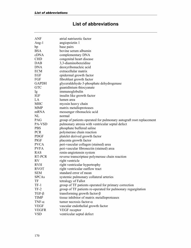
List of abbreviations
170
List of abbreviations ANF atrial natriuretic factor Ang-1 angiopoietin 1 bp base pairs BSA bovine serum albumin cDNA complementary DNA CHD congenital heart disease DAB 3,3-diaminobenzidine DNA deoxyribonucleic acid ECM extracellular matrix EGF epidermal growth factor FGF fibroblast growth factor GAPDH glyceraldehyde-3-phosphate dehydrogenase GTC guanidinium thiocyanate Ig immunoglobulin IGF insulin like growth factor LA lumen area MHC myosin heavy chain MMP matrix metalloproteases mRNA messenger ribonucleic acid NL normal PAG group of patients operated for pulmonary autograft root replacement PA-VSD pulmonary atresia with ventricular septal defect PBS phosphate buffered saline PCR polymerase chain reaction PDGF platelet derived growth factor PlGF placenta growth factor PVCA peri-vascular collagen (stained) area PVFA peri-vascular fibronectin (stained) area RAS renin-angiotensin system RT-PCR reverse transcriptase polymerase chain reaction RV right ventricle RVH right ventricular hypertrophy RVOT right ventricular outflow tract SEM standard error of mean SPCAs systemic pulmonary collateral arteries TF tetralogy of Fallot TF-1 group of TF patients operated for primary correction TF-2 group of TF patients re-operated for pulmonary regurgitation TGF-β transforming growth factor-β TIMP tissue inhibitor of matrix metalloproteases TNF-α tumor necrosis factor-α VEGF vascular endothelial growth factor VEGFR VEGF receptor VSD ventricular septal defect
Summary
171
Summary
Tetralogy of Fallot (TF) is one of the most common congenital heart disease which
can cause cyanosis. Chapter 1 provides a short overview about the main features and
the treatment of TF. Patients with TF suffer from pressure overload in the right
ventricle (RV) due to a (sub)pulmonary stenosis and a ventricular septal defect
(VSD). As an adaptive response the RV mass increases, known as right ventricular
hypertrophy (RVH). To reduce the chronic RV stress, primary corrective surgery at
young age (TF-1), nowadays is the treatment of choice. Correction is aimed at
prevention of the complications such as ventricular dysfunction and arrhythmias as a
result of long standing RVH. Despite adequate surgery, some patients develop RV
failure due to volume-overload (pulmonary regurgitation) and may require redo
surgery later in the life.
It is not yet established how the different adaptations of the RV in patients with TF
take place at a cellular and molecular level. This study aimed at getting a better
understanding of the pathogenesis in those patients in order to improve the timing of
surgical intervention and post-operative prognosis of those patients. By the
examination of RV biopsies obtained during corrective surgery of patients with TF an
attempt was made to correlate the RV overload with the phenotypic changes at a
cellular, protein and mRNA level. The study focussed on: myocardial fibrosis
(synthesis of connective tissue) (chapter 2-5), myocyte hypertrophy (chapter 4 and
5) and myocardial angiogenesis (vascularization of the RV) (chapter 6 and 7).
Chapter 2 describes a microscopic study of RV biopsies of TF patients, to examine
the degree of myocardial fibrosis as an adaptive response to the RV pressure overload.
After the immuno-histological staining of the fibrotic markers, collagen and
fibronectin, TF-1 and TF-2 patients were compared with patients who underwent a
pulmonary autograft root replacement (PAG). The results showed an increased level
of myocardial fibrosis in the extracellular space and peri-vascular regions and in some
cardiomyocytes.
Summary
172
In chapter 3 the content of fibrotic markers was quantified, with specific video image
analysis software, in digitalized microscopical pictures in the interstitial space and
peri-vascular area of different patients. This quantification revealed that the PAG
patients, which were used as a control, can not be regarded as such, despite clinically
normal RV function. Like TF patients they had increased levels of fibrosis markers in
the extracellular matrix (ECM) of the RV. The TF patients appeared to have higher
contents of interstitial collagens, but less peri-vascular collagen was found in patients
who got re-do surgery. This was accompanied by a lower amount of interstitial
fibronectin and increased levels of peri-vascular fibronectin.
In chapter 4 the total collagen, collagen type I, collagen type III and fibronectin
content in TF patients were quantified. Also the mRNA expression patterns of the RV
myocardium were quantified by DNA micro-array hybridization analysis. The image
analysis data revealed an increased total collagen, collagen type I, collagen type III
and fibronectin content in the interstitial space as well as in the peri-vascular area in
TF-1 and TF-2 patients as compared to the control group. This increase is confirmed
by the gene upregulation as found by micro-array hybridization analysis. Also the
gene-expression profile of genes encoding for proteins involved in the ECM homeostasis
(MMPs, TIMPs, PAI, PA etc.) supported the finding of an ongoing fibrosis. Furthermore
the increase of the myocyte cross sectional area with the age, indicated for myocardial
hypertrophy in patients with TF.
Chapter 5 describes a study of a sub-group of TF patients. Tetralogy of Fallot is a
generic name for patients with different degrees of anomalies. Pulmonary atresia
combined with a VSD is an extreme form of TF where the RV outflow tract is
completely blocked or absent. The myocardial hypertrophy in those patients is
accompanied by increased myocardial fibrosis in the interstitial space, although the
content is limited as compared to TF patients. At the same time the collagen support of
the blood vessels decreased, although there is an increased synthesis of collagen III (at
mRNA level) as compared to the control group.
In chapter 6 and 7 the level of angiogenesis was studied by assessing the growth
factors (e.g. VEGF) which stimulate the vascularization of the heart and the vascular
density. The gene-expression profile assessed with DNA-micro-array hybridization
Summary
173
revealed the pattern that supports the observed angiogenesis in patients with TF. Further
examination showed that the increased VEGF gene expression in the RV of TF patients
resulted in an increased RV vascularization, especially represented by small vessels
without vascular remodeling. The myocardial remodeling in TF patients appears to be
associated with an increased number of blood vessels to serve the RV in oxygen and
nutrient demand.
In chapter 8 the data represented in this thesis is evaluated in a general discussion.
Furthermore it describes proposals for future research.
Samenvatting
174
Samenvatting
Tetralogie van Fallot (TF) is een van de meest voorkomende aangeboren
hartafwijkingen die tot blauwzucht kan leiden. In hoofdstuk 1 wordt een kort
overzicht gegeven van de kenmerken van TF met een beschrijving van de huidige
behandeling. De hartafwijking wordt gekenmerkt door overbelasting van de rechter
hartkamer (RV; rechter ventrikel) die veroorzaakt wordt door een vernauwing in de
longslagader en een open verbinding tussen beide hartkamers (VSD; ventrikel septum
defect). Het hart past zich hieraan aan door de hartspier te verdikken (RVH; rechter
ventrikel hypertrofie). Om de nadelige gevolgen van langdurige overbelasting van de
RV te voorkomen, is tegenwoordig een correctieve operatie in het eerste levensjaar
(TF-1) de behandeling van keuze. Door middel van deze operatie wordt onder andere
geprobeerd om hartfalen en hartritmestoornissen op lange termijn te voorkomen.
Desondanks ontwikkelen sommige patiënten op volwassen leeftijd weer een RV
volume overbelasting als gevolg van een disfunctionerende pulmonalisklep. Deze kan
operatief worden vervangen om de opnieuw ontstane overbelasting van de RV te
verhelpen (TF-2).
De wijze waarop de aanpassingen in de hartspier bij de mens wordt gereguleerd op
cellulair en moleculair niveau is nog grotendeels onbekend. Het doel van deze studie is
om een beter inzicht te verkrijgen in het verloop van de pathogenese bij deze patiënten,
om zo nauwkeuriger het tijdstip van operatie te bepalen en om een beter post-operatieve
prognose te geven in de diverse stadia en vormen van TF. Met name door bestudering
van stukjes menselijk hartweefsel, verkregen tijdens de correctieve operatieve, is
geprobeerd om een verband te leggen tussen de mate van rechter kamerbelasting en de
mate waarin de expressie-patronen, die het moleculaire fenotype bepalen, op mRNA-,
eiwit- en cellulair niveau veranderd zijn. Er is bij de bestudering van deze stukjes
hartweefsel vooral gekeken naar: de mate van fibrose (bindweefsel vorming)
(hoofdstuk 2-5), de mate van hartspiercel hypertrofie (vergroting) (hoofdstuk 4 en 5)
en de mate van vascularisatie (bloedvatvorming) van de RV (hoofdstuk 6 en 7).
Samenvatting
175
De overbelasting in de rechter hartkamer van patiënten met TF, wordt o.a.
gecompenseerd door een toename van de concentratie van de fibrose markers collageen
en fibronectine. In hoofdstuk 2 worden deze aanpassingen microscopisch bestudeerd
na (immuno)histologische aankleuring van deze markers in de hartspierweefsel-
preparaten van de RV van TF-1 en TF-2 patiënten en vergeleken met preparaten van
patiënten die een “pulmonary autograft root replacement” (PAG) hebben ondergaan.
Hieruit kwam naar voren dat er een verhoogde mate van fibrose plaats vind met name
in de intercellulaire ruimte en rond de bloedvaten.
Door gebruik te maken van speciale beeldverwerkingsoftware kunnen de fibrose
markers in gedigitaliseerde microscopische opnamen in de interstitiële ruimte en rond
de bloedvaten, van de verschillende preparaten apart worden geanalyseerd. In
hoofdstuk 3 wordt hier nader op ingegaan. Hierbij kwam onder andere naar voren dat
de groep van PAG-patiënten, die als controle werden gebruikt, geen normale
morfologie had, ondanks een normale klinische RV functie. Net als de patiënten met TF
hadden ze een verhoogde afzetting van de fibrose markers in de extra-cellulaire matrix
(ECM). De patiënten met TF bleken een hogere concentratie interstitiële collagenen te
hebben maar een minder peri-vasculaire collageen. Tegelijkertijd hadden ze een lagere
concentratie interstitiële fibronectine en meer peri-vasculair fibronectine.
In een de studie, beschreven in hoofdstuk 4, worden naast de beeldanalyse van totaal
collageen en fibronectine ook de veranderingen in de hoeveelheid collageen subtypen I
en III bepaalt. Daarnaast zijn ook de expressie patronen van deze fibrose markers op
mRNA niveau geanalyseerd met behulp van DNA micro-array hybridisatie
experimenten. Met behulp van de beeldanalyse werd er een significante toename van de
fibrose markers totaal collageen, collageen subtype I en III en fibronectine in zowel de
interstitiële ruimte als rond de bloedvaten waargenomen bij zowel TF-1 als TF-2
patiënten, ten opzichte van de controle groep. Deze toename wordt bevestigd door de
bevindingen dat de genen die coderen voor extracellulaire matrix eiwitten in TF-1
patiënten zijn opgereguleerd. Het gen-expressie profiel laat verder zien dat de genen die
coderen voor eiwitten die de ECM homeostase reguleren, de fibrose te ondersteunen.
Verder bleek de mate van hypertrofie van de hartspiercellen in de RV van patiënten met
TF te zijn toegenomen en in het eerste levensjaar gecorreleerd te zijn met de leeftijd.
Samenvatting
176
In TF kan de obstructie van de RV ook compleet zijn. Er is een groep patiënten met
pulmonales atresia in combinatie met een VSD. In hoofdstuk 5 blijkt dat de fibrose in
deze patiënten samen gaat met hypertrofie van de hartspiercellen, maar in mindere mate
in vergelijking met andere TF patiënten. Daarnaast is er microscopisch een afname in
de collageen ondersteuning van de bloedvaten in vergelijking met de controle groep
waar te nemen, ondanks een verhoogde expressie collageen III (op mRNA niveau).
De remodelering van het hart gaat ook gepaard met een verhoogde aanmaak van
bloedvaten om in de zuurstof behoefte van de RV te blijven voorzien. In hoofdstuk 6
en 7 wordt de mate van angiogenese in patiënten met TF bestudeerd, door de aanmaak
van groeifactoren die aanzetten tot vascularisatie van het hart (o.a. VEGF) en de
bloedvat-dichtheid te bepalen. Uit het gen-expressie profiel, bepaald met behulp van
DNA-micro-array hybridisatie analyse, bleek duidelijk het dat de genen die betrokken
zijn bij de vorming van bloedvaatjes een verhoogde gen-expressie vertoonde. Verder
onderzoek wees uit dat de verhoogde VEGF expressie in de RV van patiënten met TF
resulterende in een toegenomen bloedvatdichtheid van de rechter hartkamer met
voornamelijk kleine bloedvaatjes, zonder dat er spraken is van bloedvat remodelering.
In hoofdstuk 8 worden de resultaten zoals beschreven in dit proefschrift in een
algemene discussie geëvalueerd en worden er aanbevelingen gedaan voor vervolg
onderzoek.
Dankwoord
177
Dankwoord
Aan het eind gekomen van dit proefschrift kom ik aan bij één van de meest gelezen
pagina’s. Waar de inhoud van de voorgaande bladzijden voor sommigen een
aanvulling op hun kennis zal zijn, zal dit voor anderen altijd een ondoorgrondelijke
materie blijven. Maar ik hoop dat we met dit onderzoek naar tetralogie van Fallot
weer een stukje in de goede richting zijn opgeschoven met als uiteindelijk doel de
patiënten beter te kunnen behandelen.
Gelukkig doe je zo’n onderzoek niet alleen. Veel mensen van zowel binnen als buiten
de universitaire wereld hebben mij hierbij geholpen. Ik wil hen allen hiervoor
bedanken. Maar enkele mensen die het nemen van de hobbels de afgelopen jaren van
dichtbij hebben meegemaakt wil ik toch graag met name noemen.
Als eerste wil ik de twee drijvende krachten achter het TF-project, Prof. dr. A. Bogers
en Dr. H. Sharma, bedanken voor het feit dat ik ondanks mijn afwezigheid op het
eerste sollicitatiegesprek toch als AIO ben aangenomen bij de vakgroepen
Farmacologie en Thoraxchirurgie.
Hari, als mijn dagelijkse begeleider en co-promotor, zijn we samen over pieken en
door dalen gegaan. Ik wil je bedanken voor alle raad en ondersteuning, leuke
congresbezoeken, je gedrevenheid en de vele uren die je in het onderzoek stak (Hari,
Dhanyavad).
Ad, als promotor en mede-initiator van het project wil ik ook jou bedanken voor de
prettige samenwerking, voor je kritische kijk, je onaflatende steun en de tijd die je
voor mij, ondanks je drukke werkzaamheden, toch steeds weer vrij wist te maken.
Daarnaast kon ik ook altijd bij mijn tweede promotor, Prof. dr. Saxena, terecht.
Pramod, ik wil je bedanken voor de getoonde interesse (ook al stond mijn
promotieonderwerp ver van de farmacologie af), de wetenschappelijke discussies en
niet te vergeten het wijzen op de tijdsplanningen.
Terugkijkend op de leuke momenten van mijn promotie kom ik ook zeker bij mijn
kamergenoten terecht. Emine, jij was nauw bij mijn onderzoek betrokken en hebt er
bergen werk voor verzet. Helaas was je er niet tot het einde bij maar mijn dank voor
de leuke samenwerking is er niet minder om. Ik wens je het beste voor de toekomst!
Dankwoord
178
Sue, schip ahoi! Het wordt weer drukker in de haven der promovendi. Bedankt voor
de leuke tijd, opbeurende verhalen en alle hulp op het lab. De didi’s Marieke,
Mechteld en Inge wil ik niet alleen bedanken voor de leuke tijd op de afdeling, maar
ook voor de zeer geslaagde reis naar het dak van de wereld. Maar ook met Marion,
Wenxia en Wendy was het uitstekend op één kamer, op “grote hoogte” boven
Rotterdam, uit te houden. Jullie allen bedankt voor alle hulp, vriendschap, de
gezellige momenten binnen en buiten de afdeling en natuurlijk de vele kilo’s snoep.
Edwin (Eddyman), Andor en Mark wil ik bedanken voor de ontspannende uurtjes
buiten werktijd. Maar ook mijn mede (CMBL) lab-genoten; Pankaj, Anna, Vijay,
Suneet, Joy, Oka en Sherif en de andere medewerkers van De Farma; Uday, Jan H,
Martin, Ingrid, Jasper, Richard, Peter, Beril, Antoinette, Saurabh, Ria, Roeland,
Magda, Rémon, Kapil, Larissa, Toos, Jeanette, Aloys, René, Wiro, Birgitte, Regien,
Corné, Freek, Jan D, Carlos en alle studenten wil ik bedanken voor de interesse, de
adviezen, de thee- en koffie-pauzes in de bibliotheek zonder boeken, de gezellige
(evaluerende) lunches, (de FECO) voor de zeer geslaagde labuitjes en de
terrasbezoeken (meestal) na werktijd. And for all of you who are still learning Dutch,
many thanks for the nice time in- and outside the department.
“Mijn” studenten Chantal en Lennart mogen ook zeker niet in dit dankwoord
ontbreken. Ik wil jullie bedanken voor de prettige samenwerking en jullie bijdrage aan
dit proefschrift (en de ontdekkingstocht door Canada). Inmiddels zijn jullie artsen,
maar ik hoop dat jullie nog niet helemaal van het onderzoeksvirus genezen zijn.
Buiten mijn standplaats bij de vakgroep Farmacologie hebben er ook verschillende
mensen bijgedragen aan de voortgang van mijn onderzoek. Zo hebben de
secretaresses van de afdeling Thoraxchirurgie, Saskia, Petra en Simone er voor
gezorgd dat de coördinatie tussen de twee locaties prima verliep en wij allen tijdig
werden opgetrommeld voor de werkbesprekingen. Ik zou geen resultaten hebben
gehad zonder het humane materiaal dat me ter beschikking werd gesteld door de
medewerkers van de Thorax-OK, de Hartkleppenbank en Dr. R. de Krijger. Jullie
hebben mij allen geholpen bij het verkrijgen van de juiste (controle) biopten. Deze
biopten werden verder “geprocessed” in het Cytologisch lab van de afdeling
Pathologie die mij ook regelmatig van advies voorzag. Daarnaast was Alex Nigg mij
altijd behulpzaam bij het ontwikkelen van de juiste beeldanalyse-programmatuur die
Dankwoord
179
ik kon gebruiken na de dagenlange opnamens achter de microscoop bij de afdeling
Longziekte. Ik wil jullie allen bedanken voor de ondersteuning en prettige
samenwerking.
Naast de praktische ondersteuning hebben anderen mij met hun vakkennis bijgestaan.
Hiervoor gaat mijn dank uit naar Rolf Berger en Peter de Jong voor hun bijdragen in
de discussies en het tot stand komen van het “PA-VSD” onderzoek, Walter Mooi voor
het beoordelen van de biopten en Adriana Gittenberger de Groot, Rob Poelmann en
Margo Bartelings die altijd bereid waren hun licht te laten schijnen op mijn
experimenten en bevindingen.
Het leven van een AIO speelt zich niet alleen af op de universiteit. Daarom wil ik ook
mijn familie en vrienden bedanken voor alle interesse en het aanhoren van m’n
klaagzangen als het (weer) eens fout ging. Zij hebben mij de afgelopen jaren af en toe
eens van het werk vandaan getrokken, mee naar een feestje of een gezellig weekendje
weg zodat ik weer met frisse moed verder kon.
De bewoners van het Boezemhuis wil ik ook in mijn dankwoord betrekken. Jullie
hebben mijn verblijf in Rotterdam zeker veraangenaamd. Bedankt daarvoor. Veel
succes in het afronden van jullie studies en op naar glansrijke carrières.
Conny, Dirk, Saskia en Rodolfo, bedankt voor de belangstelling en voor al het geduld
dat jullie voor me hebben gehad. Dat het promotieonderzoek niet altijd even
gemakkelijk ging hadden jullie al snel door maar toch hebben jullie me altijd
gesteund.
En natuurlijk “ons Pap en ons Mam”. Bedankt dat jullie me altijd hebben gesteund en
gestimuleerd om die dingen te doen die ik leuk vond. De deur in Sambeek stond altijd
voor me open. Bedankt voor alles!
Rotterdam, november 2003.
Curriculum vitae
180
Curriculum vitae Erik (Theodorus Henderikus Fransiscus) Peters, werd op 29 januari 1969 geboren te
Sambeek waar hij tevens zijn hele jeugd doorbracht. Na het behalen van het MAVO
en HAVO-diploma op de Scholengemeenschap Stevensbeek te Stevensbeek en zijn
VWO-diploma aan het Elzendaal College te Boxmeer ging hij in 1989 Moleculaire
Wetenschappen studeren aan de Landbouw Universiteit te Wageningen. Daar
oriënteerde hij zich op het dierlijke (cel) biotechnologie profiel. Na zijn afstudeer-
onderzoeken bij de sectie Moleculaire Cel Genetica (vakgroep Erfelijkheidsleer)
onder de dagelijkse begeleiding van Dr. M. Schalk en Prof. dr. C. Heyting en bij de
sectie Celbiologie en Immunologie (vakgroep Experimentele diermorfologie en
celbiologie) onder supervisie van Dr. M.J. van Lierop en Dr. R. Stet, mocht hij in juni
1996 zijn ingenieursdiploma in ontvangst nemen. Een half jaar later begon hij in
maart 1997 aan zijn promotie-onderzoek bij de vakgroepen Cardiothoracale Chirurgie
en Farmacologie aan de Erasmus MC, te Rotterdam, onder begeleiding van
promotoren Prof. dr. A.J.J.C. Bogers en Prof. dr. P.R. Saxena en co-promotor Dr. H.S.
Sharma. Het door de Nederlandse Hartstichting gefinancierde onderzoek richtte zich
op de “Molecular phenotype of right ventricular hypertrophy in patients with
tetralogy of Fallot”. De resultaten van dat onderzoek zijn vast gelegd in dit
proefschrift.
Publications
181
Publications
Full papers: Schalk JAC, Offenberg HH, Peters E, Groot NPB, Hoovers JMN and Heyting C.
Isolation and characterization of the human SCP2 cDNA and chromosomal
localization of the gene. Mamm Genome 1999; 642-644.
Peters THF, Sharma HS, Yilmaz E and Bogers AJJC. Quantitative analysis of
collagens and fibronectin expression in human right ventricular hypertrophy. Ann N Y
Acad Sci 1999; 874: 278-285.
Opgaard OS, Adner M, Peters THF, Xu CB, Stavenow L, Gulbenkian S, Erlinge D,
Edvinsson L and Sharma HS. Endocardial expression and functional characterization
of endothelin-1. Mol Cell Biochem 2001; 224: 151-158.
Klompe L, Peters THF, de Jong PL, Sharma HS and Bogers AJJC. Myocardial
characteristics in patients with pulmonary atresia and ventricular septal defect.
Thoraxcenter J 2002; 14:2, 33-36.
Peters THF, Sharma HS and Bogers AJJC. Computerized image analysis in the
quantitative assessment of interstitial fibrosis late after correction of tetralogy of Fallot.
Cardiovasc Eng 2003; 8: 114-120.
Peters THF, de Jong PL, Klompe L, Saxena PR, Berger RMF, Sharma HS and Bogers
AJJC. Right ventricular collagen and fibronectin levels in patients with pulmonary
atresia and ventricular septum defect. Mol Cell Biochem 2003; 251: 27-32.
Peters THF, Sharma HS, Yilmaz E, Saxena PR, and Bogers AJJC. DNA micro-array
and quantitative analysis reveal up-regulation of genes encoding extracellular matrix
proteins in patients with tetralogy of Fallot. (Submitted).
Peters THF, Bogers AJJC, Saxena PR and Sharma HS. Right ventricular hypertrophy in
human tetralogy of Fallot is associated with enhanced vascularization and expression of
vascular endothelial growth factor. (Submitted).
Publications
182
Chapters: Bogers AJJC, Peters THF, Hokken RB, Saxena PR and Sharma HS. Myocardial
phenotype of right ventricular hypertrophy in human tetralogy of Fallot. In: Imai Y,
Momma K. eds: Proceedings of 2nd World Congress of PCCS 1998; 746-749: Futura,
Armonk, New York, USA (ISBN: 0879936991).
Peters THF, Sharma HS, Yilmaz E, Saxena PR and Bogers AJJC. Quantitative
evaluation of interstitial fibrosis in patients with tetralogy of Fallot. In: Bogers AJJC,
Bartelings MM. Eds.: The Rotterdam-Leiden seminars on cardiovascular anatomy
and tetralogy 1997/1998/1999, 1999: 53-58, Erasmus University Rotterdam,
Rotterdam.
Bogers AJJC, Peters THF and Jong de PL. Tetralogy of Fallot, surgical aspects. In:
Bartelings MM, Wenink ACG eds: Congential Heart Disease. Boerhaave Committee
for Postgraduate Medical Education. 1999; 59-65: Leiden (ISBN 90-6767-411-7).
Sharma HS, Peters THF and Bogers AJJC. Angiogenesis and fibrosis during right
ventricular hypertrophy in human tetralogy of Fallot. In: Takeda N, Nagano M,
Dhalla NS eds: The hypertrophied heart. 2000; 227-241, Kluwer Academic
Publishers, Boston, USA (ISBN: 0-7923-7741-9).
Peters THF, Klompe L, Bogers AJJC and Sharma HS. Molecular phenotype of the
developing heart with a congenital anomaly. In: Ostadal B, Nagano M and Dhalla NS
eds.: Cardiac Development 2002; 195-212: Kluwer Academic Publishers, Boston,
USA (ISBN 1-40207-052-7).
Abstracts: Sharma, HS, Peters THF, Yilmaz E, Hokken RB and Bogers AJJC. Right ventricular
hypertrophy in human tetralogy of Fallot is associated with increased myocardial
fibrosis and vascularization. Circulation 1997; 96 (8): I.17-18.
Peters THF, Sharma HS, Yilmaz E and Bogers AJJC. Quantitative analysis of
collagens and fibronectin expression in human right ventricular hypertrophy.
Pathophysiol 1998; 5 (1): 5.
Publications
183
Sharma HS, Yoshida T, Maulink G, Peters THF and Dipack K. Increased myocardial
tolarance to ischemia-reperfusion injury in coenzyme Q10-fed pigs is associated with
induced expression of heme oxygenase-1 and ubiquitin. Pathophysiol 1998; 5 (1): 5.
Peters THF, Sharma HS, Yilmaz E, Saxena PR and Bogers AJJC. Right ventricular
hypertrophy in human tetralogy of Fallot is accompanied with the expression of
fibrosis markers. Fundam Clin Pharmacol 2000; 14: 54.
De Jong PL, Peters THF, Klompe L, Sharma HS and Bogers AJJC. No difference in
right ventricular myocardial fibrosis at correction of PA-VSD with or without SPCAs.
Cardiol Young 2001; 11 (3) supl. 1: 385.
Peters THF, Sharma HS, Saxena PR and Bogers AJJC. Right ventricular fibrosis and
apoptosis in patients with tetralogie of Fallot. J Mol Cell Card 2001; 33, (6) A93.
Klompe L, Peters THF, de Jong PL, Bogers AJJC and Sharma HS. Myocardial
collagen and fibronectin levels in patients with pulmonary atresia and ventricular
septal defect. J Mol Cell Card 2001; 33 (6) A60.
Sharma HS, Peters THF and Bogers AJJC. Right Ventricular failure in human
tetralogy of Fallot: micro-array and expression analysis. J Mol Cell Card 2002; 34 (6)
A89.
Peters THF, Bogers AJJC, Saxena PR and Sharma HS. DNA micro-array and
quantitative analysis revealed upregulation of genes encoding extracellular matrix
proteins in patients with tetralogie of Fallot. Circulation 2002; 106 (19): II-450.
Sharma HS, Peters THF and Bogers AJJC. DNA micro-array and quantitative
expression analysis of genes involved in human right ventricular hypertrophy. J Mol
Cell Card 2003; 35 (6) A53.
Sharma HS, Peters THF, Verdouw PD and Bogers AJJC. Genetic reprogramming
during ischemia/reperfusion induced myocardial cytoprotection. Exp Clin Cardiol
2003; 8 (1) A49.
