rescue of bacteriophage t7 dna polymerase of low
TRANSCRIPT

JOURNAL OF VIROLOGY, Sept. 2009, p. 8418–8427 Vol. 83, No. 170022-538X/09/$08.00�0 doi:10.1128/JVI.00855-09Copyright © 2009, American Society for Microbiology. All Rights Reserved.
Rescue of Bacteriophage T7 DNA Polymerase of Low Processivity bySuppressor Mutations Affecting Gene 3 Endonuclease�
Seung-Joo Lee, Kajal Chowdhury,† Stanley Tabor, and Charles C. Richardson*Department of Biological Chemistry and Molecular Pharmacology, Harvard Medical School, Boston, Massachusetts 02115
Received 27 April 2009/Accepted 11 June 2009
The DNA polymerase encoded by gene 5 (gp5) of bacteriophage T7 has low processivity, dissociating afterthe incorporation of a few nucleotides. Upon binding to its processivity factor, Escherichia coli thioredoxin(Trx), the processivity is increased to approximately 800 nucleotides per binding event. Several interactionsbetween gp5/Trx and DNA are required for processive DNA synthesis. A basic region in T7 DNA polymerase(residues K587, K589, R590, and R591) is located in proximity to the 5� overhang of the template strand.Replacement of these residues with asparagines results in a threefold reduction of the polymerization activityon primed M13 single-stranded DNA. The altered gp5/Trx exhibits a 10-fold reduction in its ability to supportgrowth of T7 phage lacking gene 5. However, T7 phages that grow at a similar rate provided with eitherwild-type or altered polymerase emerge. Most of the suppressor phages contain genetic changes in or aroundthe coding region for gene 3, an endonuclease. Altered gene 3 proteins derived from suppressor strains showreduced catalytic activity and are inefficient in complementing growth of T7 phage lacking gene 3. Results fromthis study reveal that defects in processivity of DNA polymerase can be suppressed by reducing endonucleaseactivity.
The efficient replication of the chromosome of bacterio-phage T7 is accomplished by a replisome consisting of rela-tively few components, thus making it an excellent model sys-tem for studies on DNA replication. Phage T7 encodes most ofthe essential proteins required for DNA replication includinggene 5 DNA polymerase (gp5), gene 4 helicase-primase, andgene 2.5 single-stranded DNA (ssDNA) binding protein (SSB)(39). One protein, thioredoxin (Trx), is encoded by the hostEscherichia coli and serves as the processivity factor for T7DNA polymerase. The three-dimensional structures of all fourproteins have been determined (10, 20, 21, 43). Within thereplisome, the helicase domain located in the C-terminal halfof the gene 4 protein unwinds the double-stranded DNA(dsDNA) to provide an ssDNA template for the continuoussynthesis of the leading strand by the gp5/Trx complex. Thesingle-stranded lagging strand extruded behind the helicase isprotected by the gene 2.5 protein until the primase domainlocated in the N-terminal half of the gene 4 protein synthesizesprimers at primase recognition sites to initiate lagging-strandsynthesis. The T7 replisome, reconstituted from these fourproteins, mediates the replication of duplex DNA in a reactionwhere leading and lagging strands are coordinated (29). Coor-dination is achieved by the formation of a replication loop ofthe lagging strand containing the nascent Okazaki fragment.
A key component of the T7 replisome is the gp5/Trx com-plex. Like most other prokaryotic DNA polymerases, it con-sists of a C-terminal polymerase domain and an N-terminal
exonuclease domain (1, 28). As a member of the polymerase Ifamily of DNA polymerases, it resembles a partially closedright hand with a thumb, palm, and fingers (Fig. 1A). TheDNA binding within a crevice formed by the thumb, palm, andfingers leads to the catalytic site where the 3�-hydroxyl termi-nus of the primer strand is extended by the condensation of theproperly positioned nucleoside triphosphate (NTP) in the nu-cleotide binding site. A distinguishing feature of T7 DNApolymerase is the presence of a unique segment of 76 residuesat the tip of the thumb region; this segment is not present inother homologs of the polymerase I family. Trx binds to thisregion, designated the Trx-binding domain, with high affinity(25). The presence of Trx increases the processivity of thepolymerase from the incorporation of 1 to 50 nucleotides toapproximately 800 nucleotides per binding event (44). Twobasic loops in the Trx-binding domain interact with the acidicC termini of both gene 4 and gene 2.5 proteins, suggesting thatthis region plays a regulatory role (18).
Several X-ray crystallographic structures of gp5/Trx in com-plex with a primer-template and a nucleoside 5� triphosphateare available (2, 10, 12). The double-stranded portion of theprimer-template in these structures is at the transition fromB-form to A-form, implying significant distortions during ca-talysis (10). The DNA is held securely through numerous con-tacts with the polymerase where the 5� end of the primerinteracts with the thumb region and the 3� end interacts withthe fingers as well as the palm region (10). Such interactionsfacilitate alignment of the 3�-hydroxyl end of the primer to theactive site of the enzyme where catalytically crucial residuesreside. Further interactions with the incoming deoxy NTP(dNTP) induce conformational changes in the finger region toinsure that only a properly base-paired dNTP can be incorpo-rated (9). Thus, the structural environment around the activesite is critical for efficient incorporation of the correct nucleo-tide.
* Corresponding author. Mailing address: Department of BiologicalChemistry and Molecular Pharmacology, Harvard Medical School, 240Longwood Ave., Boston, MA 02115. Phone: (617) 432-1864. Fax: (617)432-3362. E-mail: [email protected].
† Present address: Foley and Lardner LLP, 11250 El Camino Real,Suite 200, San Diego, CA 92130.
� Published ahead of print on 17 June 2009.
8418
by on August 10, 2009
jvi.asm.org
Dow
nloaded from

Whereas the dsDNA region of the primer-template resideswithin a crevice formed by the thumb and fingers of the poly-merase, the 5� ssDNA overhang of the template strand liesoutside the fingers. This region is less defined for interactionwith the polymerase in the crystallographic structure. The 5�
nucleotides in the template strand adjacent to that hydrogenbonded with the incoming nucleotide are flipped out of theactive site, and the last two 5�-terminal bases of the templateare disordered (10). Clearly, however, this region of the tem-plate strand must be firmly anchored in order for the incoming
FIG. 1. Interaction of T7 gp5/Trx complex with primer-template DNA. (A) Structure of primer-template-bound T7 gp5/Trx complex. Apartially closed right-hand-shaped polymerase domain of gp5 (shown in surface model) in complex with E. coli Trx (gray ribbon model) (10). Trxis bound to a unique segment in the thumb subdomain of the polymerase. The primer-template (orange lines) resides in the DNA binding cleftcreated by the fingers, thumb, and palm. Colors in the polymerase represent electrostatic potential of residues exposed to the surface: red (acidic)and blue (basic). Four basic residues (K587, K589, R590, and R591) forming a positively charged patch nearby the 5� end of the template strandare indicated. (B) Leading-strand DNA synthesis at replication fork. As the helicase domain of T7 gene 4 protein generates ssDNA template byunwinding parental dsDNA, gp5/Trx continuously synthesizes leading-strand DNA by extending primer in 5� to 3� direction. TBD, Trx-bindingdomain; aa, amino acid.
VOL. 83, 2009 T7 GENE 3 SUPPRESSOR 8419
by on August 10, 2009
jvi.asm.org
Dow
nloaded from
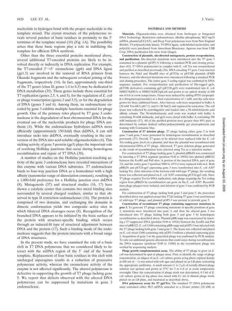
nucleotide to hydrogen bond with the proper nucleotide in thetemplate strand. The crystal structure of the polymerase re-veals several patches of basic residues in proximity to the 5�terminus of the template strand (10) (Fig. 1A). The possibilityarises that these basic regions play a role in stabilizing thetemplate for efficient DNA synthesis.
Other than the three essential proteins mentioned above,several additional T7-encoded proteins are likely to be in-volved directly or indirectly in DNA replication. For example,the T7-encoded 5�33� exonuclease (gp6) and DNA ligase(gp1.3) are involved in the removal of RNA primers fromOkazaki fragments and the subsequent covalent joining of thefragments, respectively (14). In fact, approximately one-thirdof the T7 genes (class II; genes 1.4 to 6.3) may be dedicated toDNA metabolism (35). These genes include those essential forT7 replication (genes 2.5, 4, and 5), for the regulation of E. colior phage transcription (genes 2 and 3.5), or for the degradationof DNA (genes 3 and 6). Among them, an endonuclease en-coded by gene 3 exhibits multiple functions essential for phagegrowth. One of the primary roles played by T7 gene 3 endo-nuclease is the degradation of host chromosomal DNA for theeventual use of the nucleotide products for phage DNA syn-thesis (4). While the endonuclease hydrolyzes ssDNA moreefficiently (approximately 150-fold) than dsDNA, it can stillintroduce nicks into dsDNA, eventually resulting in the con-version of the DNA into acid-soluble oligonucleotides (4). Thisnicking activity of gene 3 protein (gp3) plays the important roleof resolving Holliday junctions that occur during homologousrecombination and repair of T7 DNA (30).
A number of studies on the Holliday junction-resolving ac-tivity of the gene 3 endonuclease have revealed interactions ofthis enzyme with various DNA substrates. T7 endonucleasebinds to four-way junction DNA as a homodimer with a highaffinity (nanomolar range of dissociation constant), resulting ina 1,000-fold preference for a Holliday junction over dsDNA(8). Mutagenesis (37) and structural studies (16, 17) haveshown a catalytic center that contains two metal binding sitessurrounded by several charged residues, similar to that ob-served in type II restriction endonucleases (16). The protein iscomprised of two domains, and exchanging the domains indimeric conformation yields two composite active sites inwhich bilateral DNA cleavages occur (8). Recognition of thebranched DNA appears to be initiated by the basic surface ofthe protein with structure-specific binding, which occursthrough an induced-fit type of structural adjustment betweenDNA and the protein (17). Such a binding mode of the endo-nuclease suggests that the protein interacts with a broad rangeof DNA structures.
In the present study, we have examined the role of a basiccleft in T7 DNA polymerase that we considered likely to in-teract with the ssDNA region of the 5� end of the boundtemplate. Replacement of four basic residues in this cleft withuncharged asparagines results in a reduction of processivepolymerase activity, whereas the exonuclease activity of theenzyme is not affected significantly. The altered polymerase isdefective in supporting the growth of T7 phage lacking gene5. We report that defects observed with the altered DNApolymerase can be suppressed by mutations in gene 3endonuclease.
MATERIALS AND METHODS
Materials. Oligonucleotides were obtained from Invitrogen or IntegratedDNA Technology. Restriction endonucleases, alkaline phosphatase, M13 mp18ssDNA, plasmid pUC(AT), and Deep Vent polymerase were from New EnglandBiolabs. T4 polynucleotide kinase, T4 DNA ligase, radiolabeled nucleotides, andpoly(dA) were purchased from Amersham Bioscience. Agarose was from USBCorp. DNA purification kits were from Qiagen.
Construction of plasmids, site-directed mutagenesis, protein overproduction,and purification. Site-directed mutations were introduced into the T7 gene 5contained in a plasmid (pGP5-3) following a standard PCR and cloning proce-dure (42). T7 DNA polymerases in complex with E. coli Trx was overproducedand purified as described previously (34). DNA encoding T7 gene 3 was insertedbetween the NdeI and HindIII sites of pET24a or pET28b plasmids (EMDScience), and site-directed mutations were introduced following a standard PCRand cloning procedure. The entire gene 3 coding region was confirmed by DNAsequence analysis. For overproduction and purification of His-tagged gp3s,pET28b derivatives containing gp3 (pET28-gp3) were transformed into E. coliHMS174(DE3) or HMS174(DE3)pLysS and grown to an optical density at 600nm of 0.6 at room temperature. Genes were induced by adding IPTG (isopropyl-�-D-thiogalactopyranoside) at a final concentration of 1 mM, and the cells weregrown for three additional hours. After harvest, cells were suspended in buffer A(20 mM Tris-HCl, pH 7.5, and 0.1 M NaCl) and ruptured by sonication. The celllysate was collected by centrifugation and loaded onto a Ni-nitrilotriacetic acidaffinity column. The Ni-nitrilotriacetic acid resin was washed with buffer Acontaining 50 mM imidazole, and gp3s were eluted with buffer A containing 500mM imidazole (37). All of the purified proteins were greater than 90% pure asdetermined by sodium dodecyl sulfate-polyacrylamide gel electrophoresis andstaining with Coomassie blue.
Construction of T7 deletion phage. T7 phage lacking either gene 3 or bothgene 3 and gene 5 were generated by homologous recombination as describedpreviously (27). Overall, T7 genes to be deleted were replaced with the E. coliTrx-coding gene (trxA) in a plasmid, and then the plasmid was recombined withchromosomal DNA of T7 phage. Afterward, T7 gene deletion phage generatedas the result of recombination were selected using Trx as a selection marker.
For construction of T7 phage lacking gene 3, plasmid pBRd3t was constructedby inserting a T7 DNA segment (position 9148 to 10926) into plasmid pBR322between the EcoRI and PstI sites. A portion of the inserted DNA, part of gene2.8, and the entire gene 3 (position 9880 to 10711) were replaced with trxA. BothpBRd3t and pET24-gp3 expressing gene 3 were transformed into E. coli A307lacking Trx. After infection of the bacteria with wild-type T7 phage, the resultinglysate was collected and plated on E. coli A307 containing pET24-gp3 only. SinceT7 phage requires Trx for DNA replication, only phage acquiring the Trx codingregion from pBRd3t by recombination can grow on the E. coli A307. Recombi-nant phage plaques were isolated, and deletion of gene 3 was confirmed by PCRanalysis.
For construction of T7 phage lacking both gene 3 and gene 5, the proceduredescribed above was applied except that T7�5 lacking T7 gene 5 was used insteadof wild-type T7 phage, and plasmid pGP5-3 was present to provide gene 5.
Construction of recombinant T7 phage containing suppressor mutations ingene 3. To generate T7 phage containing mutations in specific positions of gene3, mutations were introduced into gene 3, and then the altered gene 3 wasintroduced into T7 phage lacking both gene 3 and gene 5 by homologousrecombination as described above. Plasmid pBR-supp was constructed by insert-ing a T7 suppressor DNA (position 9148 to 10926) between the EcoRI and PstIsites of pBR322. E. coli C600 containing pBR-supp and pGP5-3 was infected withthe T7 phage lacking both gene 3 and gene 5. The lysate was collected and platedon E. coli strain C600 containing only pGP5-3 without a plasmid expressing gene3. Acquisition of gene 3 in the generated phage was confirmed by PCR analysis.To rule out additional genetic alterations that could occur during recombination,the DNA sequence (positions 9148 to 11060) in the recombinant phage wasverified by sequencing analysis.
Phage growth complementation assay. The ability of T7 phage to grow on E.coli was determined by spot or plaque assay. First, to identify the range of phageconcentration, an aliquot of an E. coli culture grown at log phase (optical densityat 600 nm of �1) was mixed with soft agar and plated on an LB plate containingthe appropriate antibiotics. A small amount (1 to 2 �l) of serially diluted phagesolution was spotted and grown at 37°C for 5 to 6 h or at room temperatureovernight. Once the concentration of phage stock was determined, 0.3 ml of E.coli culture grown at log phase was mixed with 0.1 ml of diluted phage stock,plated on an LB plate, and incubated as described above.
DNA polymerase assay for T7 gp5/Trx. The standard T7 DNA polymeraseassay contained either M13 ssDNA annealed to a 24-mer primer (20 nM) or
8420 LEE ET AL. J. VIROL.
by on August 10, 2009
jvi.asm.org
Dow
nloaded from

poly(dA)390 annealed to poly(dT)25 (200 nM); 0.25 mM each of [�-3H]dTTP (5cpm/pmol), dATP, dCTP, and dGTP; 40 mM Tris-HCl, pH 7.5; 10 mM MgCl2;10 mM dithiothreitol (DTT); 50 mM potassium glutamate; and the amounts ofT7 gp5/Trx indicated in the figures. Incubation was for 10 min at 37°C for theprimed M13 template or 5 min at room temperature for the poly(dA)/oligo(dT).After the reaction was terminated by the addition of EDTA to a final concen-tration of 20 mM, aliquots were spotted onto DE-81 filters (Whatman) andwashed three times with ammonium formate. The amount of nucleotide incor-porated by gp5/Trx was determined by measuring the radioactivity retained onthe filters.
For an analysis of the products obtained from DNA synthesis reactions usingprimed M13 ssDNA, the primer was radiolabeled at its 5� end with 32P usingpolynucleotide kinase. After incubation at 37°C for 20 min, the reaction productswere separated on 0.6% agarose gel containing ethidium bromide (0.3 �g/ml)and analyzed by autoradiography.
To examine the effect of E. coli SSB protein on polymerase activity, 20 nMprimed M13 ssDNA; 4 nM T7 gp5/Trx; and 0.25 mM each of [�-3H] dTTP (5cpm/pmol), dATP, dCTP, and dGTP were incubated in the presence or absenceof 10 �M E. coli SSB protein at 37°C for the time period indicated in the figures.Incorporation of [3H]dTMP was determined as described above.
Exonuclease assay for T7 gp5/Trx. Exonuclease activity of T7 DNA polymer-ase was measured using uniformly 3H-labeled ss- or dsDNA prepared as de-scribed previously (26). The assay contained either 2 nM dsDNA or 1.2 nMssDNA, 40 mM Tris-HCl, pH 7.5, 10 mM MgCl2, 10 mM DTT, 50 mM potassiumglutamate, and the amounts of T7 gp5/Trx indicated in the figures. Incubationwas at 37°C for 10 min. The radioactivity remaining in DNA after incubation wasdetermined using a DE-81 filter as described above.
Endonuclease assay for T7 gene 3 endonuclease. The standard assay for gene3 endonuclease contained the indicated DNA (30 nM), 50 mM Tris-HCl, pH 7.9,10 mM MgCl2, 1 mM DTT, 100 mM NaCl, and the amount of gene 3 endonu-clease indicated in the figures. After incubation at 37°C for 20 min, the reactionproducts were separated on 1% agarose gel containing ethidium bromide. Theamount of hydrolysis of the DNA was determined by measuring the intensity ofDNA bands using an Alpha Imager3400.
RESULTS
As the DNA helicase bound to the lagging strand unwindsthe duplex DNA at the replication fork, the newly createdssDNA template is copied by the DNA polymerase bound tothe leading strand (Fig. 1B). Although the length of the ssDNAtemplate at the replication fork is not known, it may be dic-tated by the spatial arrangement of the helicase and polymer-ase. It seems likely that this stretch of template strand is boundto the polymerase so as to stabilize the template for properbase-pairing of the incoming NTP. Several crystallographicstructures of T7 gp5/Trx in complex with primer-template il-lustrate the interaction of the polymerase with DNA (2, 10,12), particularly at the active site where the incoming dNTP islocated. However, only a few nucleotides of the ssDNA tem-plate in these studies diffract, the remaining region probablybeing disordered. Inspection of the structure of T7 DNA poly-merase reveals a patch of four basic residues (K587, K589,R590, and R591) located in a region that has the potential tointeract with the 5� end of the template (Fig. 1A). In order toinvestigate the role of this basic region on polymerase activity,we replaced these residues with asparagines having a neutralside chain and examined the effect of these changes on T7growth and on DNA polymerase activity. The altered gp5 (gp5-4N) as well as the wild-type protein was purified as a complexwith Trx and used for biochemical assay.
DNA polymerase activity of altered gp5/Trx. We first exam-ined the ability of the altered DNA polymerase, gp5-4N/Trx, toincorporate deoxynucleoside monophosphate (dNMP) usingpoly(dA) template (average, 390-mer) to which an oligo(dT)primer of 25 nucleotides was annealed. The altered polymer-
ase exhibits polymerase activity similar to that of wild-type T7gp5/Trx (Fig. 2A). The results indicate that alteration at thisregion of the polymerase does not affect its catalytic activity asmeasured by its ability to add nucleotides to the 3�-OH of theprimer strand. However, when primed M13 ssDNA was used inthe assay, gp5-4N/Trx exhibited threefold lower activity thanthe wild-type protein (Fig. 2B). Analysis of the reaction prod-ucts on agarose gel reveals that a low level of incorporation ofdNMP by the altered polymerase is due to lower processivitythan that found with the wild-type protein (Fig. 2C). The lowprocessivity is reflected by the presence of primers not ex-tended, the absence of fully replicated M13 dsDNA, and thepresence of large amounts of aborted product of low mobility.
We have previously shown that secondary structures in M13ssDNA can hamper progression of T7 gp5/Trx and that theaddition of E. coli SSB protein can resolve the pausing of thepolymerase (26). Indeed, addition of E. coli SSB protein en-hanced incorporation of dNMP by both DNA polymerases(Fig. 2D). However, the altered polymerase still exhibitedlower activity than the wild-type enzyme.
Exonuclease activity of altered gp5/Trx. Like other DNApolymerases in the polymerase I family, T7 gp5 contains anexonuclease domain located in the N terminus of the protein.The exonuclease is active on both ssDNA and dsDNA, withTrx stimulating the latter (23). gp5-4N/Trx has somewhat lessexonuclease activity on both ds- and ssDNA (80 and 60%,respectively) than the wild-type enzyme (Fig. 3). Again, thelower activity with gp5-4N/Trx may reflect its lower processiv-ity, a parameter that affects exonuclease activity (25, 44).
Complementation for growth of T7 phage lacking gene 5.The effect of the alteration of T7 DNA polymerase on its invivo function was examined by phage complementation assay.In this assay, growth of T7 phage lacking gene 5 is dependenton DNA polymerase exogenously expressed by a plasmid in theinfected cell harboring functional gene 5. The replacement ofthe four basic residues with asparagines in gp5-4N results in areduction of its ability to complement growth of the deletionphage by more than 10-fold (Fig. 4, right). The plaques pro-duced by the infection are 10-fold smaller than those obtainedwhen the wild-type protein is used for complementation (datanot shown). Consistent with the biochemical data, the in vivocomplementation assay indicates that the alteration severelyreduces gp5 function.
Isolation of suppressor phage and analysis of geneticchanges. From the above data, it is clear that the alteration ofpolymerase yields significant defects of in vivo function of thepolymerase: reduction in both the ability to complementgrowth of the gene 5-deletion phage and the size of plaques.However, some plaques isolated from the culture with thealtered polymerase yielded phage that produced larger plaquesizes upon replating on E. coli expressing gp5-4N. Those iso-lated suppressor phages grow with gp5-4N as efficiently as withthe wild-type protein (Fig. 4).
To identify the genetic changes responsible for the gain offunction in the complementation assay, we selected 10 sup-pressor phages from three independent trials. Since class IIgenes of the T7 genome (gene 1.4 to gene 6.3) are known to beinvolved in DNA metabolism, we determined the DNA se-quence of DNA isolated from these suppressor phages. Assummarized in Table 1, two suppressors out of a total of the 10
VOL. 83, 2009 T7 GENE 3 SUPPRESSOR 8421
by on August 10, 2009
jvi.asm.org
Dow
nloaded from

phages examined do not have any mutation in the class IIgenes. The remaining eight suppressors contain genetic alter-ations in or around gene 3. Three of the suppressors havesingle (suppressors 1 and 4) or double (suppressor 5) substi-tutions in gene 3, two of them have frameshifts in gene 2.8
(suppressors 6 and 7), immediately upstream of gene 3, and theremaining three contain deletions between gene 3.5 and 3.8(suppressors 8, 9, and 10), downstream of gene 3. Five phagescontaining either frameshift or an intergenic deletion appearto originate from the same parental suppressor since they have
FIG. 2. Polymerase activity of gp5-4N/Trx. (A) DNA synthesis on poly(dA)/oligo(dT) template. The reaction mixture described in Materialsand Methods contained 200 nM poly(dA)390 annealed with oligo(dT)25; 0.25 mM each of 3H-labeled dTTP (5 cpm/pmol), dGTP, dATP, and dCTP;and the indicated amounts of gp5-4N/Trx or wild-type (wt) gp5/Trx. After incubation at room temperature for 5 min, the activity of DNApolymerase was determined by measuring the incorporation (incorp) of [3H]dTMP into DNA. (B) DNA synthesis on primed M13 ssDNA. Thereaction mixture was similar to that described for panel A except that 20 nM M13 ssDNA annealed to a primer was used as a template insteadof poly(dA)/oligo(dT). Reaction mixtures were incubated at 37°C for 10 min. (C) Products from DNA synthesis on primed M13 ssDNA template.Radioactively end-labeled 5� 32P-labeled 24-mer primer was annealed to M13 ssDNA and incubated with increasing amounts of gp5/Trx (0.3, 0.5,1, 2, 4, and 8 nM) at 37°C for 20 min. Reaction products were analyzed on 0.6% agarose gel and dried for autoradiography. Locations of the initialtemplate and full-length product M13 dsDNA are indicated. (D) Effect of E. coli SSB protein on DNA synthesis by gp5/Trx. Reaction mixturecontained 20 nM primed M13 ssDNA template, 4 nM gp5/Trx, and 0.25 mM all four dNTPs (5 cpm/pmol [3H]dTTP). The reaction mixture wasincubated in the absence or presence of 10 �M E. coli SSB protein at 37°C for the indicated time period. Amount of incorporated [3H]dTMP intoDNA was measured.
FIG. 3. Exonuclease activity of T7 DNA polymerase. Either 2 nM M13 3H-labeled dsDNA (A) or 1.2 nM M13 3H-labeled ssDNA (B) wasincubated in the reaction mixture described in Materials and Methods with the indicated concentration of either wild-type (wt) gp5/Trx orgp5-4N/Trx at 37°C for 10 min. Exonuclease activity was determined by measuring the radioactive DNA that bound to a DE-81 filter.
8422 LEE ET AL. J. VIROL.
by on August 10, 2009
jvi.asm.org
Dow
nloaded from

the same sequence. Since the majority of suppressor phageshave changes that may affect gene 3 function or expression, wechose the simplest alterations in gene 3, three substitutions(suppressors 1, 4, and 5) for further analysis.
Confirmation of gene 3 alteration in suppressor phage. Toestablish that mutations in gene 3 are the sole determinant forsuppression of the defect in gp5-4N, we constructed T7 phagelacking gene 5 but containing point mutations in gene 3 usinghomologous recombination. The recombinant phage con-tained single- or double-amino acid substitutions in gene 3identified from the suppressor DNA sequences. These recom-binant phages lacking gene 5 had similar growth when eitherwild-type gp5 or gp5-4N was provided from plasmids (data notshown). These results establish that mutations in gene 3 areresponsible for the observed phenotype.
Complementation of phage lacking gene 3. The effect of themutations in gene 3, identified from suppressor phages, onphage growth was examined by a phage growth complementa-tion assay. All of the mutations lead to gp3s that are defectivein their ability (5- to 30-fold reduction in plating efficiency) to
complement T7 phage lacking gene 3 (T7�3) (Table 2). Thesize of plaques produced by expression of the altered gp3s wasalso significantly smaller than the size produced by the wild-type protein (about threefold). We also examined the effect ofalteration of two residues (E20 and D55) previously identifiedas critical factors for catalytic activity (8) on the ability tocomplement T7 phage lacking gene 3. Alteration of thoseresidues (E20Q or D55N) also resulted in proteins with re-duced ability to complement T7�3 as well as the production ofsmall plaques (Table 2). However, these latter reductions ingene 3 function were more severe than those observed with thealterations identified in the suppressor analysis.
Endonuclease activity of gp3s. Gene 3 of bacteriophage T7is essential for phage viability (5). Gene 3 encodes an endo-nuclease that is 100-fold more active on ssDNA than ondsDNA (4). A major role of the gene 3 endonuclease is, inconcert with the gene 6 exonuclease, to degrade the host DNAso that the resulting nucleoside 5� phosphates can be used forphage DNA synthesis (35). It also participates in homologousrecombination by cleaving Holliday junctions (30). In order toexamine the endonuclease activity of the gp3s arising fromsuppressor mutations, we purified them, along with the wild-type protein, as His-tagged recombinant proteins. The pres-ence of the His tag on the N terminus of the endonuclease doesnot alter gp3 function, as judged by the ability of the modifiedgene to support growth of T7 phage lacking gene 3 (data notshown).
Gene 3 endonuclease hydrolyzes AT-rich regions with highefficiency (15). As shown in Fig. 5A, wild-type endonucleaserapidly hydrolyzes a supercoiled plasmid containing a stretchof alternating AT sequence (�20 bp) to the linear or nickedform. All three of the endonucleases derived from gene 3harboring suppressor mutations exhibited reduced activity,with the most severe defect (10-fold reduction) observed withsuppressor 1. Nucleolytic digestion on a normal dsDNA plas-mid, pUC19 (Fig. 5B), or nicked dsDNA plasmid pUC19 (Fig.5C) requires more endonuclease to achieve a similar level ofdigestion (Table 3). Again, all altered proteins exhibit lowercatalytic activity than that found with the wild-type protein
FIG. 4. In vivo complementation of phage by T7 gp5. Serial dilu-tions of either T7 gene 5-deletion phage or suppressor phages werespotted onto a LB plate containing E. coli expressing the indicated gp5.After incubation at 37°C overnight, the ability to lyse E. coli wascompared. Sup, suppressor.
TABLE 1. Genetic alterations found in T7 suppressor phagesa
Trial Suppressorno.
Geneticalteration (nt)b Outcome in gene expression
A 1 T10422C gp3 F56LB 2
3C 4 A10482G gp3 K76E
5 A10575G gp3 T107AT10666C gp3 V137A
6 �10132 Frameshift in gene 2.87 �10132 Frameshift in gene 2.88 �11195–11196 Deletion in noncoding region
between genes 3.5 and 3.89 �11195–11196 Deletion in noncoding region
between genes 3.5 and 3.810 �11195–11196 Deletion in noncoding region
between genes 3.5 and 3.8
a A total 10 suppressor phages were isolated from three independent trials,and their DNA sequences in class II genes (gene 1.4 to gene 6.3) were analyzed.Genetic changes found from the phages and their consequences in gene expres-sion are indicated.
b nt, nucleotide.
TABLE 2. Complementation of T7 gene 3-deletion phage growthby recombinant proteinsa
Protein expressedb Alteration Efficiency of platingc
None 0.01*gp5 wt wt 6 � 104*gp5-4N K587N, K589N, R590N,
R591N5 � 104*
gp3 wt wt 1.0gp3 (Supp 1) F56L 0.1*gp3 (Supp 4) K76E 0.2*gp3 (Supp 5) T107A, V137A 0.03*gp3 (E20Q) E20Q 0.003*gp3 (D55N) D55N 0.007*
a Recombinant proteins were expressed from plasmid under the control of aT7 promoter in E. coli C600. After infection with T7 gene 3-deletion phage(T7�3), the number of plaques were counted, normalized to the value obtainedwith wild-type gene 3, and results are presented as efficiency of plating. Datawere obtained from at least duplicated experiments. E. coli cells expressing noprotein or gp5 were used as negative controls. wt, wild-type; Supp, suppressor.
b Alterations/mutations are given in parentheses.c *, small size of plaque.
VOL. 83, 2009 T7 GENE 3 SUPPRESSOR 8423
by on August 10, 2009
jvi.asm.org
Dow
nloaded from

(Fig. 5B and C). The most significant reduction of activity wasobserved with suppressor 1. The amount of each enzyme re-quired to convert approximately 50% of the initial DNA intohydrolyzed forms is presented in Table 3. Catalytically inactiveendonucleases, gp3 with the mutation E20Q (gp3-E20Q) andgp3-D55N, have severe reductions in activity, far more thanenzymes derived from the suppressors.
General effect of suppressor-derived gene 3 mutations ongp5/Trx having low processivity. Is the ability of gene 3 endo-nuclease having reduced activity to suppress the phenotype ofgp5-4N specific for the alterations in the basic patch of T7 gp5
or for low processivity of gp5/Trx in general? We have exam-ined the ability of the gene 3 endonuclease mutants to suppressthe defect in growth of T7 phage arising from another DNApolymerase of low processivity.
Gene 5 DNA polymerase normally forms a tight complexwith its processivity factor E. coli Trx via a hydrophobic inter-action (24). If E. coli Trx is covalently linked to gp5 through adisulfide bond (T327C in gp5 and C32 in Trx), the processivityis reduced (26). This DNA polymerase, in covalent complexwith Trx, has a 100-fold reduction in its ability to complementthe growth of T7 phage lacking gene 5 (26), considerably
FIG. 5. Catalytic activity of gene 3 endonuclease. (A) Double-stranded plasmid pUC(AT) (30 nM) containing a sequence of approximately 20bp of alternating AT sequence was incubated with increasing amounts of the indicated gp3s (0.1, 0.3, 1, 3, and 10 nM for wild type and suppressor4; 0.1, 0.3, 1, 3, 10, and 30 nM for suppressors 1 and 5) at 37°C for 20 min. Reaction products were analyzed on 1% agarose gel. Initial supercoiledsubstrate and linear or nicked products are indicated on the left side of the gel. (B) Double-stranded plasmid pUC19 (30 nM) was incubated withincreasing amounts of the indicated gp3s (1, 3, 10, 30, and 100 nM) at 37°C for 20 min. Reaction products were analyzed on 1% agarose gel.(C) Nicked plasmid was prepared by digesting pUC19 with endonuclease Nt.BstNBI. After inactivation of Nt.BstNBI by heating, the resultingnicked DNA (30 nM) was incubated with increasing amounts of gp3 (13, 25, 50, and 100 nM for the wild-type; 125, 250, 500, and 1,000 nM forthe suppressor endonucleases) at 37°C for 20 min. Reaction products were analyzed on 1% agarose gel. Locations of initial substrate and cleavageproduct are indicated on the left side of the gel. wt, wild type; Supp, suppressor.
8424 LEE ET AL. J. VIROL.
by on August 10, 2009
jvi.asm.org
Dow
nloaded from

weaker complementation than the 10-fold reduction observedwith gp5-4N (Table 4). However, suppressor phages lackinggene 5 but containing a defective gene 3 grow equally wellwhen complemented with either wild-type gp5/Trx or the co-valently linked gp5/Trx. These results suggest that the alter-ations of gene 3 contained in suppressors are beneficial to gp5sof low processivity.
DISCUSSION
Factors contributing to processive polymerization by T7DNA polymerase. Processive DNA polymerases maintain astable binding to DNA in order to prevent their dissociationfrom the DNA after the condensation of each nucleotide.DNA polymerases of the polymerase I family resemble a par-tially closed right-handed structure containing a DNA bindingcrevice to accommodate primer-template. Numerous interac-tions within the crevice align the 3�-hydroxyl of the primerstrand at the active site for incorporation of an incoming dNTPbound to the template strand. To achieve such stable bindingto the DNA, most replicative DNA polymerases associate withprocessivity factors such as the �-sliding clamp or PCNA thatencircles DNA (38). Topological confinement of the DNA withthe ring-shaped processivity factor provides for efficient poly-merization of nucleotides without dissociation from the DNA.T7 DNA polymerase is unique in that its processivity factor, E.coli Trx, binds to the polymerase with nanomolar binding af-finity (24). However, it does not appear to encircle DNA, andthe crystal structure of the complex reveals that it positionsover the duplex region of the DNA (10). Disruptions of theassociation between the polymerase and Trx by altering eitherof the proteins leads to significant loss in processivity of thepolymerizing complex (19, 24, 26, 46).
Stable contacts of the polymerase with the primer-templatewithin the DNA binding crevice and in the pocket containingthe active site are also likely to be another major contributor toprocessivity. For example, the occupancy of the nucleotidebinding site by the properly hydrogen-bonded incoming NTPshould be provided for considerable stability of the complex.At this point in the polymerization cycle, the fingers of thepolymerase have rotated inward 41° so that several residuesgrasp the NTP prior to condensation and position it for nu-cleophilic attack by the 3� hydroxyl of the primer (10). We havenow shown that a basic patch in the polymerase is also impor-
tant in processivity. This basic patch is in close proximity to thetemplate strand as it exits the nucleotide binding site. Elimi-nation of the positive charge of this patch by replacing thebasic residues with asparagines leads to a significant decreasein processivity without affecting the other catalytic activities ofthe enzyme. It is reasonable to postulate that the elimination ofcharge results in a loss of contact of the negatively chargedDNA with this patch and thus accounts for the loss in proces-sivity. Unfortunately, at present there is no definitive way toexamine specific binding of the template strand to the poly-merase as it exits from the active site other than a crystalstructure of the wild-type and altered polymerase in which theexiting template strand diffracts. Thus far, this has not beenpossible, perhaps due to the length of the primer-templateused for crystallization. The duplex region of the primer-tem-plate binds within the DNA binding crevice of T7 DNA poly-merase and is secured by its processivity factor Trx. Conse-quently, it is likely that this much tighter binding will mask anybinding by the ssDNA template strand. However, we did ex-amine the binding affinity of DNA polymerase to short DNA(either 65-mer ssDNA or 65-mer template annealed to a 22-mer primer) using a gel mobility shift assay. It seemed possiblethat binding of ssDNA alone to this basic patch could bedetected in the absence of the primer-template structure. In-deed, gp5-4N binds to ssDNA slightly less than does wild-typepolymerase (data not shown). As expected, no difference wasobserved between polymerases in binding to primer-templatecomposed of the 65-mer annealed to a 22-mer primer.
Structural impedance such as chemically induced lesions orsecondary structure in the template leads to dissociation of thepolymerase and thus lower processivity (12, 36, 45). The struc-tural hindrance can, in some cases, be bypassed to producenewly synthesized DNA lacking a few nucleotides found in thetemplate. In other instances, the polymerase may continue byincorporating an incorrectly base-paired nucleotide (2). Addi-tion of SSB proteins such as E. coli SSB protein can resolve thesecondary structure (26). From the above, it is clear that theprocessivity of a polymerase is the result of at least two pa-rameters. The structure of the DNA itself, as just discussed,provides one parameter. Another is the affinity of the polymer-ase for the primer-template. The affinity of most replicativeDNA polymerases is significantly increased by processivity fac-tors that by themselves bind tightly to DNA or induce confor-
TABLE 4. Ability of gene 3 mutations to suppress T7 DNApolymerases of low processivity
Phage
Complementation efficiency with the indicatedmutant protein(s) no. of PFU with mutated
protein(s)/no. of PFU with wt protein(s)�a
gp5-4N/gp5 wt gp5(T327C)/Trx(C35S)�/(gp5 wt/Trx wt)
T7�5 0.08 0.006Suppressor 1 0.6 0.5Suppressor 4 0.9 0.2Suppressor 5 0.6 0.5
a Recombinant proteins were expressed from plasmids under the control of aT7 promoter in either E. coli strain C600 (for single gene 5 expression) or E. coliA307 (for both gene 5 and Trx expression). After infection with the indicated T7phage, the number of plaques was counted, and PFU were determined. Mutatedresidues are in parentheses. Data were obtained from at least two experiments.wt, wild-type protein.
TABLE 3. Nuclease activity of T7 gene 3 endonucleases
SubstrateActivity of the indicated gene 3 endonuclease (nM)b
wt Supp 1 supp 4 Supp 5 E20Q D55N
pUC(AT)a 1 � 0.2 12 � 1 5 � 1 2 � 0.4 210 � 26 1000pUC19 4 � 1 116 � 16 13 � 3 16 � 2 ND NDNicked
pUC1924 � 7 775 � 127 338 � 59 369 � 75 ND ND
a pUC(AT) contains a sequence of approximately 20 bp of alternating ATresidues.
b Nuclease reactions were carried out as described in the legend of Fig. 5.Numbers are nanomolar concentrations at which the indicated T7 gene 3 endo-nuclease converts approximately 50% of the initial DNA substrate into hydro-lyzed forms. Data were obtained from at least duplicated experiments. wt, wild-type gp3; Supp, gp3 containing amino acid substitution derived from indicatedsuppressor phage; E20Q and D55N, gp3s containing the indicated single aminoacid substitution; ND, not determined.
VOL. 83, 2009 T7 GENE 3 SUPPRESSOR 8425
by on August 10, 2009
jvi.asm.org
Dow
nloaded from

mation changes in the polymerase to increase the affinity forDNA. The low processivity of the DNA polymerase examinedin this study appears to arise from a decreased affinity of thepolymerase for the DNA. The finding that SSB protein doesnot overcome the low processivity supports this conclusion.
Molecular basis of suppression of polymerase defect by re-duced gene 3 endonuclease activity. Our results suggest that T7phage can overcome defects in the processivity of DNA poly-merase by suppressing the activity of gene 3 endonuclease.Among the T7 class II genes examined in our identification ofsuppressors, three out of six suppressors contained alterationsexclusively in gene 3. All of the mutations reduced gene 3function in vivo and reduced the endonuclease activity of theprotein in vitro. The altered residues are either very close toresidues critical to catalysis (F56L) or interact directly with theHolliday junction (K76E and T107A) (16, 17). Among theother suppressors not located in gene 3, two contain a deletioneither immediately upstream (gene 2.8) or downstream (be-tween gene 3.5 and 3.8) of gene 3. It has been proposed thatalterations in the gene 2.8 region affect the translation of gp3since the ribosomal binding site for gene 3 overlaps with thegene 2.8 coding region (40). Similarly, changes in the down-stream intergenic region could modify the translation level ofgp3 since it contains a potential RNase III cleavage site (11).Therefore, it is reasonable to speculate that the alterationsidentified near gene 3 influence the expression of gene 3,thereby reducing the level of endonuclease. Nevertheless, thefact that two suppressors do not contain any genetic alterationsin the class II genes indicates another pathway can also over-come the defect in DNA polymerase.
How do mutations in gene 3 suppress the defects arisingfrom mutations in the basic patch of T7 DNA polymerase? Themost obvious explanation centers around the function of gene3 and the biochemical defect in gp5-4N. Gene 3 encodes anendonuclease, and, indeed, all of the suppressor mutationsdecrease the endonuclease activity. gp5-4N/Trx is defective inprocessive DNA synthesis, and it is logical to suggest that thelower endonuclease activity in phage-infected cells somehowrestores processivity. However, it is difficult to envision a mech-anism by which an endonuclease with any level of activity couldinteract with a DNA template in vitro to increase processivity.We consider that it is more likely that the low processivity ofthe polymerase gives rise to DNA structures in the templatethat are prone to be cleaved by the gene 3 endonuclease. Suchcleavage of the template strand could account for the poorgrowth of T7 phage relying upon gp5-4N for DNA replication.As described earlier, a DNA polymerase of low processivityhas difficulty in copying a template containing secondary struc-ture, often pausing and eventually dissociating during DNAsynthesis. The paused replicating complex can be regarded asa stalled replication fork, a vulnerable target for the endonu-clease activity of gene 3 protein (22). An endonuclease actionon the stalled fork generated by a DNA polymerase of lowprocessivity would be detrimental to DNA replication. There-fore, an endonuclease having reduced activity might not cleaveat the stalled complex. Although this protective effect of thereduced endonuclease activity on DNA polymerization by gp5-4N/Trx was observed, our attempts to detect enhanced DNAsynthesis in the presence of endonuclease were unsuccessful
(data not shown). Additional proteins may be required to res-cue low-processive polymerase.
It is noteworthy that all of the alterations in gp3 derivedfrom suppressors retain their catalytic activity, which is not asurprising result since gene 3 endonuclease is important indegrading the host DNA in order to supply the T7 replicationsystem with nucleotides (35) and in recombination in phage-infected cells (30). A replication fork whose progression isblocked due to damage of the DNA can be reactivated throughrecombination, replication restart, and resolution of the result-ing DNA products (7). Recombination and repair proteinsrecognize and cleave Holliday junctions formed at stalled rep-lication forks (32). Therefore, junction-resolving enzymes suchas T7 gene 3 endonuclease that specifically recognize andcleave aberrant DNA structures are crucial for recombination-dependent replication (8). Although it is not yet demonstratedin the T7 replication system, it is likely that gene 3 endonucle-ase, known to recognize a variety of aberrant structures and tocleave Holliday junctions, plays a similar role. Thus, DNApolymerases of low processivity could be rescued through arecombination-mediated mechanism. For example, DNA syn-thesis at a stalled complex could be reinitiated by interactionwith other recombination components such as T7 gene 2.5ssDNA binding protein or gene 4 helicase-primase.
Compensatory evolution of T7 phage. The self-sufficient ge-nome organization of bacteriophage T7 and its robust lytic lifecycle have made it an attractive model to investigate geneticadaptation under environmental pressure (13). One study hasshown that T7 phage modifies expression of several genes inthe course of its adaptation to compensate for loss of DNAligase (40). Alterations in gene 3 were previously observed inT7 phage that suppresses the loss of the ligase gene, gene 1.3,when grown on a ligase-deficient host (41). These studies sug-gested that T7 phage modifies genes whose products are in-volved in DNA replication and/or recombination in order toadapt to alterations in an essential gene. However, since theenzymatic activity of some proteins is agonistic or antagonisticto other proteins, compensatory modification of genes must beprecisely regulated in order to achieve effective overall func-tion. For example, we found that mutations in gene 3 thatsuppress the phenotype of gp5-4N arise when gene 3 is ex-pressed from phage. When both gene 3 and gene 5 are over-expressed from supplied plasmids, compensation of phagegrowth by mutated gene 3 is not observed (data not shown).This observation supports the suggestion that compensatorychanges need to be balanced in a DNA metabolism network (3,41). We have also observed mutations in gene 3 that cansuppress defects in T7 phage that have mutations in otherreplication proteins such as gene 2.5 ssDNA binding protein(B. Marintcheva, unpublished results) or gene 4 helicase-pri-mase (S.-J. Lee, unpublished results).
Although alteration of the basic region in T7 gp5 clearlyresults in DNA polymerase of low processivity, it is still elusiveif the low processivity is solely responsible for reduction in itsability to complement T7 phage lacking gene 5. Consideringthe complexity of the replisome, the altered gp5 could haveother defects that involve interactions with other replicationproteins. Indeed, we have observed that gp5-4N/Trx does notproperly interact with gene 4 helicase to carry out strand dis-placement synthesis (S.-J. Lee, unpublished results).
8426 LEE ET AL. J. VIROL.
by on August 10, 2009
jvi.asm.org
Dow
nloaded from

ACKNOWLEDGMENTS
This work was supported by Public Health Service Grant GM 54397from National Institutes of Health.
We thank Steve Moskowitz for preparing the figures.
REFERENCES
1. Bernad, A., L. Blanco, J. M. Lazaro, G. Martin, and M. Salas. 1989. Aconserved 3�35� exonuclease active site in prokaryotic and eukaryotic DNApolymerases. Cell 59:219–228.
2. Brieba, L. G., B. F. Eichman, R. J. Kokoska, S. Doublie, T. A. Kunkel, andT. Ellenberger. 2004. Structural basis for the dual coding potential of 8-oxoguanosine by a high-fidelity DNA polymerase. EMBO J. 23:3452–3461.
3. Bull, J. J., and I. J. Molineux. 2008. Predicting evolution from genomics:experimental evolution of bacteriophage T7. Heredity 100:453–463.
4. Center, M. S., and C. C. Richardson. 1970. An endonuclease induced afterinfection of Escherichia coli with bacteriophage T7. I. Purification and prop-erties of the enzyme. J. Biol. Chem. 245:6285–6291.
5. Center, M. S., F. W. Studier, and C. C. Richardson. 1970. The structuralgene for a T7 endonuclease essential for phage DNA synthesis. Proc. Natl.Acad. Sci. USA 65:242–248.
6. Reference deleted.7. Cox, M. M., M. F. Goodman, K. N. Kreuzer, D. J. Sherratt, S. J. Sandler,
and K. J. Marians. 2000. The importance of repairing stalled replicationforks. Nature 404:37–41.
8. Declais, A. C., J. Hadden, S. E. Phillips, and D. M. Lilley. 2001. The activesite of the junction-resolving enzyme T7 endonuclease I. J. Mol. Biol. 307:1145–1158.
9. Doublie, S., M. R. Sawaya, and T. Ellenberger. 1999. An open and closedcase for all polymerases. Structure 7:R31–R35.
10. Doublie, S., S. Tabor, A. M. Long, C. C. Richardson, and T. Ellenberger.1998. Crystal structure of a bacteriophage T7 DNA replication complex at2.2 Å resolution. Nature 391:251–258.
11. Dunn, J. J., and F. W. Studier. 1983. Complete nucleotide sequence ofbacteriophage T7 DNA and the locations of T7 genetic elements. J. Mol.Biol. 166:477–535.
12. Dutta, S., Y. Li, D. Johnson, L. Dzantiev, C. C. Richardson, L. J. Romano,and T. Ellenberger. 2004. Crystal structures of 2-acetylaminofluorene and2-aminofluorene in complex with T7 DNA polymerase reveal mechanisms ofmutagenesis. Proc. Natl. Acad. Sci. USA 101:16186–16191.
13. Endy, D., L. You, J. Yin, and I. J. Molineux. 2000. Computation, prediction,and experimental tests of fitness for bacteriophage T7 mutants with per-muted genomes. Proc. Natl. Acad. Sci. USA 97:5375–5380.
14. Engler, M. J., and C. C. Richardson. 1983. Bacteriophage T7 DNA replica-tion. Synthesis of lagging strands in a reconstituted system using purifiedproteins. J. Biol. Chem. 258:11197–11205.
15. Guan, C., and S. Kumar. 2005. A single catalytic domain of the junction-resolving enzyme T7 endonuclease I is a non-specific nicking endonuclease.Nucleic Acids Res. 33:6225–6234.
16. Hadden, J. M., M. A. Convery, A. C. Declais, D. M. Lilley, and S. E. Phillips.2001. Crystal structure of the Holliday junction resolving enzyme T7 endo-nuclease I. Nat. Struct. Biol. 8:62–67.
17. Hadden, J. M., A. C. Declais, S. B. Carr, D. M. Lilley, and S. E. Phillips.2007. The structural basis of Holliday junction resolution by T7 endonucle-ase I. Nature 449:621–624.
18. Hamdan, S. M., B. Marintcheva, T. Cook, S. J. Lee, S. Tabor, and C. C.Richardson. 2005. A unique loop in T7 DNA polymerase mediates thebinding of helicase-primase, DNA binding protein, and processivity factor.Proc. Natl. Acad. Sci. USA 102:5096–5101.
19. Himawan, J. S., and C. C. Richardson. 1992. Genetic analysis of the inter-action between bacteriophage T7 DNA polymerase and Escherichia colithioredoxin. Proc. Natl. Acad. Sci. USA 89:9774–9778.
20. Hollis, T., J. M. Stattel, D. S. Walther, C. C. Richardson, and T. Ellenberger.2001. Structure of the gene 2.5 protein, a single-stranded DNA bindingprotein encoded by bacteriophage T7. Proc. Natl. Acad. Sci. USA 98:9557–9562.
21. Holmgren, A., B. O. Soderberg, H. Eklund, and C. I. Branden. 1975. Three-dimensional structure of Escherichia coli thioredoxin-S2 to 2.8 Å resolution.Proc. Natl. Acad. Sci. USA 72:2305–2309.
22. Hong, G., and K. N. Kreuzer. 2003. Endonuclease cleavage of blocked
replication forks: an indirect pathway of DNA damage from antitumordrug-topoisomerase complexes. Proc. Natl. Acad. Sci. USA 100:5046–5051.
23. Hori, K., D. F. Mark, and C. C. Richardson. 1979. Deoxyribonucleic acidpolymerase of bacteriophage T7. Characterization of the exonuclease activ-ities of the gene 5 protein and the reconstituted polymerase. J. Biol. Chem.254:11598–11604.
24. Huber, H. E., M. Russel, P. Model, and C. C. Richardson. 1986. Interactionof mutant thioredoxins of Escherichia coli with the gene 5 protein of phageT7. The redox capacity of thioredoxin is not required for stimulation of DNApolymerase activity. J. Biol. Chem. 261:15006–15012.
25. Huber, H. E., S. Tabor, and C. C. Richardson. 1987. Escherichia coli thiore-doxin stabilizes complexes of bacteriophage T7 DNA polymerase andprimed templates. J. Biol. Chem. 262:16224–16232.
26. Johnson, D. E., and C. C. Richardson. 2003. A covalent linkage between thegene 5 DNA polymerase of bacteriophage T7 and Escherichia coli thiore-doxin, the processivity factor: fate of thioredoxin during DNA synthesis.J. Biol. Chem. 278:23762–23772.
27. Kim, Y. T., and C. C. Richardson. 1993. Bacteriophage T7 gene 2.5 protein:an essential protein for DNA replication. Proc. Natl. Acad. Sci. USA 90:10173–10177.
28. Kornberg, A., and T. A. Baker. 1992. DNA replication, 2nd ed. W. H.Freeman, New York, NY.
29. Lee, J., P. D. Chastain II, T. Kusakabe, J. D. Griffith, and C. C. Richardson.1998. Coordinated leading and lagging strand DNA synthesis on a minicir-cular template. Mol. Cell 1:1001–1010.
30. Lilley, D. M., and M. F. White. 2001. The junction-resolving enzymes. Nat.Rev. Mol. Cell Biol. 2:433–443.
31. Reference deleted.32. McGlynn, P., R. G. Lloyd, and K. J. Marians. 2001. Formation of Holliday
junctions by regression of nascent DNA in intermediates containing stalledreplication forks: RecG stimulates regression even when the DNA is nega-tively supercoiled. Proc. Natl. Acad. Sci. USA 98:8235–8240.
33. Reference deleted.34. Modrich, P., and C. C. Richardson. 1975. Bacteriophage T7 deoxyribonu-
cleic acid replication in vitro. A protein of Escherichia coli required forbacteriophage T7 DNA polymerase activity. J. Biol. Chem. 250:5508–5514.
35. Molineux, I. 2006. The T7 group, p. 277–301. In R. Calendar (ed.), Thebacteriophages, 2nd ed., Oxford University Press, New York, NY.
36. Myers, T. W., and L. J. Romano. 1988. Mechanism of stimulation of T7 DNApolymerase by Escherichia coli single-stranded DNA binding protein (SSB).J. Biol. Chem. 263:17006–17015.
37. Parkinson, M. J., J. R. Pohler, and D. M. Lilley. 1999. Catalytic and bindingmutants of the junction-resolving enzyme endonuclease I of bacteriophageT7: role of acidic residues. Nucleic Acids Res. 27:682–689.
38. Pomerantz, R. T., and M. O’Donnell. 2007. Replisome mechanics: insightsinto a twin DNA polymerase machine. Trends Microbiol. 15:156–164.
39. Richardson, C. C. 1983. Bacteriophage T7: minimal requirements for thereplication of a duplex DNA molecule. Cell 33:315–317.
40. Rokyta, D., M. R. Badgett, I. J. Molineux, and J. J. Bull. 2002. Experimentalgenomic evolution: extensive compensation for loss of DNA ligase activity ina virus. Mol. Biol. Evol. 19:230–238.
41. Sadowski, P. D. 1974. Suppression of a mutation in gene 3 of bacteriophageT7 (T7 endonuclease I) by mutations in phage and host polynucleotideligase. J. Virol. 13:226–229.
42. Sambrook, J., E. F. Fritsch, and T. Maniatis. 1989. Melocular cloning: alaboratory manual, 2nd ed. Cold Spring Harbor Laboratory Press, ColdSpring Harbor, NY.
43. Singleton, M. R., M. R. Sawaya, T. Ellenberger, and D. B. Wigley. 2000.Crystal structure of T7 gene 4 ring helicase indicates a mechanism forsequential hydrolysis of nucleotides. Cell 101:589–600.
44. Tabor, S., H. E. Huber, and C. C. Richardson. 1987. Escherichia coli thiore-doxin confers processivity on the DNA polymerase activity of the gene 5protein of bacteriophage T7. J. Biol. Chem. 262:16212–16223.
45. Thrall, B. D., D. B. Mann, M. J. Smerdon, and D. L. Springer. 1992. DNApolymerase, RNA polymerase and exonuclease activities on a DNA se-quence modified by benzo[a]pyrene diolepoxide. Carcinogenesis 13:1529–1534.
46. Yang, X. M., and C. C. Richardson. 1997. Amino acid changes in a uniquesequence of bacteriophage T7 DNA polymerase alter the processivity ofnucleotide polymerization. J. Biol. Chem. 272:6599–6606.
VOL. 83, 2009 T7 GENE 3 SUPPRESSOR 8427
by on August 10, 2009
jvi.asm.org
Dow
nloaded from