research article agonist wy-14643 induces sirt1 activity...
TRANSCRIPT

Research ArticlePPAR𝛼 Agonist WY-14643 Induces SIRT1 Activity inRat Fatty Liver Ischemia-Reperfusion Injury
Eirini Pantazi,1 Emma Folch-Puy,1 Mohamed Bejaoui,1
Arnau Panisello,1 Ana Teresa Varela,2 Anabela Pinto Rolo,3
Carlos Marques Palmeira,2 and Joan Roselló-Catafau1,4
1Department of Experimental Pathology, Institute of Biomedical Research of Barcelona (IIBB-CSIC), IDIBAPS,Barcelona, 08036 Catalonia, Spain2Department of Life Sciences and Center for Neuroscience and Cell Biology, University of Coimbra, 3004-517 Coimbra, Portugal3Department of Biology, University of Aveiro, 3810-193 Aveiro, Portugal4Centro de Investigacion Biomedica en Red de Enfermedades Hepaticas y Digestivas (CIBEREHD), Barcelona, 08036 Catalonia, Spain
Correspondence should be addressed to Joan Rosello-Catafau; [email protected]
Received 28 May 2015; Accepted 11 August 2015
Academic Editor: Andrea Ferrigno
Copyright © 2015 Eirini Pantazi et al. This is an open access article distributed under the Creative Commons Attribution License,which permits unrestricted use, distribution, and reproduction in any medium, provided the original work is properly cited.
Ischemia-reperfusion injury (IRI) remains a frequent complication in surgery, especially in case of steatotic livers that presentdecreased tolerance towards IRI. Apart from its major role in metabolism, activation of peroxisome proliferator-activated receptor𝛼 (PPAR𝛼) has been relatedwith positive effects on IRI. In addition, the deacetylase enzyme sirtuin 1 (SIRT1) has recently emerged asa promising target for preventing IRI, through its interaction with stress-related mechanisms, such as endoplasmic reticulum stress(ERS). Taking this into account, this study aims to explore whether PPAR𝛼 agonist WY-14643 could protect steatotic livers againstIRI through sirtuins and ERS signaling pathway. Obese Zucker rats were pretreated or not pretreated with WY-14643 (10mg/kgintravenously) and then submitted to partial (70%) hepatic ischemia (1 hour) followed by 24 hours of reperfusion. Liver injury(ALT levels), lipid peroxidation (MDA), SIRT1 activity, and the protein expression of SIRT1 and SIRT3 and ERS parameters (IRE1𝛼,peIF2, caspase 12, andCHOP)were evaluated. TreatmentwithWY-14643 reduced liver injury in fatty livers, enhanced SIRT1 activity,and prevented ERS. Together, our results indicated that PPAR𝛼 agonist WY-14643 may exert its protective effect in fatty livers, atleast in part, via SIRT1 induction and ERS prevention.
1. Introduction
Ischemia-reperfusion injury (IRI) is a limiting factor forthe outcome of many clinical conditions and although theextensive investigations, the underlying mechanisms remainlargely unclear [1]. Moreover, the increased rates of obesityhave resulted in the augmented number of livers withsevere steatosis [2]. Steatotic livers present an exaggeratedaccumulation of lipids which contributes to the activationof various cellular stress signaling pathways and finally toincreased vulnerability against IRI [3]. Consequently, thereis an augmented interest for identifying mechanisms able toreduce IRI in steatotic livers.
Peroxisome proliferator-activated receptor 𝛼 (PPAR𝛼) isa nuclear receptor that is highly expressed in metabolicallyactive tissues, such as liver, and functions as a lipid sensor.Upon the binding of various lipids, it forms heterodimerswith the retinoid X receptor (RXR) and activates the tran-scription of various genes that regulate lipid homeostasisand metabolism, including genes involved in mitochondrial𝛽-oxidation, fatty acid uptake and binding, and lipoproteintransport [4]. In fact, PPAR𝛼 activation is associated withreduced hepatic steatosis [5, 6] through the regulation of awide variety of genes involved in peroxisomal,mitochondrial,and microsomal fatty acid 𝛽-oxidation systems in the liver[7].
Hindawi Publishing CorporationBioMed Research InternationalVolume 2015, Article ID 894679, 7 pageshttp://dx.doi.org/10.1155/2015/894679
2 BioMed Research International
Various studies have evidenced the antioxidant andanti-inflammatory effects of PPAR𝛼 agonists against IRI invarious organs; WY-14643 efficiently decreased neutrophilinfiltration and proinflammatory cytokine expression (TNF-𝛼 and IL-1𝛽) and prevented the formation of ROS [8–10]. Inaddition, PPAR𝛼 has also been associatedwith the preventionof endoplasmic reticulum stress (ERS), a common featureof IRI [11]. Pretreatment with PPAR𝛼 agonist WY-14643protected liver HepG2 cells against ERS-induced apopto-sis by downregulating the expression of BiP and C/EBPhomologous protein (CHOP), two components of the ERS-mediated apoptosis pathway. Moreover, ERS has been linkedto a number of downstream pathways that contribute to thepathogenesis of nonalcoholic fatty liver disease [12].
Sirtuin 1 (SIRT1), NAD+-dependent protein deacetylase,is involved in numerous physiological processes includingcellular stress response, glucose homeostasis, and immuneresponse. In accordance with its role as a metabolic mediator,SIRT1 is known to regulate genes involved in fatty acidoxidation and lipolysis [13]. Among them, PPAR𝛼 is a well-known factor that is activated by SIRT1 [14, 15]. SIRT1 deletionin hepatocytes impaired the activity of PPAR𝛼, resulting indevelopment of hepatic steatosis, whereas SIRT1 hepatic over-expression suppressed the expression of gluconeogenic genesand attenuated obesity-induced ERS [16, 17]. Furthermore,sirtuin 3 (SIRT3), another member of the sirtuin’s family,has also been involved in metabolic regulation [18, 19] andboth SIRT1 and SIRT3 have emerged as potential targets todiminish IRI [20].
This study aims to explore new mechanisms by which aPPAR𝛼 agonist,WY-14643, exerts its beneficial effects againsthepatic IRI in a genetic model of obese rats. SIRT1 and ERSsignaling appear to be potential targets of WY-14643.
2. Materials and Methods
2.1. Experimental Animals. Homozygous obese (Ob) Zuckerrats (Charles River, France) aged 16 weeks were used; Obrats lack the cerebral leptin receptor and are characterizedby severe macro- and microvesicular fatty infiltration inhepatocytes. Animals had free access to water and standardlaboratory food ad libitum and were kept under constantenvironmental conditions with a 12-hour light-dark cycle.All procedures were performed under isoflurane inhalationanesthesia. This study was performed in accordance withEuropeanUnion regulations (Directive 86/609 EEC). Animalexperiments were approved by the Ethics Committees forAnimal Experimentation (CEEA, Directive 396/12), Univer-sity of Barcelona.
2.2. Experimental Design. Rats were randomly divided intothree experimental groups: (1) Sham, 𝑛 = 6; (2) ischemia-reperfusion (IR), 𝑛 = 6; and (3) WY-14643 + IR, 𝑛 = 6. Amodel of partial (∼70%) hepatic warm ischemia was applied.Briefly, a midline laparotomy was performed and the portaltriad was dissected free of surrounding tissue.Then, an atrau-matic clip was placed across the portal vein and hepaticartery to interrupt the blood supply to the left lateral and
median lobes of the liver. After 60min of partial hepaticischemia, the clip was removed to recover hepatic reperfusionfor 24 hours. Sham control rats underwent the same protocolwithout vascular occlusion. In the group of WY-14643 + IR,rats were treated with WY-14643 (10mg/kg intravenously) 1hour before the induction of IR [21]. After 24 h of reperfusion,rats were sacrificed; blood samples were drawn from aortaand ischemic lobes were collected and stored at −80∘C untilassayed.
2.3. Biochemical Determinations
2.3.1. Transaminases Assay. Hepatic injury was assessed interms of transaminases levels with a commercial kit fromRAL (Barcelona, Spain). Briefly, blood samples were cen-trifuged at 4∘C for 10min at 3000 rpm and then were keptat −20∘C. In order to assay transaminase activity, 200𝜇L ofthe supernatant was added to the substrate provided by thecommercial kit. ALT levels were determined at 365 nm withan UV spectrometer and calculated following the supplierinstructions.
2.3.2. Lipid Peroxidation Assay. Lipid peroxidation in liverwas used as an indirect measurement of the oxidative injuryinduced by ROS. Lipid peroxidation was determined bymeasuring the formation of malondialdehyde (MDA) withthe thiobarbiturate reaction. MDA in combination with thio-barbituric acid (TBA) forms a pink chromogen compoundwhose absorbance at 540 nm was measured. The result wasexpressed as nmols/mg protein.
2.3.3. SIRT1 Activity Assay. SIRT1 activity was determinedaccording to themethod described by Becatti et al. with somemodifications [22]. Protein extracts were obtained using amild lysis buffer (50mM Tris-HCl pH 8, 125mM NaCl,1mM DTT, 5mM MgCl
2, 1 mM EDTA, 10% glycerol, and
0.1% NP40). SIRT1 activity was measured using a deacetylasefluorometric assay kit (CY-1151, CycLex, MBL InternationalCorp.), following the manufacturer’s protocol. A total of25 𝜇L of assay buffer containing the same quantity of proteinextracts (10 𝜇g/𝜇L) was added to all wells, and the fluores-cence intensity was monitored every 2min for 1 h using thefluorescence plate reader Spectramax Gemini, applying anexcitation wavelength of 355 nm and an emission wavelengthof 460 nm.The results are expressed as the rate of reaction forthe first 30min, when there was a linear correlation betweenthe fluorescence and this period of time.
2.3.4. ATP Quantification. Tissue samples (20mg) were pul-verized in liquid N
2and homogenized in ice-cold 25𝜇L of
KOHbuffer (KOH2.5M,K2HPO41.5M).Homogenateswere
then vortexed and centrifuged at 14,000×g at 4∘C for 2min.The supernatants were collected and dissolved in 100 𝜇L ofK2HPO41M. Following this, pH was adjusted to 7 and sam-
ples were frozen at−80∘C for posterior use. Finally, adenosinenucleotides were quantified with an ATP bioluminescentassay kit (Sigma-Aldrich) on a Victor 3 (PerkinElmer,Waltham, MA) plate reader.
BioMed Research International 3
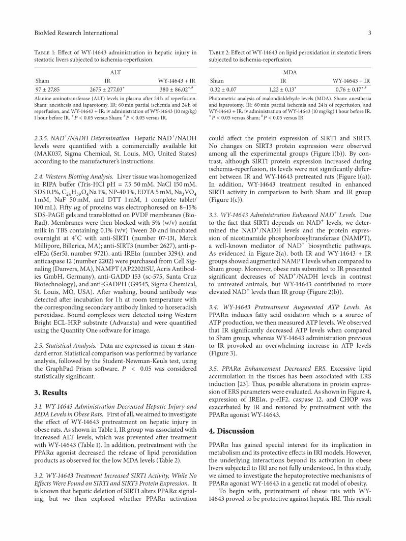
Table 1: Effect of WY-14643 administration in hepatic injury insteatotic livers subjected to ischemia-reperfusion.
ALTSham IR WY-14643 + IR97 ± 27,85 2675 ± 277,03∗ 380 ± 86,02∗,#
Alanine aminotransferase (ALT) levels in plasma after 24 h of reperfusion.Sham: anesthesia and laparotomy, IR: 60min partial ischemia and 24 h ofreperfusion, andWY-14643 + IR: iv administration of WY-14643 (10mg/kg)1 hour before IR. ∗𝑃 < 0.05 versus Sham; #𝑃 < 0.05 versus IR.
2.3.5. NAD+/NADH Determination. Hepatic NAD+/NADHlevels were quantified with a commercially available kit(MAK037, Sigma Chemical, St. Louis, MO, United States)according to the manufacturer’s instructions.
2.4. Western Blotting Analysis. Liver tissue was homogenizedin RIPA buffer (Tris-HCl pH = 7.5 50mM, NaCl 150mM,SDS 0.1%, C
24H39O4Na 1%, NP-40 1%, EDTA 5mM,Na
3VO4
1mM, NaF 50mM, and DTT 1mM, 1 complete tablet/100mL). Fifty 𝜇g of proteins was electrophoresed on 8–15%SDS-PAGE gels and transblotted on PVDF membranes (Bio-Rad). Membranes were then blocked with 5% (w/v) nonfatmilk in TBS containing 0.1% (v/v) Tween 20 and incubatedovernight at 4∘C with anti-SIRT1 (number 07-131, MerckMillipore, Billerica, MA); anti-SIRT3 (number 2627), anti-p-eIF2a (Ser51, number 9721), anti-IRE1𝛼 (number 3294), andanticaspase 12 (number 2202) were purchased from Cell Sig-naling (Danvers, MA), NAMPT (AP22021SU, Acris Antibod-ies GmbH, Germany), anti-GADD 153 (sc-575, Santa CruzBiotechnology), and anti-GADPH (G9545, Sigma Chemical,St. Louis, MO, USA). After washing, bound antibody wasdetected after incubation for 1 h at room temperature withthe corresponding secondary antibody linked to horseradishperoxidase. Bound complexes were detected using WesternBright ECL-HRP substrate (Advansta) and were quantifiedusing the Quantity One software for image.
2.5. Statistical Analysis. Data are expressed as mean ± stan-dard error. Statistical comparison was performed by varianceanalysis, followed by the Student-Newman-Keuls test, usingthe GraphPad Prism software. 𝑃 < 0.05 was consideredstatistically significant.
3. Results
3.1. WY-14643 Administration Decreased Hepatic Injury andMDALevels inObese Rats. First of all, we aimed to investigatethe effect of WY-14643 pretreatment on hepatic injury inobese rats. As shown in Table 1, IR group was associated withincreased ALT levels, which was prevented after treatmentwith WY-14643 (Table 1). In addition, pretreatment with thePPAR𝛼 agonist decreased the release of lipid peroxidationproducts as observed for the low MDA levels (Table 2).
3.2. WY-14643 Treatment Increased SIRT1 Activity, While NoEffectsWere Found on SIRT1 and SIRT3 Protein Expression. Itis known that hepatic deletion of SIRT1 alters PPAR𝛼 signal-ing, but we then explored whether PPAR𝛼 activation
Table 2: Effect of WY-14643 on lipid peroxidation in steatotic liverssubjected to ischemia-reperfusion.
MDASham IR WY-14643 + IR0,32 ± 0,07 1,22 ± 0,13∗ 0,76 ± 0,17∗,#
Photometric analysis of malondialdehyde levels (MDA). Sham: anesthesiaand laparotomy, IR: 60min partial ischemia and 24 h of reperfusion, andWY-14643 + IR: iv administration of WY-14643 (10mg/kg) 1 hour before IR.∗𝑃 < 0.05 versus Sham; #𝑃 < 0.05 versus IR.
could affect the protein expression of SIRT1 and SIRT3.No changes on SIRT3 protein expression were observedamong all the experimental groups (Figure 1(b)). By con-trast, although SIRT1 protein expression increased duringischemia-reperfusion, its levels were not significantly differ-ent between IR and WY-14643 pretreated rats (Figure 1(a)).In addition, WY-14643 treatment resulted in enhancedSIRT1 activity in comparison to both Sham and IR group(Figure 1(c)).
3.3. WY-14643 Administration Enhanced NAD+ Levels. Dueto the fact that SIRT1 depends on NAD+ levels, we deter-mined the NAD+/NADH levels and the protein expres-sion of nicotinamide phosphoribosyltransferase (NAMPT),a well-known mediator of NAD+ biosynthetic pathways.As evidenced in Figure 2(a), both IR and WY-14643 + IRgroups showed augmentedNAMPT levels when compared toSham group. Moreover, obese rats submitted to IR presentedsignificant decreases of NAD+/NADH levels in contrastto untreated animals, but WY-14643 contributed to moreelevated NAD+ levels than IR group (Figure 2(b)).
3.4. WY-14643 Pretreatment Augmented ATP Levels. AsPPAR𝛼 induces fatty acid oxidation which is a source ofATP production, we then measured ATP levels. We observedthat IR significantly decreased ATP levels when comparedto Sham group, whereas WY-14643 administration previousto IR provoked an overwhelming increase in ATP levels(Figure 3).
3.5. PPAR𝛼 Enhancement Decreased ERS. Excessive lipidaccumulation in the tissues has been associated with ERSinduction [23]. Thus, possible alterations in protein expres-sion of ERS parameters were evaluated. As shown in Figure 4,expression of IRE1𝛼, p-eIF2, caspase 12, and CHOP wasexacerbated by IR and restored by pretreatment with thePPAR𝛼 agonist WY-14643.
4. Discussion
PPAR𝛼 has gained special interest for its implication inmetabolism and its protective effects in IRImodels. However,the underlying interactions beyond its activation in obeselivers subjected to IRI are not fully understood. In this study,we aimed to investigate the hepatoprotective mechanisms ofPPAR𝛼 agonist WY-14643 in a genetic rat model of obesity.
To begin with, pretreatment of obese rats with WY-14643 proved to be protective against hepatic IRI. This result

4 BioMed Research International
IR WY-14643 + IRSham
SIRT1
∗
∗
GADPH
0.0
0.1
0.2
0.3
0.4
0.5
0.6
0.7
SIRT
1 (a
.u.)
(a)
IR WY-14643 + IRSham
SIRT3GADPH
0.0
0.1
0.2
0.3
SIRT
3 (a
.u.)
(b)
IR WY-14643 + IRSham
∗, #
0.0
2.5
5.0
7.5
SIRT
1 ac
tivity
(a.u
.)
(c)
Figure 1: WY-14643 pretreatment and sirtuins protein expression and activity in steatotic livers after ischemia-reperfusion. Western blot anddensitometric analysis of (a) SIRT1 and (b) SIRT3. (c) SIRT1 enzymatic activity. Sham: anesthesia and laparotomy, IR: 60min partial ischemiaand 24 h of reperfusion, and WY-14643 + IR: iv administration of WY-14643 (10mg/kg) 1 hour before IR. ∗𝑃 < 0.05 versus Sham; #𝑃 < 0.05versus IR.
IR WY-14643 + IRSham0.00
0.05
0.10
0.15
0.20
0.25
0.30
0.35
NA
MPT
(a.u
.)
NAMPT
∗
∗
GADPH
(a)
WY-14643 + IRIRSham0.00
0.25
0.50
0.75
1.00
1.25
1.50
1.75
NA
D+/
NA
DH
(rel
ativ
e cha
nge)
∗, #
∗
(b)
Figure 2: Effect of WY-14643 administration in NAMPT protein expression and NAD+/NADH levels. (a) Western blot and densitometricanalysis of NAMPT. (b) Photometric analysis of NAD+/NADH levels in steatotic livers after 24 hours of reperfusion. Sham: anesthesia andlaparotomy, IR: 60min partial ischemia and 24 h of reperfusion, andWY-14643 + IR: iv administration ofWY-14643 (10mg/kg) 1 hour beforeIR. ∗𝑃 < 0.05 versus Sham; #𝑃 < 0.05 versus IR.

BioMed Research International 5
IR WY-14643 + IRSham
#
∗
0
2e − 009
5e − 009
7e − 009
1e − 008
ATP
tissu
e (nm
ol/m
g)
Figure 3: Role of WY-14643 treatment in ATP levels in steatotic livers subjected to ischemia-reperfusion. Sham: anesthesia and laparotomy,IR: 60min partial ischemia and 24 h of reperfusion, andWY-14643 + IR: iv administration ofWY-14643 (10mg/kg) 1 hour before IR. ∗𝑃 < 0.05versus Sham; #𝑃 < 0.05 versus IR.
IR WY-14643 + IRSham
GADPHIRE1𝛼
#
∗
0.0
0.1
0.2
0.3
0.4
0.5
0.6
IRE1
𝛼 (a
.u.)
(a)
IR WY-14643 + IRSham
p-eIf2
∗, #
∗
GADPH
0.0
0.1
0.2
0.3
0.4p-
eIf2
(a.u
.)
(b)
#
IR WY-14643 + IRSham
Caspase 12
∗
GADPH
0.0
0.1
0.2
0.3
0.4
0.5
0.6
0.7
0.8
Casp
ase 1
2 (a
.u.)
(c)
#
IR WY-14643 + IRSham
CHOP
∗
GADPH
0.0
0.1
0.2
0.3
0.4
0.5
CHO
P (a
.u.)
(d)
Figure 4: Implication ofWY-14643 administration in ERS parameters in steatotic livers subjected to ischemia-reperfusion. Western blot anddensitometric analysis of (a) IRE1𝛼, (b) p-eIf2, (c) caspase 12, and (d) CHOP. Sham: anesthesia and laparotomy, IR: 60min partial ischemiaand 24 h of reperfusion, and WY-14643 + IR: iv administration of WY-14643 (10mg/kg) 1 hour before IR. ∗𝑃 < 0.05 versus Sham; #𝑃 < 0.05versus IR.

6 BioMed Research International
is consistent with other studies using genetic and dietarymodels of steatohepatitis. In nonalcoholic steatohepatitis(NASH) and simple steatosis, treatment of mice with WY-14643 protected steatotic livers against IRI [24]. Given thefact that oxidative injury is known to be more exaggeratedin fatty livers [3, 25], we also determined lipid peroxidationand we found a significant reduction in MDA formation inthe liver of rats pretreated with WY-14643. In this sense,PPAR𝛼 beneficial effects against oxidative stress have alsobeen demonstrated in other models [26, 27].
Various studies have evidenced the protective role ofSIRT1 against IRI, which in most cases has been associatedwith attenuation of oxidative stress [28, 29]. In the liver,a major target of SIRT1 is the PPAR𝛼 signaling pathway.Loss of hepatic SIRT1 impairs PPAR𝛼 mediated fatty acidmetabolism and decreases fatty acid 𝛽-oxidation [17]. Addi-tionally, cell culture experiments suggested that PPAR𝛼 mayalso positively regulate SIRT1 expression [30, 31]. In ourstudy, we observed that WY-14643 administration did notaffect either SIRT1 or SIRT3 protein expression, whereas itstrengthened SIRT1 activity.
As SIRT1 requires NAD+ for its enzymatic activity, theaugmented NAD+ levels that we observed in WY-14643treated group led us to suggest that SIRT1 enhanced activitymay be, at least in part, a result of the increased levels of itscofactor NAD+. However, the fact that NAD+ levels were notin accordance with NAMPT proteins levels may be attributedto the existence of other NAD+ precursors, like tryptophanand nicotinic acid [32].
It has been shown that PPAR𝛼 stimulates the 𝛽-oxidativedegradation of fatty acids, provoking thus a high yield ofATP production [33, 34]. Indeed, in our study, we observedthat administration of PPAR𝛼 agonist WY-14643 resulted inoverwhelming ATP increases. Fatty livers are characterizedby a reduced ATP content, which limits the resistance and thesurvival of hepatocytes against stress conditions, includingIRI [3].Thus, PPAR𝛼 activation diminished IRI in fatty liversby enhancing energy production. Furthermore, ATP formspart of the NAD+ biosynthesis pathways [35, 36] and in thisway may promote SIRT1 activity.
Various studies have associated steatotic livers with ERS[12, 37]. IR affects the ability of the endoplasmic reticulum tosynthesize and fold proteins, leading to the exaggerated accu-mulation of unfolded proteins and the initiation of ERS.UponERS, various proteins localized in the endoplasmic reticulumare activated, such as inositol requiring enzyme 1𝛼 (IRE1𝛼)and pancreatic ER kinase- (PKR-) like ER kinase (PERK).Activated PERK phosphorylates eukaryotic initiation factor2 (eIF2) in order to block protein synthesis and activatedIRE1𝛼 controls genes involved in protein degradation. Thisconsists of a cell attempt to restrain the accumulation of newlysynthesized proteins in the ER lumen but can also initiateproapoptotic events, including the activation of caspase 12and enhanced protein expression of CHOP [38]. Here, weobserved that the IRE1𝛼, p-eIF2, and CHOP signaling path-ways of the ERS, as well as caspase 12 levels, were significantlyabrogated after WY-14643 treatment. Due to the fact thatROS impairs the protein folding, the attenuation of oxidativestress by PPAR𝛼 activation could contribute to a more proper
folding of the proteins and thus to lessened ERS. ERSprevention by PPAR𝛼 activation has also been evidenced invitro in H
2O2-treated HepG2 cells [11]. Another potential
mechanism to prevent ERS might be based on the SIRT1augmented activity, as SIRT1 hepatocyte loss has been shownto be crucial for the development of ERS in a high-fat diet [16].
5. Conclusion
In conclusion, our study gives a new insight into the hepato-protective mechanisms of the PPAR𝛼 agonist WY-14643 insteatotic livers, implying that SIRT1 might be an importantmediator of these beneficial effects. However, more efforts arerequired to elucidate the exact mechanisms that define theobserved interactions.
Conflict of Interests
The authors declare that there is no conflict of interestsregarding the publication of this paper.
Acknowledgments
Eirini Pantazi thanks the fellowship from AGAUR(2012FI B00382), Generalitat de Catalunya, Barcelona,Catalonia, Spain. The authors would like to thank theFondo de Investigaciones Sanitarias (FIS PI12/00519) for theeconomic support.
References
[1] L. Y. Guan, P. Y. Fu, P. D. Li et al., “Mechanisms of hepaticischemia-reperfusion injury and protective effects of nitricoxide,” World Journal of Gastrointestinal Surgery, vol. 6, no. 7,pp. 122–128, 2014.
[2] J. D. Browning and J. D. Horton, “Molecular mediators ofhepatic steatosis and liver injury,” The Journal of ClinicalInvestigation, vol. 114, no. 2, pp. 147–152, 2004.
[3] C. Peralta and J. Rosello-Catafau, “The future of fatty livers,”Journal of Hepatology, vol. 41, no. 1, pp. 149–151, 2004.
[4] M. M. Masternak and A. Bartke, “PPARs in calorie restrictedand genetically long-lived mice,” PPAR Research, vol. 2007,Article ID 28436, 2007.
[5] Y. Harano, K. Yasui, T. Toyama et al., “Fenofibrate, a peroxi-some proliferator-activated receptor 𝛼 agonist, reduces hepaticsteatosis and lipid peroxidation in fatty liver Shionogimice withhereditary fatty liver,” Liver International, vol. 26, no. 5, pp. 613–620, 2006.
[6] T. Hashimoto,W. S. Cook, C. Qi, A. V. Yeldandi, J. K. Reddy, andM. S. Rao, “Defect in peroxisome proliferator-activated receptoralpha-inducible fatty acid oxidation determines the severity ofhepatic steatosis in response to fastin,”The Journal of BiologicalChemistry, vol. 275, no. 37, pp. 28918–28928, 2000.
[7] J. K. Reddy, “Nonalcoholic steatosis and steatohepatitis. III. Per-oxisomal 𝛽-oxidation, PPAR𝛼, and steatohepatitis,” AmericanJournal of Physiology—Gastrointestinal and Liver Physiology,vol. 281, no. 6, pp. G1333–G1339, 2001.
[8] N. S. A. Patel, R. Di Paola, E. Mazzon, D. Britti, C. Thiemer-mann, and S. Cuzzocrea, “Peroxisome proliferator-activated

BioMed Research International 7
receptor-𝛼 contributes to the resolution of inflammation afterrenal ischemia/reperfusion injury,”The Journal of Pharmacologyand Experimental Therapeutics, vol. 328, no. 2, pp. 635–643,2009.
[9] Z. Gao and Y.-H. Li, “Antioxidant stress and anti-inflammationof PPAR𝛼 on warm hepatic ischemia-reperfusion injury,” PPARResearch, vol. 2012, Article ID 738785, 8 pages, 2012.
[10] M. Collino, M. Aragno, R. Mastrocola et al., “Oxidativestress and inflammatory response evoked by transient cere-bral ischemia/reperfusion: effects of the PPAR-alpha agonistWY14643,” Free Radical Biology & Medicine, vol. 41, no. 4, pp.579–589, 2006.
[11] W.-X. Tang, L.-K. Wang, Y.-Q. Wang et al., “Peroxisomeproliferator-activated receptor-𝛼 activation protects againstendoplasmic reticulum stress-induced HepG2 cell apoptosis,”Molecular and Cellular Biochemistry, vol. 385, no. 1-2, pp. 179–190, 2014.
[12] M. J. Pagliassotti, “Endoplasmic reticulum stress in nonalco-holic fatty liver disease,” Annual Review of Nutrition, vol. 32, pp.17–33, 2012.
[13] J. T. Rodgers and P. Puigserver, “Fasting-dependent glucose andlipid metabolic response through hepatic sirtuin 1,” Proceedingsof the National Academy of Sciences of the United States ofAmerica, vol. 104, no. 31, pp. 12861–12866, 2007.
[14] A. Planavila, R. Iglesias,M.Giralt, andF.Villarroya, “Sirt1 acts inassociation with PPAR𝛼 to protect the heart from hypertrophy,metabolic dysregulation, and inflammation,” CardiovascularResearch, vol. 90, no. 2, pp. 276–284, 2011.
[15] S. A. Khan, A. Sathyanarayan, M. T. Mashek, K. T. Ong, E.E. Wollaston-Hayden, and D. G. Mashek, “ATGL-catalyzedlipolysis regulates SIRT1 to control PGC-1𝛼/PPAR-𝛼 signaling,”Diabetes, vol. 64, no. 2, pp. 418–426, 2015.
[16] Y. Li, S. Xu, A. Giles et al., “Hepatic overexpression of SIRT1in mice attenuates endoplasmic reticulum stress and insulinresistance in the liver,” The FASEB Journal, vol. 25, no. 5, pp.1664–1679, 2011.
[17] A. Purushotham,T. T. Schug,Q.Xu, S. Surapureddi, X.Guo, andX. Li, “Hepatocyte-specific deletion of SIRT1 alters fatty acidmetabolism and results in hepatic steatosis and inflammation,”Cell Metabolism, vol. 9, no. 4, pp. 327–338, 2009.
[18] M. D. Hirschey, T. Shimazu, E. Goetzman et al., “SIRT3 regu-lates mitochondrial fatty-acid oxidation by reversible enzymedeacetylation,” Nature, vol. 464, no. 7285, pp. 121–125, 2010.
[19] J. S. Teodoro, F. V. Duarte, A. P. Gomes et al., “Berberinereverts hepatic mitochondrial dysfunction in high-fat fed rats:a possible role for SirT3 activation,”Mitochondrion, vol. 13, no.6, pp. 637–646, 2013.
[20] E. Pantazi, M. A. Zaouali, M. Bejaoui, E. Folch-Puy, H.B. Abdennebi, and J. Rosello-Catafau, “Role of sirtuins inischemia-reperfusion injury,” World Journal of Gastroenterol-ogy, vol. 19, no. 43, pp. 7594–7602, 2013.
[21] T. Okaya and A. B. Lentsch, “Peroxisome proliferator-activatedreceptor-𝛼 regulates postischemic liver injury,” American Jour-nal of Physiology—Gastrointestinal and Liver Physiology, vol.286, no. 4, pp. G606–G612, 2004.
[22] M. Becatti, N. Taddei, C. Cecchi, N. Nassi, P. A. Nassi, andC. Fiorillo, “SIRT1 modulates MAPK pathways in ischemic-reperfused cardiomyocytes,” Cellular and Molecular Life Sci-ences, vol. 69, no. 13, pp. 2245–2260, 2012.
[23] U. Ozcan, Q. Cao, E. Yilmaz et al., “Endoplasmic reticulumstress links obesity, insulin action, and type 2 diabetes,” Science,vol. 306, no. 5695, pp. 457–461, 2004.
[24] N. C. Teoh, J. Williams, J. Hartley, J. Yu, R. S. McCuskey, and G.C. Farrell, “Short-term therapy with peroxisome proliferation-activator receptor-𝛼 agonist Wy-14,643 protects murine fattyliver against ischemia-reperfusion injury,” Hepatology, vol. 51,no. 3, pp. 996–1006, 2010.
[25] S. K. Mantena, A. L. King, K. K. Andringa, H. B. Eccleston, andS. M. Bailey, “Mitochondrial dysfunction and oxidative stressin the pathogenesis of alcohol- and obesity-induced fatty liverdiseases,” Free Radical Biology and Medicine, vol. 44, no. 7, pp.1259–1272, 2008.
[26] M. A. Abdelmegeed, K.-H.Moon, J. P. Hardwick, F. J. Gonzalez,and B.-J. Song, “Role of peroxisome proliferator-activatedreceptor-𝛼 in fasting-mediated oxidative stress,” Free RadicalBiology and Medicine, vol. 47, no. 6, pp. 767–778, 2009.
[27] M. A. Abdelmegeed, S.-H. Yoo, L. E. Henderson, F. J. Gonzalez,K. J. Woodcroft, and B.-J. Song, “PPAR𝛼 expression protectsmale mice from high fat-induced nonalcoholic fatty liver,”Journal of Nutrition, vol. 141, no. 4, pp. 603–610, 2011.
[28] C.-P. Hsu, P. Zhai, T. Yamamoto et al., “Silent informationregulator 1 protects the heart from ischemia/reperfusion,” Cir-culation, vol. 122, no. 21, pp. 2170–2182, 2010.
[29] E. Pantazi, M. A. Zaouali, M. Bejaoui et al., “Silent informationregulator 1 protects the liver against ischemia-reperfusioninjury: implications in steatotic liver ischemic preconditioning,”Transplant International, vol. 27, no. 5, pp. 493–503, 2014.
[30] S. Hayashida, A. Arimoto, Y. Kuramoto et al., “Fasting promotesthe expression of SIRT1, an NAD+-dependent protein deacety-lase, via activation of PPAR𝛼 in mice,” Molecular and CellularBiochemistry, vol. 339, no. 1-2, pp. 285–292, 2010.
[31] W.Wang, Q. Lin, R. Lin et al., “PPAR𝛼 agonist fenofibrate atten-uates TNF-𝛼-induced CD40 expression in 3T3-L1 adipocytesvia the SIRT1-dependent signaling pathway,” Experimental CellResearch, vol. 319, no. 10, pp. 1523–1533, 2013.
[32] A. Nikiforov, C. Dolle, M. Niere, andM. Ziegler, “Pathways andsubcellular compartmentation of NAD biosynthesis in humancells: from entry of extracellular precursors to mitochondrialNAD generation,”The Journal of Biological Chemistry, vol. 286,no. 24, pp. 21767–21778, 2011.
[33] K. Schoonjans, B. Staels, and J. Auwerx, “The peroxisomeproliferator activated receptors (PPARs) and their effects onlipid metabolism and adipocyte differentiation,” Biochimica etBiophysica Acta, vol. 1302, no. 2, pp. 93–109, 1996.
[34] S. Sarma, H. Ardehali, and M. Gheorghiade, “Enhancing themetabolic substrate: PPAR-alpha agonists in heart failure,”Heart Failure Reviews, vol. 17, no. 1, pp. 35–43, 2012.
[35] J. S. Smith, C. B. Brachmann, I. Celic et al., “A phylogeneticallyconserved NAD+-dependent protein deacetylase activity in theSir2 protein family,” Proceedings of the National Academy ofSciences of the United States of America, vol. 97, no. 12, pp. 6658–6663, 2000.
[36] T. Yang and A. A. Sauve, “NAD metabolism and sirtuins:metabolic regulation of protein deacetylation in stress andtoxicity,”The AAPS Journal, vol. 8, no. 4, pp. E632–E643, 2006.
[37] X.-Q. Zhang, C.-F. Xu, C.-H. Yu, W.-X. Chen, and Y.-M. Li,“Role of endoplasmic reticulum stress in the pathogenesis ofnonalcoholic fatty liver disease,”World Journal of Gastroenterol-ogy, vol. 20, no. 7, pp. 1768–1776, 2014.
[38] I. Kim, W. Xu, and J. C. Reed, “Cell death and endoplasmicreticulum stress: disease relevance and therapeutic opportuni-ties,”Nature ReviewsDrugDiscovery, vol. 7, no. 12, pp. 1013–1030,2008.

Submit your manuscripts athttp://www.hindawi.com
Stem CellsInternational
Hindawi Publishing Corporationhttp://www.hindawi.com Volume 2014
Hindawi Publishing Corporationhttp://www.hindawi.com Volume 2014
MEDIATORSINFLAMMATION
of
Hindawi Publishing Corporationhttp://www.hindawi.com Volume 2014
Behavioural Neurology
EndocrinologyInternational Journal of
Hindawi Publishing Corporationhttp://www.hindawi.com Volume 2014
Hindawi Publishing Corporationhttp://www.hindawi.com Volume 2014
Disease Markers
Hindawi Publishing Corporationhttp://www.hindawi.com Volume 2014
BioMed Research International
OncologyJournal of
Hindawi Publishing Corporationhttp://www.hindawi.com Volume 2014
Hindawi Publishing Corporationhttp://www.hindawi.com Volume 2014
Oxidative Medicine and Cellular Longevity
Hindawi Publishing Corporationhttp://www.hindawi.com Volume 2014
PPAR Research
The Scientific World JournalHindawi Publishing Corporation http://www.hindawi.com Volume 2014
Immunology ResearchHindawi Publishing Corporationhttp://www.hindawi.com Volume 2014
Journal of
ObesityJournal of
Hindawi Publishing Corporationhttp://www.hindawi.com Volume 2014
Hindawi Publishing Corporationhttp://www.hindawi.com Volume 2014
Computational and Mathematical Methods in Medicine
OphthalmologyJournal of
Hindawi Publishing Corporationhttp://www.hindawi.com Volume 2014
Diabetes ResearchJournal of
Hindawi Publishing Corporationhttp://www.hindawi.com Volume 2014
Hindawi Publishing Corporationhttp://www.hindawi.com Volume 2014
Research and TreatmentAIDS
Hindawi Publishing Corporationhttp://www.hindawi.com Volume 2014
Gastroenterology Research and Practice
Hindawi Publishing Corporationhttp://www.hindawi.com Volume 2014
Parkinson’s Disease
Evidence-Based Complementary and Alternative Medicine
Volume 2014Hindawi Publishing Corporationhttp://www.hindawi.com