research article assessment of cell-cycle arrest...
TRANSCRIPT

Research ArticleAssessment of Cell-Cycle Arrest Biomarkers to Predict Early andDelayed Acute Kidney Injury
Max Bell,1 Anders Larsson,2 Per Venge,2 Rinaldo Bellomo,3,4 and Johan Mårtensson1,3
1Section of Anaesthesia and Intensive Care Medicine, Department of Physiology and Pharmacology,Karolinska Institutet, 17176 Stockholm, Sweden2Clinical Chemistry, Department of Medical Sciences, Uppsala University, 751 85 Uppsala, Sweden3Department of Intensive Care, Austin Hospital, Heidelberg, Melbourne, VIC 3084, Australia4Australian and New Zealand Intensive Care Research Centre, School of Preventive Medicine and Public Health,Monash University, Melbourne, VIC 3800, Australia
Correspondence should be addressed to Johan Martensson; [email protected]
Received 20 December 2014; Accepted 5 March 2015
Academic Editor: Shih-Ping Hsu
Copyright © 2015 Max Bell et al.This is an open access article distributed under the Creative Commons Attribution License, whichpermits unrestricted use, distribution, and reproduction in any medium, provided the original work is properly cited.
Purpose. To assess urinary tissue inhibitor of metalloproteinases-2 and insulin-like growth factor binding protein 7 ([TIMP-2]⋅[IGFBP7]), urinary neutrophil gelatinase-associated lipocalin (NGAL), and urinary cystatin-C as acute kidney injury predictors(AKI) exploring the association of nonrenal factors with elevated biomarker levels. Methods. We studied 94 patients with urinecollected within 48 hours of ICU admission and no AKI at sampling. AKI was defined by the Kidney Disease: Improving GlobalOutcomes criteria. Predictive performance was assessed by the area under the receiver operating characteristics (ROC) curve.Associations between biomarkers and clinical factors were assessed by multivariate linear regression. Results. Overall, 19 patients(20%) developed AKI within 48 hours. [TIMP-2]⋅[IGFBP7], NGAL, or cystatin-C admission levels did not differ between patientswithout AKI and patients developing AKI. [TIMP-2]⋅[IGFBP7], NGAL, and cystatin-Cwere poor AKI predictors (ROC areas 0.34–0.51). Diabetes was independently associated with higher [TIMP-2]⋅[IGFBP7] levels (𝑃 = 0.02) but AKI was not (𝑃 = 0.24). Sepsiswas independently associated with higher NGAL (𝑃 < 0.001) and cystatin-C (𝑃 = 0.003) levels. Conclusions. Urinary [TIMP-2]⋅[IGFBP7], NGAL, and cystatin-C should be used cautiously as AKI predictors in general ICU patients since urine levels of thesebiomarkers are affected by factors other than AKI and their performance can be poor.
1. Introduction
Acute kidney injury (AKI) commonly complicates criticalillness and is associated with high mortality and morbidity[1–4]. Early diagnosis of AKI, preferably within 24 hoursafter ICU admission, is likely pivotal to the development ofeffective therapies. Traditional biomarkers like creatinine arelate indicators of AKI, delaying diagnosis by days [5]. Thisshortcoming has led to multiple attempts to identify novelbiomarkers that can predict the subsequent development ofAKI at a much earlier stage [6–8]. However, suggested novelAKI biomarkers, such as neutrophil gelatinase-associatedlipocalin (NGAL), have been unreliable in the real worldsetting of general ICU patients [9].
A recent multicenter international investigation, the Sap-phire study, reported the discovery and validation of twoG1 cell-cycle arrest biomarkers (CCABs), tissue inhibitor ofmetalloproteinases (TIMP-2) and insulin-like growth fac-tor binding protein (IGFBP-7) [6]. When combined, theseCCABs detected AKI with a high level of accuracy andwere excellent in identifying patients at imminent risk ofsevere AKI [6]. Risk for death, dialysis, or persistent renaldysfunction, that is, major adverse kidney events (MAKE),also increased with increasing test values [6].
Two other studies have assessed the predictive value of[TIMP-2]⋅[IGFBP7], a multicenter study in a general ICUsetting and a single centre study of AKI following cardiacsurgery [10, 11]. In both settings, these CCABs predicted
Hindawi Publishing CorporationDisease MarkersVolume 2015, Article ID 158658, 9 pageshttp://dx.doi.org/10.1155/2015/158658

2 Disease Markers
AKI with areas under the receiver operating characteristic(ROC) curve of over 0.8. In a separate investigation, donesimultaneously but independently of the Sapphire study,IGFBP-7 was identified by proteomics as an early prognosticmarker of AKI severity, duration, and mortality [12].
In the present study, we assessed the bedside Nephro-check Astute 140R Meter (Astute Medical, San Diego,CA), which simultaneously quantifies [TIMP-2]⋅[IGFBP7]in urine, in a subselected cohort of critically ill patientswithout evidence of AKI on ICU admission. We comparedurinary [TIMP-2]⋅[IGFBP7]with two other proposed urinarymarkers of AKI, urinary NGAL and urinary cystatin C. Weaimed to investigate the diagnostic value of these markersto detect AKI within 12 to 48 hours of biomarker analysis.Additionally, we intended to explore the potential impact ofnonrenal factors with elevated [TIMP-2]⋅[IGFBP7], NGAL,and cystatin C in urine.
2. Methods
This study was approved by the regional ethical review boardin Stockholm. Written informed consent was obtained frompatients or their next of kin.
2.1. Inclusion and Classification of Patients. We studied 138patients admitted to the general intensive care unit (ICU)at the Karolinska University Hospital, Solna, Sweden, withcreatinine clearance >60mL/min/1.73m2, estimated by themodification of diet in renal disease (MDRD) equation, andan expected length of stay of at least 24 hours. Urine wascollected in the morning and in the afternoon until ICU-discharge or initiation of renal replacement therapy (RRT).Patients were included from August 2007 to November 2010.During this time frame, approximately 3500 patients weretreated in the ICU. The relatively low number of patientsincluded in this study is mainly explained by the restrictiveinclusion criteria. In addition, we were unable to includepatients on weekends, nights/on-call hours and during hol-idays.
The presence or absence of AKI, defined by the KidneyDisease: Improving Global Outcomes (KDIGO) criteria, wasrecorded on a daily basis [13]. KDIGO grades AKI severityby increase of creatinine from baseline or decrease of urineoutput. The lowest creatinine level obtained within threemonths prior to ICU admission was used as baseline forthe KDIGO classification. When pre-ICU creatinine waslacking, it was imputed using the MDRD formula, assumingan essentially normal baseline glomerular filtration rate of75mL/min/1.73m2. For the purpose of this studywe excludedpatients who had their first urine sample obtained >48 hoursafter ICU admission and patients with AKI on the day beforeand/or on the day when the first study sample was obtained.The primary outcome was development of AKI within 48hours. Secondary outcomes were development of AKI within12 hours, within 12 to 24 hours, and within 24 to 48 hours,respectively.
2.2. Clinical Data Collection. Acute Physiology and ChronicHealth Evaluation (APACHE) II score, demographic data,
ICU admission diagnosis, ICU, and 30-day mortality wererecorded. Data on comorbid conditions were collected fromthe hospital-based electronic case-record system. In theseelectronic journals, prior diagnoses like diabetes, cardio-vascular disease, pulmonary disease, gastrointestinal/liverdisease, and malignancies are recorded.
Sepsis was defined as a suspected or confirmed infectiontogether with the presence of at least three systemic inflam-matory response syndrome (SIRS) criteria [14, 15].
2.3. Urine Collection and Biochemical Analyses. After cen-trifugation at 2000 rpm at 4∘C for 10min, the supernatanturine was stored at −80∘C. Samples were analyzed at theDepartment of Clinical Chemistry, Uppsala University Hos-pital, Uppsala, Sweden. NGAL was measured by a researchenzyme-linked immunosorbent assay (ELISA), as previouslydescribed [16, 17]. Monoclonal antibody clones 763 and764 (Diagnostics Development, Uppsala, Sweden) were usedas catching and detecting antibodies, respectively, in theELISA. Urinary [TIMP2]⋅[IGFBP7] was quantified by theNephrocheck point-of-care analyzer (Astute Medical, SanDiego, CA), a device that simultaneously measures [TIMP-2] and [IGFBP7] giving a reading based on the multiplicationof both biomarkers. Based on the Sapphire study, the manu-facturer suggests using >0.3 (ng/mL)2/1000 as moderate riskand >2.0 (ng/mL)2/1000 as high risk of severe AKI. The testrequires 100 𝜇L of urine and 20 minutes to provide results.Urine cystatin C was measured with a particle-enhancedturbidimetric immunoassay on the Architect Ci8200 ana-lyzer (Abbott Laboratories, Abbott Park, IL) with cystatin Creagents from Gentian (Moss, Norway).
2.4. Statistical Analysis. Data were analyzed using STATAversion 11.2 software (Stata Corporation, College Station, TX,USA). Continuous variables were summarized using medianand interquartile range (IQR) and categorical variables asnumbers (%). The Kruskal-Wallis test and 𝜒2 test were usedfor comparison between continuous and categorical vari-ables, respectively, across multiple groups. Mann-Whitneytest and Fisher’s exact test or 𝜒2 test were used for two-groupcomparison between continuous and categorical variables,respectively. The predictive performance for AKI was eval-uated by calculating the area under the ROC curve for eachurinary biomarker at different time-points. The relationshipsof [TIMP2]⋅[IGFBP7], NGAL, and cystatin C with AKI andnonrenal factors were investigated by multivariate linearregression analyses. The following predictor variables wereconsidered: age, gender, APACHE II score, AKI within 48hours, comorbidities, and admission diagnosis. Predictorvariables were included in the multivariate models if theywere statistically significant at 𝑃 < 0.1 in the univariateanalyses. Two-sided 𝑃 values below 0.05 were consideredstatistically significant in the final analyses.
3. Results
We screened 138 patients. Of these, 16 patients withoutavailable urine sample within 48 hours of ICU admission and
Disease Markers 3
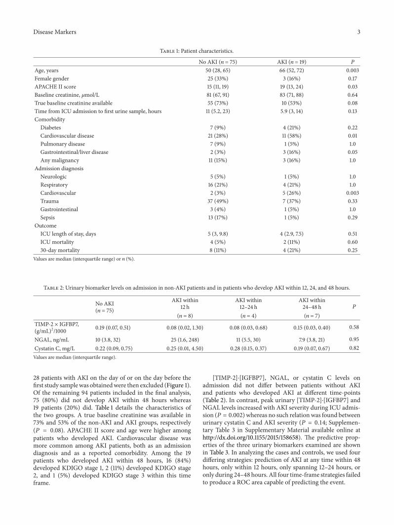
Table 1: Patient characteristics.
No AKI (𝑛 = 75) AKI (𝑛 = 19) 𝑃
Age, years 50 (28, 65) 66 (52, 72) 0.003Female gender 25 (33%) 3 (16%) 0.17APACHE II score 15 (11, 19) 19 (13, 24) 0.03Baseline creatinine, 𝜇mol/L 81 (67, 91) 83 (71, 88) 0.64True baseline creatinine available 55 (73%) 10 (53%) 0.08Time from ICU admission to first urine sample, hours 11 (5.2, 23) 5.9 (3, 14) 0.13Comorbidity
Diabetes 7 (9%) 4 (21%) 0.22Cardiovascular disease 21 (28%) 11 (58%) 0.01Pulmonary disease 7 (9%) 1 (5%) 1.0Gastrointestinal/liver disease 2 (3%) 3 (16%) 0.05Any malignancy 11 (15%) 3 (16%) 1.0
Admission diagnosisNeurologic 5 (5%) 1 (5%) 1.0Respiratory 16 (21%) 4 (21%) 1.0Cardiovascular 2 (3%) 5 (26%) 0.003Trauma 37 (49%) 7 (37%) 0.33Gastrointestinal 3 (4%) 1 (5%) 1.0Sepsis 13 (17%) 1 (5%) 0.29
OutcomeICU length of stay, days 5 (3, 9.8) 4 (2.9, 7.5) 0.51ICU mortality 4 (5%) 2 (11%) 0.6030-day mortality 8 (11%) 4 (21%) 0.25
Values are median (interquartile range) or 𝑛 (%).
Table 2: Urinary biomarker levels on admission in non-AKI patients and in patients who develop AKI within 12, 24, and 48 hours.
No AKI(𝑛 = 75)
AKI within12 h
AKI within12–24 h
AKI within24–48 h 𝑃
(𝑛 = 8) (𝑛 = 4) (𝑛 = 7)TIMP-2 × IGFBP7,(g/mL)2/1000 0.19 (0.07, 0.51) 0.08 (0.02, 1.30) 0.08 (0.03, 0.68) 0.15 (0.03, 0.40) 0.58
NGAL, ng/mL 10 (3.8, 32) 25 (1.6, 248) 11 (5.5, 30) 7.9 (3.8, 21) 0.95Cystatin C, mg/L 0.22 (0.09, 0.75) 0.25 (0.01, 4.50) 0.28 (0.15, 0.37) 0.19 (0.07, 0.67) 0.82Values are median (interquartile range).
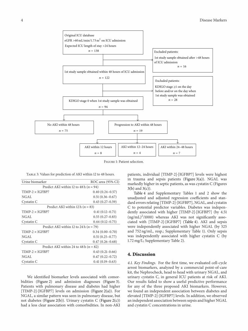
28 patients with AKI on the day of or on the day before thefirst study samplewas obtainedwere then excluded (Figure 1).Of the remaining 94 patients included in the final analysis,75 (80%) did not develop AKI within 48 hours whereas19 patients (20%) did. Table 1 details the characteristics ofthe two groups. A true baseline creatinine was available in73% and 53% of the non-AKI and AKI groups, respectively(𝑃 = 0.08). APACHE II score and age were higher amongpatients who developed AKI. Cardiovascular disease wasmore common among AKI patients, both as an admissiondiagnosis and as a reported comorbidity. Among the 19patients who developed AKI within 48 hours, 16 (84%)developed KDIGO stage 1, 2 (11%) developed KDIGO stage2, and 1 (5%) developed KDIGO stage 3 within this timeframe.
[TIMP-2]⋅[IGFBP7], NGAL, or cystatin C levels onadmission did not differ between patients without AKIand patients who developed AKI at different time-points(Table 2). In contrast, peak urinary [TIMP-2]⋅[IGFBP7] andNGAL levels increased with AKI severity during ICU admis-sion (𝑃 = 0.002) whereas no such relationwas found betweenurinary cystatin C and AKI severity (𝑃 = 0.14; Supplemen-tary Table 3 in Supplementary Material available online athttp://dx.doi.org/10.1155/2015/158658). The predictive prop-erties of the three urinary biomarkers examined are shownin Table 3. In analyzing the cases and controls, we used fourdiffering strategies: prediction of AKI at any time within 48hours, only within 12 hours, only spanning 12–24 hours, oronly during 24–48 hours. All four time-frame strategies failedto produce a ROC area capable of predicting the event.
4 Disease Markers
Original ICU database
n = 138
1st study sample obtained within 48 hours of ICU admission
n = 122
KDIGO stage 0 when 1st study sample was obtained
n = 94
No AKI within 48 hours
n = 75
Progression to AKI within 48 hours
n = 19
AKI within 12 hours
n = 8
AKI within 12–24 hours
n = 4
AKI within 24–48 hours
n = 7
Excluded patients:
of ICU admissionn = 16
Excluded patients:
before and/or on the day when1st study sample was obtained
n = 28
eGFR >60mL/min/1.73m2 on ICU admission
KDIGO stage ≥1 on the day
1st study sample obtained after >48 hours
Expected ICU length of stay >24 hours
Figure 1: Patient selection.
Table 3: Values for prediction of AKI within 12 to 48 hours.
Urine biomarker ROC area (95% CI)Predict AKI within 12 to 48 h (𝑛 = 94)
TIMP-2 × IGFBP7 0.40 (0.24–0.57)NGAL 0.51 (0.36–0.67)Cystatin C 0.43 (0.27–0.59)
Predict AKI within 12 h (𝑛 = 83)TIMP-2 × IGFBP7 0.41 (0.12–0.71)NGAL 0.55 (0.27–0.83)Cystatin C 0.44 (0.12–0.75)
Predict AKI within 12 to 24 h (𝑛 = 79)TIMP-2 × IGFBP7 0.34 (0.00–0.70)NGAL 0.51 (0.25–0.77)Cystatin C 0.47 (0.26–0.68)
Predict AKI within 24 to 48 h (𝑛 = 82)TIMP-2 × IGFBP7 0.43 (0.21–0.66)NGAL 0.47 (0.22–0.72)Cystatin C 0.41 (0.19–0.63)
We identified biomarker levels associated with comor-bidities (Figure 2) and admission diagnoses (Figure 3).Patients with pulmonary disease and diabetes had higher[TIMP-2]⋅[IGFBP7] levels on admission (Figure 2(a)). ForNGAL, a similar pattern was seen in pulmonary disease, butnot diabetes (Figure 2(b)). Urinary cystatin C (Figure 2(c))had a less clear association with comorbidities. In non-AKI
patients, individual [TIMP-2]⋅[IGFBP7] levels were highestin trauma and sepsis patients (Figure 3(a)). NGAL wasmarkedly higher in septic patients, as was cystatin C (Figures3(b) and 3(c)).
Table 4 and Supplementary Tables 1 and 2 show theunadjusted and adjusted regression coefficients and stan-dard errors relating [TIMP-2]⋅[IGFBP7], NGAL, and cystatinC to potential predictor variables. Diabetes was indepen-dently associated with higher [TIMP-2]⋅[IGFBP7] (by 4.51(ng/mL)2/1000) whereas AKI was not significantly asso-ciated with [TIMP-2]⋅[IGFBP7] (Table 4). AKI and sepsiswere independently associated with higher NGAL (by 525and 752 ng/mL, resp.; Supplementary Table 1). Only sepsiswas independently associated with higher cystatin C (by1.72mg/L; Supplementary Table 2).
4. Discussion
4.1. Key Findings. For the first time, we evaluated cell-cyclearrest biomarkers, analyzed by a commercial point-of-carekit, the Nephrocheck, head to head with urinary NGAL, andurinary cystatin C, in general ICU patients at risk of AKI.Our results failed to show a useful predictive performancefor any of the three proposed AKI biomarkers. However,we found an independent association between diabetes andelevated [TIMP-2]⋅[IGFBP7] levels. In addition, we observedan independent association between sepsis and higherNGALand cystatin C concentrations in urine.

Disease Markers 5
0
1
2
3
4TI
MP-2×
IGFB
P7
No
com
orbi
ditie
s
G1
/live
r
Mal
igna
ncy
Card
iova
scul
ar
Dia
bete
s
Pulm
onar
y(a)
0
200
400
600
800
1,000
No
com
orbi
ditie
s
G1
/live
r
Mal
igna
ncy
Card
iova
scul
ar
Dia
bete
s
Pulm
onar
y
NG
AL
(ng/
mL)
(b)
0
2
4
6
8
10
Cysta
tin C
(mg/
L)
No
com
orbi
ditie
s
G1
/live
r
Mal
igna
ncy
Card
iova
scul
ar
Dia
bete
s
Pulm
onar
y
(c)
Figure 2: Urinary biomarker levels on ICU admission in non-AKI patients with different comorbidities.
4.2. Relation to Previous Studies. The etiology of AKI iscomplex and may often be multifactorial, with both ischemicand inflammatory events. Conservation of energy may bean adaptive response of stressed tubular epithelial cells andexplain the clinical phenotype of sepsis-induced AKI [18].One way for cells to save energy is to avoid mitosis. Indeed,it has been shown that renal tubular cells can enter G1cell-cycle arrest after sepsis [19] and ischemia [20]. Thus,a combination of urinary CCABs like [TIMP-2]⋅[IGFBP7]has biological plausibility and may have clinical value inthese settings. The Sapphire study found that urine [TIMP-2]⋅[IGFBP7] levels were superior to all previously describedearly markers of AKI [6]. In a further US-only multicenterstudy, the high-sensitivity cut-off value of 0.3 (ng/mL)2/1000was tested against clinical nephrologists, who, blinded tothe biomarker findings, were asked to determine whethermoderate to severe AKI was reached for each patient [10].Critically ill patients with urinary [TIMP-2]⋅[IGFBP7] levels> 0.3 (ng/mL)2/1000 had seven times the risk for AKI (95%
CI 4–22) compared to critically ill patients with a test resultbelow 0.3 [10]. In addition, a study of 50 patients at highrisk of AKI following cardiac surgery found that maximumurinary [TIMP-2]⋅[IGFBP7] concentrations in the 24 hourspostoperatively were a sensitive and specific predictor of AKI[11].
Notable differences exist when comparing the originaldiscovery and validation investigations with our study, eventhough both were done in general ICU cohorts. Moderate tosevere AKI (KDIGO stages 2 and 3) on ICU admission was anexclusion criterion in the Sapphire study. However, 32 of 101(31.7%) had already developed KDIGO 2 or 3 at the time ofurine sampling in that study. Moreover, baseline creatininewas significantly higher in patients who developed AKIwithin 12 hours. In their aggregate, these baseline differencesmay, to a degree, have inflated the ROC area in the Sapphirestudy. In contrast, inclusion criteria for our study werestricter. Firstly, we only included patients with a glomerularfiltration rate >60mL/min/1.73m2 on admission. Secondly,

6 Disease Markers
0
1
2
3
4
G1
Card
iova
scul
ar
Neu
rolo
gic
Trau
ma
Resp
irato
ry
Seps
is
TIM
P-2×
IGFB
P7
(a)
0
200
400
600
800
1,000
NG
AL
(ng/
mL)
G1
Card
iova
scul
ar
Neu
rolo
gic
Trau
ma
Resp
irato
ry
Seps
is
(b)
0
2
4
6
8
10
Cysta
tin C
(mg/
L)
G1
Card
iova
scul
ar
Neu
rolo
gic
Trau
ma
Resp
irato
ry
Seps
is
(c)
Figure 3: Urinary biomarker levels on ICU admission in non-AKI patients with different admission diagnoses.
we excluded patients with KDIGO ≥ 1 at or immediatelybefore first sampling time. We chose this strategy to ensurethat no patient had the disease (AKI) when studying itspredictors. This methodological distinction is important, asit may explain the relatively low number of AKI cases, thepredominance ofmildAKI, the scarcity of sepsis as the reasonfor ICU admission, and the low illness severity scores. Moreimportantly, it may be the main explanation of why all threeinjury biomarkers failed to predictAKI andwhymortality didnot differ significantly between AKI and non-AKI patients(Table 1).
The study center is the main trauma referral centre in theregion. This explains the relatively higher number of traumapatients in our cohort compared to the Sapphire and Topazstudies. Another difference is that samples were not sent toa central laboratory but were analyzed with a commercialpoint-of-care analyzer. To our knowledge, this is the first timethis has been done in a cohort of general ICU patients.
In contrast to the Sapphire trial, we found an inde-pendent association between urinary [TIMP-2]⋅[IGFBP7]
and diabetes, but not with evolving AKI. The associationbetween sepsis and elevated NGAL reported here, however,is not new [21, 22]. Different NGAL forms are releasedby neutrophils and by epithelial cells from various tissuesduring systemic inflammation and sepsis [9]. The lackof assays to specifically quantify kidney-specific forms ofthe NGAL molecule limits its use as an AKI diagnos-tic tool in undifferentiated ICU patients with sepsis [23].Interestingly, when NGAL was tested in an emergencydepartment setting, where the prevalence and possibly con-founding effects of sepsis were low, the urinary NGALlevels showed excellent predictive values for subsequentAKI [24].
The association between sepsis and urinary cystatin Chas also been observed by others [25]. Despite a constantproduction rate and secretion to the circulation, seeminglyunaffected by sepsis [26], urinary cystatin C levels areamplified in septic patients. Impaired tubular reabsorption ofmiddle-sized molecules, such as cystatin C, during sepsis isone likely explanation to this phenomenon [25].

Disease Markers 7
Table 4: Association between change in level of urinary TIMP-2 × IGFBP7 and clinical factors.
Variable Not adjusted AdjustedCoefficient (SE) 𝑃 Coefficient (SE) 𝑃
Age (per year) 0.03 (0.03) 0.32Female gender −0.85 (1.42) 0.55APACHE II score (per point) 0.22 (0.09) 0.02 0.15 (0.09) 0.10AKI within 48 hours 3.05 (1.59) 0.06 1.86 (1.59) 0.24Comorbidities
GI/liver −1.07 (2.90) 0.71Malignancy −0.77 (1.83) 0.68Cardiovascular 1.96 (1.36) 0.15Diabetes 5.51 (1.94) 0.006 4.51 (1.96) 0.02Pulmonary −0.19 (2.33) 0.94
Admission diagnosisGastrointestinal −0.73 (3.23) 0.82Cardiovascular −1.08 (2.48) 0.67Neurologic −1.04 (2.90) 0.72Trauma 1.22 (1.30) 0.35Respiratory −0.70 (1.59) 0.66Sepsis −0.23 (1.83) 0.90
The adjusted regression coefficients, standard errors (SE), and𝑃 values were estimated from amultiple linear regressionmodel.Multivariate regression includesvariables chosen from univariate comparison where 𝑃 < 0.10.
4.3. Implications of Study Findings. Adding biomarkers to theclinical assessment of critically ill patients at risk of AKI willbe an important step to improve outcomes in such patients.In particular, biomarkers with the ability to predict AKIwithin 48 hourswill be important tools to guide development,validation, and clinical implementation of future strategies toprevent and treat AKI. Our data suggest that when measuredby a commercial point-of-care device, the novel CCABs,NGAL, andurinary cystatinC fail to predict evolvingAKI in ageneral cohort of ICU patients. Our findings also suggest thattheir performance may decrease markedly in general ICUpatients, with heterogeneous diagnoses, differing comorbidi-ties, and multiple sources of inflammation.
The associations detected between comorbidities andthese biomarkers warrant further exploration. They maypartly be explained by concomitant systemic inflammation,which can affect TIMP-2- and IGFBP7-production [18]. Theassociation between elevated CCABs and diabetes mightreflect mild- or subclinical, subacute, or chronic diabetes-associated renal injury and creates a major confounder.Further assessment of cell-cycle arrest biomarkers in diabeticpatients without AKI seems desirable.
4.4. Strengths and Limitations. This study has several st-rengths. It is prospective in design and detailed in infor-mation with daily information on septic state, level of AKI,and biomarker level as well as demographic and comorbiditydata. A further strength is that it applies to a generalICU population and, for the first time, used a point-of-care commercial kit for [TIMP-2]⋅[IGFBP7] measurement.Study limitations include the relatively limited sample sizeand the inability to measure IGFBP7- and TIMP-2-levelsseparately. Time from ICU admission to biomarker samplingdiffered and was sometimes long and was not standardized.
However, this is likely to reflect real world practice. Our useof the commercial point-of-care tester could theoretically be adisadvantage; the sandwich immunoassay technique and thefluorescence detection technology might not be as accurateas the central laboratory results. However, it is point-of-caretechnology that will have to be used in ICU. In addition,we collected urine samples between 2007 and 2010 whereasCCAB analyses were performed during 2013. Such long-term storage may theoretically affect sample concentrations.However, we stored samples at −80∘C. It was previouslyshown that urinary NGAL concentrations remained stableduring long-term storage at −80∘C [27]. Logically, it islikely that CCABs would remain stable during such similarconditions. Finally, this is a single center study, which limitsits external validity.
5. Conclusions
In a subset of general ICU patients, the Nephrocheck point-of-care analyzer readings of [TIMP-2]⋅[IGFBP7] or measure-ment of the previously suggested urinary biomarkers NGALand cystatin C did not predict AKI within 12 to 48 hours.Biomarker values were significantly affected by comorbiditieseven in the absence of AKI. Our findings challenge therobustness and utility of CCABs for the prediction of AKI ingeneral ICU patients.
Conflict of Interests
Dr. Venge is the owner of a worldwide granted patent formeasuring NGAL in human disease and, in the United States,for measuring inflammation licensed to Phadia AB (Uppsala,Sweden). Dr. Venge owns shares in Diagnostics Development

8 Disease Markers
(Uppsala, Sweden).Max Bell has received grants and consult-ing fees fromGambro-Baxter and consulting fees fromAstuteMedical. The Nephrocheck Astute 140 Meter, reagents, andcartridges were supplied by Astute Medical. Astute Medicalhas not been involved in the design of the study, data analysis,or data interpretation.
Acknowledgments
The authors are grateful for the financial support of theKarolinska Institutet and from Gambro-Baxter. The authorsalso wish to extend their gratitude to Astute Medical forproviding themwithNephrocheck kits.The authors also wishto thank the research nurses at Karolinska central ICU, OlaFriman and Elisabeth Hellgren, as well as the laboratoryassistants in Uppsala.
References
[1] T. Z. Ali, I. Khan,W. Simpson et al., “Incidence and outcomes inacute kidney injury: a comprehensive population-based study,”Journal of the American Society of Nephrology, vol. 18, no. 4, pp.1292–1298, 2007.
[2] M. Bell, F. Granath, S. Schon, A. Ekbom, and C.-R. Martling,“Continuous renal replacement therapy is associated with lesschronic renal failure than intermittent haemodialysis after acuterenal failure,” Intensive CareMedicine, vol. 33, no. 5, pp. 773–780,2007.
[3] S. G. Coca, S. Singanamala, and C. R. Parikh, “Chronic kidneydisease after acute kidney injury: a systematic review andmeta-analysis,” Kidney International, vol. 81, no. 5, pp. 442–448, 2012.
[4] S. Uchino, J. A. Kellum, R. Bellomo et al., “Acute renal failurein critically ill patients: a multinational, multicenter study,”TheJournal of the American Medical Association, vol. 294, no. 7, pp.813–818, 2005.
[5] S. G. Coca, R. Yalavarthy, J. Concato, and C. R. Parikh,“Biomarkers for the diagnosis and risk stratification of acutekidney injury: a systematic review,” Kidney International, vol.73, no. 9, pp. 1008–1016, 2008.
[6] K. Kashani, A. Al-Khafaji, T. Ardiles et al., “Discovery andvalidation of cell cycle arrest biomarkers in human acute kidneyinjury,” Critical Care, vol. 17, article R25, 2013.
[7] J. Martensson, C.-R. Martling, and M. Bell, “Novel biomarkersof acute kidney injury and failure: clinical applicability,” BritishJournal of Anaesthesia, vol. 109, no. 6, pp. 843–850, 2012.
[8] J. Mishra, C. Dent, R. Tarabishi et al., “Neutrophil gelatinase-associated lipocalin (NGAL) as a biomarker for acute renalinjury after cardiac surgery,”The Lancet, vol. 365, no. 9466, pp.1231–1238, 2005.
[9] J. Martensson and R. Bellomo, “The rise and fall of NGAL inacute kidney injury,” Blood Purification, vol. 37, no. 4, pp. 304–310, 2014.
[10] A. Bihorac, L. S. Chawla, A. D. Shaw et al., “Validation of cell-cycle arrest biomarkers for acute kidney injury using clinicaladjudication,”The American Journal of Respiratory and CriticalCare Medicine, vol. 189, no. 8, pp. 932–939, 2014.
[11] M. Meersch, C. Schmidt, H. van Aken et al., “Urinary TIMP-2 and IGFBP7 as early biomarkers of acute kidney injury andrenal recovery following cardiac surgery,” PLoS ONE, vol. 9, no.3, Article ID e93460, 2014.
[12] F. Aregger, D. E. Uehlinger, J. Witowski et al., “Identification ofIGFBP-7 by urinary proteomics as a novel prognostic markerin early acute kidney injury,” Kidney International, vol. 85, no.4, pp. 909–919, 2014.
[13] J. A. Kellum, N. Lameire, P. Aspelin et al., “Diagnosis, eval-uation, and management of acute kidney injury: a KDIGOsummary (part 1),” Critical Care, vol. 17, no. 1, article 204, 2013.
[14] G. R. Bernard, J.-L. Vincent, P.-F. Laterre et al., “Efficacy andsafety of recombinant human activated protein C for severesepsis,” The New England Journal of Medicine, vol. 344, no. 10,pp. 699–709, 2001.
[15] R. C. Bone, R. A. Balk, F. B. Cerra et al., “Definitions forsepsis and organ failure and guidelines for the use of innovativetherapies in sepsis. The ACCP/SCCM Consensus ConferenceCommittee. American College of Chest Physicians/Society ofCritical Care Medicine,” Chest, vol. 101, no. 6, pp. 1644–1655,1992.
[16] L. Cai, J. Borowiec, S. Xu,W. Han, and P. Venge, “Assays of urinelevels of HNL/NGAL in patients undergoing cardiac surgeryand the impact of antibody configuration on their clinicalperformances,” Clinica Chimica Acta, vol. 403, no. 1-2, pp. 121–125, 2009.
[17] S. Y. Xu, M. Carlson, A. Engstrom, R. Garcia, C. G. B. Peterson,and P. Venge, “Purification and characterization of a humanneutrophil lipocalin (HNL) from the secondary granules ofhuman neutrophils,” Scandinavian Journal of Clinical and Lab-oratory Investigation, vol. 54, no. 5, pp. 365–376, 1994.
[18] H. Gomez, C. Ince, D. de Backer et al., “A unified theoryof sepsis-induced acute kidney injury: Inflammation, micro-circulatory dysfunction, bioenergetics, and the tubular celladaptation to injury,” Shock, vol. 41, no. 1, pp. 3–11, 2014.
[19] Q.-H. Yang, D.-W. Liu, Y. Long, H.-Z. Liu, W.-Z. Chai, and X.-T. Wang, “Acute renal failure during sepsis: potential role of cellcycle regulation,”The Journal of Infection, vol. 58, no. 6, pp. 459–464, 2009.
[20] R. Witzgall, D. Brown, C. Schwarz, and J. V. Bonventre,“Localization of proliferating cell nuclear antigen, vimentin, c-Fos, and clusterin in the postischemic kidney. Evidence for aheterogenous genetic response among nephron segments, anda large pool of mitotically active and dedifferentiated cells,”TheJournal of Clinical Investigation, vol. 93, no. 5, pp. 2175–2188,1994.
[21] J. Martensson, M. Bell, A. Oldner, S. Xu, P. Venge, and C.-R.Martling, “Neutrophil gelatinase-associated lipocalin in adultseptic patients with and without acute kidney injury,” IntensiveCare Medicine, vol. 36, no. 8, pp. 1333–1340, 2010.
[22] J. Martensson, M. Bell, S. Xu et al., “Association of plasmaneutrophil gelatinase-associated lipocalin (NGAL) with sepsisand acute kidney dysfunction,” Biomarkers, vol. 18, no. 4, pp.349–356, 2013.
[23] J. Martensson, N. J. Glassford, S. Jones et al., “Urinary neu-trophil gelatinase-associated lipocalin to hepcidin ratio as abiomarker of acute kidney injury in intensive care unit patients,”Submitted toMinerva Anestesiologica.
[24] T. L. Nickolas, M. J. O’Rourke, J. Yang et al., “Sensitivity andspecificity of a single emergency department measurement ofurinary neutrophil gelatinase-associated lipocalin for diagnos-ing acute kidney injury,” Annals of Internal Medicine, vol. 148,no. 11, pp. 810–819, 2008.
[25] M. Nejat, J. W. Pickering, R. J. Walker et al., “Urinary cystatinC is diagnostic of acute kidney injury and sepsis, and predicts

Disease Markers 9
mortality in the intensive care unit,” Critical Care, vol. 14, no. 3,article R85, 2010.
[26] J. Martensson, C.-R. Martling, A. Oldner, and M. Bell, “Impactof sepsis on levels of plasma cystatin C in AKI and non-AKIpatients,”Nephrology Dialysis Transplantation, vol. 27, no. 2, pp.576–581, 2012.
[27] A. Haase-Fielitz, M. Haase, and R. Bellomo, “Instability ofurinary NGAL during long-term storage,” American Journal ofKidney Diseases, vol. 53, no. 3, pp. 564–565, 2009.

Submit your manuscripts athttp://www.hindawi.com
Stem CellsInternational
Hindawi Publishing Corporationhttp://www.hindawi.com Volume 2014
Hindawi Publishing Corporationhttp://www.hindawi.com Volume 2014
MEDIATORSINFLAMMATION
of
Hindawi Publishing Corporationhttp://www.hindawi.com Volume 2014
Behavioural Neurology
EndocrinologyInternational Journal of
Hindawi Publishing Corporationhttp://www.hindawi.com Volume 2014
Hindawi Publishing Corporationhttp://www.hindawi.com Volume 2014
Disease Markers
Hindawi Publishing Corporationhttp://www.hindawi.com Volume 2014
BioMed Research International
OncologyJournal of
Hindawi Publishing Corporationhttp://www.hindawi.com Volume 2014
Hindawi Publishing Corporationhttp://www.hindawi.com Volume 2014
Oxidative Medicine and Cellular Longevity
Hindawi Publishing Corporationhttp://www.hindawi.com Volume 2014
PPAR Research
The Scientific World JournalHindawi Publishing Corporation http://www.hindawi.com Volume 2014
Immunology ResearchHindawi Publishing Corporationhttp://www.hindawi.com Volume 2014
Journal of
ObesityJournal of
Hindawi Publishing Corporationhttp://www.hindawi.com Volume 2014
Hindawi Publishing Corporationhttp://www.hindawi.com Volume 2014
Computational and Mathematical Methods in Medicine
OphthalmologyJournal of
Hindawi Publishing Corporationhttp://www.hindawi.com Volume 2014
Diabetes ResearchJournal of
Hindawi Publishing Corporationhttp://www.hindawi.com Volume 2014
Hindawi Publishing Corporationhttp://www.hindawi.com Volume 2014
Research and TreatmentAIDS
Hindawi Publishing Corporationhttp://www.hindawi.com Volume 2014
Gastroenterology Research and Practice
Hindawi Publishing Corporationhttp://www.hindawi.com Volume 2014
Parkinson’s Disease
Evidence-Based Complementary and Alternative Medicine
Volume 2014Hindawi Publishing Corporationhttp://www.hindawi.com