research article assessment of clinical diagnosis
TRANSCRIPT

Hindawi Publishing CorporationMalaria Research and TreatmentVolume 2013, Article ID 308069, 5 pageshttp://dx.doi.org/10.1155/2013/308069
Research ArticleAssessment of Clinical Diagnosis, Microscopy, RapidDiagnostic Tests, and Polymerase Chain Reaction inthe Diagnosis of Plasmodium falciparum in Nigeria
Olusola Ojurongbe,1 Olunike Olayeni Adegbosin,1 Sunday Samuel Taiwo,1
Oyebode Armstrong Terry Alli,2 Olugbenga Adekunle Olowe,1 Taiwo Adetola Ojurongbe,3
Oloyede Samuel Bolaji,1 and Oluwaseyi Adegboyega Adeyeba1
1 Department of Medical Microbiology & Parasitology, Ladoke Akintola University of Technology, PMB 4400, Osogbo,Osun State, Nigeria
2 Department of Biomedical Science, Ladoke Akintola University of Technology, PMB 4400, Osogbo, Nigeria3 Department of Mathematical and Physical Sciences, Osun State University, PMB 4494, Osogbo, Nigeria
Correspondence should be addressed to Olusola Ojurongbe; [email protected]
Received 3 June 2013; Revised 29 September 2013; Accepted 30 September 2013
Academic Editor: Neena Valecha
Copyright © 2013 Olusola Ojurongbe et al. This is an open access article distributed under the Creative Commons AttributionLicense, which permits unrestricted use, distribution, and reproduction in any medium, provided the original work is properlycited.
This study compares the performance of clinical diagnosis and three laboratory diagnostic methods (thick filmmicroscopy (TFM),rapid diagnostic test (RDT), and polymerase chain reaction (PCR)) for the diagnosis of Plasmodium falciparum in Nigeria. Usingclinical criteria, 217 children were recruited into the study out of which 106 (48.8%) were positive by TFM, 84 (38.7%) by RDT,and 125 (57.6%) by PCR. Using a composite reference method generated from the three diagnostic methods, 71 (32.7%) patientswere found to be truly infected and 90 (41.5%) truly uninfected, while 56 (25.8%) were misidentified as infected or noninfected.When each of the 3 diagnostic methods was compared with the composite reference, PCR had sensitivity of 97.3%, specificity of62.5%, positive predictive value (PPV) of 56.8%, and negative predictive value (NPV) of 97.8%; microscopy had sensitivity of 77.2%,specificity of 72%, PPV of 66.9%, and NPV of 81.1%, while RDT had sensitivity of 62.3%, specificity of 87.4%, PPV of 67.7%, andNPV of 84.5%. PCR test performed best among the three methods followed by TFM and RDT in that order.The result of this studyshows that clinical diagnosis cannot be relied upon for accurate diagnosis of P. falciparum in endemic areas.
1. Introduction
Malaria remains an important public health concern in coun-tries where transmission occurs regularly as well as in areaswhere transmission has been largely controlled or eliminated.It was estimated that there are 39 million children under 5years of age who experience 33.7 million malaria episodesand 152,000 childhood deaths from malaria each year inareas suitable for seasonal malaria chemoprevention [1].Factors such as drug pressure, strain variation, or approachesto blood collection affect the morphological appearance ofmalaria species which have created diagnostic problems thatinvariably had a negative effect on malaria control [2]. Withthe introduction of high cost antimalarial (artemisinin based
therapies) the need for accurate diagnostic tools for monitor-ingmalaria elimination/eradication successes becomes a taskthat must be achieved [3, 4].
In most endemic countries malaria diagnosis dependsmainly on clinical evidence and in some cases thick filmmicroscopy (TFM) and rapid diagnostic technique (RDT)may be used for laboratory confirmation. Microscopyremains the gold standard for malaria diagnosis and it isless costly with a threshold sensitivity of 5 to 50 parasite/𝜇L(depending on the microscopist expertise) [5]. Microscopycan also characterize the infecting species and also deter-mine their relative densities [6]. The major constraints ofmicroscopy include the requirement of considerable tech-nical expertise and the fact that it is time-consuming for

2 Malaria Research and Treatment
optimal blood film preparation, examination and interpre-tation [6]. RDT, an immunochromatographic capture pro-cedure was developed to improve the timeless sensitivity,and objectivity of malaria diagnosis through less relianceon expert microscopy [2]. Preferred targeted antigens forRDTs are those which are abundant in all asexual andsexual stages of the parasite. Currently the focus of RDTis on the detection of Histidine-Rich Protein2 (HRP-2)from Plasmodium falciparum and Parasite-Specific LactateDehydrogenase (pLDH) or Plasmodium aldolase from theparasite glycolytic pathway found in all species [7]. However,several factors in the manufacturing process as well asenvironmental conditions may affect RDT performance, andthese include suboptimal sensitivity at low parasite densities,inability to accurately identify parasites to the species level orquantify infection density, and a higher unit cost relative tomicroscopy [8]. Polymerase chain reaction (PCR), anotherdiagnostic technique, detects specific nucleic-acid sequenceand its values lie in its sensitivity, with the ability to detect fiveparasites or less/𝜇L of blood [9]. PCR is useful both for initialparasite diagnosis and for followup during drug efficacystudy [10]. It is also useful as a sensitive standard againstwhich other non-molecular methods can been evaluated [11].However it is expensive and time-consuming and because ofthe amount of resources needed in the running of the PCRlaboratory, it is used more for research purposes.
Clinical diagnosis is imprecise but remains the basis fortherapeutic care for the majority of febrile patients in malariaendemic areas, where laboratory support is often out of reach.Clinical diagnosis also referred to as presumptive diagnosis isthe least expensive and most commonly used method and isthe basis for self-treatment in endemic countries. Overlap ofmalaria symptoms with other tropical diseases like typhoidfever, respiratory tract infections and viral infections impairsthe specificity of presumptive diagnosis thereby encouragingindiscriminate use of antimalarials in endemic areas. Accu-racy of clinical diagnosis varies with the level of endemicity,malaria season, and age group. Therefore no single clinicalalgorithm can be regarded as a universal predictor [12].
This paper reports the comparative performance of clin-ical diagnosis, TFM, RDT, and PCR in the diagnosis of P.falciparummalaria in Nigeria.
2. Methods
2.1. Study Areas and Patients. The study was carried out inOsogbo located in theWestern part of Nigeria. Osogbo is thestate capital of Osun State, Nigeria, and it represents a typicalurban setting in Nigeria. Malaria is present throughout theyear with a marked increase during the raining season.Patients (ages 4 months to 20 years) who were clinically diag-nosed for malaria at the outpatient departments of GeneralHospital Asubiaro and LAUTECH Health Centre in Osogbowere recruited into the study. Exclusion criteria used werecomplete absence of malaria symptoms and unwillingnessto participate. All the patients that were clinically diag-nosed were subsequently confirmed using TFM, RDT, andPCR before treatment. Ethical approval was obtained from
the ethical committee of Osun State Hospital ManagementBoard, Osogbo.
2.2. Clinical Diagnosis. Clinical diagnosis based on fever(temperature≥ 37.5∘C) and/or history of fever was carried outby physicians at the outpatient departments of the hospitals.Other symptoms considered for clinical diagnosis includeheadache, joint pains, body weakness, cough, diarrhea, lossof appetite/refusal of feeds, abdominal pain, and generalizedbody weakness.
2.3. Blood Collection and Analysis of RDT and Microscopy.5mL of blood was collected aseptically from antecubitalvein of consenting febrile patients, into EDTA bottle. RDTwas performed on about 5𝜇L of blood using Paracheck(Orchid Biomedical System, Verna, Goa, India) accordingto manufacturer’s instruction. A drop of blood was usedfor microscopic examination of malaria parasites using thickfilms method stained with 5% Giemsa for 30 minutes.Parasites were counted against 200 white blood cells (WBCs)from the thick film. The parasite density was obtained byassuming a total WBC count of 8000/mL and 4.5 millionRBC/mL and at least 200 fields were examined before beingtaken as a negative result [13].
2.4. Stevor PCRMethod. 10 𝜇L of blood was dotted onWhat-man 3mm filter paper and air-dried at room temperaturefor PCR. Parasite genomic DNA was extracted from bloodsamples collected on filter paper using methanol extractionmethod as previously described [14]. PCR was carried outusing primer pairs that target the multicopy P. falciparumstevor gene. The PCR reaction involves a primary and nestedreaction to enhance specificity. Primary amplification wasperformed with reaction mixture of 25 𝜇L containing 2.5 𝜇L10x reaction buffer, 5 𝜇L of Magnesium chloride, 0.75𝜇L ofeach primers (P5, P18, P20, P19), 0.2 𝜇L of DNTPs, 9.05 𝜇L ofwater, 0.25𝜇L of Taq polymerase, and 5 𝜇L of DNA extract.The PCR programme was as follows: 93∘C for 3 minutes, 22cycles of 30 seconds at 93∘C, 50 sec at 50∘C, and 30 sec at 72∘and final extension period of 3 minutes at 72∘C. 2.0 𝜇L of thefirst PCR product was used in the second round amplificationwhich was performed with a reaction mixture of 25𝜇Lcontaining 2.5 𝜇L 10x reaction buffer, 2.5𝜇L of magnesiumchloride, 0.4 𝜇L of each DNTPs, 0.25 𝜇L of Taq polymerase,1.0 𝜇L of each primers (P24, P17), and 15.35 𝜇L of water.DNA extracted from FCR P. falciparum laboratory adaptedstrain was used as positive control and water as negativecontrol. PCR products were subjected to electrophoresis on1.5% agarose gels and visualized using Syngene gel documen-tation system (Syngene, Cambridge, UK) after staining withethidium bromide. The primer sequences for the stevor PCRare as previously described [15].
2.5. Data Analysis. Data obtained was analyzed using SPSSpackage version 16.0. The sensitivity, specificity, and predic-tive values of each of the three testmethodswere calculated bycomparing to a composite reference gold standard generatedfrom the three methods. The composite reference method
Malaria Research and Treatment 3
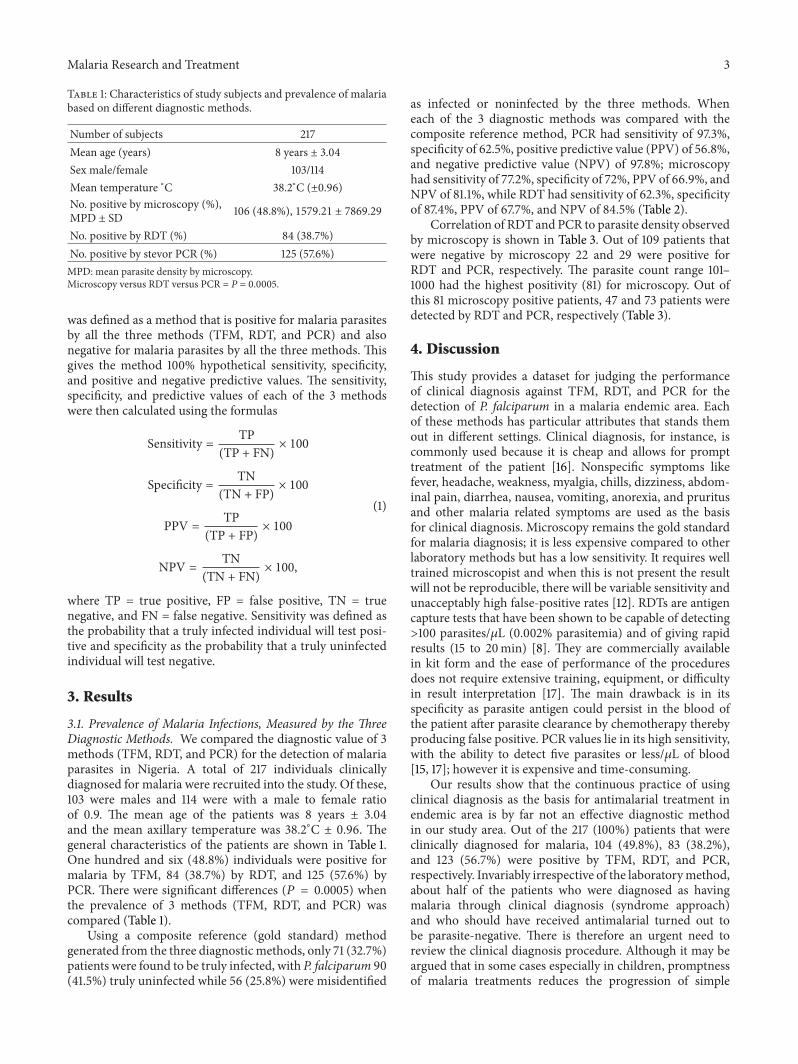
Table 1: Characteristics of study subjects and prevalence of malariabased on different diagnostic methods.
Number of subjects 217Mean age (years) 8 years ± 3.04Sex male/female 103/114Mean temperature ∘C 38.2∘C (±0.96)No. positive by microscopy (%),MPD ± SD 106 (48.8%), 1579.21 ± 7869.29
No. positive by RDT (%) 84 (38.7%)No. positive by stevor PCR (%) 125 (57.6%)MPD: mean parasite density by microscopy.Microscopy versus RDT versus PCR = P = 0.0005.
was defined as a method that is positive for malaria parasitesby all the three methods (TFM, RDT, and PCR) and alsonegative for malaria parasites by all the three methods. Thisgives the method 100% hypothetical sensitivity, specificity,and positive and negative predictive values. The sensitivity,specificity, and predictive values of each of the 3 methodswere then calculated using the formulas
Sensitivity = TP(TP + FN)
× 100
Specificity = TN(TN + FP)
× 100
PPV = TP(TP + FP)
× 100
NPV = TN(TN + FN)
× 100,
(1)
where TP = true positive, FP = false positive, TN = truenegative, and FN = false negative. Sensitivity was defined asthe probability that a truly infected individual will test posi-tive and specificity as the probability that a truly uninfectedindividual will test negative.
3. Results
3.1. Prevalence of Malaria Infections, Measured by the ThreeDiagnostic Methods. We compared the diagnostic value of 3methods (TFM, RDT, and PCR) for the detection of malariaparasites in Nigeria. A total of 217 individuals clinicallydiagnosed for malaria were recruited into the study. Of these,103 were males and 114 were with a male to female ratioof 0.9. The mean age of the patients was 8 years ± 3.04and the mean axillary temperature was 38.2∘C ± 0.96. Thegeneral characteristics of the patients are shown in Table 1.One hundred and six (48.8%) individuals were positive formalaria by TFM, 84 (38.7%) by RDT, and 125 (57.6%) byPCR. There were significant differences (𝑃 = 0.0005) whenthe prevalence of 3 methods (TFM, RDT, and PCR) wascompared (Table 1).
Using a composite reference (gold standard) methodgenerated from the three diagnostic methods, only 71 (32.7%)patients were found to be truly infected, with P. falciparum 90(41.5%) truly uninfected while 56 (25.8%) were misidentified
as infected or noninfected by the three methods. Wheneach of the 3 diagnostic methods was compared with thecomposite reference method, PCR had sensitivity of 97.3%,specificity of 62.5%, positive predictive value (PPV) of 56.8%,and negative predictive value (NPV) of 97.8%; microscopyhad sensitivity of 77.2%, specificity of 72%, PPV of 66.9%, andNPV of 81.1%, while RDT had sensitivity of 62.3%, specificityof 87.4%, PPV of 67.7%, and NPV of 84.5% (Table 2).
Correlation of RDT and PCR to parasite density observedby microscopy is shown in Table 3. Out of 109 patients thatwere negative by microscopy 22 and 29 were positive forRDT and PCR, respectively. The parasite count range 101–1000 had the highest positivity (81) for microscopy. Out ofthis 81 microscopy positive patients, 47 and 73 patients weredetected by RDT and PCR, respectively (Table 3).
4. Discussion
This study provides a dataset for judging the performanceof clinical diagnosis against TFM, RDT, and PCR for thedetection of P. falciparum in a malaria endemic area. Eachof these methods has particular attributes that stands themout in different settings. Clinical diagnosis, for instance, iscommonly used because it is cheap and allows for prompttreatment of the patient [16]. Nonspecific symptoms likefever, headache, weakness, myalgia, chills, dizziness, abdom-inal pain, diarrhea, nausea, vomiting, anorexia, and pruritusand other malaria related symptoms are used as the basisfor clinical diagnosis. Microscopy remains the gold standardfor malaria diagnosis; it is less expensive compared to otherlaboratory methods but has a low sensitivity. It requires welltrained microscopist and when this is not present the resultwill not be reproducible, there will be variable sensitivity andunacceptably high false-positive rates [12]. RDTs are antigencapture tests that have been shown to be capable of detecting>100 parasites/𝜇L (0.002% parasitemia) and of giving rapidresults (15 to 20min) [8]. They are commercially availablein kit form and the ease of performance of the proceduresdoes not require extensive training, equipment, or difficultyin result interpretation [17]. The main drawback is in itsspecificity as parasite antigen could persist in the blood ofthe patient after parasite clearance by chemotherapy therebyproducing false positive. PCR values lie in its high sensitivity,with the ability to detect five parasites or less/𝜇L of blood[15, 17]; however it is expensive and time-consuming.
Our results show that the continuous practice of usingclinical diagnosis as the basis for antimalarial treatment inendemic area is by far not an effective diagnostic methodin our study area. Out of the 217 (100%) patients that wereclinically diagnosed for malaria, 104 (49.8%), 83 (38.2%),and 123 (56.7%) were positive by TFM, RDT, and PCR,respectively. Invariably irrespective of the laboratorymethod,about half of the patients who were diagnosed as havingmalaria through clinical diagnosis (syndrome approach)and who should have received antimalarial turned out tobe parasite-negative. There is therefore an urgent need toreview the clinical diagnosis procedure. Although it may beargued that in some cases especially in children, promptnessof malaria treatments reduces the progression of simple

4 Malaria Research and Treatment
Table 2: Sensitivity, specificity, and predictive values of the three diagnostic methods.
Diagnostic methodsParameter for assessment
TP(no)
FP(no)
TN(no)
FN(no)
Sensitivity(%)
Specificity(%)
PPV(%)
NPV(%)
TFM 71 35 90 21 77.2 72 67 81.1RDT 71 13 90 43 62.3 87.4 84.5 67.7PCR 71 54 90 2 97.3 62.5 56.8 97.8Composite reference 71 0 90 0 100 100 100 100TP: true positive; FP: false positive; TN: true negative; FN: false negative; TFM: thick film microscopy;RDT: rapid diagnostic test; PCR: polymerase chain reaction; no: number; %: percent.
Table 3: Stratification by parasite density in thick blood smear and correlation with rapid diagnostic test (RDT) and stevor PCR.
Parasite count range0 1–100 101–1000 >1000
No. observed 109 10 81 17Mean parasite count/𝜇L (range) 0 91 (41.6–100) 408 (110–948) 8,101 (1,050–81,600)No. positive for clinical 109 (100%) 10 (100%) 81 (100%) 17 (100%)No. negative for clinical 0 0 0 0No. positive for RDT 22 (20.2%) 2 (20.0%) 47 (58.0%) 12 (70.6%)No. negative for RDT 87 (79.8%) 8 (80.0%) 34 (42.0%) 4 (23.5%)No. positive for PCR 29 (26.6%) 8 (80.0%) 73 (90.1%) 13 (76.5)No. negative for PCR 80 (73.4%) 2 (20.0) 8 (9.9%) 4 (25.5%)
malaria to severe malaria, which still encourages syndromicapproach to malaria diagnosis. Nevertheless malaria overdiagnosis is still a major public health problem in Africa withstudies suggesting between 50% and 99% of those prescribedantimalarial to be test negatives depending on endemicityof the clinical setting [5, 18, 19]. The ability to rule outmalaria can help to better diagnose and treat other causes offever such as acute respiratory infection, typhoid fever, andmeningitis and also avoid exposing those without malaria todrug and restricting antimalarial use to true test-positives.Till date,many clinicians in this study area still depend largelyon clinical diagnosis. Our study confirmed that continualdependence on this method will lead to overdiagnosis ofmalaria which will result into drug wastage and encourageantimalarial drug resistance.
In this study routinemicroscopic examination ofGiemsa-stained blood smearswhich is considered as the gold standardfor malaria diagnosis had a sensitivity of 77.2% and was ableto detect more parasites than the RDT (sensitivity 62.3%).Though the specificity of microscopy (72%) was not as highas that of RDT (87.4%); nevertheless, it has high sensitiv-ity, possibility for quantification of parasitemia, and easyhandling which is a good advantage. Detection of parasitesdepends on many factors including the amount of bloodprocessed and the competence of the microscopist, amongothers [20]. Also the information obtained by microscopyis limited when parasite levels are very low or when par-asite morphology is altered [8]. The development of rapiddiagnostic assays has attempted to address some of theseshortcomings of microscopy. However it has low sensitivityat parasitemia below 100 parasites/𝜇L and have insufficient
accuracy [21]. RDTs have the potential to improve the accu-racy and time needed for malaria diagnosis particularly forlaboratories in low or nonendemic countries, where expertisewith microscopy may be limited. Major advantages of RDTsinclude the fact that it can be performed close to home insettings with no sophisticated infrastructure, and they do notrequire much skill although some level of training is neededin order for RDTs to be used properly.
Different PCR based methods have been constantlyshown to be powerful tools for malaria diagnosis with bettersensitivity than conventional microscopy and antigen-baseddiagnostic tests [18, 22]. Most positive cases were detectedby the stevor PCR in this study and this method has beenreported to be at least 100-foldmore sensitive than other PCRassays [15, 23]. Generally, PCR has proven to be a sensitivemethod for diagnosis of all four species of human malariaparasites. The detection of <5 parasite/𝜇L and identificationto the species level make this an excellent technique againstwhich to compare the sensitivity and specificity of othernonmolecular methods [24].
Greater percentage of children presented at general out-patient department of the hospital in our study with feverwere diagnosed formalaria (PCR—56.7%,microscopy 49.8%,and RDT 38.2%). Available records also show that at least50% of the population of Nigeria suffer from at least oneepisode of malaria each year accounting for over 45% of allout-patient visits [25]. The implication of this is that malariais still a public health problem in this area. More concertedeffort is needed by government and all stake holders involvedin malaria control if the goal of eradicating malaria by 2015 isto be achieved.

Malaria Research and Treatment 5
In conclusion our study revealed the need for completeshift from symptom-based diagnosis to parasite-based diag-nosis. This can bring significant improvement to tropicalfever management and reduce drug wastage and also help tocurtail development of malaria drug resistance.
Conflict of Interests
The authors have no conflict of interests to declare.
Acknowledgment
The authors are grateful to Mr. Akeem Abiodun and Mr.Adeola Ayileka for their technical support. Sincere appreci-ation also goes to all consenting participants and parents ofparticipants for their cooperation.
References
[1] M. Cairns, A. Roca-Feltrer, T. Garske et al., “Estimating thepotential public health impact of seasonal malaria chemopre-vention in African children,” Nature Communications, vol. 3,article 881, 2012.
[2] C. Beadle, G. W. Long, W. R. Weiss et al., “Diagnosis of malariaby detection of Plasmodium falciparum HRP-2 antigen with arapid dipstick antigen-capture assay,” The Lancet, vol. 343, no.8897, pp. 564–568, 1994.
[3] I.M. Bashir,N.Otsyula,G.Awinda,M. Spring, P. Schneider, andJ. N. Waitumbi, “Comparison of PfHRP-2/pLDH ELISA, qPCRand microscopy for the detection of Plasmodium events andprediction of sick visits during a malaria vaccine study,” PLoSONE, vol. 8, no. 3, Article ID e56828, 2013.
[4] S. Shillcutt, C. Morel, C. Goodman et al., “Cost-effectivenessof malaria diagnostic methods in sub-Saharan Africa in anera of combination therapy,” Bulletin of the World HealthOrganization, vol. 86, no. 2, pp. 101–110, 2008.
[5] M. Amexo, R. Tolhurst, G. Barnish, and I. Bates, “Malariamisdiagnosis: effects on the poor and vulnerable,” The Lancet,vol. 364, no. 9448, pp. 1896–1898, 2004.
[6] M. T. Makler, C. J. Palmer, and A. L. Ager, “A review of practicaltechniques for the diagnosis of malaria,” Annals of TropicalMedicine and Parasitology, vol. 92, no. 4, pp. 419–433, 1998.
[7] F. A. Abanyie, P. M. Arguin, and J. Gutman, “State of malariadiagnostic testing at clinical laboratories in the United States,2010: a nationwide survey,”Malaria Journal, vol. 10, no. 1, article340, 2011.
[8] A.Moody, “Rapid diagnostic tests formalaria parasites,”ClinicalMicrobiology Reviews, vol. 15, no. 1, pp. 66–78, 2002.
[9] Y. Wataya, F. Kubochi, C. Mizukoshi et al., “DNA diagnosis offalciparummalaria,”Nucleic Acids Symposium Series, no. 25, pp.155–156, 1991.
[10] O. Ojurongbe, T. O. Ogungbamigbe, A. F. Fagbenro-Beyioku,R. Fendel, P. G. Kremsner, and J. F. J. Kun, “Rapid detection ofPfcrt and Pfmdr1 mutations in Plasmodium falciparum isolatesby FRET and in vivo response to chloroquine among childrenfrom Osogbo, Nigeria,”Malaria Journal, vol. 6, article 41, 2007.
[11] F. Kawamoto, H. Miyake, O. Kaneko et al., “Sequence variationin the 18S rRNA gene, a target for PCR-basedmalaria diagnosis,in Plasmodium ovale from Southern Vietnam,” Journal ofClinical Microbiology, vol. 34, no. 9, pp. 2287–2289, 1996.
[12] C. Wongsrichanalai, M. J. Barcus, S. Muth, A. Sutamihardja,and W. H. Wernsdorfer, “A review of malaria diagnostic tools:microscopy and rapid diagnostic test (RDT),” The AmericanJournal of Tropical Medicine and Hygiene, vol. 77, no. 6, supple-ment, pp. 119–127, 2007.
[13] WHO, Basic Malaria Microscopy, Part II: Tutor’s Guide, WorldHealth Organization, Lyon, France, 2010.
[14] Z. Lin, J. G. Suzow, J. M. Fontaine, and E. W. Naylor, “Asimple automated DNA extraction method for dried bloodspecimens collected on filter paper,” Journal of the Associationfor Laboratory Automation, vol. 10, no. 5, pp. 310–314, 2005.
[15] S. I. Oyedeji, H. O. Awobode, G. C. Monday, E. Kendjo, P. G.Kremsner, and J. F. Kun, “Comparison of PCR-based detec-tion of Plasmodium falciparum infections based on single andmulticopy genes,”Malaria Journal, vol. 6, no. 1, article 112, 2007.
[16] N. Tangpukdee, C. Duangdee, P. Wilairatana, and S. Krudsood,“Malaria diagnosis: a brief review,” Korean Journal of Parasitol-ogy, vol. 47, no. 2, pp. 93–102, 2009.
[17] C. J. M.Whitty, M. Armstrong, and R. H. Behrens, “Self-testingfor falciparum malaria with antigen-capture cards by travelerswith symptoms of malaria,” The American Journal of TropicalMedicine and Hygiene, vol. 63, no. 5-6, pp. 295–297, 2000.
[18] O. Ojurongbe, S. I. Oyedeji, W. A. Oyibo, A. F. Fagbenro-Beyioku, and J. F. Kun, “Molecular surveillance of drug-resistantPlasmodium falciparum in two distinct geographical areas ofNigeria,”Wiener KlinischeWochenschrift, vol. 122, no. 23-24, pp.681–685, 2010.
[19] H. Reyburn, R. Mbatia, C. Drakeley et al., “Overdiagnosis ofmalaria in patients with severe febrile illness in Tanzania: aprospective study,” The British Medical Journal, vol. 329, no.7476, pp. 1212–1215, 2004.
[20] J. E. Rosenblatt, L. B. Reller, and M. P. Weinstein, “Laboratorydiagnosis of infections due to blood and tissue parasites,”Clinical Infectious Diseases, vol. 49, no. 7, pp. 1103–1108, 2009.
[21] C. K. Murray, R. A. Gasser Jr., A. J. Magill, and R. S. Miller,“Update on rapid diagnostic testing formalaria,”ClinicalMicro-biology Reviews, vol. 21, no. 1, pp. 97–110, 2008.
[22] R. E. Coleman, J. Sattabongkot, S. Promstaporm et al., “Com-parison of PCR and microscopy for the detection of asymp-tomatic malaria in a Plasmodium falciparum/vivax endemicarea inThailand,”Malaria Journal, vol. 5, article 121, 2006.
[23] Q. Cheng, N. Cloonan, K. Fischer et al., “stevor and rifare Plasmodium falciparum multicopy gene families whichpotentially encode variant antigens,”Molecular and BiochemicalParasitology, vol. 97, no. 1-2, pp. 161–176, 1998.
[24] D. T. McNamara, L. J. Kasehagen, B. T. Grimberg, J. Cole-Tobian, W. E. Collins, and P. A. Zimmerman, “Diagnosinginfection levels of four human malaria parasite species by apolymerase chain reaction/ligase detection reaction fluorescentmicrosphere-based assay,” The American Journal of TropicalMedicine and Hygiene, vol. 74, no. 3, pp. 413–421, 2006.
[25] T. O. Ogungbamigbe, O. O. Ojurongbe, P. S. Ogunro, O. A.Olowe, and P. O. Elemile, “Prevalence and transmission patternof Plasmodium falciparum infection in Osogbo metropolis,Southwest, Nigeria,” African Journal of Medicine and MedicalSciences, vol. 36, no. 4, pp. 305–310, 2007.

Submit your manuscripts athttp://www.hindawi.com
Stem CellsInternational
Hindawi Publishing Corporationhttp://www.hindawi.com Volume 2014
Hindawi Publishing Corporationhttp://www.hindawi.com Volume 2014
MEDIATORSINFLAMMATION
of
Hindawi Publishing Corporationhttp://www.hindawi.com Volume 2014
Behavioural Neurology
EndocrinologyInternational Journal of
Hindawi Publishing Corporationhttp://www.hindawi.com Volume 2014
Hindawi Publishing Corporationhttp://www.hindawi.com Volume 2014
Disease Markers
Hindawi Publishing Corporationhttp://www.hindawi.com Volume 2014
BioMed Research International
OncologyJournal of
Hindawi Publishing Corporationhttp://www.hindawi.com Volume 2014
Hindawi Publishing Corporationhttp://www.hindawi.com Volume 2014
Oxidative Medicine and Cellular Longevity
Hindawi Publishing Corporationhttp://www.hindawi.com Volume 2014
PPAR Research
The Scientific World JournalHindawi Publishing Corporation http://www.hindawi.com Volume 2014
Immunology ResearchHindawi Publishing Corporationhttp://www.hindawi.com Volume 2014
Journal of
ObesityJournal of
Hindawi Publishing Corporationhttp://www.hindawi.com Volume 2014
Hindawi Publishing Corporationhttp://www.hindawi.com Volume 2014
Computational and Mathematical Methods in Medicine
OphthalmologyJournal of
Hindawi Publishing Corporationhttp://www.hindawi.com Volume 2014
Diabetes ResearchJournal of
Hindawi Publishing Corporationhttp://www.hindawi.com Volume 2014
Hindawi Publishing Corporationhttp://www.hindawi.com Volume 2014
Research and TreatmentAIDS
Hindawi Publishing Corporationhttp://www.hindawi.com Volume 2014
Gastroenterology Research and Practice
Hindawi Publishing Corporationhttp://www.hindawi.com Volume 2014
Parkinson’s Disease
Evidence-Based Complementary and Alternative Medicine
Volume 2014Hindawi Publishing Corporationhttp://www.hindawi.com