research article cellcounter: novel open-source software
TRANSCRIPT
Research ArticleCELLCOUNTER: Novel Open-Source Software for Counting CellMigration and Invasion In Vitro
Xiaoni Li,1 Hongshun Yang,2 Hailiang Huang,3,4 and Tao Zhu1
1 Hefei National Laboratory for Physical Sciences at Microscale and School of Life Sciences, USTC, Hefei, Anhui, China2 School of Information Science and Technology, USTC, Hefei, Anhui, China3 Analytic and Translational Genetics Unit, Massachusetts General Hospital, Boston, MA, USA4Program in Medical and Population Genetics, Broad Institute of MIT and Harvard, Cambridge, MA, USA
Correspondence should be addressed to Hailiang Huang; [email protected] and Tao Zhu; [email protected]
Received 16 April 2014; Revised 7 June 2014; Accepted 7 June 2014; Published 26 June 2014
Academic Editor: Calvin Yu-Chian Chen
Copyright © 2014 Xiaoni Li et al.This is an open access article distributed under the Creative CommonsAttribution License, whichpermits unrestricted use, distribution, and reproduction in any medium, provided the original work is properly cited.
Transwell Boyden chamber based migration/invasion assay is a simple and extensively used approach for the characterization ofcell motility in vitro. Cell motility is quantified by counting the number of cells that pass through the filter membrane.The countingis usually performed manually, which is laborious and error prone. We have therefore developed CELLCOUNTER, an applicationthat is capable of recognizing and counting the total number of cells through an intuitive graphical user interface.The counting canbe performed in batch, and the counting results can be visualized and further curated manually. CELLCOUNTER will be helpfulin streamlining the experimental process and improving the reliability of the data acquisition.
1. Introduction
Cell migration is the movement of cells from one locationto another generally in response to and toward specificexternal chemical signals. Cell invasion is similar to cellmigration, except that it requires the cell to migrate throughan extracellular matrix or basement membrane barrier byenzymatically degrading the barrier. Cell migration/invasionis central to many physiological and pathological processessuch as embryonic development, wound repair, and tumormetastasis [1–3].
Transwell Boyden chamber [4] based cell migration/in-vasion assay is a simple and extensively used approach for thequantitation of cell motility in vitro [3, 5].The number of cellsthat pass through the filter membrane from Boyden chamberis usually counted manually from the inverted microscopicimages. Such images may contain hundreds of cells andmanually counting the number of them is not a trivial work.Existing image analysis programs, for example, CELLPRO-FILER and IMAGEJ [6–8], require pipeline/macro/pluginfiles that are specific to the cell/assay types, which is not
available yet for the Transwell assays. Although it is possibleto create a CELLPROFILER/IMAGEJ pipeline/macro/pluginfor the Transwell assays, an independent program takinginto account the specific characteristics of the assay canmost likely perform better [9]. Another related application[10], although available for some migration assays, requiresfluorescently stained cells and does notwork for theTranswellassays.
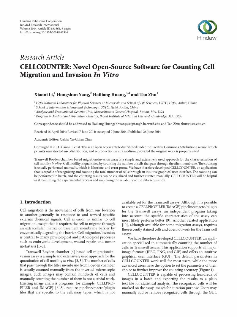
We have therefore developed CELLCOUNTER, an appli-cation specialized in automatically counting the number ofcells in Transwell assays. This application supports all majorimage formats (JPEG, PNG, and GIF) and offers an intuitivegraphical user interface (GUI). The default parameters inCELLCOUNTER work well for most users, while the moreadvanced users have the option to set the parameters of theirchoice to further improve the counting accuracy (Figure 1).
CELLCOUNTER is capable of processing hundreds ofimages in a batch and exporting the results to a plaintext file for statistical analysis. The recognized cells will bemarked on the assay images for curation purpose. Users maymanually add or remove recognized cells through the GUI.
Hindawi Publishing CorporationBioMed Research InternationalVolume 2014, Article ID 863564, 6 pageshttp://dx.doi.org/10.1155/2014/863564

2 BioMed Research International
1 2 3 4 5
67
Figure 1: The graphical user interface of CELLCOUNTER. Green circles indicate automatically recognized cells and red circles indicatemanually labeled cells. Functions for buttons/inputs are (1) manual counting/curation; (2) zooming in; (3) zooming out; (4) restoring to theoriginal image size; (5) saving the counting results (including images); (6) setting the radius of the label (for visualization only); (7) settingthe radius of small wells (in px, see the main text for more details, default recommended).
The counting process, including the automatic recognitionand manual curations, can be saved to a disk file and loadedback to resume the work. CELLCOUNTER will prove tobe useful in streamlining the analysis of Transwell assays,reducing human errors, and improving the reliability of theassay quantitation.
2. Materials and Methods
2.1. Cell Lines. 20 images from 6 human cancer cell lineswere used in this study (Table 1): 17 images are from breastcancer cell lines (1 x T47D, 13 x MCF-7, 2 x MDA-MB-231,and 1 xMDA-MB-435s), 2 images are fromhepatic carcinoma(Hep3B), and 1 image is from prostatic cancer (pc-3). Allcell lines were obtained from the American Type CultureCollection (Rockville, MD, USA).
2.2. Transwell Assay. Assays were performed in BioCoatMatrigel invasion chambers (Corning Costar, Acton, MA) asdescribed previously [11–14]. The assay images were capturedusing a Nikon camera with 5M pixels.
2.3. Biology Stimulation. Five images (out of the 20 totalimages) are from stimulated cell (4 x MCF-7 and 1 x MDA-MB-231). We stimulated the cells with 20 ng/mL epidermalgrowth factor (EGF) when the confluency reached 95%. For
the non-stimulated cells, we added ethyl alcohol as a controlcompound. After the stimulation we incubated the cells for 4hours.
2.4. Existing Software. CELLPROFILER and IMAGEJ areversatile and flexible image processing software for scientificpurposes. For comparison purpose, we used a naıve setup andchoice of parameters.
CELLPROFILER uses a customized pipeline, includinginput and analysis modules, to process images. The inputmodules in our pipeline are “Images,” “Metadata,” “Name-sAndType,” and “Groups.” The analysis modules are “Col-orToGray,” “ApplyThreshold,” and “IdentifyPrimaryObjects.”There are many tunable parameters in each module. Wechose the parameters that make the most sense to us.The project configuration file used in this study (CellPro-filer.cpproj) with all parameters is available to download athttps://bitbucket.org/linora/cellcounter/downloads.
For IMAGEJ, after loading the images, we first adjustedthe color threshold using the default thresholding method(Image→Adjust→Color Threshold→ select). We then an-alyzed the images using Analyze→Analyze particle→ sum-marize.
2.5. Implementation. CELLCOUNTER was developed usingthe C++ programming language. Some image processing

BioMed Research International 3
Table 1: Images used in this study. Cells were stained with 0.1% crystal violet for 20 minutes for the deep staining group and 8 minutes forthe light staining group. High density images refer to those having over 400 cells and low density images refer to those having less than 200cells. Biology stimulation of cells was discussed in Section 2.
ID Cell line Stimulation Stain Density Number of cells
Expert CELLCOUNTER Beginner
1 T47D No — High 931 957 955
2 MCF-7 No Deep High 792 748 538
3 MDA-MB-231 No — High 475 479 413
4 pc-3 No — — 523 507 390
5 MCF-7 — Deep — 314 322 263
6 MCF-7 Yes Deep — 359 364 300
7 MDA-MB-231 Yes — High 566 572 366
8 Hep3B — Light — 284 279 249
9 MDA-MB-435s — — — 464 461 430
10 Hep3B — — — 186 178 144
11 MCF-7 — Light Low 165 159 —
12 MCF-7 Yes — — 403 403 —
13 MCF-7 No Deep — 417 395 —
14 MCF-7 Yes Light — 189 187 —
15 MCF-7 — Light Low 94 98 —
16 MCF-7 Yes — Low 120 118 —
17 MCF-7 — — Low 139 140 —
18 MCF-7 — Light — 8 8 —
19 MCF-7 — Deep High 554 553 —
20 MCF-7 — — Low 191 192 —
algorithms were implemented with the help of Open SourceComputer Vision Library [15], and the graphical user inter-face was designed using Nokia Qt (a cross platform appli-cation and UI framework). CELLCOUNTER is compatiblewith all major platforms including Windows (XP or newer),Mac OSX (10.6+), and Linux (Kernel 3.x). Assays imagesfor CELLCOUNTER have to have an aspect ratio of 4 : 3(default in most digital cameras) and a minimal resolution of1280 px by 960 px. Images that have the correct aspect ratiobut in higher resolutions will be resized to 1280 px by 960 px.Resizing to a lower resolution reduces the use of memoryand CPU time in analyzing images. We have verified that theresolution of 1280 px by 960 px is high enough to retain all thenecessary details for cell counting.
In CELLCOUNTER, the original assay image is firstconverted to a grayscale image, and further partitioned intocell areas and background areas using an adaptive threshold.Areas that have grayscale below the threshold (close to white)
are background and areas that have grayscale above thethreshold (close to black) are cells. The threshold is estab-lished by using the maximum entropy method [16]. A smallvalue can be added to the threshold (threshold adjustment)to fine-tune the counting accuracy. Users may adjust thisvalue in GUI and use the visual feedback to choose theoptimal value. Alternately, the default valueworks verywell inmost circumstances. Standard image processing procedures,such as contrast enhancement, smoothing, eroding, anddilating [17], are also performed to remove noises in theimage.
Due to the design of the assay, small wells in theequipment can be captured in the images and counted falselyas cells. We solve this problem by further partitioning thecell areas into small wells and true cells. Another adaptivethreshold is established by applying the OSTU method[18] to only the cell areas identified in the previous step.Because the small wells are generally darker than the true

4 BioMed Research International
0
200
400
600
800
1000
1200
1 2 3 4 5 6 7 8 9 10
ExpertsCELLCOUNTERBeginners
Cel
l cou
ntin
g re
sults
Figure 2: Cell counting results for 10 assay images from 3 experts,CELLCOUNTER, and 3 beginners. Error bars are standard devia-tions (only available for human counting results).
cells, applying this adaptive threshold, followed by imagesmoothing, eroding, and dilating, can successfully removethe small wells from the cell areas.
One cell area may contain multiple overlapping cells. Wecount the number of cells in a particular cell area by using theradius of itsmaximum inscribed circle. If the radius is smallerthan the empirical threshold of the cell radius r (default to6 pixels) and greater than the empirical threshold for noise(default to 4 pixels), we count this cell area as a single cell. Ifthe radius is greater than the empirical threshold (r), we countthe number of cells in this cell area as
ℎ × [
𝐿
2𝑟
+ 𝑎] × [
𝑊
2𝑟
+ 𝑎] , (1)
in which L andW are the length and width of the minimumbounding rectangle, h is the number of layers that the cellsstack, and a is a parameter to account for cells at theboundary of the rectangle. h and a are unknown and havebeen estimated using the human counted cell numbers as thetraining dataset (ℎ ≈ 4.0 and 𝑎 ≈ 0.5). The total number ofcells in the image is then the summation of cell numbers ineach cell area.
3. Results and Discussion
The user interface and a counting example using CELL-COUNTER is provided in Figure 1. We used CELL-COUNTER to analyze 10 assay images (randomly selectedfrom the 20 images in Section 2). The cell numbers countedby the experts (3 researchers that are proficient in cell count-ing) are used as the gold standard and compared with the cellnumbers counted by CELLCOUNTER (default parameters,without manual curation) and beginners (3 researchers withbasic trainings in cell counting).
As shown in Figure 2, we found that the cell numberscounted by CELLCOUNTER are statistically the same as
the numbers counted by experts (2-way ANOVA 𝑃 value =0.91). In contrast, the beginners gave slightly smaller cellcounts (2-way ANOVA 𝑃 value = 0.04), and these cell countshave a significantly higher standard deviation (Wilcoxon test𝑃 value = 0.004) than the cell counts from the experts.Therefore, we conclude that CELLCOUNTER is able toperform accurate cell counting and improve the stabilityof the counting results. Additional tests using unpublishedimages have confirmed this conclusion (results not shown).
Systematic biases can lead to false positive findings. Forexample, if software systematically reports a larger (com-paring with the true) number of cells if they are stainedfor a longer time, and if the person who stains the casesamples tends to stain them longer, we might falsely claim asignificant case/control difference. Therefore, we determinedCELLCOUNTER for systematic biases towards the staining,the density of cells, and the biology stimulation. We foundno systematic bias in all 3 conditions (Table 1). The 𝑃 valuesare 0.42 for deep versus light stains, 0.91 for high versus lowcell densities, and 0.96 for stimulated versus nonstimulatedcells.
CELLPROFILER and IMAGEJ are powerful scientificimage processing software. They are versatile and flexiblebut also have steep learning curves and require efforts toset up the right pipeline/parameters. Using a naıve setupas discussed in Section 2, we noticed that both methodsperform worse comparing with CELLCOUNTER (Figure 3).The correlation coefficients (𝑅2) for CELLPROFILER andIMAGEJ are 0.49 and 0.12, respectively, while the 𝑅2 forCELLCOUNTER reached almost 1. CELLCOUNTER per-forms better because it was designed for images from theseassays, while CELLPROFILER and IMAGEJ have more gen-eral purposes. We are aware that more sophisticated setupor choices of parameters for CELLPROFILER and IMAGEJmay improve their performance. However, we argue that ourchoices are representative of average users with basic trainingand knowledge in image processing.
4. Conclusion
We have developed CELLCOUNTER, a program that fea-tures an intuitive graphical user interface to count thenumber of cells in Transwell Boyden chamber based migra-tion/invasion assays. This program allows high-throughputanalysis of a large number of assay images. The countedcells are visibly marked on the assay images and can bemanually curated. The accuracy of the counting results hasbeen validated using expert counted cell numbers as the goldstandard.
CELLCOUNTER significantly simplifies the data acqui-sition in the Transwell assays, reduces human errors, andimproves the stability of counting results. It will prove to bea helpful tool in the study of cell invasion and metastasis invitro.
CELLCOUNTER is currently available forWindows,MacOSX, and Linux platforms and can be downloaded fromhttps://bitbucket.org/linora/cellcounter/downloads.

BioMed Research International 5
0
200
400
600
800
1000
1200
0 200 400 600 800 1000
CELLCOUNTER
Cell counting results of experts
Cel
l cou
ntin
g re
sults
of
CELL
COU
NTE
R
R2= 0.996
(a)
0
200
400
600
800
1000
1200
1400
0 200 400 600 800 1000Cell counting results of experts
Cell
coun
ting
resu
lts o
f CE
LLPR
OFI
LER
R2= 0.490
CELLPROFILER
(b)
0
200
400
600
800
1000
1200
1400
1600
1800
2000
0 200 400 600 800 1000Cell counting results of experts
Cell
coun
ting
resu
lts o
f IM
AGEJ
IMAGEJ
R2= 0.117
(c)
Figure 3: Counting results from CELLCOUNTER (a), CELLPROFILER (b), and IMAGEJ (c) comparing with human counting results.
Conflict of Interests
The authors declare that there is no conflict of interests.
Acknowledgments
The authors thank Dr. Zhiming Pi for helpful advice toimprove this software. They would also like to thank themembers of their laboratories for contributing to the devel-opment of the software and this paper. This work was
supported by the National Key Scientific Program of China(2012CB934002, 2010CB912804) and National Natural Sci-ence Foundation of China (81272925).
References
[1] A. J. Ridley, M. A. Schwartz, K. Burridge et al., “Cell migration:integrating signals from front to back,” Science, vol. 302, no.5651, pp. 1704–1709, 2003.
6 BioMed Research International
[2] S. A. Courtneidge, “Cell migration and invasion in humandisease: the Tks adaptor proteins,” Biochemical Society Trans-actions, vol. 40, no. 1, pp. 129–132, 2012.
[3] E. A. Goncharova, D. A. Goncharov, and V. P. Krymskaya,“Assays for in vitromonitoring of human airway smoothmuscle(ASM) and human pulmonary arterial vascular smooth muscle(VSM) cell migration,” Nature Protocols, vol. 1, no. 6, pp. 2933–2939, 2006.
[4] H. Chen, “Boyden chamber assay,” Methods in Molecular Biol-ogy, vol. 294, pp. 15–22, 2005.
[5] X. H. Liu, K. H. Lu, K. M. Wang et al., “MicroRNA-196apromotes non-small cell lung cancer cell proliferation andinvasion through targeting HOXA5,” BMC Cancer, vol. 12,article 348, 2012.
[6] A. E.Carpenter, T. R. Jones,M.R. Lamprecht et al., “CellProfiler:image analysis software for identifying and quantifying cellphenotypes,” Genome Biology, vol. 7, no. 10, article R100, 2006.
[7] M. R. Lamprecht, D. M. Sabatini, and A. E. Carpenter, “Cell-Profiler: free, versatile software for automated biological imageanalysis,” BioTechniques, vol. 42, no. 1, pp. 71–75, 2007.
[8] C. A. Schneider, W. S. Rasband, and K. W. Eliceiri, “NIH Imageto ImageJ: 25 years of image analysis,” Nature Methods, vol. 9,no. 7, pp. 671–675, 2012.
[9] N. N. Kachouie, L. Kang, and A. Khademhosseini, “Arraycount,an algorithm for automatic cell counting in microwell arrays,”BioTechniques, vol. 47, no. 3, pp. 1–100, 2009.
[10] B. K. Al-Khazraji, P. J. Medeiros, N. M. Novielli, and D. N. Jack-son, “An automated cell-counting algorithm for fluorescently-stained cells in migration assays,” Biological Procedures Online,vol. 13, no. 1, article 9, pp. 1–9, 2011.
[11] S. Mukhina, H. C. Mertani, K. Guo, K. Lee, P. D. Gluckman,and P. E. Lobie, “Phenotypic conversion of human mammarycarcinoma cells by autocrine human growth hormone,”Proceed-ings of the National Academy of Sciences of the United States ofAmerica, vol. 101, no. 42, pp. 15166–15171, 2004.
[12] X. Li, X. Liu, W. Xu et al., “C-MYC-regulated miR-23a/24-2/27a cluster promotes mammary carcinoma cell invasion andhepatic metastasis by targeting sprouty2,” Journal of BiologicalChemistry, vol. 288, no. 25, pp. 18121–18133, 2013.
[13] P. Qian, Z. Zuo, Z. Wu et al., “Pivotal role of reduced let-7gexpression in breast cancer invasion and metastasis,” CancerResearch, vol. 71, no. 20, pp. 6463–6474, 2011.
[14] J. Chen, Y. Yao, C. Gong et al., “CCL18 from tumor-associatedmacrophages promotes breast cancermetastasis via PITPNM3,”Cancer Cell, vol. 19, no. 4, pp. 541–555, 2011.
[15] G. Bradski, “TheOpenCV library,”DrDobbs Journal, vol. 25, no.11, pp. 120–126, 2000.
[16] S. F. Gull and J. Skilling, “Maximum-entropy method in image-processing,” IEE Proceedings F: Communications, Radar andSignal Processing, vol. 131, no. 6, pp. 646–659, 1984.
[17] M. Sonka, V. Hlavac, and R. Boyle, Image Processing, Analysis,and Machine Vision, PWS, Pacific Grove, Calif, USA, 2ndedition, 1999.
[18] H. Ng, “Automatic thresholding for defect detection,” PatternRecognition Letters, vol. 27, no. 14, pp. 1644–1649, 2006.

Submit your manuscripts athttp://www.hindawi.com
Hindawi Publishing Corporationhttp://www.hindawi.com Volume 2014
Anatomy Research International
PeptidesInternational Journal of
Hindawi Publishing Corporationhttp://www.hindawi.com Volume 2014
Hindawi Publishing Corporation http://www.hindawi.com
International Journal of
Volume 2014
Zoology
Hindawi Publishing Corporationhttp://www.hindawi.com Volume 2014
Molecular Biology International
GenomicsInternational Journal of
Hindawi Publishing Corporationhttp://www.hindawi.com Volume 2014
The Scientific World JournalHindawi Publishing Corporation http://www.hindawi.com Volume 2014
Hindawi Publishing Corporationhttp://www.hindawi.com Volume 2014
BioinformaticsAdvances in
Marine BiologyJournal of
Hindawi Publishing Corporationhttp://www.hindawi.com Volume 2014
Hindawi Publishing Corporationhttp://www.hindawi.com Volume 2014
Signal TransductionJournal of
Hindawi Publishing Corporationhttp://www.hindawi.com Volume 2014
BioMed Research International
Evolutionary BiologyInternational Journal of
Hindawi Publishing Corporationhttp://www.hindawi.com Volume 2014
Hindawi Publishing Corporationhttp://www.hindawi.com Volume 2014
Biochemistry Research International
ArchaeaHindawi Publishing Corporationhttp://www.hindawi.com Volume 2014
Hindawi Publishing Corporationhttp://www.hindawi.com Volume 2014
Genetics Research International
Hindawi Publishing Corporationhttp://www.hindawi.com Volume 2014
Advances in
Virolog y
Hindawi Publishing Corporationhttp://www.hindawi.com
Nucleic AcidsJournal of
Volume 2014
Stem CellsInternational
Hindawi Publishing Corporationhttp://www.hindawi.com Volume 2014
Hindawi Publishing Corporationhttp://www.hindawi.com Volume 2014
Enzyme Research
Hindawi Publishing Corporationhttp://www.hindawi.com Volume 2014
International Journal of
Microbiology