research article determining the pharmacokinetics of
TRANSCRIPT

RESEARCH ARTICLE
Determining the pharmacokinetics of nicotinic drugsin the endoplasmic reticulum using biosensorsAmol V. Shivange1,2*, Philip M. Borden2*, Anand K. Muthusamy1,4*, Aaron L. Nichols1, Kallol Bera1, Huan Bao3, Ishak Bishara1, Janice Jeon1,Matthew J. Mulcahy1, Bruce Cohen1, Saidhbhe L. O'Riordan1, Charlene Kim1, Dennis A. Dougherty4, Edwin R. Chapman3, Jonathan S. Marvin2,Loren L. Looger2, and Henry A. Lester1,2
Nicotine dependence is thought to arise in part because nicotine permeates into the endoplasmic reticulum (ER), where itbinds to nicotinic receptors (nAChRs) and begins an “inside-out” pathway that leads to up-regulation of nAChRs on the plasmamembrane. However, the dynamics of nicotine entry into the ER are unquantified. Here, we develop a family of geneticallyencoded fluorescent biosensors for nicotine, termed iNicSnFRs. The iNicSnFRs are fusions between two proteins: a circularlypermutated GFP and a periplasmic choline-/betaine-binding protein engineered to bind nicotine. The biosensors iNicSnFR3a andiNicSnFR3b respond to nicotine by increasing fluorescence at [nicotine] <1 µM, the concentration in the plasma andcerebrospinal fluid of a smoker. We target iNicSnFR3 biosensors either to the plasma membrane or to the ER and measurenicotine kinetics in HeLa, SH-SY5Y, N2a, and HEK293 cell lines, as well as mouse hippocampal neurons and human stemcell–derived dopaminergic neurons. In all cell types, we find that nicotine equilibrates in the ER within 10 s (possibly within1 s) of extracellular application and leaves as rapidly after removal from the extracellular solution. The [nicotine] in the ER iswithin twofold of the extracellular value. We use these data to run combined pharmacokinetic and pharmacodynamicsimulations of human smoking. In the ER, the inside-out pathway begins when nicotine becomes a stabilizingpharmacological chaperone for some nAChR subtypes, even at concentrations as low as ∼10 nM. Such concentrations wouldpersist during the 12 h of a typical smoker’s day, continually activating the inside-out pathway by >75%. Reducing nicotineintake by 10-fold decreases activation to ∼20%. iNicSnFR3a and iNicSnFR3b also sense the smoking cessation drugvarenicline, revealing that varenicline also permeates into the ER within seconds. Our iNicSnFRs enable optical subcellularpharmacokinetics for nicotine and varenicline during an early event in the inside-out pathway.
IntroductionExisting data show that nicotine evokes two processes at neu-ronal nicotinic acetylcholine (ACh) receptors (nAChRs). His-torically, the best-characterized process is activation of nAChRson the plasma membrane (PM). If one considers events at thescale of a neuron, activation of nAChRs at the PMmay be termedthe “outside-in” pathway. Like activation by the endogenousneurotransmitter ACh, activation by exogenous nicotine via theoutside-in activation pathway involves an influx of Na+ and Ca2+
ions, depolarization and therefore increased frequency of neu-ronal action potentials. The outside-in pathway, and perhaps thesubsequent desensitization of nAChRs, leads to the acute effectsafter nicotine enters the airways either from tobacco combus-tion (smoking) or from an electronic nicotine delivery system(ENDS; “vaping”). These acute effects, beginning within <1 minafter inhalation and lasting for dozens ofminutes include a sense
of well-being, a cognitive boost, appetite suppression, increasedtolerance of stressful stimuli, and suppression of withdrawal(Miwa et al., 2011; Naude et al., 2015; Nees, 2015; Picciotto et al.,2015).
Since approximately 2005, evidence has been accumulatingfor a second process. We term this the “inside-out” pathway,because it begins when nicotine permeates into the ER. In theER, nicotine binds to nascent nAChRs and becomes a stabilizingpharmacological chaperone for α4- and β2-subunit–containing(α4β2*) nAChRs, increasing their exit from the ER (Fig. 1 A;Kuryatov et al., 2005; Sallette et al., 2005; Lester et al., 2009).The inside-out pathway leads to up-regulation of nAChRs on thePM. The inside-out pathway results, jointly, from three prop-erties (Fig. 1 A). (1) Like ACh, nicotine activates nAChRs (apharmacological property). (2) In contrast to ACh, nicotine has a
.............................................................................................................................................................................1Division of Biology and Biological Engineering, California Institute of Technology, Pasadena, CA ; 2Janelia Research Campus, Howard Hughes Medical Institute, Ashburn, VA;3Howard Hughes Medical Institute and Department of Neuroscience, University of Wisconsin, Madison, WI; 4Division of Chemistry and Chemical Engineering, CaliforniaInstitute of Technology, Pasadena, CA .
*A.V. Shivange, P.M. Borden, and A.K.Muthusamy contributed equally to this paper; Correspondence toHenryA. Lester: [email protected]; Loren Looger: [email protected].
© 2019 Shivange et al. This article is distributed under the terms of an Attribution–Noncommercial–Share Alike–No Mirror Sites license for the first six months after thepublication date (see http://www.rupress.org/terms/). After six months it is available under a Creative Commons License (Attribution–Noncommercial–Share Alike 4.0International license, as described at https://creativecommons.org/licenses/by-nc-sa/4.0/).
Rockefeller University Press https://doi.org/10.1085/jgp.201812201 738
J. Gen. Physiol. 2019 Vol. 151 No. 6 738–757
Dow
nloaded from http://w
ww
.rupress.org/jgp/article-pdf/151/6/738/1236809/jgp_201812201.pdf by guest on 19 Decem
ber 2021

neutral, membrane-permeant form (a pharmacokinetic prop-erty). (3) In contrast to hydrolysis of ACh within <1 ms by Achsterase, catabolic oxidation of nicotine proceeds on a time scaleof ∼30 min, primarily by cytochrome P450 (another pharma-cokinetic property; Henderson and Lester, 2015; Tanner et al.,2015).
The research community has not yet reported decisive testsfor the relative importance of the inside-out and outside-inpathways in nicotine dependence. Several laboratories aretesting the hypothesis that the selective up-regulation of specificnAChR subtypes via the inside-out pathway is necessary andsufficient for some early events (days to weeks) of nicotine de-pendence (Govind et al., 2009; Henderson and Lester, 2015).Nicotine-induced up-regulation of nAChRs via chaperoning ispost-translational, involving neither gene activation nor mRNAstability of nAChR subunits (Henderson and Lester, 2015).
A satisfactory comparison of the outside-in and inside-outpathways during smoking/vaping has been hampered by lackof information about the pharmacokinetics of nicotine at the
subcellular scale: How long does it take nicotine to permeate intothe ER, and what is its concentration there (Lester et al., 2009;Rollema et al., 2010; Hussmann et al., 2012)? Previous data showthat, when nicotine is applied for more than several hours, it up-regulates nAChRs. This up-regulation has an EC50 of ∼30 nM(Kuryatov et al., 2005). It has not been known (1) whethernicotine concentration in the ER reaches the EC50 for up-regulation; (2) if so, how quickly; and (3) how quickly nicotineleaves the ER.
To approach these questions quantitatively, we have devel-oped a series of genetically encoded intensity-based nicotine-sensing fluorescent reporters (iNicSnFRs). These biosensorscomprise a fusion between a bacterial periplasmic-bindingprotein (PBP) moiety (276 amino acids), a circularly permutedGFP (cpGFP) moiety (244 amino acids), joining regions (“link-ers”), and epitope tags. The development of the glutamate bio-sensor iGluSnFR (Marvin et al., 2013) has provided a model forthis study. We report on use of a novel PBP moiety and onmutations that allow the PBP moiety of the biosensor to bindnicotine. We have directed these iNicSnFRs either to the PM (atthe start of the outside-in pathway) or to the lumen of the ER (atthe start of the inside-out pathway; Fig. 1 B). We have alsoverified the compartmentalization of the iNicSnFRs by showingthat the fluorescence response to nicotine contrasts with re-sponses to membrane-impermeant quaternary amines (Fig. 1 C).
Smoking cessation has become desirable for individual healthand for public health (Royal College of Physicians, 2016). Vare-nicline (shown in Fig. 1 A), a partial agonist for α4β2 nAChRs andfull agonist for α7 nAChRs, is now the most effective syntheticdrug for smoking cessation. Nonetheless, varenicline therapysucceeds in only a minority of people who aspire to quit smoking(Fagerstrom and Hughes, 2008). Varenicline also permeatesmodestly well into the central nervous system (Rollema et al.,2010). Varenicline has a half-life of ∼24 h in human plasma(Faessel et al., 2006) and presumably in human brain. Exposureto varenicline also up-regulates α4β2 nAChRs (Turner et al.,2011; Marks et al., 2015; Govind et al., 2017), an indication thatit too participates in the inside-out pathway. We thereforesought to determine whether varenicline also enters the ER and,if so, how much and how quickly. Serendipitously, an iNicSnFRalso binds to and senses varenicline, and we suggest how vare-nicline’s entry into the ER may limit its therapeutic actions.
Materials and methodsDirected evolution of iNicSnFR proteins usingbacterial-expressed protein assaysThe Results section, below, begins by describing the overallstrategy in constructing the iNicSnFRs. Class F PBPs consist oftwo domains that move relative to each other when the ligandbinds at the interdomain interface (Berntsson et al., 2010).Previous biosensor constructs have placed a cpGFP moleculewithin the PBP. We constructed and measured ∼12,000 mutantsiteratively, using fluorescence measurements as described be-low. We incorporated four additional linker1 residues before theN terminus of the “superfolder” cpGFP gene (Pedelacq et al.,2006; Marvin et al., 2018) and four additional linker2 residues
Figure 1. Strategy of the experiments. (A) Nicotine (pKa, 7.5–8.1) andvarenicline (pKa, 9.5–10) are weak bases. They interconvert on a millisecondtime scale between protonated and deprotonated forms; these are respec-tively membrane impermeant and permeant. (B) The tactic of confining agenetically encoded fluorescent nicotinic drug sensor to the PM or the ER. (C)Choline, ACh, and N’MeNic exist only as charged, membrane-impermeantforms near physiological pH.
Shivange et al. Journal of General Physiology 739
Biosensor for optical subcellular pharmacokinetics https://doi.org/10.1085/jgp.201812201
Dow
nloaded from http://w
ww
.rupress.org/jgp/article-pdf/151/6/738/1236809/jgp_201812201.pdf by guest on 19 Decem
ber 2021

after the C terminus of the superfolder cpGFP, because thiscpGFP variant functions well in the ER (Aronson et al., 2011). Weinserted linker1-cpGFP-linker2 at candidate positions withinOpuBC sequence at positions near the interdomain interface ofthe PBP, based on previous structural data cited in Results forcholine- and betaine-binding class F PBPs. We optimized linker1and linker2 with site-saturated mutagenesis (SSM).
The ligand-binding site (originally for choline and/or beta-ine) lies at the interdomain interface of the PBP. To optimize theligand site for nicotine, we performed SSM on several residuesnear the possible cation-π residues (first-shell residues that liewithin 7 A of the ligand binding pocket; Fig. S1), as well as on“second-shell” residues (residues that showed intraprotein in-teractions with the first-shell residues). Mutagenesis was per-formed by slight modifications to the Quikchange mutagenesisprotocol (Agilent). Each round of SSM used NN(C/G) oligonu-cleotides that provided > 96% residue coverage for a collection of188 randomly chosen clones.
One design goal was a 30% increase in fluorescence (ΔF/F0 =0.3) at [nicotine] = 1 µM, a concentration thought to lie in therange of the peak [nicotine] in the plasma and brain of a smoker(Benowitz et al., 1991; Rollema et al., 2010). In preliminarycharacterization, lysates were tested with excitation at 485 nmand emission at 535 nm. Automated 96-well fluorescence platereaders were used to measure resting and nicotine-inducedfluorescence (F0 and ΔF, respectively; Tecan M1000, equippedwith monocomators; and Tecan Spark M10, equipped with fil-ters). Promising clones were amplified and sequenced. Thebeneficial substitutions identified were combined with side-directed mutagenesis (SDM), and the “best” combination ineach round of evolution was used as a template for the nextround of SSM (Fig. S2).
We conducted one or more rounds of SSM experiments ateach of the 25 codons shown in Fig. S1. In sum, these experi-ments improved the ΔF/F0 at 1 µM nicotine by a factor ∼105
(Fig. S2). In early experiments on weakly responding constructs,we measured responses to much higher concentrations (up to10mM).We extrapolated to responses at 1 µM nicotine, based onthe EC50, on the maximal ΔF (ΔFmax) and the observed Hill co-efficient of ∼1. We used automated liquid-handling devices atseveral stages of mutagenesis and quantification.
Measurements on purified iNicSnFRsBiosensors selected for further studywere purified with the His6sequence (Fig. S1). Proteins were purified by Ni-NTA affinitychromatography as described (Marvin et al., 2013), using PBS,pH 7.4, and elution in an imidazole gradient (10–200 mM).Proteins were concentrated by centrifugation through a 10- or30-kD cutoff column and by dialysis against PBS. The dialyzedprotein was quantified, and 50 or (preferably) 100 nMwas usedin dose–response studies to characterize responses to variousligands.
We conducted isothermal titration calorimetry experi-ments with a Malvern Microcal iTC200 instrument. PurifiediNicSnFR3a (100 µM) was titrated with 1 mM nicotine in PBS at25°C. Analyses used the Origin software bundled with thatinstrument.
Proteins purified by size-exclusion chromatography weresubjected to high-throughput crystallization trials in the pres-ence of nicotine. Crystals were grown with hanging drop vapordiffusion at room temperature. Promising crystals of iNicSnFR1were obtained with 15 mg/ml protein, 6 mM nicotine, 50 mMMgCl2, 10 mM HEPES, pH 7.5, and 30% vol/vol polyethyleneglycol monomethyl ether 550. The diffraction datasets werecollected at Stanford Synchrotron Radiation Laboratory. Thedata were reduced using Mosflm (Powell et al., 2017) and Scala(Evans, 2011). The structure was solved using the CCP4 softwaresuite (Evans, 2011) to carry out molecular replacement using asolved unliganded, open structure of an earlier version ofiAChSnFR construct (Borden et al., in preparation). The struc-ture was iteratively rebuilt using Coot (Emsley et al., 2010) andrefined using PHENIX. The maximum resolution was 2.4 A.After refinement (Table S1), the electron density for the ligandwas incompletely resolved; therefore the molecular dockingprogram SwissDock was used to study protein–ligand interac-tions (Fig. 2 B) and to design further SSM libraries. After weobtained the modeled-liganded, partially closed structure ofFig. 2 B, we concentrated on mutating the residues noted; thisstructural information accelerated progress toward the criterionresponses of ΔF/F0 = 0.3 at [nicotine] = 1 µM.
Spectrally resolved fluorescence measurements of pH de-pendence (Fig. 3, A–C) were conducted with an ISS (Champaign)K2 fluorometer running under MS-DOS. Excitation and emis-sion bandwidths were 2 nm. Data were exported as ASCII files.Dose–response relations for ligands were conducted with theM1000 (Fig. 2 C) or Spark 10M (Fig. 3, D–H; and Fig. 9 A) platereader. For excitation wavelength (λex) of 400 nm, an idealemission filter would have been centered at ∼500 nm; but nonewas available, so wemeasured the∼2-fold lower emissionwith afilter centered at an emission wavelength (λem) of 535 nm.
Stopped-flow experiments were conducted on an AppliedPhotophysics SX-18MV instrument at 25°C. Equal volumes ofiNicSnFR3a solution (100 nM) and ligand solution in PBS weremixed, yielding the final nicotine and varenicline concentrationsgiven in Fig. 2 D and Fig. 9 B, respectively. The samples wereexcited at 470 nm via a monochromator (9.3 nm slit width), andthe emission was collected at 520 nm using a 10-nm band-passfilter. Waveforms were fitted to single exponential waveforms,using Applied Photophysics software.
Expression in mammalian cellsWe constructed two variants of the iNicSnFR biosensors forexpression in mammalian cells. The constructs were cloned intovectors designed for expression either on the PM (iNicSnFR_PM)or in the ER (iNicSnFR_ER). For iNicSnFR3a_PM andiNicSnFR3b_PM, we cloned the bacterial constructs intopCMV(MinDis), a variant of pDisplay (Invitrogen) lacking thehemagglutinin tag (Marvin et al., 2013). To generate iN-icSnFR3a_ER and iNicSnFR3b_ER, we replaced the 14 C-terminalamino acids (QVDEQKLISEEDLN, including the Myc tag; Fig. S1,final line) with an ER-retention motif, QTAEKDEL.
We conducted cDNA transfection experiments on iNiCSnFR3a_PMand iNicSnFR3a_ER expressed in HeLa cells, in SH-SY5Y cell,in HEK293 cells, and in N2a cells. All cell lines were purchased
Shivange et al. Journal of General Physiology 740
Biosensor for optical subcellular pharmacokinetics https://doi.org/10.1085/jgp.201812201
Dow
nloaded from http://w
ww
.rupress.org/jgp/article-pdf/151/6/738/1236809/jgp_201812201.pdf by guest on 19 Decem
ber 2021

from ATCC and cultured according to ATCC protocols.Chemical transfection was achieved by combining 0.5 or 1 µgof plasmid with 1 µl of Lipofectamine 2000 (1 mg/ml; In-vitrogen) in 500 µl OptiMEM (Gibco), incubating at roomtemperature for 30 min, and adding to dishes with freshOptiMEM. Cells were incubated in the transfection mediumfor 24 h and then in growth media for ∼24 h before imaging.
For experiments on cultured mouse hippocampal neurons,an effective expression procedure used adeno-associatedviral (AAV2) constructs. As stated in Results, iNicSnFR3a andiNicSnFR3b gave identical ΔF in solution experiments and intransfection experiments. After we obtained preliminary datawith AAV2/1 constructs for iNiCSnFR3bwith a synapsin promoter(in this paper, usually abbreviated AAV2_iNicSnFR3b_PM andAAV2_iNicSnFR3b_ER); we therefore judged that it was anunnecessary expense to generate the analogous iNicSnFR3aAAV constructs. The cells were grown on circular coverslips
(1 cm diameter) glued to the bottom of 35-mm culture dishes(MatTek).
GFP immunoblot quantitation of biosensor levelsTransfected HeLa cells were lysed using 50 mM NaCl, 50 mMNaH2PO4, 2 mM EDTA, 2 mM EGTA, and 2% Triton X-100, pH7.4 with 40 strokes of a disposable polypropylene pestle andincubated for 3 h at 4°C with agitation to solubilize membrane-bound proteins. The membrane fraction was pelleted via cen-trifugation (21,130 g for 10 min at 4°C) and the supernatantcontaining the protein fraction was isolated. Isolated proteinswere incubated at 95°C for 5 min in 1× Laemmli sample bufferand 355 nM β-mercaptoethanol (BioRad). The pH of each samplewas adjusted with 1 M Tris base and then alkylated using100 mM iodoacetamide at 20–25°C for 1 h in the dark. Proteinswere separated by SDS-PAGE using a 6–18% bis-Tris gel andtransferred to Immun-Blot low fluorescence polyvinylidene
Figure 2. The genetically encoded family ofbiosensors for nicotinic drugs, iNicSnFRs. (A)Cartoon of the x-ray crystallographic structure ofiNicSnFR1, crystallized in the presence of nico-tine. The structure is available as PDB file 6EFR.The iNiCSnFR family are fusion proteins. A su-perfolder cpGFP (shown in green) has been in-serted into the coding sequence of OpuBC, acholine/betaine PBP from T. spX513. The linkersequences (shown in dark blue; see Fig. S1) wereselected for optimal ΔF/F. One poorly resolvedlinker residue, Pro323, is shown as a dashedbackbone. The engineered OpuBC is shown incyan, except that the backbone residues near theincompletely resolved nicotine ligand are shownin gray. The nicotine-binding site lies betweenthe two lobes of the PBP; these move relative toeach other. (B) To generate later iNicSnFRs, thebinding site of OpuBC was further engineered bymutagenesis for acceptable sensitivity to nico-tine. ∼12,000 mutants were screened during thedesign of iNicSnFR3a and iNicSnFR3b. The imageshows redesign of the nicotine-binding site ofiNicSnFR1, naming the additional mutationspresent in iNicSnFR3a and iNicSnFR3b. Portionsof the cartoon shown in gray are identical to thegray regions of OpuBC in A. The α-carbon atomsremain at the positions in the x-ray crystallo-graphic data (PDB file 6EFR), and the conformersof the mutated side chains were selected basedon the best-fit rotamer using University of Cali-fornia, San Francisco (San Francisco, CA) Chi-mera software. The pyrrolidine group of nicotineis at left, seen edge-on; the pyridine group is atright, seen from an acute angle. Also shown isY357, which remains wild type in all iAChSnFRand iNicSnFR constructs (Fig. S1). The Y357Amutation renders all iNicSnFR and iAChSnFRconstructs insensitive to the ligand. Also shownis F391, which remains unchanged in all iNicSnFRand iAChSnFR constructs, but differs from the
glutamate in OpuBC. (C) Dose–response relations for purified iNicSnFR3a. Data were fitted to a single Hill equation with an assumed Hill coefficient of 1. Dataare mean ± SEM (n ≤ 3). (D) Stopped-flow analysis. The rate constant for fluorescence decay, koff, was measured most consistently by extrapolation of the konvalue to zero [nicotine] (koff-ext). The ratio, koff-ext/kon, gives an equilibrium-binding constant Kd = 29 µM. The blue symbols and label showmeasurements of koffin experiments that diluted an iNicSnFR3a-nicotine solution by 25-fold to [nicotine] values < 1 µM (koff-dil). (E) Isothermal titration calorimetry for nicotine. Thedata yield an equilibrium-binding constant Kd = 10 ± 2.5 µM, and a stoichiometry of 0.65 ± 0.03 moles of nicotine per mole of protein.
Shivange et al. Journal of General Physiology 741
Biosensor for optical subcellular pharmacokinetics https://doi.org/10.1085/jgp.201812201
Dow
nloaded from http://w
ww
.rupress.org/jgp/article-pdf/151/6/738/1236809/jgp_201812201.pdf by guest on 19 Decem
ber 2021

fluoride membranes (BioRad). Membranes were blocked usingOdyssey Tris-buffered saline (TBS; Li-Cor) for 1 h at roomtemperature and then incubated with mouse anti-GFP anti-bodies (2955S; Cell Signaling), diluted 1:1,000 in Odyssey TBS-blocking buffer, supplemented with 0.1% Tween-20 (“antibodybuffer”) overnight at 4°C. After washing, the membrane wasincubated with anti-mouse secondary antibodies (925-32212;Li-Cor) diluted 1:10,000 in antibody buffer. The blot waswashed and visualized using an Odyssey scanner (Li-Cor).Anti-glyceraldehyde-3-phosphate dehydrogenase (GAPDH)antibodies (1:1,000 in antibody buffer; Ab9483; Abcam) wereused as a loading control. GAPDH immunoreactivity wasvisualized with anti-goat secondary antibodies (1:10,000; 926-68074; Li-Cor). Anti-GAPDH primary and secondary anti-bodies were added concurrently with anti-GFP primary and
secondary antibodies. To quantify biosensor levels, a standardcurve of purified soluble iNicSnFR3b protein (0.23, 1.15,and 5.76 ng protein, in duplicate) was used in each blot. Datawere analyzed using Odyssey application software (version3.0; Li-Cor).
Preparation and transduction of hippocampal neuronsA pregnant mouse was euthanized at embryonic day 16. Thepups were removed from the uterine sac and decapitated beforedissection. The hippocampi from several pups were combinedand digested in 50 units of papain for 15 min. After DNasetreatment, cells were triturated in Hanks balanced salt solution(HBSS; ThermoFisher; GIBCO) with 5% equine serum and spundown through a 4% BSA solution. The pellet was resuspendedin plating medium, and the cells were plated onto glass bottom
Figure 3. The pH dependence of purifiediNicSnFR3a in solution, over the range pH5.5–9.0, at 25°C. Measurements were per-formed in 3× PBS, adjusted to nominal pH within0.1 pH unit. (A–C) Measurements in a spectro-fluorometer (see Materials and methods).100 nM protein. Fluorescence is measured in theabsence of ligand and termed F0. (A) Excitationspectra at various pH values, measured at a λemof 535 nm. (B) Emission spectrum at a λex of400 nm. Note that F0 depends to only a limitedextent on the pH. (C) Emission spectrum at λex =485 nm. Note the strong dependence on pH.(D–H) Measurements in a 96-well plate reader,bandwidth 20 nm for excitation and 25 nm foremission; 50 nM protein, 25°C. (D) Data analo-gous to those of B and C. pH dependence of F0,with λex = 400 nm (open symbols) or 485 nm(closed symbols). Arbitrary units on the y axis(log scale; 30–3,000 arbitrary units; a.u.) differfrom those taken with the instrument of A–C.Values for SEM are smaller than the size of thesymbols. (E and F) Fluorescence dose–responserelations for nicotine (E) and for N’MeNic (F).Excitation at 485 nm, at varying pH between 7.5and 9.0. The data have been fitted to a singleHill equation, with parameters given in the leg-end. Error bars give SEM. Uncertainties for theΔFmax/F0 and EC50 values are <10%, and for theHill coefficient (nH) value are <0.2. (G) Summaryof E and F to show both the unliganded fluoresce(F0) and maximal fluorescence (F0 + ΔFmax) inthe pH range measured. (H) Exemplar dose–response relations from excitation at 400 nm, atpH 6.0. As expected (Barnett et al., 2017), nico-tine produces a decrease in fluorescence inten-sity at 535 nm. The data have been fitted to asingle Hill equation, with parameters given inthe legend. Error bars give SEM. (I) Comparisonof EC50 values for nicotine (squares) versusN’MeNic (circles). Data from experiments likethose in B and C. Excitation at 400 and 485 nmare given by the open and closed symbols, re-spectively. Data were included if they were wellfitted by a Hill coefficient between 0.75 and 1.2,if the observed ΔF at 1,000 µM ligand reached>85% of the fitted ΔFmax, and if the curve-fittingalgorithm provided error bounds of EC50 < 10%.
Shivange et al. Journal of General Physiology 742
Biosensor for optical subcellular pharmacokinetics https://doi.org/10.1085/jgp.201812201
Dow
nloaded from http://w
ww
.rupress.org/jgp/article-pdf/151/6/738/1236809/jgp_201812201.pdf by guest on 19 Decem
ber 2021

35-mm imaging dishes (MatTek) coated with poly-D-lysine,poly-L-ornithine, and laminin. After 1 h, the cells were floodedwith 3 ml of culture medium, and half of the culture mediumwas changed every 3 d. At 3 d in vitro, the cells were infectedwith AAV2_iNicSnFR3b_PM or AAV2_iNicSnFR3b_ER at amultiplicity of infection of 100,000 or 50,000, respectively.
Expression in dopaminergic neurons differentiated fromhuman induced pluripotent stem cells (iPSCs)Fujifilm CDI (formerly named Cellular Dynamics International;CDI) furnished iCell DopaNeurons. These are human dopamin-ergic neurons differentiated from iPSCs. The supplier hasmeasured that 89% of the cells are positive for tyrosine hy-droxylase (TH) by fluorescence-activated cell sorting. The iCellDopaNeurons were maintained in 95% BrainPhys Neuronalmedium (StemCell Technologies), 2% iCell Neural Supplement B(CDI), 1% iCell Nervous System Supplement (CDI), 0.1% of 1 mg/mllaminin (Sigma), and 1% N-2 Supplement 100× (ThermoFisher)and supplemented with penicillin and streptomycin. iCellDopaNeurons were maintained on dishes for 17–24 d beforeimaging. Glass bottoms of the 35-mm imaging dishes (MatTek)were coated with ∼0.07% poly(ethyleneimine) solution andincubated at 37°C for 1 h. Dishes were rinsed with PBS, thenrinsed with water and air dried overnight. Glass bottoms werethen coated with 80 µg/ml laminin solution for 30 min at 37°Cbefore cells were plated. We confirmed that ≥40% of the cellsstained for TH by immunocytochemistry using a previouslydescribed assay (Srinivasan et al., 2016).
iCell DopaNeurons were transfected after either 13 or 21 d inculture using the Viafect kit (E4981; Promega) at 4:1 transfectionreagent (µl) to DNA (μg) ratio. The transfection mixture wasprepared in 100 µl OptiMEM (ThermoFisher), containing 4 μLof Viafect transfection reagent and 1 μg of either iNicSnFr3a_ERor iNicSnFr3b_PM cDNA. The mixture was incubated for10–15 min and then added directly to fresh maintenance me-dium in the culture dish. Transfection medium was removedafter 24 h, and cells were incubated for 48–72 h further beforeimaging.
iCell DopaNeurons were transduced with AAV2_i-NicSnFr3b_ER virus particles after 7 d in culture. 1 µl of the stock(2 × 10¹² genome copies/ml) was mixed with 100 µl of mainte-nance medium, and then the mixture was added to 2 ml ofmaintenance medium in the culture dish. Cells were studiedafter 24 d in culture.
Time-resolved fluorescence measurements in livemammalian cellsExperiments have been conducted at room temperature withfour inverted microscope systems; each produced useful fluo-rescence increases when nicotine or varenicline was perfusedinto the chamber. Early experiments used a Zeiss 510 spectrallyresolved laser-scanning confocal microscope, previously usedfor other biosensors that use cpGFP moieties. This group in-cludes the GCaMP sensors and iGluSnFR (Marvin et al., 2013).We find that signals with the iNiCSnFR constructs havebrightness and dynamic range similar to those of the previouscpGFP-based biosensors.
Most datasets were taken on an Olympus IX-81 microscope,in wide-field epifluorescence mode. Images were acquired at 3–4fps with a back-illuminated EM charge-coupled device camera(iXon DU-897; Andor Technology; Pantoja et al., 2009), con-trolled by Andor IQ2 software. Initial experiments used excita-tion by the 488-nm line of an argon laser (IMA101040ALS;Melles Griot; Richards et al., 2011); however, this producedspeckles and also excessive bleaching when the iNicSnFR wasactivated by ligands (Fig. S3). Therefore, we installed a consid-erably weaker incoherent light source: a light-emitting diode(LED). Although a peak at ∼485 nm would have been optimal,the closest available LED had peak emission at 470 nm (LZ1-10DB00; Led Engin). We used a 40-nm band-pass filter, centeredat 470 nm (ET 470/40X; Chroma Technology), at currents of40–800 mA. The epifluorescence cube was previously described(Srinivasan et al., 2011). We obtained useful signals with 20×(numerical aperture [NA] 0.4), 40× (NA 1.0; oil), 63×, and 100×(NA 1.45) lenses. The 40× lens proved most convenient forimaging several adjacent cells and was relatively insensitive tomodest drift of the focus. For HeLa cells, the PM-directed con-structs were measured with a region of interest (ROI) that in-cluded only the cell periphery.
Solutions were delivered from elevated reservoirs by gravityflow, through solenoid valves (Automate Scientific), thenthrough tubing fed into a manifold at a rate of 1–2 ml/min. Ex-periments were performed with HBSS buffer, except that iPSC-derived neurons were studied in PBS plus D-glucose (5.56 mM),MgCl2 (0.49 mM), MgSO4 (0.4 mM), KCl (5.33 mM), and CaCl2(1.26 mM). The most robust datasets were taken with gas-impermeable fluorinated ethylene propylene-lined Versilontubing. This minimized loss of CO2, which in turn would pro-duce transient pH increases that artifactually increased bio-sensor fluorescence in the absence of ligand (Fig. S5). Culturedishes were placed on a Warner Instruments SA-TS100 adapterthat supported a DH-40i perfusion ring. With this arrangement,the stainless steel tubes for the inlet and aspiration were sepa-rated by 5 mm. Videos of dye solutions showed that local solu-tion changes proceeded with a time constant of <1 s (CaliforniaInstitute of Technology “Katz” station). As usual in fluorescenceimaging experiments, we excluded data from the brightest cells,because these may have fluorescent impurities or aggregatesthat produce a rapidly bleaching baseline (an example is theblack trace in Fig. S5). Data analysis procedures included sub-traction of blank (extracellular) areas and corrections for base-line drifts.
Experiments with micro-iontophoretic nicotine application(Fig. 7) used the following optical and electrophysiological in-struments. A Nikon Diaphot 300 wide-field microscope wasequipped with an Hg arc lamp (100 W), a 40× (NA 0.33) ob-jective lens, and a fluorescein/GFP epifluorescence cube. Imageswere acquired with a Hamamatsu Orca 03G camera, controlledby Hamamatsu software. Iontophoretic pipettes were filled with1 M nicotine HCl, had resistance of ∼50 MΩ and were mountedon a micromanipulator. Current was supplied by an Axon In-struments Geneclamp 500, commanded by Axon InstrumentspCLAMP software (California Institute of Technology “Erlanger”station).
Shivange et al. Journal of General Physiology 743
Biosensor for optical subcellular pharmacokinetics https://doi.org/10.1085/jgp.201812201
Dow
nloaded from http://w
ww
.rupress.org/jgp/article-pdf/151/6/738/1236809/jgp_201812201.pdf by guest on 19 Decem
ber 2021

Structured illumination microscopy and confocalfluorescence imagesCells were cotransfected with cDNA for DsRed2-ER (Srinivasanet al., 2012) and with iNicSnFR3a_ER or iNicSnFR3a_PM (0.5 µgof each construct combined with 1 µl of Lipofectamine 2000 wasadded to the cells in OptiMEM and incubated for 24 h, followedby incubation in growth media for ∼24 h). The image in Fig. 4 C1was acquired as Z stacks with a Zeiss ELYRA S.1 microscope,equipped with a 63× NA 1.4 objective lens. GFP illumination wasat 488 nm, observed through a 495–550 nm band-pass + 750 nmlong-pass filter. DsRed2-ER was illuminated at 561 nm and ob-served through a 570–620 band-pass + 750 long-pass filter. Thestructured illumination grating was rotated five times andprocessed using Zeiss ZEN software to produce the final image.
The image in Fig. 4 C2 was acquired with a Zeiss LSM710 laser-scanning confocal microscope, equipped with a 63× NA1.4 objective lens. Neither microscope has a perfusion system;solutions were changed with a pipette. Nicotine (15 µM nicotine
in HBSS) was used to wash and replace the growth medium inthe dishes before imaging.
Reagents(—)–Nicotine salts or free base (>98% purity) were obtainedfrom several suppliers, with no detectable difference in prop-erties. Samples of N’-methylnicotinium (N’MeNic; Fig. 1 C) wereobtained from two sources, with no detectable difference inproperties. A sample was synthesized by M.R. Post (CaliforniaInstitute of Technology), as described (Post et al., 2017), andpurified by P.S. Lee (Janelia Research Campus). Other sampleswere purchased from Toronto Research ((S)-1’-methylnicotiniumIodide; M323280), and purity was verified byNMR by D.P.Walton(California Institute of Technology).
Pharmacokinetic and pharmacodynamic simulationsSimulations of nicotine and varenicline in human subjects wereconstructed and run in Matlab (2017a and later releases) using
Figure 4. Protein levels and subcellular imag-ing of iNicSnFR3a_PM and iNicSnFR3a_ER inHeLa cells. (A) Typical immunoblots with anti-GFPimmunoreactivity to lysates (1 µg protein) fromHeLa cells transfected with either iNicSnFR3a_PM(top) or iNicSnFR3a_ER (bottom). In each panel,the leftmost lane is the 75-kD molecular weightmarker (MWM). The two middle lanes are du-plicate samples of purified, diluted iNicSnFR3b(∼61 kD). The two rightmost lands are duplicatesamples from the transfected cells. Bands foriNicSnFR3a_PM appear at ∼68 kD (slightly be-low the MWM), and bands for iNicSnFR3a_ERappear at ∼61 kD (markedly below the MWMand comparable with purified biosensor), con-firming the predicted size difference of 7 kDbetween the two constructs. (B1) Absolutequantitation of iNicSnFR3a expression, basedon GFP immunoblots, from five paired trans-fections, analyzed 7–23 h after transfection.Data are mean ± SEM (two duplicate lanesloaded with 1 µg protein per blot; one blot pertransfection). Significance was determined us-ing an unpaired Student’s t test. *, P ≤ 0.05; n.s.,not significant. (B2) Ratio of iNicSnFR3a_PM toiNicSnFR3a_ER levels, within each set oftransfections. The ratio varied from 0.40 to 0.95with an average of 0.56 ± 0.10. The bar givesSEM. (C) Typical fluorescence microscopy im-ages from cells cotransfected with iNicSnFR3aand with DsRed2-ER (red channel) and exposedto nicotine (15 µM). (C1) iNicSnFR3a_ER (greenchannel) and DsRed2-ER (red channel) andmerged image. Structured illumination micros-copy. (C2) iNicSnFR3a_PM (only the greenchannel is shown). Confocal microscopy.
Shivange et al. Journal of General Physiology 744
Biosensor for optical subcellular pharmacokinetics https://doi.org/10.1085/jgp.201812201
Dow
nloaded from http://w
ww
.rupress.org/jgp/article-pdf/151/6/738/1236809/jgp_201812201.pdf by guest on 19 Decem
ber 2021
the SimBiology App. The detailed parameters of the simulationare given in Table S2. The code is available in SimBiology format(.sbproj, which presents the formatting of the diagrams in Fig. 8 Aand Fig. 10 A, and in Systems Biology Markup Language [.xml].See the ZIP file contained in the Supplemental material).
Data analysis softwareImage video files, spectral data, and dose–response data wereanalyzed further and presented with general purpose software.These programs include ImageJ, Excel (Microsoft), and Origin(OriginLab).
Online supplemental materialOnline supplementary information includes text informationthat amine-containing buffers produce anomalous results andthat acidic vesicles are candidates for the “sequestered com-partment;” Table S1 shows steps in structure and refinementof iNicSnFR1 crystallized with nicotine; Table S2 shows pa-rameters for nicotine and varenicline Matlab/SimBiology mod-els; Fig. S1 shows sequences of PBPs and constructs described inthis paper and/or studied in preliminary experiments; Fig. S2shows directed evolution of the iNicSnFR family; Fig. S3shows photoswitching noticeable at high [nicotine] with focusedlaser illumination; Fig. S4 shows responses to nicotine withiNicSnFR_ER in SH-SY5Y cells and HEK293 cells; Fig. S5 showshuman iPSCs, differentiated to dopaminergic neurons, trans-duced with AAV_iNicSnFR3b_ER; and Fig. S6 shows vareniclineat iNicSnFR3a expressed in HeLa cells. Additional online sup-plementary information includes a ZIP file containing MatlabSimBiology models for nicotine and varenicline in .sproj andSystems Biology Markup Language (.xml) format. Additionalonline supplementary files include a guide to the online videos,of which there are nine.
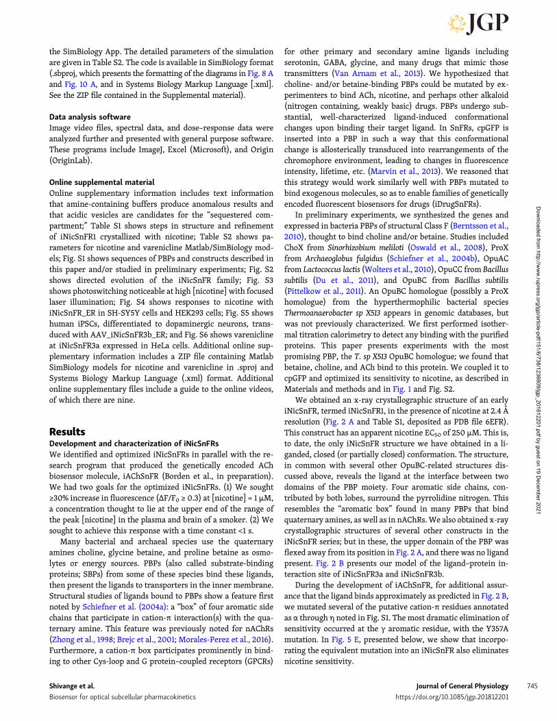
ResultsDevelopment and characterization of iNicSnFRsWe identified and optimized iNicSnFRs in parallel with the re-search program that produced the genetically encoded AChbiosensor molecule, iAChSnFR (Borden et al., in preparation).We had two goals for the optimized iNicSnFRs. (1) We sought≥30% increase in fluorescence (ΔF/F0 ≥ 0.3) at [nicotine] = 1 µM,a concentration thought to lie at the upper end of the range ofthe peak [nicotine] in the plasma and brain of a smoker. (2) Wesought to achieve this response with a time constant <1 s.
Many bacterial and archaeal species use the quaternaryamines choline, glycine betaine, and proline betaine as osmo-lytes or energy sources. PBPs (also called substrate-bindingproteins; SBPs) from some of these species bind these ligands,then present the ligands to transporters in the inner membrane.Structural studies of ligands bound to PBPs show a feature firstnoted by Schiefner et al. (2004a): a “box” of four aromatic sidechains that participate in cation-π interaction(s) with the qua-ternary amine. This feature was previously noted for nAChRs(Zhong et al., 1998; Brejc et al., 2001; Morales-Perez et al., 2016).Furthermore, a cation-π box participates prominently in bind-ing to other Cys-loop and G protein–coupled receptors (GPCRs)
for other primary and secondary amine ligands includingserotonin, GABA, glycine, and many drugs that mimic thosetransmitters (Van Arnam et al., 2013). We hypothesized thatcholine- and/or betaine-binding PBPs could be mutated by ex-perimenters to bind ACh, nicotine, and perhaps other alkaloid(nitrogen containing, weakly basic) drugs. PBPs undergo sub-stantial, well-characterized ligand-induced conformationalchanges upon binding their target ligand. In SnFRs, cpGFP isinserted into a PBP in such a way that this conformationalchange is allosterically transduced into rearrangements of thechromophore environment, leading to changes in fluorescenceintensity, lifetime, etc. (Marvin et al., 2013). We reasoned thatthis strategy would work similarly well with PBPs mutated tobind exogenous molecules, so as to enable families of geneticallyencoded fluorescent biosensors for drugs (iDrugSnFRs).
In preliminary experiments, we synthesized the genes andexpressed in bacteria PBPs of structural Class F (Berntsson et al.,2010), thought to bind choline and/or betaine. Studies includedChoX from Sinorhizobium meliloti (Oswald et al., 2008), ProXfrom Archaeoglobus fulgidus (Schiefner et al., 2004b), OpuACfrom Lactococcus lactis (Wolters et al., 2010), OpuCC from Bacillussubtilis (Du et al., 2011), and OpuBC from Bacillus subtilis(Pittelkow et al., 2011). An OpuBC homologue (possibly a ProXhomologue) from the hyperthermophilic bacterial speciesThermoanaerobacter sp X513 appears in genomic databases, butwas not previously characterized. We first performed isother-mal titration calorimetry to detect any binding with the purifiedproteins. This paper presents experiments with the mostpromising PBP, the T. sp X513 OpuBC homologue; we found thatbetaine, choline, and ACh bind to this protein. We coupled it tocpGFP and optimized its sensitivity to nicotine, as described inMaterials and methods and in Fig. 1 and Fig. S2.
We obtained an x-ray crystallographic structure of an earlyiNicSnFR, termed iNicSnFR1, in the presence of nicotine at 2.4 Aresolution (Fig. 2 A and Table S1, deposited as PDB file 6EFR).This construct has an apparent nicotine EC50 of 250 µM. This is,to date, the only iNicSnFR structure we have obtained in a li-ganded, closed (or partially closed) conformation. The structure,in common with several other OpuBC-related structures dis-cussed above, reveals the ligand at the interface between twodomains of the PBP moiety. Four aromatic side chains, con-tributed by both lobes, surround the pyrrolidine nitrogen. Thisresembles the “aromatic box” found in many PBPs that bindquaternary amines, as well as in nAChRs.We also obtained x-raycrystallographic structures of several other constructs in theiNicSnFR series; but in these, the upper domain of the PBP wasflexed away from its position in Fig. 2 A, and there was no ligandpresent. Fig. 2 B presents our model of the ligand–protein in-teraction site of iNicSnFR3a and iNicSnFR3b.
During the development of iAChSnFR, for additional assur-ance that the ligand binds approximately as predicted in Fig. 2 B,we mutated several of the putative cation-π residues annotatedas α through η noted in Fig. S1. The most dramatic elimination ofsensitivity occurred at the γ aromatic residue, with the Y357Amutation. In Fig. 5 E, presented below, we show that incorpo-rating the equivalent mutation into an iNicSnFR also eliminatesnicotine sensitivity.
Shivange et al. Journal of General Physiology 745
Biosensor for optical subcellular pharmacokinetics https://doi.org/10.1085/jgp.201812201
Dow
nloaded from http://w
ww
.rupress.org/jgp/article-pdf/151/6/738/1236809/jgp_201812201.pdf by guest on 19 Decem
ber 2021

We used excitation near the absorption peak at 485 nm, andwe made emission measurements at wavelengths >510 nm.Fig. 2 C shows dose–response relations for ΔF induced atiNicSnFR3a by three nicotinic agonists: nicotine, ACh, choline,and cotinine. The EC50 for choline is more than fourfold greaterthan for nicotine, showing a greater-than-desired sensitivity,but higher than the usual value for choline in brain (∼10 µM;Klein et al., 1992). The near-zero ΔF values for cotinine are toosmall for systematic study. The iNicSnFR3a and iNicSnFR3bproteins differ by one amino acid substitution at codon 11 (Asnvs. Glu, respectively; Fig. S1) and have no detectable photo-physical differences.
The stopped-flow data (Fig. 2 D) show that the fluorescenceincrease reached steady-state with time constants extrapolatingto a value of koff-ext = 2.0 s−1 at the lowest [nicotine]. This sat-isfied the criterion that the kinetics should be substantiallycomplete within 1 s. In other experiments that diluted premixedsolutions of 200 nM iNicSnFR3a plus nicotine into PBS, wemeasured modestly higher values of koff-dil = 3.3 ± 0.3 s−1.However, we consider koff-dil measurements less satisfactorybecause the resulting iNicSnFR3a concentrations were just8 nM, producing small and noisy signals. The isothermal titra-tion calorimetry data (Fig. 2 E) also show that nicotine binds toiNicSnFR3a (Kd = 10 ± 2.5 µM). Thus, all the available data in-dicate a noncooperative interaction between iNicSnFR3a andnicotine, with a Kd between 10 and 30 µM, that is completewithin ∼1 s.
pH dependence of an iNicSnFRWe based additional photophysical studies on previous data withthe GCaMP family. In the inactive conformation of cpGFP, thefluorophore has a pKa of 8–9. At neutral pH, the fluorophore isalmost fully protonated, decreasing the absorption in the bandcentered at λex∼485 nm (Barnett et al., 2017). In the active form,the pKa is ∼7, so that some of the fluorophore molecules aredeprotonated. This allows absorption and fluorescence (Barnettet al., 2017). Our pH studies have the additional feature thatnicotine itself is a weak base (Fig. 1, A and B), as are vareniclineand many other neural drugs. One expects both the pH depen-dence of the biosensor, and that of the ligand, to affect meas-urements with iNicSnFRs. We also sought to determine whetherthe charged or the uncharged form of nicotine binds to theiNicSnFRs.
We investigated the pH dependence of both ligand-independent and ligand-induced fluorescence (F0 and ΔF, re-spectively, Fig. 3). Throughout the pH range from 5.5 to 9.0,excitation at λex = 400 nm produces detectable F0 at an λem of535 nm, and this is nearly independent of pH (Fig. 3, A and B).This agrees with previous data on cpGFP-based biosensors(Barnett et al., 2017). The F0 values for λex = 485 nm show theexpected, contrasting strong pH dependence (Barnett et al.,2017): F0 is approximately inversely proportional to [H+](Fig. 3 C).
We determined the ΔF dose–response relations of iNicSnFR3ausing excitation at λex = either 400 or 485 nm. At basic pH, wefound the most reliable signals at λex= 485 nm, where nicotineinduces increased fluorescence (ΔF > 0; Fig. 3 E). The ΔFmax
decreases with increasing pH. Thus, ΔFmax/F0 at pH 7.5 is 14.5;but at pH 9.0, ΔFmax/F0 is only 3.5. This analysis is summarizedby plotting both F0 and F0 + ΔFmax in the measured range(Fig. 3 G). The pH dependence of the F0 measurements is con-sistent with those measured more precisely in a spectrofluo-rometer (Fig. 3 C). The pH dependence of the fully saturatedbiosensor (F0 + ΔFmax) resembles that for the fully saturatedGCaMP sensors (Barnett et al., 2017), reaching a plateau underbasic conditions; however, the apparent pKa of iNicSnFR3a maybe shifted to the right by ∼0.5 pH units from that for GCaMP6m(Barnett et al., 2017).
At acidic conditions for λex = 485 nm, nicotine evokes little orno ΔF; we therefore excited at λex = 400 nm and measured thenicotine-induced fluorescence decrease (ΔF < 0; Fig. 3 H). At pH= 7.5 and 8.0, where the two measurement modes both haveadequate signals, the two modes yield good agreement in EC50values (Fig. 3 I), consistent with the idea that the two modes aremeasuring the same nicotine-iNicSnFR binding. Nicotine ex-hibits a ∼20-fold decrease in EC50 as the pH is increased from6.0 to 9.0 (Fig. 3 I). Both this pH dependence of the response tonicotine and the large pH dependence of F0 are complicatingfactors in our live-cell experiments.
A special environment in the iNicSnFR3-binding siteThe nicotine derivative N’MeNic (Fig. 1 C) has a quaternaryammonium moiety at the pyrrolidine (N’) nitrogen. LikenAChRs (Beene et al., 2002; Post et al., 2017), iNicSnFR3 re-sponds robustly to N’MeNic. Comparing N’MeNic-induced withnicotine-induced ΔF over a pH range is expected to reveal fur-ther mechanistic details about the ligand–protein interaction,independent of the pH dependence of other regions and/ortransitions of the biosensor protein.We consider that systematicerrors render the data most reliable for λex = 400 nm at pH ≤ 7.5,and for λex = 485 nm at pH ≥ 7.5. Fig. 3 (E–G) show that iN-iCSnFR3a responds similarly to nicotine and to N’MeNic, withrespect both to EC50 and to ΔFmax/F0, at basic pH. Fig. 3 G showsthat the fully liganded state has a similar pH dependencewhether the ligand is nicotine or N’MeNic. We found a similartrend under acidic conditions. Fig. 3 I shows that, over almostthe entire measurable range from pH 6.0 to 9.0, the EC50 valuefor nicotine is 1.25- to 2-fold higher than that for N’MeNic. Themodestly higher sensitivity to N’MeNic is not surprising, con-sidering that the natural ligand of OpuBC is either choline orbetaine, both quaternary compounds. Many nicotine-bindingsites, such as those in AChBP and nicotinic receptors, toleratethe differences in charge density among protonated secondaryamines (such as cytisine and varenicline), protonated tertiaryamines (such as nicotine and ABT-418), and quaternary amines(such as choline and N’MeNic; Daly, 2005; Van Arnam andDougherty, 2014).
How do we explain that the EC50 for nicotine remains a smallmultiple of the N’MeNic EC50 over nearly the entire measurablepH range, even though the concentration of protonated nicotinein free solution decreases for pH values above its pKa (7.5–8)? Ina straightforward explanation, the binding site of iNicSnFR3,probably including a cation-π box, stabilizes diffuse positivecharges such as those in quaternary amines and in protonated
Shivange et al. Journal of General Physiology 746
Biosensor for optical subcellular pharmacokinetics https://doi.org/10.1085/jgp.201812201
Dow
nloaded from http://w
ww
.rupress.org/jgp/article-pdf/151/6/738/1236809/jgp_201812201.pdf by guest on 19 Decem
ber 2021

tertiary amines. Previous pH dependence studies of nAChRsshow this phenomenon (Petersson et al., 2002). Regardless ofthe underlying mechanism, the data suggest that the pH de-pendence of the cpGFP moiety exerts a stronger effect than thepH dependence of the weakly basic ligand, nicotine.
Studies with nicotine in live cellsiNicSnFR3a_PM and iNiCSnFR3a_ER in HeLa cellsWe conducted many of our optical and biochemical experimentsin HeLa cells, which have a relatively large, flat appearanceand prominent ER. Fig. 4 (A and B) shows protein expressionof iNicSnFR3a_PM and iNiCSnFR3a_ER after transfection inHeLa cells. We performed immunoblotting using an anti-GFPantibody. Observed GFP immunoreactivity for proteins fromiNicSnFR3a_PM- and iNiCSnFR3a_ER-transfected cells appearsnear the predicted values of ∼68 and ∼61 kD, respectively. Im-portantly, immunoblots demonstrated that the PM targetedbiosensor is larger than the ER targeted biosensor, accounted forby the addition of the PM targeting sequence. We found no otherbands with GFP immunoreactivity. In experiments conducted at7–23 h post-transfection, we found that iNicSnFR3a_PM pro-duced lower levels of protein than iNicSnFR3a_ER (22 ± 6 nM/mgprotein compared with 43 ± 14 nM/mg protein, respectively).This expression level difference was not significant whenwe averaged absolute protein levels across the entire dataset(Fig. 4 B1), but was consistent and significant when assessedwithin each paired (same day) set of transfections (Fig. 4 B2).No detectable dependence on the time since transfectionwas observed.
We also imaged the transfected cells with higher-resolutionfluorescence microscopy (Fig. 4 C). We cotransfected someof these samples with DsRed2-ER to assess localization with theER. As expected from the included targeted sequences, theiNiCSnFR3a_ER construct showed the expected ER structurestypical of HeLa cells, and also colocalized well with DsRed2-ER(Fig. 4 C1). The iNiCSnFR3a_PM fluorescence was most intenseat the periphery of the cell, as expected for a PM protein(Fig. 4 C2).
Fig. 5 presents a typical time-resolved fluorescence experiment,∼24 h after transfection. For reasons reported in Section 1 above,the time-resolved imaging experiments were performed withillumination near the 485 nm absorption peak (see Materialsand methods). Images were gathered at 4 Hz while cells wereexposed to 20-s pulses of nicotine at 40-s intervals at fourfoldconcentration increments between 0.25 and 256 µM. Nicotine-induced fluorescence increases are well resolved, even at0.25 µM. We note good reproducibility among cells: the coef-ficient of variation of ΔF/F0 < 10% within an experiment.Within a few seconds after the nicotine pulse begins in theextracellular solution, nicotine-induced fluorescence reachesan approximate plateau and changes by <10% over the next 20 s(the small increase in Fig. 5 was observed in only some ex-periments). Within a few seconds after [nicotine] is stepped tozero in the external solution, ΔF returns to zero. The temporalresolution of these experiments is limited by the speed ofthe solution change; we detected no difference between theiNicSnFR3a_PM and iNicSnFR3a_ER waveforms.
Fig. 5 exemplifies an idiosyncrasy of the HeLa cell nicotine-induced fluorescence increases. The dose–response relation atthe higher [nicotine] shows less saturation than expected fromexperiments on purified iNicSnFR3a (Fig. 2 C and Fig. 3 E). Wedevoted little attention to this phenomenon, because [nicotine]never exceeds ∼10 µM during smoking or vaping.
iNicSnFR3_ER detects only membrane-permeant moleculesA key goal for our experiments is to distinguish ER nicotine li-gands from extracellular molecules. Therefore we comparedresponses to nicotine itself versus two quaternary amines,thought to be membrane-impermeant molecules: N’MeNic andACh (Fig. 5 E). In cells transfected with iNicSnFR3a_PM,we found comparable fluorescence increases among the threeligands. In contrast, for cells transfected with iNicSnFR3a_ER,only nicotine evoked fluorescence increases. In solution,iNicSnFR3a has comparable ΔF to nicotine, N’MeNic, and ACh(Fig. 2 C and Fig. 3, E and F). Therefore it may be concluded thatiNicSnFR3a_ER samples only intracellular molecules. Giventhe predominant ER localization of iNicSnFR3a_ER (Fig. 4 C1),it may be concluded that this biosensor measures primarilyligands in the ER. We provide, in the section “Studies withvarenicline in vitro and in live cells” of this paper, evidence thatiNicSnFR3a_ER responds strongly to varenicline, an additionalmembrane-permeant ligand.
The data of Fig. 5, by themselves, cannot rule out the possi-bility that iNicSnFR3a_PM samples some intracellular nicotine.We argue against this possibility by noting the images of Fig. 4C2, showing that iNicSnFR3a_PM fluorescence occurs only onthe periphery of the cell. To investigate further, we conductednicotine exposure experiments on iNicSnFR3a_PM with a 100×objective lens, and we chose ROIs including only the periphery.We found that ΔF/F0 measurements had comparable values tothose of ROIs that include the entire cell (data not shown). Oneobjection to these “periphery-only” experiments is that someiNicSnFR3a_PMmight remain within endosomes that cannot bedistinguished from the PM by light microscopy, as found forsome transporters (Chiu et al., 2002; Moss et al., 2009). How-ever such compartments have luminal pH values ∼5.5, and in-traluminal cpGFP would fail to fluoresce. In summary, there isgood evidence that iNicSnFR3a_PM and iNicSnFR3a_ER mea-sure the nicotine concentration in the extracellular solution andin the ER, respectively.
Transfection of iNicSnFR3a_PM and iNicSnFR3a_ER into twoother human clonal cell types, SH-SY5Y (Fig. S4 A) and HEK293(Fig. S4 B) produced similar ΔF values in response to nicotineperfusion. We also obtained preliminary data in the only mousecell line tested, N2a (data not shown).
We also tested whether eliminating a crucial cation-π inter-action, at the γ amino acid (Fig. S1), eliminates nicotine-inducedΔF. In experiments on purified iAChSnFR, we found that theF357A mutation abolished sensitivity to ACh. In the presentexperiments, the equivalent mutation, iNiSnFR3a_Y357A_PM,was constructed and tested in HeLa cells. We found no detect-able nicotine-induced ΔF at concentrations ≤300 µM (Fig. 5 F).This observation also provides assurance that the nicotine-induced ΔF has little or no nonselective component.
Shivange et al. Journal of General Physiology 747
Biosensor for optical subcellular pharmacokinetics https://doi.org/10.1085/jgp.201812201
Dow
nloaded from http://w
ww
.rupress.org/jgp/article-pdf/151/6/738/1236809/jgp_201812201.pdf by guest on 19 Decem
ber 2021

Nicotine enters the ER of neuronsFig. 6 analyzes fluorescence induced by nicotine at two variantsof iNicSnFR3 expressed in neurons. An AAV2 construct yieldedbasal fluorescence in >50% of cultured mouse hippocampalneurons, and each fluorescent cell also showed responses tonicotine within a few seconds after an increase or decrease ofextracellular nicotine. Interestingly, neurons yield roughlythe same ΔF/F0 values for iNicSnFR3b_PM and iNicSnFR3b_ER.A similar pattern of roughly equal nicotine-induced ΔF/F0in neurons was observed for cDNA transfection, withiNicSnFR3a_PM and iNicSnFR3a_ER. As usual for neuronalcultures, cDNA transfection led to sparser expression (<10% ofcells) than viral transduction.
Dopaminergic neurons of the reward pathway located in theventral tegmental area play a role(s) in nicotine addiction(Subramaniyan and Dani, 2015). Studies also suggest that nico-tine protects dopaminergic neurons of the substantia nigra parscompacta during the initial stages of Parkinson’s disease, via aninside-out pathway (Srinivasan et al., 2014, 2016; Henderson
et al., 2016). Therefore, we assessed the entry of nicotine intothe ER of human dopaminergic neurons differentiated fromiPSCs.
It is not yet routinely possible to specifically induce eitherventral tegmental area–like or substantia nigra pars compacta–like dopaminergic neurons from iPSCs; therefore, we recordedfluorescence from all neurons expressing iNicSnFR3a_ER (Fig. 6,E and F). The data show recordings resembling those recordedfrom other cell types. The fluorescence reaches steady-statewithin a few seconds after nicotine appears near the cells anddecays to F0 within a few seconds after removal. The ΔF/F0values reach a maximum of 3–4, and the [nicotine] giving half-ΔFmax/F0 is on the order of 20 µM.
We also found that viral transduction with AAV2-iNicSnFR3b_ER proceeded efficiently in the induced iPSC cul-tures (Fig. S5). >90% of the neurons were fluorescent, and all ofthese gave detectable increases in the presence of nicotine. Theresponses resembled those for transfected dopaminergic neu-rons but had lower ΔF/F0, rarely exceeding 2. In two cells, we
Figure 5. Dose–response relations fornicotine-induced ΔF in HeLa cells. Exemplardata for iNicSnFR3a_PM and iNicSnFR3a_ERexpressed in transfected HeLa cells. (A and C)20-s nicotine dose application followed by 20-swash in HBSS. The average response for threecells at each dose is overlaid in a 30-s windowfor the PM and ER traces in A and C, respectively.The SEM is shown as colored bands. (B and D)The averaged ΔF/F0 at each response in A and C,respectively, is plotted against the logarithmicconcentration scale with SEM given as errorbars. (E) Comparisons among nicotine itself,N’MeNic, and ACh; experiments in transfectedHeLa cells. The iNicSnFR3a_PM responses toACh and to N’MeNic were normalized toNicSnFR3a_PM responses for nicotine (100 µM).The iNicSnFR3b_ER responses to ACh and toN’MeNic were normalized to iNicSnFR3b_ERresponses for nicotine (100 µM). Note thatiNIcSnFR3a_PM responds robustly to all threeligands, but only iNicSnFR3b_ER responds ro-bustly only to nicotine, the only permeant mol-ecule among the three tested. (F) Mutating aprobable cation-π interacting residue, Tyr357,eliminates nicotine-induced ΔF. Exemplar datafrom cells transfected in parallel and tested onthe same day.
Shivange et al. Journal of General Physiology 748
Biosensor for optical subcellular pharmacokinetics https://doi.org/10.1085/jgp.201812201
Dow
nloaded from http://w
ww
.rupress.org/jgp/article-pdf/151/6/738/1236809/jgp_201812201.pdf by guest on 19 Decem
ber 2021

found that ΔF induced by N’MeNic (100 µM) was <0.2 timesas large as ΔF induced by 1 µM nicotine. This confirms thatiNicSnFR3b_ER senses only intracellular nicotine, as found forexpression in HeLa cells (Fig. 5).
[Nicotine] in the ER approximately equals [nicotine] at the PMTo assess the relationship between [nicotine] in the ER versus[nicotine] applied in the extracellular solution, we compared theincrease of ΔF/F0 versus applied [nicotine], as measured by the_ER and _PM constructs. Two cell types (HeLa and hippocampalneurons) provided complete datasets with both _ER and _PMconstructs (presented in Fig. 5 and Fig. 6, A–D). For this analysis,we accepted data for applied [nicotine] ≤ 5 µM, because thisconcentration range is most pharmacologically relevant, wellbelow the EC50, and least subject to pH perturbation in the ER.The appropriate metric is defined as [ΔF/F0]/[nicotine] and hasthe units µM−1. For measurements with the _ER constructs andfor the _PM constructs, the metric is 0.075 ± 0.019 µM−1 and0.063 ± 0.13 µM−1, respectively (mean ± SEM, n = 8 cells in eachcase; Fig. 6 F inset, gives an exemplar plot). This similarity
shows that [nicotine] in the ER is approximately equal to [nic-otine] applied in the external solution. Less complete data showa similar pattern in other cell types: SH-SY5Y (Fig. S4 A),HEK293 (Fig. S4 B), and human dopaminergic neurons differ-entiated from iPSCs (Fig. 6, E and F; and Fig. S5).
For purposes of the simulations described in a later section,we assumed that [nicotine] is equal in the ER and in the extra-cellular solution. We interpret “approximately equal” as a dif-ference of less-than twofold. The uncertainty arises primarilybecause of differences between the pH of the ER and extracel-lular solution. In most estimates, this difference is <0.2 pH units(Casey et al., 2010), but has not been measured for the cell typeswe investigated. As noted, it is unlikely that nicotine and vare-nicline at the sub-µM concentrations of most interest perturbthe pH of the ER. Such perturbation (for instance, by mito-chondrial uncouplers; Mitchell, 1966) requires that both theuncharged and charged form of the drug can permeate throughmembranes, either passively or via transporters(s). All previousstudies on nicotine conclude that only the uncharged form ismembrane-permeant (Lester et al., 2009).
Figure 6. Nicotine in the ER of neurons.(A–D) Exemplar nicotine-induced fluorescenceincreases for cultured hippocampal neuronstransduced with AAV2/1.sin1.iNicSnFR3b_PM (Aand B) or with AAV2.sin1.iNicSnFR3b_ER (C and D).(A and C) 20-s nicotine pulses, followed by 20-swash in HBSS. The average waveform for five cellsat each [nicotine] is overlaid for the PM and ERtraces in A and C, respectively. The SEM is shown ascolored bands around each line. Dose–responserelations are shown in B and D. (E and F) Humandopaminergic neurons transfected with iNicSnFR_ER3a.(E) Typical nicotine-induced fluorescence during20-s pulses of nicotine at the indicated concen-trations. Data were subjected to a triangle filter(half-time, 1 s). (F) Full dose–response data from20 transfected human dopaminergic neurons.Inset, start of the dose relation at [nicotine] ≤5 µM. The slope of the line, [ΔF/F0]/[nicotine], is0.087 µM−1.
Shivange et al. Journal of General Physiology 749
Biosensor for optical subcellular pharmacokinetics https://doi.org/10.1085/jgp.201812201
Dow
nloaded from http://w
ww
.rupress.org/jgp/article-pdf/151/6/738/1236809/jgp_201812201.pdf by guest on 19 Decem
ber 2021

Micro-iontophoresis of nicotine: membrane permeation does notslow fluorescence increasesResults presented above show that a “jump” of [nicotine] in theexternal solution results in ΔF in the ER. We asked whether thekinetics of ΔF reveal any delay due to diffusion of nicotine acrosseither the PM or the ER membrane. To decide this point, weobtained more rapid application of nicotine without complica-tions from solution changes or pH changes. We delivered nico-tine from micro-iontophoretic pipettes (Del Castillo and Katz,1955). The data (Fig. 7) show that, for a cell within 10 µm ofthe pipette tip, iNicSnFR3b_ER produces fluorescence increaseswithin <1 s, approximately equal to the response time of theiNicSnFR itself (Fig. 2 D). This result shows that the PM and theER membrane do not present a detectable diffusion barrier onthis time scale. For more distant cells, the fluorescence increaseis slower and smaller. For instance, the cell in ROI 3, ∼150 µmfrom the tip of the micro-iontophoretic pipette, respondedcompletely on a time scale of ∼10 s. This is consistent with adiffusion constant on the order of 1 µm2/ms.
It is not possible to quantify the [nicotine] ejected from thetip of an iontophoretic pipette. We conducted dose–responsestudies by varying the iontophoretic current between 10 nA (asshown in Fig. 7) and 100 nA. As the current was increased, theΔF/F0 for the cell in ROI 1 did not increase; the response in thecell of ROI 2 increased modestly and became faster, to equalthe value in ROI 1. The cell in ROI 3 increased more gradually,eventually reaching ΔF/F0 ∼0.3 at an ejection current of 100 nA.
Simulations of the outside-in and inside-out pathwaysduring smokingOur data generate two insights important for understanding theinside-out pathway. First, the genetically encoded nicotine bio-sensors targeted to the ER reveal that nicotine appears in the ERwithin ≤10 s after it appears in the extracellular solution. Thedelay may be as small as 1 s, but this distinction has no impor-tance for the simulations in this section. Second, after this delay,the [nicotine] in the ER differs by less than twofold from [nic-otine] in the extracellular solution.
The most complete data on pharmacological chaperoninghave used extracellular nicotine, applied for several hours(Kuryatov et al., 2005). Therefore, a major question arising fromprevious data were whether nicotine enters the ER quicklyenough to serve as a pharmacological chaperone. The answer,based on our present data, is clearly, “yes.” It is reassuring, but
not crucial, to know that [nicotine] in the ER is rather close tothat outside the cell, so that the highest-affinity states ofnicotine-nAChR binding, which leads to pharmacologicalchaperoning, need not differ drastically from events at the PM.
A question of particular interest now arises about the exit ofnicotine from the ER. When nicotine is removed from the ex-tracellular solution, nicotine leaves the ER, again within 10 s. If[nicotine] in the ER drops below the EC50 for pharmacologicalchaperoning during the interval between cigarettes, then theinside-out pathway cannot readily account for nicotine depen-dence. We term this point the “rapid exit” problem.
The rapid exit problem may be addressed by existing data onthe pharmacokinetics of nicotine during smoking. After a personreceives a bolus of nicotine from a cigarette or an ENDS, [nic-otine] in the body decreases with two exponential terms. Duringthe slower phase, measurable nicotine endures in the plasma forseveral hours (Benowitz et al., 1991). We simulated fractionalactivation of the inside-out and outside-in pathways using theavailable data from the literature (Benowitz et al., 1991; Kuryatovet al., 2005; Rollema et al., 2007). We assume a common patternof smoking: one cigarette, yielding 1 mg of ingested nicotine,each hour, for 12 h during each day (Table 1). The detailed pa-rameters of the simulation are given in Table S2.
The simulations show how plasma/cerebrospinal fluid (CSF)/ER nicotine concentration varies on the time scale of minutes tohours (Fig. 8 B1). Clearly, [nicotine] in the ER remains greaterthan the EC50 for pharmacological chaperoning during the entire1-h interval between cigarettes. The inside-out pathway remainssubstantially activated continually during the 12–16 smoking-period hours of our simulations (Fig. 8 B2).
Studies with varenicline in vitro and in live cellsIn addition to detecting nicotine, the biosensors iNicSnFR3a andiNicSnFR3b also detect varenicline (Fig. 9 and Fig. S6). In fluo-rescence data, purified iNicSnFR3a displays a varenicline EC50approximately sixfold less than for nicotine. The maximal re-sponse, ΔFmax/F0, is at least equal to that for nicotine (Fig. 9 A).Note that the fitted Hill coefficient is significantly less thanunity, as also noted for the live-cell imaging described below.
Biochemical characterization also indicates that vareniclinebinds more strongly than nicotine to iNicSnFR. The stopped-flow kinetics (Fig. 9 B) reveal smaller pseudo–first-order for-ward and reverse rate constants (kon and koff, respectively) thanfor nicotine (Fig. 2 D), as well as an inferred equilibrium binding
Figure 7. Micro-iontophoretic nicotine ap-plication. (A) Cultured mouse hippocampalneurons transducedwith AAV_iNicSnFR3b_ER. Anicotine-containing micro-iontophoretic pipettewas positioned <10 µm above the cell in ROI 1.A 10-nA outward current pulse (32-s duration)was delivered. Most cells in the area showedfluorescence increases. (B) Fluorescence tracesrecorded simultaneously for cells at three dis-tances from the pipette.
Shivange et al. Journal of General Physiology 750
Biosensor for optical subcellular pharmacokinetics https://doi.org/10.1085/jgp.201812201
Dow
nloaded from http://w
ww
.rupress.org/jgp/article-pdf/151/6/738/1236809/jgp_201812201.pdf by guest on 19 Decem
ber 2021

constant Kd, approximately threefold less than for nicotine(compare with Fig. 2 D). The isothermal titration calorimetrydata for the varenicline–iNicSnFR3a interaction (Fig. 9 C) alsoreveal a several-fold lower Kd (3.5 µM) than for nicotine.
Varenicline enters the ERLive-cell imaging shows robust dose-dependent, varenicline-induced fluorescence increases, both in HeLa cells and in neu-rons, both at the PM and in the ER (Fig. 9 and Fig. S6). Thepharmacologically relevant varenicline concentrations are<1 µM, a range that yields varenicline-induced ΔF. Unlike thedata for nicotine, the varenicline dose–response data for bothHeLa cells and neurons do approach saturation, allowing theconclusion that the dose–response relations show an EC50 of1–4 µM. This agrees well with the data on purified biosensorprotein.
Especially in neurons, the growth and decay phases of the ERvarenicline-induced fluorescence (Fig. 9 F and Fig. S6 C) areclearly slower than either the nicotine responses presentedearlier or the varenicline responses on the PM (Fig. 9 D andFig. S6 A). The relatively slow ER varenicline responses occureven for the smallest measured [varenicline] (≤ 1 µM), whichare unlikely to perturb organellar pH. The slower ER entry andexit for varenicline than for nicotine are consistent with thelower logD7.4 (−1.27 vs. −0.04; Smith et al., 2012), as thoughthe ER entry/exit of varenicline is rate-limited by membranepermeability.
Pharmacokinetic and pharmacodynamic simulations of vareniclineThe data show that varenicline does enter the ER within <30 safter appearing near cells and that varenicline then leaves the ERwithin at most 60 s after leaving the external solution, at the
clinically relevant sub-µM concentrations. These data are ade-quate to add a subcellular dimension to pharmacokinetic simu-lations for orally administered varenicline. We used a modelappropriate to twice-daily oral administration and with pa-rameters that account for the very different absorption andmetabolism of varenicline (Fig. 10) versus nicotine (Fig. 8 andTable S2). The simulations show that the usual doses of vare-nicline only slightly activate the outside-in pathway of nAChRactivation. In contrast, varenicline activates the inside-outpathway of pharmacological chaperoning by >50% after thesecond dose of varenicline and by >70% after the fifth dose.
DiscussionThe study quantifies the dynamics and extent of an early step inthe inside-out pathway for nicotine: entry into the ER. Down-stream steps have been studied and quantified in several aprevious report using biochemistry, fluorescence microscopy,genetically alteredmice, and immunocytochemistry (Hendersonand Lester, 2015).
Development of the iNicSnFR family begins the field of op-tical subcellular pharmacokinetics for nicotine. We present datathat extend to the sub-µM nicotine concentration that exists inthe plasma and CSF of a smoker or vaper. The genetically en-coded nicotine biosensors iNicSnFR3a and -3b, trapped in theER, reveal that nicotine appears in the ER within at most 10 safter it appears in the extracellular solution (Fig. 5, Fig. 6, Fig. 7,Fig. S4, and Fig. S5). The [nicotine] in the ER is equal to that inthe extracellular solution, at a precision of twofold. These con-clusions hold for each of the five cell types we have investigated:three types of human clonal cell lines (HeLa, Fig. 5; SH-SY5Y,Fig. S4 A; and HEK293, Fig. S4 B), human dopaminergic neuronsdifferentiated from iPSCs (Fig. 6, E and F; and Fig. S5), andmouse hippocampal neurons (Fig. 6, A–D; and Fig. 7).
Fluorescent biosensors for opticalsubcellular pharmacokineticsOur strategy (Fig. 1) extends that used for iGluSnFR (Marvinet al., 2013). No known natural PBP binds nicotine; therefore,we engineered a PBP to bind a drug. That NicSnFRs also rec-ognize Ach, and more weakly, choline, is useful for studies ofcompartmentalization. Related OpuBC proteins, further opti-mized to sense ACh itself, will also find use in neuroscience(Borden et al., in preparation).
Although the directed evolution of the iNicSnFR family (Fig.S2) did not explicitly include assays for varenicline, vareniclineis a highly potent full agonist for iNicSnFR3 fluorescence. In-terestingly, varenicline is also more potent than ACh at bothα4β2 and α7 nAChRs (Coe et al., 2005; Mihalak et al., 2006).However, varenicline is a full agonist at α7, but not at α4β2nAChRs. Thus, there are differences in the details of the bindingsite of the iNicSnFR constructs versus nAChRs. We found thatthe iNicSnFR3a and iNicSnFR3b constructs are less sensitive toother α4β2 agonists: cytisine, dianicline, and A-85380 (unpub-lished data).
Other neuronal drugs may also operate via inside-out path-ways (Jong et al., 2009; Lester et al., 2012, 2015). With further
Table 1. Effect of dosing regimen variations on simulated nicotineconcentrations in plasma/CSF/ER and on nAChR activation orchaperoning
Nicotinedose (mg)percigarette
Total dailycigarettes,intervals
Average[nicotine] inplasma/CSF/ER(nM)
AveragenAChRactivationon PM
Averageactivation ofnAChRchaperoning inER
1 12/d, 1 h 112 0.1 0.7
0.5 12/d, 1 h 56 0.05 0.55
0.3 12/d, 1 h 34 0.03 0.44
0.1 12/d, 1 h 11 0.01 0.2
0.05 12/d, 1 h 6 0.006 0.13
3 12/d, 1 h 321 0.23 0.83
1 6/d, 2 h 65 0.06 0.6
1 20/d, 0.8 h 148* 0.13* 0.75*
Results have been averaged over 12 or 16 h. Asterisk (*) denotes 16-haverage. The first row (in bold) presents our definition of a standard habit,plotted in Fig. 8. The following five rows show simulations for increasingly“denicotinized” cigarettes. The row presenting a dose of 3 mg might beappropriate for a schizophrenic’s smoking strategy (Miwa et al., 2011).
Shivange et al. Journal of General Physiology 751
Biosensor for optical subcellular pharmacokinetics https://doi.org/10.1085/jgp.201812201
Dow
nloaded from http://w
ww
.rupress.org/jgp/article-pdf/151/6/738/1236809/jgp_201812201.pdf by guest on 19 Decem
ber 2021

modifications to the ligand site, preliminary data show that itmay be possible to develop families of biosensors for opticalsubcellular pharmacokinetics of several amine-containing drugclasses (Muthusamy et al., 2018; Shivange et al., 2017. AnnualMeeting of the Society of General Physiologists. Abstract no. 32.J. Gen. Physiol.). Values for logD7.4 of most neural drugs suggestthat they enter organelles (Lester et al., 2012; Nickell et al., 2013;Jong et al., 2018), and melatonin probably also enters neutralorganelles (Yu et al., 2016). The most important limitation, atpresent, is the extreme pH sensitivity of the biosensors. Thisconstrains their usefulness in acidic organelles, where someneuronal drugs may also act (Stoeber et al., 2018).
Another class of fluorescent protein-based biosensors is de-rived from GPCRs rather than from PBPs (Jing et al., 2018;Patriarchi et al., 2018). In the GPCR-based biosensors, the ligandsites face the extracellular solution; they would presumably face
the lumen of organelles like most (but not all) PBP-based SnFRs.However, in the GPCR-based biosensors, the fluorescent proteinmoiety faces the cytosol and therefore might be relatively in-sensitive to luminal pH. If GPCR-based biosensors can functionin acidic organelles, they may also find use for subcellularpharmacokinetics.
Implications for the inside-out pathwayOur data show that when [nicotine] in the extracellular solutionfalls to zero, nicotine completely leaves the ER, again within 10 s.Yet after a person receives a bolus of nicotine from a cigarette,[nicotine] in the body does not immediately fall to zero. Therather leisurely metabolism of nicotine (half-time, ∼20 min inhumans; Benowitz et al., 1991) provides that a smoker’s CSF[nicotine] decreases on the time scale shown in Fig. 8 B1. Becauseof the highly nicotine-sensitive feature of pharmacological
Figure 8. Simulations of nicotine pharmaco-kinetics and pharmacodynamics duringsmoking. (A) The pharmacokinetic/pharmaco-dynamic model, implemented in Matlab SimBi-ology. Individual parameters and structures andsmoking dosages are presented in Table S2 andin Supplemental ZIP File. (B1) Nicotine concen-trations in the plasma/CSF/ER and in the “se-questered” compartment, during 40 simulatedhours for the standard habit (Table S2 andSupplemental ZIP File). The latter compartmentwas termed the “peripheral compartment” byBenowitz et al. (1991), but that terminology isless preferable in discussions of the nervoussystem. Note the logarithmic [nicotine] scale.(B2) Effects on the two processes shown in A.Note that the standard habit nearly activatesnAChR protein chaperoning (inside-out process)>50%, but activates nAChR channel activation(outside-in process) <20%.
Shivange et al. Journal of General Physiology 752
Biosensor for optical subcellular pharmacokinetics https://doi.org/10.1085/jgp.201812201
Dow
nloaded from http://w
ww
.rupress.org/jgp/article-pdf/151/6/738/1236809/jgp_201812201.pdf by guest on 19 Decem
ber 2021

chaperoning, [nicotine] in the ER remains greater than the EC50for pharmacological chaperoning during the entire 0.75–1-h in-terval between cigarettes. The inside-out pathway remains>50%-activated continually during the 12–16 h of our simu-lations (Fig. 8 B2). We conclude that the inside-out pathway,operating via pharmacological chaperoning, can readily accountfor up-regulation, an important component of nicotinedependence.
Our simulations also lead one to reexamine the relationshipbetween the previously distinct concepts of “acute,” “repeated,”and “chronic” exposure to nicotine. In the “standard habit,” evena single cigarette, whose nicotine is fully ingested during 10min,activates the inside-out pathway >50% until the next cigarette1 h later (Fig. 8). Thus, a series of “acute” and “repeated” ex-posures, one per hour for 12 h, becomes “chronic” activation forthe 12 h of smoking activity. By the next morning, nicotine levelshave decreased below levels that activate pharmacologicalchaperoning; but the downstream trafficking and subsequentsequelae, including nAChR up-regulation, probably endure forseveral days (Marks et al., 1985).
Thus, subcellular pharmacokinetics readily explains depen-dence on smoked nicotine. Around the world at any time, sev-eral 100 million people have ingested tobacco within the past
hour; they retain nicotine in every organelle of every cell.In the ER and cis-Golgi of the small percentage of cells thatcontain α4β2 nAChRs, these levels activate the inside-outpathway >50%, and therefore maintain one aspect of nico-tine dependence.
Recently introduced ENDSs provide plasma levels with ki-netics approaching those of cigarettes (Bowen and Xing, 2015).Thus, ENDSs can also maintain a key molecular/cellular basis ofnicotine dependence: the inside-pathway at α4β2 nAChRs.
Can outside-in mechanisms, such as channel activation andCa2+ influx, also account for cellular/molecular aspects of nico-tine dependence? In contrast to pharmacological chaperoning,nAChR channels do not remain fully activated during the aver-age smoker’s day; in fact, the average activation of PM nAChRchannels is <20% (Fig. 8 B2). Some explanations of nicotinedependence assume that Ca2+ influx, either directly throughnAChRs or indirectly through neuronal activity-induced Ca2+
channel influx, can account for nicotine addiction, even con-sidering the rather modest level of nAChR channel activationshown by our simulations. Yet the rather general phenomenonof Ca2+ influx has not been shown to account for the highlyspecific aspects of nicotine addiction, such as post-translationalnAChR up-regulation. Another frequently invoked explanation
Figure 9. Varenicline activity against purifiediNicSnFR3a (A–C) and iNicSnFR3b expressedin hippocampal neurons (D–G). (A) Dose–response relations for varenicline-induced ΔF/F0, measured in 3× PBS, pH 7.4. Mean ± SEM;three measurements. (B) Stopped-flow measure-ments at various [varenicline]. (C) Isothermaltitration calorimetry. (D–G) Exemplar varenicline-induced fluorescence increases for cultured hip-pocampal neurons transduced with AAV2/1.syn1.-iNicSnFR3b_PM (D and E) or with AAV2/1.syn1.iNicSnFR3b_ER (F and G). (D and F) 30-svarenicline pulses, followed by 40-s wash in HBSS.The average waveform for five cells at each [var-enicline] is overlaid for the PM and ER traces in Aand C, respectively. The SEM is shown as coloredbands around each line. (E and G) Dose–responserelations, fitted to a single-component Hill equa-tion, including zero response at zero [varenicline].Parameters are shown.
Shivange et al. Journal of General Physiology 753
Biosensor for optical subcellular pharmacokinetics https://doi.org/10.1085/jgp.201812201
Dow
nloaded from http://w
ww
.rupress.org/jgp/article-pdf/151/6/738/1236809/jgp_201812201.pdf by guest on 19 Decem
ber 2021

for nicotine dependence is desensitization of nAChR channelsin response to the presence of nicotine. It is highly likely thatsome acute aspects of nicotine exposure arise from desensitiz-ation rather than activation (Miwa et al., 2011). However, noavailable data suggest that desensitized nAChRs begin an in-tracellular signaling pathway that could account for nicotinedependence.
Implications for varenicline-based therapyThe present data show that varenicline also enters the ER (Fig. 9and Fig. S6), and the simulations show that the resultingmaintained level of varenicline in the ER could lead to phar-macological chaperoning by varenicline (Fig. 10). This explainsthe observations that varenicline also up-regulates α4β2 nAChRs(Turner et al., 2011; Marks et al., 2015; Govind et al., 2017). The
usual explanation for varenicline as a smoking cessation agent isthat, even though it produces negligible activation of α4β2nAChR channels (Fig. 10 B2), it does desensitize these nAChRs,blocking the action of nicotine itself. Our study supports theadditional suggestion that the several-week taper regimen forvarenicline also allows nicotine users to reverse up-regulationgradually. This, in turn, could help aspiring quitters to avoidcraving and/or behavioral aspects of withdrawal. However, thepersistence of up-regulated nAChRs could also cause some of theside effects of varenicline during the first days to weeks (Ashareet al., 2017); these side effects could underlie this drug’s modesteffectiveness in smoking cessation (Fagerstrom and Hughes,2008).
Nicotine may also be neuroprotective in Parkinson’s dis-ease, and this effect may proceed via an inside-out pathwayin the ER (Srinivasan et al., 2014, 2016). Therefore, we noterecent reports that varenicline also displays neuroprotectiveeffects in animal models of Parkinson’s disease (McGregoret al., 2017) and another neurodegenerative disease (Sharmaet al., 2018).
Quantitative aspects of reducing nicotine levelsThe prominence of the inside-out pathway in nicotine de-pendence has prompted us to simulate the activation of theinside-out pathway, as well as the outside-in pathway, duringuse of modified tobacco or ENDS (Table 1). United StatesFood and Drug Administration officials recently suggesteddecreasing the nicotine content of combustible tobacco. Oursimulations show that a 3-fold reduction would nonethelessprovide >40% average activation of the inside-out pathway; a10-fold reduction (to 0.1 mg/cigarette) would reduce averageactivation to 20%. This conclusion would also hold for inges-tion via an ENDS.
Our simulations deal with a “typical” smoker (Benowitz et al.,1991). These conclusions must be modified, at a personal level,for each smoker or vaper. Polymorphisms in cytochrome P4502A6, the enzyme that primarily metabolizes nicotine, lead todramatic (up to 10-fold) differences in nicotine lifetime amongpeople (Dempsey et al., 2004). These result, in turn, to differ-ences in the frequency of smoking (Tanner et al., 2015). Futureinvestigations must simulate the consequences of these varia-tions for activation of the inside-out pathway.
If a nicotine-dependent person perceives desirable effectsfrom up-regulated nAChRs, he or she might therefore prefer toinhale nicotine from an ENDS capable of delivering 1 mg ofnicotine within a few minutes, rather than from a modifiedcigarette that, along with harmful smoke, delivers <0.1 mg, adose that does not maintain full up-regulation. The harm re-duction that results from such an individual behavioral choiceabout nicotine intake, (1) if stringently maintained by thenicotine-dependent individual and (2) if replicated in the entirenicotine-dependent population, would be considerable. One re-view suggests that such a complete transition would decreasetobacco-related diseases by 95% (Royal College of Physicians,2016). Whether and how a society wishes to encourage such atransition from smoked tobacco to ENDS involves details both ofpolicy and of science.
Figure 10. Simulated pharmacokinetics and pharmacodynamics duringoral varenicline administration. (A) In the pharmacokinetic model for orallyadministered varenicline, the lungs are replaced by the digestive tract. Theparameters derive from studies on humans (Faessel et al., 2006). The pa-rameters are given in Table S2 and in Supplemental ZIP File. (B1) Vareniclineconcentrations in two compartments: the plasma/CSF/ER and the seques-tered compartment. (B2) Effects on the two processes shown in A. Note thatrecommended treatment with varenicline almost completely activates nAChRprotein chaperoning (inside-out process), but only slightly produces nAChRchannel activation (outside-in process).
Shivange et al. Journal of General Physiology 754
Biosensor for optical subcellular pharmacokinetics https://doi.org/10.1085/jgp.201812201
Dow
nloaded from http://w
ww
.rupress.org/jgp/article-pdf/151/6/738/1236809/jgp_201812201.pdf by guest on 19 Decem
ber 2021

AcknowledgmentsHenry A. Lester and Amol V. Shivange thank the Janelia Re-search Campus for devoting hospitality and resources in theVisiting Scientist Program over a period of 3 y. We also thankCrystal N. Dilworth (early suggestion to use ProX-family PBPs),Jacob P. Keller (advice on pH and tubing), David P. Walton(analysis of N’MeNic), Michael R. Post (synthesis of N’MeNic),Peter H. Lee and Scott Sternson (purification of N’MeNic), AdelaNano and Jacqueline Barton (access to the ISS-K2 spectrofluo-rometer), Daniel Wagenaar (construction of LED light sources),Lauren M. Barnett (advice on photochemistry), Elizabeth K.Unger and Lin Tian (biosensors), Erik L. Snapp (ER imaging),Jennifer Lippincott-Schwartz (ER imaging), Eric R. Schreiter(biosensors), Tanner Lakin (cell culture), Kim Ritola and MelissaRamirez (AAV constructs), Baljit S. Khakh (advice), Mark Lobas(advice), Victoria J. Orphan and Fabai Wu (use and instruction onstructured illumination microscope), Stephen Grant (assisted inconfocal microscopy), Margaret Jefferies (excellent laboratorymanagement at Janelia), and Purnima Deshpande (excellent lab-oratory management at the California Institute of Technology).
This research was supported by grants from US National In-stitutes of Health (DA036061, DA037161, DA043829, GM123582,GM007616, MH061876, NS097362, and NS034407), the Califor-nia Tobacco-Related Disease Research Project (23XT-0007), theCalifornia Institute for Regenerative Medicine (EDUC2-08398),the Brain and Behavior Research Foundation (National Alliancefor Research on Schizophrenia and Depression), the HowardHughes Medical Institute, the Della Martin Foundation, Louisand Janet Fletcher, and California Institute of Technology SURFdonors Paraskeva N. Danailov and Maria Chan. E.R. Chapman isan Investigator of the Howard Hughes Medical Institute.
H.A. Lester previously had a consulting agreement withPfizer, who manufacture varenicline. The authors declare nofurther competing financial interests.
Author contributions: A.V. Shivange, P.M. Borden, A.K.Muthusamy, A.L. Nichols, K. Bera, H. Bao, I. Bishara, M.J. Mulcahy,B. Cohen, E.R. Chapman, J. Marvin, L. Looger, and H.A. Lester de-signed experiments. A.V. Shivange, P.M. Borden, A.K. Muthusamy,A.L. Nichols, K. Bera, H. Bao, I. Bishara, J. Jeon, M.J. Mulcahy,B. Cohen, S.L. O’Riordan, C. Kim and H.A. Lester performed ex-periments. J. Jeon performed simulations. A.V. Shivange, P.M.Borden, A.K. Muthusamy, J. Jeon, K. Bera, H. Bao, I. Bishara,M.J. Mulcahy, L. Looger, and H.A. Lester wrote the paper. A.V.Shivange, P.M. Borden, A.K. Muthusamy, A.L. Nichols, K. Bera,H. Bao, I. Bishara, J. Jeon, M.J. Mulcahy, B. Cohen, D.A. Dougherty,E.R. Chapman, L. Looger, and H.A. Lester revised the paper.
Merritt C. Maduke served as editor.
Submitted: 1 August 2018Revised: 5 November 2018Accepted: 9 January 2019
ReferencesAronson, D.E., L.M. Costantini, and E.L. Snapp. 2011. Superfolder GFP is fluor-
escent in oxidizing environments when targeted via the Sec translocon.Traffic. 12:543–548. https://doi.org/10.1111/j.1600-0854.2011.01168.x
Ashare, R.L., C. Lerman, R.F. Tyndale, L.W. Hawk, T.P. George, P. Cinciripini,and R.A. Schnoll. 2017. Sleep Disturbance During Smoking Cessation:
Withdrawal or Side Effect of Treatment? J Smok. Cessat. 12:63–70.https://doi.org/10.1017/jsc.2016.11
Barnett, L.M., T.E. Hughes, and M. Drobizhev. 2017. Deciphering the mo-lecular mechanism responsible for GCaMP6m’s Ca2+-dependent changein fluorescence. PLoS One. 12:e0170934. https://doi.org/10.1371/journal.pone.0170934
Beene, D.L., G.S. Brandt, W. Zhong, N.M. Zacharias, H.A. Lester, and D.A.Dougherty. 2002. Cation-π interactions in ligand recognition by sero-tonergic (5-HT3A) and nicotinic acetylcholine receptors: the anomalousbinding properties of nicotine. Biochemistry. 41:10262–10269. https://doi.org/10.1021/bi020266d
Benowitz, N.L., P. Jacob III, C. Denaro, and R. Jenkins. 1991. Stable isotopestudies of nicotine kinetics and bioavailability. Clin. Pharmacol. Ther. 49:270–277. https://doi.org/10.1038/clpt.1991.28
Berntsson, R.P., S.H. Smits, L. Schmitt, D.J. Slotboom, and B. Poolman. 2010. Astructural classification of substrate-binding proteins. FEBS Lett. 584:2606–2617. https://doi.org/10.1016/j.febslet.2010.04.043
Bowen, A., and C. Xing. 2015. Nicotine salt formulations for aerosol devicesand methods thereof. US Patent 9,215,895. In own. 9,215,895UnitedStates.
Brejc, K., W.J. van Dijk, R.V. Klaassen, M. Schuurmans, J. van Der Oost, A.B.Smit, and T.K. Sixma. 2001. Crystal structure of an ACh-binding proteinreveals the ligand-binding domain of nicotinic receptors. Nature. 411:269–276. https://doi.org/10.1038/35077011
Casey, J.R., S. Grinstein, and J. Orlowski. 2010. Sensors and regulators ofintracellular pH. Nat. Rev. Mol. Cell Biol. 11:50–61. https://doi.org/10.1038/nrm2820
Chiu, C.S., K. Jensen, I. Sokolova, D. Wang, M. Li, P. Deshpande, N. Davidson,I. Mody, M.W. Quick, S.R. Quake, and H.A. Lester. 2002. Number,density, and surface/cytoplasmic distribution of GABA transporters atpresynaptic structures of knock-in mice carrying GABA transportersubtype 1-green fluorescent protein fusions. J. Neurosci. 22:10251–10266.https://doi.org/10.1523/JNEUROSCI.22-23-10251.2002
Coe, J.W., P.R. Brooks, M.G. Vetelino, M.C. Wirtz, E.P. Arnold, J. Huang, S.B.Sands, T.I. Davis, L.A. Lebel, C.B. Fox, et al. 2005. Varenicline: an α4β2nicotinic receptor partial agonist for smoking cessation. J. Med. Chem.48:3474–3477. https://doi.org/10.1021/jm050069n
Daly, J.W. 2005. Nicotinic agonists, antagonists, andmodulators from naturalsources. Cell. Mol. Neurobiol. 25:513–552. https://doi.org/10.1007/s10571-005-3968-4
Del Castillo, J., and B. Katz. 1955. On the localization of acetylcholine receptors.J. Physiol. 128:157–181. https://doi.org/10.1113/jphysiol.1955.sp005297
Dempsey, D., P. Tutka, P. Jacob III, F. Allen, K. Schoedel, R.F. Tyndale, andN.L. Benowitz. 2004. Nicotine metabolite ratio as an index of cyto-chrome P450 2A6 metabolic activity. Clin. Pharmacol. Ther. 76:64–72.https://doi.org/10.1016/j.clpt.2004.02.011
Du, Y.,W.W. Shi, Y.X. He, Y.H. Yang, C.Z. Zhou, and Y. Chen. 2011. Structuresof the substrate-binding protein provide insights into the multiplecompatible solute binding specificities of the Bacillus subtilis ABCtransporter OpuC. Biochem. J. 436:283–289. https://doi.org/10.1042/BJ20102097
Emsley, P., B. Lohkamp, W.G. Scott, and K. Cowtan. 2010. Features and de-velopment of Coot. Acta Crystallogr. D Biol. Crystallogr. 66:486–501.https://doi.org/10.1107/S0907444910007493
Evans, P.R. 2011. An introduction to data reduction: space-group determi-nation, scaling and intensity statistics. Acta Crystallogr. D Biol. Crys-tallogr. 67:282–292. https://doi.org/10.1107/S090744491003982X
Faessel, H.M., M.A. Gibbs, D.J. Clark, K. Rohrbacher, M. Stolar, and A.H.Burstein. 2006. Multiple-dose pharmacokinetics of the selective nico-tinic receptor partial agonist, varenicline, in healthy smokers. J. Clin.Pharmacol. 46:1439–1448. https://doi.org/10.1177/0091270006292624
Fagerstrom, K., and J. Hughes. 2008. Varenicline in the treatment of tobaccodependence. Neuropsychiatr. Dis. Treat. 4:353–363. https://doi.org/10.2147/NDT.S927
Govind, A.P., P. Vezina, and W.N. Green. 2009. Nicotine-induced upregula-tion of nicotinic receptors: underlying mechanisms and relevance tonicotine addiction. Biochem. Pharmacol. 78:756–765. https://doi.org/10.1016/j.bcp.2009.06.011
Govind, A.P., Y.F. Vallejo, J.R. Stolz, J.Z. Yan, G.T. Swanson, and W.N. Green.2017. Selective and regulated trapping of nicotinic receptor weak baseligands and relevance to smoking cessation. eLife. 6:e25651. https://doi.org/10.7554/eLife.25651
Henderson, B.J., and H.A. Lester. 2015. Inside-out neuropharmacology ofnicotinic drugs.Neuropharmacology. 96(Pt B):178–193. https://doi.org/10.1016/j.neuropharm.2015.01.022
Shivange et al. Journal of General Physiology 755
Biosensor for optical subcellular pharmacokinetics https://doi.org/10.1085/jgp.201812201
Dow
nloaded from http://w
ww
.rupress.org/jgp/article-pdf/151/6/738/1236809/jgp_201812201.pdf by guest on 19 Decem
ber 2021

Henderson, B.J., T.R. Wall, B.M. Henley, C.H. Kim, W.A. Nichols, R. Moaddel,C. Xiao, and H.A. Lester. 2016. Menthol alone upregulates midbrainnAChRs, alters nAChR subtype stoichiometry, alters dopamine neuronfiring frequency, and prevents nicotine reward. J. Neurosci. 36:2957–2974. https://doi.org/10.1523/JNEUROSCI.4194-15.2016
Hussmann, G.P., J.R. Turner, E. Lomazzo, R. Venkatesh, V. Cousins, Y. Xiao,R.P. Yasuda, B.B. Wolfe, D.C. Perry, A.H. Rezvani, et al. 2012. Chronicsazetidine-A at behaviorally active doses does not increase nicotiniccholinergic receptors in rodent brain. J. Pharmacol. Exp. Ther. 343:441–450. https://doi.org/10.1124/jpet.112.198085
Jing, M., P. Zhang, G. Wang, J. Feng, L. Mesik, J. Zeng, H. Jiang, S. Wang, J.C.Looby, N.A. Guagliardo, et al. 2018. A genetically encoded fluorescentacetylcholine indicator for in vitro and in vivo studies. Nat. Biotechnol.36:726–737.
Jong, Y.J., V. Kumar, and K.L. O’Malley. 2009. Intracellular metabotropicglutamate receptor 5 (mGluR5) activates signaling cascades distinctfrom cell surface counterparts. J. Biol. Chem. 284:35827–35838. https://doi.org/10.1074/jbc.M109.046276
Jong, Y.I., S.K. Harmon, and K.L. O’Malley. 2018. Intracellular GPCRs Play KeyRoles in Synaptic Plasticity. ACS Chem. Neurosci. 9:2162–2172. https://doi.org/10.1021/acschemneuro.7b00516
Klein, J., A. Koppen, K. Loffelholz, and J. Schmitthenner. 1992. Uptake andmetabolism of choline by rat brain after acute choline administration.J. Neurochem. 58:870–876. https://doi.org/10.1111/j.1471-4159.1992.tb09337.x
Kuryatov, A., J. Luo, J. Cooper, and J. Lindstrom. 2005. Nicotine acts as apharmacological chaperone to up-regulate human α4β2 acetylcholinereceptors. Mol. Pharmacol. 68:1839–1851.
Lester, H.A., C. Xiao, R. Srinivasan, C.D. Son, J. Miwa, R. Pantoja, M.R.Banghart, D.A. Dougherty, A.M. Goate, and J.C. Wang. 2009. Nicotine isa selective pharmacological chaperone of acetylcholine receptor num-ber and stoichiometry. Implications for drug discovery. AAPS J. 11:167–177. https://doi.org/10.1208/s12248-009-9090-7
Lester, H.A., J.M. Miwa, and R. Srinivasan. 2012. Psychiatric drugs bind toclassical targets within early exocytotic pathways: therapeutic effects.Biol. Psychiatry. 72:907–915. https://doi.org/10.1016/j.biopsych.2012.05.020
Lester, H.A., L.D. Lavis, and D.A. Dougherty. 2015. Ketamine inside neurons?Am. J. Psychiatry. 172:1064–1066. https://doi.org/10.1176/appi.ajp.2015.14121537
Marks, M.J., J.A. Stitzel, and A.C. Collins. 1985. Time course study of the ef-fects of chronic nicotine infusion on drug response and brain receptors.J. Pharmacol. Exp. Ther. 235:619–628.
Marks, M.J., H.C. O’Neill, K.M. Wynalda-Camozzi, N.C. Ortiz, E.E. Simmons,C.A. Short, C.M. Butt, J.M. McIntosh, and S.R. Grady. 2015. Chronictreatment with varenicline changes expression of four nAChR bindingsites in mice. Neuropharmacology. 99:142–155. https://doi.org/10.1016/j.neuropharm.2015.07.019
Marvin, J.S., B.G. Borghuis, L. Tian, J. Cichon, M.T. Harnett, J. Akerboom, A.Gordus, S.L. Renninger, T.W. Chen, C.I. Bargmann, et al. 2013. An op-timized fluorescent probe for visualizing glutamate neurotransmission.Nat. Methods. 10:162–170. https://doi.org/10.1038/nmeth.2333
Marvin, J.S., B. Scholl, D.E. Wilson, K. Podgorski, A. Kazemipour, J.A. Müller,S. Schoch, F.J.U. Quiroz, N. Rebola, H. Bao, et al. 2018. Stability, affinity,and chromatic variants of the glutamate sensor iGluSnFR. Nat. Methods.15:936–939. https://doi.org/10.1038/s41592-018-0171-3
McGregor, A.L., G. D’Souza, D. Kim, and M.D. Tingle. 2017. Varenicline im-proves motor and cognitive deficits and decreases depressive-like be-haviour in late-stage YAC128 mice. Neuropharmacology. 116:233–246.https://doi.org/10.1016/j.neuropharm.2016.12.021
Mihalak, K.B., F.I. Carroll, and C.W. Luetje. 2006. Varenicline is a partialagonist at α4β2 and a full agonist at α7 neuronal nicotinic receptors.Mol. Pharmacol. 70:801–805. https://doi.org/10.1124/mol.106.025130
Mitchell, P. 1966. Chemiosmotic coupling in oxidative and photosynthetic phos-phorylation. Glynn Research, Ltd, Bodmin, Cornwall. https://doi.org/10.1111/j.1469-185X.1966.tb01501.x
Miwa, J.M., R. Freedman, and H.A. Lester. 2011. Neural systems governed bynicotinic acetylcholine receptors: emerging hypotheses. Neuron. 70:20–33. https://doi.org/10.1016/j.neuron.2011.03.014
Morales-Perez, C.L., C.M. Noviello, and R.E. Hibbs. 2016. X-ray structure ofthe human α4β2 nicotinic receptor. Nature. 538:411–415. https://doi.org/10.1038/nature19785
Moss, F.J., P.I. Imoukhuede, K. Scott, J. Hu, J.L. Jankowsky, M.W. Quick, andH.A. Lester. 2009. GABA transporter function, oligomerization state,and anchoring: correlates with subcellularly resolved FRET. J. Gen.Physiol. 134:489–521. https://doi.org/10.1085/jgp.200910314
Muthusamy, A.K., A.V. Shivange, A.L. Nichols, A. Kamajaya, J. Jeon, P.M.Borden, J.S. Marvin, E.K. Unger, H. Bao, E.R. Chapman, et al. 2018.Microscopy Using Fluorescent Drug Biosensors for “Inside-Out Phar-macology”. Biophys. J. 114:358a. https://doi.org/10.1016/j.bpj.2017.11.1990
Naude, J., M. Dongelmans, and P. Faure. 2015. Nicotinic alteration ofdecision-making. Neuropharmacology. 96(Pt B):244–254. https://doi.org/10.1016/j.neuropharm.2014.11.021
Nees, F. 2015. The nicotinic cholinergic system function in the human brain.Neuropharmacology. 96(Pt B):289–301. https://doi.org/10.1016/j.neuropharm.2014.10.021
Nickell, J.R., V.P. Grinevich, K.B. Siripurapu, A.M. Smith, and L.P. Dwoskin.2013. Potential therapeutic uses of mecamylamine and its stereo-isomers. Pharmacol. Biochem. Behav. 108:28–43. https://doi.org/10.1016/j.pbb.2013.04.005
Oswald, C., S.H. Smits, M. Hoing, L. Sohn-Bosser, L. Dupont, D. Le Rudulier,L. Schmitt, and E. Bremer. 2008. Crystal structures of the choline/acetylcholine substrate-binding protein ChoX from Sinorhizobium me-liloti in the liganded and unliganded-closed states. J. Biol. Chem. 283:32848–32859. https://doi.org/10.1074/jbc.M806021200
Pantoja, R., E.A. Rodriguez, M.I. Dibas, D.A. Dougherty, and H.A. Lester.2009. Single-molecule imaging of a fluorescent unnatural amino acidincorporated into nicotinic receptors. Biophys. J. 96:226–237. https://doi.org/10.1016/j.bpj.2008.09.034
Patriarchi, T., J.R. Cho, K. Merten, M.W. Howe, A. Marley, W.H. Xiong, R.W.Folk, G.J. Broussard, R. Liang, M.J. Jang, et al. 2018. Ultrafast neuronalimaging of dopamine dynamics with designed genetically encodedsensors. Science. 360:eaat4422. https://doi.org/10.1126/science.aat4422
Pedelacq, J.D., S. Cabantous, T. Tran, T.C. Terwilliger, and G.S. Waldo. 2006.Engineering and characterization of a superfolder green fluorescentprotein. Nat. Biotechnol. 24:79–88. https://doi.org/10.1038/nbt1172
Petersson, E.J., A. Choi, D.S. Dahan, H.A. Lester, and D.A. Dougherty. 2002. Aperturbed pK(a) at the binding site of the nicotinic acetylcholine re-ceptor: implications for nicotine binding. J. Am. Chem. Soc. 124:12662–12663. https://doi.org/10.1021/ja028206i
Picciotto, M.R., A.S. Lewis, G.I. van Schalkwyk, and Y.S. Mineur. 2015. Moodand anxiety regulation by nicotinic acetylcholine receptors: A potentialpathway to modulate aggression and related behavioral states. Neuro-pharmacology. 96(Pt B):235–243. https://doi.org/10.1016/j.neuropharm.2014.12.028
Pittelkow, M., B. Tschapek, S.H. Smits, L. Schmitt, and E. Bremer. 2011. Thecrystal structure of the substrate-binding protein OpuBC from Bacillussubtilis in complex with choline. J. Mol. Biol. 411:53–67. https://doi.org/10.1016/j.jmb.2011.05.037
Post, M.R., H.A. Lester, and D.A. Dougherty. 2017. Probing for and quanti-fying agonist hydrogen bonds in α6β2 nicotinic acetylcholine receptors.Biochemistry. 56:1836–1840. https://doi.org/10.1021/acs.biochem.7b00213
Powell, H.R., T.G.G. Battye, L. Kontogiannis, O. Johnson, and A.G.W. Leslie.2017. Integrating macromolecular X-ray diffraction data with thegraphical user interface iMosflm. Nat. Protoc. 12:1310–1325. https://doi.org/10.1038/nprot.2017.037
Richards, C.I., R. Srinivasan, C. Xiao, E.D. Mackey, J.M. Miwa, and H.A.Lester. 2011. Trafficking of α4* nicotinic receptors revealed by super-ecliptic phluorin: effects of a β4 amyotrophic lateral sclerosis-associatedmutation and chronic exposure to nicotine. J. Biol. Chem. 286:31241–31249. https://doi.org/10.1074/jbc.M111.256024
Rollema, H., L.K. Chambers, J.W. Coe, J. Glowa, R.S. Hurst, L.A. Lebel, Y. Lu,R.S. Mansbach, R.J. Mather, C.C. Rovetti, et al. 2007. Pharmacologicalprofile of the α4β2 nicotinic acetylcholine receptor partial agonistvarenicline, an effective smoking cessation aid. Neuropharmacology. 52:985–994. https://doi.org/10.1016/j.neuropharm.2006.10.016
Rollema, H., A. Shrikhande, K.M.Ward, F.D. Tingley III, J.W. Coe, B.T. O’Neill,E. Tseng, E.Q. Wang, R.J. Mather, R.S. Hurst, et al. 2010. Pre-clinicalproperties of the α4β2 nicotinic acetylcholine receptor partial agonistsvarenicline, cytisine and dianicline translate to clinical efficacy fornicotine dependence. Br. J. Pharmacol. 160:334–345. https://doi.org/10.1111/j.1476-5381.2010.00682.x
Royal College of Physicians. 2016. Nicotine without smoke: Tobacco harm re-duction. ECP, London.
Sallette, J., S. Pons, A. Devillers-Thiery, M. Soudant, L. Prado de Carvalho, J.P.Changeux, and P.J. Corringer. 2005. Nicotine upregulates its own re-ceptors through enhanced intracellular maturation. Neuron. 46:595–607. https://doi.org/10.1016/j.neuron.2005.03.029
Schiefner, A., J. Breed, L. Bosser, S. Kneip, J. Gade, G. Holtmann, K. Die-derichs, W. Welte, and E. Bremer. 2004a. Cation-pi interactions asdeterminants for binding of the compatible solutes glycine betaine and
Shivange et al. Journal of General Physiology 756
Biosensor for optical subcellular pharmacokinetics https://doi.org/10.1085/jgp.201812201
Dow
nloaded from http://w
ww
.rupress.org/jgp/article-pdf/151/6/738/1236809/jgp_201812201.pdf by guest on 19 Decem
ber 2021

proline betaine by the periplasmic ligand-binding protein ProX fromEscherichia coli. J. Biol. Chem. 279:5588–5596. https://doi.org/10.1074/jbc.M309771200
Schiefner, A., G. Holtmann, K. Diederichs, W. Welte, and E. Bremer. 2004b.Structural basis for the binding of compatible solutes by ProX from thehyperthermophilic archaeon Archaeoglobus fulgidus. J. Biol. Chem. 279:48270–48281. https://doi.org/10.1074/jbc.M403540200
Sharma, A.K., S. Gupta, R.K. Patel, and N. Wardhan. 2018. Haloperidol-induced parkinsonism is attenuated by varenicline in mice. J. BasicClin. Physiol. Pharmacol. 29:395–401. https://doi.org/10.1515/jbcpp-2017-0107
Smith, D., C. Allerton, A. Kalgutkar, H. van de Waterbeemd, and D. Walker,editors. 2012. Pharmacokinetics and Metabolism in Drug Design. Thirdedition. Wiley, Weinheim, Germany. 268 pp. https://doi.org/10.1002/9783527645763
Srinivasan, R., R. Pantoja, F.J. Moss, E.D.W. Mackey, C.D. Son, J. Miwa, andH.A. Lester. 2011. Nicotine up-regulates α4β2 nicotinic receptors and ERexit sites via stoichiometry-dependent chaperoning. J. Gen. Physiol. 137:59–79. https://doi.org/10.1085/jgp.201010532
Srinivasan, R., C.I. Richards, C. Xiao, D. Rhee, R. Pantoja, D.A. Dougherty, J.M.Miwa, and H.A. Lester. 2012. Pharmacological chaperoning of nicotinicacetylcholine receptors reduces the endoplasmic reticulum stress re-sponse. Mol. Pharmacol. 81:759–769. https://doi.org/10.1124/mol.112.077792
Srinivasan, R., B.J. Henderson, H.A. Lester, and C.I. Richards. 2014. Phar-macological chaperoning of nAChRs: a therapeutic target for Parkin-son’s disease. Pharmacol. Res. 83:20–29. https://doi.org/10.1016/j.phrs.2014.02.005
Srinivasan, R., B.M. Henley, B.J. Henderson, T. Indersmitten, B.N. Cohen,C.H. Kim, S. McKinney, P. Deshpande, C. Xiao, and H.A. Lester. 2016.Smoking-relevant nicotine concentration attenuates the unfolded pro-tein response in dopaminergic neurons. J. Neurosci. 36:65–79. https://doi.org/10.1523/JNEUROSCI.2126-15.2016
Stoeber, M., D. Jullie, B.T. Lobingier, T. Laeremans, J. Steyaert, P.W. Schiller,A. Manglik, and M. von Zastrow. 2018. A genetically encoded biosensorreveals location bias of opioid drug action. Neuron. 98:963–976.e5.https://doi.org/10.1016/j.neuron.2018.04.021
Subramaniyan, M., and J.A. Dani. 2015. Dopaminergic and cholinergiclearning mechanisms in nicotine addiction. Ann. N. Y. Acad. Sci. 1349:46–63. https://doi.org/10.1111/nyas.12871
Tanner, J.A., M.J. Chenoweth, and R.F. Tyndale. 2015. Pharmacogenetics ofnicotine and associated smoking behaviors. Curr. Top. Behav. Neurosci.23:37–86. https://doi.org/10.1007/978-3-319-13665-3_3
Turner, J.R., L.M. Castellano, and J.A. Blendy. 2011. Parallel anxiolytic-likeeffects and upregulation of neuronal nicotinic acetylcholine receptorsfollowing chronic nicotine and varenicline. Nicotine Tob. Res. 13:41–46.https://doi.org/10.1093/ntr/ntq206
Van Arnam, E.B., and D.A. Dougherty. 2014. Functional probes of drug-receptor interactions implicated by structural studies: Cys-loop re-ceptors provide a fertile testing ground. J. Med. Chem. 57:6289–6300.https://doi.org/10.1021/jm500023m
Van Arnam, E.B., E.E. Blythe, H.A. Lester, and D.A. Dougherty. 2013. Anunusual pattern of ligand-receptor interactions for the α7 nicotinicacetylcholine receptor, with implications for the binding of varenicline.Mol. Pharmacol. 84:201–207. https://doi.org/10.1124/mol.113.085795
Wolters, J.C., R.P. Berntsson, N. Gul, A. Karasawa, A.M. Thunnissen, D.J.Slotboom, and B. Poolman. 2010. Ligand binding and crystal structuresof the substrate-binding domain of the ABC transporter OpuA. PLoSOne. 5:e10361. https://doi.org/10.1371/journal.pone.0010361
Yu, H., E.J. Dickson, S.R. Jung, D.S. Koh, and B. Hille. 2016. High membranepermeability for melatonin. J. Gen. Physiol. 147:63–76. https://doi.org/10.1085/jgp.201511526
Zhong, W., J.P. Gallivan, Y. Zhang, L. Li, H.A. Lester, and D.A. Dougherty.1998. From ab initio quantum mechanics to molecular neurobiology: acation-π binding site in the nicotinic receptor. Proc. Natl. Acad. Sci. USA.95:12088–12093. https://doi.org/10.1073/pnas.95.21.12088
Shivange et al. Journal of General Physiology 757
Biosensor for optical subcellular pharmacokinetics https://doi.org/10.1085/jgp.201812201
Dow
nloaded from http://w
ww
.rupress.org/jgp/article-pdf/151/6/738/1236809/jgp_201812201.pdf by guest on 19 Decem
ber 2021