research article modulation of hyaluronan synthesis by the...
TRANSCRIPT

Research ArticleModulation of Hyaluronan Synthesis by the Interaction betweenMesenchymal Stem Cells and Osteoarthritic Chondrocytes
Eliane Antonioli1 Carla A Piccinato1 Helena B Nader2 Moiseacutes Cohen3
Anna Carla Goldberg1 and Mario Ferretti1
1Hospital Israelita Albert Einstein No 627701 05652-900 Sao Paulo SP Brazil2Department of Biochemistry Molecular Biology Program Federal University of Sao Paulo No 100 04044-020 Sao Paulo SP Brazil3Orthopedic Division Federal University of Sao Paulo No 783 04038-031 Sao Paulo SP Brazil
Correspondence should be addressed to Mario Ferretti marioferrettieinsteinbr
Received 13 November 2014 Revised 11 December 2014 Accepted 2 January 2015
Academic Editor Mark F Pittenger
Copyright copy 2015 Eliane Antonioli et alThis is an open access article distributed under theCreativeCommonsAttribution Licensewhich permits unrestricted use distribution and reproduction in any medium provided the original work is properly cited
Bone marrow mesenchymal stem cells (BM-MSCs) are considered a good source for cellular therapy in cartilage repair Buttheir potential to repair the extracellular matrix in an osteoarthritic environment is still controversial In osteoarthritis (OA)anti-inflammatory action and extracellular matrix production are important steps for cartilage healing This study examined theinteraction of BM-MSC and OA-chondrocyte on the production of hyaluronan and inflammatory cytokines in a Transwell systemWe compared cocultured BM-MSCs and OA-chondrocytes with the individually cultured controls (monocultures) There wasa decrease in BM-MSCs cell count in coculture with OA-chondrocytes when compared to BM-MSCs alone In monocultureBM-MSCs produced higher amounts of hyaluronan than OA-chondrocytes and coculture of BM-MSCs with OA-chondrocytesincreased hyaluronan production per cell Hyaluronan synthase-1 mRNA expression was upregulated in BM-MSCs after coculturewith OA-chondrocytes whereas hyaluronidase-1 was downregulated After coculture lower IL-6 levels were detected in BM-MSCscompared with OA-chondrocytes These results indicate that in response to coculture with OA-chondrocytes BM-MSCs changetheir behavior by increasing production of hyaluronan and decreasing inflammatory cytokines Our results indicate that BM-MSCsper se could be a potential tool for OA regenerative therapy exerting short-term effects on the local microenvironment even whencellcell contact is not occurring
1 Introduction
Osteoarthritis (OA) is a pathology accompanied by anincreased secretion of inflammatory cytokines and prote-olytic molecules into the surrounding tissue leading toextracellularmatrix degeneration and functional impairment[1] The capacity of adult chondrocytes to maintain cartilagehomeostasis declines with age with loss of the ability tosecrete the extracellular matrix components responsible forthe characteristic viscoelastic properties of the cartilage [2]Hyaluronan and aggrecan act as themajor aggregating factorsfor collagen proteoglycans and water playing a key role inthe maintenance of the cartilage structure and the ability toresist to compressive loads [3 4]
Hyaluronan is a glycosaminoglycan composed of repeat-ed disaccharide units synthesized by hyaluronan synthases
(HASs) which are membrane-bound enzymes There arethree isoforms in humans HAS-1 HAS-2 and HAS-3 whichproduce hyaluronan molecules of different molecular sizesHAS-1 andHAS-2 produce higher molecular weight hyaluro-nan molecules (gt2 times 106Da) [5 6] High molecular weighthyaluronan has been described as an anti-inflammatoryand immunosuppressive molecule whereas low hyaluronanfragments exhibit immunostimulatory and proinflammatoryeffects [7]
Degradation of hyaluronan is regulated by hyaluronidas-es There are six hyaluronidase-like sequences in the humangenome [7] however only three hyaluronidases (HYAL-1 HYAL-2 and HYAL-3) have been described in cartilage[8] Each enzyme acts upon molecules of different molec-ular weight paving the way for hyaluronan turnover in
Hindawi Publishing CorporationStem Cells InternationalVolume 2015 Article ID 640218 11 pageshttpdxdoiorg1011552015640218
2 Stem Cells International
the cartilage HYAL-1 degrades hyaluronan of all molecularweights to smaller oligomers HYAL-2 cleaves only high andintermediate molecular weight hyaluronan yielding productsof approximately 20 kDa while little is known about HYAL-3enzymatic activity [7 9] Of the three hyaluronidase genesHYAL-2 is the most expressed in normal chondrocytesCurrently there are indications that in OA there are lowerhyaluronan levels and with altered molecular weight and thathyaluronidases are upregulated in response to inflammatorycytokines [10ndash12]
Inflammation has been described as an important fac-tor in the development and progression of OA The mainproinflammatory cytokines described in the pathophysiologyof OA are interleukin- (IL-) 1 beta TNF IL-6 and also IL-8 [13] These cytokines contribute to the pathogenesis ofOA through several mechanisms leading to a shift in chon-drocytes phenotype In an inflammatory environment chon-drocytes become activated and increase further the expres-sion of proinflammatory cytokines and factors involved intissue catabolism namely matrix metalloproteinases andother proteolytic enzymes which degrade hyaluronan aggre-can collagen and fibromodulin Such fragments of matrixcomponents in turn also help maintain the productionof inflammatory cytokines [1 14] Alternative therapies forcartilage regeneration in OA should ideally reduce inflam-mation and promote tissue remodeling In this context theuse of mesenchymal stem cells (MSCs) has been pointedout as an interesting therapeutic option [14 15] due totheir distinct immunomodulatory anti-inflammatory andregenerative properties [16 17] MSCs anti-inflammatoryproperties might also be able to change chondrocytes pheno-type decreasing production of inflammatory molecules andfavoring the renewal of extracellular components
Indeed interaction between MSCs and chondrocytes hasbeen studied in vitro especially in the context of cartilagedevelopment evidencing a role of MSCs in forming carti-lage tissue [18ndash20] However the effects of MSCs on OA-chondrocytes and on their capacity to repair the extracel-lular matrix have not yet been fully examined Likewisethe potential effect of OA-chondrocytes on MSCs has sofar been overlooked In vitro coculture of MSCs and OA-chondrocytes represents a powerful approach to distinguishthe contribution of each cell type and their interaction Usingcells from the same patients we proposed to investigate theeffects caused by the interaction with no physical contactbetween MSCs and OA-chondrocytes on the secretion ofinflammatory markers and on hyaluronan synthesis
2 Materials and Methods
21 Culture and Isolation of Human Bone Marrow StemCell and Chondrocytes Both OA-chondrocytes and BM-MSCs were obtained from six patients undergoing total kneereplacement (TKR) surgery All patients were women (ages63ndash80 years average age 70 years) with Grade III or IV kneeOA according to the Kellgren and Lawrence classification[21] Articular cartilage and bone marrow were harvestedfrom the distal femur during TKR procedure The study wascarried out in full accordance with local ethical guidelines
(CEPEinstein 101268 Hospital Israelita Albert EinsteinCAAE 00060028000-10) and samples were collected afterobtaining written informed consent from all donors
For isolation of chondrocytes slices of OA knee cartilagefrom each donor were separately incubated in 025 type Icollagenase in Dulbeccorsquos modified Eaglersquos medium (DMEM)(Sigma St Louis MO) overnight at 37∘C in 5 CO
2
The cells were then seeded onto tissue culture flasks forexpansion and maintained as subconfluent monolayers inDMEM with low glucose (DMEM-LG) supplemented with1mM of L-glutamine 10 fetal bovine serum (FBS) and1 antibiotic-antimycotic solution (GibcoLife TechnologiesCarlsbad CA)
A small volume of bone marrow was drawn from thedistal femur to obtain BM-MSCs [22] and diluted in equalvolume of phosphate buffered saline (PBS) The cells werethen layered over Ficoll (density 103 to 112 gmL GE)and centrifuged at 500 g for 30 minutes Mononuclear cellswere collected seeded onto tissue flasks and cultivatedwith DMEM-LG supplemented with 15 fetal bovine serum1mM of L-glutamine and 1 antibiotic-antimycotic solution(GibcoLife Technologies) All incubations occurred in a 5CO2atmosphere at 37∘C Medium was replaced 3 times a
week until cells reached confluence At 80 confluence cellswere harvested with a trypsinEDTA solution (025 trypsin4mMEDTA GibcoLife Technologies) and seeded onto newflasks
BM-MSCs were expanded until the fifth passage and ana-lyzed by flow cytometry to determine the expression profileof stem cell markers as defined by the International Societyof Cell Therapy All BM-MSCs samples expressed CD90CD73 and CD105 on at least 95 of all cells with very low(or absent) expression of CD45 CD34 CD14 and HLA-DRDifferentiation of BM-MSCs into three cell types (adipocytesosteocytes and chondrocytes) was successfully achieved afterculture with specific media (StemPro Adipogenesis Chon-drogenesis and Osteogenesis Differentiation Kit GibcoLifeTechnologies) and confirmation after specific staining withOil Red for adipocytes Alcian Blue for chondrocytes andAlizarin Red S for osteocytes
22 Coculture of BM-MSCs and OA-Chondrocytes Cocul-tures (119899 = 6) were performed by seeding the paired BM-MSCs and OA-chondrocytes at a 1 1 cell ratio (50000 cellseach) fromeachdonor BM-MSCswere seeded onto the lowerchamber of a 6-well plate andOA-chondrocytes ontoMillicellhanging cell culture inserts (04 120583m pore size MilliporeBillerica MA USA) in 5mL of DMEM-LG supplementedwith 10 FBS 1mM of L-glutamine and 1 antibiotic-antimycotic solution Controls were monocultures of BM-MSCs and OA-chondrocytes (50000 cells each 5mL ofmedium) On days 3 and 6 bothmonocultures and cocultureswere detached with trypsin-EDTA solution Viable cells werecounted using the Trypan blue exclusion technique usinga Neubauer chamber Total RNA from the cells harvestedon both days 3 and 6 was extracted for quantitative reversetranscription-polymerase chain reaction (qRT-PCR) analysisand culture supernatants were stored at minus80∘C
Stem Cells International 3
23 Hyaluronan Measurement Hyaluronan measurementwas performed using a highly specific enzyme-linkedimmunosorbent assay- (ELISA-) like fluorometric method[23] Supernatants were boiled for 30min in order to inac-tivate all proteolytic activity Boiled sample triplicates andhyaluronan standards (ranging from 0 to 500mgL) wereincubated in plates coated with biotinylated hyaluronan-binding protein followed by a washing process addingof europium-marked streptavidin and an enhancementsolution Final fluorescence was measured in a fluorome-ter (Perkin-Elmer Life Sciences-Wallac Oy) Individual cellnumbers were used to normalize the absolute amounts ofhyaluronan of each sample including when coculture wasperformed In this last case we considered the sum of bothcells types added in the coculture Thus data are presentedas mean of hyaluronan concentration in pgmL per cell(pgmLcell)
24 Inflammatory Cytokine Analysis The concentrations ofinflammatory molecules in the culture supernatants weresimultaneously evaluated using the Cytokine Beads ArrayKit (Human Inflammation IL-8 IL-1120573 IL-6 IL-10 TNF andIL-12p70) (BD Biosciences) by flow cytometry (FACSAriaBD Biosciences San Jose CA) following the manufacturerrsquosinstructions and analyzed with FlowJo (TreeStar AshlandOR) andBDCBA software Concentrationwas normalized bycell count in each culture group and the relative productionof inflammatory cytokines was expressed as ngmLcell
25 Gene Expression Analysis Relative quantification ofmRNA expression of hyaluronan enzymes of extracellularmolecules and of inflammatory cytokines was performedusing qRT-PCR Total RNA was extracted with TriZol (LifeTechnologies) and a reverse transcriptase reaction (Quan-tiTect Reverse Transcription Kit QIAGEN) was performedqRT-PCR was carried out using the ABI7500 thermocycler(Applied Biosystems Carlsbad CA) and the Maxima SYBRGreen qPCR Master Mix (Thermo Fisher Scientific IncWaltham MA) for hyaluronan enzymes and extracellularmolecules according to the manufacturerrsquos recommenda-tions Primer sequences are shown in Table 1 Expressionof target genes was normalized by 120573-actin mRNA levelsThe level of expression was then calculated as 2minusΔΔCt andexpressed as the mean The results are presented as meanfold change relative to a calibration sample (Reference RNAfor Real-Time qPCR 636690 Clontech Mountain ViewCA USA) For coculture analysis fold change is presentedas gene expression relative to each BM-MSC or chondrocytemonoculture
26 Statistical Analysis All data analyses were performedusing GraphPad Prism version 6 (GraphPad Software IncLa Jolla CA) Statistically significant differences per cell inhyaluronan (pgmLcell) and cytokine (ngmLcell) concen-tration were evaluated using two-way ANOVA with Tukeyrsquospost hoc test Comparison of gene expression values betweentwo groups was performed using unpaired t-test or the non-parametric Mann-Whitney test and multiple comparisons
Table 1 Sequence of primers
Gene name Sequence
Beta-actin 51015840-GGCACCCAGCACAATGAAG-31015840
51015840-CCGATCCACACGGAGTACTTG-31015840
HAS-1 51015840-CAAGGCGCTCGGAGATTC-31015840
51015840-CCAACCTTGTGTCCGAGTCA-31015840
HAS-2 51015840-CAGACAGGCTGAGGACGACTTTAT-31015840
51015840-GGATACATAGAAACCTCTCACAATGC-31015840
HAS-3 51015840-GGCGATTCGGTGGACTACAT-31015840
51015840-CGATGGTGCAGGCTGGAT-31015840
HYAL-1 51015840-GGTGAGCTGGGAAAATACAAGAA-31015840
51015840-GCCCCAGTGTAGTGTCCATATACTC-31015840
HYAL-2 51015840-GGCGCAGCTGGTGTCATC-31015840
51015840-CCGTGTCAGGTAATCTTTGAGGTA-31015840
HYAL-3 51015840-TGTGCAGTCCATTGGTGTGA-31015840
51015840-AAGGTGTCCACCAGGTAGTCATG-31015840
Collagen type I 51015840-CCGCCGCTTCACCTACAGC-31015840
51015840-TTTGTATTCAATCACTGTCTTGCC-31015840
Collagen type II 51015840-CCGAATAGCAGGTTCACGTACA-31015840
51015840-CGATAACAGTCTTGCCCCACTT-31015840
Aggrecan 51015840-TTCAGTGGCCTACCAAGTGG-31015840
51015840-AGCCTGGGTTACAGATTCCA-31015840
Sox-9 51015840-TGCTAGAAGATGAGGCTTCTGG-31015840
51015840-GGCACTTTGTCCAGACCCA-31015840
were performed with Kruskal-Wallis and Dunnrsquos post hoctests Values are presented as mean plusmn SEM of triplicate wellsIn all analyses the level of significance was considered as119875 le 005
3 Results
31 The Number of BM-MSCs Is Decreased When Cocul-tured with OA-Chondrocytes To determine effects of the cellcoculture we counted the cell number of both BM-MSCsand OA-chondrocytes remaining at the end of the cocultureand compared them to the corresponding cell number whencultured alone (Figures 1(a) and 1(b)) After three days incoculture with chondrocytes BM-MSCs count decreased28 compared to BM-MSCs cultured alone (76 times 104 versus106 times 104) The difference increased as time in cultureprogressed and after six days BM-MSCs numbers were only53 of the cells cultured alone (63times 104 versus 135times 104)Weobserved that the number of BM-MSCs decreased after 6 daysin coculture with OA-chondrocytes in comparison to 3 days(76 times 104 versus 63 times 104) but no statistical difference wasobservedThe number ofOA-chondrocytes cultured togetherwith BM-MSCs however did not change significantly atboth time points analyzed (Figure 1(b)) In spite of thesechanges no significant variation in cell ratio was observedbetween BM-MSCs and OA-chondrocytes in coculture after3 days (75920 BM-MSCs and 63574 OA-chondrocytes 12 1cell ratio) and 6 days (65000 BM-MSCs and 71944 OA-chondrocytes after 3 and 6 days resp ie 09 1 cell ratio)
4 Stem Cells International
0 3 60
50000
100000
150000
200000
BM-MSCDays
Num
ber o
f cel
ls
BM-MSC Coc
lowast
lowastlowastlowast
(a)
0 3 60
25000
50000
75000
100000
ChondrocyteDays
Num
ber o
f cel
ls
Chondro Coc
(b)
Figure 1 BM-MSCs decrease cell number after coculture with OA-chondrocytes Number of cells cultivated for 3 and 6 days in monoculture(119899 = 6 bone marrow mesenchymal stem cells (BM-MSCs) and chondrocytes) or in coculture (119899 = 6 BM-MSC Coc and Chondro Coc)Error bars represent the SEM for the mean value Statistical significance (two-way ANOVA) is set according to the number of asterisks asfollows lowast119875 le 005 lowastlowastlowast119875 le 00001
3 6Days
0
1
2
3
4
5
Cell numberBM-MSC
Chondrocyte
BM-MSCChondrocyte
CocultureExpected coculture
Hya
luro
nan
(pg
mL
cell)
105 times 104
124 times 104
76 times 104
65 times 104
66 times 104
63 times 104
mdashmdash
mdashmdash 83 times 10
472 times 10
4
lowast
lowast
Figure 2 Coculture upregulates hyaluronan production Hyaluro-nan concentration (pgmLcell) after 3 and 6 days in monoculture(BM-MSCs and chondrocytes) or in coculture Bars represent themean and SEM bars with gray line (expected coculture) showldquoexpected hyaluronan production in coculturerdquo based on produc-tion of monoculture and cell number counted Table shows thecell number in each group The asterisk (lowast) indicates a significant(119875 le 005) difference between cell culture groups based on a two-way ANOVA followed by Tukeyrsquos posttest
32 BM-MSCs and OA-Chondrocyte Coculture ModulatesHyaluronan Production To determine whether hyaluronansynthesis could be altered by coculture wemeasured hyaluro-nan secreted by the cells in monoculture and when cocul-tured Data on hyaluronan concentration obtained were
normalized by cell number in order to account for the greaternumber of cells in the coculture To evaluate individualOA-chondrocyte and BM-MSC contribution in cocultureexpected values were calculated based on the sum of val-ues obtained from individual OA-chondrocyte and BM-MSC (monoculture) normalized by cell number of eachcell type We anticipated that the comparison between theexpected and observed values could clarify whether the crosstalk between OA-chondrocytes and BM-MSCs in cocultureresults in changes in hyaluronan production Our resultsshowed that both cells were able to synthesize hyaluronanalbeit BM-MSCs produced 2-foldmore hyaluronan thanOA-chondrocytes (280 pgmLcell versus 15 pgmLcell resp)after 3 days in monoculture (Figure 2)
Hyaluronan production per cell was increased in cocul-ture when compared with OA-chondrocytes cultivated alone(Figure 2) On a per cell basis after 6 days in our cocul-ture system hyaluronan present in the supernatant was215-fold higher than that from OA-chondrocytes alone(309 pgmLcell versus 145 pgmLcell Figure 2) but wassimilar to the levels detected in BM-MSCs monocultures(26 pgmLcell) Though after 3 days hyaluronan levelsshow intermediate values the same is not true for valuesobtained after 6 days in coculture clearly much higher Theexpected values if hyaluronan production ratio was main-tained after 6 days would be 27 pgmLcell (13 pgmLcellBM-MSC + 14 pgmLcell OA-chondrocyte) in contrast tothe value of 309 pgmLcell observed
Coculture is an important experiment when assessingcellcell interactions In our study cells were cocultured ina Transwell system that permitted harvesting of cells at thetwo defined time points and showed that though presentproliferation rate of BM-MSCs was significantly decreased(cell number Figure 2) On the other hand products are
Stem Cells International 5
BM-MSCChondrocyte
HAS-1 HAS-2 HAS-3 HYAL-1 HYAL-2 HYAL-30
1
2
3
4
3 days
Relat
ive e
xpre
ssio
n of
HA
enzy
mes
lowast
lowast
lowast
150
100
50
(a)
HAS-1 HAS-2 HAS-3 HYAL-1 HYAL-2 HYAL-30
1
2
50100150
6 days
BM-MSCChondrocyte
Relat
ive e
xpre
ssio
n of
HA
enzy
mes
lowast
lowastlowast
(b)
Figure 3 mRNA expression of hyaluronan enzymes by BM-MSCs and OA-chondrocytes Relative mRNA expression of hyaluronan-relatedenzymes after 3 days (a) or 6 days (b) in monoculture Bars represent the mean plusmn SEM of hyaluronan synthase- (HAS-) 1 HAS-2 and HAS-3and hyaluronidase- (HYAL-) 2 and HYAL-3 mRNA expression All fold changes were calculated relative to a calibrator sample Statisticalsignificance based on unpaired t-test was set according to the number of asterisks as follows lowast119875 le 005 lowastlowast119875 le 0001
continuously secreted into the supernatant making thisanalysis more difficult Alternatively in BM-MSC and OA-chondrocyte cultured alone cell number increased but nochange in hyaluronan production was observed during thetime points In coculture hyaluronan concentration increasedduring time points without changing the total cell number
33 Hyaluronan-Related Enzymes Are Differentially Expressedin OA-Chondrocytes and BM-MSCs To further clarify thecontribution of each cell type to hyaluronan production wefirst measured hyaluronan synthase- (HAS-) 1 HAS-2 andHAS-3 and hyaluronidases- (HYAL-) 1 HYAL-2 and HYAL-3 mRNA expression in monocultures as mean fold changerelative to a calibration sample After 3 and 6 days in culture adistinct expression pattern of HAS was observed BM-MSCspresented significantly greater HAS-1 expression than OA-chondrocytes (Figures 3(a) and 3(b)) in the latter HAS-1 waspractically absent after 6 days in culture HAS-2 also variedwith higher relative values exhibited by BM-MSCs HAS-3 gene expression was low in both cells and at both timepoints On the other hand mRNA expression of the threehyaluronidases was low at both time points (Figures 3(a) and3(b))
After 3 days in coculture relative HAS-1 mRNA expres-sion by BM-MSCs was further increased compared to BM-MSCs cultured individually (367-fold Figure 4(a)) After6 days we observed a trend towards increase of HAS-1 mRNA expression by BM-MSC (Figure 4(b)) In con-trast we observed a downregulation (sim25-fold) of HYAL-1 mRNA expression in BM-MSC after interaction withOA-chondrocyte at both time points (after 3 and 6 daysFigures 4(c) and 4(d)) HYAL-2 mRNA expression was alsoreduced after 6 days (15-fold) (Figure 4(d)) The expressionof other enzymes was unaltered in BM-MSCs and no changein the expression of any of the enzymes was detected in
the cocultured OA-chondrocytes in comparison with OA-chondrocytes in monoculture (Figures 5(a)ndash5(d))
34 Gene Expression of Extracellular Matrix ComponentsAlteration in genes related to extracellular matrix com-ponents may reflect cartilage regeneration and differentia-tion status Thus we chose to evaluate whether cocultureaffects the mRNA expression of type I and type II colla-gen Sox-9 and aggrecan Our findings showed that OA-chondrocytes maintained the expression of chondrogenicmarkers throughout the experiment No significant differ-ence in expression of extracellular matrix genes (type Iand II collagen aggrecan) and Sox-9 was observed after 3or 6 days of coculture system (Supplementary Figures 1(a)and 1(b) in Supplementary Material available online athttpdxdoiorg1011552015640218)
35 Coculture Alters Cytokine Production To evaluate theeffects on the inflammatorymicroenvironment wemeasuredsix cytokines (IL-8 IL-1120573 IL-6 IL-10 TNF and IL-12p70) inthe culture supernatants of OA-chondrocytes and BM-MSCscocultures Only IL-6 and IL-8 were present in detectablelevels and there was no evidence of production of theremaining cytokines investigated Similarly to hyaluronansynthesis we normalized cytokine production by the cellnumber measured at 3 and 6 days
As expected OA-chondrocytes produced 8-fold greateramounts of IL-6 than BM-MSC after 3 days (113 ngmLcellversus 14 ngmLcell) a difference maintained at 9-fold after6 days (238 ngmLcell versus 25 ngmLcell Figure 6(a))Similarly OA-chondrocytes produced more IL-8 than BM-MSCs a 14-fold increase after 3 days (130 ngmLcellversus 9 ngmLcell) and a 21-fold increase after 6 days(107 ngmLcell versus 5 ngmLcell Figure 6(b)) Interest-ingly as a result of coculture IL-6 secretion per cell was
6 Stem Cells International
0
2
4
6
8
10
BM-MSC after 3 days in coculture
BM-MSCRelat
ive e
xpre
ssio
n of
HA
S(fo
ld B
M-M
SC)
HAS-1HAS-2HAS-3
lowast
(a)
0
2
4
6
8
10
HAS-1HAS-2HAS-3
BM-MSC after 6 days in coculture
BM-MSCRela
tive e
xpre
ssio
n of
HA
S(fo
ld B
M-M
SC)
(b)
00
05
10
15
20
25
BM-MSC after 3 days in coculture
BM-MSC
Relat
ive e
xpre
ssio
n of
HYA
L(fo
ld B
M-M
SC)
HYAL-1
HYAL-3HYAL-2
lowastlowastlowast
(c)
Relat
ive e
xpre
ssio
n of
HYA
L(fo
ld B
M-M
SC)
00
05
10
15
20
25
HYAL-1BM-MSC after 6 days in coculture
BM-MSC
HYAL-3HYAL-2
lowast
lowastlowastlowast
(d)
Figure 4 Hyaluronan enzyme mRNA expression in BM-MSC after coculture mRNA expression of hyaluronan synthase- (HAS-) 1 HAS-2 and HAS-3 (a-b) and hyaluronidase- (HYAL-) 1 HYAL-2 and HYAL-3 (c-d) after coculture relative to time-matched and cell-matchedcontrols Expression of HAS in cocultivated BM-MSC relative to BM-MSCmonoculture after 3 days (a) and 6 days (119899 = 5) (b) expression ofhyaluronidases in cocultivated BM-MSC relative to BM-MSC monoculture after 3 days (c) and 6 days (d) (119899 = 5) Bars represent the meanfold change value plusmn SEM relative to BM-MSC in monoculture of hyaluronan enzymes Statistical significance based on unpaired t-test wasset according to the number of asterisks as follows lowast119875 le 005 lowastlowast119875 le 0001
of lower levels when compared to chondrocytes in mono-culture (55 ngmLcell versus 113 ngmLcell) and of greaterlevels when compared to BM-MSCs (55 ngmLcell versus14 ngmLcell) This pattern was also observed after 6 days(130 ngmLcell versus 238 ngmLcell coculture versus OA-chondrocyte and 25 ngmLcell BM-MSC resp Figure 6(a))In contrast to the increase in hyaluronan observed the secre-tion of IL-6 by OA-chondrocytes was clearly downregulatedThe expected values if secretion levels weremaintainedwouldbe 249 ngmLcell (12 ngmLBM-MSC + 237 ngmLOA-chondrocyte = 249 ngmLcell) but were only 130 ngmLcellThe same occurred with IL-8 where expected values would be109 ngmLcell (ie 23 ngmLBM-MSC + 107 ngmLOA-chondrocyte) but reached only 30 ngmLcell indicating atrend for lower production
IL-8 measured after coculture was also lower thanwhen OA-chondrocytes were cultured individually but didnot reach statistical difference after both 3 (130 ngmLcellversus 51 ngmLcell) and 6 days (107 ngmLcell versus30 ngmLcell Figure 6(b)) Although the IL-8 concentrationin coculture observed (30 ngmLcell) was different fromthe expected (109 ngmLcell ie 23 ngmLBM-MSC +107 ngmLOA-chondrocyte) we did not reach statisticaldifference between coculture and OA-chondrocytes
4 Discussion
In the present study we sought to establish the impact ofautologous BM-MSC on hyaluronan production and theireffects on the secretion profile of chondrocytes from patients
Stem Cells International 7
00
05
10
15
20
Chondrocyte after 3 days in coculture
Chon
Rela
tive e
xpre
ssio
n of
HA
S(fo
ld ch
ondr
ocyt
es)
HAS-1HAS-2HAS-3
(a)
00
05
10
15
20
HAS-1HAS-2HAS-3
Chondrocyte after 6 days in coculture
Chon
Rela
tive e
xpre
ssio
n of
HA
S(fo
ld ch
ondr
ocyt
es)
(b)
Relat
ive e
xpre
ssio
n of
HYA
L(fo
ld ch
ondr
ocyt
es)
0
1
2
3
4
Chondrocyte after 3 days in coculture
Chon
HYAL-1HYAL-2HYAL-3
(c)
0
1
2
3
4
HYAL-1HYAL-2HYAL-3
Chondrocyte after 6 days in coculture
Chon
Relat
ive e
xpre
ssio
n of
HYA
L(fo
ld ch
ondr
ocyt
es)
(d)
Figure 5 Hyaluronan enzyme mRNA expression in OA-chondrocytes after coculture mRNA expression of hyaluronan synthase- (HAS-)1 HAS-2 and HAS-3 (a-b) and hyaluronidase- (HYAL-) 1 HYAL-2 and HYAL-3 (c-d) after coculture relative to time-matched and cell-matched controls Expression of HAS in cocultivated chondrocytes (Chon) relative to Chon monoculture after 3 days (a) and 6 days (b)(119899 = 5) expression of hyaluronidases in cocultivated chondrocyte relative to Chon monoculture after 3 days (c) and 6 days (d) (119899 = 5) Barsrepresent the mean fold change value plusmn SEM relative to chondrocyte in monoculture of hyaluronan enzymes Statistical significance basedon unpaired t-test was set according to the number of asterisks as follows lowast119875 le 005 lowastlowast119875 le 0001
with OA Our coculture experiments suggest that an inter-action occurs between BM-MSC and OA-chondrocytes ina Transwell system which favors hyaluronan productionMoreover we were able to show that BM-MSCs aloneproduce high amounts of hyaluronan and exhibit abundantHAS-1 mRNA expression
Hyaluronan is a key component of the cartilage matrixand is used widely as an anti-inflammatory and antinocicep-tive agent in the treatment of OA improving joint lubricationand shock absorbance [24 25] Intra-articular hyaluronaninjection has been employed in the management of patientswith OA The anti-inflammatory anabolic and chondropro-tective action of hyaluronan has been increasingly evidencedsuggesting that hyaluronan helps to reduce pain and improve
cartilage function [26] Therefore insights into mechanismsthat can change hyaluronan levels in OA are relevant
Several studies have investigated the effects of MSCs onchondrocytes [18 27ndash29] but few have provided data toshow that MSCs and not only chondrocytes might alsobe affected by the cellcell interactions Our study showsthat coculture of BM-MSCs with OA-chondrocytes led to adecrease in BM-MSCs cell numbers which can be explainedby reduced cell proliferation or cell death However celldeath was not directly measured in the present study Theselow BM-MSCs numbers after 3 or 6 days in coculturesuggest that OA-chondrocytes are capable of altering BM-MSCs behavior Similar results have been shown in studiesusing coculture of BM-MSCs with chondrocyte pellets from
8 Stem Cells International
3 60
50
100
150
200
250
300
IL-6
secr
etio
n (n
gm
Lce
ll)
Days
ChondrocyteCocultureBM-MSCExpected coculture
lowast
lowastlowastlowast
lowastlowast
lowastlowast
lowastlowast
lowast
Cell numberBM-MSC
Chondrocyte105 times 10
4124 times 10
476 times 10
465 times 10
4
66 times 104
63 times 104
mdashmdash
mdashmdash 83 times 10
472 times 10
4
(a)
0
100
200
300
ChondrocyteCocultureBM-MSCExpected coculture
IL-8
secr
etio
n (n
gm
Lce
ll)
3 6Days
lowastlowast
Cell numberBM-MSC
Chondrocyte105 times 10
4124 times 10
476 times 10
465 times 10
4
66 times 104
63 times 104
mdashmdash
mdashmdash 83 times 10
472 times 10
4
(b)
Figure 6OA-chondrocytes produce higher IL-6 and IL-8 levels thanBM-MSCs and cocultured cells anddecrease these cytokines Interleukin(IL) production for 3 and 6 days inmonoculture (119899 = 5 BM-MSCs and chondrocytes) or in coculture (119899 = 5) IL-6 concentration (ngmLcell)(a) IL-8 concentration (ngmLcell) (b) Overall bars represent the mean value with the SEM bars with gray line (expected coculture) showexpected ILs synthesis in coculture based on production of monoculture and cell number counted Table shows cell number in each groupStatistical significance based on a two-way ANOVA with Tukeyrsquos posttest was set according to the number of asterisks as follows lowast119875 le 005lowastlowast119875 le 0001 and lowastlowastlowast119875 le 00001 Tendency of statistical differences between cell culture groups was identified by
different sources during 3- to 4-week periods of culturein which BM-MSCs numbers decreased progressively anddifferentiated into chondrocytes [18 30] Thus it is possiblethat in the present short-term cultures the trend towardsdifferentiation into chondrocytes with a slowdown of theproliferation rate was already present and that an extendedculture time would eventually favor chondrogenic markeralterations In fact in a previous study employing coculturewith direct contact between MSCs and OA-chondrocytesthe increase in chondrogenic markers was observed laterbeginning only after day 7 [31] Nevertheless our findingsshow that a short culture period is necessary and sufficientto change hyaluronan production in this microenvironment
Hyaluronan production by MSCs has been recentlydescribed [32] but we have shown that an at least twicegreater hyaluronan production occurs by BM-MSCs fromthe same patient than by OA-chondrocytes themselves Inaddition when BM-MSCs were cultured together with OA-chondrocytes the pattern of increased production was stillmaintained although at lower rates than when BM-MSCswere cultured alone Reduction in BM-MSC cell numberconcomitant with no change in OA-chondrocyte numbermost probably affected hyaluronan production Given thatthe expected and observed values of hyaluronan showedsignificant differences it can be concluded that BM-MSCscontribute to hyaluronan production in our coculture system
Thus the present study provides evidence for a role ofBM-MSCs in hyaluronan synthesis in OA Our values forhyaluronan production were similar to the short-term (1day) BM-MSCs monolayer cultures (range between 225 and4974 pgmLcell [32]) even though we cultured the cells forlonger periods (6 days)
Hyaluronan enzymes HAS-1 and HAS-2 have previouslybeen shown to be downregulated in chondrocytes collectedfrom OA cartilage when compared to the primary chon-drocytes or cartilage [33 34] We measured mRNA expres-sion levels of hyaluronan synthases to clarify which and towhat extent BM-MSCs and OA-chondrocytes contribute tohyaluronan levels produced Our data show that HAS-1 butnot other isoforms is increased inBM-MSCswhen comparedto OA-chondrocytes suggesting that HAS-1 may have animportant role in the hyaluronan production in our systemHAS-1 is the preponderant hyaluronan synthase presentin native cartilage which synthesized the high molecularweight hyaluronan which is found in normal cartilage [33]The greater HAS-1 expression in BM-MSCs after coculturewith OA-chondrocytes indicates that BM-MSCs might becapable of upregulating hyaluronan production in OA car-tilage though The cocultured OA-chondrocytes exhibitedunchanged expression of HASs after 3 or 6 days in cultureIt is possible that 6 days is not enough time to induce thischange It is also important to note that a 3D culture couldeventually induce more rapid changes as a result of the BM-MSCs secreted factors
Hyaluronan levels in the extracellular matrix and ulti-mately the regenerative potential in cartilage are deter-mined not only by hyaluronan synthesis and but also by itsdegradation Therefore expression of enzymes involved inhyaluronan degradation was analyzed in OA-chondrocytesand in BM-MSCs Among these enzymes HYAL-1 has beendescribed as an enzyme critical for cartilage development[8] HYAL-1 mRNA was detected at higher levels in OA-chondrocytes than in BM-MSCs cultured alone To ourknowledge the present study is the first to show mRNA
Stem Cells International 9
expression profile of HYALs in BM-MSCs in comparisonwith OA-chondrocytes Our results show that HYAL-1 andHYAL-2 mRNA expression are downregulated in BM-MSCsafter coculture with OA-chondrocytes in concordance withthe higher amounts of hyaluronan found in this systemBecause HYAL-1 and HYAL-2 hydrolyze hyaluronan frag-ments of different sizes and have been suggested as the mostabundant hyaluronan-degrading enzymes these enzymesmay be working together to degrade hyaluronan in OA[10 11] The expression of other enzymes did not changein BM-MSCs and OA-chondrocytes after coculture Thusour data suggest that OA-chondrocytes modulate BM-MSCsby increasing HAS-1 and inhibiting HYAL-1 and HYAL-2expression in order to synthesize higher molecular weighthyaluronan and consequently improve the local microenvi-ronment
The beneficial effect of hyaluronan on cartilage regener-ation was demonstrated in an in vivo study using hyaluro-nan hydrogel combined with MSC [35] The hyaluronanproduction by BM-MSCs might also have a direct anti-inflammatory role Hyaluronan injection in the knees of OApatients has been associatedwith decreased IL-6 but not withIL-8 levels in the synovial fluid which correlated with clinicalimprovement [36] Another study suggested that the presenceof hyaluronan reduces TNF-120572 and IL-6 concentration incoculture of OA-cartilage explants with synoviocytes [37]
Osteoarthritic cartilage is typically characterized by thepresence of cytokines associated with inflammation such asinterleukin-1 beta (IL-1120573) IL-10 IL-6 and TNF-120572 besidesproteolytic molecule MMPs These cytokines are secreted bychondrocytes and contribute to OA development [13 38]Our results show that OA-chondrocytes in culture maintainsecretion of high levels of inflammatory molecules even inthe presence of interfering factors such as fetal calf serumThese OA-chondrocytes produced large amounts of IL-6 andIL-8 even though kept for long periods in culture suggestingthat they preserve an ldquoinflammatory memoryrdquo In contrastBM-MSCs obtained from the same individual showed lowlevels of IL-6 and IL-8 production In fact coculture ofOA-chondrocytes with the paired BM-MSCs reduced IL-6 secretion on a ldquoper cellrdquo basis These observations areconsistent with a report showing an anti-inflammatory effectof adipose-derived allogeneicMSConOA-chondrocyteswitha decrease of IL-6 and IL-8 production [27] A differentialproduction of IL-8was however not detected in our analyses
The beneficial hyaluronan production and anti-inflammatory role of BM-MSCs indicate that the crosstalk with OA-chondrocytes may stimulate synthesis of othersoluble molecules creating a more propitious environmentfor cartilage regeneration Experimental models that permitcell contact (using OA cartilage explants) suggested thatthe microenvironment of OA cartilage does affect thechondrogenic potential of BM-MSCs [39]
The fact that in our study coculture was establishedwithout cellcell contact opens new avenues of cell therapyusing even allogeneic MSCs which would induce short-termchanges by adding hyaluronan and blockading IL-6 andIL-8 to induce a more regenerative and less inflammatorymicroenvironment in the affected OA cartilage
5 Conclusion
BM-MSCs produce hyaluronan and modulate this produc-tion in response to cross talk with OA-chondrocytes
In conclusion our data support the hypothesis that BM-MSCs produce hyaluronan in response to OA-chondrocytesincreasing mRNA expression of HAS-1 associated withHYAL-1 downregulation andhyaluronan synthesisThe inter-action promoted also an overall lower IL-6 production Takentogether these results indicate that BM-MSCs per se can be apotential tool for OA regenerative therapy Our study offersinsights into the mechanisms whereby treatment with BM-MSCs would exert beneficial effects on the diseased cartilageas a therapeutic strategy to increase hyaluronan productionand decrease inflammation locally More importantly ourdata point to a strategic role of MSCs in differentiating intomore active specialized cells and not only in remodelingchondrocytes However more basic and preclinical studiesthat considerMSC as an alternativeOA treatment are needed
Conflict of Interests
The authors declare that they have no conflict of interests
Authorsrsquo Contribution
All authors read and approved the final paper Eliane Anto-nioliMario Ferretti andAnnaCarlaGoldberg contributed tothe conception and design Eliane Antonioli Mario Ferrettiand Moises Cohen contributed to collection and assemblyof data Eliane Antonioli and Helena B Nader performedexperiments and data acquisition Eliane Antonioli Carla APiccinato andMario Ferretti contributed to data analysis andinterpretation Eliane Antonioli Carla A Piccinato MarioFerretti and Anna Carla Goldberg contributed to writingHelena B Nader and Anna Carla Goldberg contributed toediting Mario Ferretti contributed to financial and admin-istrative support
Acknowledgments
This work was supported by FAPESP (Proc 201200831-7)The authors would like to thank the technical assistanceof A Mendes MS from the Department of Biochemistryof the Federal University of Sao Paulo for collaboration inhyaluronan measurement and Luiz Sardinha PhD fromHospital Israelita Albert Einstein for help with flow cytome-try analysis Special thanks are due toHospital Israelita AlbertEinstein for supporting research
References
[1] MMaldonado and J Nam ldquoThe role of changes in extracellularmatrix of cartilage in the presence of inflammation on thepathology of osteoarthritisrdquo BioMed Research International vol2013 Article ID 284873 10 pages 2013
[2] R F Loeser ldquoAging and osteoarthritis the role of chondro-cyte senescence and aging changes in the cartilage matrixrdquoOsteoarthritis and Cartilage vol 17 no 8 pp 971ndash979 2009
10 Stem Cells International
[3] J Dudhia ldquoAggrecan aging and assembly in articular cartilagerdquoCellular andMolecular Life Sciences vol 62 no 19-20 pp 2241ndash2256 2005
[4] A Aspberg ldquoThe Different Roles of Aggrecan InteractionDomainsrdquoThe Journal of Histochemistry and Cytochemistry vol60 no 12 pp 987ndash996 2012
[5] N Itano and K Kimata ldquoMammalian hyaluronan synthasesrdquoIUBMB Life vol 54 no 4 pp 195ndash199 2002
[6] P H Weigel and P L DeAngelis ldquoHyaluronan synthases adecade-plus of novel glycosyltransferasesrdquo Journal of BiologicalChemistry vol 282 no 51 pp 36777ndash36781 2007
[7] K S Girish and K Kemparaju ldquoThemagic glue hyaluronan andits eraser hyaluronidase a biological overviewrdquo Life Sciencesvol 80 no 21 pp 1921ndash1943 2007
[8] S B Nicoll O Barak A B Csoka R S Bhatnagar and R SternldquoHyaluronidases and CD44 undergo differential modulationduring chondrogenesisrdquo Biochemical and Biophysical ResearchCommunications vol 292 no 4 pp 819ndash825 2002
[9] R Stern and M J Jedrzejas ldquoHyaluronidases their genomicsstructures and mechanisms of actionrdquo Chemical Reviews vol106 no 3 pp 818ndash839 2006
[10] G Chow C B Knudson and W Knudson ldquoExpression andcellular localization of human hyaluronidase-2 in articularchondrocytes and cultured cell linesrdquo Osteoarthritis and Carti-lage vol 14 no 9 pp 849ndash858 2006
[11] M Yoshida S Sai K Marumo et al ldquoExpression analysis ofthree isoforms of hyaluronan synthase and hyaluronidase in thesynovium of knees in osteoarthritis and rheumatoid arthritis byquantitative real-time reverse transcriptase polymerase chainreactionrdquo Arthritis Research ampTherapy vol 6 no 6 pp R514ndashR520 2004
[12] C R Flannery C B Little C E Hughes and B CatersonldquoExpression and activity of articular cartilage hyaluronidasesrdquoBiochemical and Biophysical Research Communications vol 251no 3 pp 824ndash829 1998
[13] M Kapoor J Martel-Pelletier D Lajeunesse J-P Pelletier andH Fahmi ldquoRole of proinflammatory cytokines in the patho-physiology of osteoarthritisrdquo Nature Reviews Rheumatologyvol 7 no 1 pp 33ndash42 2011
[14] F Montoya F Martınez M Garcıa-Robles et al ldquoClinical andexperimental approaches to knee cartilage lesion repair andmesenchymal stem cell chondrocyte differentiationrdquo BiologicalResearch vol 46 no 4 pp 441ndash451 2013
[15] A I Caplan ldquoAdult mesenchymal stem cells for tissue engineer-ing versus regenerative medicinerdquo Journal of Cellular Physiol-ogy vol 213 no 2 pp 341ndash347 2007
[16] AMDimarino A I Caplan and T L Bonfield ldquoMesenchymalstem cells in tissue repairrdquo Frontiers in Immunology vol 4article 201 2013
[17] M B Murphy K Moncivais and A I Caplan ldquoMesenchymalstem cells environmentally responsive therapeutics for regener-ative medicinerdquo Experimental and Molecular Medicine vol 45no 11 article e54 2013
[18] C Acharya A Adesida P Zajac et al ldquoEnhanced chondrocyteproliferation and mesenchymal stromal cells chondrogenesisin coculture pellets mediate improved cartilage formationrdquoJournal of Cellular Physiology vol 227 no 1 pp 88ndash97 2012
[19] J H Lai G Kajiyama R L Smith W Maloney and F YangldquoStem cells catalyze cartilage formation by neonatal articularchondrocytes in 3D biomimetic hydrogelsrdquo Scientific Reportsvol 3 article 3553 2013
[20] M A Gonzalez E Gonzalez-Rey L Rico D Buscher andM Delgado ldquoTreatment of experimental arthritis by inducingimmune tolerance with human adipose-derived mesenchymalstem cellsrdquo Arthritis and Rheumatism vol 60 no 4 pp 1006ndash1019 2009
[21] J H Kellgren and J S Lawrence ldquoRadiological assessment ofosteo-arthrosisrdquo Annals of the Rheumatic Diseases vol 16 no4 pp 494ndash502 1957
[22] F Garcıa-Alvarez E Alegre-Aguaron P Desportes et alldquoChondrogenic differentiation in femoral bonemarrow-derivedmesenchymal cells (MSC) from elderly patients sufferingosteoarthritis or femoral fracturerdquo Archives of Gerontology andGeriatrics vol 52 no 2 pp 239ndash242 2011
[23] J RMMartins C C Passerotti RM BMaciel L O SampaioC P Dietrich and H B Nader ldquoPractical determinationof hyaluronan by a new noncompetitive fluorescence-basedassay on serum of normal and cirrhotic patientsrdquo AnalyticalBiochemistry vol 319 no 1 pp 65ndash72 2003
[24] L W Moreland ldquoIntra-articular hyaluronan (hyaluronic acid)and hylans for the treatment of osteoarthritis mechanisms ofactionrdquo Arthritis Research and Therapy vol 5 no 2 pp 54ndash672003
[25] M Ishijima T Nakamura K Shimizu et al ldquoIntra-articularhyaluronic acid injection versus oral non-steroidal anti-inflammatory drug for the treatment of knee osteoarthritisa multi-center randomized open-label non-inferiority trialrdquoArthritis Research andTherapy vol 16 no 1 article R18 2014
[26] E J Strauss J A Hart M D Miller R D Altmanand J E Rosen ldquoHyaluronic acid viscosupplementation andosteoarthritis current uses and future directionsrdquoTheAmericanJournal of Sports Medicine vol 37 no 8 pp 1636ndash1644 2009
[27] C Manferdini M Maumus E Gabusi et al ldquoAdipose-derivedmesenchymal stem cells exert antiinflammatory effects onchondrocytes and synoviocytes from osteoarthritis patientsthrough prostaglandin E2rdquo Arthritis and Rheumatism vol 65no 5 pp 1271ndash1281 2013
[28] P K Gupta A K Das A Chullikana and A S MajumdarldquoMesenchymal stem cells for cartilage repair in osteoarthritisrdquoStem Cell Research andTherapy vol 3 no 4 article 25 2012
[29] Q Zuo W Cui F Liu Q Wang Z Chen and W Fan ldquoCo-cultivatedmesenchymal stem cells support chondrocytic differ-entiation of articular chondrocytesrdquo InternationalOrthopaedicsvol 37 no 4 pp 747ndash752 2013
[30] L Wu J C H Leijten N Georgi J N Post C A Van Blitter-swijk andM Karperien ldquoTrophic effects of mesenchymal stemcells increase chondrocyte proliferation and matrix formationrdquoTissue Engineering Part A vol 17 no 9-10 pp 1425ndash1436 2011
[31] A Aung G Gupta G Majid and S Varghese ldquoOsteoarthriticchondrocyte-secreted morphogens induce chondrogenic dif-ferentiation of human mesenchymal stem cellsrdquo Arthritis andRheumatism vol 63 no 1 pp 148ndash158 2011
[32] C Qu K Rilla R Tammi M Tammi H Kroger and MJ Lammi ldquoExtensive CD44-dependent hyaluronan coats onhuman bone marrow-derived mesenchymal stem cells pro-duced by hyaluronan synthases HAS1 HAS2 and HAS3rdquo TheInternational Journal of Biochemistry and Cell Biology vol 48no 1 pp 45ndash54 2014
[33] Y Ono T Sakai H Hiraiwa et al ldquoChondrogenic capacityand alterations in hyaluronan synthesis of cultured humanosteoarthritic chondrocytesrdquo Biochemical and BiophysicalResearch Communications vol 435 no 4 pp 733ndash739 2013
Stem Cells International 11
[34] A D Recklies C White L Melching and P J Roughley ldquoDif-ferential regulation and expression of hyaluronan synthases inhuman articular chondrocytes synovial cells and osteosarcomacellsrdquoThe Biochemical Journal vol 354 no 1 pp 17ndash24 2001
[35] J Y Chung M Song C-W Ha J-A Kim C-H Lee and Y-B Park ldquoComparison of articular cartilage repair with differenthydrogel-human umbilical cord blood-derived mesenchymalstem cell composites in a rat modelrdquo Stem Cell Research andTherapy vol 5 no 2 article 39 2014
[36] M Sezgin A C Demirel C Karaca et al ldquoDoes hyaluronanaffect inflammatory cytokines in knee osteoarthritisrdquoRheuma-tology International vol 25 no 4 pp 264ndash269 2005
[37] E A Sundman B J Cole V Karas et al ldquoThe anti-inflammatory and matrix restorative mechanisms of platelet-rich plasma in osteoarthritisrdquo The American Journal of SportsMedicine vol 42 no 1 pp 35ndash41 2014
[38] M B Goldring and M Otero ldquoInflammation in osteoarthritisrdquoCurrent Opinion in Rheumatology vol 23 no 5 pp 471ndash4782011
[39] M Leyh A Seitz L Durselen et al ldquoOsteoarthritic cartilageexplants affect extracellularmatrix production and compositionin cocultured bone marrow-derived mesenchymal stem cellsand articular chondrocytesrdquo Stem Cell Research ampTherapy vol5 no 3 article 77 2014
Submit your manuscripts athttpwwwhindawicom
Hindawi Publishing Corporationhttpwwwhindawicom Volume 2014
Anatomy Research International
PeptidesInternational Journal of
Hindawi Publishing Corporationhttpwwwhindawicom Volume 2014
Hindawi Publishing Corporation httpwwwhindawicom
International Journal of
Volume 2014
Zoology
Hindawi Publishing Corporationhttpwwwhindawicom Volume 2014
Molecular Biology International
GenomicsInternational Journal of
Hindawi Publishing Corporationhttpwwwhindawicom Volume 2014
The Scientific World JournalHindawi Publishing Corporation httpwwwhindawicom Volume 2014
Hindawi Publishing Corporationhttpwwwhindawicom Volume 2014
BioinformaticsAdvances in
Marine BiologyJournal of
Hindawi Publishing Corporationhttpwwwhindawicom Volume 2014
Hindawi Publishing Corporationhttpwwwhindawicom Volume 2014
Signal TransductionJournal of
Hindawi Publishing Corporationhttpwwwhindawicom Volume 2014
BioMed Research International
Evolutionary BiologyInternational Journal of
Hindawi Publishing Corporationhttpwwwhindawicom Volume 2014
Hindawi Publishing Corporationhttpwwwhindawicom Volume 2014
Biochemistry Research International
ArchaeaHindawi Publishing Corporationhttpwwwhindawicom Volume 2014
Hindawi Publishing Corporationhttpwwwhindawicom Volume 2014
Genetics Research International
Hindawi Publishing Corporationhttpwwwhindawicom Volume 2014
Advances in
Virolog y
Hindawi Publishing Corporationhttpwwwhindawicom
Nucleic AcidsJournal of
Volume 2014
Stem CellsInternational
Hindawi Publishing Corporationhttpwwwhindawicom Volume 2014
Hindawi Publishing Corporationhttpwwwhindawicom Volume 2014
Enzyme Research
Hindawi Publishing Corporationhttpwwwhindawicom Volume 2014
International Journal of
Microbiology
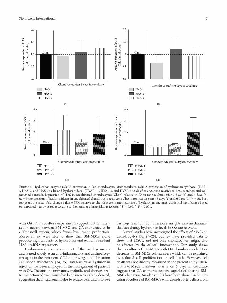
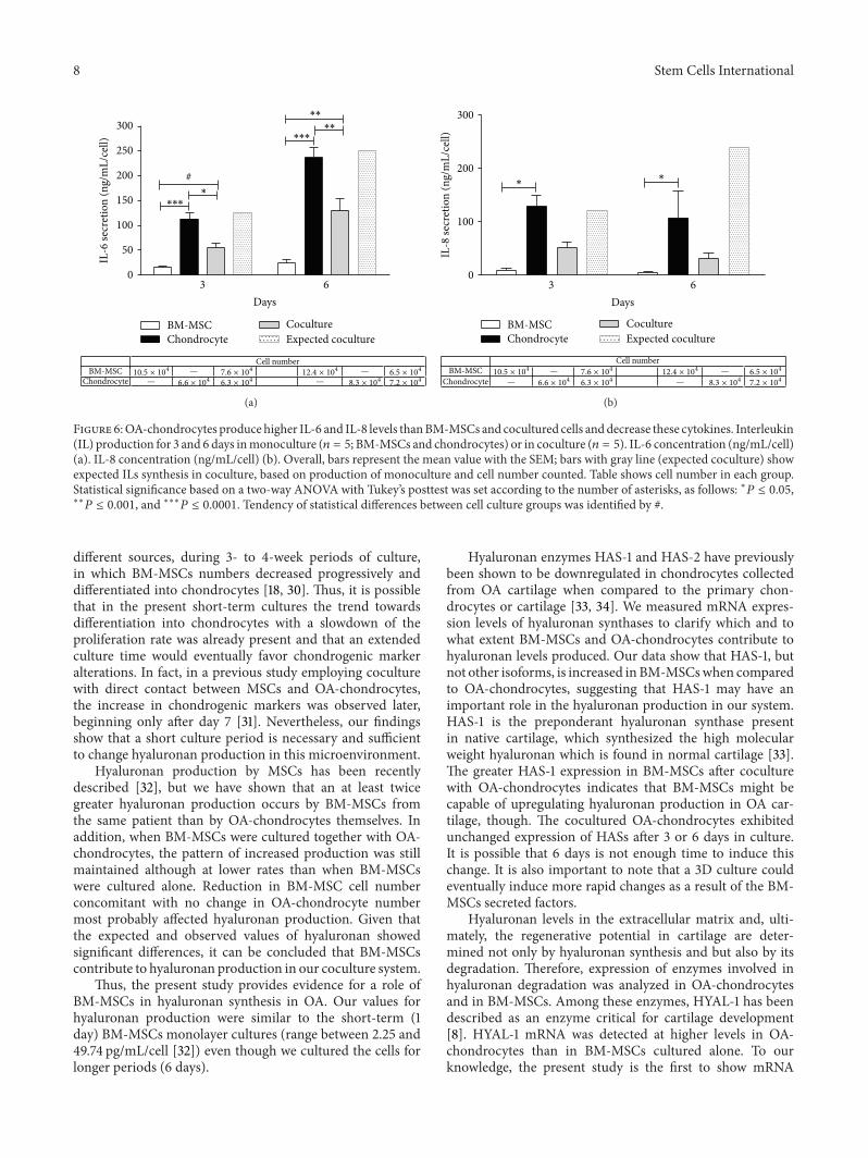