research article nerve dysfunction in hallux valgus is ......hallux valgus the most common forefoot...
TRANSCRIPT

CentralBringing Excellence in Open Access
JSM Foot and Ankle
Cite this article: Yassin M, Heller E, Rath-Wolfson L, Robinson D (2017) Nerve Dysfunction in Hallux Valgus is Correlated with Degree of Deformity - a Retro-spective Analysis of a Consecutive Series of Patients. JSM Foot Ankle 2(2): 1025.
*Corresponding authorDror Robinson, Department of Orthopedics, Hasharon Hospital, 7 Keren Kayemet Str., Petah Tikwa 49372, Israel, Tel.: 972-54-3342899; Fax: 972-8-9206013; Email:
Submitted: 12 October 2016
Accepted: 12 April 2017
Published: 14 April 2017
ISSN: 2475-9112
Copyright© 2017 Robinson et al.
OPEN ACCESS
Keywords•Hallux valgus•Neuropathy•Nerve injury
Research Article
Nerve Dysfunction in Hallux Valgus is Correlated with Degree of Deformity - a Retrospective Analysis of a Consecutive Series of PatientsMustafa Yassin1, Eyal Heller2, Lea Rath-Wolfson3, and Dror Robinson2*1Department of Orthopedics, Hasharon Hospital, Israel2Foot and Ankle Unit, Hasharon Hospital, Israel3Department of Pathology, Hasharon Hospital, Israel
Abstract
Background: Hallux valgus the most common forefoot deformity stretches the medial cutaneous nerve. There is a paucity of information regarding neuropathy associated with this often-progressive deformation.
Methods: The current study involves two groups: 30 patients examined prior to hallux valgus surgery and 30 foot surgery candidates without hallux valgus that were scheduled for a different operation of the hindfoot or ankle. The patients were assessed for quantitative vibration sensation using a Biothesiometer and for light sensation detection using a standardized monofilament. The hallux valgus angle (HVA) and intermetatarsal angle (IMA) were measured on routine standing radiographs.
Results: There were no significant differences between the groups regarding demographic data. There were significant differences in vibration thresholds in some examined points, and in light sensation measurement findings. Pearson’s correlation tests showed good statistically significant correlation between the Hallux Valgus angle and both light touch and vibration threshold measurement, with r ranges between 0.6 to 0.7 and P<0.05.A significant correlation was found between the two sensation modalities (vibration and light touch). Clinical neuropathy and the HVA and IMA.
Interpretation: It appears that nerve dysfunction is correlated with the degree of deformity in hallux valgus. Such nerve dysfunction is rare in patients undergoing hindfoot surgery (nerve release procedures were excluded).The current study does not resolve the issue of whether the more severe deformity is the reason for the nerve dysfunction due to digital nerve traction or alternatively the nerve dysfunction is the reason for deformity development.
INTRODUCTIONHallux valgus is probably the most common forefoot
pathology. It is usually slowly progressive. It is highly correlated with shoe wear, but occurs also in non-shoe wearing populations [1,2]. There is a strong genetic predeliction as demonstrated by Coughlin [3]. While a neuromuscular misbalance is obviously responsible for the deformity in most instances, little is known about the neurological status of hallux valgus patients. It has been reported that about twenty percent of hallux valgus patients suffer from sensory disturbances prior to surgery [4]. A prior study has shown that there is no correlation between the presence of sensory deficit and the degree of hallux valgus angle in a specific patient. The current study was undertaken to assess whether the amount of lateral hallux deviation correlates with the presence of neuropathy as determined both by light touch sensation and quantitative vibration assessment.
METHODSPatient Population
Experimental group: This is a retrospective analysis of pa-tient treated at the foot and ankle surgery unit of the Hasharon Hospital, Rabin Medical Center, in Petah Tikwa, Israel. All pa-tients treated from February 2009 to May 2010 were included in the study. The patient records were evaluated for the presence of data regarding the preoperative sensory state. All patients rou-tinely undergo standing radiographs in the anteroposterior and lateral positions. These radiographs were used in order to assess the HVA as previously described [5]. 52 patients being scheduled for hallux valgus surgery during the relevant period and having an adequate preoperative evaluation met the inclusion criteria. The patients reported here are a consecutive series of thirty pa-tients treated during this period.

CentralBringing Excellence in Open Access
Robinson et al. (2017)Email:
JSM Foot Ankle 2(2): 1025 (2017) 2/4
Exclusion Criteria:
I. Known nerve injury at a location more proximal than the hallux including lumbar radiculopathy or compression syndromes.
II. Prior surgical procedure of the foot
III. Prior lumbar spine operation
IV. Known neuropathy either diabetic or another type
Thirty patients met both inclusion and exclusion criteria and served as the experimental group.
Control group
Thirty consecutive patients were included. Inclusion criteria were: patients scheduled for foot or ankle operation at our unit at the same period, no forefoot pathology, no prior history of foot or ankle surgery, no history of neuropathy, no known medical history of nerve compression syndromes of the lower limb, no prior surgery of the lumbar spine or known radiculopathy.
Classsification of hallux valgus grade
A weight-bearing antero-posterior radiograph was taken during the pre-operative assessment. The AAOS classification was used(5). The hallux valgus deformity was graded as mild, moderate or severe according to the hallux valgus angle(HVA), the intermetatarsal angle (IMA)and the degree of sesamoid subluxation (the latter two parameters are not reported herein).
Light touch assessment
Light touch assessment was performed using Semmes-Weinstein mono filament size 5.07 (Connecticut Bio-instruments Inc, Danbury, Connecticut). The test assesses the sensation to 10 grams point pressure. The sensation was evaluated at five points (Figure 1):
1. Hallux’ tip 5 millimeters caudal to the nail.
2. At the center of deformity (bunion).
3. At the first web space over the dorsal aspect 5 millimeters proximal to the web space.
4. At the dorsal aspect of the metatarso-cuneiform joint.
5. At the medial malleolus.
Three measurements were performed over each of the points. If the patient reported feeling in one of the tests, this point was considered as positive. The result was recorded as either negative – no feeling or positive – sensation at the measured point. Each patient was graded on a 0 to 5 scale. Grade zero (0) indicates no sensation at any of the points and grade five (5) indicates light-touch feeling at all points.
Vibration sense assessement
Vibration was assessed using a BIOTHESIOMETER® (Bio-Medical Instrument Company, Newbury, Ohio(. This instrument allows definition of vibration sense at different strengths of vibration. The measurement was done according to the two steps threshold method. The vibration is set to 0 volts and increased by 5 volts every 2 seconds. The examinee reports the first stage
when vibration feeling is felt. Then the examiner continues to raise the threshold up to 10 volts higher than the threshold. Then the examiner lowers the voltage by 5 volts each until the examinee reports he can no longer feel the vibration. For the test to be valid the points should coincide or differ at most by one step. The lower voltage is reported as the threshold. The exam was repeated twice for each point and the results were averaged. Four different points were assessed in each examinee: 1. The tip of the hallux 5 millimeters below the nail. 2. At the center of the bunion. 3. Half a centimeter proximal to the fist web space. 4. Anterior to the medial malleolus, so that the probe does not lie directly on bone (Figure 2).
Statistical Analysis
The statistical analysis was performed using Analyse-it for Microsoft Excel (version 2.20) Analyse-it Software, Ltd. http://www.analyse-it.com/; 2009. The following statistical tests were used: Student’s t-test for continuous variables (age, vibration threshold, light sensation score), Mann-Whitney’s test for ordinal or nominal variables (gender, limb side).
RESULTSDemographics
Control group: Consisted of 30 patients treated at our unit. There were 19 females and 11 males, average age 49 years (27-76 years). In eleven patients, the limb treated was the right one and in the rest the left one. The average hallux valgus angle was moderate in size according to published classifications (Pique-Vidal and Vila, 2009).
Experimental group: consisted of 22 females and 8 male patients. The average age was 57.6 years (23-87 years). The right leg was operated on in 22 patients and the left one in the rest. The deformation was bilateral in 23 patients but no bilateral procedures were performed (Table 1) includes the demographic data of both groups).
Figure 1 Areas of assessment of light touch using a Semmes-Weinstein filament.

CentralBringing Excellence in Open Access
Robinson et al. (2017)Email:
JSM Foot Ankle 2(2): 1025 (2017) 3/4
Figure 2 Vibration examination points.
Figure 3 Vibration threshold is higher in the experimental group.
Table 1: Demographic characteristics of both groups.
Parameter Experimen-tal Group
Control Group
Student's t-test, p value
Males 8 11Mann-Whitney's statistic 495.0, Z statistic -0.83,2-tailed p value
0.4090
Females 22 19
Median Age 57 50Average Age ±
Standard Deviation 57.66±16.89 49±17.11 t statistic 1.5, p>0.12
Average HAV ± Standard Deviation
36.167o ± 6.88o
11.267o± 3.83o
t statistic -6.5, P<0.001
Light touch assessment
The difference in light touch score was significant between the experimental group (2.6 ± 0.4) as compared with the control group (1.4 ± 0.1, t-test p<0.05). In no patient in either group was sensation at the medial malleolus impaired. A good correlation was found between the vibration threshold and the light touch assessment (Table 2).
Vibration threshold
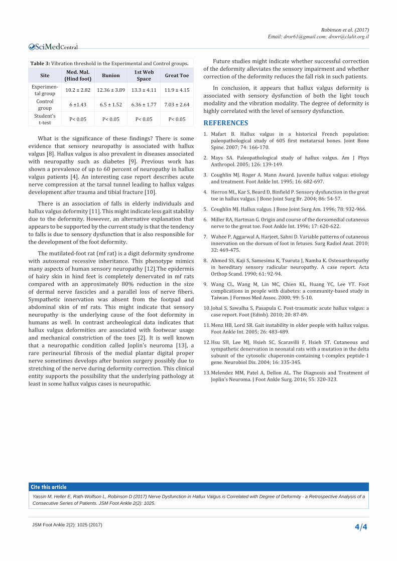
The vibration threshold is lower in control group’s feet than in hallux valgus afflicted feet (Figure 3). It is interesting to note that the difference in the threshold is not only at the hallux area but also in the medial malleolus area (representing the hindfoot). The average threshold in the three forefoot areas was compared with the average threshold at the fourth point (hind foot). The difference within the experimental group was significant (t statistic, p<0.05). In a more detailed analysis, the difference between the bunion site and the 1st web space site is significant as compared with the hind foot point (t statistic, p<0.05), but the difference between the great toe site and the hind foot was not significant (t statistic, p>0.05). In the control group, there is no significant difference between hind foot and forefoot measurements (Table 3).
A significant correlation was found between the light touch sensation grade and the hallux valgus angle ((Pearson Correlation 0.54, p>0.001). The correlation with the vibration threshold was even higher slightly less than 0.7 at the 1st web space (Pearson Correlation 0.69, p>0.001) and the bunion site (Pearson Correlation 0.694, p>0.001) and slightly less at the toe site (Pearson Correlation 0.58, p>0.001). An interesting finding was the high correlation between vibration threshold levels at the hind foot site and the hallux valgus angle (Pearson Correlation 0.58, p>0.001).
DISCUSSIONThe current work aimed at defining the relationship
between the hallux valgus angle and a possible neuropathy of the dorsomedial cutaneous branch. This is the last branch of the peroneal nerve [6] and it supplies cutaneous sensation to three of the forefoot points tested. The differences in vibration threshold between the groups at the bunion point and the 1st Webspace point were significant. However, at the great toe site vibration sensation was similar. This is presumably due to the double innervation of this area with branches of the deep peroneal nerve [7].
The difference in vibration threshold and light touch was significant at all forefoot sites between the groups. An interesting observation is that there is a significant difference in vibration threshold between the groups at the medial malleolus. This might indicate diffuse nerve damage around the foot in these patients. Alternatively, neuropathic damage underlying the disorder involving other nerves in the foot is possible. Perhaps a further study could answer this question. Another limitation of this study is the lack of a qualitative grading for light sensation at each site. However, there was found a strong correlation between high vibration threshold and damage to light sensation at the examined points.
Table 2: Light Sensation Assessment (score 0-5) as correlated with Vibration Threshold (arbitrary units).
Site Med. Mal. (Hind foot) Bunion 1st Web
Space Great Toe
Pearson's Correlation .581 .669 .654 .667
p-value P<0.05 P<0.05 P<0.05 P<0.05
CentralBringing Excellence in Open Access
Robinson et al. (2017)Email:
JSM Foot Ankle 2(2): 1025 (2017) 4/4
What is the significance of these findings? There is some evidence that sensory neuropathy is associated with hallux valgus [8]. Hallux valgus is also prevalent in diseases associated with neuropathy such as diabetes [9]. Previous work has shown a prevalence of up to 60 percent of neuropathy in hallux valgus patients [4]. An interesting case report describes acute nerve compression at the tarsal tunnel leading to hallux valgus development after trauma and tibial fracture [10].
There is an association of falls in elderly individuals and hallux valgus deformity [11]. This might indicate less gait stability due to the deformity. However, an alternative explanation that appears to be supported by the current study is that the tendency to falls is due to sensory dysfunction that is also responsible for the development of the foot deformity.
The mutilated-foot rat (mf rat) is a digit deformity syndrome with autosomal recessive inheritance. This phenotype mimics many aspects of human sensory neuropathy [12].The epidermis of hairy skin in hind feet is completely denervated in mf rats compared with an approximately 80% reduction in the size of dermal nerve fascicles and a parallel loss of nerve fibers. Sympathetic innervation was absent from the footpad and abdominal skin of mf rats. This might indicate that sensory neuropathy is the underlying cause of the foot deformity in humans as well. In contrast archeological data indicates that hallux valgus deformities are associated with footwear usage and mechanical constriction of the toes [2]. It is well known that a neuropathic condition called Joplin’s neuroma [13], a rare perineurial fibrosis of the medial plantar digital proper nerve sometimes develops after bunion surgery possibly due to stretching of the nerve during deformity correction. This clinical entity supports the possibility that the underlying pathology at least in some hallux valgus cases is neuropathic.
Future studies might indicate whether successful correction of the deformity alleviates the sensory impairment and whether correction of the deformity reduces the fall risk in such patients.
In conclusion, it appears that hallux valgus deformity is associated with sensory dysfunction of both the light touch modality and the vibration modality. The degree of deformity is highly correlated with the level of sensory dysfunction.
REFERENCES1. Mafart B. Hallux valgus in a historical French population:
paleopathological study of 605 first metatarsal bones. Joint Bone Spine. 2007; 74: 166-170.
2. Mays SA. Paleopathological study of hallux valgus. Am J Phys Anthropol. 2005; 126: 139-149.
3. Coughlin MJ. Roger A. Mann Award. Juvenile hallux valgus: etiology and treatment. Foot Ankle Int. 1995; 16: 682-697.
4. Herron ML, Kar S, Beard D, Binfield P. Sensory dysfunction in the great toe in hallux valgus. J Bone Joint Surg Br. 2004; 86: 54-57.
5. Coughlin MJ. Hallux valgus. J Bone Joint Surg Am. 1996; 78: 932-966.
6. Miller RA, Hartman G. Origin and course of the dorsomedial cutaneous nerve to the great toe. Foot Ankle Int. 1996; 17: 620-622.
7. Wahee P, Aggarwal A, Harjeet, Sahni D. Variable patterns of cutaneous innervation on the dorsum of foot in fetuses. Surg Radiol Anat. 2010; 32: 469-475.
8. Ahmed SS, Kaji S, Samesima K, Tsuruta J, Namba K. Osteoarthropathy in hereditary sensory radicular neuropathy. A case report. Acta Orthop Scand. 1990; 61: 92-94.
9. Wang CL, Wang M, Lin MC, Chien KL, Huang YC, Lee YT. Foot complications in people with diabetes: a community-based study in Taiwan. J Formos Med Assoc. 2000; 99: 5-10.
10. Johal S, Sawalha S, Pasapula C. Post-traumatic acute hallux valgus: a case report. Foot (Edinb). 2010; 20: 87-89.
11. Menz HB, Lord SR. Gait instability in older people with hallux valgus. Foot Ankle Int. 2005; 26: 483-489.
12. Hsu SH, Lee MJ, Hsieh SC, Scaravilli F, Hsieh ST. Cutaneous and sympathetic denervation in neonatal rats with a mutation in the delta subunit of the cytosolic chaperonin-containing t-complex peptide-1 gene. Neurobiol Dis. 2004; 16: 335-345.
13. Melendez MM, Patel A, Dellon AL. The Diagnosis and Treatment of Joplin’s Neuroma. J Foot Ankle Surg. 2016; 55: 320-323.
Table 3: Vibration threshold in the Experimental and Control groups.
Site Med. Mal. (Hind foot) Bunion 1st Web
Space Great Toe
Experimen-tal group 10.2 ± 2.82 12.36 ± 3.89 13.3 ± 4.11 11.9 ± 4.15
Control group 6 ±1.43 6.5 ± 1.52 6.36 ± 1.77 7.03 ± 2.64
Student's t-test P< 0.05 P< 0.05 P< 0.05 P< 0.05
Yassin M, Heller E, Rath-Wolfson L, Robinson D (2017) Nerve Dysfunction in Hallux Valgus is Correlated with Degree of Deformity - a Retrospective Analysis of a Consecutive Series of Patients. JSM Foot Ankle 2(2): 1025.
Cite this article