research article oleuropein-enriched olive leaf...
TRANSCRIPT
Research ArticleOleuropein-Enriched Olive Leaf Extract AffectsCalcium Dynamics and Impairs Viability of MalignantMesothelioma Cells
Carla Marchetti,1 Marco Clericuzio,2 Barbara Borghesi,3 Laura Cornara,3
Stefania Ribulla,2 Fabio Gosetti,2 Emilio Marengo,2 and Bruno Burlando1,2
1 Istituto di Biofisica, Consiglio Nazionale delle Ricerche, Via De Marini 6, 16149 Genova, Italy2Dipartimento di Scienze e Innovazione Tecnologica (DISIT), Universita del Piemonte Orientale, Viale Teresa Michel 11,15121 Alessandria, Italy3Dipartimento di Scienze della Terra dell’Ambiente e della Vita (DISTAV), Universita degli Studi di Genova,Corso Europa 26, 16132 Genova, Italy
Correspondence should be addressed to Bruno Burlando; [email protected]
Received 14 September 2015; Revised 3 November 2015; Accepted 11 November 2015
Academic Editor: I-Min Liu
Copyright © 2015 Carla Marchetti et al.This is an open access article distributed under the Creative CommonsAttribution License,which permits unrestricted use, distribution, and reproduction in any medium, provided the original work is properly cited.
Malignant mesothelioma is a poor prognosis cancer in urgent need of alternative therapies. Oleuropein, the major phenolic ofolive tree (Olea europaea L.), is believed to have therapeutic potentials for various diseases, including tumors. We obtained anoleuropein-enriched fraction, consisting of 60%w/w oleuropein, from olive leaves, and assessed its effects on intracellular Ca2+ andcell viability in mesothelioma cells. Effects of the oleuropein-enriched fraction on Ca2+ dynamics and cell viability were studied inthe RENmesothelioma cell line, using fura-2microspectrofluorimetry andMTT assay, respectively. Fura-2-loaded cells, transientlyexposed to the oleuropein-enriched fraction, showed dose-dependent transient elevations of cytosolic Ca2+ concentration ([Ca2+]i).Application of standard oleuropein and hydroxytyrosol, and of the inhibitor of low-voltage T-type Ca2+ channels NNC-55-0396,suggested that the effect is mainly due to oleuropein acting through its hydroxytyrosol moiety on T-type Ca2+ channels. Theoleuropein-enriched fraction and standard oleuropein displayed a significant antiproliferative effect, as measured on REN cellsby MTT cell viability assay, with IC
50of 22 𝜇g/mL oleuropein. Data suggest that our oleuropein-enriched fraction from olive leaf
extract could have pharmacological application in malignant mesothelioma anticancer therapy, possibly by targeting T-type Ca2+channels and thereby dysregulating intracellular Ca2+ dynamics.
1. Introduction
Malignant mesothelioma is a poor prognosis cancer withworldwide increasing insurgence, arising from the pleura andother mesothelial tissue and showing close association withasbestos exposure. An effective therapeutic approach for thedisease is currently lacking, while alternative treatments areurgently needed [1]. Among alternative remedies for cancertreatment, there is a growing interest in the anticancer actionof natural substances, some of which are present in largeamounts in byproducts from agrofood chains.
Olive (Olea europaea L.) is a main temperate fruit crop,mostly destined to oil production [2].Olive oil is reputed to be
an important health promoting factor in the Mediterraneandiet, especially because of its alleged prevention of cardio-vascular problems, metabolic syndrome, cancer, alleviationof inflammatory and autoimmune conditions, and woundhealing [3]. However, olive tree leaves have a several-century-long tradition in the folk medicines of the Mediterraneanbasin and are currently contemplated in the PharmacopoeiaPh. Eur. 5 [4].
Leaves and other materials from olive tree pruningaccumulate in yearly amounts as high as about 25 kg per tree[5]. This material is generally disposed by olive tree growers,representing an overhead cost of olive and oil production,while the possibility of using it as biomass for thermal energy
Hindawi Publishing CorporationEvidence-Based Complementary and Alternative MedicineVolume 2015, Article ID 908493, 9 pageshttp://dx.doi.org/10.1155/2015/908493
2 Evidence-Based Complementary and Alternative Medicine
is under study [6]. Olive leaves are used in the form of herbalextracts, tea, and powder for nutritional supplement andcosmetics [4], but these products represent aminimal portionwith respect to the huge amount of disposed materials.
The major leaf secondary metabolite is the secoiridoidoleuropein, a glycosylated ester of elenolic acid with hydrox-ytyrosol (2-(3,4-dihydroxyphenyl)ethanol). Oleuropein leafcontent has been reported to vary about tenfold among differ-ent olive varieties, rating on average between 10 and 100mg/gof dry matter [7, 8]. Many other biologically active phenolicsare present in olive leaves, including hydroxytyrosol itself [9].
Oleuropein is deemed to have great potential as an antiox-idant and food additive, but also as a possible therapeutictool. A wide range of studies on oleuropein have been carriedout using in vitro assays, animal models of disease, or humanvolunteers, in order to explore possible beneficial effects forhuman health [10]. The reported findings mainly includeantioxidant and anti-inflammatory effects, hepatoprotection,neuroprotection, hypoglycemic and hypolipidemic activities,and cardiovascular protection [11, 12]. Antiproliferative activ-ities on cancer cell lines, antitumor effects in animals, andantimicrobial and antiviral effects have also been shown [13].
Hydroxytyrosol in olive leaves, fruits, and extracts isbelieved to derive from oleuropein hydrolysis, both by spe-cific enzymes and as an extraction artifact [9]. In vitro andin vivo studies suggest that this compound shares remarkableantioxidant and anti-inflammatory power with oleuropein,allegedly related to antiatherogenic, antithrombotic, cardio-protective, and anticancer effects [14, 15].
The value of olive leaf extract for possible use in healthproducts and medical food has been previously assessed interms of antioxidant and antimicrobial power [16]. However,the profitable reutilization of agrofood chain byproductsneeds to overcome several problems, including the costsof transport and processing and the seasonal availability ofthese materials. In this study, we have developed a rapidmethod for obtaining olive leaf extract enriched in oleu-ropein, involving raw extraction, extract partitioning, andchromatographic separation. The material has been tested inpharmacologically relevant, intracellular Ca2+ mobilizationassay and antiproliferative assay on mesothelioma cells. Inthese experiments, standard oleuropein has been used ascontrol, while hydroxytyrosol has been also used in Ca2+assays to provide insight into the Ca2+ mobilizing propertiesof the oleuropein molecule.
2. Materials and Methods
2.1. Plant Material. Branch specimens of Olea europaea L.cultivar “Taggiasca” [17] were obtained from olive groveslocated at Imperia, Italy, owned by the Pietro Isnardi S.r.l.food company. The material was taxonomically identifiedby one of us (LC), and a voucher specimen was depositedat DISTAV, University of Genova (cat.: GE-Olea europaeacultivar “Taggiasca”). Leaves were separated from twigs, air-dried for one week, minced to <1mm using a grinder, andthen subjected to extraction procedure.
2.2. Reagents and Solutions. HPLC grade methanol Chro-masolv (>99.9%) and glacial acetic acid were purchasedfrom Sigma-Aldrich (Milwaukee, USA). Ammonium acetate(99%) was acquired from Fluka (Buchs, Switzerland). Ultra-pure water was produced by a Millipore Milli-Q system(Milford, USA). Analytical grade chemicals were purchasedfrom Sigma-Aldrich, unless otherwise specified.
For LC/MS analysis, standard stock solutions of oleuro-pein and hydroxytyrosol (each at concentration of100.0mg L−1) were prepared in methanol and diluted asrequired with a buffer solution of ammonium acetate10.0mM brought to pH 4.0 for acetic acid. For in vitrobioassays, stock solutions of oleuropein and hydroxytyrosol(each at 100mM) were prepared in DMSO and dilutedas indicated with cell medium or loading buffer for Ca2+measurements. Stock solutions were preserved at −20∘C indark conditions and were stable for three months.
2.3. Olive Leaf Extraction Procedure. The minced leaveswere extracted with a mixture of 2-propanol : water of 9 : 1.Aliquots of 10 g of leaves were extracted twice with 200mLof solvent each time. The extraction was carried out at RTfor about 1.5 h. The resulting solutions were mixed and thesolvent was eliminated under vacuum, yielding an amount of2.35 g of driedmaterial.This raw extract was then partitionedbetween a mixture of methanol : water of 3 : 1 and a mixtureof toluene : petroleum ether of 2 : 1 (100mL each phase). Thehydrocarbon phase became deep green, indicating that itcontained all the chlorophyll of the raw extract. The aqueousmethanol phase was dried under vacuum, yielding 1.8 g ofsubextract. This material was then again partitioned betweenwater and 2-butanone (100mL each phase), to remove sugarsand other water-soluble compounds. The organic phase wasdried out, obtaining a partitioned subextract that amountedto 1.03 g (43% recovery from the first raw extract, 10% fromthe plant material). A control TLC showed that in the formerpartition no oleuropein passed in the hydrocarbon phase,while in the latter only a tiny amount of it was lost in thewaterphase.
2.4. Chromatographic Separation. Liquid chromatographywas performed by means of an MPLC system, consisting ofan Alltech 426 HPLC pump equipped with a VWR LaPrep3101 detector. Glass columns from Omnifit were used, home-packed with Fluka silica gel 100 RP-18 (15–35 𝜇m) stationaryphase. TLC was performed with Merck F
254glass plates (RP-
18); the spots were detected using a UV lamp (254 and366 nm) and additionally by spraying with sulfovanillin andheating at 100∘C, obtaining an oleuropein coral pink spot onTLC.
The above subextract was loaded on a RP-18 preparativecolumn and eluted with 80% water and 20% methanol. Frac-tioningwasmonitored through theUVdetector (𝜆= 250 nm)and with TLC. Finally, an oleuropein-enriched fraction wascollected and subjected to further study.
Evidence-Based Complementary and Alternative Medicine 3
2.5. LC/MS Analysis
2.5.1. Apparatus. LC/MS analysis was performed on theabove raw extract and subfractions by Nexera Liquid Chro-matography Shimadzu (Kyoto, Japan) system equipped witha DGU-20A3R Degasser, two LC-30AD Pumps, a SIL-30ACAutosampler, a CTO-20AC column compartment, and aCMB-20A Lite system controller. The system was interfacedwith a 3200 QTrap LC-MS/MS system (AB Sciex, Concord,Canada) by a Turbo V interface equipped with an ESI probe.The 3200QTrap data were processed byAnalyst 1.5.2 software(Toronto, Canada).
2.5.2. UHPLC-MS/MS Conditions. The stationary phase wasa Kinetex C18 column (2.1mm × 100mm, 2.6 𝜇m) (Phe-nomenex, Italy). The mobile phase was a mixture of ammo-nium acetate 10.0mM in ultrapure water with the additionof 0.1% acetic acid (A), and aqueous ammonium acetate10.0mM/methanol 5/95 (v/v) with the addition of 0.1% aceticacid (B), eluting at flow-rate 0.400mLmin−1 in the followinggradient conditions: 0.0–0.5min 5% B, 0.6–8.0min 65% B,8.1–12.0min 100% B, and 12.1–15.0min 5% B. The injectionvolume was 5.0 𝜇L. Oven temperature was set at 40∘C.
The turbo ion spray (TIS) ionization was obtained usingthe Turbo V interface working in negative ion mode. Theinstrumental parameters were set as follows: curtain gas (N
2)
at 30 psig, nebulizer gas GS1 and GS2 at 50 and 40 psig,respectively, desolvation temperature (TEM) at 550∘C, col-lision activated dissociation gas (CAD) at 6 units of thearbitrary scale of the instrument, and ion spray voltage (IS)at −4500V.
The 3200 QTrap was used in scheduled multiple reactionmonitoring (sMRM) considering the transitions of eachspecies at a prefixed retention time. Unit mass resolutionwas established and maintained in each mass-resolvingquadrupole by keeping a full width at half maximum(FWHM) of about 0.7 u.
The analytes were previously subjected toMS/MS charac-terization study, to identify the fragmentation patterns takingplace under increasing collisional energy. Characterizationexperiments were carried out for direct infusion of 1.0mg L−1standard solutions of each analyte connected through a Tvalve to the syringe pump (syringe flow-rate 20.0𝜇Lmin−1;chromatographic pump flow-rate 200 𝜇Lmin−1). For eachspecies, the most intense transition was used for the quan-titative analysis and referred to as “quantifier” transition,while the second intense one (the “qualifier” transition) wasemployed in the identification step, as a confirmation. The“quantifier” and “qualifier” transitions and the instrumentalpotential values for each compound are reported in Table 1.
Calibration plots of five concentration levels (betweenLOQ value and 1000.0 𝜇g L−1), relating the peak area of the“quantifier” transition signal (𝑦) versus standard concentra-tion (𝑥), were built for all the analytes. To avoid a possibleinfluence of the experimental error on the execution order ofanalyses, the standard solutions were injected in randomizedorder. For all the analytes a linear regression fit was used witha weighting factor 1/𝑥 and for all the calibration plots a good
linearity with regression coefficients (𝑅2) always greater than0.9982 was obtained.
Detection limit values expressed as the concentration ofthe analyte that gives a signal equal to the average background(𝑆blank) plus 3 times the standard deviation of the blank(LOD = 𝑆blank + 3𝑠blank) are 0.3 𝜇g L−1 for hydroxytyrosoland 0.4 𝜇g L−1 for oleuropein.The quantitation limits (LOQ),evaluated as LOQ= 𝑆blank + 10𝑠blank, are 1.0 𝜇g L
−1 for hydrox-ytyrosol and 1.2 𝜇g L−1 for oleuropein.
2.6. In Vitro Cell Culture. In vitro experiments were carriedout using the tumorigenic malignant mesothelioma REN cellline [1]. Cells were grown in DMEM, supplemented with 10%foetal bovine serum (FBS, Euroclone), at 37∘C, in a 5% CO
2,
fully humidified atmosphere.
2.7. Cell Viability Assay. Cell viability was determined onREN cells by the MTT assay. Cells were settled in 96-wellplates for 24 h, exposed to various agents for 48 h as specified,incubated with 100𝜇L/mL tetrazolium salt (MTT, 5mg/mLin PBS) in cell culture mediumwithout serum for 3 h at 37∘C,and treated with a solution of 1 N HCl-isopropanol (1 : 24,v/v) followed by mixing to dissolve the dark-blue formazancrystals formed. After a few minutes at room temperature,the plates were read at 550 nm in a VMax microplate reader(Molecular Devices, Sunnyvale, CA).
2.8. Intracellular Ca2+ Measurements. Cytosolic free Ca2+concentration was measured in fura-2-loaded REN cellsusing a microspectrophotometry fluorescence-ratio setupequipped with a perfusion system, as previously described[18]. Cells were incubated in loading buffer containing 10 𝜇Mfura-2-AM at 37∘C for 30 minutes, washed with buffer, andmounted on the stage of an inverted microscope (AxiovertZeiss, Germany), where they were continually superfusedwith different solutions. Cells were illuminated by a xenonlamp through a wavelength selector monochromator; emis-sionwas observed through×40 quartz objective and recordedby a photomultiplier.The ratio𝐸
340/𝐸380
was calculated every40msec to acquire a time-dependent internal Ca2+ sensitivesignal.
At the end of each experiment, cells were incubated with10 𝜇M ionomycin in a Ca-free solution containing 2mMEGTA for 20−40min until the ratio reached a minimumvalue (𝑅min); then EGTA was washed out, normal externalCa2+ (1mM) was restored, and the ratio readily reached amaximum value (𝑅max). Finally, MnCl
2(5mM) was added to
the bath to quench the fura-2 fluorescence and determine thebackground (cell autofluorescence) values. The fluorescenceemissions relative to each excitation wavelength (𝐸
340and
𝐸380
, resp.) were corrected for this background signal beforeratio 𝑅 = 𝐸
340/𝐸380
determination. Internal Ca2+ was cal-culated according to the Grynkiewicz equation [19].
2.9. Statistics. Statistics were obtained with the 𝑅 package,version 3.0.1 (http://www.r-project.org/foundation/), using 𝑡-test with Bonferroni’s correction for multiple comparisons.
4 Evidence-Based Complementary and Alternative Medicine
Table 1: sMRM NI transitions (Q1 and Q3 masses) and mass spectrometry parameters. The two most sensitive transitions for each specieswere monitored.
Analytes Q1 (𝑚/𝑧) Q3 (𝑚/𝑧) 𝑡𝑅(min) DP (V) EP (V) CEP (V) CE (V) CXP (V)
Hydroxytyrosol 153 123/93 3.4 −34 −75 −17.32 −20/−31 −1.08/−1.35Oleuropein 539 275/307 7.9 −40 −5 −31.61 −28/−29 −1.89/−2.12𝑡𝑅: retention time; DP: Declustering Potential; EP: Entrance Potential; CEP: Collision cell Entrance Potential; CE: Collision Energy; CXP: Collision cell eXitPotential.
Table 2: Percent content (w/w) of oleuropein and hydroxytyrosol indifferent olive leaf extracts.
RE SE EFOleuropein 21.4 ± 0.3 43.2 ± 1.4 60.1 ± 0.3Hydroxytyrosol 0.37 ± 0.04 0.91 ± 0.03 n.d.Data are means ± s.d. of three independent measures. RE: raw extract; SE:subextract; EF: oleuropein-enriched fraction; n.d.: not detected.
Cytotoxicity was determined using a logistic dose-responsecurve as reported in Ranzato et al. [20]. Analysis of data fromCa2+ measurements and determination of EC
50values were
done by Sigmaplot 8 (Systat Software Inc., San Jose, CA).
3. Results
Multistep processing of olive leaves consisted of the following:(1) extraction of dried, minced leaves in 2-propanol : water(9 : 1), to obtain a raw extract; (2) partitioning betweenmethanol : water (3 : 1) and toluene : petroleum ether (2 : 1),followed by partitioning of the dried aqueousmethanol phasebetween water and 2-butanone, to obtain a subextract; (3)liquid (column) chromatography separation of the driedorganic phase, to obtain an oleuropein-enriched fraction (seeMethods). Oleuropein and hydroxytyrosol quantification inthe raw extract and fractions was achieved by LC-MS tech-nique. Data showed a progressive enrichment in oleuropein,up to a value of 60% w/w in the final fraction obtainedthrough chromatographic separation (Table 2).
In order to evaluate the pharmacological potentials of theoleuropein-enriched, leaf extract fraction, and of its majorcomponent oleuropein, we tested the ability of mobilizingcell Ca2+ in mesothelioma REN cells. Intracellular Ca2+ is amain regulator of cell growth, and therefore agents able toderegulate Ca2+ dynamics can be possibly used to hinder cellproliferation.
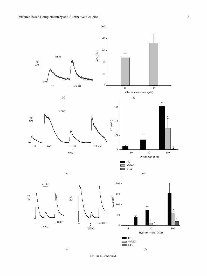
Fura-2-loaded cells were transiently exposed by perfusionto the oleuropein fraction. This treatment induced [Ca2+]irises followed by a prompt recovery of the baseline level uponwashout (Figure 1(a)). Data indicate that the observed [Ca2+]ispikes are not due to cell membrane injury but rather dependon the influx of Ca2+ through membrane Ca2+ channels.Moreover, the [Ca2+]i transient elevations were dependenton the nominal concentration of oleuropein (10–30 𝜇M)contained in the extract (Figure 1(b)).
To explore more in depth the mechanism of action, wealso studied the Ca2+ mobilizing property of standard oleu-ropein. Similar to the oleuropein fraction, increasing doses of
standard oleuropein, in the range 10–100 𝜇M, induced dose-dependent [Ca2+]i spikes (Figures 1(c) and 1(d)). Removalof Ca2+ from the external solution almost abolished theoleuropein effect (Figure 1(d)). In addition, the use of theT-type Ca2+ channel inhibitor NNC-55-0396 (Sigma, ≥98%,HPLC) [21] reversibly reduced the Ca2+ spike induced bythe highest dose of 100 𝜇M oleuropein (Figures 1(c) and1(d)), suggesting at least a partial involvement of T-type Ca2+channels.
We tried to provide some hint as to which oleuropeinmoiety could be active on cell Ca2+ mobilization. Hydrox-ytyrosol is known to induce modulatory effects on [Ca2+]i[22]. We therefore used standard hydroxytyrosol on fura-2-loaded REN cells, observing the induction of dose-dependent[Ca2+]i spikes that also in this case were inhibited by Ca2+-free medium. Moreover, NNC-55-0396 also hindered thehydroxytyrosol-induced [Ca2+]i spikes, with 80% inhibitionat 10 𝜇M hydroxytyrosol and 65% inhibition at 100𝜇Mhydroxytyrosol (Figures 1(e) and 1(f)).
As for comparative quantification of the effect on [Ca2+]i,dose-response data allowed estimating an EC
50of 12 𝜇M
for hydroxytyrosol and of 53𝜇M for oleuropein, showingthat hydroxytyrosol is more powerful than oleuropein ininducing Ca2+ spikes. No established drug is known toinduce T-type Ca2+ channel opening. We therefore usedthe natural phenolic epigallocatechin-3-gallate (EGCG) aspositive control, since we had previously shown that thiscompound induces Ca2+ rise in REN cells via T-type Ca2+channels [23]. Presently, EGCG was found to induce dose-dependent [Ca2+]i spikes with an estimated EC
50of 69 𝜇M
(Figures 1(e) and 1(f)).The oleuropein-enriched fraction and standard oleu-
ropein were separately used on REN cells at nominallyequivalent oleuropein concentrations, in order to verify theirantiproliferative activity. EGCG was used as positive controlalso in this case, since we had previously assessed its cytotox-icity on REN cells [24]. The cytotoxic effect of the oleuropeinfraction (IC
50= 22𝜇g/mL expressed as oleuropein) was
slightly, but not significantly, stronger than that of standardoleuropein (IC
50= 25 𝜇g/mL). Both substances were slightly
more cytotoxic than EGCG (IC50= 33 𝜇g/mL) (Figure 2).
4. Discussion
The oleuropein yield in our enriched fraction is very close tothe highest concentrations present in commercially availableolive leaf extract formulations [25], suggesting its possibledirect use in medicinal preparations. Despite a vast array of
Evidence-Based Complementary and Alternative Medicine 5
10 30 ole
30nM
1min
(a)Δ
Ca(n
M)
100
80
60
40
20
010 30
Oleuropein content (𝜇M)
(b)
10 100 100
NNC
100 ole
30nM
2min
(c)
ΔCa
(nM
)
0
Oleuropein (𝜇M)
150
100
50
10 30 100
∗
∗
Ole+NNC0 Ca
(d)
NNCNNC
20nM 30
nM
10 HT 100 HT
4min
(e)
ΔCa
(nM
)
0
∗
∗
∗∗
+NNC0 Ca
200
150
100
50
1 10 100
Hydroxytyrosol (𝜇M)
HT
(f)
Figure 1: Continued.
6 Evidence-Based Complementary and Alternative Medicine
20nM
10 30 100 EGCG
1min
(g)
ΔCa
(nM
)
120
90
60
30
010 30 100
EGCG (𝜇M)
(h)
Figure 1: Time course of [Ca2+]i level in mesothelioma REN cells loaded with fura-2 and alternatively exposed to the oleuropein-enrichedfraction from olive leaf extract (a, b), to standard oleuropein (c, d) or to standard hydroxytyrosol (e, f). The oleuropein fraction and standardoleuropein have been used at nominal concentrations of oleuropein, expressed in𝜇moles/L. (a) Induction of [Ca2+]i spikes in RENcells loadedwith fura-2 by transient exposure to the oleuropein fraction (ole) at 10 and 30 𝜇Moleuropein. (b) Quantification of dose-dependent inductionof [Ca2+]i spikes by the oleuropein fraction. Data are means ± s.e.m. (𝑛 ≥ 3) of Δ[Ca2+]i (difference between peak value and basal level of[Ca2+]i). (c) Induction of [Ca2+]i spikes in REN cells by transient exposure to standard oleuropein (ole) at 10 and 100 𝜇M. The inhibitoryeffect of the T-type Ca2+ channel blocker NNC-55-0396 (5 𝜇M) and recovery after washout are shown. (d) Quantification of dose-dependentinduction of [Ca2+]i spikes by standard oleuropein (EC
50= 53 𝜇M), and inhibition by Ca2+-free medium (0 Ca) or NNC-55-0396 measured
at 100 𝜇M oleuropein. Data as above; ∗ = 𝑝 ≤ 0.01, 𝑡 test. (e) Induction of [Ca2+]i spikes by standard hydroxytyrosol (HT) at 10 and 100𝜇M,inhibitory effect of NNC-55-0396, and recovery after washout. (f) Quantification of dose-dependent effect of hydroxytyrosol (EC
50= 12 𝜇M)
and inhibition by Ca2+-free medium (0 Ca) or NNC-55-0396 measured at 10 and 100 𝜇M hydroxytyrosol. Data and statistics as above. (g, h)Induction of [Ca2+]i spikes by EGCG and statistics of dose-dependent effect (EC
50= 69 𝜇M). Data as above.
data on possible oleuropein cellular targets, our data aboutthe effects on intracellular Ca2+ are new. The activity ofoleuropein was slightly lower than that of the oleuropein-enriched fraction, possibly due to the occurrence in thefraction of other secoiridoid esters known to be present inolive leaves, such as demethyloleuropein, ligstroside, andoleuroside [26], allegedly coeluting with oleuropein in theHPLC separation.
We have previously found that low-voltage-activated T-type Ca2+ channels, specifically the isoform Cav3.2, medi-ate the Ca2+ rise induced in REN cells by the green teapolyphenol epigallocatechin-3-gallate [23]. We have alsoshown that Cav3.2 is responsible for Ca2+ spikes induced byepigallocatechin-3-gallate in MCF-7 breast cancer cells [20].In the present study, an involvement of T-type Ca2+ channelsis called into question also for the Ca2+ mobilizing effect ofoleuropein by the use of Ca2+-freemedium, indicating a Ca2+entry process, and of the specific T-type Ca2+ channel blockerNNC-55-0396.
The stronger activity of hydroxytyrosol with respect tooleuropein and the inhibitory effect of NNC-55-0396 suggestthat the hydroxytyrosol moiety could be the active sitemediating the oleuropein effect on T-type Ca2+ channels.The almost complete inhibition exerted by NNC-55-0396
on hydroxytyrosol at lower dose, compared with the partialinhibition at higher dose, seems to indicate that the effectof olive phenolics mostly involves a T-type Ca2+ channel-dependent mechanism at low doses (≤10 𝜇M). At higherdoses, these phenolics could instead act through amore com-plex mechanism, possibly including other Ca2+ mobilizingsystems.
T-type Ca2+ channels have been linked to various kindsof epilepsy [27], while they are thought to play a role intumor cell cycle progression andproliferation [28, 29].Hence,much interest has been raised in their possible therapeutictargeting with more or less specific inhibitors [30, 31].However, the blockage of these channels does not seem to bean exclusive strategy in antitumor therapy. Antiproliferativeeffects obtained with mibefradil and pimozide, two T-typeCa2+ channel inhibitors, have been partially ascribed tosome unidentified mechanism [32]. Also, cell cycle progres-sion needs a fine tuning of [Ca2+]i oscillations, suggestingthat intracellular Ca2+ dysregulation determining transient[Ca2+]i rise may also interfere with tumor development, pos-sibly leading to cell death [33]. Accordingly, pharmacologicalagents like olive phenolics, able to abnormally activate T-type Ca2+ channel activity, could be promising tools for theinhibition of cancerous cell growth.

Evidence-Based Complementary and Alternative Medicine 7
0
20
40
60
80
100
120
1 10 100
Cel
l via
bilit
y (%
)
Oleuropein (𝜇g/mL)
IC50 = 22 (17–29)
(a)
0
20
40
60
80
100
120
1 10 100
Cel
l via
bilit
y (%
)
Oleuropein (𝜇g/mL)
IC50 = 25 (20–33)
(b)
0
20
40
60
80
100
120
1 10 100
Cel
l via
bilit
y (%
)
EGCG (𝜇g/mL)
IC50 = 33 (26–41)
(c)
Figure 2: Dose-response data of cell viability obtained with the MTT assay after exposure of REN cells for 48 h to the oleuropein-enrichedfraction (60% oleuropein) (a), standard oleuropein (b), and EGCG (c). The charts show means ± s.d. of percent MTT-formazan absorbance,logistic regression lines, and IC
50values expressed as 𝜇g/mL (95% CI). Concentrations are normalized as the logarithm of 𝜇g/mL.
In apparent contrast with the present data, it has beenpreviously shown that an olive leaf extract exerts antago-nistic effects on high-voltage-activated L-type Ca2+ chan-nel, thereby inducing blood pressure lowering [34, 35]. Acomparison between these previous reports and our study isdifficult tomake, owing to differences in both the extracts andthe experimental models. However, such a complex of dataadvocates an intriguing scenario, entailing divergent effects ofmajor olive phenolics on low-voltage and high-voltage Ca2+channels.
More insight into the possibility of using the oleuropeinfraction in antitumor treatment formesothelioma is providedby our cytotoxicity tests, indicating that the antiproliferativeactivity of the oleuropein fraction essentially depends onits major constituent oleuropein. The United States NationalCancer Institute plant screening program has established thata plant extract can be deemed to have a cytotoxic effect ifIC50
for incubations of 48–72 h is 20𝜇g/mL or lower [36].Our oleuropein-enriched fraction has an oleuropein contentof 60% and a cytotoxicity IC
50of 22 (17–29)𝜇g/mL in terms
of oleuropein. We can then estimate IC50
of 36.7 (28.3–48.3) 𝜇g/mL for the oleuropein-enriched fraction as a whole.Hence, our oleuropein fraction is close to the cytotoxicity
threshold established by NCI and could therefore be appro-priate for the development of synergistic treatments allowingreducing the doses of highly toxic conventional drugs, aspreviously suggested for EGCG [1]. Such an approach couldovercome critical drawbacks in the treatment of chemoresis-tant tumors like malignant mesothelioma [37].
There is no established standard dose of oleuropeinfor humans, but in acute toxicity studies, oleuropein andhydroxytyrosol have shown no lethality or adverse effectsin mice up to doses of 1000 and 2000mg/kg, respec-tively. In addition, studies conducted on humans witholive extract or its polyphenolics have shown no undesiredeffects [38, 39]. Pharmacokinetics of oral oleuropein andits metabolite hydroxytyrosol in human plasma indicatespeaks of about 0.004 𝜇g/mL (0.01𝜇M) for oleuropein, whileits main metabolite hydroxytyrosol can reach values ofabout 0.15𝜇g/mL (1 𝜇M) [40]. These values are close tothe effective concentrations observed in our experimentsfor hydroxytyrosol, but intravenous delivery of oleuropeinshould allow reaching much higher blood peaks. Hence,pharmacokinetics data indicate that plasma concentrationscompatible with dysregulating effects on tumor cell Ca2+could be easily achieved upon clinical administrations of
8 Evidence-Based Complementary and Alternative Medicine
oleuropein, followed by its conversion into the more effectivemetabolite hydroxytyrosol.
5. Conclusions
We have shown a method to obtain oleuropein-enrichedolive leaf extract fractions that could be of industrial interest,because extraction is generally competitive with chemicalsynthesis for complex molecules like oleuropein. In addi-tion, we have highlighted a novel mechanism of action foroleuropein and its metabolite hydroxytyrosol, consisting ina dysregulation of Ca2+ dynamics occurring via T-type Ca2+channels. This kind of effect suggests a possible use of theoleuropein fraction from olive leaves in the treatment ofmesothelioma.
Conflict of Interests
The authors declare no conflict of interests.
Acknowledgments
Bruno Burlando was supported by Fondazione Buzzi Uni-cem (Casale Monferrato, Italy), Grant no. FBU-P27. LauraCornara was supported by a PO-CRO-FSE Project, AsseIV Capitale Umano, ob. Specifico I/6, 2007–2013 (RegioneLiguria, Italy). Barbara Borghesi was recipient of a ResearchFellowship from the PO-CRO-FSE Project. Stefania Ribullawas recipient of a Ph.D. scholarship from the ItalianMinistryof University and Research (MIUR). The authors wish tothank the Pietro Isnardi S.r.l. food company (Imperia, Italy)for providing olive leaves.
References
[1] V. Volta, E. Ranzato, S.Martinotti et al., “Preclinical demonstra-tion of synergistic Active Nutrients/Drug (AND) combinationas a potential treatment for malignant pleural mesothelioma,”PLoS ONE, vol. 8, no. 3, Article ID e58051, 2013.
[2] C. M. Diez, I. Trujillo, N. Martinez-Urdiroz et al., “Olivedomestication and diversification in the Mediterranean basin,”The New Phytologist, vol. 206, no. 1, pp. 436–447, 2015.
[3] C. Alarcon de la Lastra, M. D. Barranco, V. Motilva, andJ. M. Herrerıas, “Mediterranean diet and health: biologicalimportance of olive oil,” Current Pharmaceutical Design, vol. 7,no. 10, pp. 933–950, 2001.
[4] S. N. El and S. Karakaya, “Olive tree (Olea europaea) leaves:potential beneficial effects on human health,”Nutrition Reviews,vol. 67, no. 11, pp. 632–638, 2009.
[5] M. Delgado-Pertınez, A. Chesson, G. J. Provan, A. Garrido,and A. Gomez-Cabrera, “Effect of different drying systems forthe conservation of olive leaves on their nutritive value forruminants,” Animal Research, vol. 47, no. 2, pp. 141–150, 1998.
[6] A. Garcia-Maraver, D. Salvachua, M. J. Martınez, L. F. Diaz, andM. Zamorano, “Analysis of the relation between the cellulose,hemicellulose and lignin content and the thermal behavior ofresidual biomass from olive trees,” Waste Management, vol. 33,no. 11, pp. 2245–2249, 2013.
[7] C. Savournin, B. Baghdikian, R. Elias, F. Dargouth-Kesraoui,K. Boukef, and G. Balansard, “Rapid high-performance liquid
chromatography analysis for the quantitative determination ofoleuropein in Olea europaea leaves,” Journal of Agricultural andFood Chemistry, vol. 49, no. 2, pp. 618–621, 2001.
[8] M. Ansari, M. Kazemipour, and S. Fathi, “Development ofa simple green extraction procedure and HPLC method fordetermination of oleuropein in olive leaf extract applied to amulti-source comparative study,” Journal of the Iranian Chemi-cal Society, vol. 8, no. 1, pp. 38–47, 2011.
[9] D. Ryan, M. Antolovich, P. Prenzler, K. Robards, and S. Lavee,“Biotransformations of phenolic compounds in Olea europaeaL.,” Scientia Horticulturae, vol. 92, no. 2, pp. 147–176, 2002.
[10] S. H. Omar, “Oleuropein in olive and its pharmacologicaleffects,” Scientia Pharmaceutica, vol. 78, no. 2, pp. 133–154, 2010.
[11] S. Bulotta, M. Celano, S. M. Lepore, T. Montalcini, A. Pujia,and D. Russo, “Beneficial effects of the olive oil phenoliccomponents oleuropein and hydroxytyrosol: focus on protec-tion against cardiovascular and metabolic diseases,” Journal ofTranslational Medicine, vol. 12, article 219, 2014.
[12] F. Durlu-Ozkaya and M. T. Ozkaya, “Oleuropein using as anadditive for feed and products used for humans,” Journal of FoodProcessing & Technology, vol. 2, no. 3, article 113, 2011.
[13] B. Barbaro, G. Toietta, R. Maggio et al., “Effects of the olive-derived polyphenol oleuropein on human health,” InternationalJournal of Molecular Sciences, vol. 15, no. 10, pp. 18508–18524,2014.
[14] S. Granados-Principal, J. L. Quiles, C. L. Ramirez-Tortosa, P.Sanchez-Rovira, and M. C. Ramirez-Tortosa, “Hydroxytyrosol:from laboratory investigations to future clinical trials,”NutritionReviews, vol. 68, no. 4, pp. 191–206, 2010.
[15] R. Bernini, N. Merendino, A. Romani, and F. Velotti, “Naturallyoccurring hydroxytyrosol: synthesis and anticancer potential,”Current Medicinal Chemistry, vol. 20, no. 5, pp. 655–670, 2013.
[16] O.-H. Lee and B.-Y. Lee, “Antioxidant and antimicrobial activi-ties of individual and combined phenolics inOlea europaea leafextract,” Bioresource Technology, vol. 101, no. 10, pp. 3751–3754,2010.
[17] T. Bracci, L. Sebastiani, M. Busconi, C. Fogher, A. Belaj, and I.Trujillo, “SSR markers reveal the uniqueness of olive cultivarsfrom the Italian region of Liguria,” Scientia Horticulturae, vol.122, no. 2, pp. 209–215, 2009.
[18] M.Mazzolini, S. Traverso, andC.Marchetti, “Multiple pathwaysof Pb2+ permeation in rat cerebellar granule neurones,” Journalof Neurochemistry, vol. 79, no. 2, pp. 407–416, 2001.
[19] G. Grynkiewicz, M. Poenie, and R. Y. Tsien, “A new generationof Ca2+ indicators with greatly improved fluorescence proper-ties,” Journal of Biological Chemistry, vol. 260, no. 6, pp. 3440–3450, 1985.
[20] E. Ranzato, V.Magnelli, S.Martinotti et al., “Epigallocatechin-3-gallate elicits Ca2+ spike in MCF-7 breast cancer cells: essentialrole of Cav3.2 channels,” Cell Calcium, vol. 56, no. 4, pp. 285–295, 2014.
[21] M. Li, J. B. Hansen, L. Huang, B. M. Keyser, and J. T. Taylor,“Towards selective antagonists of T-type calcium channels:design, characterization and potential applications of NNC 55-0396,” Cardiovascular Drug Reviews, vol. 23, no. 2, pp. 173–196,2005.
[22] C. A. Palmerini, E. Carlini, C. Saccardi, M. Servili, G. Monte-doro, and G. Arienti, “Activity of olive oil phenols on lym-phomonocyte cytosolic calcium,” The Journal of NutritionalBiochemistry, vol. 16, no. 2, pp. 109–113, 2005.

Evidence-Based Complementary and Alternative Medicine 9
[23] E. Ranzato, S. Martinotti, V. Magnelli et al., “Epigallocatechin-3-gallate induces mesothelioma cell death via H
2O2-dependent
T-typeCa2+ channel opening,” Journal of Cellular andMolecularMedicine, vol. 16, no. 11, pp. 2667–2678, 2012.
[24] S. Martinotti, E. Ranzato, and B. Burlando, “In vitro screeningof synergistic ascorbate-drug combinations for the treatment ofmalignant mesothelioma,” Toxicology in Vitro, vol. 25, no. 8, pp.1568–1574, 2011.
[25] D. T. Kremastinos, “Olive and oleuropein,” Hellenic Journal ofCardiology, vol. 49, no. 4, pp. 295–296, 2008.
[26] Y. Wang, S. Q. Wang, W. H. Cui, J. J. He, Z. F. Wang, and X. L.Yang, “Olive leaf extract inhibits lead poisoning-induced braininjury,” Neural Regeneration Research, vol. 8, no. 22, pp. 2021–2029, 2013.
[27] E. Tringham, K. L. Powell, S. M. Cain et al., “T-type calciumchannel blockers that attenuate thalamic burst firing and sup-press absence seizures,” Science translationalmedicine, vol. 4, no.121, Article ID 121ra19, 2012.
[28] A. Panner, L. L. Cribbs, G. M. Zainelli, T. C. Origitano, S. Singh,and R. D.Wurster, “Variation of T-type calcium channel proteinexpression affects cell division of cultured tumor cells,” CellCalcium, vol. 37, no. 2, pp. 105–119, 2005.
[29] J. T. Taylor, X.-B. Zeng, J. E. Pottle et al., “Calcium signaling andT-type calcium channels in cancer cell cycling,” World Journalof Gastroenterology, vol. 14, no. 32, pp. 4984–4991, 2008.
[30] W. Li, S.-L. Zhang, N. Wang, B.-B. Zhang, andM. Li, “Blockadeof T-type Ca2+ channels inhibits human ovarian cancer cellproliferation,” Cancer Investigation, vol. 29, no. 5, pp. 339–346,2011.
[31] K. R. Loughlin, “Calcium channel blockers and prostate cancer,”Urologic Oncology, vol. 32, no. 5, pp. 537–538, 2014.
[32] G. E. Bertolesi, C. Shi, L. Elbaum et al., “The Ca2+ channelantagonists mibefradil and pimozide inhibit cell growth viadifferent cytotoxic mechanisms,” Molecular Pharmacology, vol.62, no. 2, pp. 210–219, 2002.
[33] G. R. Monteith, D. McAndrew, H. M. Faddy, and S. J. Roberts-Thomson, “Calcium and cancer: targeting Ca2+ transport,”Nature Reviews Cancer, vol. 7, no. 7, pp. 519–530, 2007.
[34] A. Scheffler, H. W. Rauwald, B. Kampa, U. Mann, F. W. Mohr,and S. Dhein, “Olea europaea leaf extract exerts L-type Ca2+channel antagonistic effects,” Journal of Ethnopharmacology,vol. 120, no. 2, pp. 233–240, 2008.
[35] A. H. Gilani, A.-U. Khan, A. J. Shah, J. Connor, and Q.Jabeen, “Blood pressure lowering effect of olive is mediatedthrough calcium channel blockade,” International Journal ofFood Sciences and Nutrition, vol. 56, no. 8, pp. 613–620, 2005.
[36] R. I. Geran, N. H. Greenberg, M. M. Macdonald, A. M.Shumacher, and B. J. Abbott, “Protocols for screening chemicalagents and natural products against animal tumors and otherbiological systems,” Cancer Chemotherapy Reports Part 3, vol. 3,pp. 1–103, 1972.
[37] M. Suganuma, A. Saha, and H. Fujiki, “New cancer treatmentstrategy using combination of green tea catechins and anti-cancer drugs,” Cancer Science, vol. 102, no. 2, pp. 317–323, 2011.
[38] F. Visioli and C. Galli, “Phenolics from olive oil and its wasteproducts. Biological activities in in vitro and in vivo studies,”World Review of Nutrition and Dietetics, vol. 88, pp. 233–237,2001.
[39] P. Gonzalez, F. Florido, B. Saenz de San Pedro, F. de la Torre, P.Rico, and S. Martın, “Immunotherapy with an extract of Oleaeuropaea quantified in mass units: evaluation of the safety and
efficacy after one year of treatment,” Journal of InvestigationalAllergology and Clinical Immunology, vol. 12, no. 4, pp. 263–271,2002.
[40] M. deBock, E. B.Thorstensen, J. G. B.Derraik,H.V.Henderson,P. L. Hofman, and W. S. Cutfield, “Human absorption andmetabolism of oleuropein and hydroxytyrosol ingested as olive(Olea europaea L.) leaf extract,” Molecular Nutrition and FoodResearch, vol. 57, no. 11, pp. 2079–2085, 2013.

Submit your manuscripts athttp://www.hindawi.com
Stem CellsInternational
Hindawi Publishing Corporationhttp://www.hindawi.com Volume 2014
Hindawi Publishing Corporationhttp://www.hindawi.com Volume 2014
MEDIATORSINFLAMMATION
of
Hindawi Publishing Corporationhttp://www.hindawi.com Volume 2014
Behavioural Neurology
EndocrinologyInternational Journal of
Hindawi Publishing Corporationhttp://www.hindawi.com Volume 2014
Hindawi Publishing Corporationhttp://www.hindawi.com Volume 2014
Disease Markers
Hindawi Publishing Corporationhttp://www.hindawi.com Volume 2014
BioMed Research International
OncologyJournal of
Hindawi Publishing Corporationhttp://www.hindawi.com Volume 2014
Hindawi Publishing Corporationhttp://www.hindawi.com Volume 2014
Oxidative Medicine and Cellular Longevity
Hindawi Publishing Corporationhttp://www.hindawi.com Volume 2014
PPAR Research
The Scientific World JournalHindawi Publishing Corporation http://www.hindawi.com Volume 2014
Immunology ResearchHindawi Publishing Corporationhttp://www.hindawi.com Volume 2014
Journal of
ObesityJournal of
Hindawi Publishing Corporationhttp://www.hindawi.com Volume 2014
Hindawi Publishing Corporationhttp://www.hindawi.com Volume 2014
Computational and Mathematical Methods in Medicine
OphthalmologyJournal of
Hindawi Publishing Corporationhttp://www.hindawi.com Volume 2014
Diabetes ResearchJournal of
Hindawi Publishing Corporationhttp://www.hindawi.com Volume 2014
Hindawi Publishing Corporationhttp://www.hindawi.com Volume 2014
Research and TreatmentAIDS
Hindawi Publishing Corporationhttp://www.hindawi.com Volume 2014
Gastroenterology Research and Practice
Hindawi Publishing Corporationhttp://www.hindawi.com Volume 2014
Parkinson’s Disease
Evidence-Based Complementary and Alternative Medicine
Volume 2014Hindawi Publishing Corporationhttp://www.hindawi.com