research article open access abnormalities of cell …
TRANSCRIPT
RESEARCH ARTICLE Open Access
Abnormalities of cell packing density anddendritic complexity in the MeCP2 A140Vmouse model of Rett syndrome/X-linkedmental retardationGarilyn M Jentarra1, Shannon L Olfers1, Stephen G Rice1, Nishit Srivastava1, Gregg E Homanics2, Mary Blue3,SakkuBai Naidu4, Vinodh Narayanan1,5*
Abstract
Background: Rett syndrome (RTT), a common cause of mental retardation in girls, is associated with mutations inthe MECP2 gene. Most human cases of MECP2 mutation in girls result in classical or variant forms of RTT. Whenthese same mutations occur in males, they often present as severe neonatal encephalopathy. However, someMECP2 mutations can also lead to diseases characterized as mental retardation syndromes, particularly in boys. Oneof these mutations, A140V, is a common, recurring missense mutation accounting for about 0.6% of all MeCP2mutations and ranking 21st by frequency. It has been described in familial X-linked mental retardation (XLMR),PPM- X syndrome (Parkinsonism, Pyramidal signs, Macroorchidism, X-linked mental retardation) and in otherneuropsychiatric syndromes. Interestingly, this mutation has been reported to preserve the methyl-CpG bindingfunction of the MeCP2 protein while compromising its ability to bind to the mental retardation associated proteinATRX.
Results: We report the construction and initial characterization of a mouse model expressing the A140V MeCP2mutation. These initial descriptive studies in male hemizygous mice have revealed brain abnormalities seen in bothRTT and mental retardation. The abnormalities found include increases in cell packing density in the brain and asignificant reduction in the complexity of neuronal dendritic branching. In contrast to some MeCP2 mutationmouse models, the A140V mouse has an apparently normal lifespan and normal weight gain patterns with noobvious seizures, tremors, breathing difficulties or kyphosis.
Conclusion: We have identified various neurological abnormalities in this mouse model of Rett syndrome/X-linkedmental retardation which may help to elucidate the manner in which MECP2 mutations cause neuronal changesresulting in mental retardation without the confounding effects of seizures, chronic hypoventilation, or other Rettsyndrome associated symptoms.
BackgroundRett syndrome (RTT; OMIM #312750) is an X-linkeddominant neurological disorder affecting predominantlyfemales [1,2] and has an estimated prevalence of 1 in10,000-15,000 females [3]. In patients with RTT, symp-toms are generally recognized around 6 to 18 months ofage following an apparently normal period of early
development. Regression of previously acquired skillsand deceleration of head growth are key features of thedisease in conjunction with stereotypic hand move-ments. Ultimately, affected individuals frequently displaycognitive, social, and language impairments as well as avariety of serious motor and breathing abnormalities,seizures, microcephaly, gastrointestinal dysfunction, sco-liosis, kyphosis, and rigidity [2-4]. Pathological studies ofhuman RTT cases have shown a reduction in brain size,and an increase in cell packing density [5,6]. These stu-dies have also noted a significant reduction in the
* Correspondence: [email protected] Research Department, Barrow Neurological Institute,350 W Thomas Rd, NRC 438, Phoenix, AZ, 85013, USA
Jentarra et al. BMC Neuroscience 2010, 11:19http://www.biomedcentral.com/1471-2202/11/19
© 2010 Jentarra et al; licensee BioMed Central Ltd. This is an Open Access article distributed under the terms of the Creative CommonsAttribution License (http://creativecommons.org/licenses/by/2.0), which permits unrestricted use, distribution, and reproduction inany medium, provided the original work is properly cited.

dendritic branching of neurons in specific layers of thefrontal, motor, temporal and limbic cortex [6-9].Most cases of RTT are caused by point mutations,
insertions, duplications, or deletions in the MECP2(methyl-CpG binding protein 2) gene [1,4,10]. Over 300distinct MECP2 mutations have been described and cer-tain mutations occur with high frequency (T158M,R168X, R255X, R270X, R294X, R306C, R133C, andR106W) (IRSA MeCP2 Variation Database, http://mecp2.chw.edu.au/mecp2/). At least two importantfunctional domains within the MeCP2 protein havebeen identified. The first is a methyl-CpG bindingdomain (MBD) which binds specifically to methylatedCpG- dinucleotides and the second is a transcriptionalrepressor domain (TRD), which interacts with a tran-scriptional repressor complex [11,12]. While widelydescribed in the past as a transcriptional repressor, arecent report indicates that MeCP2 may also be a tran-scriptional activator, and may actually function moreoften as an activator than a repressor of transcription[13]. It is also believed to participate in recruiting chro-matin remodeling and co-repressor proteins [11,12], and[14]. MeCP2 acts to modulate the transcription of manygenes, one of which, BDNF [13,15,16], is particularlyrelevant to diseases causing mental retardation as it isheavily involved in synaptic plasticity [17] and influencesdisease progression in Mecp2 mutant mice [18].In addition to the classic or variant RTT phenotypes
in females, MECP2 gene mutation has also beendescribed in males [19-21], sometimes causing a severeand fatal early onset encephalopathy [22]. One specificmutation of MeCP2, A140V, does not result in a severephenotype or in early lethality. While not resulting inclassical RTT features, this mutation has been reportedto cause X-linked mental retardation (XLMR) and neu-ropsychiatric syndromes as well as PPM-X syndrome(OMIM #300055; Parkinsonism, Pyramidal signs,Macroorchidism, X-linked mental retardation) [23-26].The A140V mutation is a recurring mutation within theMBD of MeCP2 accounting for about 0.6% of allreported MeCP2 mutations (IRSA MeCP2 VariationDatabase, http://mecp2.chw.edu.au/mecp2/). This muta-tion was first reported in a female with mild mentalretardation, her daughter with mild mental retardation,and her four sons with severe mental retardation [23].In a cohort of mentally retarded patients negative forexpansion of the CGG repeat at the FMR1 locus, Cou-vert et al found two sporadic cases of A140V mutation[21]. A three-generation family with five affected males(MRX 79) was shown to carry the A140V mutation [26].The A140V mutation has also been reported in a youngboy with language disorder and schizophrenia [27]. Inaddition to the previously listed symptoms, dysarthricspeech, mild dysmorphic features, spasticity, gait
abnormalities, mild microcephaly, trophic changes of thelegs, distal atrophy in the limbs, kyphoscoliosis, ankleclonus, and abnormal deep tendon reflexes are alsosymptoms associated with this mutation [23]. Thus, theA140V MeCP2 mutation is an important cause ofXLMR and developmental disorders and is somewhatdistinct clinically from classic RTT.It has been predicted based on structural data that the
A140V mutation, located in the a1 helix of the MBD,could potentially affect the structure of the MeCP2MBD and therefore the methyl-CpG DNA binding func-tion. Ohki, et al. [28] speculate that due to its bulky sidechain, the substituted valine may cause reduced DNAbinding affinity and Orrico, et al., [25] using secondarystructure prediction programs, proposed that the A140Vmutation shortens the a1 helix by half, potentiallyresulting in an alteration of function. Although manyMECP2 gene mutations do affect the methyl-CpG DNAbinding function of the MeCP2 protein, the A140Vmutation has been reported not to, despite predictionsbased on its location within that functional domain [29].Indeed, MeCP2 A140V protein localizes to heterochro-matic foci, containing abundant methyl- CpG sites, asefficiently as wild type (WT) MeCP2. In addition, DNAbinding assays in cultured cells have demonstrated thatthe A140V mutation does not affect binding to methy-lated DNA [29,30]. Nevertheless, another interestingproperty of MeCP2 A140V protein was discovered instudies of transcriptional activity in transfected cells.MeCP2 A140V repressed transcriptional activity frommethylated promoters at levels comparable to WT andfrom unmethylated promoters at a level approximately40% higher than WT MeCP2 [30]. This raises the possi-bility that higher repressive activity by MeCP2 A140Vmay affect the normal expression of unmethylatedgenes.The A140V mutation is also reported to directly affect
the ability of MeCP2 to bind to the ATRX protein [29],a helicase/ATPase believed to be involved in chromatinremodeling [22]. Alpha-thalassemia X-linked mentalretardation syndrome (ATRX; OMIM #301040) iscaused by mutation of the ATRX gene. In addition tothe hematologic abnormality alpha-thalassemia, ATRXgene mutations also result in mental retardation, micro-cephaly, dysmorphic features, genital and renal abnorm-alities, growth deficiency, seizures, spasticity,gastrointestinal dysfunction, and kyphoscoliosis andother skeletal abnormalities [31-34]. Many of the symp-toms of ATRX overlap those seen in patients with aclassic RTT mutation of the MECP2 gene. There arealso clear differences, the presence of dysmorphic fea-tures in ATRX mutations for example. Interestingly, theA140V mutation reproduces symptoms of both condi-tions while still being somewhat distinct from each of
Jentarra et al. BMC Neuroscience 2010, 11:19http://www.biomedcentral.com/1471-2202/11/19
Page 2 of 15

them. This may provide a glimpse into the mechanismunderlying each of these conditions. Currently there areno complete ATRX knock out mouse models due likelyto the described embryonic lethality in males [35]. Tis-sue specific conditional inactivation of ATRX results inenhanced cell death during corticogenesis with earlylethality (forebrain specific inactivation) [36] and inter-neuron defects in the retina (retina specific inactivation)[37].A variety of mouse models of RTT have been pre-
viously developed and tested. One of the first Mecp2mutant mouse models generated lacked exons 3 and 4.This model demonstrated that MeCP2-null animalswere viable and did not have a specific initial phenotype.Between the ages of 3 and 8 weeks these animals (nullmales) developed abnormalities of gait and breathing,ultimately leading to early death at around 2 months ofage [38]. A second MeCP2-null mouse was generated bydeletion of Exon 3. Male mice appeared healthy for thefirst few weeks of age but developed abnormal move-ments, motor control, and breathing by 5 weeks of agewith death occurring around 10 weeks. Female mice inthis study exhibited similar symptoms but with a timecourse delayed by months [39].Shahbazian et al. [40] generated mice with truncation
of the C-terminal third of the coding region (308/ymouse) by introducing a stop codon following aminoacid 308, which is located at the C terminal end of thetranscriptional repressor domain. Male mutant micesurvived to at least 1 year of age and developed a varietyof symptoms including tremor, myoclonic seizures,kyphosis and stereotyped forelimb movements. Themotor performance of these mice declined with age.Histological studies of the brain were normal and fearconditioning and spatial learning were normal, however,the mice did display enhanced anxiety and an abnormalcorticosterone stress response [41]. Recently, a knock-inmodel of RTT (R168X) was generated, reproducing acommon non-sense mutation of MeCP2 found inhuman cases. As with other RTT model mice, maleR168X mice die early at around 12 weeks of age, butdisplay symptoms suggestive of RTT such as breathingirregularities, hypoactivity, and hind limb atrophy andclasping. Female heterozygous mice show similar symp-toms but not until around 6 months of age [42]. Asreported in pathological studies of human RTT braintissues [5,6], abnormalities of brain size [39,43] and cellpacking density have been observed in mouse models ofRTT [39,44,45]. Abnormalities of dendrite branchinghave also been reported in some Rett syndrome mousemodels [44,45]. Much of the testing in mouse modelshas been done using hemizygous males to avoid thephenotype variability resulting from random X inactiva-tion in females, which is considered to influence
phenotype in human RTT patients. We have performedour initial tests in males for this reason and becauseA140V is more commonly recognized in longer surviv-ing male patients.The pathogenesis of the neurological phenotype in
RTT remains unclear, and continued study of mousemodels will improve our understanding. The variousmouse models recapitulate the symptoms of RTT inmany ways but are often distinct from each other.Although much has been learned about RTT throughthe study of existing mouse models, the A140V modelwe have created is unique. It reproduces a recurringmissense mutation seen in humans and, in males,results in an XLMR phenotype (rather than classicalRTT). In addition, due to the long life span of mutantmale mice, developmental studies can be performed.We expect that by a careful analysis of the A140Vmouse, we will better understand the important rolesthat MeCP2 plays in the pathogenesis of developmentalbrain disorders that result in mental retardation. Herewe describe the construction and initial characterizationof brain abnormalities in the MeCP2 A140V mutantmouse model of RTT/XLMR. In addition to being amodel for these two diseases, the A140V mutant mousepermits the study of a novel mechanism by whichMeCP2 mutation might affect neural development; itsinteraction with the ATRX chromatin remodelingprotein.
ResultsGeneration of the MeCP2 A140V mutant mouseThe A140V mouse model of RTT/XLMR was generatedby introducing a point mutation (a C to T change atnucleotide position 601 of the MeCP2-e2 mRNA, NCBIReference Sequence: NM_010788.3) that results in theA140V missense mutation of MeCP2 protein. The WTlocus, targeting vector, and recombined locus, are dia-grammed in Figure 1A; also shown in Figure 1A is thelocation of Probe A (external to the targeting vector)used for Southern blot analysis as well as the Southernblot Bam HI fragments. In addition, the location of thegenotyping primers (F30/B34) is indicated. Results of aPCR based test of ES cell DNA to detect homologousrecombination at the 3’-end of the targeting vector areshown in Figure 1B (Upper Panel). The WT allele yieldsa 322 bp fragment while the mutant allele yields a 362bp fragment. As a confirmatory test these fragmentswere digested with Acl I which cuts the mutant allelebut not the WT allele, yielding a double band of 227 bpand 135 bp (Figure 1B Lower Panel). Southern blot ana-lysis of Bam HI digested ES cell DNAs detects a 5.5 kbpband, confirming homologous recombination at the 5’-end of the targeting vector (Figure 1C). ES cell clonesthat were positive by PCR (3’-end) and Southern blot
Jentarra et al. BMC Neuroscience 2010, 11:19http://www.biomedcentral.com/1471-2202/11/19
Page 3 of 15

(5’-end) were then sequenced to verify that they werehemizygous for the A140V mutation (data not shown)and two of these clones were used for blastocyst injec-tion. Shown in Figure 1D are PCR genotyping results ofF1 females: heterozygous females produce two bandsand WT females produce a single (smaller) band. Plas-mid controls show either the single smaller band (WT)or single larger band (mutant). A chimeric male createdfrom the 177U9 ES cell clone was used to produce het-erozygous female founders for the MeCP2 A140Vmouse colony.
Mutant Mecp2 mRNA is expressed in brain tissueWe confirmed that only mutant Mecp2 mRNA was pre-sent in brain tissue of male hemizygous animals byreverse transcription PCR (RT-PCR). cDNA was pre-pared from cerebellar total RNA extracted from maleanimals (2 A140V mutant and 2 WT controls), andtested by PCR (with primers F30 and B34, locatedwithin the 3’-UTR). As shown in Figure 2A (Left Panel)mutant animals display only the larger fragment whileWT animals display only the smaller fragment. Thisresult also confirms the presence of the Mecp2 3’-UTR.
Figure 1 Generation of the MeCP2 A140V “knock-in” mouse. (1A) Diagram showing the WT Mecp2 locus, the A140V targeting vector, andthe recombined MeCP2 A140V locus used to create the knock-in mouse. (1B) PCR demonstrating ES cell DNA positive (177U9, W7, and A6) andnegative (G3) for homologous recombination at the 3’ end of the targeting vector. Lanes “WT” and “Mut” are plasmid DNA controlsdemonstrating the WT and A140V mutant PCR fragments (Upper Panel). The PCR fragments were digested with Acl I which cuts only themutant PCR fragments, confirming the presence of the A140V mutation and producing a double band (1B Lower Panel). (1C) Southern blotanalysis Bam HI digested ES cell DNA. A 5.5 kilobase pair (kbp) band was detected confirming homologous recombination at the 5’ end of thetargeting vector. The ladder on the left is in kbp. (1D) PCR genotyping results of F1 generation females. DNA from a WT female produces asingle band while DNA from a heterozygous female (Het) produces two bands. The WT and Mut lanes show control plasmid reactions.
Jentarra et al. BMC Neuroscience 2010, 11:19http://www.biomedcentral.com/1471-2202/11/19
Page 4 of 15
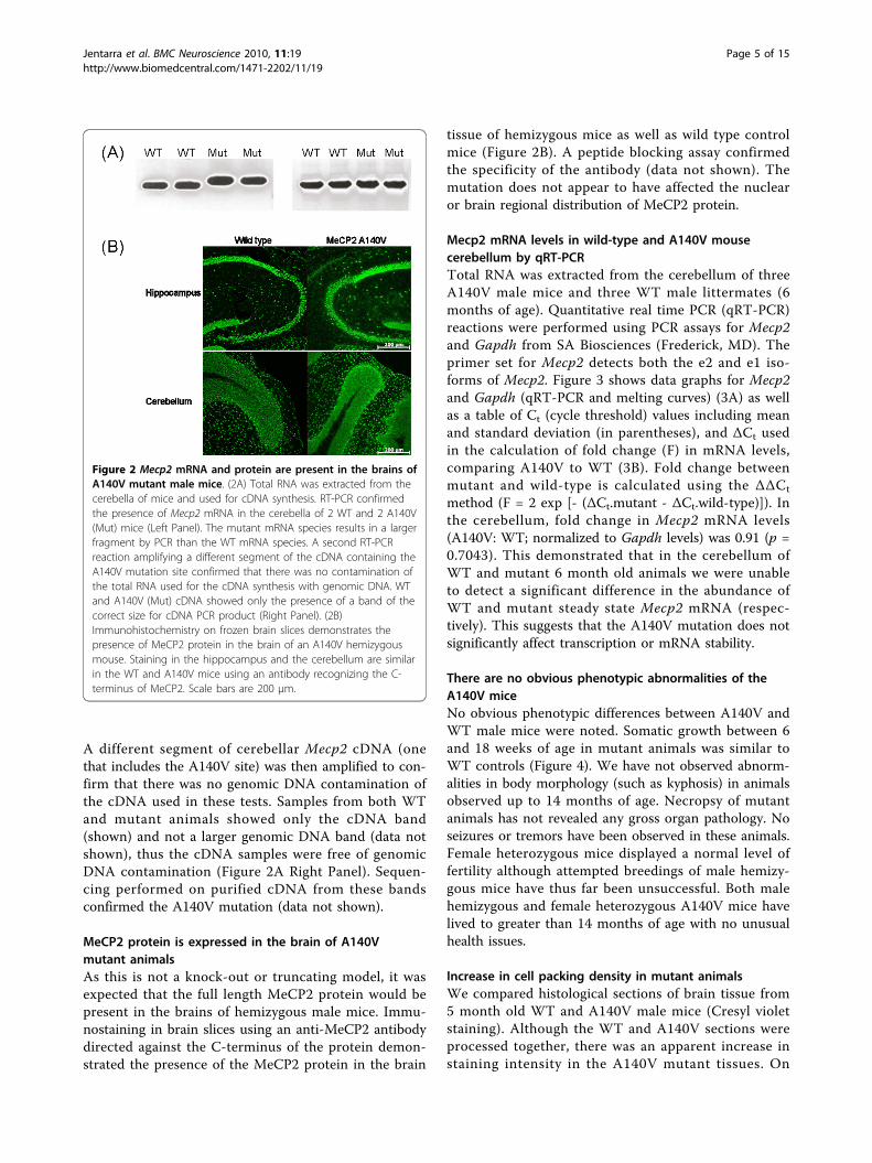
A different segment of cerebellar Mecp2 cDNA (onethat includes the A140V site) was then amplified to con-firm that there was no genomic DNA contamination ofthe cDNA used in these tests. Samples from both WTand mutant animals showed only the cDNA band(shown) and not a larger genomic DNA band (data notshown), thus the cDNA samples were free of genomicDNA contamination (Figure 2A Right Panel). Sequen-cing performed on purified cDNA from these bandsconfirmed the A140V mutation (data not shown).
MeCP2 protein is expressed in the brain of A140Vmutant animalsAs this is not a knock-out or truncating model, it wasexpected that the full length MeCP2 protein would bepresent in the brains of hemizygous male mice. Immu-nostaining in brain slices using an anti-MeCP2 antibodydirected against the C-terminus of the protein demon-strated the presence of the MeCP2 protein in the brain
tissue of hemizygous mice as well as wild type controlmice (Figure 2B). A peptide blocking assay confirmedthe specificity of the antibody (data not shown). Themutation does not appear to have affected the nuclearor brain regional distribution of MeCP2 protein.
Mecp2 mRNA levels in wild-type and A140V mousecerebellum by qRT-PCRTotal RNA was extracted from the cerebellum of threeA140V male mice and three WT male littermates (6months of age). Quantitative real time PCR (qRT-PCR)reactions were performed using PCR assays for Mecp2and Gapdh from SA Biosciences (Frederick, MD). Theprimer set for Mecp2 detects both the e2 and e1 iso-forms of Mecp2. Figure 3 shows data graphs for Mecp2and Gapdh (qRT-PCR and melting curves) (3A) as wellas a table of Ct (cycle threshold) values including meanand standard deviation (in parentheses), and ΔCt usedin the calculation of fold change (F) in mRNA levels,comparing A140V to WT (3B). Fold change betweenmutant and wild-type is calculated using the ΔΔCt
method (F = 2 exp [- (ΔCt.mutant - ΔCt.wild-type)]). Inthe cerebellum, fold change in Mecp2 mRNA levels(A140V: WT; normalized to Gapdh levels) was 0.91 (p =0.7043). This demonstrated that in the cerebellum ofWT and mutant 6 month old animals we were unableto detect a significant difference in the abundance ofWT and mutant steady state Mecp2 mRNA (respec-tively). This suggests that the A140V mutation does notsignificantly affect transcription or mRNA stability.
There are no obvious phenotypic abnormalities of theA140V miceNo obvious phenotypic differences between A140V andWT male mice were noted. Somatic growth between 6and 18 weeks of age in mutant animals was similar toWT controls (Figure 4). We have not observed abnorm-alities in body morphology (such as kyphosis) in animalsobserved up to 14 months of age. Necropsy of mutantanimals has not revealed any gross organ pathology. Noseizures or tremors have been observed in these animals.Female heterozygous mice displayed a normal level offertility although attempted breedings of male hemizy-gous mice have thus far been unsuccessful. Both malehemizygous and female heterozygous A140V mice havelived to greater than 14 months of age with no unusualhealth issues.
Increase in cell packing density in mutant animalsWe compared histological sections of brain tissue from5 month old WT and A140V male mice (Cresyl violetstaining). Although the WT and A140V sections wereprocessed together, there was an apparent increase instaining intensity in the A140V mutant tissues. On
Figure 2 Mecp2 mRNA and protein are present in the brains ofA140V mutant male mice. (2A) Total RNA was extracted from thecerebella of mice and used for cDNA synthesis. RT-PCR confirmedthe presence of Mecp2 mRNA in the cerebella of 2 WT and 2 A140V(Mut) mice (Left Panel). The mutant mRNA species results in a largerfragment by PCR than the WT mRNA species. A second RT-PCRreaction amplifying a different segment of the cDNA containing theA140V mutation site confirmed that there was no contamination ofthe total RNA used for the cDNA synthesis with genomic DNA. WTand A140V (Mut) cDNA showed only the presence of a band of thecorrect size for cDNA PCR product (Right Panel). (2B)Immunohistochemistry on frozen brain slices demonstrates thepresence of MeCP2 protein in the brain of an A140V hemizygousmouse. Staining in the hippocampus and the cerebellum are similarin the WT and A140V mice using an antibody recognizing the C-terminus of MeCP2. Scale bars are 200 μm.
Jentarra et al. BMC Neuroscience 2010, 11:19http://www.biomedcentral.com/1471-2202/11/19
Page 5 of 15

Figure 3 Mecp2 mRNA levels are similar in WT and A140V mouse cerebellum by qRT-PCR. (3A) Quantitative real time PCR (qRT-PCR)reactions were performed using PCR assays for Mecp2 and Gapdh. qRT-PCR data graphs (Upper Panels) and melting curves (Lower Panels) forMecp2 and Gapdh are shown. (3B) A table of Ct (cycle threshold) values is shown including mean and standard deviation (in parentheses) andΔCt used in the calculation of fold change (F) in mRNA levels. F (Mecp2:Gapdh) = 0.91.
Figure 4 MeCP2 A140V male mice show similar somatic growth patterns to WT controls. Male WT and MeCP2 A140V mice were weighedweekly between the ages of 6 and 18 weeks. No significant difference was found between the mean weights of the two groups. Error barsindicate one standard deviation.
Jentarra et al. BMC Neuroscience 2010, 11:19http://www.biomedcentral.com/1471-2202/11/19
Page 6 of 15
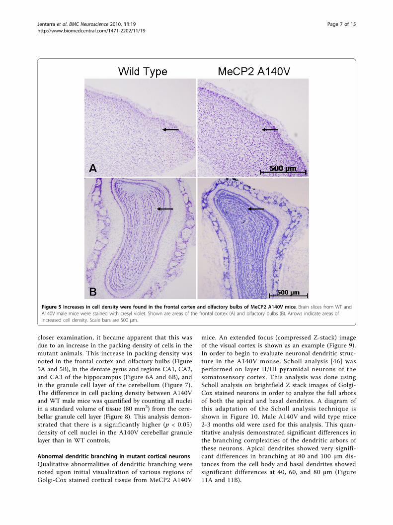
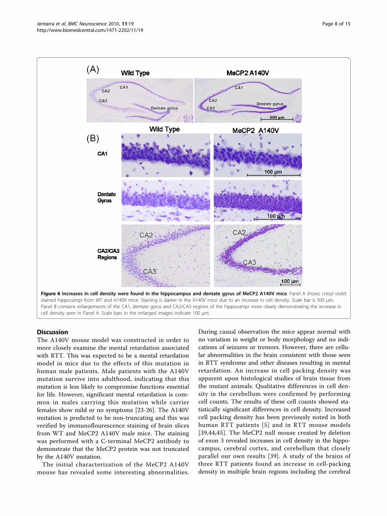
closer examination, it became apparent that this wasdue to an increase in the packing density of cells in themutant animals. This increase in packing density wasnoted in the frontal cortex and olfactory bulbs (Figure5A and 5B), in the dentate gyrus and regions CA1, CA2,and CA3 of the hippocampus (Figure 6A and 6B), andin the granule cell layer of the cerebellum (Figure 7).The difference in cell packing density between A140Vand WT male mice was quantified by counting all nucleiin a standard volume of tissue (80 mm3) from the cere-bellar granule cell layer (Figure 8). This analysis demon-strated that there is a significantly higher (p < 0.05)density of cell nuclei in the A140V cerebellar granulelayer than in WT controls.
Abnormal dendritic branching in mutant cortical neuronsQualitative abnormalities of dendritic branching werenoted upon initial visualization of various regions ofGolgi-Cox stained cortical tissue from MeCP2 A140V
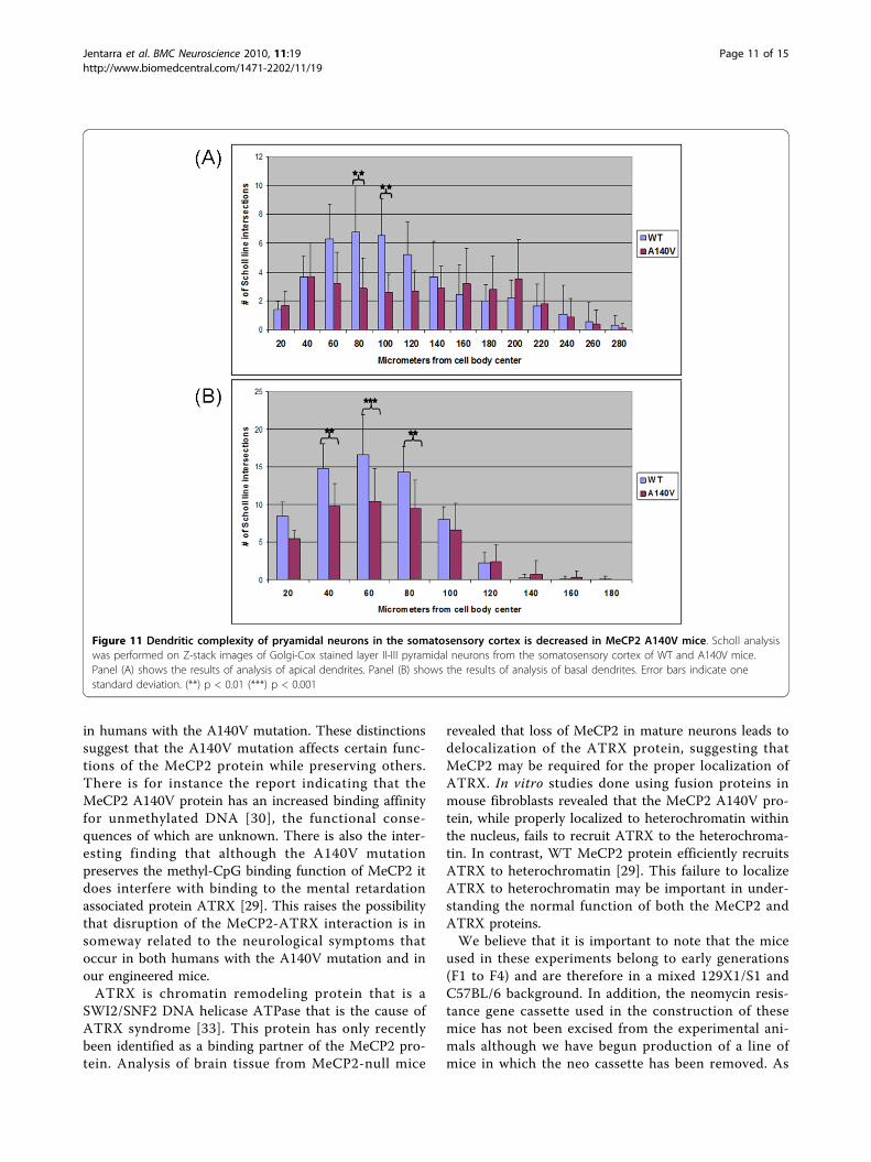
mice. An extended focus (compressed Z-stack) imageof the visual cortex is shown as an example (Figure 9).In order to begin to evaluate neuronal dendritic struc-ture in the A140V mouse, Scholl analysis [46] wasperformed on layer II/III pyramidal neurons of thesomatosensory cortex. This analysis was done usingScholl analysis on brightfield Z stack images of Golgi-Cox stained neurons in order to analyze the full arborsof both the apical and basal dendrites. A diagram ofthis adaptation of the Scholl analysis technique isshown in Figure 10. Male A140V and wild type mice2-3 months old were used for this analysis. This quan-titative analysis demonstrated significant differences inthe branching complexities of the dendritic arbors ofthese neurons. Apical dendrites showed very signifi-cant differences in branching at 80 and 100 μm dis-tances from the cell body and basal dendrites showedsignificant differences at 40, 60, and 80 μm (Figure11A and 11B).
Figure 5 Increases in cell density were found in the frontal cortex and olfactory bulbs of MeCP2 A140V mice. Brain slices from WT andA140V male mice were stained with cresyl violet. Shown are areas of the frontal cortex (A) and olfactory bulbs (B). Arrows indicate areas ofincreased cell density. Scale bars are 500 μm.
Jentarra et al. BMC Neuroscience 2010, 11:19http://www.biomedcentral.com/1471-2202/11/19
Page 7 of 15
DiscussionThe A140V mouse model was constructed in order tomore closely examine the mental retardation associatedwith RTT. This was expected to be a mental retardationmodel in mice due to the effects of this mutation inhuman male patients. Male patients with the A140Vmutation survive into adulthood, indicating that thismutation is less likely to compromise functions essentialfor life. However, significant mental retardation is com-mon in males carrying this mutation while carrierfemales show mild or no symptoms [23-26]. The A140Vmutation is predicted to be non-truncating and this wasverified by immunoflourescence staining of brain slicesfrom WT and MeCP2 A140V male mice. The stainingwas performed with a C-terminal MeCP2 antibody todemonstrate that the MeCP2 protein was not truncatedby the A140V mutation.The initial characterization of the MeCP2 A140V
mouse has revealed some interesting abnormalities.
During casual observation the mice appear normal withno variation in weight or body morphology and no indi-cations of seizures or tremors. However, there are cellu-lar abnormalities in the brain consistent with those seenin RTT syndrome and other diseases resulting in mentalretardation. An increase in cell packing density wasapparent upon histological studies of brain tissue fromthe mutant animals. Qualitative differences in cell den-sity in the cerebellum were confirmed by performingcell counts. The results of these cell counts showed sta-tistically significant differences in cell density. Increasedcell packing density has been previously noted in bothhuman RTT patients [5] and in RTT mouse models[39,44,45]. The MeCP2 null mouse created by deletionof exon 3 revealed increases in cell density in the hippo-campus, cerebral cortex, and cerebellum that closelyparallel our own results [39]. A study of the brains ofthree RTT patients found an increase in cell-packingdensity in multiple brain regions including the cerebral
Figure 6 Increases in cell density were found in the hippocampus and dentate gyrus of MeCP2 A140V mice. Panel A shows cresyl violetstained hippocampi from WT and A140V mice. Staining is darker in the A140V mice due to an increase in cell density. Scale bar is 500 μm.Panel B contains enlargements of the CA1, dentate gyrus and CA2/CA3 regions of the hippocampi more clearly demonstrating the increase incell density seen in Panel A. Scale bars in the enlarged images indicate 100 μm.
Jentarra et al. BMC Neuroscience 2010, 11:19http://www.biomedcentral.com/1471-2202/11/19
Page 8 of 15

cortex, basal ganglia, thalamus, hippocampus, and amyg-dala. Abnormalities were also reported in the cerebellumalthough an increase in cell packing density was notreported [5].We find no previous descriptions of cell density
abnormalities in the olfactory bulbs of Rett mouse mod-els. We are uncertain if this is because cell densityabnormalities have not previously been seen in olfactorybulbs or if the olfactory bulbs were not examined in
these mice. There have been reports of defects in neuro-nal development in olfactory neurons from the nasalepithelium of RTT patients [47]. In addition, defects ofolfactory neuron maturation and protein expressionhave been found in two previously described RTTmouse models [48-51], indicating that olfactory defectsare not unusual in RTT or in RTT mouse models. Inaddition, a recent and very thorough study of brainmorphology found changes in the volume and shape of
Figure 7 Increases in cell density were found in the cerebellum of MeCP2 A140V mice. Left panels show low power images of cresylviolet stained cerebelli from WT and A140V mice. The A140V cerebellum has noticeably darker staining due to an increase in cell density. Scalebar is 500 μm. Right panels show enlargement of these images demonstrating the differences in cell density. Scale bar is 100 μm. Arrowsindicate areas of increased cell density.
Figure 8 Quantitative analysis of cell density in the cerebellum of MeCP2 A140V mice. Cresyl violet stained cell nuclei in cerebellarsections from WT and A140V mice were counted in two separate areas of the granule cell layer in the cerebellum. In each cerebellar area, threeadjacent regions were counted and the mean taken. The means for each area for each genotype are shown on the bar graph. The differencesbetween the means were significant, (*) p < 0.05, indicating an increase in cell density in the A140V cerebellum.
Jentarra et al. BMC Neuroscience 2010, 11:19http://www.biomedcentral.com/1471-2202/11/19
Page 9 of 15

olfactory bulbs in two MeCP2 null mouse models [43].We have seen abnormalities in the shape of olfactorybulbs and the morphology of glomeruli in some theA140V male hemizygous mice although this has notbeen quantified and will be the subject of further study.A lack of branching complexity in cortical pyramidal
neurons in many brain regions has been reported inhuman cases of RTT [6-9] as well as in mouse models[44,45]. In fact, dendritic branching anomalies are often
found in conjunction with mental retardation of manycauses [52,53]. The A140V mutation is strongly asso-ciated with mental retardation [23-26]; therefore, we feltthat abnormal dendritic branching patterns of neuronswould likely occur in this new mouse model. Weexplored the branching complexity of neurons in theA140V mouse model and found differences betweenWT and MeCP2 A140V mice. We noted significant dif-ferences in the branching complexity of both the apicaland basal dendrites of layer II/III pyramidal neurons ofthe somatosensory cortex as measured by Scholl analy-sis. This correlates closely with other reports ofdecreases in branching complexity of cortical pyramidalneurons in tissue from both human RTT patients and inMecp2 mutant mouse models. We also found apparentdifferences in branching complexity in the visual cortexalthough this has not yet been quantified.The MeCP2 A140V mouse has proven to be distinct
from other mouse models of MeCP2 mutation. Like itshuman counterpart, the A140V mutation in mice pro-duces symptoms both correlating with and disparatefrom the classical symptoms of RTT. In humans, theA140V mutation causes a mental retardation syndromethat has associated with it abnormalities of gait, kyphos-coliosis, mild microcephaly, spasticity, dysarthric speech,and occasionally mental illness such as schizophrenia[21,23,26,27]. However certain features of classic Rettsyndrome such as stereotypic hand movements, breath-ing abnormalities and seizures [2,3] are notably absent
Figure 9 Qualitative abnormalities of neuronal dendritic branching were seen in the layer II/III pyramidal neurons of the visual cortexof MeCP2 A140V mice. Extended focus images of Golgi-Cox stained brain sections from WT and A140V mice are shown. Arrows indicate areasof apparent decreased dendritic branching in the A140V mouse in comparison to the WT mouse. Scale bar is 100 μm.
Figure 10 The Scholl analysis technique for assessing dendriticcomplexity was adapted for use in brightfield Z-stack imagesof Golgi-Cox stained neurons. The diagram shows an illustrationof Scholl lines superimposed at 20 μm increments on a Z-stack ofimages of a stained neuron. This was done using Zeiss AxioVisionsoftware. Each Z-stack image is separated by only 0.280 μmallowing all dendritic intersections with Scholl lines to be clearlyseen and counted. Drawing not to scale.
Jentarra et al. BMC Neuroscience 2010, 11:19http://www.biomedcentral.com/1471-2202/11/19
Page 10 of 15
in humans with the A140V mutation. These distinctionssuggest that the A140V mutation affects certain func-tions of the MeCP2 protein while preserving others.There is for instance the report indicating that theMeCP2 A140V protein has an increased binding affinityfor unmethylated DNA [30], the functional conse-quences of which are unknown. There is also the inter-esting finding that although the A140V mutationpreserves the methyl-CpG binding function of MeCP2 itdoes interfere with binding to the mental retardationassociated protein ATRX [29]. This raises the possibilitythat disruption of the MeCP2-ATRX interaction is insomeway related to the neurological symptoms thatoccur in both humans with the A140V mutation and inour engineered mice.ATRX is chromatin remodeling protein that is a
SWI2/SNF2 DNA helicase ATPase that is the cause ofATRX syndrome [33]. This protein has only recentlybeen identified as a binding partner of the MeCP2 pro-tein. Analysis of brain tissue from MeCP2-null mice
revealed that loss of MeCP2 in mature neurons leads todelocalization of the ATRX protein, suggesting thatMeCP2 may be required for the proper localization ofATRX. In vitro studies done using fusion proteins inmouse fibroblasts revealed that the MeCP2 A140V pro-tein, while properly localized to heterochromatin withinthe nucleus, fails to recruit ATRX to the heterochroma-tin. In contrast, WT MeCP2 protein efficiently recruitsATRX to heterochromatin [29]. This failure to localizeATRX to heterochromatin may be important in under-standing the normal function of both the MeCP2 andATRX proteins.We believe that it is important to note that the mice
used in these experiments belong to early generations(F1 to F4) and are therefore in a mixed 129X1/S1 andC57BL/6 background. In addition, the neomycin resis-tance gene cassette used in the construction of thesemice has not been excised from the experimental ani-mals although we have begun production of a line ofmice in which the neo cassette has been removed. As
Figure 11 Dendritic complexity of pryamidal neurons in the somatosensory cortex is decreased in MeCP2 A140V mice. Scholl analysiswas performed on Z-stack images of Golgi-Cox stained layer II-III pyramidal neurons from the somatosensory cortex of WT and A140V mice.Panel (A) shows the results of analysis of apical dendrites. Panel (B) shows the results of analysis of basal dendrites. Error bars indicate onestandard deviation. (**) p < 0.01 (***) p < 0.001
Jentarra et al. BMC Neuroscience 2010, 11:19http://www.biomedcentral.com/1471-2202/11/19
Page 11 of 15
genetic background and the presence of extra DNAsequences such as the neo cassette can contribute tophenotype we will be repeating these experiments inlater generation mice lacking the neo cassette in orderto confirm our experimental results. However, webelieve that, given the close parallels of our data withthat seen in other mouse models of Mecp2 gene disrup-tion, the phenotype we have observed is in fact due tothe A140V mutation itself.
ConclusionsWe believe that this new mouse model will provideinsight into the functions of MeCP2, including its asso-ciation with the ATRX protein. The A140V mouse hasproven to reproduce some of the abnormalities of otherMecp2 mouse models (increased cell packing densityand decreased neuronal dendritic arbors) while failing toreproduce others (seizures, tremors, kyphosis, breathingabnormalities, shortened life span). With this knowledgewe can continue dissecting the functions of MeCP2 inthe hopes of explaining its relationship to mental retar-dation and other clinical symptoms.
MethodsGeneration of A140V “knock-in” miceBreeding protocols and all animal experiments wereapproved by the Institutional Animal Care and UseCommittee (IACUC) of St. Joseph’s Hospital and Medi-cal Center. An 11 kbp Bam HI fragment of the mouseMeCP2 gene, that included exon 3 and part of exon 4,was isolated by restriction digestion of a BAC clone(mBAC B22804). The 5’- end of the insert was firsttrimmed by restriction digestion with Pml I. A pGK-Neo cassette (flanked by two Frt sites and a single LoxPsite, gift of Dr. Dymecki (Department of Genetics, Har-vard Medical School), was inserted into the Nde I sitelocated about 1.7 kbp upstream of exon 3. A secondLoxP site and a novel Acl I restriction were insertedinto the 3’-UTR of exon 4 (into an Mfe I site locatedabout 400 bp downstream of the translation terminationcodon). A single base mutation was introduced usingthe QuikChangeXL kit (Stratagene Inc., Cedar Creek,Texas): a C to T change at nucleotide position 601 ofthe Mecp2-e2 mRNA, resulting in the A140V missensemutation. Pml I linearized targeting vector was electro-porated into the R1 line of ES cells [54] (male genotype,Strain 129X1/S1) under previously described conditions[55]. G418 (neomycin analogue) resistant clones weretested for proper recombination at the 3’-integration siteby a PCR based assay using the following primers:F30: 5’ CGACAAGCACAGTCAGGTTGAAGAC 3’B34: 5’ TCCCAAGAGTCCCATAGTTTCTCC 3’The wild-type allele yields an amplimer that is 322 bp
long, while the mutant allele yields an amplimer that is
362 bp long. Random integration of the targeting vectorinto the mouse genome results in both wild-type andmutant bands. The PCR product was also digested withAcl I, which cuts the mutant amplimer into two seg-ments (227 bp and 135 bp), leaving the wild-type ampli-mer uncut. Selected ES clone DNAs were tested bySouthern blot for correct recombination at the 5’end,using Probe A. Homologous recombination at the 5’end results in a 5.5 kbp band, and clones that retain thewild-type locus produce an 11 kbp band. Finally,selected positive ES cell clones were verified by sequen-cing to confirm the presence of the desired point muta-tion. Two ES cell clones (169J6 and 177U9) were theninjected into blastocysts from C57BL/6 mice (doneunder contract by Ingenious Targeting Laboratories,Inc., Stony Brook, New York) and implanted intopseudo-pregnant females. Male chimeras were harembred with wild-type C57BL/6 mice and tail DNAs fromall female F1 pups were tested for the presence of thetransgene. Founder females (derived from ES clone177U9) were identified by PCR, Southern blot, andDNA sequencing. These founder females were used toestablish our colony of A140V mice by backcrossingwith WT C57BL/6NCrL male mice (Charles River, Wil-mington, Massachusetts), yielding mutant hemizygousmales, heterozygous females, and WT controls. Hetero-zygous females produced in the colony were back-crossed with WT C57BL/6NCrL male mice to produceexperimental mice as well as additional breeding stock.
MeCP2 RT-PCRTotal RNA was extracted from the cerebella of 2 mutantand 2 WT male mice and treated with DNase I toremove traces of genomic DNA. cDNA was synthesizedusing random hexamers (Superscript III kit, Invitrogen,Inc., Carlsbad, California). cDNA from each of thesefour animals was used in PCR reactions with the F30and B34 primers described above. The amplimer frommutant animals is 40 bp larger than the amplimer fromWT animals. A second cDNA fragment (729 bp) thatincluded a portion of exon 3 and exon 4, containing theA140V point mutation was then amplified using the fol-lowing primers:U256 (located within exon 3): 5’ GTATGATGACCC-
CACCTTGC 3’L1452 (located within exon 4): 5’ TTCAGTCCC-
TTCCCGCTTTT 3’The cDNA amplimers from WT and mutant RNAs
were gel purified and sequenced to verify WT or mutantstatus at the codon for amino acid 140. When genomicDNA was used as a template for PCR using these sameprimers, the amplimer was 1216 bp long (providing acontrol for contamination of the RNA by genomicDNA).
Jentarra et al. BMC Neuroscience 2010, 11:19http://www.biomedcentral.com/1471-2202/11/19
Page 12 of 15
qRT-PCRTotal RNA was extracted from cerebella of three mutantmale mice and three WT male littermates (6 monthsold) using Trizol (Invitrogen, Inc., Carlsbad, California)and cleaned up using an RNA extraction kit (SA Bios-ciences, Frederick, MD). First strand cDNA was synthe-sized (kit from SA Biosciences) and used in qRT-PCRreactions using PCR assays and SYBR Green master mixfrom SA Biosciences (Mecp2, cat no. PPM31215E, andGapdh, cat no. PPM02946E). Controls were first donewith mouse reference RNA to verify comparable effi-ciency of PCR reactions, and negative controls includedno-reverse transcriptase (NRT) reactions for each RNA.Triplicate reactions were done for each primer assayand each of the 6 cDNA samples. Threshold level offluorescence was set across the entire run within theexponential phase of amplification. The correspondingCt values were then determined for each sample in therun. The means, standard deviations, and p value werecalculated using GraphPad InStat3 software.
Histology: Cresyl Violet Staining of Brain TissueMice were anesthetized and transcardially perfused withsaline followed by 4% buffered paraformaldehyde (PFA).The brains were then post-fixed in 4% PFA overnight at4°C and then cryoprotected in 30% sucrose for 1-2 days.Subsequently, brains were frozen in an isopentane/dryice bath. The brains were immediately sectioned into 30μm slices using a cryostat, slices were placed on slides,and then kept frozen at -20°C until staining. Alternatingbrain slices were then thawed briefly and stained withcresyl violet (Sigma, St. Louis, Missouri), cover slippedwith Permount (Fisher Scientific, Pittsburgh, PA) andwere visualized with either an Olympus microscope or aZeiss AxioImager microscope (for brightfield Z-stackimages).
Cell Nuclei Count AnalysisCell nuclei counts were performed in the cerebellum(using brightfield z-stack microscopy) in order to quan-tify differences in cell nuclei number. This was doneusing brightfield Z-stack images of the granule cell layerof the cerebellum in cresyl violet stained tissue. A rec-tangular counting area was drawn on the Z-stack imagesusing Axiovision software. This demarcated area carriedthrough the entire Z-stack. Calculating the distancebetween the top and bottom slices of the stack in con-junction with the demarcated area allowed for a volumemeasurement to be made. All cell nuclei counts weredone in a total volume of 80 mm3. Using this countingmethod, nuclei were clear and easily distinguishablefrom each other. Triplicate cell counts were performedin each region of the cerebellar granule cell layer fromWT and MeCP2 A140V mice. The mean, standard
deviation and p values were calculated using GraphPadInStat3 software.
Histology: Golgi-Cox Staining of Brain TissueThe Rapid Golgi-Cox staining kit (FD Neurotechnologies,Ellicott City, Maryland) was used for staining whole mousebrains. Mice were euthanized by overdose of isofluoraneand the brains removed. After rinsing in de-ionized waterthe brains were incubated for 2 weeks in a Solution A/Bmixture (impregnation) following the manufacturer’s pro-tocol. The brains were then moved to Solution C (cryopro-tection) for 4-5 days and subsequently frozen in anisopentane/dry ice bath. The brains were immediately sec-tioned into 100-200 μm slices using a cryostat and theslices were mounted on gelatin-coated slides. After beingallowed to dry for 3-5 days, the staining protocol for devel-opment of the black precipitate was completed using kitreagents. The tissue was counterstained with cresyl violet(Sigma, St. Louis, Missouri). Cover slips were mountedusing Permount (Fisher Scientific, Pittsburgh, Pennsylva-nia) and slides were visualized using a Zeiss AxioImagermicroscope capable of brightfield Z-stack.
Scholl AnalysisScholl analysis [46] was adapted for use on brightfieldZ-stack images using Axiovision software. Concentriccircles (Scholl lines) were drawn at 20 μm incrementsfrom the center of the cell body. The Scholl lines weresuperimposed on the entire stack of images allowing theanalysis to be conducted in a 3-dimensional fashion.The dendritic arbor was followed through the Z-stackand the number of intersections made by dendrites withthe Scholl lines was measured as each dendrite traveledaway from the cell body. The Z-stack image slices wereseparated by a depth of only 0.280 μm preventing inter-sections from being missed.The apical and basal arbors of 10 neurons from a
MeCP2 A140V mouse and 10 neurons from a WTmouse were captured in Z-stack images. All neuronswere layer II/III pyramidal neurons of similar morphol-ogy, located in the somatosensory cortex at similar coor-dinates [56]. Scholl lines were placed on the imagesevery 20 μm until no more dendrites crossed the Scholllines. Axiovision software was then used to manuallymark crossings of each Scholl line by the dendrites ofthe neurons. Crossings at each Scholl line were summedand the mean and standard deviation calculated. pvalues comparing the wild type and A140V mouse neu-rons at each Scholl line were then determined. Graph-Pad InStat3 software was used for statistical analysis.
ImmunohistochemistryBrain tissue from WT and A140V mutant mice was fro-zen in an isopentane/dry ice bath and 10 μm frozen
Jentarra et al. BMC Neuroscience 2010, 11:19http://www.biomedcentral.com/1471-2202/11/19
Page 13 of 15
sections were cut on a cryostat, placed on slides, andfrozen at -20°C until staining. Tissue was then fixedwith 4% PFA, blocked and permeabilized with a mixtureof 10% goat serum, 1% BSA, and 0.2% Triton X100, andincubated overnight at 4°C with anti-MeCP2 antibody(PA1-888 rabbit anti-MeCP2 antibody, Affinity BioRea-gents, Rockford, Illinois). This is a polyclonal antibodythat recognizes the C-terminus of the MeCP2 protein.Sections were then incubated with secondary antibody(Alexa Fluor 488 goat anti-rabbit antibody, Invitrogen,Carlsbad, California) for 2 hours at room temperature.Following a wash step, sections were coverslipped withPro-long Gold (Invitrogen, Carlsbad, California) mount-ing medium. MeCP2 staining in the slices was visualizedusing a Zeiss confocal microscope.
AcknowledgementsThis work was supported by funds from the NIH (to SN; P01-HD024448) andthe Barrow Neurological Foundation (to VN). The authors would like tothank Ed Mallick for expert technical assistance with gene targeting in EScells.
Author details1Neurology Research Department, Barrow Neurological Institute,350 W Thomas Rd, NRC 438, Phoenix, AZ, 85013, USA. 2Departments ofAnesthesiology and Pharmacology & Chemical Biology, University ofPittsburgh, 6060 Biomedical Science Tower 3, Pittsburgh, PA, 15261, USA.3Neuroscience Laboratory, Kennedy Krieger Institute, 707 North Broadway,Baltimore, MD, 21205, USA. 4Departments of Neurology and Pediatrics,Kennedy Krieger Institute, 707 North Broadway, Baltimore, MD, 21205, USA.5School of Life Sciences, Arizona State University, University Drive and MillAve, Tempe, AZ, 85287, USA.
Authors’ contributionsVN, GH and SO constructed and verified the mouse model. SO genotypedand maintained the mouse colony. GJ performed the experiments. SRassisted with tissue processing and microscope technique. NS provided freshfrozen mouse brain slices and tested antibodies for immunohistochemistry.GJ and VN wrote the manuscript with editing from GH, SN, and SR. MBprovided experimental advice as well as MeCP2 null mouse tissue forexperimental controls. All authors read and reviewed the final manuscript.
Received: 14 October 2009Accepted: 17 February 2010 Published: 17 February 2010
References1. Amir RE, Veyver Van Den IB, Wan M, Tran CQ, Franke U, Zoghbi HY: Rett
syndrome is caused by mutations in X-linked MECP2, encoding methyl-CpG-binding protein 2. Nature Genetics 1999, 23:185-188.
2. Hagberg B, Aicardi J, Dias K, Ramos O: A progressive syndrome of autism,dementia, ataxia, and loss of purposeful hand use in girls: Rett’ssyndrome: report of 35 cases. Annals of Neurology 1983, 14:471-479.
3. Hagberg B: Rett’s Syndrome: Prevalence and Impact on ProgressiveSevere Mental Retardation in Girls. Acta Paediatrica Scandinavica 1985,74:405-408.
4. Dunn HG, MacLeod PM: Rett syndrome: review of biologicalabnormalities. Can J Neurol Sci 2001, 28:16-29.
5. Bauman ML, Kemper TL, Arin DM: Pervasive neuroanatomic abnormalitiesof the brain in three cases of Rett’s syndrome. Neurology 1995,45:1581-1586.
6. Belichenko PV: Rett syndrome: 3-D confocal microscopy of corticalpyramidal dendrites and afferents. NeuroReport 1994, 5:1509-1513.
7. Armstrong D, Dunn JK, Antalffy B, Trivedi R: Selective dendritic alterationsin the cortex of Rett syndrome. Journal of Neuropathology andExperimental Neurology 1995, 54:195-201.
8. Armstrong DD: Neuropathology of Rett syndrome. J Child Neurol 2005,20:747-753.
9. Armstrong DD, Dunn K, Antalffy B: Decreased dendritic branching infrontal, motor and limbic cortex in Rett syndrome compared withtrisomy 21. J Neuropathol Exp Neurol 1998, 57:1013-1017.
10. Neul JL, Zoghbi HY: Rett syndrome: a prototypical neurodevelopmentaldisorder. Neuroscientist 2004, 10:118-128.
11. Nan X, Campoy FJ, Bird A: MeCP2 is a transcriptional repressor withabundant binding sites in genomic chromatin. Cell 1997, 88:471-481.
12. Nan X, Tate P, Li E, Bird A: DNA methylation specifies chromosomallocalization of MeCP2. Mol Cell Biol 1996, 16:414-421.
13. Chahrour M, Jung SY, Shaw C, Zhou X, Wong ST, Qin J, Zoghbi HY: MeCP2,a key contributor to neurological disease, activates and repressestranscription. Science 2008, 320:1224-1229.
14. Jones PL, Veenstra GJ, Wade PA, Vermaak D, Kass SU, Landsberger N,Strouboulis J, Wolffe AP: Methylated DNA and MeCP2 recruit histonedeacetylase to repress transcription. Nat Genet 1998, 19:187-191.
15. Sun YE, Wu H: The ups and downs of BDNF in Rett syndrome. Neuron2006, 49:321-323.
16. Zhou Z, Hong EJ, Cohen S, Zhao W, Ho HH, Schmidt L, Chen WG, Lin Y,Savner E, Griffith EC, et al: Brain-specific phosphorylation of MeCP2regulates activity-dependent Bdnf transcription, dendritic growth, andspine maturation. Neuron 2006, 52:255-269.
17. Poo MM: Neurotrophins as synaptic modulators. Nat Rev Neurosci 2001,2:24-32.
18. Chang Q, Khare G, Dani V, Nelson S, Jaenisch R: The disease progressionof Mecp2 mutant mice is affected by the level of BDNF expression.Neuron 2006, 49:341-348.
19. Meloni I, Bruttini M, Longo I, Mari F, Rizzolio F, D’Adamo P, Denvriendt K,Fryns JP, Toniolo D, Renieri A: A mutation in the rett syndrome gene,MECP2, causes X-linked mental retardation and progressive spasticity inmales. Am J Hum Genet 2000, 67:982-985.
20. Imessaoudene B, Bonnefont JP, Royer G, Cormier-Daire V, Lyonnet S,Lyon G, Munnich A, Amiel J: MECP2 mutation in non-fatal, non-progressive encephalopathy in a male. J Med Genet 2001, 38:171-174.
21. Couvert P, Bienvenu T, Aquaviva C, Poirier K, Moraine C, Gendrot C,Verloes A, Andres C, Le Fevre AC, Souville I, et al: MECP2 is highly mutatedin X-linked mental retardation. Hum Mol Genet 2001, 10:941-946.
22. Bienvenu T, Chelly J: Molecular genetics of Rett syndrome: when DNAmethylation goes unrecognized. Nature Reviews: Genetics 2006, 7:415-426.
23. Dotti MT, Orrico A, De Stephano N, Battisti C, Sicurelli F, Severi S, Lam CW,Galli L, Sorrentino V, Federico A: A Rett syndrome MECP2 mutation thatcauses mental retardation in men. Neurology 2002, 58:226-230.
24. Klauck SM, Lindsay S, Beyer KS, Splitt M, Burn J, Poustka A: A mutation hotspot for nonspecific X-linked mental retardation in the MECP2 genecauses the PPM-X syndrome. American Journal of Medical Genetics 2002,70:1034-1037.
25. Orrico A, Lam C, Galli L, Dotti MT, Hayek G, Tong SF, Poon PM, Zappella M,Federico A, Sorrentino V: MECP2 mutation in male patients with non-specific X-linked mental retardation. FEBS Lett 2000, 481:285-288.
26. Winnepenninckx B, Errijgers V, Hayez-Delatte F, Reyniers E, Kooy RF:Identification of a family with nonspecific mental retardation (MRX79)with the A140V mutation in the MECP2 gene: Is there a need for routinescreening?. Human Mutation 2002, 20:249-252.
27. Cohen D, Lazar G, Couvert P, Desportes V, Lippe D, Mazet P, Heron D:MECP2 mutation in a boy with language disorder and schizophrenia.Am J Psychiatry 2002, 159:148-149.
28. Ohki I, Shimotake N, Fujita N, Jee J, Ikegami T, Nakao M, Shirakawa M:Solution structure of the methyl-CpG binding domain of human MBD1in complex with methylated DNA. Cell 2001, 105:487-497.
29. Nan X, Hou J, Maclean A, Nasir J, Lafuente MJ, Shu X, Kriaucionis S, Bird A:Interaction between chromatin proteins MECP2 and ATRX is disruptedby mutations that cause inherited mental retardation. Proceedings of theNational Academy of Sciences USA 2007, 104:2709-2714.
30. Kudo S, Nomura Y, Segawa M, Fujita N, Nakao M, Hammer S, Schanen C,Terai I, Tamura M: Functional characterisation of MeCP2 mutations foundin male patients with X linked mental retardation. J Med Genet 2002,39:132-136.
31. Stevenson RE: Alpha-Thalassemia X-Linked Mental Retardation Syndrome.GeneReviews 2009http://www.ncbi.nlm.nih.gov/bookshelf/br.fcgi?book=gene&part=xlmr.
Jentarra et al. BMC Neuroscience 2010, 11:19http://www.biomedcentral.com/1471-2202/11/19
Page 14 of 15
32. Villard L, Fontes M: Alpha-thalassemia/mental retardation syndrome, X-linked (ATR-X, MIM #301040, ATR-X/XNP/XH2 gene MIM #300032.European Journal of Human Genetics 2002, 10:223-225.
33. Gibbons RJ, Higgs DR: Molecular-clinical spectrum of the ATR-Xsyndrome. American Journal of Medical Genetics 2000, 97:204-212.
34. Badens C, Lacoste C, Philip N, Martini N, Courrier S, Giuliano F, Verloes A,Munnich A, Leheup B, Burglen L, et al: Mutations in PHD-like domain ofthe ATRX gene correlate with severe psychomotor impairment andsevere urogenital abnormalities in patients with ATRX syndrome. ClinicalGenetics 2006, 70:57-62.
35. Garrick D, Sharpe JA, Arkell R, Dobbie L, Smith AJ, Wood WG, Higgs DR,Gibbons RJ: Loss of Atrx affects trophoblast development and thepattern of X-inactivation in extraembryonic tissues. PLoS Genet 2006, 2:e58.
36. Berube NG, Mangelsdorf M, Jagla M, Vanderluit J, Garrick D, Gibbons RJ,Higgs DR, Slack RS, Picketts DJ: The chromatin-remodeling protein ATRXis critical for neuronal survival during corticogenesis. J Clin Invest 2005,115:258-267.
37. Medina CF, Mazerolle C, Wang Y, Berube NG, Coupland S, Gibbons RJ,Wallace VA, Picketts DJ: Altered visual function and interneuron survivalin Atrx knockout mice: inference for the human syndrome. Hum MolGenet 2009, 18:966-977.
38. Guy J, Hendrich B, Holmes M, Martin JE, Bird A: A mouse Mecp2 -nullmutation causes neurolgical symptoms that mimic Rett syndrome.Nature Genetics 2001, 27:322-326.
39. Chen RZ, Akbarian S, Tudor M, Jaenisch R: Deficiency of methyl-CpGbinding protein-2 in CNS neurons results in a Rett-like phenotype inmice. Nature Genetics 2001, 27:327-331.
40. Shahbazian MD, Young JI, Yuva-Paylor LA, Spencer CM, Antalffy BA,Noebels JL, Armstrong DL, Paylor R, Zoghbi HY: Mice with truncatedMeCP2 recapitulate many Rett syndrome features and displayhyperacetylation of histone H3. Neuron 2002, 35:243-254.
41. McGill BE, Bundle SF, Yaylaoglu MB, Carson JP, Thaller C, Zoghbi HY:Enhanced anxiety and stress-induced corticosterone release areassociated with increased Crh expression in a mouse model of Rettsyndrome. Proc Natl Acad Sci USA 2006, 103:18267-18272.
42. Lawson-Yuen A, Liu D, Han L, Jiang ZI, Tsai GE, Basu AC, Picker J, Feng J,Coyle JT: Ube3a mRNA and protein expression are not decreased inMecp2R168X mutant mice. Brain Research 2007, 1180:1-6.
43. Belichenko NP, Belichenko PV, Li HH, Mobley WC, Francke U: Comparativestudy of brain morphology in Mecp2 mutant mouse models of Rettsyndrome. J Comp Neurol 2008, 508:184-195.
44. Fukuda T, Itoh M, Ichikawa T, Washiyama K, Goto Y: Delayed maturation ofneuronal architecture and synaptogenesis in cerebral cortex of Mecp2-deficient mice. J Neuropathol Exp Neurol 2005, 64:537-544.
45. Kishi N, Macklis JD: MECP2 is progressively expressed in post-migratoryneurons and is involved in neuronal maturation rather than cell fatedecisions. Mol Cell Neurosci 2004, 27:306-321.
46. Scholl DA: Dendritic organization in the neurons of the visual and motorcortices of the cat. Journal of Anatomy 1953, 87:387-406.
47. Ronnett GV, Leopold D, Cai X, Hoffbuhr KC, Moses L, Hoffman EP, Naidu S:Olfactory biopsies demonstrate a defect in neuronal development inRett’s syndrome. Ann Neurol 2003, 54:206-218.
48. Degano AL, Pasterkamp RJ, Ronnett GV: MeCP2 deficiency disrupts axonalguidance, fasciculation, and targeting by altering Semaphorin 3Ffunction. Mol Cell Neurosci 2009, 42:243-254.
49. Matarazzo V, Cohen D, Palmer AM, Simpson PJ, Khokhar B, Pan SJ,Ronnett GV: The transcriptional repressor Mecp2 regulates terminalneuronal differentiation. Mol Cell Neurosci 2004, 27:44-58.
50. Matarazzo V, Ronnett GV: Temporal and regional differences in theolfactory proteome as a consequence of MeCP2 deficiency. Proc NatlAcad Sci USA 2004, 101:7763-7768.
51. Palmer A, Qayumi J, Ronnett G: MeCP2 mutation causes distinguishablephases of acute and chronic defects in synaptogenesis andmaintenance, respectively. Molecular and Cellular Neuroscience 2008,37:794-807.
52. Machado-Salas JP: Abnormal dendritic patterns and aberrant spinedevelopment in Bourneville’s disease–a Golgi survey. Clin Neuropathol1984, 3:52-58.
53. Kaufmann WE, Moser HW: Dendritic anomalies in disorders associatedwith mental retardation. Cerebral Cortex 2000, 10:981-991.
54. Nagy A, Rossant J, Nagy R, Abramow-Newerly W, Roder JC: Derivation ofcompletely cell culture-derived mice from early-passage embryonic stemcells. Proc Natl Acad Sci USA 1993, 90:8424-8428.
55. Homanics GE, Ferguson C, Quinlan JJ, Daggett J, Snyder K, Lagenaur C,Mi ZP, Wang XH, Grayson DR, Firestone LL: Gene knockout of the alpha6subunit of the gamma-aminobutyric acid type A receptor: lack of effecton responses to ethanol, pentobarbital, and general anesthetics. MolPharmacol 1997, 51:588-596.
56. Paxinos G, Franklin K: The Mouse Brain in Stereotaxic Coordinates San Diego,CA 92101 USA: Academic Press, 2 2001.
doi:10.1186/1471-2202-11-19Cite this article as: Jentarra et al.: Abnormalities of cell packing densityand dendritic complexity in the MeCP2 A140V mouse model of Rettsyndrome/X-linked mental retardation. BMC Neuroscience 2010 11:19.
Submit your next manuscript to BioMed Centraland take full advantage of:
• Convenient online submission
• Thorough peer review
• No space constraints or color figure charges
• Immediate publication on acceptance
• Inclusion in PubMed, CAS, Scopus and Google Scholar
• Research which is freely available for redistribution
Submit your manuscript at www.biomedcentral.com/submit
Jentarra et al. BMC Neuroscience 2010, 11:19http://www.biomedcentral.com/1471-2202/11/19
Page 15 of 15