research article open access ... - bmc biotechnology
TRANSCRIPT

RESEARCH ARTICLE Open Access
Physicochemical and biological characterizationof 1E10 Anti-Idiotype vaccineYoan J Machado1*, Yamilet Rabasa1, Raquel Montesinos2, José Cremata2, Vladimir Besada2, Dasha Fuentes4,Adolfo Castillo1, Kathya R de la Luz1, Ana M Vázquez3 and Martin Himly4
Abstract
Background: 1E10 monoclonal antibody is a murine anti-idiotypic antibody that mimics N-glycolyl-GM3gangliosides. This antibody has been tested as an anti-idiotypic cancer vaccine, adjuvated in Al(OH)3, in severalclinical trials for melanoma, breast, and lung cancer. During early clinical development this mAb was obtainedin vivo from mice ascites fluid. Currently, the production process of 1E10 is being transferred from the in vivo to abioreactor-based method.
Results: Here, we present a comprehensive molecular and immunological characterization of 1E10 produced bythe two different production processes in order to determine the impact of the manufacturing process in vaccineperformance. We observed differences in glycosylation pattern, charge heterogeneity and structural stabilitybetween in vivo-produced 1E10 and bioreactor-obtained 1E10. Interestingly, these modifications had no significantimpact on the immune responses elicited in two different animal models.
Conclusions: Changes in 1E10 primary structure like glycosylation; asparagine deamidation and oxidation affected1E10 structural stability but did not affect the immune response elicited in mice and chickens when compared to1E10 produced in mice.
BackgroundAnti-idiotype vaccination represents an innovativeapproach to target tumor-associated antigen-expressingcells. This approach comes directly from Jerne’s idiotypicnetwork theory, which postulates that due to the hugepotentiality for diversity of the immunoglobulin variableregions, the idiotype repertoire can mimic the universe ofself and foreign epitopes [1].NeuGc-containing gangliosides are attractive targets
for cancer immunotherapy because these glycolipids arenon-self antigens in humans [2,3]. In contrast, they havebeen detected in different human tumors by antibodiesand chemical analysis [4-6]. Recent experimental datasuggest that N-glycolyl-GM3 ganglioside (NeuGcGM3) isrelevant for tumor biology [7].mAb-1E10 [8] is an IgG1 anti-idiotype (Ab2) mAb
obtained by immunizing Balb/c mice with mAb-P3 (Ab1)[9] coupled to keyhole limpet hemocyanin (KLH) in the
presence of Freund’s adjuvant. This Ab2 inhibited thebinding of mAb-P3 to NeuGcGM3 ganglioside. mAb-1E10 induced an idiotype-positive antigen-negative (Id+Ag-) Ab3 response in syngeneic, allogeneic and xeno-geneic models, where NeuGc-containing gangliosides arenormally expressed [8,10]. In contrast, in chicken, wherelike in humans NeuGc-containing gangliosides are notexpressed in normal tissues, mAb-1E10 was capable ofinducing a specific Ab3 antibody response against thesegangliosides (Id+Ag+) [10]. Similar results have beenobtained in cancer patients immunized with Al(OH)3-pre-cipitated mAb-1E10 [11-14]. The results of these clinicaltrials evidenced that the vaccine was well-tolerated andimmunologically active. In addition, Al(OH)3-precipitatedmAb-1E10 immunization induced a pronounced anti-metastatic effect in different murine tumor models [15,16].For phase I and II clinical trials, mAb-1E10 was pro-
duced in mice ascites, a common practice in the 1990’sfor small scale antibody production.We developed a new bioreactor-based method using
protein-free media for the production of mAb-1E10.The mAb-1E10 produced from bioreactors (1E10-ST)
* Correspondence: [email protected] for Molecular Immunology, 216 St and 15th Ave., Atabey Siboney,Playa, P.O. Box 16040, Havana 11600, CubaFull list of author information is available at the end of the article
Machado et al. BMC Biotechnology 2011, 11:112http://www.biomedcentral.com/1472-6750/11/112
© 2011 Machado et al; licensee BioMed Central Ltd. This is an Open Access article distributed under the terms of the CreativeCommons Attribution License (http://creativecommons.org/licenses/by/2.0), which permits unrestricted use, distribution, andreproduction in any medium, provided the original work is properly cited.

has to be bioequivalent to ascites fluid-produced 1E10(1E10-AF) in order to ensure the same effect in thepatients. In this case, this bioequivalence has to bedemonstrated by a set of physicochemical and biologicalmethods as required by regulatory authorities for char-acterization of mAbs [17]. As mAb-1E10 is used as anadjuvated vaccine additional characteristics should betaken in to account.Defining the molecular similarity of two mAbs can be
difficult due to their inherent heterogeneity. Apart fromthe primary sequence, it has been established that glyco-sylation can be critical for the biological function ofmAbs [18-21]. Product-related substances or impuritiessuch as deamidated, isomerized, and oxidized forms, orprotein aggregates [22-25] that may be introduced duringcloning and production processes can affect, both, themAbs’ tertiary structure and antigen-binding properties.Therefore, a detailed characterization has special rele-vance for idiotypic vaccines, where the correct spatialatomic distribution in the Complementarity-DeterminingRegions (CDRs) is critical for their biological activity.
Here, we present the detailed molecular and immuno-logical characterization of mAb-1E10 obtained by twodifferent production methods in order to determine theimpact of the manufacturing process in vaccineperformance.
Results and DiscussionN-terminal pyroglutamic acid, Asn glycosylation andthree deamidation sites common for 1E10-AF and ST,while oxidized methionine found only in 1E10-STPrimary structure was determined by mass spectrometryusing both MALDI-TOF2 and ESI-QTOF for MS2 mea-surements. The 1E10 amino acid sequence remainedunaltered during stirred tank fermentation or productionin ascites fluid. Post-translational modifications detectedwere heavy chain N-terminal pyroglutamic acid, N-glyco-sylation and the oxidation of methionine 396 (Figure 1),as summarized in Table 1.One common post-translational modification observed
in mAbs is a cyclized N-terminal glutamine [24,26-28]when either the heavy and/or light chain sequences begin
Figure 1 Representative MALDI-TOF spectrum of 1E10 ST (upper panel) and 1E10 AF (lower panel). Sequences containing N-terminalpyroglutamic acid and methionine 396 are shown at their corresponding m/z. The 16 Da mass shifts corresponding to methionine oxidation isillustrated. Mass values corresponding to methionine oxidation were not detected in 1E10 LAM.
Machado et al. BMC Biotechnology 2011, 11:112http://www.biomedcentral.com/1472-6750/11/112
Page 2 of 13

with glutamine. This reaction involves the cyclization ofthe N-terminal amine and the subsequent loss of NH3
(17 Da). The gene sequence of the heavy and light chainsof many mAbs codes for an N-terminal glutamine, butupon sequencing the N-terminal amino acid is predomi-nantly found as the cyclized pyro-glutamic acid form[24,27,29]. Figure 2 shows the MS2 mass spectrum of theN-terminal peptide from heavy chain which bears the
cyclized pyro-glutamic acid form of glutamine. Regardlessthe source, ascites or stirred tank, all the heavy chain N-terminal glutamines were found as pyro-glutamic acid,exclusively.Methionine oxidation is a chemical modification
known to change the stability and conformation of mAbsthrough altering CH2 and CH3 domain structure [30,31].Figure 1 shows the oxidation in residue Met396, which isa conserved residue located at CH3 of IgG1 antibodies,near to the region of interaction between CH2 and CH3domains. This oxidation affects the overall charge andstability of the antibody [32].Deamidation has been shown to be one of the major
chemical degradation mechanisms in protein pharmaceu-ticals during production and storage [33] Three sites ofAsn deamidation were found for mAb-1E10, two of thesewere found in the constant region of the light chain andthe other one in the constant region of heavy chain, twoof these sites were also observed by Harris et al. [25]studying MMA383 anti-idiotypic antibody.
Table 1 PTMs of mAb-1E10 detected by massspectrometry
Modifications 1E10 AF 1E10 ST
N-term glutamine to pyroglutamic acid + +
Glycosylation of Asn294 + +
Deamidation of Asn141 (heavy chain) + +
Deamidation of Asn157 (light chain) + +
Deamidation of Asn161 (light chain) + +
Oxidation of M396 + -
+ Modified.-Non Modified
Figure 2 Deconvoluted ms/ms spectrum of heavy chain N-terminal peptide containing pyroglutamic acid. y and b fragment ionsassigned are shown in the table and spectrum.
Machado et al. BMC Biotechnology 2011, 11:112http://www.biomedcentral.com/1472-6750/11/112
Page 3 of 13

Very low amounts of aggregates in 1E10-AF and ST, butlow molecular weight fragments present only in 1E10-AFAggregation analysis (Figure 3) revealed one major peakcorresponding to > 98% monomeric 1E10-ST eluting at aretention volume of 7.6 mL. Additionally, a small amountof dimers (< 2%) seemed to be present (vret = 6.9 mL).1E10-AF showed two major peaks corresponding to > 91%
monomeric 1E10 eluting at a vret of 7.7 mL and 7.9% ofmAb fragments (vret = 8.6 ml) were determined. Dimersseemed to be < 1% (vret = 6.9 ml).Aggregates have been reported to cause immunogenicity
in a T cell-independent pathway [34]. As aggregates com-prise a large number of individual protein molecules, thereis the potential for the surface to display a “repetitive”
Figure 3 Aggregation analysis of 1E10. HPSEC-TDA chromatograms of 1E10 ST (upper) and 1E10 AF (bottom). Refractive index (red), LogMolecular Weight (black), Right Angle Light Scattering (green) and Log Radius (pale green) signals are shown. 1E10 ST revealed one major peakcorresponding to > 98% of monomers and a small amount of dimmers (< 2%). Two major peaks corresponding to > 91% monomeric 1E10eluting and 7.9% of fragments with masses around 75 kDa were determined for 1E10 AF. Dimmers seemed to be < 1% for this mAb.
Machado et al. BMC Biotechnology 2011, 11:112http://www.biomedcentral.com/1472-6750/11/112
Page 4 of 13

array of epitopes that may be seen by the immune systemas resembling the external surfaces of pathogens, trigger-ing the pattern recognition receptors (Toll-like receptors)on APCs. This T-cell-independent pathway leads to anIgM response. However, B cells activated by protein aggre-gates could function as APCs to recruit T cell help andthereby switch to an IgG response. Although B cells arerelatively poor APCs, their presentation potential isenhanced following specific recognition of antigen, thuscontributing to initiation of T cell responses [35].Among the various product-related impurities and
degradation products, aggregation is considered a strongrisk factor for immunogenicity. It is also the attributemost difficult to control, because there are multiple factorsthat can lead to the formation of aggregates in a product[36].
Charge profile of 1E10-ST different from 1E10-AF evenafter C-terminal lysine removalCation-exchange chromatography has been introduced tomeasure charge heterogeneity, in particular caused bylysine variants, more than 15 years ago [37] and now iswidely accepted in pharmaceutical analysis. We detected acomplex charge variety for both molecules consisting ofclusters of charged species (Figure 4). 1E10-AF remainedunaltered after enzymatic removal of C-terminal lysine
while in 1E10-ST the charged species changed dramati-cally. The shape of the WCX chromatograms and thechanges observed after enzymatic treatment for both1E10-AF and 1E10-ST suggested a complete processing ofC-terminal lysine in 1E10-AF, while the 1E10 obtainedfrom the stirred tank process was found to yield threecharged populations of C-terminal lysine processingdenoted in Figure 4 as 0 (mAb-1E10 with no C-terminallysine), 1 (mAb-1E10 with C-terminal lysine partiallyremoved), and 2 (intact mAb-1E10). Charged species pre-sent on each cluster (a to e in Figure 4) can result fromposttranslational modifications like asparagine deamida-tion, N-teminal pyroglutamic acid and methionine oxida-tion determined by mass spectrometry. Harris et al [25]reported similar findings as potential sources for the sig-nificant charge heterogeneities in mAb-MMA383, an anti-idiotypic murine IgG1 mAb. In that study C-terminallysine variants, proteolysis, and deamidation of light chainAsn161 and heavy chain Asn141 were identified to beresponsible for mAb-MMA383 charge heterogeneity.
Same sugar structures attached to Fc of 1E10-AF and STbut not their relative amountDetailed N-glycosylation was investigated by N-glycanmapping of fluorescence-labeled oligosaccharidesreleased from the mAbs. The 2-AB-labeled glycans,
Figure 4 WCX chromatograms of 1E10-ST (blue) and 1E10-AF (red). Gradient is represented in white (% A) and blue (% B), units are in theright axis. Inverted chromatograms were recorded after carboxypeptidase b treatment. Charged species are denoted with letters; prefix numbersindicate C-terminal lysine content for each peak. 1E10-ST gave rise to a continuous population of peaks due to the presence of terminal lysinewhile 1E10-AF showed almost no C-terminal lysine.
Machado et al. BMC Biotechnology 2011, 11:112http://www.biomedcentral.com/1472-6750/11/112
Page 5 of 13

released from mAbs by PNGase F, were separated usinghydrophilic interaction chromatography (HILIC) withfluorescence detection for quantification. The N-glycansidentified were core-fucosylated biantennary-complex-type oligosaccharides. Using GU values [38], the mostintense peaks were identified as agalactosylated core-fucosylated biantennary (G0F) and monogalactosylatedcore-fucosylated biantenary (G1F) structures, respec-tively. Minor peaks with higher retention times weredetermined to be core-fucosylated mono- and di-sialy-lated biantennary structures, respectively.
Oligosaccharide structures were determined to besimilar for both mAb molecules. Six major glycans(G0F, G1Fa, G1Fb, G2F, G2FS1 and G2FS2) were identi-fied and quantified. Figure 5a shows a relative quantifi-cation based on relative peak areas of 2AB-labeledHPLC chromatograms. As expected, G0F, G1F and G2were the most highly represented glycoforms in bothmAb-1E10, while sialylated glycoforms were less. Glyco-sylation patterns of 1E10-ST and AF were determinedto be different in their ratios of glycan structures. How-ever, they exhibited the same glycan structures attached
Figure 5 Comparison of HPLC separation and quantification of 2-AB-labeled glycans released from 1E10 ST and AF. a) Relative contentsof 2-AB-labeled glycans quantified by peak integration of HPLC-fluorescence signals.
Machado et al. BMC Biotechnology 2011, 11:112http://www.biomedcentral.com/1472-6750/11/112
Page 6 of 13

to Asn297 of heavy chain. Compared to 1E10-AF, bior-eactor-produced mAb contained more G0 and displayeda two-fold increase in sialylated forms. The glycan pat-tern of 1E10-AF can be a result from enzymatic trim-ming of oligosaccharides by glycosidases present inascites fluid. However, the 1E10-ST glycan pattern mayhave resulted from differences in specific productionrates of hybridoma cells during fermentation. At highproduction rates, mAb oligosaccharides result incom-plete, while at low production rates, more completestructures are produced.IgG glycosylation plays a crucial role in antibody effec-
tor functions like cell-dependent cytotoxicity (CDC),antibody-dependent cell cytotoxicity (ADCC), and FcgRrecognition. As the intended use of mAb-1E10 is to beadministered as a vaccine, adsorbed to alum, and injectedsubcutaneously, it is very unlikely that Fc-mediated effec-tor functions plays a critical role in the biological activityof this mAb. However, conformation of IgG is affected byglycosylation [32]. On the other hand, it is well knownthat murine cells can attach highly immunogenic sugarepitopes like aGal-Gal and N-glycolyl sialic acid [21]. Asthe N-glycan structures of 1E10-AF and ST were identi-cal, and so far none of these epitopes has been detected,differences in immunogenicity and safety between 1E10-AF and ST are not expected.
Reduced thermal stability of 1E10-ST as compared to1E10-AFThermally induced structural transitions in the secondarystructure monitored by infrared spectroscopy have beenpreviously described [39-42] to analyze the unfoldingbehavior of proteins and to determine a melting tempera-ture (TmFTIR). As structural transition to intermolecularb-sheets occur regardless of the initial secondary structurecomposition of the native protein, these bands can be usedto monitor protein aggregation in aqueous solution andsolid state [33]. The melting point can be determined byplotting the ratio of the intensities of characteristic amideI bands vs. temperature [43]. 1E10-AF and 1E10-STdisplayed a different behavior under temperature stress(Figure 6). While 1E10-AF remained in solution, 1E10-STstarted melting at approximately 73°C. One explanationfor the lower thermal stability of 1E10-ST might be theoxidized Met396 in 1E10-ST, which was not detectable in1E10-AF. This residue is located on CH3 domain, near tothe interaction site of CH2 and CH3, therefore, it may behypothesized that oxidation of this residue can affect thethree dimensional structures of CH3 and CH2 and here-with the overall stability of the antibody. In summary, thethermal stability for mAb-1E10 obtained in stirred tankfermentation was lower than mAb-1E10 obtained inascites fluid, supported by several factors including
methionine oxidation, differences in glycan patterns,charge state heterogeneities, and protein conformation.
Similar Ab3 antibody responses elicited by 1E10-AF andST vaccination in chickenWe compared the specificity of the antibodies generatedin chicken by immunization with 1E10-AF and ST. Anti-body responses were tested in the sera obtained from theanimals after the immunization. All chicken developed astrong humoral response against the whole mAb-1E10molecules. The titers of Ab3 responses were higher than1/400,000, as measured by ELISA (Figure 7). The reactiv-ity of animal sera with the murine isotype-matched mAbwas also tested, and a higher binding to the immunizingmAb-1E10 was observed in the chicken sera indepen-dently of the source of anti-idiotype mAb (Figure 7) (p <0:001, one way analysis of variance and Bonferroni multi-ple comparisons tests). This result demonstrated theimmunodominance of the idiotype of both mAb-1E10products in the antibody response induced in immunizedchicken. Moreover, there was no difference in the serolo-gical antibody titers obtained when the animals wereimmunized with 1E10-AF or 1E10-ST (p > 0.05) (Figure7), evidencing the same immunogenicity for these twomAb-1E10 molecules. Thus, the structural changesobserved did neither affect overall immunogenicity northe idiotypic immunodominance reported for this mAb[10].
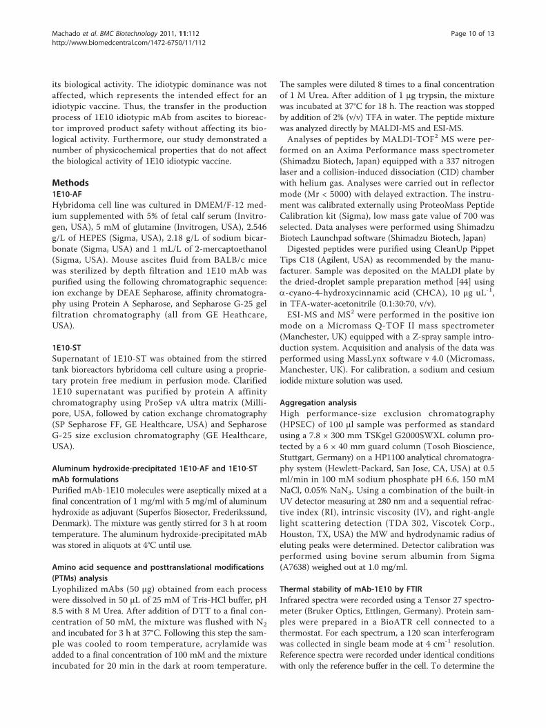
Co-administration of 1E10-AF/Alum or 1E10 ST/Alum withlow-dose Cyclophosphamide induced anti-tumor effectsin a mammary carcinoma modelAs shown in Figure 8, no antitumor effect was observedwhen low-dose Cyclophosphamide or 1E10-ST/Alum wasadministered alone at the evaluated dose. Interestingly,when 1E10-AF/Alum or 1E10-ST/Alum was co-adminis-tered with low-dose Cyclophosphamide F3II mammarycarcinoma growth was reduced significantly. No statisticaldifferences in tumor growth between these groups of micewere determined and the observed anti-tumor responseswere comparable with the effect upon co-administrationof the standard high-dose chemotherapy for breast cancerbased on 60 mg/m2 of Doxorubicin and 600 mg/m2 ofCyclophosphamide.In vivo experiments resulted in similar immunogeni-
city, idiotypic dominance, and antitumor effects of 1E10-AF and 1E10-ST. Thus, the observed posttranslationalmodifications did most likely not affect the Fab structure,as the biological activity of 1E10 vaccine was not altered.Moreover, deamidation of asparagine and methionineoxidation were restricted to Fc regions in 1E10 mAb, faraway from CDRs. Therefore, it remains unlikely thatthese amino acid modifications might affect the spatial
Machado et al. BMC Biotechnology 2011, 11:112http://www.biomedcentral.com/1472-6750/11/112
Page 7 of 13

conformation of the respective CDRs enabling structuralmimicry as postulated for anti-idiotypic mAbs.
ConclusionsEffector functions of classic therapeutic monoclonalantibodies can be divided in Fab-dependent functionsand Fc-dependent functions. The Fab region containsvariable regions of light and heavy chains which areresponsible for antigen recognition, while the Fc regionmediates effector functions like CDC, ADCC and FcgRrecognition. Therefore, any change in their physico-chemical profile could eventually affect the efficacy ofmAbs.It is well known that physicochemical properties of
recombinant proteins can be heavily affected duringchanges in the production process. Thus, it is manda-tory to carefully investigate the similarities and differ-ences of therapeutic mAbs when the production processhas been changed. Comparability must be demonstrated
by an array of physicochemical and biological methodsas required by the regulatory authorities.Regulatory aspects regarding physicochemical charac-
teristics of idiotypic monoclonal antibodies used foridiotypic vaccination bear some differences from classictherapeutic mAbs. For idiotypic mAbs, Fc-related func-tions are not critical for the intended biological effect asthey are administered adsorbed to alum and via intra-dermis. Antigen mimicry is restricted to variable regionCDRs which are located in Fab.Here, we demonstrate that change of 1E10 production
from ascites to bioreactor generates a molecule with dif-ferent physicochemical properties. Changes wereobserved in the degree of asparagine deamidation, C-terminal lysine processing, methionine oxidation, andglycosylation pattern. All these modifications had animpact on the charge profile and conformation of the1E10 molecule, as revealed by an altered thermal stabi-lity of 1E10-ST. However, these changes did not affect
Figure 6 Thermal transition curves of the 1E10-AF (dotted line) and 1E10-ST (solid line). Curves were obtained by plotting the ratio of theintensities of the increasing to the decreasing amide I band vs. temperature. 1E10-ST showed different thermal stability in respect to 1E10-AF.
Machado et al. BMC Biotechnology 2011, 11:112http://www.biomedcentral.com/1472-6750/11/112
Page 8 of 13
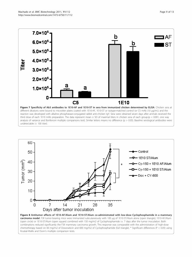
Figure 7 Specificity of Ab3 antibodies to 1E10-AF and 1E10-ST in sera from immunized chicken determined by ELISA. Chicken sera atdifferent dilutions were bound to microtiter plates coated with 1E10-AF, 1E10-ST or isotype-matched control ior C5 mAb (10 μg/mL) and thereaction was developed with alkaline phosphatase-conjugated rabbit anti-chicken IgY. Sera were obtained seven days after animals received thethird dose of each 1E10 mAb preparation. The data represent mean ± SD of maximal titers in chicken sera of each group.(p < 0:001, one wayanalysis of variance and Bonferroni multiple comparisons test). Similar letters means no difference (p > 0.05). Baseline serological antibodies wereundetectable (< 100 titer).
Figure 8 Antitumor effects of 1E10-AF/Alum and 1E10-ST/Alum co-administrated with low-dose Cyclophosphamide in a mammarycarcinoma model. F3II tumor-bearing mice were immunized subcutaneously with 100 μg of 1E10-ST/Alum alone (open triangle); 1E10-AF/Alum(open circle) or 1E10-ST/Alum (open square) combined with 150 mg/m2 of Cyclophosphamide i.v. 7 days after the tumor inoculation. Bothcombinations reduced significantly the F3II mammary carcinoma growth. This response was comparable with the administration of high-dosechemotherapy based on 60 mg/m2 of Doxorubicin and 600 mg/m2 of Cyclophosphamide (full triangle). * Significant differences (P < 0.05) usingKruskal-Wallis and Dunn’s multiple comparison tests.
Machado et al. BMC Biotechnology 2011, 11:112http://www.biomedcentral.com/1472-6750/11/112
Page 9 of 13
its biological activity. The idiotypic dominance was notaffected, which represents the intended effect for anidiotypic vaccine. Thus, the transfer in the productionprocess of 1E10 idiotypic mAb from ascites to bioreac-tor improved product safety without affecting its bio-logical activity. Furthermore, our study demonstrated anumber of physicochemical properties that do not affectthe biological activity of 1E10 idiotypic vaccine.
Methods1E10-AFHybridoma cell line was cultured in DMEM/F-12 med-ium supplemented with 5% of fetal calf serum (Invitro-gen, USA), 5 mM of glutamine (Invitrogen, USA), 2.546g/L of HEPES (Sigma, USA), 2.18 g/L of sodium bicar-bonate (Sigma, USA) and 1 mL/L of 2-mercaptoethanol(Sigma, USA). Mouse ascites fluid from BALB/c micewas sterilized by depth filtration and 1E10 mAb waspurified using the following chromatographic sequence:ion exchange by DEAE Sepharose, affinity chromatogra-phy using Protein A Sepharose, and Sepharose G-25 gelfiltration chromatography (all from GE Heathcare,USA).
1E10-STSupernatant of 1E10-ST was obtained from the stirredtank bioreactors hybridoma cell culture using a proprie-tary protein free medium in perfusion mode. Clarified1E10 supernatant was purified by protein A affinitychromatography using ProSep vA ultra matrix (Milli-pore, USA, followed by cation exchange chromatography(SP Sepharose FF, GE Healthcare, USA) and SepharoseG-25 size exclusion chromatography (GE Healthcare,USA).
Aluminum hydroxide-precipitated 1E10-AF and 1E10-STmAb formulationsPurified mAb-1E10 molecules were aseptically mixed at afinal concentration of 1 mg/ml with 5 mg/ml of aluminumhydroxide as adjuvant (Superfos Biosector, Frederikssund,Denmark). The mixture was gently stirred for 3 h at roomtemperature. The aluminum hydroxide-precipitated mAbwas stored in aliquots at 4°C until use.
Amino acid sequence and posttranslational modifications(PTMs) analysisLyophilized mAbs (50 μg) obtained from each processwere dissolved in 50 μL of 25 mM of Tris-HCl buffer, pH8.5 with 8 M Urea. After addition of DTT to a final con-centration of 50 mM, the mixture was flushed with N2
and incubated for 3 h at 37°C. Following this step the sam-ple was cooled to room temperature, acrylamide wasadded to a final concentration of 100 mM and the mixtureincubated for 20 min in the dark at room temperature.
The samples were diluted 8 times to a final concentrationof 1 M Urea. After addition of 1 μg trypsin, the mixturewas incubated at 37°C for 18 h. The reaction was stoppedby addition of 2% (v/v) TFA in water. The peptide mixturewas analyzed directly by MALDI-MS and ESI-MS.Analyses of peptides by MALDI-TOF2 MS were per-
formed on an Axima Performance mass spectrometer(Shimadzu Biotech, Japan) equipped with a 337 nitrogenlaser and a collision-induced dissociation (CID) chamberwith helium gas. Analyses were carried out in reflectormode (Mr < 5000) with delayed extraction. The instru-ment was calibrated externally using ProteoMass PeptideCalibration kit (Sigma), low mass gate value of 700 wasselected. Data analyses were performed using ShimadzuBiotech Launchpad software (Shimadzu Biotech, Japan)Digested peptides were purified using CleanUp Pippet
Tips C18 (Agilent, USA) as recommended by the manu-facturer. Sample was deposited on the MALDI plate bythe dried-droplet sample preparation method [44] usinga-cyano-4-hydroxycinnamic acid (CHCA), 10 μg uL-1,in TFA-water-acetonitrile (0.1:30:70, v/v).ESI-MS and MS2 were performed in the positive ion
mode on a Micromass Q-TOF II mass spectrometer(Manchester, UK) equipped with a Z-spray sample intro-duction system. Acquisition and analysis of the data wasperformed using MassLynx software v 4.0 (Micromass,Manchester, UK). For calibration, a sodium and cesiumiodide mixture solution was used.
Aggregation analysisHigh performance-size exclusion chromatography(HPSEC) of 100 μl sample was performed as standardusing a 7.8 × 300 mm TSKgel G2000SWXL column pro-tected by a 6 × 40 mm guard column (Tosoh Bioscience,Stuttgart, Germany) on a HP1100 analytical chromatogra-phy system (Hewlett-Packard, San Jose, CA, USA) at 0.5ml/min in 100 mM sodium phosphate pH 6.6, 150 mMNaCl, 0.05% NaN3. Using a combination of the built-inUV detector measuring at 280 nm and a sequential refrac-tive index (RI), intrinsic viscosity (IV), and right-anglelight scattering detection (TDA 302, Viscotek Corp.,Houston, TX, USA) the MW and hydrodynamic radius ofeluting peaks were determined. Detector calibration wasperformed using bovine serum albumin from Sigma(A7638) weighed out at 1.0 mg/ml.
Thermal stability of mAb-1E10 by FTIRInfrared spectra were recorded using a Tensor 27 spectro-meter (Bruker Optics, Ettlingen, Germany). Protein sam-ples were prepared in a BioATR cell connected to athermostat. For each spectrum, a 120 scan interferogramwas collected in single beam mode at 4 cm-1 resolution.Reference spectra were recorded under identical conditionswith only the reference buffer in the cell. To determine the
Machado et al. BMC Biotechnology 2011, 11:112http://www.biomedcentral.com/1472-6750/11/112
Page 10 of 13

melting temperature (TmFTIR), temperature-dependentspectra were acquired every 5°C in a ranging from 25 to90°C. Recorded infrared spectra were analyzed by the Pro-tein Dynamics software for Opus 4.2 (Bruker Optics) anddisplayed as vector-normalized second-derivative amide Ispectra. Melting temperature was calculated by plotting the1639 cm-1 to 1625 cm-1 signal ratio vs. temperature.
Analysis of charge heterogeneity1E10-ST and 1E10-AF (approximately 100 μg each) werediluted five-fold with buffer A (10 mM sodium acetate pH5) loaded onto a ProPac10 WCX column (Dionex, Hous-ton, USA) and eluted with buffer B (10 mM sodium acet-ate, 1 M NaCl pH 5). The gradient was performed in twosteps, from 8 to 13% B in 5 min and 13 to 20% B in 20min. Protein elution was monitored by absorbance at 280nm.The mAb solutions (1.5 mg/ml in 15 mM sodium
phosphate pH 7) were treated with carboxypeptidase B(Boehringer, Germany) at an enzyme-to-substrate ratioof 1:5000 (w/w) at 25°C. Reaction was stopped by add-ing 1 μl of acetic acid. Digested samples were analyzedby WCX HPLC as described above.
N-Glycosylation analysisN-glycans were released by digestion with peptide-N4-(N-acetyl-b-d-glucosaminyl) asparagine amidase F (PNGase F,BioLabs, Beverly, MA, USA), using the method describedby the manufacturer. Briefly, mAbs-1E10 were denaturedat 100°C for 10 min in 0.1% SDS, 5% b-mercaptoethanol.Nonidet P-40 (NP-40) was added to a final concentrationof 1% before enzyme addition. The digestion was carriedout at a ratio of 5 U of PNGase F per milligram of glyco-protein at 37°C for 2 h. The protein was precipitated byadding three volumes of cold ethanol and the mixture waskept at -20°C for 30 min. The oligosaccharides were con-centrated under vacuum and subjected to 2-aminobenza-mide (2AB) labeling.Oligosaccharides were fluorescently labeled with 2AB by
reductive amination [45]. Briefly, oligosaccharides weredissolved in 5 μL DMSO-acetic acid (7:3) containing 2AB(0.35 M) and 1 M NaCNBH3 and incubated at 65°C for 2h. Excess of fluorophore was removed by 2 h vertical chro-matography on Whatman 3 MM paper using acetonitrile.The paper bearing the oligosaccharide signal (applicationpoint) was cut. The oligosaccharides were eluted from thepaper by adding double-distilled water (2 × 500 μL). Theeluate was filtrated in a syringe through a 0.45 μm PTFEfilter (Millex-LCR, Millipore) and then concentratedunder vacuum.Normal-phase HPLC (NP-HPLC) of the labeled por-
tion was performed using a TSK-Gel Amide-80 4.6 × 250mm column (Tosoh BioSep, Japan) on a separation mod-ule (Merck-Hitachi, Japan) equipped with a fluorescence
detector. Labeled N-glycans were separated by a lineargradient of 20-58% of 50 mM ammonium formiate pH4.4 against acetonitrile over 152 min at a flow rate of 0.4mL/min. Samples were injected in 80% acetonitrile. Thefluorescence detection was carried out using an excita-tion wavelength of 330 nm and an emission wavelengthof 420 nm [46]. The elution positions of the N-glycanswere determined in glucose units (GU) by comparisonwith a standard dextran hydrolysate 2AB labeled (dextranladder) [47].
AnimalsFemale BALB/c/cenp mice (ten weeks old) and Leghornchicken (10 weeks old) were purchased from the Centerfor Laboratory Animal Breeding (CENPALAB, Havana,Cuba). Animals were housed under conventional condi-tions with free access to water and food and maintainedin accordance with the guidelines stipulated by the Ani-mal Subject Committee Review Board of CENPALAB.Animal studies were performed with approval fromCENPALAB’s and CIM’s Institutional Animal Care andUse Committees.
Antitumor experimentsThe sarcomatoid mammary carcinoma cell line F3II is ahighly invasive and metastatic variant established from aclone of a spontaneous BALB/c mouse mammary tumor.F3II cells grow as spindle-cell carcinoma tumors with ahigh local invasiveness and a 90-100% incidence of lungmetastases [48]. F3II cells were maintained in minimalessential medium (MEM 41500, Gibco, BRL) supplementedwith 10% fetal bovine serum, 2 mM glutamine, 80 mg/mlGentamycin, and 20 mg/ml tetracycline in monolayerculture.All mice were injected subcutaneously with 2 × 10⁵ F3II
cells in the right flank. Seven days after tumor inoculation,the animals were separated in different groups andinjected as follows: subcutaneously with normal saline;subcutaneously with 100 μg of 1E10-ST/Alum; intrave-nously with 150 mg/m2 of Cyclophosphamide (Cy-150);intravenously with 150 mg/m2 of Cyclophosphamide andsubcutaneously with 100 μg of 1E10-AF/Alum; intrave-nously with 150 mg/m2 of Cyclophosphamide and subcu-taneously with 100 μg of 1E10-ST/Alum or intravenouslywith 60 mg/m2 of Doxorubicin (Dox) and 600 mg/m2 ofCyclophosphamide (Cy-600). The time of appearance oflocal tumors was monitored by palpation and further con-firmed by histopathology. In all cases, tumors were diag-nosed as spindle-cell carcinomas. Tumor size wasmeasured with a caliper twice a week.
Induction of anti-anti-idiotype antibody (Ab3) responseChicken were immunized subcutaneously with 100 μg of1E10 vaccine in the days 0, 7 and 21. Blood extractions
Machado et al. BMC Biotechnology 2011, 11:112http://www.biomedcentral.com/1472-6750/11/112
Page 11 of 13
were done before and one week after the last immuniza-tion (day 28).The presence of anti-1E10 antibodies in chicken sera
was determined in a solid-phase ELISA Polystyrene Maxi-sorp microtiter plates (Nunc, Roskilde, Denmark) werecoated with 50 μl of a solution of 10 μg/ml of mAb-1E10in carbonate buffer pH 9.6 and incubated overnight at 4°C.After washing with PBS containing 0.05% Tween 20, theplates were blocked for 1 h at room temperature with PBScontaining 1% BSA. Then, diluted serum samples wereadded to each well and the plates were incubated for 2 hat 37°C. Chicken antibodies were detected with an AP-conjugated rabbit anti-chicken IgY (Sigma). The plateswere washed four times and the reaction was developedwith a substrate solution consisting of 1 mg/ml p-nitro-phenylphosphate (Sigma) in diethanolamine buffer, pH9.8. Absorbance was measured at 405 nm in an ELISAreader (Tekan, Salzburg, Austria).The highest serum dilution giving OD values ≥0.2 and
being at least three times the value corresponding to thepreimmune serum at the same dilution was consideredas titer. Assays were performed in triplicate for each sam-ple and the coefficient of variation (CV) was less than15%. The ODs of the blanks were less than 0.1.
Acknowledgements and FundingThe authors like to thank Dr. Aniel Sánchez for his critical revision of thepaper.YJM’s stay at Salzburg University for HPSEC-TDA and FTIR experiments wasfunded by a Boehringer Ingelheim Fonds Travel Grant.
Author details1Center for Molecular Immunology, 216 St and 15th Ave., Atabey Siboney,Playa, P.O. Box 16040, Havana 11600, Cuba. 2Center for Genetic Engineeringand Biotechnology, 31st Ave and 158 St, Playa, P.O. Box 6162, Havana 10600.Cuba. 3Centro Nacional Para la producción de Animales de Laboratorio, LaHabana, Cuba. 4Christian Doppler Laboratory for Allergy Diagnosis andTherapy, University of Salzburg, Austria.
Authors’ contributionsYJM conceived the experiments, carried out the MALDI-MS and chargeheterogeneity determinations, the FTIR experiments, the immunogenicitystudies, performed the overall data treatments and has drafted themanuscript. YR performed the ELISA determinations for all theimmunogenicity studies. RM and JC performed glycans structure analysis; VBperformed ESI-QTOF determinations and supported YJM in massspectrometry data analysis. DF performed antitumor experiments andsupported YJM in experimental design of animal experiments. AMV, AC, KRLand MH has been involved in the experiments planning, the drafting of themanuscript and for revising it critically for content and general supervisionof the project. MH performed HPSEC-TDA determinations. All the authorshave read and approved the final manuscript.
Received: 21 September 2011 Accepted: 22 November 2011Published: 22 November 2011
References1. Jerne NK: Towards a network theory of the immune system. Ann
Immunol (Paris) 1974, 125C(1-2):373-389.2. Olson MV, Varki A: Sequencing the chimpanzee genome: insights into
human evolution and disease. Nat Rev Genet 2003, 4(1):20-28.
3. Irie A, Koyama S, Kozutsumi Y, Kawasaki T, Suzuki A: The molecular basisfor the absence of N-glycolylneuraminic acid in humans. J Biol Chem1998, 273(25):15866-15871.
4. Malykh YN, Schauer R, Shaw L: N-Glycolylneuraminic acid in humantumours. Biochimie 2001, 83(7):623-634.
5. Marquina G, Waki H, Fernandez LE, Kon K, Carr A, Valiente O, Perez R,Ando S: Gangliosides expressed in human breast cancer. Cancer Res 1996,56(22):5165-5171.
6. Miyake M, Hashimoto K, Ito M, Ogawa O, Arai E, Hitomi S, Kannagi R: Theabnormal occurrence and the differentiation-dependent distribution ofN-acetyl and N-glycolyl species of the ganglioside GM2 in human germcell tumors. A study with specific monoclonal antibodies. Cancer 1990,65(3):499-505.
7. de Leon J, Fernandez A, Mesa C, Clavel M, Fernandez LE: Role of tumour-associated N-glycolylated variant of GM3 ganglioside in cancerprogression: effect over CD4 expression on T cells. Cancer ImmunolImmunother 2006, 55(4):443-450.
8. Vazquez AM, Perez A, Hernandez AM, Macias A, Alfonso M, Bombino G,Perez R: Syngeneic anti-idiotypic monoclonal antibodies to an anti-NeuGc-containing ganglioside monoclonal antibody. Hybridoma 1998,17(6):527-534.
9. Vazquez AM, Alfonso M, Lanne B, Karlsson KA, Carr A, Barroso O,Fernandez LE, Rengifo E, Lanio ME, Alvarez C, et al: Generation of a murinemonoclonal antibody specific for N-glycolylneuraminic acid-containinggangliosides that also recognizes sulfated glycolipids. Hybridoma 1995,14(6):551-556.
10. Hernandez AM, Rodriguez M, Lopez-Requena A, Beausoleil I, Perez R,Vazquez AM: Generation of anti-Neu-glycolyl-ganglioside antibodies byimmunization with an anti-idiotype monoclonal antibody: A self versusnon-self-matter. Immunobiology 2005, 210(1):11-21.
11. Alfonso M, Diaz A, Hernandez AM, Perez A, Rodriguez E, Bitton R, Perez R,Vazquez AM: An anti-idiotype vaccine elicits a specific response to N-glycolyl sialic acid residues of glycoconjugates in melanoma patients. JImmunol 2002, 168(5):2523-2529.
12. Diaz A, Alfonso M, Alonso R, Saurez G, Troche M, Catala M, Diaz RM,Perez R, Vazquez AM: Immune responses in breast cancer patientsimmunized with an anti-idiotype antibody mimicking NeuGc-containinggangliosides. Clin Immunol 2003, 107(2):80-89.
13. Neninger E, Diaz RM, de la Torre A, Rives R, Diaz A, Saurez G, Gabri MR,Alonso DF, Wilkinson B, Alfonso AM, et al: Active immunotherapy with1E10 anti-idiotype vaccine in patients with small cell lung cancer: reportof a phase I trial. Cancer Biol Ther 2007, 6(2):145-150.
14. Hernandez AM, Toledo D, Martinez D, Grinan T, Brito V, Macias A, Alfonso S,Rondon T, Suarez E, Vazquez AM, et al: Characterization of the antibodyresponse against NeuGcGM3 ganglioside elicited in non-small cell lungcancer patients immunized with an anti-idiotype antibody. J Immunol2008, 181(9):6625-6634.
15. Diaz Y, Gonzalez A, Lopez A, Perez R, Vazquez AM, Montero E: Anti-ganglioside anti-idiotypic monoclonal antibody-based cancer vaccineinduces apoptosis and antiangiogenic effect in a metastatic lungcarcinoma. Cancer Immunol Immunother 2009, 58(7):1117-1128.
16. Fuentes D, Avellanet J, Garcia A, Iglesias N, Gabri MR, Alonso DF,Vazquez AM, Perez R, Montero E: Combined therapeutic effect of amonoclonal anti-idiotype tumor vaccine against NeuGc-containinggangliosides with chemotherapy in a breast carcinoma model. BreastCancer Res Treat 2009, 120(2):379-389.
17. Reichert JM, Beck A, Iyer H: European Medicines Agency workshop onbiosimilar monoclonal antibodies: July 2, 2009, London, UK. MAbs 2009,1(5):394-416.
18. Jefferis R: Recombinant antibody therapeutics: the impact ofglycosylation on mechanisms of action. Trends Pharmacol Sci 2009,30(7):356-362.
19. Jefferis R: Glycosylation as a strategy to improve antibody-basedtherapeutics. Nat Rev Drug Discov 2009, 8(3):226-234.
20. Kaneko Y, Nimmerjahn F, Ravetch JV: Anti-inflammatory activity ofimmunoglobulin G resulting from Fc sialylation. Science 2006,313(5787):670-673.
21. Chung CH, Mirakhur B, Chan E, Le QT, Berlin J, Morse M, Murphy BA,Satinover SM, Hosen J, Mauro D, et al: Cetuximab-induced anaphylaxisand IgE specific for galactose-alpha-1,3-galactose. N Engl J Med 2008,358(11):1109-1117.
Machado et al. BMC Biotechnology 2011, 11:112http://www.biomedcentral.com/1472-6750/11/112
Page 12 of 13

22. Paborji M, Pochopin NL, Coppola WP, Bogardus JB: Chemical and physicalstability of chimeric L6, a mouse-human monoclonal antibody. PharmRes 1994, 11(5):764-771.
23. Vlasak J, Bussat MC, Wang S, Wagner-Rousset E, Schaefer M, Klinguer-Hamour C, Kirchmeier M, Corvaia N, Ionescu R, Beck A: Identification andcharacterization of asparagine deamidation in the light chain CDR1 of ahumanized IgG1 antibody. Anal Biochem 2009, 392(2):145-154.
24. Beck A, Bussat MC, Zorn N, Robillard V, Klinguer-Hamour C, Chenu S,Goetsch L, Corvaia N, Van Dorsselaer A, Haeuw JF: Characterization byliquid chromatography combined with mass spectrometry ofmonoclonal anti-IGF-1 receptor antibodies produced in CHO and NS0cells. J Chromatogr B Analyt Technol Biomed Life Sci 2005, 819(2):203-218.
25. Harris RJ, Kabakoff B, Macchi FD, Shen FJ, Kwong M, Andya JD, Shire SJ,Bjork N, Totpal K, Chen AB: Identification of multiple sources of chargeheterogeneity in a recombinant antibody. J Chromatogr B Biomed Sci Appl2001, 752(2):233-245.
26. Gadgil HS, Pipes GD, Dillon TM, Treuheit MJ, Bondarenko PV: Improvingmass accuracy of high performance liquid chromatography/electrosprayionization time-of-flight mass spectrometry of intact antibodies. J AmSoc Mass Spectrom 2006, 17(6):867-872.
27. Lewis DA, Guzzetta AW, Hancock WS, Costello M: Characterization ofhumanized anti-TAC, an antibody directed against the interleukin 2receptor, using electrospray ionization mass spectrometry by directinfusion, LC/MS, and MS/MS. Anal Chem 1994, 66(5):585-595.
28. Werner WE, Wu S, Mulkerrin M: The removal of pyroglutamic acid frommonoclonal antibodies without denaturation of the protein chains. AnalBiochem 2005, 342(1):120-125.
29. Wang S, Ionescu R, Peekhaus N, Leung JY, Ha S, Vlasak J: Separation ofpost-translational modifications in monoclonal antibodies by exploitingsubtle conformational changes under mildly acidic conditions.J Chromatogr A 2005, 1217(42):6496-6502.
30. Liu D, Ren D, Huang H, Dankberg J, Rosenfeld R, Cocco MJ, Li L, Brems DN,Remmele RL Jr: Structure and stability changes of human IgG1 Fc as aconsequence of methionine oxidation. Biochemistry 2008,47(18):5088-5100.
31. Liu H, Gaza-Bulseco G, Xiang T, Chumsae C: Structural effect ofdeglycosylation and methionine oxidation on a recombinantmonoclonal antibody. Mol Immunol 2008, 45(3):701-708.
32. Wang S, Ionescu R, Peekhaus N, Leung JY, Ha S, Vlasak J: Separation ofpost-translational modifications in monoclonal antibodies by exploitingsubtle conformational changes under mildly acidic conditions.J Chromatogr A 2010, 1217(42):6496-6502.
33. Manning MC, Patel K, Borchardt RT: Stability of protein pharmaceuticals.Pharm Res 1989, 6(11):903-918.
34. Singh SK: Impact of product-related factors on immunogenicity ofbiotherapeutics. Journal of Pharmaceutical Sciences 2010, 100(2):354-387.
35. De Groot AS, Scott DW: Immunogenicity of protein therapeutics. TrendsImmunol 2007, 28(11):482-490.
36. Bee JS, Davis M, Freund E, Carpenter JF, Randolph TW: Aggregation of amonoclonal antibody induced by adsorption to stainless steel. BiotechnolBioeng 2010, 105(1):121-129.
37. Harris RJ: Processing of C-terminal lysine and arginine residues ofproteins isolated from mammalian cell culture. J Chromatogr A 1995,705(1):129-134.
38. Quintero O, Montesino R, Cremata JA: Two-dimensional mapping of 8-amine-1,3,6-naphthalene trisulfonic acid derivatives of N-linked neutraland sialyloligosaccharides. Anal Biochem 1998, 256(1):23-32.
39. Dobson CM: Experimental investigation of protein folding andmisfolding. Methods 2004, 34(1):4-14.
40. Takeda N, Kato M, Taniguchi Y: Pressure- and thermally-induced reversiblechanges in the secondary structure of ribonuclease A studied by FT-IRspectroscopy. Biochemistry 1995, 34(17):5980-5987.
41. van de Weert M, Haris PI, Hennink WE, Crommelin DJ: Fourier transforminfrared spectrometric analysis of protein conformation: effect ofsampling method and stress factors. Anal Biochem 2001, 297(2):160-169.
42. Becktel WJ, Schellman JA: Protein stability curves. Biopolymers 1987,26(11):1859-1877.
43. Matheus S, Friess W, Mahler HC: FTIR and nDSC as analytical tools forhigh-concentration protein formulations. Pharm Res 2006,23(6):1350-1363.
44. Karas M, Hillenkamp F: Laser desorption ionization of proteins withmolecular masses exceeding 10,000 daltons. Anal Chem 1988,60(20):2299-2301.
45. Bigge JC, Patel TP, Bruce JA, Goulding PN, Charles SM, Parekh RB:Nonselective and efficient fluorescent labeling of glycans using 2-aminobenzamide and anthranilic acid. Anal Biochem 1995, 230(2):229-238.
46. Guile GR, Wong SY, Dwek RA: Analytical and preparative separation ofanionic oligosaccharides by weak anion-exchange high-performanceliquid chromatography on an inert polymer column. Anal Biochem 1994,222(1):231-235.
47. Guile GR, Rudd PM, Wing DR, Prime SB, Dwek RA: A rapid high-resolutionhigh-performance liquid chromatographic method for separating glycanmixtures and analyzing oligosaccharide profiles. Anal Biochem 1996,240(2):210-226.
48. Alonso DF, Farias EF, Urtreger A, Ladeda V, Vidal MC, Bal De Kier Joffe E:Characterization of F3II, a sarcomatoid mammary carcinoma cell lineoriginated from a clonal subpopulation of a mouse adenocarcinoma. JSurg Oncol 1996, 62(4):288-297.
doi:10.1186/1472-6750-11-112Cite this article as: Machado et al.: Physicochemical and biologicalcharacterization of 1E10 Anti-Idiotype vaccine. BMC Biotechnology 201111:112.
Submit your next manuscript to BioMed Centraland take full advantage of:
• Convenient online submission
• Thorough peer review
• No space constraints or color figure charges
• Immediate publication on acceptance
• Inclusion in PubMed, CAS, Scopus and Google Scholar
• Research which is freely available for redistribution
Submit your manuscript at www.biomedcentral.com/submit
Machado et al. BMC Biotechnology 2011, 11:112http://www.biomedcentral.com/1472-6750/11/112
Page 13 of 13