research article open access prognostic impact of … · massimo ruscazio2, luigi meloni2, sabino...
TRANSCRIPT

RESEARCH ARTICLE Open Access
Prognostic impact of coronary microcirculationabnormalities in systemic sclerosis: a prospectivestudy to evaluate the role of non-invasive testsAlessandra Vacca1*†, Roberta Montisci2†, Pietro Garau1, Paolo Siotto3, Matteo Piga1, Alberto Cauli1,Massimo Ruscazio2, Luigi Meloni2, Sabino Iliceto4 and Alessandro Mathieu1
Abstract
Introduction: Microcirculation dysfunction is a typical feature of systemic sclerosis (SSc) and represents the earliestabnormality of primary myocardial involvement. We assessed coronary microcirculation status by combining twofunctional tests in SSc patients and estimating its impact on disease outcome.
Methods: Forty-one SSc patients, asymptomatic for coronary artery disease, were tested for coronary flow velocityreserve (CFR) by transthoracic-echo-Doppler with adenosine infusion (A-TTE) and for left ventricular wall motionabnormalities (WMA) by dobutamine stress echocardiography (DSE). Myocardial multi-detector computedtomography (MDCT) enabled the presence of epicardial stenosis, which could interfere with the accuracy of thetests, to be excluded. Patient survival rate was assessed over a 6.7- ± 3.5-year follow-up.
Results: Nineteen out of 41 (46%) SSc patients had a reduced CFR (≤2.5) and in 16/41 (39%) a WMA was observedduring DSE. Furthermore, 13/41 (32%) patients showed pathological CFR and WMA. An inverse correlation betweenwall motion score index (WMSI) during DSE and CFR value (r = -0.57, P <0.0001) was observed; in addition, CFRwas significantly reduced (2.21 ± 0.38) in patients with WMA as compared to those without (2.94 ± 0.60) (P<0.0001). In 12 patients with abnormal DSE, MDCT was used to exclude macrovasculopathy. During a 6.7- ± 3.5-year follow-up seven patients with abnormal coronary functional tests died of disease-related causes, compared toonly one patient with normal tests.
Conclusions: A-TTE and DSE tests are useful tools to detect non-invasively pre-clinical microcirculationabnormalities in SSc patients; moreover, abnormal CFR and WMA might be related to a worse disease outcomesuggesting a prognostic value of these tests, similar to other myocardial diseases.
IntroductionUntil two decades ago, clinical evidence of cardiac invol-vement in systemic sclerosis (SSc) was considered aninfrequent event and it mainly resulted from autopsy stu-dies. In particular, overt manifestations of ischemic heartdisease were considered rare, cardiac failure was observedin about 10% of cases, and pericarditis in 15% [1-4].Conversely, post mortem investigations demonstratedmyocardial lesions secondary to SSc in more than 50% ofcases [2].
The discrepancy between the high prevalence of sclero-derma heart involvement (SHI) at autopsy studies and ofits lower detection by in vivo studies might be due to thelow sensitivity or to the scarce applicability of the diagnos-tic tools utilized [5]. On the other hand, when SHIbecomes clinically evident, it assumes a deeply negativeprognostic significance, with a mortality rate above 70% atfive years [6]. Detailed reviews of the clinical studies con-cerning SHI have recently been published [7-9]. However,data on the prognostic impact of sub-clinical myocardialinvolvement as detected by more sensitive tests in SScpatients are presently lacking.The pathogenesis of SHI is still debated; the most fre-
quent pathological features of SSc in the myocardium
* Correspondence: [email protected]† Contributed equally1University and A.O.U. of Cagliari, Chair and Unit of Rheumatology, S.S. 554bivio per Sestu, Monserrato 09042, ItalyFull list of author information is available at the end of the article
Vacca et al. Arthritis Research & Therapy 2013, 15:R8http://arthritis-research.com/content/15/1/R8
© 2013 Vacca et al.; licensee BioMed Central Ltd. This is an open access article distributed under the terms of the Creative CommonsAttribution License (http://creativecommons.org/licenses/by/2.0), which permits unrestricted use, distribution, and reproduction inany medium, provided the original work is properly cited.

are focal fibrosis (in more than 50% of cases) and con-traction band necrosis (CBN) (in 77% of patients) [2].Follansbee et al. [10] found a high prevalence of CBN inSSc patients with SHI, probably related to an inter-mittent vascular spasm of the coronary arteries withepisodes of ischemia-reperfusion [2].The small coronary vessels show a reduced patency or
obliteration due to intimal proliferation, fibrinoid necrosis,fibrosis and intravascular coagulation [11]. The microvas-cular structural and functional abnormalities seem to leadto the increased fibroblast activity and disseminated tissuefibrosis [12], which may progress to a clinical pattern ofrestrictive cardiomyopathy [13].The consequences of such anatomical damage and
functional disorder were reported in subsequent studies.Kahan et al. [14] first demonstrated the impairment ofcoronary vasodilator reserve using coronary catheterism,later confirmed by non-invasive adenosine transthoracicechocardiography (A-TTE) by other groups [15,16]. Inaddition, myocardial scintigraphy enabled several authorsto observe reversible myocardial perfusion defects,induced either by exposure to the cold or by physicalexercise [17-19].Dobutamine stress echocardiography (DSE) enables
evaluation of the dynamics of left ventricular wallmotion, which correlate to perfusion and oxygen supply,during chronotropic and inotropic pharmacologicalstress. This test is a well- established diagnostic andprognostic tool that has widespread applicability becauseof its clinical accuracy and cost effectiveness [20].Some authors demonstrated that the simultaneous eva-
luation of coronary flow velocity reserve (CFR) and leftventricular wall motion (LVWM) by dipyridamole stressechocardiography increases the diagnostic power of eachtest to detect coronary macro- and micro-vascular invol-vement [21,22].In order to increase the accuracy of these two methods,
the presence of epicardial artery stenosis should beexcluded, and in patients with pre-clinical SHI, where aninvasive procedure is not ethically applicable, myocardialmulti-detector computed tomography (MDCT) might bepreferable in order to avoid cardiac catheterization [23].On this basis, the presence of early myocardial func-
tional changes in SSc patients asymptomatic for coronaryartery disease (CAD) was investigated by combinedA-TTE and DSE, integrating them, when applicable, withMDCT, and the impact of such abnormalities on mortalitywas determined.
Materials and methodsThe initial population comprised 97 SSc patients, who ful-filled the American College of Rheumatology classificationcriteria [24], followed from September 2000 to June 2006at our Department. Exclusion criteria were: 1) technically
poor acoustic window precluding satisfactory imaging ofthe left ventricle (for 2-D echo) or of left anterior descend-ing coronary artery (LAD) flow by Color Doppler (for CFRassessment) (16 patients), 2) a right ventricular systolicpressure >40 mmHg by echocardiography (12 patients), 3)hemodynamic instability (2 patients), valvular disease(5 patients), CAD (3 patients), unstable angina (1 patient),life-threatening arrhythmias (1 patient), severe lung and/orpulmonary vascular disease (6 patients), 4) asthma orsevere chronic obstructive pulmonary disease (6 patients),and 5) inability or refusal to give informed consent(6 patients). None of them showed symptoms or signs ofcongestive heart failure, CAD or arrhythmia. Treatmentwith calcium channel blockers and prostanoids wassuspended 48 hours and 4 weeks before functional tests,respectively. All the studied subjects were requested toavoid xanthine-containing food and drinks for ≥24 hoursbefore evaluation. The study was approved by the ethicscommittee of Cagliari University Hospital (Italy), and writ-ten informed consent was obtained from all participants.The final study population consisted of 41 patients; amongthem, 30 represented the initial cohort who underwentDSE in our previous study [25]. Hematochemical andserological tests, chest X-ray, lung function tests and CTexamination, ECG, basal M-mode and two-dimensionalechocardiography were carried out on all subjects.
Follow-upFollowing initial cardiac evaluations, all patients werefollowed up for a mean period of 6.7 ± 3.5 years.
Adenosine Transthoracic Echochardiography (A-TTE)In all patients, CFR was assessed by A-TTE as previouslydescribed [15]. Briefly, CFR was evaluated in the LAD withtransthoracic Color Doppler during adenosine infusion.The pulsed wave Doppler examination of blood flow velo-city was recorded in the LAD at rest and after maximumvasodilation by adenosine infusion (140 μg/kg/min forthree to five minutes). When the Doppler signal wassuboptimal, Levovist® (Schering AG, Berlin, Germany), asuspension of monosaccharide (galactose) microparticlesin sterile water, was infused at a concentration of 300mg/ml, at a rate of 0.5 to 1 ml/min. The CFR value wasexpressed as the ratio of peak diastolic velocity duringhyperaemia to peak diastolic velocity at rest (baseline).CFR values ≤2.5 were considered impaired, as previouslyreported [15].
Dobutamine stress echocardiography protocolAll patients underwent DSE. After baseline data had beenacquired, dobutamine was infused, beginning at a dose of5 mcg/Kg/min, and increasing every three minutes to amaximum dose of 40 mcg/Kg/min. Echocardiogramswere recorded at baseline, low dose, peak dose and five
Vacca et al. Arthritis Research & Therapy 2013, 15:R8http://arthritis-research.com/content/15/1/R8
Page 2 of 9

minutes into recovery. When necessary, atropine (up to 1mg) was given intravenously at the higher dose levels toaugment heart rate response. Assessment was performedby two experienced investigators who had no knowledgeof CFR data. The following scoring system was used forregional wall motion: normal = 1; hypokinetic = 2;akinetic = 3; diskinetic = 4. For segmental analysis of leftventriculum (LV) function, a 16-segment model was usedas suggested by the American Society of Echocardiogra-phy [26]. A Wall Motion Score Index (WMSI) was cal-culated at baseline and at peak stress as the sum ofscores divided by the number of analyzed segments.End points for dobutamine infusion were: achievement
of target heart rate (85% of the maximal heart rate pre-dicted for age), maximal dose of dobutamine and atro-pine, extensive new wall-motion abnormalities, >2 mmST-segment depression in two or more ECG leads,chest pain, significant arrhythmias, severe hypertension(blood pressure >220 mmHg) or hypotension (a fall insystolic blood pressure >30 mmHg). ECG and bloodpressure were monitored continuously and recorded ateach stage.
Coronary multi-detector computed tomographyThe indication to perform MDCT was based on the pre-sence of WMA at the DSE test. MDCT was carried outusing a multi-detector computerized tomograph witheight lines of detectors and a rotation time of 500 msec on360° and with a slice depth of 1.3 mm (Light Speed Ultra,General Electric Medical Systems, Milwaukee, WI, USA).Retrospective gating was used. A total of 120 ml of non-ionic contrast medium was injected at 4 ml/sec. Imageswere processed as appropriate on a SUN-80 ULTRAworkstation (Sun Microsystems, Palo Alto, CA, USA). Theentire examination lasted from 20 to 30 minutes. Imageevaluation was carried out by an expert radiologist awareof patients’ diagnoses.
Statistical analysisAll data are presented as mean ± SD. Comparisons weremade with an independent t-test or a non-parametric test(Mann-Whitney, chi-square or Fisher’s exact test), whenappropriate. Relationships between CFR, WMSI and pro-tocol variables were evaluated with the simple linear corre-lation coefficient r (determination coefficient r2), P-valuesless than or equal to 0.05 were considered significant.Kaplan-Meier method survival curves were used to
summarize the follow-up experience in these patients.The differences in survival curves were tested with alog-rank statistic. The association of selected variableswith outcome was assessed with the Cox proportionalhazard model using univariate and stepwise multivariateprocedures. A significance of 0.05 was required for avariable to be included in the multivariate model,
whereas 0.1 was the exclusion cut-off value. Hazardratios with the corresponding 95% confidence intervalswere estimated. Data management and analysis wereperformed using MedCalc software (version. 12.2.1;Mariakerker, Belgium).
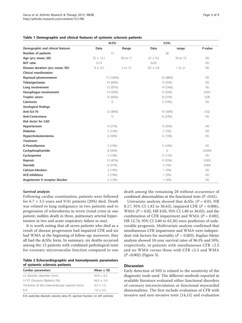
ResultsForty-one consecutive SSc patients (33 female, 8 male;mean age 54.1 y, range 28 to 73) with mean diseaseduration of 7.5 y (range 1 to 22 y) were included in thestudy. Fifteen out of the 41 SSc patients were affectedby dcSSc subtype and 26 by lcSSc form, according tothe LeRoy et al. classification [27]. Demographic, clinicaland laboratory data of patients are reported in Table 1.Echocardiographic and functional cardiac parameters
determined in SSc patients studied are listed in Tables 2and 3. Both cardiac stress tests were well tolerated andno cardiac adverse events occurred during examinations.The median interval time between A-TTE and DSE was1.5 weeks.Nineteen out of 41 (46%) patients with SSc showed
reduced CFR (≤2.5) and in 16/41 (39%) SSc patientsWMA (hypokinesia), which was absent on baseline restexamination, was observed during dobutamine infusion(Figure 1). Thirteen out of 41 patients (32%) showedboth CFR and DSE test impaired; these combined abnor-mal findings were more frequent in the subgroup withdcSSc (8/15; 53%) than in the lcSSc subset (5/26; 19%)(P <0.04), while they were irrespective of age, diseaseduration, presence of anti Scl-70 or anti-centromere anti-body, esophageal and lung involvement, digital ulcers,high cholesterol serum levels and blood pressure.WMA during DSE were limited to one segment in
10 patients, to two segments in 5 patients and to threesegments in 1 patient. In seven patients with abnormalDSE, WMA were localized in the LAD coronary arteryarea, in three patients in both the LAD and right coronaryartery area, whereas in three patients wall motion abnorm-alities were only in the right coronary artery area and inthree patients in circumflex coronary area.An inverse correlation between CFR and WMSI (r =
-0.57, P <0.0001) was observed (Figure 2). Moreover, CFRwas significantly reduced in patients with induced WMAduring DSE (2.21 ± 0.38 vs 2.94 ± 0.60, P = <0.0001); indetail, 13/16 patients (81%) with WMA during DSE alsohad impaired CFR and only 3 had normal CFR, whileamong patients without WMA only 6 (24%) showed pat-hologically reduced CFR and 19 had normal values(P <0.0001).The presence of epicardial artery stenosis, which might
affect the two functional tests’ results, could be excludedby MDCT in 12 patients with WMA, as the examinationwas declined by 3 patients and 1 died before MDCT wasperformed.
Vacca et al. Arthritis Research & Therapy 2013, 15:R8http://arthritis-research.com/content/15/1/R8
Page 3 of 9
Survival analysisFollowing cardiac examination, patients were followedfor 6.7 ± 3.5 years and 9/41 patients (20%) died. Deathwas related to lung malignancy in two patients and toprogression of scleroderma in seven (renal crisis in onepatient, sudden death in three, pulmonary arterial hyper-tension in two and acute respiratory failure in one).It is worth noting that all seven patients who died as a
result of disease progression had impaired CFR and sixhad WMA at the beginning of follow-up; moreover, theyall had the dcSSc form. In summary, six deaths occurredamong the 13 patients with combined pathological testsfor coronary microvascular function compared to one
death among the remaining 28 without occurrence ofcombined abnormalities at the functional tests (P <0.01).Univariate analysis showed that dcSSc (P = 0.01, HR
8.17, 95% CI 1.82 to 36.63), impaired CFR (P = 0.006),WMA (P = 0.02, HR 8.05, 95% CI 1.80 to 36.02), and thecombination of CFR impairment and WMA (P = 0.002,HR 12.74, 95% CI 2.60 to 62.36) were predictors of unfa-vorable prognosis. Multivariate analysis confirmed thatsimultaneous CFR impairment and WMA were indepen-dent risk factors for mortality (P = 0.003). Kaplan-Meieranalysis showed 10-year survival rates of 96.5% and 59%,respectively, in patients with simultaneous CFR >2.5and no WMA versus those with CFR ≤2.5 and WMA(P <0.002) (Figure 3).
DiscussionEarly detection of SHI is related to the sensitivity of thediagnostic tools used. The different methods reported inavailable literature evaluated either functional disordersof coronary microcirculation or functional myocardialabnormalities. The first include evaluation of CFR withinvasive and non-invasive tests [14,15] and evaluation
Table 1 Demographic and clinical features of systemic sclerosis patients
dcSSc lcSSc
Demographic and clinical features Data Range Data range P-value
Number of patients 15 26
Age (yrs; mean; SD) 53 ± 13.1 28 to 71 52 ± 9.3 39 to 73 NS
M/F ratio 2/13 6/20 NS
Disease duration (yrs; mean; SD) 8 ± 3.3 2 to 15 8.5 ± 5.8 1 to 21 NS
Clinical manifestation
Raynaud phoenomenon 15 (100%) 23 (88%) NS
Teleangectasias 10 (66%) 9 (35%) NS
Lung involvement 13 (87%) 14 (54%) NS
Oesophagus involvement 14 (93%) 9 (35%) 0.001
Trophic ulcers 10 (66%) 8 (31%) 0.06
Calcinosis 0 5 (19%) NS
Serological findings
Anti-Scl-70 12 (80%) 10 (38%) 0.02
Anti-Centromere 0 6 (23%) NS
Risk factor for CAD
Hypertension 4 (27%) 9 (35%) NS
Diabetes 2 (13%) 2 (7%) NS
Hypercholesterolemia 6 (40%) 4 (15%) NS
Treatment
D-Penicillamine 3 (19%) 5 (19%) NS
Cyclophosphamide 8 (50%) 0 0.0004
Cyclosporine 3 (19%) 3 (11%) NS
Iloprost 13 (87%) 9 (35%) 0.003
Steroids 6 (37%) 2 (7%) 0.004
Calcium-blockers 3 (19%) 1 (3%) NS
ACE-inhibitors 3 (19%) 1 (3%) NS
Angiotensin II receptor blocker 2 (13%) 1 (3%) NS
Table 2 Echocardiographic and hemodynamic parametersof systemic sclerosis patients
Cardiac parameters Mean ± SD
LV diastolic diameter (mm) 44.9 ± 6.3
LV EF (Simpson Biplano) (%) 66.0 ± 5.4
Thickness of the interventricular septum (mm) 9.5 ± 1.3
E/A 1.0 ± 0.3
E/A, early/late diastolic velocity ratio; EF, ejection fraction; LV, left ventricle
Vacca et al. Arthritis Research & Therapy 2013, 15:R8http://arthritis-research.com/content/15/1/R8
Page 4 of 9

of myocardial perfusion with SPECT and MRI [17,18,28-30]. The second include evaluation of systolic or dia-stolic dysfunction by Doppler echocardiography [31-35]and tissue-Doppler echocardiography [36].To our knowledge, this is the first report where two
non-invasive methods, one exploring coronary microvas-cular function (A-TTE test) and the other perfusion- andmetabolism-dependent contractility (DSE test), have been
combined and complementarily applied in SSc to detectpre-clinical cardiac involvement; furthermore, we sug-gested that CFR and DSE impairment might have animpact on the prognosis of such patients.Our findings can be summarized as follows. First,
impaired myocardial microcirculation has been con-firmed in SSc patients in the absence of any CAD relatedsymptoms and signs as revealed by non-invasive methods
Table 3 Cardiac parameters during dobutamine stress echocardiography (mean values ± standard deviation)
Pts with normal DSE (n = 25) Pts with abnormal DSE (n = 16) P-value
Heart rate(beats/min)
Baseline 75.3 ± 17.3 79.3 ± 8 ns
Max DSE 138 ± 12.5 129.7 ± 17.2 ns
Sistolic BP (mmHg)
Baseline 120 ± 18.7 117.5 ± 14.4 ns
Max DSE 136.1 ± 22.9 131.6 ± 20.3 ns
BP, blood pressure; DSE, dobutamine stress echocardiography test; Max, maximal; ns, not significant
Figure 1 Doppler echocardiography image of a SSc patient with WMA and impaired CFR. Doppler echocardiography image of a patientwith wall motion abnormalities (hypokinesia of apical segments) during Dobutamine Stress Echocardiography (upper panel) and abnormalCoronary Flow Reserve (lower pannel).
Vacca et al. Arthritis Research & Therapy 2013, 15:R8http://arthritis-research.com/content/15/1/R8
Page 5 of 9

whose diagnostic concordance is good [21,22]. Second,the combined CFR impairment and WMA occurrenceseem to identify a subset of SSc patients at higher risk fora worse outcome and might provide the potential to clo-sely monitor and counteract myocardial damage withmore aggressive treatments, whereas patients with nor-mal CFR and negative stress tests have showed a favour-able outcome (no deaths in this group during follow-up).Our study demonstrated the complementary use of
CFR and WMA in SSc. CFR impairment, alreadyreported in a smaller series of SSc patients [15], in theabsence of major epicardial coronary arteries stenosis[23], could explain the segmental kinetic abnormalitiesduring pharmacologic stimulus. In fact, the genesis of theLVWMA induced by dobutamine infusion is likely to belinked to the relative ischemia secondary to inotropic andchronotropic stimuli in patients with impaired CFR. Asalready demonstrated in other series of patients at highrisk of CAD, the combined result of low CFR and abnor-mal LVWM should address stenosis of epicardial arteries[22], but as is reported in the literature, macrovasculardisease is considered in SSc as frequent as expected inthe general population [37-39]. An inverse correlationbetween WMSI and CFR was found in our SSc patients,similar to other pathological conditions where microvas-cular dysfunction is present, such as after reperfusion inacute myocardial infarction [40] and dilated cardiomyo-pathy [41]. During dobutamine infusion, an increase in
oxygen consumption occurs; thus if concomitant micro-vascular dysfunction with reduced CFR is present, we canhypothesize that the mismatch between oxygen demandand supply could induce regional LVWMA. On the otherhand, the incomplete overlap between patients withabnormal DSE and with impaired CFR may be explainedby the fact that CFR is a more sensitive parameter ofmicrovascular impairment and could be an early markerof endothelial dysfunction, which does not normallyinduce regional WMA.Another interesting finding in our series is the absence
of WMA abnormalities at baseline examination, suggest-ing that they are neither directly referable to the patchyareas of fibrosis reported in the literature [2,42] nor toatherosclerotic CAD, but rather to the expression of afunctional defect, preceding the anatomical lesion, as seenduring stress [25]. Moreover, WMA did not correspond toany epicardial coronary artery distribution, as demon-strated by cardiac MDCT; therefore, they are highly sug-gestive of microvascular abnormalities [23,39].The CFR impairment and DSE pattern abnormalities
observed in our SSc patients seem to be a direct expres-sion of SHI, and it is conceivable that these alterationsmight have an unfavorable impact on the disease outcome.In the current study, we found that the presence of
abnormal CFR and/or WMA was related to a worseoutcome. In fact, the high mortality observed in thegroup with both tests abnormal suggests that coronary
Figure 2 Inverse correlation between CFR and WMSI in examined SSc patients. Relationship between coronary flow reserve (CFR) and wallmotion score index (WMSI) in systemic sclerosis patients. WMSI = difference between rest and peak WMSI (0 to 1 minute after the end of peak dose).
Vacca et al. Arthritis Research & Therapy 2013, 15:R8http://arthritis-research.com/content/15/1/R8
Page 6 of 9

microcirculation dysfunction, although clinically silent,needs a more specific and earlier therapeutic approach.Although only three of the patients with abnormal myo-cardial tests died of cardiac related causes, we cannotexclude that impaired CFR and WMA could be one ofthe early expressions of microcirculation involvementwhich might influence prognosis, independently of thecause of death. In this context, the evidence of microcir-culation abnormalities may be a manifestation of presentor future severe heart disease. It has been described thatreduced CFR, in the absence of CAD, negatively influ-ences prognosis in different myocardial diseases[41,43,44]; looking at these studies, CFR might become anew therapeutic target in the near future [45].Our data also highlighted a significantly elevated fre-
quency of both CFR and WMA in patients with dcSSc incomparison to those with lcSSc. Since the mean diseaseduration appears to be similar in patients with the twocutaneous disease subsets, this difference might be theexpression of a more rapid and/or severe progression ofmyocardial involvement in dsSSc, in agreement with the
different timing of development of visceral lesions in thetwo SSc subsets.Evaluation of coronary microcirculation with A-TTE
and DSE appears to be complementary to the standardecho-color-Doppler technique currently utilized in routineassessment of cardiac abnormalities detectable in SScpatients (pulmonary hypertension, ventricular hypertro-phy, valve lesions, alterations of diastolic or systolic func-tion); moreover, when compared to other stress imagingtechniques with comparable prognostic value, such asmyocardial perfusion scintigraphy, stress echocardiogra-phy has clear advantages: lower cost, higher specificity (noneed for coronary angiography) and lack of radiationexposure. Doppler echocardiography CFR may be a suita-ble tool in this clinical condition, while the potential roleof CT scanning has yet to be established, and its costs andlimited availability do not allow its routine use in clinicalpractice [43,44,46].Our data have some limitations as this study was carried
out in a series of patients from a single clinical center;therefore, further studies by other investigators in a larger
Figure 3 Kaplan-Meier survival curves in patients stratified according to normal CFR/no WMA and abnormal CFR/WMA. Kaplan-Meiersurvival curves in patients stratified according to normal coronary flow reserve (CFR >2.5) and no wall motion abnormalities (WMA) versusabnormal CFR (CFR ≤2.5) and WMA at Doppler echocardiography. The worst survival is observed in patients with abnormal CFR.
Vacca et al. Arthritis Research & Therapy 2013, 15:R8http://arthritis-research.com/content/15/1/R8
Page 7 of 9

cohort are required to confirm our results; the examina-tions are dependent on image quality and need an expertreader. The high radiation exposure with MDCT althoughlower than catheterization, is another aspect which shouldbe taken into account [47]. The relatively small number ofdeaths can reduce the statistical power on the mortalitydata.Further studies should also target the potential use of
A-TTE and DSE in evaluating the benefits of pharmaco-logical treatment on small coronary vessel disease inSSc. An example of that is the recent evaluation ofthe effects of a non-conventional short-term treatment(l-propionylcarnitine) on the coronary microvasculaturein SSc patients with impaired CFR [48].
ConclusionsIn summary, we confirmed pre-clinical coronary microcir-culation impairment in a large series of SSc patients,asymptomatic for CAD, by two complementary non-inva-sive tests. During a 10-year follow-up, we observed thatpatients with coronary microcirculation abnormalities hada worse prognosis.An extension of the follow-up of the patients evalu-
ated in this study is in progress in order to furtherassess the power of A-TTE and DSE in providing anearly prospective index of silent SHI progression and areliable prognostic score in SSc.
AbbreviationsA-TTE: transthoracic-echo-Doppler with adenosine infusion; CAD: coronaryartery disease; CBN: contraction band necrosis; CFR: coronary flow velocityreserve; DSE: dobutamine stress echocardiography; LAD: left anteriordescending coronary artery; LVWM: left ventricular wall motion; MDCT: multi-detector computed tomography; SHI: scleroderma heart involvement; SSc:systemic sclerosis; WMA: wall motion abnormalities; WMSI: wall motion scoreindex
Authors’ contributionsAM, SI and LM conceived of the study and participated in its design andcoordination. AV and PG participated in the design of the study, enrolledthe patients and wrote the manuscript. RM performed cardiologicexaminations, helped to draft the manuscript and performed the statisticalanalysis. PS performed MDCT examination. MP, AC and MR helped toperform the statistical analysis and draft the manuscript. All authors read andapproved the final manuscript.
Competing interestsThe authors declare that they have no competing interests.
AcknowledgementsWe are grateful to Barry Mark Wheaton for his linguistic assistance and DrGrazia Dessole for helping with statistical analysis.Supported in part by the grants n. 2478/2002 and n. 2411/2003 for aResearch Project of A.M., Chair of Rheumatology, University of Cagliari,founded by the Health Administration of the Regione Autonoma dellaSardegna, Italy.
Author details1University and A.O.U. of Cagliari, Chair and Unit of Rheumatology, S.S. 554bivio per Sestu, Monserrato 09042, Italy. 2University and A.O.U. of Cagliari,Chair and Unit of Cardiovascular Diseases, Via Ospedale, Cagliari 09100, Italy.
3Azienda Ospedaliera Brotzu, Radiology Service, Piazzale Ricchi 1, Cagliari09121, Italy. 4University and Azienda Ospedaliera of Padova, via Giustiniani 2,Padova 35128, Italy.
Received: 13 June 2012 Revised: 10 December 2012Accepted: 7 January 2013 Published: 9 January 2013
References1. Black C, Myers A: Current topics in rheumatology: Systemic Sclerosis
(Scleroderma). Proceedings of the International Conference on ProgressiveSystemic Sclerosis Austin, TX, USA, Oct; 1981, 20-23.
2. Bulkley BH, Ridolfi RL, Salyer WR, Hutchins GM: Myocardial lesions ofprogressive systemic sclerosis: a cause of cardiac dysfunction. Circulation1976, 53:483-490.
3. Deswal A, Follansbee WP: Cardiac involvement in scleroderma. Rheum DisClin North Am 1996, 22:841-861.
4. Roberts NK, Cabeen WR, Moss J, Clements PJ, Furst DE: The prevalence ofconduction defects and cardiac arrhythmias in progressive systemicsclerosis. Ann Intern Med 1981, 94:38-40.
5. Follansbee WP: The cardiovascular manifestation of systemic sclerosis(scleroderma). Curr Probl Cardiol 1986, 11:241-298.
6. Ioannidis JP, Vlachoyiannopoulos PG, Haidich AB, Medsger TA Jr, Lucas M,Michet CJ, Kuwana M, Yasuoka H, van den Hoogen F, Te Boome L, vanLaar JM, Verbeet NL, Matucci-Cerinic M, Georgountzos A,Moutsopoulos HM: Mortality in systemic sclerosis: an international meta-analysis of individual patient data. Am J Med 2005, 118:2-10.
7. Ferri C, Giuggioli D, Sebastiani M, Colaci M, Emdin M: Heart involvementand systemic sclerosis. Lupus 2005, 14:702-707.
8. Kahan A, Allanore Y: Primary myocardial involvement in systemicsclerosis. Rheumatology 2006, 45:iv14-17.
9. Ngian GS, Sahhar J, Wicks IP, Van Doornum S: Cardiovascular disease insystemic sclerosis - an emerging association? Arthritis Res Ther 2011,13:237.
10. Follansbee WP, Curtiss EI, Medsger TA Jr, Owens GR, Steen VD, Rodnan GP:Myocardial function and perfusion in the CREST Syndrome variant ofprogressive systemic sclerosis. Exercise radionuclide evaluation andcomparison with diffuse scleroderma. Am J Med 1984, 77:489-496.
11. James TN: De subitaneis mortibus. VIII. Coronary arteries and conductionsystem in scleroderma heart disease. Circulation 1974, 50:844-856.
12. Kahaleh MB: The role of the vascular endothelium in sclerodermapathogenesis. Clin Exp Rheumatol 1998, 16:360.
13. Todesco S, Gatta A, Glorioso S, Chioin R, Peserico A, Zuin R, Merkel C:Cardiac involvement in progressive systemic sclerosis. Acta Cardiol 1979,5:311.
14. Kahan A, Nitenberg A, Foult JM, Amor B, Menkes CJ, Devaux JY, Blanchet F,Perennec J, Lutfalla G, Roucayrol JC: Decreased coronary reserve inprimary scleroderma myocardial disease. Arthritis Rheum 1985, 28:637-646.
15. Montisci R, Vacca A, Garau P, Colonna P, Ruscazio M, Passiu G, Iliceto S,Mathieu A: Detection of early impairment of coronary flow reserve inpatients with systemic sclerosis. Ann Rheum Dis 2003, 69:890-893.
16. Sulli A, Ghio M, Bezante GP, Deferrari L, Craviotto C, Sebastiani V, Setti M,Barsotti A, Cutolo M, Indiveri F: Blunted coronary flow reserve in systemicsclerosis. Rheumatology 2004, 43:505-509.
17. Alexander EL, Firestein GS, Weiss JL, Heuser RR, Leitl G, Wagner HN Jr,Brinker JA, Ciuffo AA, Becker LC: Reversible cold-induced abnormalitiesmyocardial perfusion and function in systemic sclerosis. Ann Intern Med1986, 105:661-668.
18. Gustafsson R, Mannting F, Kazzam E, Waldenstrom A, Hallgren R: Coldinduced reversible myocardial ischaemia in systemic sclerosis. Lancet1989, 2:475-479.
19. Steen VD, Follansbee WP, Conte CG, Medsger TA Jr: Thallium perfusiondefects predict subsequent cardiac dysfunction in patients withsystemic sclerosis. Arthritis Rheum 1996, 39:677-681.
20. Usher BW Jr, O’Brien TX: Recent advances in dobutamine stressechocardiography. Clin Cardiol 2000, 23:560-570.
21. Rigo F, Richieri M, Pasanisi E, Cutaia V, Zanella C, Della Valentina P, DiPede F, Raviele A, Picano E: Usefulness of coronary flow reserve overregional wall motion when added to dual-imaging dipyridamoleechocardiography. Am J Cardiol 2003, 91:269-273.
22. Lim He, Shim WJ, Rhee H, Kim SM, Hwang GS, Kim YH, Seo HS, Oh DJ,Ro YM: Assessment of coronary flow reserve with transthoracic Doppler
Vacca et al. Arthritis Research & Therapy 2013, 15:R8http://arthritis-research.com/content/15/1/R8
Page 8 of 9
echocardiography: comparison among adenosine, standard-dosedipyridamole, and high-dose dipyridamole. J Am Soc Echocardiogr 2000,13:264-270.
23. Vacca A, Siotto P, Cauli A, Montisci R, Garau P, Ibba V, Mameli A, Passiu G,Iliceto S, Mathieu A: Absence of epicardial coronary stenosis in systemicsclerosis patients with severe impairment of coronary flow reserve. AnnRheum Dis 2005, 65:274-275.
24. Preliminary criteria for the classification of systemic sclerosis(scleroderma). Subcommittee for Scleroderma Criteria of the AmericanRheumatism Association Diagnostic and therapeutic Criteria Committee.Arthritis Rheum 1980, 23:581-590.
25. Vacca A, Montisci R, Cauli A, Garau P, Colonna P, Ruscazio M, Passiu G,Meloni L, Iliceto S, Mathieu A: Evaluation of cardiac functionalabnormalities in systemic sclerosis by dobutamine stressechocardiography. A myocardial echostress scleroderma pattern (MESP).Ann Rheum Dis 2006, 65:1669-1670.
26. Schiller NB, Shah PM, Crawford M, DeMaria A, Devereux R, Feigenbaum H,Gutgesell H, Reichek N, Sahn D, Schnittger I, et al: Recommendations forquantitation of the left ventricle by two-dimensional echocardiography.American Society of Echocardiography Committee on Standards,Subcommittee on Quantitation of Two-Dimensional Echocardiograms.J Am Soc Echocardiogr 1989, 2:358-367.
27. LeRoy EC, Black C, Fleischmajer R, Jablonska S, Krieg T, Medsger TA Jr,Rowell N, Wollheim F: Scleroderma (systemic sclerosis): classification,subset and pathogenesis. J Rheumatol 1988, 15:202-205.
28. Vignaux O, Allanore Y, Meune C, Pascal O, Duboc D, Weber S, Legmann P,Kahan A: Evaluation of the effect of nifedipine upon myocardialperfusion and contractility using cardiac resonance imaging and tissueDoppler echocardiography in systemic sclerosis. Ann Rheum Dis 2005,64:1268-1273.
29. Belloli L, Carlo-Stella N, Ciocia G, Chiti A, Massarotti M, Marasini B:Myocardial involvement in systemic sclerosis. Rheumatology 2008,47:1070-1072.
30. Garau P, Vacca A, Calvisi S, Matta G, De Candia G, Cauli A, Mathieu A: Thegrowing role of cardiac magnetic resonance imaging in assessment andfollow-up of pulmonary arterial hypertension associated with systemicsclerosis. Semin Arthritis Rheum 2012, 41:e7-8.
31. Hegedus I, Czirjak L: Left ventricular wall motion abnormalities in 80patients with systemic sclerosis. Clin Rheumatol 1995, 14:161-164.
32. Armstrong GP, Whalley GA, Doughty RN: Left ventricular function inscleroderma. Br J Rheumatol 1996, 35:983-988.
33. Valentini G, Vitale DF, Giunta A: Diastolic abnormalities in systemicsclerosis: evidence for associated defective cardiac functional reserve.Ann Rheum Dis 1996, 55:455-460.
34. Maione S, Cuomo G, Giunta A, Tanturri de Horatio L, La Montagna G,Manguso F, Alagia I, Valentini G: Echocardiographic alterations in systemicsclerosis: a longitudinal study. Semin Arthritis Rheum 2005, 34:721-727.
35. Meune C, Allanore Y, Devaux JY, Dessault O, Duboc D, Weber S, Kahan A:High prevalence of right ventricular sistolic dysfunction in early systemicsclerosis. J Rheumatol 2004, 31:1941-1944.
36. Meune C, Avouac J, Wahbi K, Cabanes L, Wipff J, Mouthon L, Guillevin L,Kahan A, Allanore Y: Cardiac involvement in systemic sclerosis assessedby tissue-doppler echocardiography during routine care. A controlledstudy of 100 consecutive patients. Arthritis Rheum 2008, 58:1803-1809.
37. Bulkely BH, Klacsmann PG, Hutchins GM: Angina pectoris, myocardialinfarction and sudden cardiac death with normal coronary arteries: aclinicopathologic study of 9 patients with progressive systemic sclerosis.Am Heart J 1978, 95:563-569.
38. D’ Angelo WA, Fries JF, Masi AT, Shulman LE: Pathologic observations insystemic sclerosis (scleroderma). A study of fifty-eight autopsy cases andfifty-eight matched controls. Am J Med 1969, 46:428-440.
39. Akram MR, Handler CE, Williams M, Carulli MT, Andron M, Black CM,Denton CP, Coghlan JG: Angiographically proven coronary artery diseasein scleroderma. Rheumatology 2006, 45:1395-1398.
40. Montisci R, Chen L, Ruscazio M, Colonna P, Cadeddu C, Caiati C,Montisci M, Meloni L, Sabino Iliceto: Non-invasive coronary flow reserve iscorrelated with microvascular integrity and myocardial viability afterprimary angioplasty in acute myocardial infarction. Heart 2006,92:1113-1118.
41. Rigo F, Gherardi S, Galderisi M, Pratali L, Cortigiani L, Sicari R, Picano E: Theprognostic impact of coronary flow reserve ossesse by Doppler
echocardiography in non-ischemic dilated cardiomyopathy. Eur Heart J2006, 27:1319-1323.
42. Follansbee WP, Miller TR, Curtiss EI: A controlled clinicopathologic study ofmyocardial fibrosis in systemic sclerosis (scleroderma). J Rheumatol 1990,17:656-662.
43. Neglia D, Michelassi C, Trivieri MG, Sambuceti G, Giorgetti A, Pratali L,Gallopin M, Salvadori P, Sorace O, Carpeggiani C, Poddighe R, L’Abbate A,Parodi O: Prognostic role of myocardial blood flow impairment inidiopathic left ventricular dysfunction. Circulation 2002, 105:186-193.
44. Cortigiani L, Rigo F, Galderisi M, Gherardi S, Bovenzi F, Scari R: Diagnosticand prognostic value of doppler echocardiographic coronary flowreserve in the left anterior descending artery. Heart 2011, 97:1758-1765.
45. Zamorano J, Mateos BR: Prognosis of coronary flow reserve: a newtherapeutic target? Eur Heart J 2006, 27:1266-1267.
46. Picano E: Informed consent and communication of risk from radiologicaland nuclear medicine examinations: how to escape from acommunication inferno. BMJ 2004, 329:819-851.
47. Picano E: The risk of inappropriateness in cardiac imaging. Int J EnvironRes Public Health 2009, 6:1649-1664.
48. Montisci R, Ruscazio M, Lai S, Vacca A, Cauli A, Passiu G, Montisci M,Meloni L, Mathieu A, Iliceto S: Effect of a single IV administration ofl-propionylcarnitine on myocardial microcirculation assessed bycoronary flow velocity reserve measurement in patients with systemicsclerosis: a pilot study. Clin Ther 2007, 29:163-171.
doi:10.1186/ar4136Cite this article as: Vacca et al.: Prognostic impact of coronarymicrocirculation abnormalities in systemic sclerosis: a prospective studyto evaluate the role of non-invasive tests. Arthritis Research & Therapy2013 15:R8.
Submit your next manuscript to BioMed Centraland take full advantage of:
• Convenient online submission
• Thorough peer review
• No space constraints or color figure charges
• Immediate publication on acceptance
• Inclusion in PubMed, CAS, Scopus and Google Scholar
• Research which is freely available for redistribution
Submit your manuscript at www.biomedcentral.com/submit
Vacca et al. Arthritis Research & Therapy 2013, 15:R8http://arthritis-research.com/content/15/1/R8
Page 9 of 9