research article peroxisome proliferator activator receptor (ppar
TRANSCRIPT
Research ArticlePeroxisome Proliferator Activator Receptor(PPAR)-𝛾 Ligand, but Not PPAR-𝛼, AmelioratesCyclophosphamide-Induced Oxidative Stress andInflammation in Rat Liver
Azza A. K. El-Sheikh1 and Rehab A. Rifaai2
1 Department of Pharmacology, Faculty of Medicine, Minia University, Minia 61511, Egypt2 Department of Histology, Faculty of Medicine, Minia University, Minia 61511, Egypt
Correspondence should be addressed to Azza A. K. El-Sheikh; [email protected]
Received 31 August 2013; Revised 9 March 2014; Accepted 10 March 2014; Published 2 April 2014
Academic Editor: Howard P. Glauert
Copyright © 2014 A. A. K. El-Sheikh and R. A. Rifaai. This is an open access article distributed under the Creative CommonsAttribution License, which permits unrestricted use, distribution, and reproduction in any medium, provided the original work isproperly cited.
Hepatoprotective potential of peroxisome proliferator activator receptor (PPAR)-𝛼 and -𝛾 agonists, fenofibrate (FEN), andpioglitazone (PIO), respectively, against cyclophosphamide (CP)-induced toxicity has been investigated in rat. FEN and PIO (150and 10mg/kg/day, resp.) were given orally for 4 weeks. In separate groups, CP (150mg/kg, i.p.) was injected as a single dose 5days before the end of experiment, with or without either PPAR agonist. CP induced hepatotoxicity, as it caused histopathologicalalterations, with increased serum alanine and aspartate transaminases, total bilirubin, albumin, alkaline phosphatase andlactate dehydrogenase. CP caused hepatic oxidative stress, indicated by decrease in tissue reduced glutathione, with increasein malondialdehyde and nitric oxide levels. CP also caused decrease in hepatic antioxidant enzyme levels, including catalase,superoxide dismutase, glutathione peroxidase, and glutathione S-transferase. Furthermore, CP increased serum and hepatic levelsof the inflammatory marker tumor necrosis factor (TNF)-𝛼, evaluated using ELISA. Preadministration of PIO, but not FEN,prior to CP challenge improved hepatic function and histology, and significantly reversed oxidative and inflammatory parameters.In conclusion, activation of PPAR-𝛾, but not PPAR-𝛼, conferred protection against CP-induced hepatotoxicity, via activation ofantioxidant and anti-inflammatory mechanisms, and may serve as supplement during CP chemotherapy.
1. Introduction
Cyclophosphamide (CP) is a synthetic alkylating agent thathas for long been successfully used in treatment of cancer andautoimmune diseases, as well as in the prevention of organtransplantation rejection [1]. Despite of its tumor selectivityand wide range of clinical applications, CP is known tocause multiorgan damage that result in severe morbidity andmight end fatally [2]. Most reports focused on studying CP-induced cardio- and gonadotoxicity [3–5], with much lesserattention to hepatotoxicity [6]. CP-induced hepatotoxicitymay occur at high chemotherapeutic dosage [7] or evenat lower concentrations attained during treating patientswith autoimmune diseases [8, 9]. To date, the mechanisms
involved in CP-induced hepatotoxicity are not completelyclarified. It has been proposed that administration of CPmight cause impairment of cellular respiration due to damageof mitochondrial energy converting mechanisms [10], whichmay interfere with hepatic intracellular oxidant/antioxidantbalance and lead to accumulation of reactive oxygen species[11]. The resultant oxidative stress may then trigger nuclearfactor-𝜅B (NF-𝜅B) inflammatory pathway, which increaseshepatic intracellular proinflammatory cytokines as tumornecrosis factor (TNF)-𝛼 [12].
Fenofibrate (FEN) andpioglitazone (PIO) are peroxisomeproliferator activator receptor (PPAR)-𝛼 and -𝛾 agoniststhat are used as antihyperlipidemic [13] and antidiabeticagents [14], respectively. We have recently shown that FEN
Hindawi Publishing CorporationPPAR ResearchVolume 2014, Article ID 626319, 10 pageshttp://dx.doi.org/10.1155/2014/626319
2 PPAR Research
and PIO possessed comparable antioxidant, but not anti-inflammatory, properties, and that they confer nephropro-tection against toxicity of another anticancer drug, namely,methotrexate [15]. Still, the hepatic safety of these PPARligands has been controversial. FEN was reported to havehepatic favorable effects in some studies [16], whereas inothers, FEN was reported to cause fatty liver in mice [17]and acute cholestatic hepatitis in humans [18]. Hepatic safetyof PIO is also still controversial. While long term follow-up in a 3-year human study declared that PIO have nosubstantial hazard on the liver [19]; another study reportedthat PIO might be the cause of sporadic cases of liverfailure [20]. Interestingly, both FEN [21, 22] and PIO [23,24] were suggested to modulate hepatic oxidant/antioxidantparameters and inflammatory cytokines, which may suggestthat they confer hepatoprotective effects. The objective ofthis study is to establish the potential use of PPAR-𝛼 and-𝛾 agonists, FEN, and PIO, respectively, as supplementaryadjuvant to protect against CP-induced hepatotoxicity and toinvestigate the pharmacological mechanisms involved.
2. Materials and Methods
2.1. Chemicals. FEN and PIO were kind gifts from SigmaPharmaceutical Industries and Medical Union Pharmaceu-ticals (Egypt), respectively. CP was purchased from BaxterOncology (Germany). Kits for examining total bilirubin,albumin, alanine transaminase (ALT), aspartate transami-nase (AST), alkaline phosphatase (ALP), and lactate dehy-drogenase (LDH) in serum, as well as reduced glutathione(GSH), superoxide dismutase (SOD), catalase (CAT), glu-tathione peroxidase (GPX), and glutathione S-transferase(GST) in liver homogenate were purchased from Biodiag-nostic (Egypt). TNF-𝛼 enzyme-linked immunosorbent assay(ELISA) kit was purchased fromWKEA-Med supplies Corp.(China).
2.2. Experimental Design. Forty-eight adult male albino rats(180–220 g) were purchased from the National ResearchCentre (Giza, Egypt). Rats were placed in the standard animalfacility throughout the experiments, housed 4 animals percage. Tap water and laboratory chow were freely accessed.The study protocol was consistent with the guidelines andapproved by the Research Ethical Committee of Faculty ofMedicine, Minia University. For 2 weeks before the start ofexperiments, animals were left to acclimatize. After acclima-tization period, animals were divided into 6 groups (𝑛 = 8each): control untreated group, FEN- and PIO-treated groupsreceiving single daily oral dose of 150 and 10mg/kg/day ofFEN and PIO, respectively [24, 25], by gastric gavage for 4weeks, and CP-treated group receiving a single i.p. dose of150mg/kg 5 days before the end of the experiment [26]. Twoother groups of combined CP/FEN and CP/PIO received CP,FEN, and PIO treatments as previously indicated. Total ratbody weights were recorded before the start and at the end ofthe 4-week experiment. Percent of change in body weight wasevaluated by calculating the percent of the difference betweenfinal and initial weights of each animal group compared tocontrol.
2.3. Sample Preparation and Histopathological Examinationof Liver. At the end of the 4-week experiment, rats weresacrificed. Venous blood samples were collected from thejugular vein and centrifuged at 5000 rpm for 15min andserum was collected and stored at −80∘C till used. Liver wasrapidly excised and weighed. Liver sections were taken forhistopathological examination and the rest of the liver tissuewas snap-frozen in liquid nitrogen and kept at −80∘C. Forhistopathology, liver specimens were fixed in 10% bufferedneutral formalin solution, dehydrated in gradual ethanol(70–100%), cleared in xylene, and embedded in paraffin.Five 𝜇m thick paraffin sections were prepared and thenstained with hematoxylin and eosin (H&E) dyes [27]. Stainedslides were microscopically analyzed using light microscopy(Olympus CX41). For scoring different histopathologicalparameters, 5 sections from each rat liver were examinedfor necrotic degeneration, fatty changes, and inflammatorycellular infiltration. Histopathological damage was gradedaccording to a semiquantitative scoring as no change, mild,moderate, or severe [28]. To prepare tissue homogenate,livers were homogenized (Glas-Col homogenizer) and a20% w/v homogenate was prepared in ice-cold phosphatebuffer (0.01M, pH 7.4). The homogenate was centrifuged at3000 rpm for 20min and the supernatant was then dividedover several containers to avoid sample thawing and refreez-ing and was kept at −80∘C till used.
2.4. Evaluation of Serum Markers of Liver Function andOxidant/Antioxidant Markers in Liver Homogenate. Usingcommercially available colorimetric diagnostic kits, assess-ment of liver function and hepatotoxicity were done bydetermination of total bilirubin, albumin,ALT,AST,ALP, andLDH in serum, according to the manufacturer’s instructions.Biochemical oxidative stress markers were determined inliver homogenate, including GSH, nitric oxide (NO), andlipid peroxide content assessed by malondialdehyde (MDA)level. A spectrophotometric kit was used for assessment ofGSH. In Brief, the method is based on that the sulfhydrylcomponent of GSH reacts with 5,5-dithio-bis-2-nitrobenzoicacid (Ellman’s reagent) producing 5-thio-2-nitrobenzoic acidhaving a yellow color that was measured colorimetricallyat 405 nm (Beckman DU-64 UV/VIS spectrophotometer).Results were expressed as 𝜇mol/g tissue. For NO, the stableoxidation end products of NO, nitrite and nitrate, were usedas an index of NO production, as NO has an extremelyshort half-life of few seconds, as it is readily oxidizedto nitrite then to nitrate. The method used was basedon Griess reaction that depends on measurement of totalnitrites at 540 nm after the conversion of nitrate to nitriteby copperized cadmium granules [29], using nitric acidas a standard. Results were expressed as nmol/0.1 g tissue.Tissue content of lipid peroxideswas assessed via biochemicalevaluation of thiobarbituric acid reacting substance throughspectrophotometric measurement of color at 535 nm, using1,1,3,3-tetramethoxypropane as standard. The results wereexpressed as equivalents ofmalondialdehyde (MDA) in tissuehomogenate in nmol/g tissue [30].
PPAR Research 3
(a) (b) (c)
(d) (e) (f)
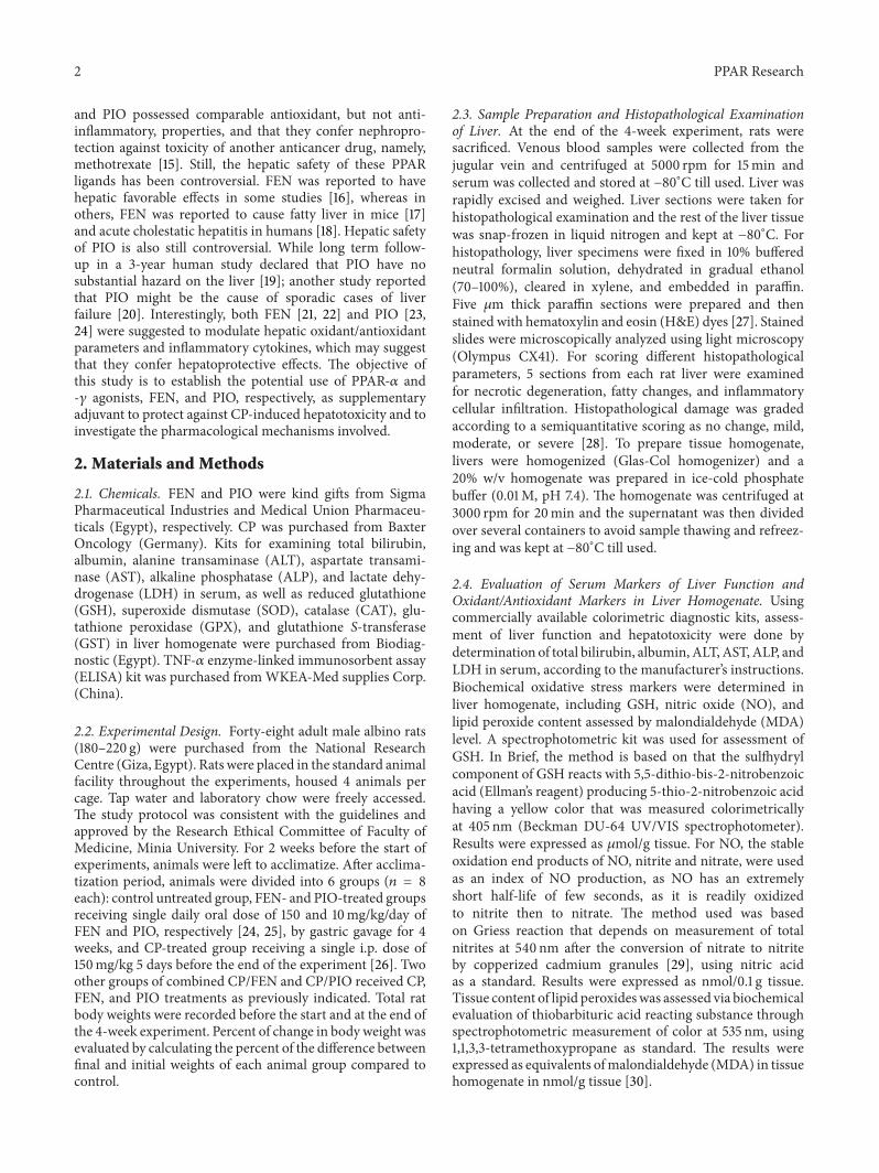
Figure 1: Effect of fenofibrate (FEN) and pioglitazone (PIO) on liver histopathological profile in cyclophosphamide (CP)-treated rats.Representative photomicrographs of liver from: ((a), (b), and (c)) control and FEN and PIO groups, respectively, showing no pathologicalchanges in hepatocytes, (d) CP-treated group presentingwith loss of normal hepatic architecture, congested dilated central vein, inflammatorycellular infiltration, and perivenular hepatocytic necrosis, (e) CP/FEN group showing congested dilated central veins with focal inflammatorycellular infiltration and degenerative necrotic cells, and (f) CP/PIO group demonstrating normal liver histology. Arrowhead: dilated centralvein, black arrow: inflammatory cellular infiltration (×100). The histological changes were scored, and results are expressed in Table 1.
2.5. Determination of Hepatic Antioxidant Enzymatic Activity.Antioxidant enzymatic activity of CAT, SOD, GPX, andGST were determined in hepatic tissue homogenate usingcommercial kits according to the manufacturer instructions.Briefly, hepatic CAT activity was calculated from the rateof decomposition of H
2O2at 510 nm after the addition
of liver homogenate and results were expressed as U/gtissue.The SOD determination assay depended on the abilityof SOD enzyme to inhibit the phenazine methosulphate-mediated reduction of nitroblue tetrazolium dye. The changein absorbance at 560 nm was measured over 5min. SODactivity results were expressed in U/0.1 g tissue. Hepatic GPXand GST activities were evaluated spectrophotometricallyusing reduced glutathione as substrate by addition of liverhomogenate measured at 340 nm. GPX and GST results wereexpressed in U/g tissue and U/mg tissue, respectively.
2.6. Assessment of Proinflammatory Cytokine, TNF-𝛼, inSerum and Liver Homogenate. According to manufacturer’sinstructions, 10 𝜇L of serum or liver homogenate were dis-pensed in 40 𝜇L of sample diluent solution, mixed, andincubated for 30min at 37∘C. After the first incubation, theplate was washed five times with 30-fold diluted wash bufferand then dried. 50𝜇L enzyme conjugate was added to eachwell, incubated, then washed as previously described. Afterdrying the plate, 50 𝜇L of substrate A and 50𝜇L of substrateB were added to each well and the plate was incubated for15min at 37∘C.The reactionwas stopped by adding 50𝜇L stopsolution. The plate was then read using ELISA plate reader at450 nm.
2.7. Statistical Analysis. The data was analyzed by one wayANOVA followed by Dunnett Multiple Comparison Test.The values are represented as means ± S.E.M. All statisticalanalysis was done using GraphPad Prism (GraphPad Prismsoftware, 2011). The differences were considered significantwhen the calculated 𝑃 value is less than 0.05.
3. Results
3.1. Effect of FEN and PIO on Hepatic HistopathologicalFindings inCP-TreatedRat. Liver sections fromcontrol, FEN,and PIO groups (Figures 1(a), 1(b), and 1(c), resp.) showednormal hepatic structure. Single administration of CP wasfollowed by loss of normal hepatic architecture (Figure 1(d)).The central vein was dilated and congested. Sections demon-strated migration of inflammatory cells from the centralvein to infiltrate the perivenular area that showed necrotichepatocytes. FEN/CP group did not show improvement com-pared to CP alone, withmultiple foci of degenerative necroticcells, fatty changes, and inflammatory cellular infiltration.On the other hand, pretreatment with PIO prior to CPchallenge (Figure 1(f)) caused marked improvement in liverhistological picture. Scoring of histological hepatic changesis summarized in Table 1.
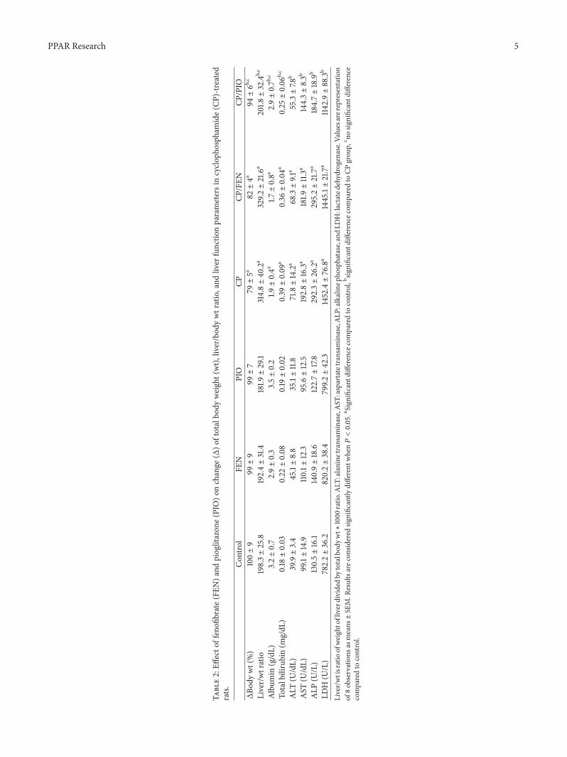
3.2. Effect of FEN and PIO onWeight Changes and Liver Func-tional Parameters in CP-Treated Rat. In CP-treated group,percent of change of body weight was significantly decreased,while liver/total body weight ratio was significantly increasedcompared to control (Table 2). Pretreatment with FEN before
4 PPAR Research
Table 1: Effect of fenofibrate (FEN) and pioglitazone (PIO) onhistological findings in cyclophosphamide (CP)-treated rats liver.
Degenerationand necrosis Fatty changes Inflammatory cell
infiltrationControl − − −
FEN + − −
PIO − − −
CP +++ ++ +++CP/FEN +++ +++ +++CP/PIO + − −
From each animal, 5 sections were examined and scored according to thefollowing criteria: (−) = absent, (+) = mild, (++) = moderate, and (+++) =severe changes.
administration of CP did not improve either parameters,whereas pretreatment with PIO improved both parametersto level not statistically significant from control. Neither FENnor PIO alone affected either parameter. After CP challenge,blood biochemical parameters indicative of liver functiondeteriorated, as evident by significant increase in serumlevels of total bilirubin, ALT, AST, ALP, and LDH, withsignificant decrease in serum albumin (Table 2). CP/FENgroup did not show any improvement in these liver functionalparameters, whereas CP/PIO group demonstrated significantimprovement compared to group treated with CP alone.
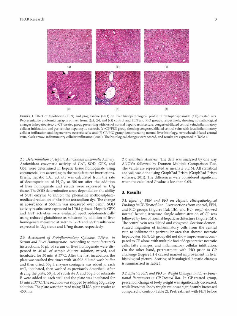
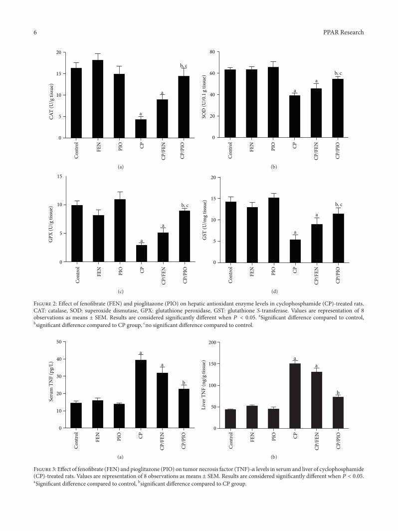
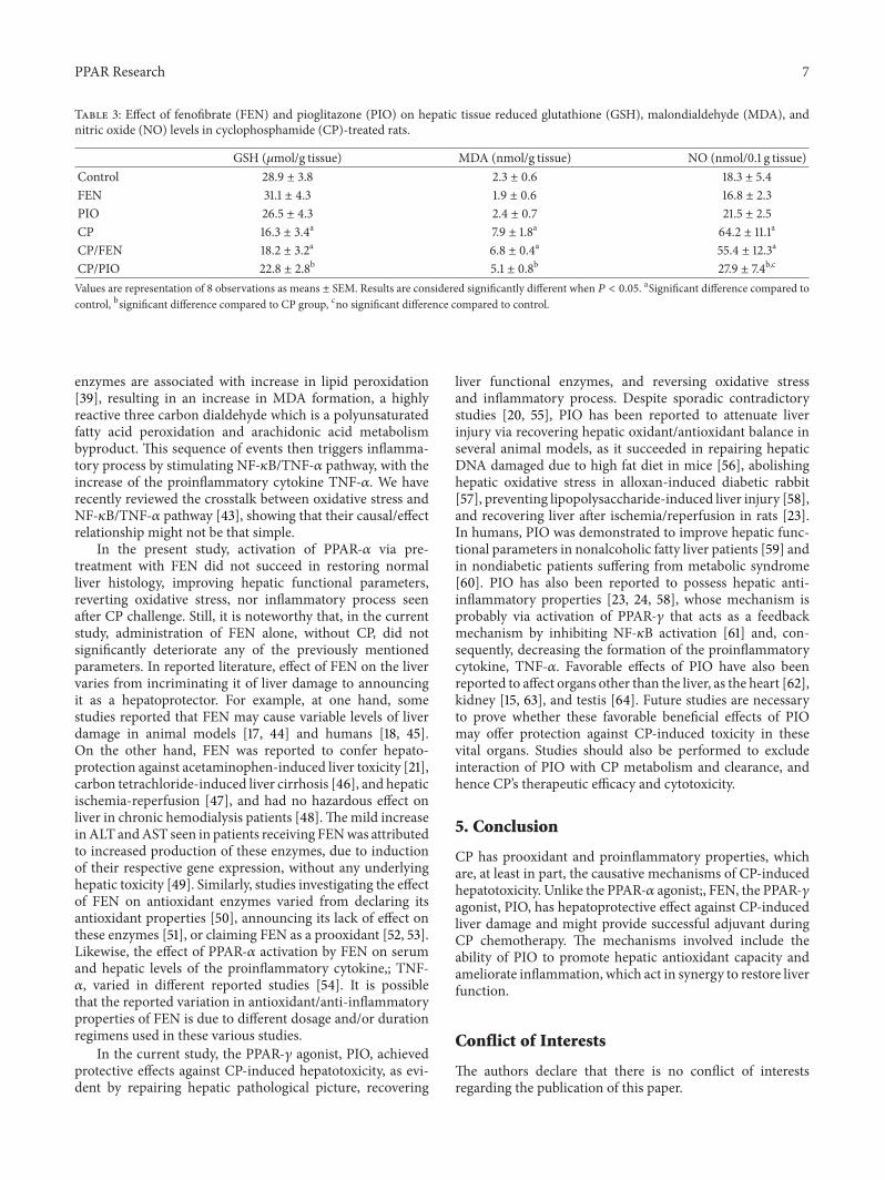
3.3. Effect of FEN and PIO on Oxidation Markers and An-tioxidant Enzymes in CP-Treated Rat Liver. Table 3 depictsthe effect of FEN and PIO, with or without CP challenge,on levels of hepatic GSH, NO, and MDA. CP treatmentsignificantly decreased hepatic GSH compared to controlgroup. FEN administration before CP failed to restore hepaticGSH level, while PIO pretreatment significantly increasedGSH level compared to CP sole treatment. In addition, liverhomogenate of CP group exhibited higher levels of lipidperoxidation as indicated by significantly higher levels ofMDA compared to control. Liver homogenate of CP-treatedgroup also showed significantly higher levels ofNOcomparedto control. Pretreatment with PIO, but not FEN, succeededin reversing MDA and NO to levels statistically significantfrom CP-treated group. As shown in Figure 2, the activityof hepatic antioxidant enzymes CAT, SOD, GPX, and GSTwere significantly less in CP group compared to control. Onlypretreatment with PIO, but not FEN, significantly increasethese enzymatic activities, compared to CP alone.
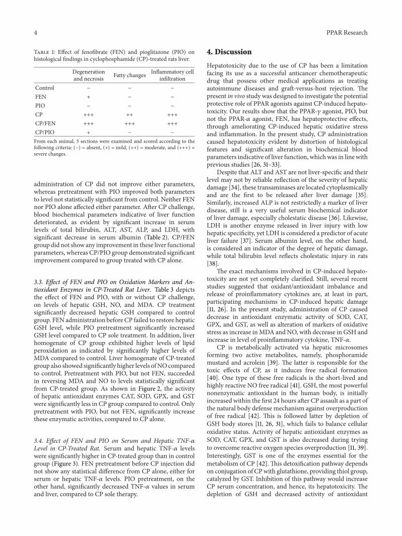
3.4. Effect of FEN and PIO on Serum and Hepatic TNF-𝛼Level in CP-Treated Rat. Serum and hepatic TNF-𝛼 levelswere significantly higher in CP-treated group than in controlgroup (Figure 3). FEN pretreatment before CP injection didnot show any statistical difference from CP alone, either forserum or hepatic TNF-𝛼 levels. PIO pretreatment, on theother hand, significantly decreased TNF-𝛼 values in serumand liver, compared to CP sole therapy.
4. Discussion
Hepatotoxicity due to the use of CP has been a limitationfacing its use as a successful anticancer chemotherapeuticdrug that possess other medical applications as treatingautoimmune diseases and graft-versus-host rejection. Thepresent in vivo study was designed to investigate the potentialprotective role of PPAR agonists against CP-induced hepato-toxicity. Our results show that the PPAR-𝛾 agonist, PIO, butnot the PPAR-𝛼 agonist, FEN, has hepatoprotective effects,through ameliorating CP-induced hepatic oxidative stressand inflammation. In the present study, CP administrationcaused hepatotoxicity evident by distortion of histologicalfeatures and significant alteration in biochemical bloodparameters indicative of liver function, whichwas in linewithprevious studies [26, 31–33].
Despite that ALT and AST are not liver-specific and theirlevel may not by reliable reflection of the severity of hepaticdamage [34], these transaminases are located cytoplasmicallyand are the first to be released after liver damage [35].Similarly, increased ALP is not restrictedly a marker of liverdisease, still is a very useful serum biochemical indicatorof liver damage, especially cholestatic disease [36]. Likewise,LDH is another enzyme released in liver injury with lowhepatic specificity, yet LDH is considered a predictor of acuteliver failure [37]. Serum albumin level, on the other hand,is considered an indicator of the degree of hepatic damage,while total bilirubin level reflects cholestatic injury in rats[38].
The exact mechanisms involved in CP-induced hepato-toxicity are not yet completely clarified. Still, several recentstudies suggested that oxidant/antioxidant imbalance andrelease of proinflammatory cytokines are, at least in part,participating mechanisms in CP-induced hepatic damage[11, 26]. In the present study, administration of CP causeddecrease in antioxidant enzymatic activity of SOD, CAT,GPX, and GST, as well as alteration of markers of oxidativestress as increase inMDA and NO, with decrease in GSH andincrease in level of proinflammatory cytokine, TNF-𝛼.
CP is metabolically activated via hepatic microsomesforming two active metabolites, namely, phosphoramidemustard and acrolein [39]. The latter is responsible for thetoxic effects of CP, as it induces free radical formation[40]. One type of these free radicals is the short-lived andhighly reactive NO free radical [41]. GSH, the most powerfulnonenzymatic antioxidant in the human body, is initiallyincreased within the first 24 hours after CP assault as a part ofthe natural body defense mechanism against overproductionof free radical [42]. This is followed latter by depletion ofGSH body stores [11, 26, 31], which fails to balance cellularoxidative status. Activity of hepatic antioxidant enzymes asSOD, CAT, GPX, and GST is also decreased during tryingto overcome reactive oxygen species overproduction [11, 39].Interestingly, GST is one of the enzymes essential for themetabolism of CP [42]. This detoxification pathway dependson conjugation of CPwith glutathione, providing thiol group,catalyzed by GST. Inhibition of this pathway would increaseCP serum concentration, and hence, its hepatotoxicity. Thedepletion of GSH and decreased activity of antioxidant
PPAR Research 5
Table2:Eff
ecto
ffenofi
brate(FEN
)and
pioglitazon
e(PIO
)onchange
(Δ)o
ftotalbo
dyweight(wt),
liver/bod
ywtratio,and
liver
functio
nparametersincyclo
phosph
amide(C
P)-tr
eated
rats.
Con
trol
FEN
PIO
CPCP
/FEN
CP/PIO
ΔBo
dywt(%)
100±9
99±9
99±7
79±5a
82±4a
94±6b
,c
Liver/wtratio
198.3±25.8
192.4±31.4
181.9±29.1
314.8±40
.2a
329.2±21.6
a201.8±32.4
b,c
Album
in(g/dL)
3.2±0.7
2.9±0.3
3.5±0.2
1.9±0.4a
1.7±0.8a
2.9±0.7b
,c
Totalbilirubin(m
g/dL
)0.18±0.03
0.22±0.08
0.19±0.02
0.39±0.09
a0.36±0.04
a0.25±0.06
b,c
ALT
(U/dL)
39.9±3.4
45.1±8.8
35.1±11.8
71.8±14.2
a68.3±9.1
a55.3±7.8
b
AST
(U/dL)
99.1±14.9
110.1±
12.3
95.6±12.5
192.8±16.3
a181.9±11.3
a144.3±8.3b
ALP
(U/L)
130.5±16.1
140.9±18.6
122.7±17.8
292.3±26.2
a295.2±21.7
a184.7±18.9
b
LDH(U
/L)
782.2±36.2
820.2±38.4
799.2±42.3
1452.4±76.8
a1445.1±21.7
a114
2.9±88.3
b
Liver/wtisratioofweightofliverdividedby
totalbod
ywt∗
1000
ratio
.ALT
:alanine
transaminase,AST
:aspartatetransaminase,ALP
:alkalinep
hosphatase,and
LDH:la
ctated
ehydrogenase.Valuesarerepresentatio
nof
8ob
servations
asmeans±SE
M.R
esultsarec
onsid
ered
significantly
different
when𝑃<0.05.aSign
ificant
differencec
omparedto
control,b significantd
ifference
comparedto
CPgrou
p,c nosig
nificantd
ifference
comparedto
control.
6 PPAR Research
Con
trol
FEN
PIO CP
CP/F
EN
CP/P
IO
0
5
10
15
20CA
T (U
/g ti
ssue
)
a
a
b, c
(a)
a
ab, c
0
20
40
60
80
Con
trol
FEN
PIO CP
CP/F
EN
CP/P
IO
SOD
(U/0.1
g tis
sue)
(b)
a
a
b, c
0
5
10
15
Con
trol
FEN
PIO CP
CP/F
EN
CP/P
IO
GPX
(U/g
tiss
ue)
(c)
a
a
b, c
Con
trol
FEN
PIO CP
CP/F
EN
CP/P
IO
0
5
10
15
20
GST
(U/m
g tis
sue)
(d)
Figure 2: Effect of fenofibrate (FEN) and pioglitazone (PIO) on hepatic antioxidant enzyme levels in cyclophosphamide (CP)-treated rats.CAT: catalase, SOD: superoxide dismutase, GPX: glutathione peroxidase, GST: glutathione S-transferase. Values are representation of 8observations as means ± SEM. Results are considered significantly different when 𝑃 < 0.05. aSignificant difference compared to control,bsignificant difference compared to CP group, cno significant difference compared to control.
0
10
20
30
40
50
a
a
b
Con
trol
FEN
PIO CP
CP/F
EN
CP/P
IO
Seru
m T
NF
(pg/
L)
(a)
0
50
100
150
200
aa
b
Con
trol
FEN
PIO CP
CP/F
EN
CP/P
IO
Live
r TN
F (n
g/g·
tissu
e)
(b)
Figure 3: Effect of fenofibrate (FEN) and pioglitazone (PIO) on tumor necrosis factor (TNF)-𝛼 levels in serum and liver of cyclophosphamide(CP)-treated rats. Values are representation of 8 observations as means ± SEM. Results are considered significantly different when 𝑃 < 0.05.aSignificant difference compared to control, bsignificant difference compared to CP group.
PPAR Research 7
Table 3: Effect of fenofibrate (FEN) and pioglitazone (PIO) on hepatic tissue reduced glutathione (GSH), malondialdehyde (MDA), andnitric oxide (NO) levels in cyclophosphamide (CP)-treated rats.
GSH (𝜇mol/g tissue) MDA (nmol/g tissue) NO (nmol/0.1 g tissue)Control 28.9 ± 3.8 2.3 ± 0.6 18.3 ± 5.4FEN 31.1 ± 4.3 1.9 ± 0.6 16.8 ± 2.3PIO 26.5 ± 4.3 2.4 ± 0.7 21.5 ± 2.5CP 16.3 ± 3.4a 7.9 ± 1.8a 64.2 ± 11.1a
CP/FEN 18.2 ± 3.2a 6.8 ± 0.4a 55.4 ± 12.3a
CP/PIO 22.8 ± 2.8b 5.1 ± 0.8b 27.9 ± 7.4b,c
Values are representation of 8 observations as means ± SEM. Results are considered significantly different when 𝑃 < 0.05. aSignificant difference compared tocontrol, bsignificant difference compared to CP group, cno significant difference compared to control.
enzymes are associated with increase in lipid peroxidation[39], resulting in an increase in MDA formation, a highlyreactive three carbon dialdehyde which is a polyunsaturatedfatty acid peroxidation and arachidonic acid metabolismbyproduct. This sequence of events then triggers inflamma-tory process by stimulating NF-𝜅B/TNF-𝛼 pathway, with theincrease of the proinflammatory cytokine TNF-𝛼. We haverecently reviewed the crosstalk between oxidative stress andNF-𝜅B/TNF-𝛼 pathway [43], showing that their causal/effectrelationship might not be that simple.
In the present study, activation of PPAR-𝛼 via pre-treatment with FEN did not succeed in restoring normalliver histology, improving hepatic functional parameters,reverting oxidative stress, nor inflammatory process seenafter CP challenge. Still, it is noteworthy that, in the currentstudy, administration of FEN alone, without CP, did notsignificantly deteriorate any of the previously mentionedparameters. In reported literature, effect of FEN on the livervaries from incriminating it of liver damage to announcingit as a hepatoprotector. For example, at one hand, somestudies reported that FEN may cause variable levels of liverdamage in animal models [17, 44] and humans [18, 45].On the other hand, FEN was reported to confer hepato-protection against acetaminophen-induced liver toxicity [21],carbon tetrachloride-induced liver cirrhosis [46], and hepaticischemia-reperfusion [47], and had no hazardous effect onliver in chronic hemodialysis patients [48].Themild increaseinALT andAST seen in patients receiving FENwas attributedto increased production of these enzymes, due to inductionof their respective gene expression, without any underlyinghepatic toxicity [49]. Similarly, studies investigating the effectof FEN on antioxidant enzymes varied from declaring itsantioxidant properties [50], announcing its lack of effect onthese enzymes [51], or claiming FEN as a prooxidant [52, 53].Likewise, the effect of PPAR-𝛼 activation by FEN on serumand hepatic levels of the proinflammatory cytokine,; TNF-𝛼, varied in different reported studies [54]. It is possiblethat the reported variation in antioxidant/anti-inflammatoryproperties of FEN is due to different dosage and/or durationregimens used in these various studies.
In the current study, the PPAR-𝛾 agonist, PIO, achievedprotective effects against CP-induced hepatotoxicity, as evi-dent by repairing hepatic pathological picture, recovering
liver functional enzymes, and reversing oxidative stressand inflammatory process. Despite sporadic contradictorystudies [20, 55], PIO has been reported to attenuate liverinjury via recovering hepatic oxidant/antioxidant balance inseveral animal models, as it succeeded in repairing hepaticDNA damaged due to high fat diet in mice [56], abolishinghepatic oxidative stress in alloxan-induced diabetic rabbit[57], preventing lipopolysaccharide-induced liver injury [58],and recovering liver after ischemia/reperfusion in rats [23].In humans, PIO was demonstrated to improve hepatic func-tional parameters in nonalcoholic fatty liver patients [59] andin nondiabetic patients suffering from metabolic syndrome[60]. PIO has also been reported to possess hepatic anti-inflammatory properties [23, 24, 58], whose mechanism isprobably via activation of PPAR-𝛾 that acts as a feedbackmechanism by inhibiting NF-𝜅B activation [61] and, con-sequently, decreasing the formation of the proinflammatorycytokine, TNF-𝛼. Favorable effects of PIO have also beenreported to affect organs other than the liver, as the heart [62],kidney [15, 63], and testis [64]. Future studies are necessaryto prove whether these favorable beneficial effects of PIOmay offer protection against CP-induced toxicity in thesevital organs. Studies should also be performed to excludeinteraction of PIO with CP metabolism and clearance, andhence CP’s therapeutic efficacy and cytotoxicity.
5. Conclusion
CP has prooxidant and proinflammatory properties, whichare, at least in part, the causative mechanisms of CP-inducedhepatotoxicity. Unlike the PPAR-𝛼 agonist;, FEN, the PPAR-𝛾agonist, PIO, has hepatoprotective effect against CP-inducedliver damage and might provide successful adjuvant duringCP chemotherapy. The mechanisms involved include theability of PIO to promote hepatic antioxidant capacity andameliorate inflammation, which act in synergy to restore liverfunction.
Conflict of Interests
The authors declare that there is no conflict of interestsregarding the publication of this paper.
8 PPAR Research
References
[1] A. Emadi, R. J. Jones, and R. A. Brodsky, “Cyclophosphamideand cancer: golden anniversary,”Nature Reviews Clinical Oncol-ogy, vol. 6, no. 11, pp. 638–647, 2009.
[2] M. Haubitz, “Acute and long-term toxicity of cyclophos-phamide,” Transplantationsmedizin, vol. 19, supplement 2, pp.S26–S31, 2007.
[3] A. H. Viswanatha Swamy, U. M. Patel, B. C. Koti, P. C. Gadad,N. L. Patel, and A. H.Thippeswamy, “Cardioprotective effect ofSaraca indica against cyclophosphamide induced cardiotoxicityin rats: a biochemical, electrocardiographic and histopatholog-ical study,” Indian Journal of Pharmacology, vol. 45, no. 1, pp.44–48, 2013.
[4] W. Kim, S. H. Kim, S. K. Park, and M. S. Chang, “Astragalusmembranaceus ameliorates reproductive toxicity induced bycyclophosphamide in male mice,” Phytotherapy Research, vol.26, no. 9, pp. 1418–1421, 2012.
[5] N. E. Aikawa, A. M. Sallum, R. M. Pereira et al., “Subclinicalimpairment of ovarian reserve in juvenile systemic lupuserythematosus after cyclophosphamide therapy,” Clinical andExperimental Rheumatology, vol. 30, no. 3, pp. 445–449, 2012.
[6] J. W. Goldberg and M. D. Lidsky, “Cyclophosphamide-associated hepatotoxicity,” SouthernMedical Journal, vol. 78, no.2, pp. 222–223, 1985.
[7] M. E. de Jonge, A. D. R. Huitema, J. H. Beijnen, and S. Roden-huis, “High exposures to bioactivated cyclophosphamide arerelated to the occurrence of veno-occlusive disease of the liverfollowing high-dose chemotherapy,” British Journal of Cancer,vol. 94, no. 9, pp. 1226–1230, 2006.
[8] H.Akay, T. Akay, S. Secilmis, Z. Kocak, and O. Donderici, “Hep-atotoxicity after low-dose cyclophosphamide therapy,” SouthernMedical Journal, vol. 99, no. 12, pp. 1399–1400, 2006.
[9] M. Martınez-Gabarron, R. Enrıquez, A. E. Sirvent, M. Garcıa-Sepulcre, I. Millan, and F. Amoros, “Hepatotoxity aftercyclophosphamide treatment in a patient with MPO-ANCAvasculitis,” Nefrologia, vol. 31, no. 4, pp. 496–498, 2011.
[10] A. K. Souid, K. A. Tacka, K. A. Galvan, and H. S. Penefsky,“Immediate effects of anticancer drugs on mitochondrial oxy-gen consumption,”Biochemical Pharmacology, vol. 66, no. 6, pp.977–987, 2003.
[11] M. Zarei and T. Shivanandappa, “Amelioration of cyclo-phosphamide-induced hepatotoxicity by the root extract ofDecalepis hamiltonii in mice,” Food and Chemical Toxicology,vol. 57, pp. 179–184, 2013.
[12] T. P. Hamsa and G. Kuttan, “Ipomoea obscura ameliorates cy-clophosphamide induced toxicity by modulating the immunesystem and levels of proinflammatory cytokine and GSH,”Canadian Journal of Physiology and Pharmacology, vol. 88, no.11, pp. 1042–1053, 2010.
[13] K. McKeage, G. M. Keating, D. Bhatnagar, M. B. Elam, M.Farnier, and S. M. Mohiuddin, “Fenofibrate: a review of its usein dyslipidaemia,” Drugs, vol. 71, no. 14, pp. 1917–1946, 2011.
[14] P. de Pablos-Velasco, “Pioglitazone: beyond glucose control,”Expert Review of CardiovascularTherapy, vol. 8, no. 8, pp. 1057–1067, 2010.
[15] M. Ibrahim, A. El-Sheikh, H. Khalaf, and A. Abdelrahman,“Protective effect of peroxisome proliferator activator receptor(PPAR)-𝛼 and -𝛾 ligands against methotrexate-induced neph-rotoxicity,” Immunopharmacology and Immunotoxicology, 2014.
[16] X. Shi, D. Yao, B. A. Gosnell, and C. Chen, “Lipidomic profilingreveals protective function of fatty acid oxidation in cocaine-induced hepatotoxicity,” Journal of Lipid Research, vol. 53, no.11, pp. 2318–2330, 2012.
[17] S. Y. Pan, Q. Yu, Y. Zhang et al., “Dietary Fructus Schisandraeextracts and fenofibrate regulate the serum/hepatic lipid-profilein normal and hypercholesterolemic mice, with attention tohepatotoxicity,” Lipids in Health and Disease, vol. 11, article 120,2012.
[18] D. Hajdu, K. Aiglova, I. Vinklerova, and K. Urbanek, “Acutecholestatic hepatitis induced by fenofibrate,” Journal of ClinicalPharmacy andTherapeutics, vol. 34, no. 5, pp. 599–602, 2009.
[19] K. G. Tolman, J. W. Freston, S. Kupfer, and P. Alfonso, “Liversafety in patients with type 2 diabetes treated with pioglitazone:Results from a 3-year, randomized, comparator-controlledstudy in the US,” Drug Safety, vol. 32, no. 9, pp. 787–800, 2009.
[20] J. S. Floyd, E. Barbehenn, P. Lurie, and S. M. Wolfe, “Case seriesof liver failure associated with rosiglitazone and pioglitazone,”Pharmacoepidemiology andDrug Safety, vol. 18, no. 12, pp. 1238–1243, 2009.
[21] A. D. Patterson, Y. M. Shah, T. Matsubara, K. W. Krausz, andF. J. Gonzalez, “Peroxisome proliferator-activated receptoralpha induction of uncoupling protein 2 protects againstacetaminophen-induced liver toxicity,” Hepatology, vol. 56, no.1, pp. 281–290, 2012.
[22] T. Karakan, M. Kerem, M. Cindoruk, D. Engin, M. Alper, andO. Akin, “PPAR-alpha agonist treatment increases trefoil factorfamily-3 expression and attenuates apoptosis in the liver tissueof bile duct-ligated rats,” The Turkish Journal of Gastroenterol-ogy, vol. 24, no. 2, pp. 134–140, 2013.
[23] M. H. Somi, B. Hajipour, N. A. Asl et al., “Pioglitazone atten-uates ischemia/reperfusion-induced liver injury in rats,” Trans-plantation Proceedings, vol. 41, no. 10, pp. 4105–4109, 2009.
[24] J. S. Zhao, F. S. Zhu, S. Liu, C. Q. Yang, and X. M. Chen, “Piogli-tazone ameliorates nonalcoholic steatohepatitis by down-regulating hepatic nuclear factor-kappa B and cyclooxygenases-2 expression in rats,”ChineseMedical Journal, vol. 125, no. 13, pp.2316–2321, 2012.
[25] M. Olukman, E. D. Sezer, S. Ulker, E. Y. Szmen, and G. M.Cnar, “Fenofibrate treatment enhances antioxidant status andattenuates endothelial dysfunction in streptozotocin-induceddiabetic rats,” Experimental Diabetes Research, vol. 2010, ArticleID 828531, 10 pages, 2010.
[26] S. Jnaneshwari, M. Hemshekhar, M. S. Santhosh et al., “Crocin,a dietary colorant mitigates cyclophosphamide-induced organtoxicity by modulating antioxidant status and inflammatorycytokines,”The Journal of Pharmacy and Pharmacology, vol. 65,no. 4, pp. 604–614, 2013.
[27] J.D. Bancroft andM.Gamble,Theory andPractice ofHistologicalTechniques, Churchill Livingstone, New York, NY, USA, 5thedition, 2008.
[28] M. Ozsoy, Y. Ozsoy, T. Coskun, K. Naml, A. Var, and B. Ozyurt,“The effects of l-arginine on liver damage in experimental acutecholestasis an immunohistochemical study,” HPB Surgery, vol.2011, Article ID 306069, 5 pages, 2011.
[29] K. V. Sastry, R. P. Moudgal, J. Mohan, J. S. Tyagi, and G. S. Rao,“Spectrophotometric determination of serumnitrite and nitrateby copper-cadmiumalloy,”Analytical Biochemistry, vol. 306, no.1, pp. 79–82, 2002.
[30] J. A. Buege and S. D. Aust, “Microsomal lipid peroxidation,”Methods in Enzymology, vol. 52, pp. 302–310, 1978.
PPAR Research 9
[31] E. Selvakumar, C. Prahalathan, Y. Mythili, and P. Varalakshmi,“Mitigation of oxidative stress in cyclophosphamide-challengedhepatic tissue by DL-𝛼-lipoic acid,” Molecular and CellularBiochemistry, vol. 272, no. 1-2, pp. 179–185, 2005.
[32] S. S. Dadarkar, L. C. Fonseca, P. B. Mishra et al., “Phenotypicand genotypic assessment of concomitant drug-induced toxiceffects in liver, kidney and blood,” Journal of Applied Toxicology,vol. 31, no. 2, pp. 117–130, 2011.
[33] E. L. Lushnikova, O. P. Molodykh, L. M. Nepomnyashchikh,A. A. Bakulina, and Y. A. Sorokina, “Ultrastructurural pictureof cyclophosphamide-induced damage to the liver,” Bulletin ofExperimental Biology and Medicine, vol. 151, no. 6, pp. 751–756,2011.
[34] M. Lazo, E. Selvin, and J. M. Clark, “Brief communication:clinical implications of short-term variability in liver functiontest results,”Annals of InternalMedicine, vol. 148, no. 5, pp. 348–352, 2008.
[35] D. H. Vroon and Z. Israili, “Aminotransferases,” in ClinicalMethods: The History, Physical, and Laboratory Examinations,H. K. Walker, W. D. Hall, and J. W. Hurst, Eds., chapter 99,Butterworths, Boston, Mass, USA, 3rd edition, 1990.
[36] N. J. Fernandez and B. A. Kidney, “Alkaline phosphatase:beyond the liver,” Veterinary Clinical Pathology, vol. 36, no. 3,pp. 223–233, 2007.
[37] K. Kotoh, M. Enjoji, M. Kato, M. Kohjima, M. Nakamuta,and R. Takayanagi, “A new parameter using serum lactatedehydrogenase and alanine aminotransferase level is useful forpredicting the prognosis of patients at an early stage of acuteliver injury: A retrospective study,” Comparative Hepatology,vol. 7, article 6, 2008.
[38] Y. Mano, H. Tsukada, T. Kurihara, M. Nomura, K. Yokogawa,and K.-I. Miyamoto, “Development of dosage design of hepaticmetabolizing drugs using serum albumin level in chronichepatic failure,” Biological and Pharmaceutical Bulletin, vol. 29,no. 8, pp. 1692–1699, 2006.
[39] K. Jyothi, A. G. Reddy, K. S. Gopi, B. A. Kumar, and G. D.Reddy, “A study on free radical-induced renal toxicity due tocyclophosphamide and its amelioration withN-acetyl cysteine,”Toxicology International, vol. 16, no. 2, pp. 137–139, 2009.
[40] F. Liu, X. L. Li, T. Lin et al., “The cyclophosphamide metabolite,acrolein, induces cytoskeletal changes and oxidative stress inSertoli cells,” Molecular Biology Reports, vol. 39, no. 1, pp. 493–500, 2012.
[41] C. C. Wang, T. I. Weng, E. T. Wu, M. H. Wu, R. S. Yang, andS. H. Liu SH, “Involvement of interleukin-6-regulated nitricoxide synthase in hemorrhagic cystitis and impaired bladdercontractions in young rats induced by acrolein, a urinarymetabolite of cyclophosphamide,” Toxicological Sciences, vol.131, no. 1, pp. 302–310, 2013.
[42] P. Abraham and E. Sugumar, “Increased glutathione levels andactivity of PON1 (phenyl acetate esterase) in the liver of ratsafter a single dose of cyclophosphamide: a defensemechanism?”Experimental and Toxicologic Pathology, vol. 59, no. 5, pp. 301–306, 2008.
[43] A. Taye and A. A. El-Sheikh, “Lectin-like oxidized low-densitylipoprotein receptor 1 pathways,” European Journal of ClinicalInvestigation, vol. 43, no. 7, pp. 740–745, 2013.
[44] T. Moustafa, P. Fickert, C. Magnes et al., “Alterations in lipidmetabolism mediate inflammation, fibrosis, and proliferationin a mouse model of chronic cholestatic liver injury,” Gastroen-terology, vol. 142, no. 1, pp. 140–151, 2012.
[45] K. Dohmen, C. Y. Wen, S. Nagaoka et al., “Fenofibrate-inducedliver injury,” World Journal of Gastroenterology, vol. 11, no. 48,pp. 7702–7703, 2005.
[46] A. Rodrıguez-Vilarrupla, B. Lavina, H. Garcıa-Caldero et al.,“PPARalpha activation improves endothelial dysfunction andreduces fibrosis and portal pressure in cirrhotic rats,” Journal ofHepatology, vol. 56, no. 5, pp. 1033–1039, 2012.
[47] V. Boshra and A.M.Moustafa, “Effect of preischemic treatmentwith fenofibrate, a peroxisome proliferator-activated receptor-𝛼ligand, on hepatic ischemia-reperfusion injury in rats,” Journalof Molecular Histology, vol. 42, no. 2, pp. 113–122, 2011.
[48] A. Makowka, P. Dryja, G. Chwatko, E. Bald, and M. Nowicki,“Treatment of chronic hemodialysis patients with low-dosefenofibrate effectively reduces plasma lipids and affects plasmaredox status,” Lipids in Health and Disease, vol. 11, article 47,2012.
[49] A. Kobayashi, Y. Suzuki, H. Kuno, S. Sugai, H. Sakakibara,and K. Shimoi, “Effects of fenofibrate on plasma and hepatictransaminase activities and hepatic transaminase gene expres-sion in rats,” Journal of Toxicological Sciences, vol. 34, no. 4, pp.377–387, 2009.
[50] M. Broncel, D. Cieslak, M. Koter-Michalak, P. Duchnow-icz, K. Mackiewicz, and J. Chojnowska-Jezierska, “The anti-inflammatory and antioxidants effects ofmicronized fenofibratein patients with visceral obesity and dyslipidemia,” PolskiMerkuriusz Lekarski, vol. 20, no. 119, pp. 547–550, 2006.
[51] J. Kowalski, J. Olejniczak, J. Błaszczyk et al., “Effect of fluvastatinand fenofibrate on the antioxidative barrier enzyme activity inpatients with dyslipidemia,” Polski Merkuriusz Lekarski, vol. 14,no. 82, pp. 285–288, 2003.
[52] J. Kowalski, J. Błaszczyk, J. Olejniczak et al., “Effect of fluvastatinand fenofibrate on reactive oxygen species generation and lipidperoxidation in patients with dyslipidaemia,” Polski MerkuriuszLekarski, vol. 14, no. 82, pp. 279–284, 2003.
[53] J. Nishimura, Y. Dewa, T. Okamura et al., “Possible involvementof oxidative stress in fenofibrate-induced hepatocarcinogenesisin rats,” Archives of Toxicology, vol. 82, no. 9, pp. 641–654, 2008.
[54] A. Zambon, P. Gervois, P. Pauletto, J. C. Fruchart, and B.Staels, “Modulation of hepatic inflammatory risk markers ofcardiovascular diseases by PPAR-𝛼 activators: clinical andexperimental evidence,” Arteriosclerosis, Thrombosis, and Vas-cular Biology, vol. 26, no. 5, pp. 977–986, 2006.
[55] A. Bedir, Y. Aliyazicioglu, B. Bilgici et al., “Assessment of geno-toxicity in rats treated with the antidiabetic agent, pioglitazone,”Environmental and Molecular Mutagenesis, vol. 49, no. 3, pp.185–191, 2008.
[56] P. J. Hsiao, T. J. Hsieh, K. K. Kuo et al., “Pioglitazone retrieveshepatic antioxidant DNA repair in a mice model of high fatdiet,” BMCMolecular Biology, vol. 9, article 82, 2008.
[57] A. Gumieniczek, “Effect of the new thiazolidinedione-pioglitazone on the development of oxidative stress in liverand kidney of diabetic rabbits,” Life Sciences, vol. 74, no. 5, pp.553–562, 2003.
[58] N. Enomoto, Y. Takei, S. Yamashima, K. Ikejima, T. Kita-mura, and N. Sato, “Protective effect of pioglitazone againstendotoxin-induced liver injury through prevention of Kupf-fer cell sensitization,” Alcoholism: Clinical and ExperimentalResearch, vol. 29, supplement 3, pp. 216S–219S, 2005.
[59] M. Razavizade, R. Jamali, A. Arj, S. M. Matini, A. Moraveji, andE. Taherkhani, “The effect of pioglitazone and metformin onliver function tests, insulin resistance, and liver fat content in
10 PPAR Research
nonalcoholic Fatty liver disease: a randomized double blindedclinical trial,”HepatitisMonthly, vol. 13, no. 5, article e9270, 2013.
[60] P. Shokouh, A. Joharimoghadam, H. Roohafza et al., “Effectsof pioglitazone on asymmetric dimethylarginine and com-ponents of the metabolic syndrome in nondiabetic patients(EPICAMP study): a double-blind, randomized clinical trial,”PPAR Research, vol. 2013, Article ID 358074, 9 pages, 2013.
[61] Y. Wang, Q. W. Lin, P. P. Zheng, J. S. Zhang, and F. R. Huang,“DHA inhibits protein degradation more efficiently than EPAby regulating the PPARgamma/NFkappaB pathway in C2C12myotubes,” BioMed Research International, vol. 2013, Article ID318981, 2013.
[62] T. Chen, X. Jin, B. H. Crawford et al., “Cardioprotectionfrom oxidative stress in the newborn heart by activation ofPPARgamma is mediated by catalase,” Free Radical Biology andMedicine, vol. 53, no. 2, pp. 208–215, 2012.
[63] C. Zou, H. Hu, X. Xi, Z. Shi, G. Wang, and X. Huang, “Piogli-tazone protects against renal ischemia-reperfusion injuryby enhancing antioxidant capacity,” The Journal of SurgicalResearch, vol. 184, no. 2, pp. 1092–1095, 2013.
[64] A. Gumieniczek, H.Hopkała, andA. Zbek, “Protective effects ofa PPAR𝛾 agonist pioglitazone on anti-oxidative system in testisof diabetic rabbits,” Pharmazie, vol. 63, no. 5, pp. 377–378, 2008.

Submit your manuscripts athttp://www.hindawi.com
Stem CellsInternational
Hindawi Publishing Corporationhttp://www.hindawi.com Volume 2014
Hindawi Publishing Corporationhttp://www.hindawi.com Volume 2014
MEDIATORSINFLAMMATION
of
Hindawi Publishing Corporationhttp://www.hindawi.com Volume 2014
Behavioural Neurology
EndocrinologyInternational Journal of
Hindawi Publishing Corporationhttp://www.hindawi.com Volume 2014
Hindawi Publishing Corporationhttp://www.hindawi.com Volume 2014
Disease Markers
Hindawi Publishing Corporationhttp://www.hindawi.com Volume 2014
BioMed Research International
OncologyJournal of
Hindawi Publishing Corporationhttp://www.hindawi.com Volume 2014
Hindawi Publishing Corporationhttp://www.hindawi.com Volume 2014
Oxidative Medicine and Cellular Longevity
Hindawi Publishing Corporationhttp://www.hindawi.com Volume 2014
PPAR Research
The Scientific World JournalHindawi Publishing Corporation http://www.hindawi.com Volume 2014
Immunology ResearchHindawi Publishing Corporationhttp://www.hindawi.com Volume 2014
Journal of
ObesityJournal of
Hindawi Publishing Corporationhttp://www.hindawi.com Volume 2014
Hindawi Publishing Corporationhttp://www.hindawi.com Volume 2014
Computational and Mathematical Methods in Medicine
OphthalmologyJournal of
Hindawi Publishing Corporationhttp://www.hindawi.com Volume 2014
Diabetes ResearchJournal of
Hindawi Publishing Corporationhttp://www.hindawi.com Volume 2014
Hindawi Publishing Corporationhttp://www.hindawi.com Volume 2014
Research and TreatmentAIDS
Hindawi Publishing Corporationhttp://www.hindawi.com Volume 2014
Gastroenterology Research and Practice
Hindawi Publishing Corporationhttp://www.hindawi.com Volume 2014
Parkinson’s Disease
Evidence-Based Complementary and Alternative Medicine
Volume 2014Hindawi Publishing Corporationhttp://www.hindawi.com