research article simulation-based mastery learning...
TRANSCRIPT

Research ArticleSimulation-Based Mastery Learning with Deliberate PracticeImproves Clinical Performance in Spinal Anesthesia
Ankeet D. Udani,1 Alex Macario,1,2 Kiruthiga Nandagopal,3
Maria A. Tanaka,1 and Pedro P. Tanaka1
1 Department of Anesthesiology, Perioperative and Pain Medicine, 300 Pasteur Drive, Room H3580, Stanford University,Stanford, CA 94305-5640, USA
2Department of Anesthesiology, Perioperative and Pain Medicine, and Department of Health Research and Policy,Stanford University, Stanford, CA, USA
3 Stanford Center for Medical Education Research and Innovation, Stanford University, Stanford, CA, USA
Correspondence should be addressed to Ankeet D. Udani; [email protected]
Received 25 January 2014; Accepted 27 April 2014; Published 16 July 2014
Academic Editor: Jean Jacques Lehot
Copyright © 2014 Ankeet D. Udani et al. This is an open access article distributed under the Creative Commons AttributionLicense, which permits unrestricted use, distribution, and reproduction in any medium, provided the original work is properlycited.
Introduction. Properly performing a subarachnoid block (SAB) is a competency expected of anesthesiology residents. We aimedto determine if adding simulation-based deliberate practice to a base curriculum improved performance of a SAB. Methods. 21anesthesia residentswere enrolled. After baseline assessment of SABon a task-trainer, all residents participated in a base curriculum.Residents were then randomized so that half received additional deliberate practice including repetition and expert-guided, real-time feedback. All residents were then retested for technique. SABs on all residents’ next three patients were evaluated in theoperating room (OR). Results. Before completing the base curriculum, the control group completed 81% of a 16-item performancechecklist on the task-trainer and this increased to 91% after finishing the base curriculum (𝑃 < 0.02). The intervention group alsoincreased the percentage of checklist tasks properly completed from 73% to 98%, which was a greater increase than observedin the control group (𝑃 < 0.03). The OR time required to perform SAB was not different between groups. Conclusions. Thebase curriculum significantly improved resident SAB performance. Deliberate practice training added a significant, independent,incremental benefit. The clinical impact of the deliberate practice intervention in the OR on patient care is unclear.
1. Introduction
Research in expert performance identifies deliberate practiceas the hallmark of superior performance. Deliberate practicetraining as described by Ericsson and colleagues entails(1) motivated learners, (2) well-defined learning objectives,(3) precise measurements of performance, (4) focused andrepetitive practice, and (5) informative real-time feedbackconcerning performance [1]. Deliberate practice has beenshown to be effective in increasing performance skills invarious domains including music, sports, and games such aschess and typing [2, 3]. Recently, educators in science andmedicine have been using principles of deliberate practiceto design training modules in an attempt to improve student
performance [4]. Simulation technologies in particular havebeen used in the deliberate practice of procedural skills at thegraduate medical education level as there is opportunity forrepeated practice and immediate feedback in controlled, safe,representative scenarios.
Simulation-based instruction of procedural skills inmedicine is becomingwidespread. Simulation-basedmedicaleducation has been shown to increase knowledge, provideopportunities for practice, and allow for assessment [4, 5].Despite these benefits, the methodology used in simulationvariesby instructor, institution, and available resources. Rig-orous evaluation of educational techniques such as simu-lation requires standardized protocols, which, to date, arelacking [6]. Deliberate practice training in simulation-based
Hindawi Publishing CorporationAnesthesiology Research and PracticeVolume 2014, Article ID 659160, 10 pageshttp://dx.doi.org/10.1155/2014/659160
2 Anesthesiology Research and Practice
instruction has been shown to be effective in promotinglearning and retention in the performance of lumbar punc-tures and central line placement [7, 8]. However usingdeliberate practice to train residents to perform subarachnoidblocks, an expected competency [9], has not been studied,especially to determinewhether it can actually change clinicalperformance on real patients. The most common methodfor learning this fundamental skill is through apprentice-ship with a faculty anesthesiologist. Additional instructionalmethods include viewing online videos and tutorials, text-books, workshops, lectures, and simulation-based training[10]. The efficacy of these various educational techniquesto achieve competency in the technical performance of asubarachnoid block is unknown.
More generally, the assessment of procedural skills inanesthesiology can be improved compared with otherdomains of learning and has fallen behind other fields [11].Thus, the goals of our study were to (1) use a Delphi methodto develop the recommended sequence of steps for placementof a subarachnoid block, (2) use this procedural checklist tocreate a base standardized curriculum consisting of writtenmaterial and a teaching video, (3) determine whether thisbase curriculum compared with the base curriculum plusmastery learning through deliberate practice could improvethe technical performance of a subarachnoid block on atask-trainer simulator, and (4) determine whether clinicalperformance of this procedure on patients having jointreplacement surgery was improved by either curriculum orboth curricula. The primary outcomes were percentage ofchecklist tasks performed correctly. We also measured theoperating room time used to place a subarachnoid block inactual patients.
2. Methods
2.1. Checklist Development. A checklist of the necessary pro-cedural steps for block placement was adapted from previousneuraxial block checklists [12–14]. Then, a modified Delphi-approach was used to refine and ensure face and contentvalidity.Thismethod is designed to achieve consensus amongexperts assembled to serve as a panel [15, 16]. Each actionwas listed in order and given equal weight using a dichoto-mous scoring system (“satisfactory” or “unsatisfactory”).Theinitial checklist was designed by 1 author, pilot-tested on agroup of 3 local faculties, and then reviewed by 5 board-certified anesthesiologists from four different hospitals toanswer specific questions and give feedback. Suggestions foradding or deleting steps were encouraged, and the checklistwas reviewed iteratively by the panel until consensus wasachieved.
Written teaching materials including the proceduralchecklist, FAQs, and technique description were producedand modified using the same Delphi-approach describedabove. A 15-minute video was also produced that providedstep-by-step instructions corresponding to the proceduralchecklist.
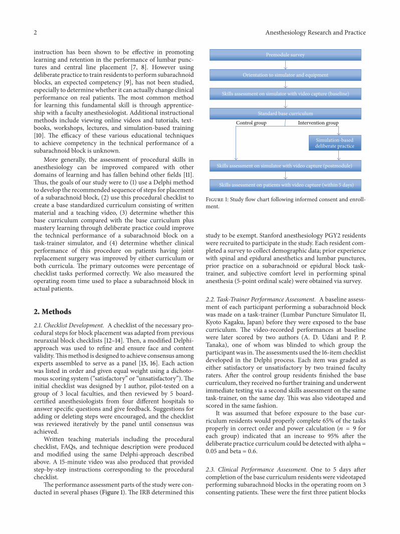
The performance assessment parts of the study were con-ducted in several phases (Figure 1). The IRB determined this
Control group Intervention group
Premodule survey
Orientation to simulator and equipment
Skills assessment on simulator with video capture (baseline)
Standard base curriculum
Simulation-baseddeliberate practice
Skills assessment on simulator with video capture (postmodule)
Skills assessment on patients with video capture (within 5 days)
Figure 1: Study flow chart following informed consent and enroll-ment.
study to be exempt. Stanford anesthesiology PGY2 residentswere recruited to participate in the study. Each resident com-pleted a survey to collect demographic data; prior experiencewith spinal and epidural anesthetics and lumbar punctures,prior practice on a subarachnoid or epidural block task-trainer, and subjective comfort level in performing spinalanesthesia (5-point ordinal scale) were obtained via survey.
2.2. Task-Trainer Performance Assessment. A baseline assess-ment of each participant performing a subarachnoid blockwas made on a task-trainer (Lumbar Puncture Simulator II,Kyoto Kagaku, Japan) before they were exposed to the basecurriculum. The video-recorded performances at baselinewere later scored by two authors (A. D. Udani and P. P.Tanaka), one of whom was blinded to which group theparticipant was in.The assessments used the 16-item checklistdeveloped in the Delphi process. Each item was graded aseither satisfactory or unsatisfactory by two trained facultyraters. After the control group residents finished the basecurriculum, they received no further training and underwentimmediate testing via a second skills assessment on the sametask-trainer, on the same day. This was also videotaped andscored in the same fashion.
It was assumed that before exposure to the base cur-riculum residents would properly complete 65% of the tasksproperly in correct order and power calculation (𝑛 = 9 foreach group) indicated that an increase to 95% after thedeliberate practice curriculum could be detected with alpha =0.05 and beta = 0.6.
2.3. Clinical Performance Assessment. One to 5 days aftercompletion of the base curriculum residents were videotapedperforming subarachnoid blocks in the operating room on 3consenting patients. These were the first three patient blocks

Anesthesiology Research and Practice 3
Table 1: Procedural checklist for subarachnoid block.
Task Satisfactory Unsatisfactory
(1) Performs a “time-out” and places monitors on patient (pulse oximetry and NIBP). — —
(2) Verifies that spinal kit tray, nonsterile and sterile gloves (correct size), and cleansing solutionare present.
— —
(3) Palpates the superior aspects of the iliac crests and identifies the intersection at the L4 spinousprocess with nonsterile gloves on. Marks position at the L3/L4 or L4/L5 interspace.
— —
(4) Cleans the overlying skin with chlorhexidine. — —
(5) Opens the spinal tray before placing sterile gloves on. — —
(6) Puts on sterile gloves with proper technique. — —
(7) Applies sterile drapes. — —
(8) Draws up lidocaine in the 3cc syringe and bupivacaine in the 5cc syringe. Administers localanesthesia in a wheal at the previously marked site.
— —
(9) Injects more anesthetic in the correct location and angle. — —
(10) Inserts the introducer needle in the middle of the interspace with a slight cephalad angulationof 10 to 15 degrees. The bevel of the spinal needle should be in the sagittal plane.
— —
(11) Advances spinal needle through anatomic structures until the subarachnoid space is reached.May experience a popping sensation as the ligamentum flavum is crossed.
— —
(12) Withdraws the stylet each time a pop is felt to assess for CSF flow. — —
(13) Confirms CSF flow by aspiration before and after injecting anesthetic. — —
(14) Removes the spinal and introducer needle together once completed. — —
(15) Applies pressure with the provided 2 × 2 gauze and assesses good hemostasis. — —
(16) Removes the drape, lays the patient, and observes vitals.Disposes of all sharps and biohazard material appropriately.
— —
placed by the resident participant since completing theireducational module. The same two faculty raters usingthe same 16-item checklist scored their performance andrecorded the time to achieve subarachnoid block. Time wasmeasured using a stopwatch for three contiguous intervals:patient positioning (from patient in room to sitting position),setup (from sitting position to injection of local anestheticwheal), and subarachnoid injection (from injection of localanesthetic wheal to completion of subarachnoid injection).
Power analysis showed that a sample size of 9 patientsin each group would provide sufficient power (alpha 0.05and beta 0.6) to detect a 3-minute decrease in time frompositioning to injection if the baseline time equaled 9minutes(SD 4mins) for these junior residents. The 9 minutes wasbased on prestudy data collected on 6 residents.
Residents were also randomized via a random numbergenerator such that, in addition to the base curriculum,half received simulation-based deliberate practice under theguidance of one faculty anesthesiologist (P. P. Tanaka).
2.4. The Mastery Learning with Deliberate Practice Model ofSAB. Fundamental principles of deliberate practice train-ing were followed, including (1) having motivated learners(residents volunteered to improve specific aspects of theirperformance), (2) giving well-defined learning objectives(goals were broken down into specific steps), (3) providing
precise measurements of performance (steps correspondedto specific actions), (4) engaging in focused and repetitivepractice (residents engaged in specific activities and stepsperformed unsatisfactorily were repeated until performedsatisfactorily), and (5) giving informative real-time feedbackconcerning performance (residents received one-on-one fac-ulty coaching).
2.5. Statistical Analysis. Proportions of participants’ genderand exposure to spinal simulator prior to the study werecompared using the Fisher exact test. Number of neurax-ial blocks prior to the study and perceptions of comfortperforming spinal anesthesia were compared among the2 groups using the nonparametric Mann-Whitney U test(http://www.socscistatistics.com/). The Cohen kappa coeffi-cient was used to assess interrater reliability. To assess theimpact of the added deliberate practice training, baselineand posttest checklist scores were compared using analysis ofcovariance to determine if the scores weremore improved forthe intervention group than for the control group.
3. Results
ThemodifiedDelphimethod resulted in a 16-item checklist ofrequired procedural tasks (Table 1). We used this procedural

4 Anesthesiology Research and Practice
Table 2: Characteristics of residents enrolled in study (mean (SD, median, and range)).
Intervention group (deliberate practice) Control group 𝑃value
𝑁 11 10
Months of anesthesia residency completed 5.2 (3.8, 5, 0.25–10) 8.5 (2, 10, 5–12) 0.05
Age (yrs) 28.5 (1.4, 28, 26–31) 30.8 (3.8, 30, 26–37) 0.22
Gender (% F) 45% 20% 0.36
Self-reported number of spinals done before study 6.1 (5, 4, 0–15) 20.0 (16, 14, 3–50) 0.02
Self-reported number of epidurals done before study 9.8 (13, 2, 0–40) 34.5 (16, 30, 15–60) 0.002
Self-reported number of lumbar punctures done before study 5.2 (3, 5, 2–12) 6.5 (5, 7, 0–15) 0.69
Have you practiced on spinal simulator before study (% yes) 55% 0% 0.01
How comfortable are you performing spinal anesthesia(1 = not comfortable, 5 = very comfortable)
2.8 (0.9, 3, 1–4) 3.7 (0.82, 4, 2–5) 0.04
checklist to create a base curriculum of teaching materials(Appendices A and B) and a 15-minute video (available athttp://www.youtube.com/watch?v=eblMcptvcAo&feature=youtu.be).
All 21 residents invited to be in the study consented. Thecontrol group had more experience as anesthesia residents,self-reported experience with epidural blocks and simulationtraining, and higher self-rated comfort performing the spinalanesthesia procedure (Table 2). Scoring of the videos of theresidents performing the subarachnoid blocks demonstratedvery good agreement (kappa = 0.938 SE = 0.044, 95% confi-dence interval: 0.852 to 1.0) between examiners.
Before completing the base curriculum, the control groupproperly completed 81% (SD = 7%, median 81%, and range69–94%) of the 16 checklist tasks on the task-trainer simulatorand this significantly increased to 91% (SD=7%,median 94%,and range 81–100%) after finishing the base curriculum (𝑃 <0.02).
The intervention group (the base curriculum plus delib-erate practice) also significantly increased the percentage ofchecklist tasks properly completed on the task-trainer from73% (SD = 15%, median 75%, and range 31–88%) to 98%(SD = 4%, median 100%, and range 88–100%), which wasa significantly greater increase than observed in the controlgroup (𝑃 < 0.03).
We were unable to study a full set of 3 patients havingsubarachnoid block placed per resident due to residentunavailability. In an average of 3.22 (SD 1.2, median 3, andrange 2–5 days) days after finishing the curriculum, thecontrol group (𝑛 = 10 residents) successfully performedspinals on 20 of 21 patients (66% female, mean age 66 SD 12,mean BMI 28 SD 4.45, and range 19–34), with 1 of the spinalsultimately done by the attending (supervising) physician afterthe resident had prolonged difficulty. The intervention group(𝑛 = 11) successfully performed spinals on 21 of 28 patients(50% female, mean age 61 yrs SD9, BMI 27 SD 4.9, and range19–42), as 7 were placed by the attending after the residenthad prolonged difficulty.
The control group properly performed 84% (SD = 7%,median 88%, and range 69–94%) of checklist tasks versus 81%
(SD = 14%, median 81%, and range 50–100%) of the interven-tion group (𝑃 = NS).
The control group on average spent 296 seconds (SD =104, median 283, and range 113–464) from patient in roomto sitting position and 252 seconds (SD = 118, median 260,and range 46–474) from sitting position to injection of localanesthetic wheel while the intervention group (the basecurriculum plus deliberate practice) on average spent 253seconds (SD = 91, median 226, and range 125–517) frompatient in room to sitting position and 338 seconds (SD = 91,median 338, and range 158–521) from sitting to injection (ineach case, 𝑃 = NS).
Excluding the cases where the attending finished thespinal, time from injection of local anesthetic wheel to finishof subarachnoid injection was also not different for the twogroups. This time equaled 253 seconds (SD = 156, median201, and range 88–627) for the control group and 232 seconds(SD= 158,median 142, and range 86–527) for the interventiongroup (𝑃 = NS).
4. Discussion
Thebase curriculumwe developed for teaching subarachnoidblocks significantly increased correct performance of thetechnical aspects of the block in the control group. Impor-tantly, the addition of deliberate practice led to a significantlyhigher increase in performance in the intervention group.As the depth and breadth of anesthesiology grow and ashouse staff time and resources are limited, it is important todeterminewhich teachingmethods yield the greatest learning[17]. In the current study, we used a rigorous procedure(the Delphi method) to establish a base curriculum and alsoexamined the additional benefits of 1 : 1 mentoring accordingto predetermined guidelines of deliberate practice.
Although overall benefits persisted several days later onactual patients, no differences in time required to place theblocks or checklist scoreswere observed between groups.Thiscan be attributed to differences in learning climate betweenthe simulated and operating room environments. Learning
Anesthesiology Research and Practice 5
climate is defined as the tone or atmosphere of the teachingsetting. Some key components of the learning climate mayhave impacted performance of residents.The operating roommay be a challenging educational environment.There is noisefrom surgical instruments being set up, production pressurefrom the surgical team, and inherent patient characteristicsmay complicate subarachnoid block placement. An influen-tial factor may be specific teaching behaviors by attendinganesthesia faculty. For example, the time required for thesubarachnoid injection was the most difficult of the threeperiods measured because attendings decided based on theirown judgment when to intervene. Instead of encouragingresidents to take a different approach to perform the SAB, weobserved that the attendings would take over the procedureafter a short period of time.The study protocol set no criteriaa priori on how the attending should assist the resident andif and when the attending could intervene. The differences inlearning climate between the simulated and operating roomenvironments hindered consistency most ideal for residenteducation and research.
The current study yielded several informative productsand results. First, we used a rigorous, iterative methodologyto establish a standard base curriculum. This curriculum iscurrently available to all residents to access via the Internet atany time. Second, we developed a training module using thebase curriculum that significantly improved block placementperformance. Third, we implemented a deliberate practicetraining component, which yielded additional performancebenefits. Although we were unable to detect a difference inclinical performance between groups trained with the basecurriculum and those with the additional deliberate practice,the gaps identified via the current study are being usedto design future structured training. For example, we willemphasize the importance of the safety time-out andwashinghands before wearing sterile gloves. Residency programscan also use our checklist evaluation to identify deficienciesin trainees and those who require extra instruction. Fur-thermore, our observations regarding potential confoundingvariables when transferring to clinical settings can informfuture training initiatives.
Simulation-basedmedical education translational studiesattempt to demonstrate that results achieved in the edu-cational laboratory (T1) transfer to improved downstreampatient care practices (T2) and improved patient and publichealth (T3) [18]. Designing and implementing a T2 studycomes with inherent difficulties. In fact a recent literaturereview of hundreds of simulation studies found that only10% included follow-up data from the clinical environment[19]. It is therefore perhaps not surprising that although weobtained a positive T1 result (improved performance in asimulated subarachnoid block for both groups, particularlythe deliberate practice group), this did not translate into apositive T2 result.
This study has several limitations. First, the study enrolleda relatively small group of residents, 21 (although this figurerepresented 88% of the 24 residents in the PGY2 class). Thishighlights the challenge of single institution graduatemedicaleducation studies as there is usually too few available house
staff to enroll to get larger sample sizes. Also, the residentsenrolled in the study were at varying points in trainingand on average were approximately half way through PGY2year. This variation led to differences in resident neuraxialblock placement and simulator experience prior to studyenrollment. It is likely that the impact of the teachingresidents received on performance improvement is greatestfor beginner residents in their first months of residency.The difference between the control and intervention group’straining prior to enrollmentmay also explain the difference inbaseline SAB skills, 81% versus 73%, respectively. The impactof starting at a lower baseline score may overstate the overallchange described from deliberate practice in the interventiongroup. However, the control group’s prior experience withthe spinal simulator may also have influenced the higherbaseline performance score. Finally, attending physicians aremore likely to take over procedures from junior residents,those with little training experience. This may have resultedin 7 incomplete blocks in the intervention group versus 1in the control group. Multi-institution studies may be a wayto enroll subjects more quickly and increase sample size,although it may be even more difficult to control for priorexperience and skills. Another limitation is that posttestingoccurred immediately after training potentially enhancingrecall.
5. Conclusions
The current study represents an advance in simulation-basededucation, particularly in anesthesia, due to the developmentand implementation of a base curriculum and the formalizedmethodology of deliberate practice training. Moreover, thisstudy is one of few investigations examining transfer toclinical settings. Our results and observations have iden-tified specific considerations and areas for improvementin subsequent training modules. For example, this studyfocused on the technical skill required to place a subarach-noid block but there is more to this in actual practice,including the decision of whether a block is indicated andobtaining consent and postoperative follow-up. The bestway to teach all of those elements together deserves fur-ther study. More generally, this study provides an attemptat rigorous methodology for designing, implementing,and evaluating simulation-based learning interventions inmedicine.
Appendices
A. Spinal Anesthesia Teaching Module:FAQs about Spinal Anesthesia
A.1. What Agent Should Be Used to Cleanse the Skin for Spi-nal Anesthesia? The skin is prepared with an appropriateantiseptic solution and draped. All antiseptic solutions areneurotoxic, and care must be taken not to contaminate spinalneedles or local anesthetics with the antiseptic solution.

6 Anesthesiology Research and Practice
Chlorhexidine-alcohol antiseptic prevents colonization ofpercutaneous catheters better than does 10% povidone-iodine. Consequently, the American Society of RegionalAnesthesia currently recommends chlorhexidine for skinantisepsis prior to regional anesthesia procedures. How onedrapes is a matter of personal preference, but clear plasticdrapes offer the important advantage of permitting visual-ization of the entire back, which makes it easier to identifya rotated or inadequately flexed spine.
A.2. Should a Mask, Hat, Handwashing, and Gown Be Usedin addition to Sterile Gloves? Full sterile precautions arerequired for spinal anesthesia.Thorough handwashing great-ly reduces the risk of cross-contamination and should occurprior to performing any regional anesthetic technique.Alcohol-based antiseptic solutions will provide the maximaldegree of antimicrobial activity with extended duration whencompared to nonalcoholic antimicrobial or nonantimicrobialpreparations.The duration andmethod of washing (standardhandwashing versus full surgical scrub) required to reduceinfectious complications are currently unknown (grade A).Higher microbial counts have been identified in health-careworkers who do not remove jewelry prior to handwashing.Therefore, it may be prudent to remove all jewelry items(rings, watches, etc.) prior to handwashing to reduce the riskof contamination. Sterile surgical gloves should be used andconsidered a supplement to, not replacement for, handwash-ing (grade B). The use of surgical gloves is advocated notonly to protect patients from cross-contamination but alsoto protect health-care workers from blood-borne pathogenexposure as required by the Occupational Safety and HealthAdministration (OSHA). The use of surgical masks duringregional anesthesia will maximize sterile barrier precautions(grade A). In particular, surgical masks have been foundto significantly reduce the likelihood of site contaminationfrom microorganisms grown in the upper airway of clin-icians. Although the routine use of masks has not beenfound to reduce infectious complications related to regionalanesthesia, they do remain a vital protective measure againstblood-borne pathogen exposure as recommended by theOccupational Safety and Health Administration (OSHA)(grade B).
A.3. What Type of Needle Is “Best” to Perform a Spinal Anes-thesia? Spinal needles are classified by the design of their tips.TheWhitacre, Eldor, Marx, and Sprotte spinal needles have a“pencil-point” tip with one or two (Eldor) apertures on theside of the shaft proximal to the tip. The Greene, Atraucan,and Quincke needles have beveled tips with cutting edges.The pencil-point needles require more force to insert thanthe bevel-tip needles but provide a better tactile “feel” ofthe various tissues encountered as the needle is inserted. Inaddition, the bevel has been shown to cause the needle to bedeflected from the intended path as it passes through tissueswhile the pencil-point needles are not deflected. Larger gauge(i.e., smaller diameter) spinal needles are less likely to causepostdural puncture headaches (PDPH) but are more read-ily deflected than smaller gauge needles. Spinal needles are
typically sized 22 to 29 gauge. Spinal needles smaller than22 gauge are often easier to insert if an introducer needleis used. The introducer is inserted into the interspinousligament in the intended direction of the spinal needle andthe spinal needle is then inserted through the shaft of theintroducer. The introducer prevents the spinal needle frombeing deflected or bent as it passes through the interspinousligament. Needles of the same outside diameter may havedifferent inside diameters. This is important because insidediameter determines how large a catheter can be insertedthrough the needle and determines how rapidly CSF willappear at the needle hub during spinal needle insertion.All spinal needles come with a tight-fitting stylet. The styletprevents the needle from being plugged with skin or fatand, importantly, prevents dragging skin into the epiduralor subarachnoid spaces, where the skin may grow and formdermoid tumors.
A.4. What Other Measures May Reduce PDPH? Orientatingthe bevel of the standard spinal needle parallel versus per-pendicular to the fibers of the cauda equina has been shownto reduce the risk of postprocedure headache. The reason isnot well understood. Bed rest postspinal anesthesia does notreduce the incidence of postdural puncture headache. See[20].
A.5. Is There an Advantage to a Sitting versus Lateral Recum-bent Position in Performing Spinal Anesthesia? Careful atten-tion to patient positioning is critical to successful spinalblock. Poor positioning can turn an otherwise easy spinalanesthetic into a challenge for both the anesthesiologist andthe patient. Studies of the intervertebral distance show thatthe space is increased slightly (by about 0.2 cm) when thepatient is in the sitting position, with the feet supported,compared to the lateral recumbent position. There are nostudies comparing “success rates,” ease of puncture, need forsecond attempts, or other clinical outcomes with the sittingversus recumbent position.
A.6. What If My Patient Is Febrile, Can I Place a Spinal Block?Serious central neuraxial infections such as arachnoiditis,meningitis, and abscess following spinal or epidural anesthe-sia are rare. Available data suggest that patients with evidenceof systemic infection may safely undergo spinal anesthesia,provided appropriate antibiotic therapy is initiated prior todural puncture, and the patient has demonstrated a responseto therapy, such as a decrease in fever (placement of anindwelling epidural or intrathecal catheter in this group ofpatients remains controversial) (grade B). Epidural cathetersshould be removed in the presence of local erythema and/ordischarge; there are no convincing data to suggest thatconcomitant infection at remote sites and the absence ofantibiotic therapy are risk factors for infection (grade A). Adelay in diagnosis and treatment of major CNS infectionsof even a few hours may significantly worsen neurologicoutcome (grade B).

Anesthesiology Research and Practice 7
A.7. What Factors Increase the Risk of Spinal Hematomaafter Spinal Block? Bleeding disorders may increase therisk of spinal hematoma related to spinal puncture. Spinalhematoma may in turn result in spinal cord compressionand permanent paralysis. Most of the literature on the risksof spinal hematoma after lumbar puncture comes fromcase reports and case series in patients receiving spinal orepidural anesthesia. Because of the variability in the cases andprocedures reported in the literature, many experts refrainfrom specific recommendations about levels of platelets orINR that are “safe” for lumbar puncture.
Additional risk factors for spinal hematoma and neu-rologic compromise include patient factors (female sex,increased age, ankylosing spondylitis or spinal stenosis, andrenal insufficiency), factors related to technique (traumaticneedle/catheter placement), and dosing factors (“high” or“low” dose LMWH, timing of LMWH, and concomitantantiplatelet or anticoagulants). There are some guidelines,described below, for specific situations.
A.8. Can Spinal Anesthesia Be Performed Safely in Patientswith Thrombocytopenia? A recent extensive review conclud-edwith the recommendation that LPmay be safely performedin patients with a platelet count ≥40K, as long as the patientis not receiving antiplatelet agents or anticoagulants, thereare no other coagulopathies, the platelets are functioningnormally, and the platelet count is stable. This is consistentwith the recommendations of the American National RedCross (http://www.redcross.org/www-files/Documents/WorkingWiththeRedCross/practiceguidelinesforbloodtrans.pdf); see [21].
A.9. Can Spinal Anesthesia Be Performed Safely in Patientson Antiplatelet Agents (Aspirin and/or Plavix)? Patients tak-ing nonsteroidal anti-inflammatory drugs with antiplateleteffects (e.g., cyclooxygenase-1 inhibitors) or receiving sub-cutaneous unfractionated heparin for deep vein thrombosisprophylaxis are not viewed as being at increased risk of spinalhematoma.
In contrast, other classes of antiplatelet drugs, like thieno-pyridine derivatives (e.g., ticlopidine, clopidogrel) and glyco-protein IIb/IIIa antagonists (e.g., abciximab, eptifibatide, andtirofiban), have a more potent effect on platelet aggregation,and neuraxial block should generally not be performed inpatients taking these or similar medications. Further, theconsensus statement recommends that ticlopidine be discon-tinued for 2 weeks and clopidogrel for 1 week before per-forming central neuraxial blocks. The glycoprotein IIb/IIIaantagonists have a shorter duration of action; thus, it isrecommended that abciximab should be discontinued 24 to48 hours before central neuraxial block, and eptifibatide andtirofiban should be discontinued 4 to 8 hours beforehand; see[22, 23].
A.10. Can Spinal Anesthesia Be Performed Safely in PatientsReceiving Prophylactic Heparin or Low-Molecular-WeightHeparin? Low-molecular-weight heparin increases the riskof spinal hematoma sufficiently to warrant a “black box
warning” about use of neuraxial (spinal or epidural) anes-thesia in patients on LMWH. Practitioners are urged to“consider risk versus benefit” and observe the patient closelyfor bleeding and signs of neurological impairment if therapyis administered during or immediately following lumbarpuncture.
Patients receiving fractionated low-molecular-weightheparin (e.g., enoxaparin, dalteparin, and tinzaparin) areconsidered to be at increased risk of spinal hematoma.Patients receiving these drugs preoperatively at thrombo-prophylactic doses should have the drug held for 10 to 12hours before central neuraxial block. At higher doses, suchas those used to treat established deep vein thrombosis,central neuraxial block should be delayed for 24 hours afterthe last dose. For patients in whom low-molecular-weightheparin is begun after surgery, single-shot central neuraxialblocks are not contraindicated provided that the first low-molecular-weight heparin dose is not administered until 24hours postoperatively if using a twice-daily dosing regimenand 6 to 8 hours if using a once-daily dosing regimen. If anindwelling central neuraxial catheter is in place, it should notbe removed until 10 to 12 hours after the last low-molecular-weight heparin dose, and the subsequent doses should notbegin until at least 2 hours after catheter removal.
There is no contraindication to spinal anesthesia inpatients receiving prophylactic unfractionated heparin aslong as the total 24-hour dose is ≤10,000 units; see [22].
A.11. Can Spinal Anesthesia Be Performed Safely in Patientswith Elevated INR due to Coumadin Use? Can Spinal Anes-thesia Be Performed Safely in Patients with Elevated INR dueto Their Underlying Medical Condition (e.g., Liver Disease)?The American National Red Cross recommends using freshfrozen plasma to treatmultiple coagulation deficiencies (suchas liver disease) prior to an invasive procedure, includinglumbar puncture. The goal is an INR ≤ 1.5. FFP may alsobe used to correct INR in patients on Coumadin if urgentreversal of anticoagulation is needed. The usual dose toprovide 30% of plasma factor concentrate is 10–20mL/kg. A“unit” of FFP is approximately 250 cc, though the volumemayvary. In a 70 kg patient, 3–6 units of FFP is recommended tocorrect INR sufficiently. Complete normalization of INR isoften not possible in patients with liver disease.
Patients with hemophilia or von Willebrand’s diseaseshould have factors normalized prior to lumbar puncture; see[24, 25].
B. Spinal Anesthesia Teaching Module:Spinal Anesthesia Technique Description
B.1. Midline Approach. From the midline approach to thesubarachnoid space, the skin overlying the desired interspaceis infiltrated with a small amount of local anesthetic toprevent pain when inserting the spinal needle. Additionallocal anesthetic (1 to 2mL) is then deposited along theintended path of the spinal needle to a depth of 1 to 2 inches.This deeper infiltration provides additional anesthesia forspinal needle insertion and helps identify the correct path

8 Anesthesiology Research and Practice
Spinous process, L-2Interspinous ligament
(A)
(B)
(C)
Figure 2: Midline approach to the subarachnoid space. The spinalneedle is inserted with a slight cephalad angulation and shouldadvance in the midline without contacting bone (B). If bone iscontacted, it may be either the caudad (A) or the cephalad spinousprocess (C). The needle should be redirected slightly with cephaladand reinserted. If bone is encountered at a shallower depth, theneedle is likely walking up the cephalad spinous process. If bone isencountered at a deeper depth, the needle is likely walking downthe inferior spinous process. If bone is repeatedly contacted at thesame depth, the needle is likely off the midline and walking alongthe lamina.
for the spinal needle. Infiltrating local anesthetic lateral to themidline is painful and generally unnecessary.
The spinal needle or introducer needle is inserted in themiddle of the interspace with a slight cephalad angulationof 10 to 15 degrees (Figure 2). The needle is then advanced,in order, through the subcutaneous tissue, supraspinous lig-ament, interspinous ligament, ligamentum flavum, epiduralspace, duramater, and finally arachnoidmater.The ligamentsproduce a characteristic “feel” as the needle is advancedthrough them, and the anesthesiologist should develop theability to distinguish a needle that is advancing through thehigh-resistance ligaments fromone that is advancing throughlower-resistance paraspinous muscle. This will allow earlydetection and correction of needles that are not advancing inthe midline. Penetration of the dura mater often produces asubtle “pop” that is most easily detected with the pencil-pointneedles. Detection of dural penetration will prevent insertingthe needle all the way through the subarachnoid space andcontacting the vertebral body. In addition, learning to detectdural penetration will allow one to insert the spinal needlequickly without having to stop every few millimeters andremove the stylet to look for CSF at the needle hub.
Once the needle tip is believed to be in the subarachnoidspace, the stylet is removed to see if CSF appears at theneedle hub.With small-diameter needles (26 to 29 gauge) thisgenerally requires 5 to 10 seconds. If CSF does not appear, theneedle orifice may be obstructed by a nerve root and rotatingthe needle 90 degrees may result in CSF flow. Alternatively,the needle orifice may not be completely in the subarachnoidspace and advancing an additional 1 to 2mm may resultin brisk CSF flow. This is particularly true of pencil-pointneedles, which have their orifice on the side of the needleshaft proximal to the needle tip. Finally, failure to obtain CSFsuggests that the needle orifice is not in the subarachnoidspace and the needle should be reinserted.
If bone is encountered during needle insertion, the anes-thesiologist must develop a reasoned, systematic approachto redirect the needle. Simply withdrawing the needle andrepeatedly reinserting it in different directions are not appro-priate. When contacting bone, the depth should be immedi-ately noted and the needle redirected slightlywith cephalad. Ifbone is again encountered at a greater depth, then the needleis most likely walking down the inferior spinous processand it should be redirected more with cephalad until thesubarachnoid space is reached. If bone is encountered againat a shallower depth, then the needle ismost likely walking upthe superior spinous process and it should be redirectedmorewith caudad. If bone is repeatedly encountered at the samedepth, then the needle is likely off the midline and walkingalong the vertebral lamina (Figure 2).
When redirecting a needle it is important to withdraw thetip into the subcutaneous tissue. If the tip remains embeddedin one of the vertebral ligaments, attempts at redirecting theneedle will simply bend the shaft and will not reliably changeneedle direction. When using an introducer needle, it alsomust be withdrawn into the subcutaneous tissue before beingredirected. Changes in needle direction should be made insmall increments because even small changes in needle angleat the skin may result in fairly large changes in position of theneedle tip when it reaches the spinal meninges at a depth of 4to 6 cm. Care should be exercised when gripping the needleto ensure that it does not bow. Insertion of a curved needlewill cause it to veer off course.
If the patient experiences a paresthesia, it is important todetermine whether the needle tip has encountered a nerveroot in the epidural space or in the subarachnoid space.When the paresthesia occurs, immediately stop advancingthe needle, remove the stylet, and look for CSF at theneedle hub. The presence of CSF confirms that the needleencountered a cauda equina nerve root in the subarachnoidspace and the needle tip is in good position. Given how tightlypacked the cauda equina nerve roots are, it is surprising thatall spinal punctures do not produce paresthesias. If CSF isnot visible at the hub, then the paresthesia may have resultedfrom contact with a spinal nerve root traversing the epiduralspace. This is especially true if the paresthesia occurs in thedermatome corresponding to the nerve root that exits thevertebral canal at the same level in which the spinal needle isinserted. In this case the needle has most likely deviated fromthemidline and should be redirected toward the side oppositethe paresthesia. Occasionally, pain experienced when theneedle contacts bone may be misinterpreted by the patient asa paresthesia and the anesthesiologist should be alert to thispossibility.
Once the needle is correctly inserted into the subarach-noid space, it is fixed in position and the syringe containinglocal anesthetic is attached. CSF is gently aspirated to confirmthat the needle is still in the subarachnoid space and the localanesthetic slowly injected (≤0.5mL/sec). After completingthe injection, a small volume of CSF is again aspirated to con-firm that the needle tip remained in the subarachnoid spacewhile the local anesthetic was deposited. This CSF is thenreinjected and the needle, syringe, and any introducer wereremoved together as a unit. If the surgical procedure is to be
Anesthesiology Research and Practice 9
performed in the supine position, the patient is helped ontohis or her back. To prevent excessive cephalad spread ofhyperbaric local anesthetic, care should be taken to ensurethat the patient’s hips are not raised off the bed as theyturn.
Once the block is placed, strict attention must be paidto the patient’s hemodynamic status with blood pressureand/or heart rate supported as necessary. Block height shouldalso be assessed early by pin prick or temperature sensation.Temperature sensation is tested by wiping the skin withalcohol and may be preferable to pin prick because it isnot painful. If, after a few minutes, the block is not risinghigh enough or is rising too high, the table may be tiltedas appropriate to influence further spread of hypobaric orhyperbaric local anesthetics.
Ethical Approval
IRB determined this study not to be human study research,StanfordUniversity Institutional ReviewBoard,December 19,2011, Protocol 22632.
Disclosure
Meetings preliminary results and abstract were presented atPost Graduate Assembly in Anesthesiology, December 16,2012, New York, NY, and at Innovations in Medical Educa-tion, February 23, 2013, Los Angeles, CA.
Conflict of Interests
The authors declare that there is no conflict of interestsregarding the publication of this paper.
Acknowledgment
Funding was received from Department of Anesthesiology,Perioperative and Pain Medicine, Departmental Grant, Stan-ford University, 300 Pasteur Drive, Room H3589, Stanford,CA.
References
[1] K. Ericsson, “The influence of experience and deliberate prac-tice on the development of superior expert performance,” inCambridge Handbook of Expertise and Expert Performance, K.Ericsson, N. Charness, P. Feltovich, and R. Hoffman, Eds., pp.685–706, Cambridge University Press, Cambridge, UK, 2006.
[2] K. A. Ericsson, “Deliberate practice and acquisition of expertperformance: a general overview,” Academic EmergencyMedicine, vol. 15, no. 11, pp. 988–994, 2008.
[3] K. A. Ericsson, “Deliberate practice and the acquisition andmaintenance of expert performance in medicine and relateddomains,”AcademicMedicine, vol. 79, no. 10, pp. S70–S81, 2004.
[4] S. B. Issenberg, W. C. McGaghie, E. R. Petrusa, D. L. Gordon,and R. J. Scalese, “Features and uses of high-fidelity medicalsimulations that lead to effective learning: a BEME systematicreview,”Medical Teacher, vol. 27, no. 1, pp. 10–28, 2005.
[5] R. K. Latif, A. F. Bautista, S. B. Memon et al., “Teachingaseptic technique for central venous access under ultrasoundguidance: a randomized trial comparing didactic training aloneto didactic plus simulation-based training,” Anesthesia andAnalgesia, vol. 114, no. 3, pp. 626–633, 2012.
[6] J. H. Barsuk, W. C. McGaghie, E. R. Cohen, J. S. Balachandran,and D. B. Wayne, “Use of simulation-based mastery learning toimprove the quality of central venous catheter placement in amedical intensive care unit,” Journal of Hospital Medicine, vol.4, no. 7, pp. 397–403, 2009.
[7] R. Aggarwal and A. Darzi, “Technical-skills training in the 21stcentury,”The New England Journal of Medicine, vol. 355, no. 25,pp. 2695–2696, 2006.
[8] D. O. Kessler, M. Auerbach, M. Pusic, M. G. Tunik, and J. C.Foltin, “A randomized trial of simulation-based deliberate prac-tice for infant lumbar puncture skills,” Simulation in Healthcare,vol. 6, no. 4, pp. 197–203, 2011.
[9] Accreditation Council for Graduate Medical Education andAnesthesiology Program Requirements, 2008, https://www.acgme.org/acgmeweb/Portals/0/PFAssets/ProgramRequire-ments/040 anesthesiology 07012014.pdf.
[10] B. D. Sites, B. C. Spence, J. D. Gallagher, C. W. Wiley, M.L. Bertrand, and G. T. Blike, “Characterizing novice behaviorassociated with learning ultrasound-guided peripheral regionalanesthesia,” Regional Anesthesia and Pain Medicine, vol. 32, no.2, pp. 107–115, 2007.
[11] M. Bould, N. Crabtree, and V. Naik, “Assessment of proceduralskills in anaesthesia,” British Journal of Anaesthesia, vol. 103, no.4, pp. 472–483, 2009.
[12] P. Wathen, M. Johnson, J. O’Rorke, and V. Lawrence, “LumbarPuncture Procedure Module,” 2011, https://www.mededportal.org/publication/8201.
[13] M. A. Hayter, Z. Friedman,M. D. Bould et al., “Validation of theimperial college surgical assessment device (ICSAD) for labourepidural placement,”Canadian Journal of Anesthesia, vol. 56, no.6, pp. 419–426, 2009.
[14] M. S. Ellenby, K. Tegtmeyer, S. Lai, and D. Braner, “Videos inclinical medicine: lumbar puncture,” The New England journalof medicine, vol. 355, no. 13, article e12, 2006.
[15] B. Graham, G. Regehr, and J. G. Wright, “Delphi as a method toestablish consensus for diagnostic criteria,” Journal of ClinicalEpidemiology, vol. 56, no. 12, pp. 1150–1156, 2003.
[16] M. Clayton, “Delphi: a technique to harness expert opinionfor critical decision-making tasks in education,” EducationalPsychology, vol. 17, no. 4, pp. 373–386, 1997.
[17] Z. Friedman, N. Siddiqui, R. Katznelson, I. Devito,M. D. Bould,and V. Naik, “Clinical impact of epidural anesthesia simulationon short-and long-term learning curve high-versus low-fidelitymodel training,” Regional Anesthesia and PainMedicine, vol. 34,no. 3, pp. 229–232, 2009.
[18] W. C.McGaghie, T. J. Draycott,W. F. Dunn, C.M. Lopez, andD.Stefanidis, “Evaluating the impact of simulation on translationalpatient outcomes,” Simulation in Healthcare, vol. 6, no. 7, pp.S42–S47, 2011.
[19] A. J. Ross, N. Kodate, J. E. Anderson, L. Thomas, and P.Jaye, “Review of simulation studies in anaesthesia journals,2001–2010: mapping and content analysis,” British Journal ofAnaesthesia, vol. 109, no. 1, pp. 99–109, 2012.
[20] S. E. Straus, K. E. Thorpe, and J. Holroyd-Leduc, “How do Iperform a lumbar puncture and analyze the results to diagnose
10 Anesthesiology Research and Practice
bacterial meningitis?” Journal of the American Medical Associa-tion, vol. 296, no. 16, pp. 2012–2022, 2006.
[21] J. J. van Veen, T. J. Nokes, and M. Makris, “The risk ofspinal haematoma following neuraxial anaesthesia or lumbarpuncture in thrombocytopenic individuals,” British Journal ofHaematology, vol. 148, no. 1, pp. 15–25, 2009.
[22] K. F. Layton, D. F. Kallmes, and T. T. Horlocker, “Recommen-dations for anticoagulated patients undergoing image-guidedspinal procedures,” AJNR, vol. 27, no. 3, pp. 467–471, 2006.
[23] T. T. Horlocker, D. J. Wedel, J. C. Rowlingson et al., “Regionalanesthesia in the patient receiving antithrombotic or throm-bolytic therapy,” Regional Anesthesia and Pain Medicine, vol. 35,no. 1, pp. 64–101, 2010.
[24] C. Armon and R. W. Evans, “Addendum to assessment: pre-vention of post-lumbar puncture headaches: report of theTherapeutics and Technology Assessment Subcommittee of theAmerican Academy of Neurology,”Neurology, vol. 65, no. 4, pp.510–512, 2005.
[25] J. Williams, D. C. B. Lye, and T. Umapthi, “Diagnostic lumbarpuncture: minimizing complications,” Internal Medicine Jour-nal, vol. 38, no. 7, pp. 587–591, 2008.

Submit your manuscripts athttp://www.hindawi.com
Stem CellsInternational
Hindawi Publishing Corporationhttp://www.hindawi.com Volume 2014
Hindawi Publishing Corporationhttp://www.hindawi.com Volume 2014
MEDIATORSINFLAMMATION
of
Hindawi Publishing Corporationhttp://www.hindawi.com Volume 2014
Behavioural Neurology
EndocrinologyInternational Journal of
Hindawi Publishing Corporationhttp://www.hindawi.com Volume 2014
Hindawi Publishing Corporationhttp://www.hindawi.com Volume 2014
Disease Markers
Hindawi Publishing Corporationhttp://www.hindawi.com Volume 2014
BioMed Research International
OncologyJournal of
Hindawi Publishing Corporationhttp://www.hindawi.com Volume 2014
Hindawi Publishing Corporationhttp://www.hindawi.com Volume 2014
Oxidative Medicine and Cellular Longevity
Hindawi Publishing Corporationhttp://www.hindawi.com Volume 2014
PPAR Research
The Scientific World JournalHindawi Publishing Corporation http://www.hindawi.com Volume 2014
Immunology ResearchHindawi Publishing Corporationhttp://www.hindawi.com Volume 2014
Journal of
ObesityJournal of
Hindawi Publishing Corporationhttp://www.hindawi.com Volume 2014
Hindawi Publishing Corporationhttp://www.hindawi.com Volume 2014
Computational and Mathematical Methods in Medicine
OphthalmologyJournal of
Hindawi Publishing Corporationhttp://www.hindawi.com Volume 2014
Diabetes ResearchJournal of
Hindawi Publishing Corporationhttp://www.hindawi.com Volume 2014
Hindawi Publishing Corporationhttp://www.hindawi.com Volume 2014
Research and TreatmentAIDS
Hindawi Publishing Corporationhttp://www.hindawi.com Volume 2014
Gastroenterology Research and Practice
Hindawi Publishing Corporationhttp://www.hindawi.com Volume 2014
Parkinson’s Disease
Evidence-Based Complementary and Alternative Medicine
Volume 2014Hindawi Publishing Corporationhttp://www.hindawi.com