research article transcutaneous intraluminal...
TRANSCRIPT
Research ArticleTranscutaneous Intraluminal Impedance Measurement forMinimally Invasive Monitoring of Gastric Motility: Validation inAcute Canine Models
Michael D. Poscente,1 Gang Wang,1 Dobromir Filip,2 Polya Ninova,3 Gregory Muench,4
Orly Yadid-Pecht,2 Martin P. Mintchev,1,2,5 and Christopher N. Andrews6
1Centre for Bioengineering and Research, University of Calgary, Engineering Complex, 2500 University Drive NW,Calgary, AB, Canada T2N 1N42Department of Electrical and Computer Engineering, University of Calgary, Engineering Complex, 2500 University Drive NW,Calgary, AB, Canada T2N 1N43Division of Pediatrics, Faculty of Medicine, University of Calgary, Calgary, AB, Canada T2N 1N44Faculty of Veterinary Medicine, University of Calgary, Calgary, AB, Canada T2N 1N45Department of Surgery, Faculty of Medicine, University of Alberta, Edmonton, AB, Canada T6G 2B76Division of Gastroenterology, Faculty of Medicine, University of Calgary, Calgary, AB, Canada T2N 1N4
Correspondence should be addressed to Martin P. Mintchev; [email protected]
Received 12 October 2014; Revised 18 November 2014; Accepted 18 November 2014; Published 9 December 2014
Academic Editor: Paul Enck
Copyright © 2014 Michael D. Poscente et al. This is an open access article distributed under the Creative Commons AttributionLicense, which permits unrestricted use, distribution, and reproduction in any medium, provided the original work is properlycited.
Transcutaneous intraluminal impedance measurement (TIIM) is a new method to cutaneously measure gastric contractions byassessing the attenuation dynamics of a small oscillating voltage emitted by a battery-powered ingestible capsule retained in thestomach. In the present study, we investigated whether TIIM can reliably assess gastric motility in acute canine models.Methods.Eight mongrel dogs were randomly divided into 2 groups: half received an active TIIM pill and half received an identically sizedsham capsule. After 24-hour fasting and transoral administration of the pill (active or sham), two force transducers (FT) weresutured onto the antral serosa at laparotomy. After closure, three standard cutaneous electrodes were placed on the abdomen,registering the transluminally emitted voltage. Thirty-minute baseline recordings were followed by pharmacological inductionof gastric contractions using neostigmine IV and another 30-minute recording. Normalized one-minute baseline and post-neostigmine gastric motility indices (GMIs) were calculated and Pearson correlation coefficients (PCCs) between cutaneous andFT GMIs were obtained. Statistically significant GMI PCCs were seen in both baseline and post-neostigmine states. There were nosignificant GMI PCCs in the sham capsule test. Further chronic animal studies of this novel long-term gastricmotilitymeasurementtechnique are needed before testing it on humans.
1. Introduction
Distal postprandial gastricmotility involves contractions thatmechanically crush ingested food and mix it with secretionsto prepare it for absorption, whilemaintaining an appropriatepressure gradient across the pyloric sphincter to regulategastric emptying [1]. The two most prevalent gastric motilitydisorders are functional dyspepsia and gastroparesis, thecause and physiology of which are diverse [2]. Both havesimilar symptoms, including upper abdominal pain, nausea,
and early satiety, and can be associated with delayed gastricemptying [2, 3] which can make it difficult to distinguishbetween them clinically [2]. Both are chronic disordersthat generate significant health care costs [4], and in bothcases new approaches are needed for minimally invasive,long-term, ambulatory monitoring to potentially improvediagnosis and management.
Gastric function studies that can identify delayed gastricemptying include scintigraphy, C13 breath tests, wirelessmotility capsule tests, and combinations thereof [5]. All
Hindawi Publishing CorporationGastroenterology Research and PracticeVolume 2014, Article ID 691532, 9 pageshttp://dx.doi.org/10.1155/2014/691532
2 Gastroenterology Research and Practice
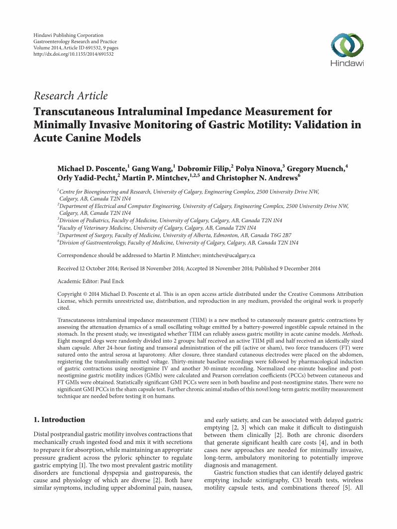
Intraluminal electrodes
Bioelectric amplifier
Cutaneous electrodesTIIM
transducerLong-term gastricretentive enclosure
Stomach wall
Extraluminal tissue and space
Skin
Human torso
Ground
(a)
1cm
1 cm
(b)
Figure 1: (a) The TIIM principle shown schematically: a small electrical signal is emitted from the TIIM transducer intraluminally fromwithin the stomach after ingestion. The transducer is contained within an expandable pill that swells within the stomach, thus preventingit from being expelled through the pylorus for a prolonged amount of time (days). The attenuation dynamics of the electrical signal acrossthe gastric and extraluminal tissue is measured from the skin via external cutaneous electrodes. The TIIM signals are measured using astandard multichannel bioelectric amplifier (electrogastrograph). After a predetermined amount of time (depending on the materials used),the long-term gastric-retentive enclosure disintegrates and the resulting smaller constituents of the pill individually exit the gastrointestinaltract via natural peristalsis.TheTIIM components in the figure are deliberately zoomed in. (b) TIIM gastric-retentive pill design: (1) electronicoscillator circuit; (2) capsule body; (3) assembled capsule; (4) superabsorbent granules; (5) capsule and granules inside a liquid-permeablemesh; (6) dissolvable pill containing themeshed capsule; (7) TIIMgastric-retentive pill; (8) pill expanded inwater; and (9) test dish.Horizontaland vertical 1 cm scales are depicted at the bottom left corner.
these methods have benefits and drawbacks. Scintigraphyis currently the gold standard [3], which is limited by 4-hour stationary testing [6], and exhibits high intrapatientvariability, largely dependent on body position [7]. C13 breathtests overcome the radiation issues of scintigraphy but can beinfluenced by other factors in the body and are limiting interms of the meal that can be assessed [5]. Wireless motilitycapsule-based tests overcome the radiation limitations as wellbut do not offer gastric retention and therefore are bettersuited to assess colonic motility [8]. The method still has apotential issue of unintended retention, occasionally requir-ing endoscopic removal [5]. Gastric motility has been shownto be relevant to functional dyspepsia in clinical studies,with the gastric emptying rate exhibiting significant negativecorrelation with the severity of symptoms [9]. FunctionalMRI may be capable of providing information about antralcontractile activity but is limited due to its short duration ofassessment and high cost [10]. Antroduodenal manometryis another avenue for assessing foregut motor function andcan be employed in long-term (>24 h) ambulatory tests[11] but is catheter-based (transnasal or transoral) and hasdemonstrated inconsistent performance in the larger antrallumen, and in many cases its results can be difficult tointerpret. Noninvasive electrical-based measurements suchas electrogastrography [12] (EGG) or bioimpedance [13]currently lack reliability, and so far the use of electricityin gastric motility studies has been considered to be notadequately validated for clinical use [14].
Transcutaneous intraluminal impedance measurement(TIIM) works by emitting a small high-frequency electricalsignal from within the stomach, with parameters (50 kHz,1.5 V peak-to-peak) chosen for their optimal transmissionproperties through smooth and abdominal muscles [15]
and other extraluminal tissues, thus reaching the skin withpotentially strong residual energy (Figures 1(a) and 1(b)).Thesteady, stable, and rectangular voltage signal is generated by awatch battery-supplied miniature electronic oscillator. Sincethe frequency and the originally generated amplitude of thisbroad-spectrum rectangular signal are known, it is possibleto quantify its amplitude attenuation and related modulationby measuring the resultant signal at the surface of the skinusing a plain electrogastrograph [16]. It is hypothesized thatgastric contractions are amplitude-modulated on the high-frequency carrier, and TIIM reflects these contractions afterappropriate amplitude demodulation. Two implementationsof the gastric TIIMoscillator have been developed: a catheter-based system and an ingestible pill form that expands in thestomach and is retained for a long duration [16]. In both cases,pilot studies [16] have demonstrated that TIIM is capableof measuring gastric motility with comparable precision toinvasively implanted force transducers on the stomach.
TIIM is a substantially different concept from stan-dard electrogastrography (EGG), where cutaneous electrodesmeasure the intrinsic spontaneous electrical activity of thestomach. EGG has substantial drawbacks due to dynamicartifacts and lack of direct correlation with gastric muscularfunction, disease, or symptoms [14]. Conversely, the gastric-retentive TIIM measures the attenuation dynamics of a pre-determined high-frequency signal originatingwithin the hol-low viscus of the stomach. Based on theTIIM signal’s parame-ters, its measurement is completely independent of and unaf-fected by any intrinsic electrical activity, whether originatingfrom the gut or elsewhere (e.g., the heart). Thus, for gastricapplications, the major driver for the variations in TIIMsignal attenuation is directly related to gastric contractions.
Gastroenterology Research and Practice 3
The aim of the present study was to evaluate the effective-ness of the minimally invasive, ingestible, gastric-retentiveTIIM capsule formeasuring gastricmotility in comparison toa sham gastric-retentive, ingestible capsule, with both beingreferenced to force transducers attached to the serosa of thestomach in acute canine models [17]. This validation can beregarded as a necessary precursor to future long-term chronicambulatory tests involving TIIM.
2. Methods
2.1. TIIM Capsule Design. For the present study, the TIIMminiature electronic oscillator was integrated into a gastric-retentive pill, which further lowers the already minimalimpact of the catheter-based design [16]. Each custom-designed TIIM transducer contained a miniature, surface-mount, battery-supplied, 50 kHz/1.5 V electronic oscillator(Linear Technology, Milpitas, CA, USA). The length of thetransducer was 18mmwith a diameter of 11mm.The oscillat-ing TIIM capsule had a battery life of 62 ± 3 hours. It wasembedded in dry, biocompatible superabsorbent polymergranules contained in a nonwoven, permeable, 40-degree, 20gsm polyvinyl alcohol (PVA) mesh (Qingdao TSKY Chemi-cal Co., Ltd., Qingdao, China) inside a size AAA DB capsule(Capsugel, Morristown, NJ, USA). The polymer granulesswelled to 30–40 times their dry size in gastric liquid. Theparameters of the expandable capsule were chosen to haveminimal impact on the stomach and to maintain ease ofswallowing, while still being able to ensure gastric retention,even during interdigestive periods [8].Thepermeable gastric-retentive capsule design has self-disintegration capabilitiesin 2-3 days, with no adverse mucosal impact or evacua-tion/obstruction issues, as previously described [18]. In addi-tion, in this design we utilized temperature-controllable PVAmesh, which can be immediately disintegrated on demand byadministering hot (>40∘C) water.
2.2. Experimental Setup
2.2.1. Animals and Animal Preparation. This study wasapproved by the Life and Environmental Sciences AnimalCare Committee, University of Calgary, Calgary, Alberta,Canada.
Experiments were performed on eight mongrel dogs(6 F) with a mean weight of 23.8 kg ± 3.3 kg, four of whichwere administered an active TIIM capsule, while the restwere given a deactivated (battery-removed) capsule. After24 h fasting and 12 h water deprivation, each animal ingestedtransorally a single capsule as described above (TIIM orsham) with 500 cc of room-temperature water. The pillswelled to its maximum size in the stomach within 15minutes after ingestion to dimensions exceeding 1.5 cm inany direction and subsequently was unable to pass the pyloricsphincter even when subjected to propulsive peristalsis. Theanimals then underwent induction with an intravenousinjection of thiopental (Thiotal 15mg kg−1 IV, VetoquinolCanada, Lavaltrie, QC, Canada) and were continuouslymaintained on inhalant isoflurane and oxygen (HalocarbonLaboratories, River Edge, New Jersey, USA) with a vaporizer
setting of 1%–3% until the end of the experiment, so thatgastric motility was minimally affected. The anesthesia waschosen because it did not influence gastric neurotransmittersand as such would not affect gastric contractions [19].Individually, the animals were then positioned supinely, andtheir abdomens were shaved, cleaned, and sterilized withalcohol before performing laparotomy via a median incisionvertically along the linea alba to gain access to the stomach.
After the incision the location of the ingested pill in thestomach was verified endoscopically using an EPK-700 vet-erinary endoscope (Pentax, Tokyo, Japan), and the serosa ofthe stomach was measured using an oscilloscope (Tektronix,Beaverton, OR, USA) to confirm the presence of an activatedor deactivated pill. After this verification, two 90W24 forcetransducers (RB Products, Stillwater, MN, USA), specificallydesigned for gastric motility monitoring, were surgicallysutured to the serosal side of the antral stomach along thegastric axis [20]. The first force transducer was positioned 1-2 cm from the pylorus, and the second was affixed proximally5-6 cm from the pylorus, along the gastric axis (Figure 2(a)).The mesenteric innervation and the blood supply of thestomach were carefully preserved. The intraluminal positionof the gastric-retentive pill is shown in Figure 2(b).
The signals from the force transducers were ampli-fied using a custom-designed multichannel bridge ampli-fier and digitized using a PCMCIA DAQ Card-AL-16XE-50 (National Instruments, Austin, TX, USA). The forcetransducer (FT) signals were monitored and analyzed withcustom-designed signal processing and visualization soft-ware (GAS-6.2, Biomedical Instrumentation Laboratory,University of Calgary, Calgary, Alberta, Canada). Once theforce transducers were in place, their functionality was veri-fied mechanically by manual palpation of variable strength,and the offsets and gains were calibrated accordingly formaximal sensitivity. The intragastric position of the pill wasthen verifiedmechanically by palpating it to ensure that it hadnot been compromised during surgery.
Following the FT implantation the abdomen was closed,and after appropriate skin cleaning and preparation, threepediatric ECG electrodes (Conmed, Utica, NY, USA) wereplaced cutaneously over the stomach along the abdominalprojection of the gastric axis, with a ground electrodepositioned closer to the left hip of the animal [16]. Theposition of the electrodes was similar to the one associatedwith impedance epigastrography, since previous studies havesuggested optimal electrode placement [21].
The cutaneous electrodes were connected to a custom-designedmultichannel electrogastrograph (EGG, James LongCompany, Caroga Lake, NY, USA), which measured thesurface electrical activity relative to ground. The cut-offfrequencies of the bandpass filter of the EGG amplifierwere set to the commonly used 0.03–0.1Hz following thehypothesis that gastric motility signals in the animals will notexceed 6 cycles per minute (cpm) [22] and will amplitude-modulate the intraluminal oscillator frequency of 50 kHz,with the latter acting only as their carrier.The 0.1Hz low-passfilter would thus act as a demodulator for this transcutaneoussignal transmission and would prevent higher frequency
4 Gastroenterology Research and Practice
Gastric axis
Fundus
FT1FT2
Pylorus
Esophagus
1-2 cm
(a) Serosal view
Gastric retentivepill
TIIMoscillator
(b) Internal view
Figure 2: Position of the force transducers sutured to the serosa of the stomach (a) and the intraluminal position of the expanded gastric-retentive pill carrying the TIIM oscillator (b).
electrophysiological and mechanical processes (e.g., electro-cardiographic activity and respiration) from interfering withthe signal originating from within the stomach. The signalswere then digitized using the same PCMCIA card DAQCard-AL-16XE-50 (National Instruments, Austin, TX, USA)simultaneously with the FT signals and were subsequentlymonitored and stored for further analysis using the samecustom software.
2.3. Experimental Procedure. Immediately after the exper-imental setup was completed [16], a baseline recordingwas performed with no pharmacological stimulant for30minutes. Following this recording, bolus neostigmine(0.04mg kg−1, APP Pharmaceuticals, Schaumburg, IL) wasadministered intravenously as a smooth muscle stimulant toinvoke contractions [20].Thirtyminutes of post-neostigminerecordings were subsequently obtained. The total recordedtime from each animal was one hour, 1/2 hour basal stateand 1/2 hour post-neostigmine state, with a one-minute timeinterval between them for the intravenous (IV) administra-tion of the bolus neostigmine.
At the end of the experiments the animals were sacrificedby an IV injection of Euthanyl, 480mg/4.5 kg (Bimeda-MTCAnimal Health Inc., Cambridge, ON, Canada). Subsequentretrieval of the expanded pill was performed in order toverify its retention within the stomach and confirm thepresence of the signal in the active TIIM pills or the lackthereof in the inactive sham pills using an oscilloscope. Thepostadministration volume of each gastric-retentive pill wasmeasured to quantify expansion dimensions.
2.4. Signal Processing and Statistics. Since the cutaneous andthe force transducermeasurements were relativistic in nature,all measurements (2 cutaneous and 2 FT channels) werenormalizedwith themaximal amplitude in 1-minute intervalsbecoming unity and the minimal set to zero. Thirty one-minute gastric motility indices [23] were calculated for each
channel per test (basal or post-neostigmine) from the nor-malized measurements by calculating the total signal powerof the normalized signal for each minute. The correspond-ing motility indices (TIIM versus FT) were comparativelyassessed using the Pearson correlation coefficient (PCC) [24].For each 30-sample correlation, a PCC of 0.349 or greater wasconsidered to be statistically significant (𝑃 < .05) [24].
In addition, the frequency spectra of the signals werecomputed at 3.4-minute intervals with 64% overlap (2048-point fast Fourier transform, 10Hz sampling frequency), andthe frequencies of the dominant peaks per spectrum permodality (FT, TIIM, or sham-based EGG) were recordedand averaged utilizing the custom-designed software packageGAS 6.2.
Furthermore, the dominant peaks of the frequencyspectra for each measuring modality (basal and post-neostigmine) were subjected to a comprehensive statisticalanalysis using the paired Student’s 𝑡-test [24] to evaluate therelationship between the frequency dynamics of TIIM, sham,and FT measurements.
3. Results
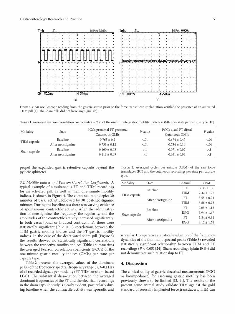
3.1. Gastric Retention. Prior to the force transducer implanta-tion, the electrical activity or lack thereof of the ingested pillwas verified with an oscilloscope. Figure 3(a) shows sampletracing of an active pill in the stomach measured from theserosa, while Figure 3(b) depicts sample oscilloscope tracingof an inactive sham pill. Final verification of the pill’s activityat the conclusion of each experiment revealed that all TIIMpills had remained active for the entire duration of the test,and conversely, sham pills remained inactive as anticipated.
In each animal the expanded pill was retrieved fromthe stomach at the end of the experiment, with an aver-age postretrieval volume of 12.1 ± 0.4mL and dimensionsexceeding 1.5 cm in all directions, indicating that even theneostigmine-invoked contractions had not been able to
Gastroenterology Research and Practice 5
(a) (b)
Figure 3: An oscilloscope reading from the gastric serosa prior to the force transducer implantation verified the presence of an activatedTIIM pill (a). The sham pills did not have any signal (b).
Table 1: Averaged Pearson correlation coefficients (PCCs) of the one-minute gastric motility indices (GMIs) per state per capsule type [17].
Modality State PCCs proximal FT-proximal𝑃 value PCCs distal FT-distal
𝑃 valueCutaneous GMIs Cutaneous GMIs
TIIM capsule Baseline 0.763 ± 0.2 <.01 0.674 ± 0.47 <.01After neostigmine 0.731 ± 0.12 <.01 0.734 ± 0.14 <.01
Sham capsule Baseline 0.160 ± 0.03 >.1 0.071 ± 0.02 >.1After neostigmine 0.113 ± 0.09 >.1 0.051 ± 0.03 >.1
propel the expanded gastric-retentive capsule beyond thepyloric sphincter.
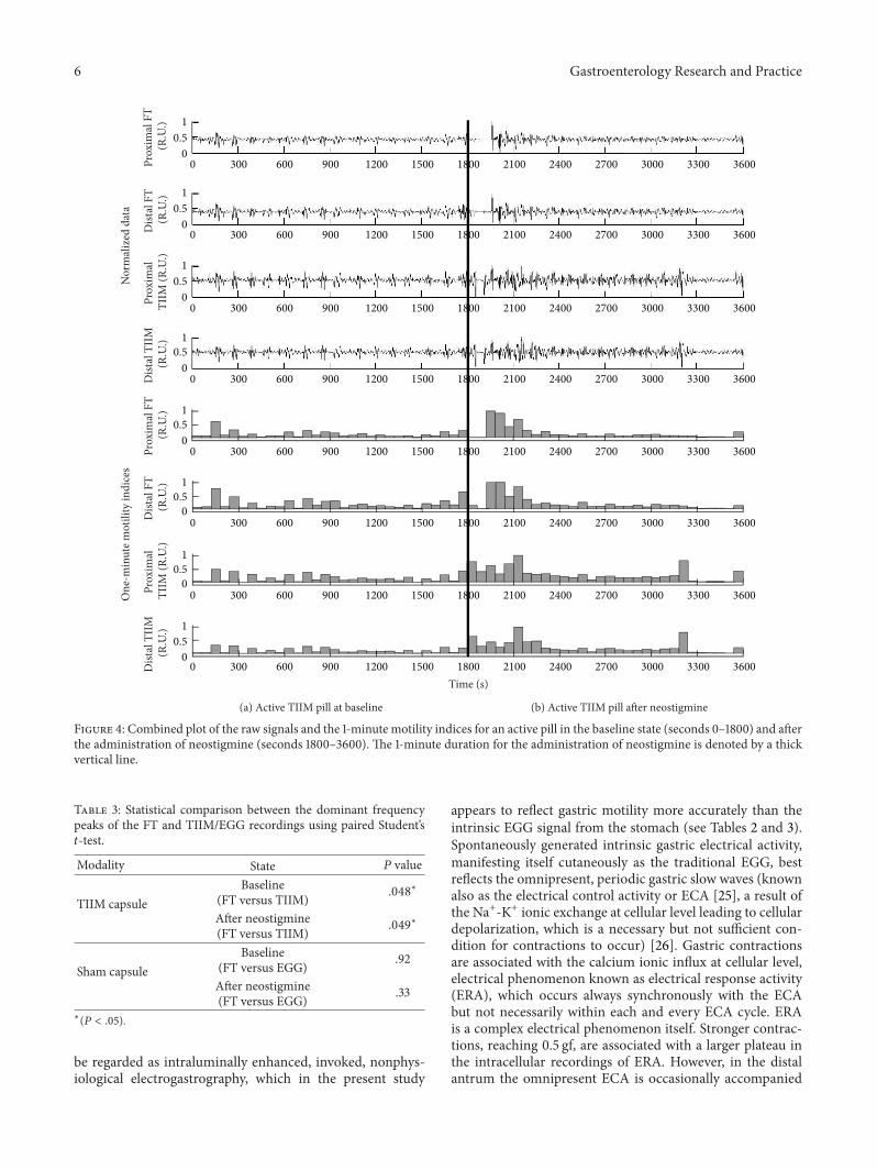
3.2. Motility Indices and Pearson Correlation Coefficients. Atypical example of simultaneous FT and TIIM recordingsfor an activated pill, as well as their one-minute motilityindices, is shown in Figure 4. The combined plots depict 30minutes of basal activity, followed by 30 post-neostigmineminutes. During the baseline test there was varying evidenceof spontaneous contractile activity. After the administra-tion of neostigmine, the frequency, the regularity, and theamplitudes of the contractile activity increased significantly.In both cases (basal or induced contractions), there werestatistically significant (𝑃 < 0.05) correlations between theTIIM gastric motility indices and the FT gastric motilityindices. In the case of the deactivated sham pill (Figure 5)the results showed no statistically significant correlationsbetween the respective motility indices. Table 1 summarizesthe averaged Pearson correlation coefficients (PCCs) of theone-minute gastric motility indices (GMIs) per state percapsule type.
Table 2 presents the averaged values of the dominantpeaks of the frequency spectra (frequency range 0.03–0.1Hz)of all recorded signals permodality (FT, TIIM, or sham-basedEGG). The substantial dissociation between the averageddominant frequencies of the FT and the electrical recordingsin the sham capsule study is clearly evident, particularly dur-ing baseline when the contractile activity was sporadic and
Table 2: Averaged cycles per minute (CPM) of the raw forcetransducer (FT) and the cutaneous recordings per state per capsuletype.
Modality State Channel CPM
TIIM capsuleBaseline FT 2.38 ± 1.2
TIIM 2.42 ± 1.27
After neostigmine FT 3.55 ± 0.94
TIIM 3.58 ± 0.95
Sham capsuleBaseline FT 2.65 ± 1.15
EGG 3.94 ± 1.67
After neostigmine FT 3.84 ± 0.91
EGG 4.12 ± 1.56
irregular. Comparative statistical evaluation of the frequencydynamics of the dominant spectral peaks (Table 3) revealedstatistically significant relationship between TIIM and FTrecordings (𝑃 < 0.05) [24]. Sham recordings (plain EGG) didnot demonstrate such relationship to FT.
4. Discussion
The clinical utility of gastric electrical measurements (EGGor bioimpedance) for assessing gastric motility has beenpreviously shown to be limited [12, 14]. The results of thepresent acute animal study validate TIIM against the goldstandard of serosally implanted force transducers. TIIM can
6 Gastroenterology Research and Practice
Time (s)
Prox
imal
FT
(R.U
.)D
istal
FT
(R.U
.)Pr
oxim
al
TIIM
(R.U
.)D
istal
TII
M
(R.U
.)Pr
oxim
al F
T (R
.U.)
Dist
al F
T (R
.U.)
Prox
imal
TI
IM (R
.U.)
Dist
al T
IIM
(R
.U.)
(a) Active TIIM pill at baseline
Nor
mal
ized
dat
aO
ne-m
inut
e mot
ility
indi
ces
(b) Active TIIM pill after neostigmine
10.5
0
10.5
0
10.5
0
10.5
0
10.5
0
10.5
0
10.5
0
10.5
00 300 600 900 1200 1500 1800 2100 2400 2700 3000 3300 3600
0 300 600 900 1200 1500 1800 2100 2400 2700 3000 3300 3600
0 300 600 900 1200 1500 1800 2100 2400 2700 3000 3300 3600
0 300 600 900 1200 1500 1800 2100 2400 2700 3000 3300 3600
0 300 600 900 1200 1500 1800 2100 2400 2700 3000 3300 3600
0 300 600 900 1200 1500 1800 2100 2400 2700 3000 3300 3600
0 300 600 900 1200 1500 1800 2100 2400 2700 3000 3300 3600
0 300 600 900 1200 1500 1800 2100 2400 2700 3000 3300 3600
Figure 4: Combined plot of the raw signals and the 1-minute motility indices for an active pill in the baseline state (seconds 0–1800) and afterthe administration of neostigmine (seconds 1800–3600). The 1-minute duration for the administration of neostigmine is denoted by a thickvertical line.
Table 3: Statistical comparison between the dominant frequencypeaks of the FT and TIIM/EGG recordings using paired Student’s𝑡-test.
Modality State 𝑃 value
TIIM capsuleBaseline
(FT versus TIIM) .048∗
After neostigmine(FT versus TIIM) .049∗
Sham capsuleBaseline
(FT versus EGG) .92
After neostigmine(FT versus EGG) .33
∗(𝑃 < .05).
be regarded as intraluminally enhanced, invoked, nonphys-iological electrogastrography, which in the present study
appears to reflect gastric motility more accurately than theintrinsic EGG signal from the stomach (see Tables 2 and 3).Spontaneously generated intrinsic gastric electrical activity,manifesting itself cutaneously as the traditional EGG, bestreflects the omnipresent, periodic gastric slow waves (knownalso as the electrical control activity or ECA [25], a result ofthe Na+-K+ ionic exchange at cellular level leading to cellulardepolarization, which is a necessary but not sufficient con-dition for contractions to occur) [26]. Gastric contractionsare associated with the calcium ionic influx at cellular level,electrical phenomenon known as electrical response activity(ERA), which occurs always synchronously with the ECAbut not necessarily within each and every ECA cycle. ERAis a complex electrical phenomenon itself. Stronger contrac-tions, reaching 0.5 gf, are associated with a larger plateau inthe intracellular recordings of ERA. However, in the distalantrum the omnipresent ECA is occasionally accompanied

Gastroenterology Research and Practice 7
Time (s)
Prox
imal
FT
(R.U
.)D
istal
FT
(R.U
.)Pr
oxim
al
TIIM
(R.U
.)D
istal
TII
M
(R.U
.)Pr
oxim
al F
T (R
.U.)
Dist
al F
T (R
.U.)
Prox
imal
TI
IM (R
.U.)
Dist
al T
IIM
(R
.U.)
Nor
mal
ized
dat
aO
ne-m
inut
e mot
ility
indi
ces
10.5
0
10.5
0
10.5
0
10.5
0
10.5
0
10.5
0
10.5
0
10.5
00 300 600 900 1200 1500 1800 2100 2400 2700 3000 3300 3600
0 300 600 900 1200 1500 1800 2100 2400 2700 3000 3300 3600
0 300 600 900 1200 1500 1800 2100 2400 2700 3000 3300 3600
0 300 600 900 1200 1500 1800 2100 2400 2700 3000 3300 3600
0 300 600 900 1200 1500 1800 2100 2400 2700 3000 3300 3600
0 300 600 900 1200 1500 1800 2100 2400 2700 3000 3300 3600
0 300 600 900 1200 1500 1800 2100 2400 2700 3000 3300 3600
0 300 600 900 1200 1500 1800 2100 2400 2700 3000 3300 3600
(a) Inactive sham pill at baseline (b) Inactive sham pill after neostigmine
Figure 5: Combined plot of the raw signals and the 1-minute motility indices for an inactive pill in the baseline state (seconds 0–1800) andafter the administration of neostigmine (seconds 1800–3600). The 1-minute duration for the administration of neostigmine is denoted by athick vertical line.
not only by the ERA plateau but also by an additional high-frequency electrical signal superimposed over the latter. Theappearance of this higher frequency electrical signal, knownas “spikes,” has been associated with strong contractionsexceeding 0.5 gf [27, 28]. Therefore, it has been suggested toseparate the transmembrane dynamics of calcium ions intoslow calcium transmembranic channels (the plateau or type Ielectrical response activity, ERA-I) and fast calcium channels(spikes or type II electrical response activity, ERA-II) [27].Both ERA-I and ERA-II are not omnipresent, particularlyin fasting. Moreover, extracellularly the plateau (ERA-I) andthe spikes (ERA-II) get nonlinearly differentiated due tothe properties of the cellular membranes resembling “leakycapacitors” [28]. Thus, extracellularly, both ERA-I and ERA-II, although associatedwith contractions, becomeof low aver-age electrical power which is easily dissipated intraluminally
in the body and cannot consistently significantly contributeto the power dynamics of the cutaneously recorded EGG toa point of being reliably detected. Therefore, in the complexinteraction between the cutaneous representations of ECA,ERA-I and ERA-II, and the influence of numerous externalfactors including the body mass index of the patient, theskin impedance, andmotion andmotility artifacts, amplitudeand power dynamics of EGG cannot consistently and reliablyreflect amplitude and power dynamics of gastric contractions.In order to overcome this limitation of the otherwise attrac-tive and noninvasive EGG technique, we decided to positionwithin the lumen of the stomach a man-made oscillator ofelectrical power and frequency that would not be able todissipate in the body in such inconsistent fashion as theintrinsic, spontaneously generated gastric electrical activitydoes. Thus, the TIIM-based electrical signal reaches the
8 Gastroenterology Research and Practice
abdominal skin with easily detectable and reliably recordableparameters. The demodulation of this cutaneously recordedsignal does not present any technological problem and canbe achieved with an existing standard EGG amplifier. It isimportant to note that TIIM would not pick up or reflectERA of any type, since the electrical dissipation of the latterwould remain within the body. Instead, due to the movementof the gastric-retainable oscillator within the gastric lumencaused by contractions, the signal from this intraluminaloscillator would be amplitude-modulated in TIIM. So, TIIMis an electromechanical rather than an electrophysiologicalsignal.
It is worth noting that although the aim of the presentstudy was to further validate the TIIM technique, the shampill study was fundamentally a direct comparison betweengastric motility indices obtained via routine EGG and forcetransducers, since standard EGG bandpass filtering parame-ters (0.03–0.1 Hz) were used. In four animals no statisticallysignificant correlation was observed in the motility indicesbetween any of the channel pairs (FT-EGG) examined,further supporting our understanding that EGG cannotreliably monitor gastric motility due to its important and,in our opinion, unsurpassable limitation; the relatively lowaverage electric-power of extracellular ERA-I and ERA-IIpreceding mechanically relevant gastric contractions dissi-pates intraluminally in an inconsistent and nonlinear fashionbefore reaching the abdominal skin surface and thus cannotbe reliably recorded by traditional cutaneous EGG. Withthe electrically isolated, gastric-retentive pill-based designpresented in this study, it is possible that environmentalelectrical phenomena such as capsule tumbling due tomotionnot associated with gastric contractions, static electricity,and external electromagnetic signals may affect the measure-ments in an ambulatory setting, particularly if the patientis mobile in an electromagnetically dynamic environment.In addition, in an ambulatory setting it is possible thatabdominal muscle contractions and motion artifacts mightalso be registered. In such cases, adaptive filtering procedures[29] might help to eliminate the unwanted phenomena butat the price of increasing the complexity of the method andthe number of recorded channels. Further considerationsand potential advanced digital signal processing might berequired postprandially, since solid or liquidmeals could alterthe base impedance and affect the efficacy of TIIM, callingfor more powerful digital filtering and dynamic amplificationof the TIIM signal. The limitations of the present study andindeed of this newTIIM technique require further evaluationto examine their impact on the clinical efficacy of this novelidea.
It should be noted that in the case of the inactive shamstudy small noise artifacts appear dramatic since there isno 50 kHz carrier signal as a reference. In fact, these noiseartifacts were much lower (10–100 times) than the measure-ments from the active TIIM study. However, since the signalswere normalized (the highest peaks were equated to unityand the lowest were equated to zero) before visualizationand subsequent presentation, electrostatic noises affectedthe cutaneous recordings of the sham TIIM signals, andthey appeared as enlarged white noise. Unfortunately, we
cannot avoid normalization, because there is no exact way todetermine the basal impedance the TIIM electrical oscillatorfaces, which can also be dynamically changing, particularlyin chronic studies of longer duration. Regardless, it shouldbe noted that if the sham TIIM tracings were displayed onthe same scale as the active TIIM figure, they would appearas virtually flat lines. In future long-term (e.g., 24 h) chronicgastric motility studies utilizing TIIM, we do not perceive thenormalization of the TIIM data to pose a significant problem,since the interdigestive motor complex and postprandialcontractility would be clearly seen, particularly if a dataloggermarks themeal periodswhile recording the TIIMdata.
The next steps for TIIM involve the development of aportable data-logger to enable ambulatory measurements inlong-term chronic tests, which can examine the effectivenessof themethod inmore clinically relevant settings, in dynamic,noise-ridden environments and during meals. Further long-term chronic animal studies (>24 h) should be considered,potentially evaluating gastric motility and gastric emptyingusing TIIM, before moving to chronic human trials.
5. Conclusion
Transcutaneous intraluminal impedance measurement(TIIM) is a minimally invasive gastric motility monitoringtechnique which has now been validated in a sham-comparative study. Acute canine models confirmed thatTIIM was able to measure gastric motility with comparableprecision to force transducers implanted invasively to theserosa of the stomach.
Disclosure
There are no financial arrangements to disclose at this time.
Conflict of Interests
The authors declare that there is no conflict of interestsregarding the publication of this paper.
Acknowledgment
This paper was supported in part by the Natural Sciences andEngineering Research Council of Canada.
References
[1] J. Tack, “Gastric motility disorders,” in Textbook of ClinicalGastroenterology and Hepatology, C. J. Hawkey, J. Bosch, J. E.Richter, G. Garcia-Tsao, and F. K. Chan, Eds., pp. 265–271, JohnWiley & Sons, Hoboken, NJ, USA, 2012.
[2] B. E. Lacy, “Functional dyspepsia and gastroparesis: one diseaseor two,” American Journal of Gastroenterology, vol. 107, no. 11,pp. 1615–1620, 2012.
[3] H. P. Parkman, W. L. Hasler, and R. S. Fisher, “American gas-troenterological association technical review on the diagnosisand treatment of gastroparesis,”Gastroenterology, vol. 127, no. 5,pp. 1592–1622, 2004.
Gastroenterology Research and Practice 9
[4] H. P. Parkman, M. Camilleri, G. Farrugia et al., “Gastroparesisand functional dyspepsia: excerpts from theAGA/ANMSmeet-ing,”Neurogastroenterology&Motility, vol. 22, no. 2, pp. 113–133,2010.
[5] S. S. C. Rao, M. Camilleri, W. L. Hasler et al., “Evaluation ofgastrointestinal transit in clinical practice: position paper of theAmerican and European Neurogastroenterology and MotilitySocieties,” Neurogastroenterology and Motility, vol. 23, no. 1, pp.8–23, 2011.
[6] T. L. Abell, M. Camilleri, K. Donohoe et al., “Consensusrecommendations for gastric emptying scintigraphy: a jointreport of the American neurogastroenterology and motilitysociety and the society of nuclear medicine,” American Journalof Gastroenterology, vol. 103, no. 3, pp. 753–763, 2008.
[7] L. A. Szarka and M. Camilleri, “Methods for measurementof gastric motility,” The American Journal of Physiology—Gastrointestinal and Liver Physiology, vol. 296, no. 3, pp. G461–G475, 2009.
[8] D. Cassilly, S. Kantor, L. C. Knight et al., “Gastric emptying ofa non-digestible solid: assessment with simultaneous SmartPillpH and pressure capsule, antroduodenal manometry, gastricemptying scintigraphy,” Neurogastroenterology & Motility, vol.20, no. 4, pp. 311–319, 2008.
[9] N. M. Devanarayana, S. Rajindrajith, M. S. Perera, S. W. Nis-hanthanie, and M. A. Benninga, “Gastric emptying and antralmotility parameters in children with functional dyspepsia:association with symptom severity,” Journal of Gastroenterologyand Hepatology, vol. 28, no. 7, pp. 1161–1166, 2013.
[10] A. E. Bharucha and R. C. Grimm, “Magnetic resonance imagingfor gastric motility,” in Gastroparesis, H. P. Parkman and R. W.McCallum, Eds., pp. 139–151, Humana Press, Chicago, Ill, USA,2012.
[11] O. Borrelli, V. Giorgio, andN.Thapar, “Antroduodenalmanom-etry,” in Pediatric Neurogastroenterology, C. Faure, C. DiLorenzo, and N. Thapar, Eds., pp. 91–105, Humana Press,Chicago, Ill, USA, 2013.
[12] A. J. P. M. Smout, H. J. A. Jebbink, L. M. A. Akkermans, and P. P.M. Bruijs, “Role of electrogastrography and gastric impedancemeasurements in evaluation of gastric emptying and motility,”Digestive Diseases and Sciences, vol. 39, supplement 12, pp. 110S–113S, 1994.
[13] R. Huerta-Franco, M. Vargas-Luna, E. Hernandez, K. Capac-cione, and T. Cordova, “Use of short-term bio-impedance forgastric motility assessment,” Medical Engineering and Physics,vol. 31, no. 7, pp. 770–774, 2009.
[14] A. J. Bredenoord and A. J. P. M. Smout, “Advances in motilitytesting—current and novel approaches,” Nature Reviews Gas-troenterology and Hepatology, vol. 10, no. 8, pp. 463–472, 2013.
[15] A. J. Arriagada, A. S. Jurkov, E. Neshev, G. Muench, C. N.Andrews, and M. P. Mintchev, “Design, implementation andtesting of an implantable impedance-based feedback-controlledneural gastric stimulator,” Physiological Measurement, vol. 32,no. 8, pp. 1103–1115, 2011.
[16] M. D. Poscente, G. Wang, D. Filip et al., “Real-time gas-tric motility monitoring using transcutaneous intraluminalimpedance measurements (TIIM),” Physiological Measurement,vol. 35, no. 2, pp. 217–229, 2014.
[17] G. Wang, M. D. Poscente, D. Filip et al., “Mo1302 gastric-retentive transcutaneous intraluminal impedance measure-ment (TIIM): sham controlled, minimally-invasive assessmentof gastric motility in acute canine models,” Gastroenterology,vol. 146, no. 5, p. S-613, 2014.
[18] M. G. Deneva, O. Yadid-Pecht, M. Fattouche et al., “Utilizationof temporary controllable intragastric pseudobezoars for thetreatment of obesity,” Current Obesity Reports, vol. 1, no. 2, pp.68–74, 2012.
[19] J. A. Hall, C. I. Dunlop, T. N. Solie, D. S. Hodgson, and D. C.Twedt, “Gastric myoelectric and motor activity in dogs afterisoflurane anesthesia,” Veterinary Surgery, vol. 24, no. 5, pp.456–463, 1995.
[20] D. A. Reinke, A. H. Rosenbaum, and D. R. Bennett, “Patternsof dog gastrointestinal contractile activity monitored in vivowith extraluminal force transducers,” The American Journal ofDigestive Diseases, vol. 12, no. 2, pp. 113–141, 1967.
[21] W. C. Kee, Y. J. Kingma, M. P. Mintchev, and K. L. Bowes,“Optimal placement of impedance epigastrography electrodes,”Annals of Biomedical Engineering, vol. 24, no. 2, pp. 328–332,1996.
[22] K. A. Kelly, C. F. Code, and L. R. Elveback, “Patterns of caninegastric electrical activity,” American Journal of Physiology, vol.217, no. 2, pp. 461–470, 1969.
[23] T. J. Stemper and A. R. Cooke, “Gastric emptying and itsrelationship to antral contractile activity,” Gastroenterology, vol.69, no. 3, pp. 649–653, 1975.
[24] G. W. Snedecor and W. G. Cochran, Statistical Methods, IowaState University Press, Ames, Iowa, USA, 1967.
[25] S. K. Sarna andM. F. Otterson, “Gastrointestinal motility: somebasic concepts,” Pharmacology, vol. 36, no. 1, pp. 7–14, 1988.
[26] J. H. Szurszewski, “Electrical basis of gastrointestinal motility,”in Physiology of the Gastrointestinal Tract, L. R. Johnson, Ed.,Raven Press, New York, NY, USA, 1987.
[27] A. J. P. M. Smout, E. J. van der Schee, and J. L. Grashius, “Post-prandial and interdigestive gastric electrical activity in the dogrecorded by means of cutaneous electrodes,” in GastrointestinalMotility, J. Christensen, Ed., Raven Press, New York, NY, USA,1980.
[28] A. J. P. M. Smout,Myoelectric activity of the stomach: gastroelec-tromyography and electrogastrography [Ph.D. thesis], ErasmusMC: University Medical Center Rotterdam, 1980.
[29] N. I. Adochiei, V. David, and I. Tudosa, “Methods of electro-magnetic interference reduction in electrocardiographic signalacquisition,” Environmental Engineering and Management Jour-nal, vol. 10, no. 4, pp. 553–559, 2011.

Submit your manuscripts athttp://www.hindawi.com
Stem CellsInternational
Hindawi Publishing Corporationhttp://www.hindawi.com Volume 2014
Hindawi Publishing Corporationhttp://www.hindawi.com Volume 2014
MEDIATORSINFLAMMATION
of
Hindawi Publishing Corporationhttp://www.hindawi.com Volume 2014
Behavioural Neurology
EndocrinologyInternational Journal of
Hindawi Publishing Corporationhttp://www.hindawi.com Volume 2014
Hindawi Publishing Corporationhttp://www.hindawi.com Volume 2014
Disease Markers
Hindawi Publishing Corporationhttp://www.hindawi.com Volume 2014
BioMed Research International
OncologyJournal of
Hindawi Publishing Corporationhttp://www.hindawi.com Volume 2014
Hindawi Publishing Corporationhttp://www.hindawi.com Volume 2014
Oxidative Medicine and Cellular Longevity
Hindawi Publishing Corporationhttp://www.hindawi.com Volume 2014
PPAR Research
The Scientific World JournalHindawi Publishing Corporation http://www.hindawi.com Volume 2014
Immunology ResearchHindawi Publishing Corporationhttp://www.hindawi.com Volume 2014
Journal of
ObesityJournal of
Hindawi Publishing Corporationhttp://www.hindawi.com Volume 2014
Hindawi Publishing Corporationhttp://www.hindawi.com Volume 2014
Computational and Mathematical Methods in Medicine
OphthalmologyJournal of
Hindawi Publishing Corporationhttp://www.hindawi.com Volume 2014
Diabetes ResearchJournal of
Hindawi Publishing Corporationhttp://www.hindawi.com Volume 2014
Hindawi Publishing Corporationhttp://www.hindawi.com Volume 2014
Research and TreatmentAIDS
Hindawi Publishing Corporationhttp://www.hindawi.com Volume 2014
Gastroenterology Research and Practice
Hindawi Publishing Corporationhttp://www.hindawi.com Volume 2014
Parkinson’s Disease
Evidence-Based Complementary and Alternative Medicine
Volume 2014Hindawi Publishing Corporationhttp://www.hindawi.com