research article use of mechanical turk as a mapreduce...
TRANSCRIPT
Research ArticleUse of Mechanical Turk as a MapReduce Framework forMacular OCT Segmentation
Aaron Y. Lee,1,2 Cecilia S. Lee,1,2 Pearse A. Keane,2,3,4 and Adnan Tufail2,3,4
1Department of Ophthalmology, University of Washington, Seattle, WA 98104, USA2Medical Retina Service, Moorfields Eye Hospital NHS Foundation Trust, London EC1V 2PD, UK3Institute of Ophthalmology, University College London, London WC1E 6BT, UK4National Institute for Health Research Biomedical Research Centre for Ophthalmology,Moorfields Eye Hospital NHS Foundation Trust, London SE1 4TT, UK
Correspondence should be addressed to Adnan Tufail; [email protected]
Received 5 January 2016; Accepted 19 April 2016
Academic Editor: Yannis Athanasiadis
Copyright © 2016 Aaron Y. Lee et al. This is an open access article distributed under the Creative Commons Attribution License,which permits unrestricted use, distribution, and reproduction in any medium, provided the original work is properly cited.
Purpose. To evaluate the feasibility of using Mechanical Turk as a massively parallel platform to perform manual segmentations ofmacular spectral domain optical coherence tomography (SD-OCT) images using a MapReduce framework. Methods. A macularSD-OCT volume of 61 slice images was map-distributed to Amazon Mechanical Turk. Each Human Intelligence Task was set to$0.01 and required the user to draw five lines to outline the sublayers of the retinal OCT image after being shown example images.Each image was submitted twice for segmentation, and interrater reliability was calculated.The interface was created using customHTML5 and JavaScript code, and data analysis was performed using R. An automated pipeline was developed to handle the mapand reduce steps of the framework. Results. More than 93,500 data points were collected using this framework for the 61 imagessubmitted. Pearson’s correlation of interrater reliability was 0.995 (𝑝 < 0.0001) and coefficient of determination was 0.991. Thecost of segmenting the macular volume was $1.21. A total of 22 individual Mechanical Turk users provided segmentations, eachcompleting an average of 5.5 HITs. Each HIT was completed in an average of 4.43 minutes. Conclusions. AmazonMechanical Turkprovides a cost-effective, scalable, high-availability infrastructure for manual segmentation of OCT images.
1. Introduction
Crowdsourcing is a relatively novel technique involving thedistribution of work to a large group of people, typicallythrough online frameworks [1]. It allows the subdivision oftedious tasks into discrete tasks that can be completed indi-vidually. Amazon Mechanical Turk is the largest and mostpopular of the online crowdsourcing systems [2]. In this sys-tem, simple Human Intelligence Tasks (HITs) are submittedto online untrained users for a small compensation. Recentlyin computer science, the MapReduce programming modelhas caused a paradigm shift in the way that large data sets aredistributed in parallel within a computing cluster [3]. NotablyGoogle used the MapReduce framework to regenerate theirindex of the Internet, and the MapReduce framework hasbecome popularized as a generic framework to solve big data
problems in multicore cluster systems. In this study, our goalwas to utilize human intelligence as aMapReduce frameworkfor the segmentation of a macular optical coherence tomog-raphy (OCT) volume [4–6].
OCT is an important noninvasive diagnostic tool in thefield of ophthalmology [6] and in the management of age-related macular degeneration (AMD), the commonest causeof blindness in the developed world [7, 8]. OCT measure-ments such as retinal thickness, subretinal fluid, and pigmentepithelial detachment are important parameters in the diag-nosis and monitoring of various retinal diseases [9, 10] andare thus integral in both large-scale clinical trials and routineclinical practice [11]. However, automated measurementsprovided by the OCT software result in frequent errors inquantifying critical parameters such asmacular thickness andvolume [12, 13]. Errors of retinal boundary detection and
Hindawi Publishing CorporationJournal of OphthalmologyVolume 2016, Article ID 6571547, 6 pageshttp://dx.doi.org/10.1155/2016/6571547

2 Journal of Ophthalmology
(a)
(b) (c)
(d)
Figure 1: Examples of incorrect segmentations by automated software and user-interface for Mechanical Turk. Panel (a) is an example of amacula SD-OCT image with missing information (arrow) causing a sudden jump in the identification of the Internal-Limiting Membrane(ILM) by automated software included with Heidelberg Spectralis. Panels (b and c) show two similar macular OCT images with differentautomated segmentations caused by pigment epithelial detachment (arrow) and subretinal fibrosis (arrowhead). Panel (d) is a screenshot ofweb-based user-interface submitted to Amazon Mechanical Turk for manual segmentations.
thickness measurements have been reported as high as 92%in segmentation performed by the Stratus OCT system (CarlZeiss Meditec, Germany) [14]. Although spectral domainOCT (SD-OCT) is expected to produce more accurate mea-surements with higher resolution and less artifacts, the seg-mentation errors continue to be a significant problem inmea-suring macular thickness, particularly in eyes with pathology[15–17].
There has been increasing interest in ways to overcomeautomated segmentation errors. Publicly available imageanalysis software, OCTOR (Doheny Image Reading Center,Los Angeles), quantifies OCT-derived parameters after atrained OCT grader delineates the retinal boundaries ofinterest manually. The software calculates the distance inpixels between two manually drawn layers. Then using thedimensions of the B-scan image, the data is converted intoa thickness measurement [18]. Even though OCTOR is lesssubject to segmentation errors, it is time-consuming andimpractical for use in large-scale clinical trials.
Automated segmentations have been attempted usingdual-scale gradient or intensity information. Then the edgesof the boundary were optimized using a shortest path searchmethod [19]. Statistical models have been utilized for a morereliable automatic segmentation system [20]. Retinal layershave been segmented using seven features extracted fromthe OCT data with a random forest classifier [21]. Despitethese achievements in the field of automated segmentations,macular OCT images with complex subretinal pathology,intraretinal/subretinal fluid, or low signal to noise ratiocontinue to pose a challenge for computer vision (Figures1(a)–1(c)).
AmazonMechanical Turk and othermodalities of crowd-sourcing have been previously used in medical applicationsand demonstrated high level of accuracy in diagnostic accu-racy [22–24]. In ophthalmology, retinal fundus photographshave been recently analyzed and showed an accuracy level atleast comparable to automated programs and some trainedgraders [25]. To our knowledge, it has not been used to

Journal of Ophthalmology 3
attain segmentations in macular OCT images with complexpathology.We sought to achieve highly reliable segmentationby designing a system for distributedOCT segmentation overa scalable, human based infrastructure and to show proof ofconcept results.
2. Materials and Methods
Patient identifiers were stripped out completely and pseu-doanonymized, and on this basis and for retrospective use ofanonymized data in the UK formal ethics committee reviewis not required. However, consent was still obtained from allpatients in this study to use their OCT images for research.This study was conducted in accordance with the Declarationof Helsinki and the United Kingdom’s Data Protection Act.
A total of 61 individual macular SD-OCT images weretaken using a commercially available SD-OCT device (Spec-tralis, Heidelberg Engineering, Heidelberg, Germany) as partof routine medical care for AMD.The images were extractedusing commercially provided software (HeyexDICOM Inter-face, Heidelberg Engineering, Heidelberg, Germany), and noimage manipulation was performed.
A custom web-based user-interface was created withHyper Text Markup Language 5 (HTML5) and JavaScript toallow Mechanical Turk users to directly draw on the imagesthrough their web browser (Figure 1(d)). This interface gaveeach user an example image of segmentation by an expertretina trained physician. The JavaScript interface allowedcapture of the mouse input to draw segmentation lines on theprovided OCT image and captured timing data as the userdrew segmentation lines. Each user was instructed to draw 5lines to segment the provided image and they were requiredto spend at least an average of 15 seconds per line before theywere allowed to submit their work. No image enlargementor zoom was allowed and users were given 3 example seg-mentations provided by a trainedOCT grader using the samesystem. Each image was created as a separate HIT and thereward was set to $0.01 (USD). Mechanical Turk users wererequired to have a prior approval rate of 80% before beingallowed to participate in these HITs. In addition to the linesdrawn, data was collected on the time spent drawing each linesegment and time to completion of segmentation, and eachimage was submitted twice for segmentation.
After all segmentations were performed, the data wascollected and image processing was performed to enhancethe accuracy of the manual segmentations. This automatedanalysis pipeline used adjustments based on finding theconsistently highest contrast value within 5 pixels of wherethe segmentation line was drawn. If there were no improvedchanges detected, then data from the original manual seg-mentation was used. Automated quality control heuristicswere implemented to ensure that no two segmentations fromthe same user of the same image crossed paths.The reductionstep of combining consensus segmentations of the sameimage was classified using a linear correlation heuristic, andthese segmentation data were used to calculate interraterreliability. The final reduction step was utilized to recreate athree-dimensional segmented model of the retina. CustomRuby and R code was created to automate the creation,
submission, collection, image processing, and data analysis.All custom software is available upon request.
3. Results
The automated analysis pipeline, using a MapReduce frame-work, was able to create, submit, collect, collate, process,and analyze a total of over 92,500 data points from the 61macular OCT images that were manually segmented twiceover Amazon Mechanical Turk. Time of submission of the122 HITs to completion of all tasks was 3 days with greaterthan 75% of HITs finished within the first 24 hours. A total of22 individual Mechanical Turk users provided segmentationseach completing an average of 5.5 HITs.
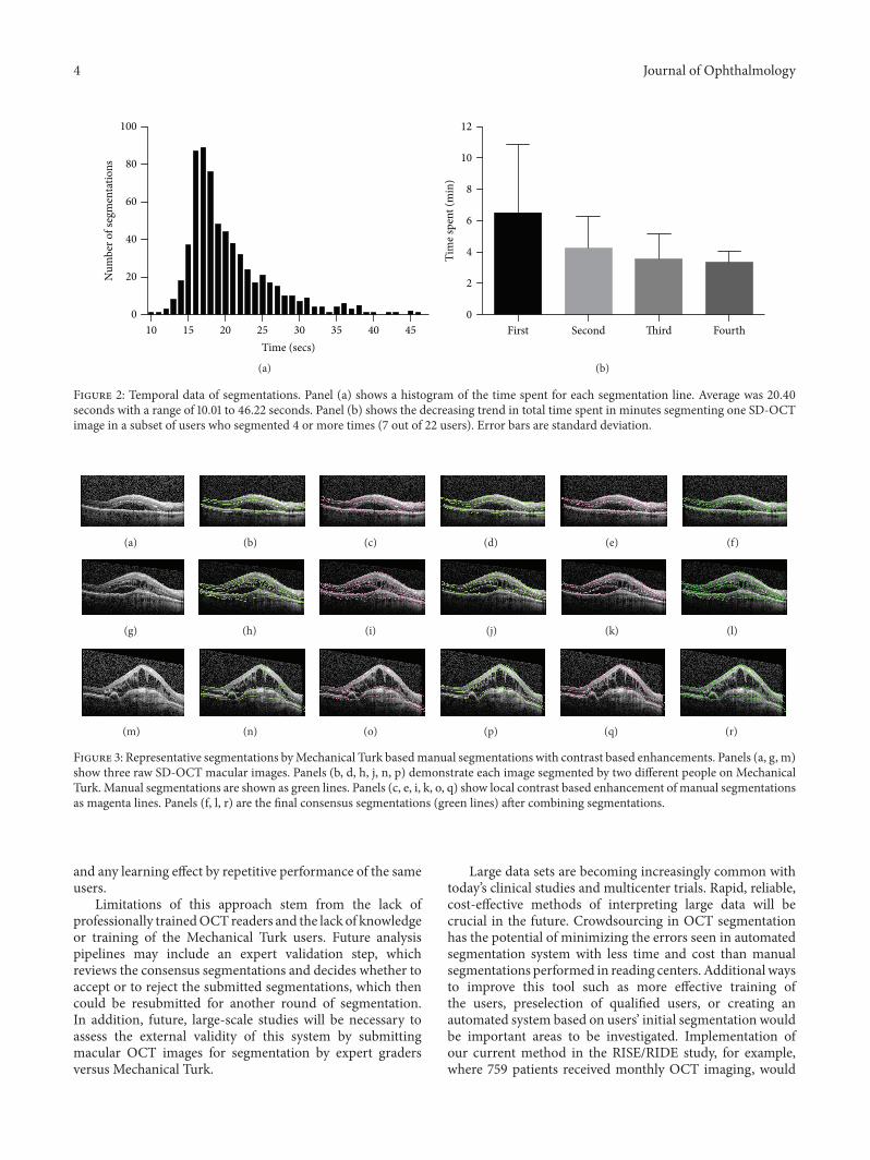
Each HIT was completed on average of 4.43 minutes(range: 1.83–24.45 minutes) with each segmentation linecompleted on average of 20.40 seconds (Figure 2(a)). In asubset of users who had segmented four or more HITs, wenoted that there was a trend in decreasing time to completionof the task (Figure 2(b)). A total of 646 segmentationswere collected, and an average of 5.30 segmentations permacular OCT was provided (range: 5 to 7). The total cost ofsegmentations of all images was $1.22 (USD).
Representative segmentations with the associated imageprocessing are shown in Figure 3. All slices from bothmanualsegmentation and the combined final segmentations areshown in the Supplementary Materials (see SupplementaryMaterial available online at http://dx.doi.org/10.1155/2016/6571547). Pearson’s correlation of interrater reliability was0.995 (𝑝 < 0.0001) and coefficient of determination was0.991. A Bland-Altman plot was calculated to estimate inter-rater agreement based on the consensus segmentation lines(Figure 4).
4. Discussion
OCT is a critical tool in clinical practice for ophthalmology,and objective, quantitative OCT parameters have the poten-tial of guiding clinical practice and establishing new end-points for clinical trials. Automated segmentation approacheshave traditionally suffered in the setting of complex retinalpathology such as pigment epithelial detachments, subretinalfibrosis, or intraretinal and subretinal fluid. Indeed theautomated segmentation that is provided with the com-mercial device used in this study failed in many situations(Figures 1(a)–1(c)). With the advent of Mechanical Turkand programming APIs, automating simple human visiontasks through a MapReduce framework has become not onlyfeasible but also cost-effective. The advantages of utilizingmanual segmentations using human vision include the abilityto complete areas of macular OCT where there is poorsignal to noise ratio (Figure 3(a)) or complex pathology(Figure 3(c)).
Next steps of this study would be to compare the accuracyof the Mechanical Turk based segmentation to the onesperformed by trained experts. Using the segmentation linesperformed by trained experts as the gold standard, we willplan to evaluate the correlation between the accuracy andthe time spent by the users, previous experience of the users,
4 Journal of Ophthalmology
10 15 20 25 30 35 40 450
20
40
60
80
100
Time (secs)
Num
ber o
f seg
men
tatio
ns
(a)
First Second Third Fourth0
2
4
6
8
10
12
Tim
e spe
nt (m
in)
(b)
Figure 2: Temporal data of segmentations. Panel (a) shows a histogram of the time spent for each segmentation line. Average was 20.40seconds with a range of 10.01 to 46.22 seconds. Panel (b) shows the decreasing trend in total time spent in minutes segmenting one SD-OCTimage in a subset of users who segmented 4 or more times (7 out of 22 users). Error bars are standard deviation.
(a) (b) (c) (d) (e) (f)
(g) (h) (i) (j) (k) (l)
(m) (n) (o) (p) (q) (r)
Figure 3: Representative segmentations byMechanical Turk basedmanual segmentations with contrast based enhancements. Panels (a, g, m)show three raw SD-OCT macular images. Panels (b, d, h, j, n, p) demonstrate each image segmented by two different people on MechanicalTurk. Manual segmentations are shown as green lines. Panels (c, e, i, k, o, q) show local contrast based enhancement of manual segmentationsas magenta lines. Panels (f, l, r) are the final consensus segmentations (green lines) after combining segmentations.
and any learning effect by repetitive performance of the sameusers.
Limitations of this approach stem from the lack ofprofessionally trainedOCT readers and the lack of knowledgeor training of the Mechanical Turk users. Future analysispipelines may include an expert validation step, whichreviews the consensus segmentations and decides whether toaccept or to reject the submitted segmentations, which thencould be resubmitted for another round of segmentation.In addition, future, large-scale studies will be necessary toassess the external validity of this system by submittingmacular OCT images for segmentation by expert gradersversus Mechanical Turk.
Large data sets are becoming increasingly common withtoday’s clinical studies and multicenter trials. Rapid, reliable,cost-effective methods of interpreting large data will becrucial in the future. Crowdsourcing in OCT segmentationhas the potential of minimizing the errors seen in automatedsegmentation system with less time and cost than manualsegmentations performed in reading centers. Additional waysto improve this tool such as more effective training ofthe users, preselection of qualified users, or creating anautomated system based on users’ initial segmentation wouldbe important areas to be investigated. Implementation ofour current method in the RISE/RIDE study, for example,where 759 patients received monthly OCT imaging, would

Journal of Ophthalmology 5
−40
−20
0
20
900 1000 1100 1200 1300Mean y-coordinate (𝜇m)
Diff
eren
ce (𝜇
m)
Figure 4: Bland-Altman plot showing agreement between seg-mentations. Consensus segmentations of the same image betweentwo independent Mechanical Turk users were used to determineinterrater reliability. The average 𝑦-coordinate value in microns foreach consensus line was used and the Bland-Altman plot wascreated.
cost approximately $273.24 per study month for a standard18-slice macular OCT.
This study has applied a novel proof of concept of applyingmanual segmentation of OCT images in a distributed way tononexpert graders. The retinas with various pathologies pro-vide challenge to currently available automated segmentationsystems. Mechanical Turk provides a cost-effective, scalable,high-availability infrastructure for manual segmentation ofOCT images of the type which are difficult for automatedalgorithms to handle. The resulting images can be recom-bined for high-resolution 3D analysis. This approach may beapplied to the analysis of high volumes of OCT images inclinical studies or training of future automated segmentationalgorithms.
Competing Interests
The authors declare that they have no competing interests.
Acknowledgments
Cecilia S. Lee was supported by NIH Grant no. 1K23EY-024921.
References
[1] D. C. Brabham,K.M. Ribisl, T. R. Kirchner, and J.M. Bernhardt,“Crowdsourcing applications for public health,” American Jour-nal of Preventive Medicine, vol. 46, no. 2, pp. 179–187, 2014.
[2] M. J. C. Crump, J. V. McDonnell, and T. M. Gureckis, “Eval-uating Amazon’s Mechanical Turk as a tool for experimentalbehavioral research,” PLoS ONE, vol. 8, no. 3, Article ID e57410,2013.
[3] R. Bellazzi, “Big data and biomedical informatics: a challengingopportunity,” Yearbook of Medical Informatics, vol. 9, pp. 8–13,2014.
[4] R. C. Taylor, “An overview of the Hadoop/MapReduce/HBaseframework and its current applications in bioinformatics,” BMCBioinformatics, vol. 11, supplement 12, article S1, 2010.
[5] D. Huang, E. A. Swanson, C. P. Lin et al., “Optical coherencetomography,” Science, vol. 254, no. 5035, pp. 1178–1181, 1991.
[6] I. Voo, E. C. Mavrofrides, and C. A. Puliafito, “Clinical applica-tions of optical coherence tomography for the diagnosis andmanagement of macular diseases,” Ophthalmology Clinics ofNorth America, vol. 17, no. 1, pp. 21–31, 2004.
[7] R. Klein, T. Peto, A. Bird, andM. R. Vannewkirk, “The epidemi-ology of age-relatedmacular degeneration,”American Journal ofOphthalmology, vol. 137, no. 3, pp. 486–495, 2004.
[8] N. Congdon, B. O’Colmain, C. C. W. Klaver et al., “Causes andprevalence of visual impairment among adults in the UnitedStates,” Archives of Ophthalmology, vol. 122, no. 4, pp. 477–485,2004.
[9] D. M. Brown, P. K. Kaiser, M. Michels et al., “Ranibizumab ver-sus verteporfin for neovascular age-related macular degenera-tion,”The New England Journal of Medicine, vol. 355, no. 14, pp.1432–1444, 2006.
[10] P. J. Rosenfeld, D. M. Brown, J. S. Heier et al., “Ranibizumabfor neovascular age-related macular degeneration,” The NewEngland Journal ofMedicine, vol. 355, no. 14, pp. 1419–1431, 2006.
[11] D. M. Brown, Q. D. Nguyen, D. M. Marcus et al., “Long-termoutcomes of ranibizumab therapy for diabetic macular edema:the 36-month results from two phase III trials: RISE and RIDE,”Ophthalmology, vol. 120, no. 10, pp. 2013–2022, 2013.
[12] P. J. Patel, F. K. Chen, L. da Cruz, and A. Tufail, “Segmentationerror in Stratus optical coherence tomography for neovascularage-related macular degeneration,” Investigative Ophthalmologyand Visual Science, vol. 50, no. 1, pp. 399–404, 2009.
[13] P. A. Keane, P. J. Patel, S. Liakopoulos, F. M. Heussen, S. R.Sadda, and A. Tufail, “Evaluation of age-related macular degen-eration with optical coherence tomography,” Survey of Ophthal-mology, vol. 57, no. 5, pp. 389–414, 2012.
[14] P. A. Keane, S. Liakopoulos, R. V. Jivrajka et al., “Evaluation ofoptical coherence tomography retinal thickness parameters foruse in clinical trials for neovascular age-related macular degen-eration,” Investigative Ophthalmology andVisual Science, vol. 50,no. 7, pp. 3378–3385, 2009.
[15] Y. Song, B. R. Lee, Y. W. Shin, and Y. J. Lee, “Overcoming seg-mentation errors in measurements of macular thickness madeby spectral-domain optical coherence tomography,” Retina(Philadelphia, Pa), vol. 32, no. 3, pp. 569–580, 2012.
[16] M. Kim, S. Lee, J. Han, S.-Y. Yu, and H. Kwak, “Segmenta-tion error and macular thickness measurements obtained withspectral-domain optical coherence tomography devices in neo-vascular age-related macular degeneration,” Indian Journal ofOphthalmology, vol. 61, no. 5, pp. 213–217, 2013.
[17] P. A. Keane, P. S. Mand, S. Liakopoulos, A. C. Walsh, and S. R.Sadda, “Accuracy of retinal thickness measurements obtainedwith Cirrus optical coherence tomography,” British Journal ofOphthalmology, vol. 93, no. 11, pp. 1461–1467, 2009.
[18] S. R. Sadda, S. Joeres, Z. Wu et al., “Error correction and quan-titative subanalysis of optical coherence tomography data usingcomputer-assisted grading,” Investigative Ophthalmology &Visual Science, vol. 48, no. 2, pp. 839–848, 2007.

6 Journal of Ophthalmology
[19] Q. Yang, C. A. Reisman, Z. Wang et al., “Automated layer seg-mentation of macular OCT images using dual-scale gradientinformation,” Optics Express, vol. 18, no. 20, pp. 21293–21307,2010.
[20] V. Kajic, B. Povazay, B. Hermann et al., “Robust segmentationof intraretinal layers in the normal human fovea using a novelstatistical model based on texture and shape analysis,” OpticsExpress, vol. 18, no. 14, pp. 14730–14744, 2010.
[21] A. Lang, A. Carass, E. Sotirchos, P. Calabresi, and J. L. Prince,“Segmentation of retinal OCT images using a random forestclassifier,” in Proceedings of the Medical Imaging: Image Process-ing, vol. 8669 ofProceedings of SPIE, Lake BuenaVista, Fla,USA,February 2013.
[22] R. R. Carter, A. DiFeo, K. Bogie, G.-Q. Zhang, and J. Sun,“Crowdsourcing awareness: exploration of the ovarian cancerknowledge gap through AmazonMechanical Turk,” PLoS ONE,vol. 9, no. 1, Article ID e85508, 2014.
[23] S. Mavandadi, S. Dimitrov, S. Feng et al., “Distributed medicalimage analysis and diagnosis through crowd-sourced games: amalaria case study,” PLoS ONE, vol. 7, no. 5, Article ID e37245,2012.
[24] T. B. Nguyen, S. Wang, V. Anugu et al., “Distributed humanintelligence for colonic polyp classification in computer-aideddetection for CT colonography,” Radiology, vol. 262, no. 3, pp.824–833, 2012.
[25] D. Mitry, T. Peto, S. Hayat, J. E. Morgan, K.-T. Khaw, and P. J.Foster, “Crowdsourcing as a novel technique for retinal fundusphotography classification: analysis of images in the EPIC nor-folk cohort on behalf of the UKBiobank eye and vision consor-tium,” PLoS ONE, vol. 8, no. 8, Article ID e71154, 2013.

Submit your manuscripts athttp://www.hindawi.com
Stem CellsInternational
Hindawi Publishing Corporationhttp://www.hindawi.com Volume 2014
Hindawi Publishing Corporationhttp://www.hindawi.com Volume 2014
MEDIATORSINFLAMMATION
of
Hindawi Publishing Corporationhttp://www.hindawi.com Volume 2014
Behavioural Neurology
EndocrinologyInternational Journal of
Hindawi Publishing Corporationhttp://www.hindawi.com Volume 2014
Hindawi Publishing Corporationhttp://www.hindawi.com Volume 2014
Disease Markers
Hindawi Publishing Corporationhttp://www.hindawi.com Volume 2014
BioMed Research International
OncologyJournal of
Hindawi Publishing Corporationhttp://www.hindawi.com Volume 2014
Hindawi Publishing Corporationhttp://www.hindawi.com Volume 2014
Oxidative Medicine and Cellular Longevity
Hindawi Publishing Corporationhttp://www.hindawi.com Volume 2014
PPAR Research
The Scientific World JournalHindawi Publishing Corporation http://www.hindawi.com Volume 2014
Immunology ResearchHindawi Publishing Corporationhttp://www.hindawi.com Volume 2014
Journal of
ObesityJournal of
Hindawi Publishing Corporationhttp://www.hindawi.com Volume 2014
Hindawi Publishing Corporationhttp://www.hindawi.com Volume 2014
Computational and Mathematical Methods in Medicine
OphthalmologyJournal of
Hindawi Publishing Corporationhttp://www.hindawi.com Volume 2014
Diabetes ResearchJournal of
Hindawi Publishing Corporationhttp://www.hindawi.com Volume 2014
Hindawi Publishing Corporationhttp://www.hindawi.com Volume 2014
Research and TreatmentAIDS
Hindawi Publishing Corporationhttp://www.hindawi.com Volume 2014
Gastroenterology Research and Practice
Hindawi Publishing Corporationhttp://www.hindawi.com Volume 2014
Parkinson’s Disease
Evidence-Based Complementary and Alternative Medicine
Volume 2014Hindawi Publishing Corporationhttp://www.hindawi.com