research article visual parsing after recovery from...
TRANSCRIPT

Research Article
Visual Parsing After RecoveryFrom BlindnessYuri Ostrovsky,1 Ethan Meyers,1 Suma Ganesh,2 Umang Mathur,2 and Pawan Sinha1
1Department of Brain and Cognitive Sciences, Massachusetts Institute of Technology, and 2Dr. Shroff’s Charity Eye
Hospital, Daryaganj, New Delhi, India
ABSTRACT—How the visual system comes to bind diverse
image regions into whole objects is not well understood. We
recently had a unique opportunity to investigate this issue
when we met three congenitally blind individuals in India.
After providing them treatment, we studied the early
stages of their visual skills. We found that prominent fig-
ural cues of grouping, such as good continuation and
junction structure, were largely ineffective for image
parsing. By contrast, motion cues were of profound sig-
nificance in that they enabled intraobject integration and
facilitated the development of object representations that
permitted recognition in static images. Following 10 to 18
months of visual experience, the individuals’ performance
improved, and they were able to use the previously in-
effective static figural cues to correctly parse many static
scenes. These results suggest that motion information
plays a fundamental role in organizing early visual expe-
rience and that parsing skills can be acquired even late in
life.
Individuals who acquire sight late in life provide a unique
window into several aspects of visual development. Such cases,
however, are extremely rare; fewer than 30 have been studied in
any detail over the course of the past 1,000 years (Valvo, 1971).
Through a concerted effort to locate such individuals in un-
derprivileged enclaves in India, a country with an estimated
25% of the world’s blind, we have been able to conduct longi-
tudinal studies from sight onset up to 18 months later with 3 such
patients, S.K., J.A., and P.B. These studies provide an oppor-
tunity to add to this important but sparse body of work (Fine et
al., 2003; Gregory & Wallace, 1963; Maurer, Lewis, & Mond-
loch, 2005; Von Senden, 1932/1960).
Real-world images typically comprise many regions of
different colors and luminances (see Fig. 1). The human visual
system is adept at integrating subsets of these regions into
meaningful entities. How this is achieved is a fundamental
question, and has been researched extensively in the domains of
experimental and computational neuroscience (Brady & Ker-
sten, 2003; Hummell & Biederman, 1992; Hupe et al., 1998;
Marr, 1982; Needham, 2001; Tu, Chen, Yuille, & Zhu, 2003;
Ullman, 1996; Wertheimer, 1938). Much of this work has fo-
cused on the use of heuristics, such as alignment of contours and
similarity of texture statistics (August, Siddiqi, & Zucker, 1999;
Field, Hayes, & Hess, 1993; Grossberg & Mingolla, 1985; Ko-
vacs & Julesz, 1993; Leung & Malik, 1998; Mumford & Shah,
1985). In circumscribed domains, these heuristics can account
rather well for human performance (Elder & Zucker, 1998;
Kanizsa, 1979; Koffka, 1935), but using them for analyzing real-
world imagery remains an open challenge (Borenstein & Ull-
man, 2002; Shi & Malik, 1997). Furthermore, although it is
evident that a mature visual system makes use of these heuris-
tics, it is unclear whether they serve to organize visual infor-
mation during the early stages of development. Determining the
nature of the cues that are active at that time is important for
elucidating the principles of visual learning and bootstrapping.
Our studies with 3 individuals immediately after they first
experienced patterned vision provided a rare opportunity to
examine the bootstrapping mechanisms for visual parsing and
the progression of visual abilities due to visual experience.
These studies were undertaken as part of Project Prakash, our
initiative in India to identify, and provide medical care to, in-
dividuals with treatable congenital blindness (Mandavilli,
2006). In working with these individuals, we also have an op-
portunity to examine how time bound the development of parsing
skills is, and whether it is subject to a critical period. Earlier
case reports have demonstrated that individuals who acquire
sight late in life show a profound deficit in interpreting the visual
confusion that they suddenly encounter (Fine et al., 2003;
Gregory & Wallace, 1963; Valvo, 1971; Von Senden, 1932/
Address correspondence to Pawan Sinha, 46-4077, Department ofBrain and Cognitive Sciences, Massachusetts Institute of Technology,Cambridge, MA 02139, e-mail: [email protected].
PSYCHOLOGICAL SCIENCE
Volume ]]]—Number ]] 1Copyright r 2009 Association for Psychological Science

1960). These results appear to suggest that visual parsing
might be subject to a critical period in the first few years of life.
Despite the lack of conclusive evidence for the permanence of
deprivation-induced deficits, individuals who have been blind
past the age of 5 or 6 years but have treatable conditions (a
situation that is virtually nonexistent in developed nations, but
unfortunately not as rare in the developing world) are often
passed over for treatment owing to the assumed poor prognosis
for recovery.
It is worth noting that working with a noninfant population
provides us with both advantages and disadvantages. On the one
hand, because the brain is otherwise almost fully mature in the
individuals we work with, visual learning can be segregated from
development of the other senses and from real-world knowledge.
On the other hand, a mature brain may not undergo the same
progression as an infant brain. Thus, it is appropriate to consider
this work as complementary to, rather than a replacement for,
traditional studies with infants.
METHOD
Participants
S.K. is a 29-year-old male, born in Bihar, India. By the time he
was 4 months old, members of his family noticed his inability to
fixate and a lack of visually guided behaviors. Because of fi-
nancial and logistical constraints, medical intervention was not
sought until S.K. was an adolescent. At the age of 12 years, he
was examined by an ophthalmologist, who recommended sur-
gery to correct his sight. However, the operation was canceled
because S.K.’s father had an illness that completely depleted the
family’s finances. S.K. was admitted to the State School for the
Blind in Darbangha, Bihar, where he studied for 12 years and
learned Braille. In 2000, he moved to a hostel for the blind in
New Delhi and enrolled in a correspondence course; he earned a
master’s degree in political science in April 2006. It was during
a visit to this hostel that we met S.K. in January 2004.
Examinations by three independent ophthalmologists in New
Delhi yielded identical assessments: S.K. has secondary con-
genital bilateral aphakia (B.L. Johnson & Cheng, 1997; Pratt &
Richards, 1968), with the lenses almost completely absorbed in
the anterior and posterior chambers of the right and left eye
respectively. The optical pathways in the eyes are clear. S.K.’s
acuity was assessed to be 20/900. He had never been able to
afford a pair of eyeglasses that could compensate for his apha-
kia. During our next visit to India, in July 2004, we had S.K.
reexamined by optometrists and ophthalmologists in New Delhi
and purchased a pair of eyeglasses for him. Postcorrection
acuity was determined to be 20/120. The residual acuity im-
pairment is likely due to neural amblyopia (Kiorpes & McKee,
1999).
Beginning 2 weeks after the refractive correction, we per-
formed a series of experiments to assess S.K.’s visual abilities.
Tests of low-level visual function revealed that he had near-
normal ability to discriminate among colors, luminances, and
motion directions.
We also had the opportunity to work with 2 male children, P.B.
and J.A., whom we studied beginning 1 and 3 months, respec-
tively, after surgery to correct their dense bilateral congenital
cataracts. P.B. received treatment at the age of 7 years, and J.A.
at the age of 13 years. P.B. was born in a village near Panipat,
Haryana. His family has a long history of congenital blindness.
Both P.B. and his older sister T.B. (age 12) were congenitally
blind, as were his father, his paternal grandmother, his great-
grandmother, two aunts, and an uncle. P.B. has been enrolled in
the Blind Relief Association’s school in Delhi since the age of
4½ years. His parents did not pursue treatment for him (or T.B.)
because a doctor incorrectly told them that his condition was
untreatable because of the development of nystagmus. A
botched eye surgery that P.B.’s uncle had undergone a few years
earlier further dampened the parents’ desire to seek treatment
for their children. We came across P.B. in an outreach eye-
screening session we had organized in his school. His condition
was determined to be treatable. In December 2005, P.B. un-
derwent a small-incision cataract surgery with intraocular lens
implantation in both eyes; as a result, his acuity improved from
light perception to 20/100.
Fig. 1. Example illustrating how a natural image (a) is typically a collection of many regions of different huesand luminances (b). The human visual system has to accomplish the task of integrating subsets of these regionsinto coherent objects, illustrated in (c).
2 Volume ]]]—Number ]]
Visual Parsing After Recovery From Blindness

J.A. was born in Bijnor, Uttar Pradesh. He has five siblings,
three sighted (ages 21, 19, and 8) and two congenitally blind
(ages 17 and 7). Both parents are illiterate, and J.A. has never
received any education. J.A. received cataract surgery and an
intraocular lens implant in both eyes (right eye: September
2005; left eye: October 2005); as a result, his acuity improved
from light perception to 20/80.
In what follows, we describe results from all 3 individuals.
Practical constraints allowed us to work with S.K. more thor-
oughly than with P.B. and J.A., preventing us from replicating
every experiment from the battery with the children. For con-
venience, we refer to S.K., J.A., and P.B. collectively as the re-
cently treated group.
S.K. volunteered his participation and was not paid, other
than being compensated for transportation costs. The families of
P.B. and J.A. were compensated for their transportation costs
and also for part of the wages they lost while in the hospital.
Subjects were free to take as many rest breaks as they wished
during the course of the testing. We also enlisted 4 normally
sighted adult control subjects. These subjects came from a so-
cial tier similar to that of our experimental group and had re-
ceived a basic education through high school.
Procedure
Tests of Static Visual Parsing
Our studies of static visual parsing comprised seven tests, which
assessed the subjects’ responses to images of simple shapes.
These tests were administered 2 weeks after S.K. received his
glasses and 1 month and 3 months postsurgery for P.B. and J.A.,
respectively. Their task was to say how many objects there were
in each image, point to where they were, and (whenever possible)
name them. (The recently treated group was already familiar
with common shape names through touch.) Figure 2a illustrates
the specific tasks and representative stimuli. Tests A, B, F, and G
were readministered to S.K., J.A., and P.B. 18 months, 12
months, and 10 months posttreatment, respectively (S.K. was
also tested 6 and 12 months posttreatment). Each of the seven
tests comprised 10 distinct trials. The recently treated subjects’
viewing distance averaged 40 cm. Control subjects’ viewing
distance was scaled to simulate image information loss in the
recently treated group. All stimuli were presented until a re-
sponse was given.
Test of Object Recognition
For this test, subjects were shown a set of 50 images of common
objects and were asked to name the objects they recognized. The
images were in color and had different backgrounds (see Fig. 3).
The images subtended 25 degrees of visual angle, on average.
There were no constraints on viewing time. As were the tests of
visual parsing, the recognition test was administered 2 weeks
after S.K. received his glasses and 1 month and 3 months
postsurgery for P.B. and J.A., respectively.
Tests of Dynamic Visual Parsing
The stimuli used to test dynamic visual parsing were similar to
those in the tests of visual parsing, but incorporated motion cues.
These tests were administered at the same time as the initial
tests of static visual parsing and the recognition test.
RESULTS
Figure 2b shows all subjects’ performance in the seven tests of
visual parsing. The responses of the recently treated group ex-
hibited a consistent pattern. They had no difficulty in enumer-
ating individual geometric shapes presented by themselves or in
the presence of other shapes that were nonoverlapping (Test A).
However, when the shapes overlapped, regardless of whether the
shapes were presented as line drawings or as filled transparent
surfaces (Test B and C), the recently treated subjects’ responses
were very different from control subjects’. They perceived all
closed loops and regions of uniform luminance as distinct ob-
jects. All errors we observed were such errors of overfragmen-
tation. Thus, for instance, when viewing two overlapping
squares, the recently treated subjects invariably parsed them as
three objects. Using lines of different colors or luminances as
potential aids for segmenting the component objects did not
change this pattern of results. Note that to ensure that these
subjects understood the task, we told them at the start that the
figures might be overlapping (a notion they were familiar with
from prior haptic experience) and that they had to indicate the
number of ‘‘objects,’’ rather than ‘‘regions.’’
When the images showed opaque overlapping shapes, S.K.
was able to correctly indicate their number (Test D), but per-
formed at chance in determining their depth ordering (Test E).
Extended contours made up of a series of separated line seg-
ments embedded in a field of randomly oriented line segments
(Test F) were only infrequently detected by the recently treated
subjects. When three-dimensional shapes, such as cubes or
pyramids, were shown with surfaces of different luminance
consistent with lighting and shadows (Test G), the recently
treated subjects reported perceiving multiple objects, one cor-
responding to each facet. They were unable to integrate the
facets into the percept of a single three-dimensional object.
In summary, the recently treated subjects’ performance in-
dicated a profound inability to use cues of contour continuation,
junction structure, and figural symmetry to analyze the images
presented. The subjects’ tendency to perceive the stimuli in a
fragmented manner was also reflected in their tracings of simple
figures (e.g., see Fig. 2c).
Next, we investigated the functional significance of the re-
cently treated subjects’ atypical image-parsing skills. Given
their pronounced tendency to overfragment images, we reasoned
that their ability to veridically segment and recognize real-world
images would be compromised. To test this hypothesis, we as-
sessed their naming performance using a set of 50 images of
common objects. S.K. was able to recognize only 26% of the
Volume ]]]—Number ]] 3
Y. Ostrovsky et al.

images, J.A. recognized 34%, and P.B. recognized only 18%. We
asked subjects to point to objects in these images and also to
indicate their extent, even if they could not name the objects. We
found that subjects’ responses were driven by low-level image
attributes; they pointed to regions of different hues and lumi-
nances as distinct objects. This approach greatly oversegmented
the images and partitioned them into meaningless regions,
which would be unstable across different views and uninfor-
mative regarding object identity. A robust object representation
is difficult to construct on the basis of such fragments. Figure 2d,
which shows S.K.’s responses to three sample images, illustrates
this tendency toward overfragmentation. In separate computa-
tional simulations, we found that the recently treated subjects’
parsing could be largely accounted for by a simple computa-
tional algorithm based on luminance and hue (Fig. 2d, lower
row).
100
a
b
c d
50
0
Per
form
ance
(% c
orre
ct)
ControlGroup
S.K.
J.A.
P.B.
NA NANA NA
How manyobjects?
How manyobjects?
How manyobjects?
How manyobjects?
Which objectis in front?
Trace thelong curve
How manyobjects?
A B C D E F G
A B C D E F G
Fig. 2. Subjects’ parsing of static images. Seven tasks (a) were used to assess the recently treated subjects’ ability to perform simple image seg-mentation and shape analysis. The graph (b) shows the performance of these subjects relative to the control subjects on these tasks. ‘‘NA’’ indicatesthat data are not available for a subject. S.K.’s tracing of a pattern drawn by one of the authors (c) illustrates the fragmented percepts of the recentlytreated subjects. In the upper row of (d), the outlines indicate the regions of real-world images that S.K. saw as distinct objects. He was unable torecognize any of these images. For comparison, the lower row of (d) shows the segmentation of the same images according to a simple algorithm thatagglomerated spatially adjacent regions that satisfied a threshold criterion of similarity in their hue and luminance attributes.
4 Volume ]]]—Number ]]
Visual Parsing After Recovery From Blindness
So far, we have described the recently treated subjects’ per-
formance with static imagery. In order to make our experiments
more representative of everyday visual experience, which typ-
ically involves dynamic inputs, we used a set of stimuli that
incorporated motion cues (Fig. 4, Tests A and B). The task was
the same as for the tests of static visual parsing—to indicate the
number of objects shown. The individual shapes underwent
independent smooth translational motion. For overlapping fig-
ures, the movement was constrained such that the overlap was
maintained at all times.
The inclusion of motion brought about a dramatic change in
the recently treated subjects’ responses. As Figure 4 indicates,
they responded correctly on a majority of the trials. Motion also
allowed S.K. to better perceive shapes embedded in noise
(Fig. 4, Tests C and D). Motion thus appeared to be instrumental
for enabling the recently treated subjects to link together parts
of an object and segregate them from the background.
The recently treated subjects’ recognition results with real-
world images, already summarized, provide evidence of another
role that motion might play in their object perception skills.
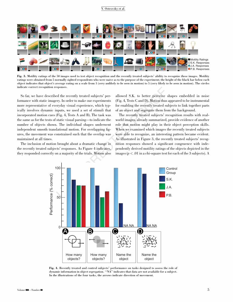
When we examined which images the recently treated subjects
were able to recognize, an interesting pattern became evident.
As illustrated in Figure 3, the recently treated subjects’ recog-
nition responses showed a significant congruence with inde-
pendently derived motility ratings of the objects depicted in the
images (p< .01 in a chi-square test for each of the 3 subjects). A
100 ControlGroup
50
0
Per
form
ance
(% c
orre
ct)
S.K.
J.A.
P.B.
NA NA NA NA
How manyobjects?
How manyobjects?
Name theobject
Name theobject
A B C D
Fig. 4. Recently treated and control subjects’ performance on tasks designed to assess the role ofdynamic information in object segregation. ‘‘NA’’ indicates that data are not available for a subject.In the illustrations of the four tasks, the arrows indicate direction of movement.
Fig. 3. Motility ratings of the 50 images used to test object recognition and the recently treated subjects’ ability to recognize these images. Motilityratings were obtained from 5 normally sighted respondents who were naive as to the purpose of the experiment; the height of the black bar below eachobject indicates that object’s average rating on a scale from 1 (very unlikely to be seen in motion) to 5 (very likely to be seen in motion). The circlesindicate correct recognition responses.
Volume ]]]—Number ]] 5
Y. Ostrovsky et al.

plausible, though not definitive, explanation of this congruence
is that motion of objects helps bind their constituent regions into
cohesive representations, which can then be used to recognize
instances in new inputs that may be static. It appears, however,
that motion information is not used to the exclusion of figural
cues, as preliminary tests with point-light walkers of the kind
devised by Johansson (1973) proved ineffective for conveying
the impression of a person: None of the 3 subjects in the recently
treated group was able to perceive such displays as anything
other than a collection of moving dots.
We also examined changes in the recently treated subjects’
performance as a function of time after treatment. S.K.’s per-
formance pattern was unaltered when we tested him 6 and 12
months after treatment. Given the relatively mature age at which
he had received treatment, we were not hopeful of observing
much visual recovery. However, follow-up tests conducted at 18
months posttreatment demonstrated that S.K.’s visual skills,
although still not normal, had registered a significant improve-
ment. The results are summarized in Figure 5. Essentially, at
this time, S.K. could perform tasks with static images that he
previously could perform only if motion cues were added. As
Figure 5 shows, P.B. and J.A. exhibited a similar improvement in
their ability to parse static images when tested several months
after initial treatment (10 months for P.B. and 12 months
for J.A.).
DISCUSSION
Taken together, these results provide a longitudinal glimpse into
the development of visual parsing skills several years postin-
fancy. They suggest that the early stages of this process are
characterized by integrative impairments. These impairments
lead to perceptual overfragmentation of images, and thus com-
promise recognition performance. However, the use of motion
information effectively mitigates these integrative difficulties.
During the early stages of visual learning, motion appears to be
instrumental both in segregating objects and in binding their
constituents into representations for recognition.
We derive confidence in the generality of the results described
here from the consistency among the 3 subjects, and also their
congruence with findings from previously reported case studies
of sight recovery. Although the earlier studies on sight recovery
in adulthood (Fine et al., 2003; Gregory & Wallace, 1963) did
not specifically focus on skills involved in region integration,
they reported difficulties consistent with impairments in these
skills during recognition of natural images. For instance,
Gregory and Wallace (1963), in describing their patient SB,
wrote: ‘‘We formed the impression that he saw [the natural
scenes as] little more than patches of colour’’ (p. 24). Similarly,
as regards simple image parsing, Fine et al. (2003) wrote that
MM ‘‘described two overlapping transparent squares as three
surfaces with the central square in front’’ (p. 915). Furthermore,
in these past cases, as in the present one, motion sensitivity was
evident soon after treatment.
The privileged status of motion observed with our recently
treated individuals is reminiscent of results reported in the in-
fant literature. Although infants eventually become able to use
static figural cues for object segregation (Needham, 1998),
segmentation from motion arises at least 2 months prior to the
ability to segment from static cues (Arterberry & Yonas, 2000;
S.P. Johnson, 2003), and infants’ ability to link spatially sepa-
rated parts of a partially occluded object is initially driven
strongly by common motion (S.P. Johnson, Bremner, Slater,
Mason, & Foster, 2002; Kellman & Spelke, 1983). It is inter-
esting to find this point of overlap between the developmental
progressions of infants and of our recently treated group, given
that maturational processes would presumably have already
completed their time course in our subjects. The neural
underpinnings of this similarity are unclear. However, the per-
ceptual utility of an early sensitivity to motion for both popu-
100
50
0
Per
form
ance
(% c
orre
ct)
S.K. (1 mo.) S.K. (18 mo.)J.A. (3 mo.) J.A. (12 mo.)P.B. (1 mo.) P.B. (10 mo.)
How manyobjects?
How manyobjects?
Name theobject
Trace thelong curve
Fig. 5. The recently treated subjects’ performance on four tasks with static displays soon aftertreatment and at follow-up testing after the passage of several months (indicated in the key).
6 Volume ]]]—Number ]]
Visual Parsing After Recovery From Blindness

lations allows a conjecture. It is possible that the early avail-
ability of motion sensitivity in the primate brain (Kiorpes &
Movshon, 2003, 2004) serves an adaptive purpose by providing
a scaffolding for acquiring skills for analyzing static figural in-
formation. By observing the correlations between motion-based
groupings and static cues, such as aligned contours, the visual
system might learn to use the latter by themselves as proxies for
grouping (Cavanagh, 1993). This conjecture regarding potential
dependencies between early- and later-developing visual skills
has significant implications for theoretical models of visual
learning (Sinha, Balas, Ostrovsky, & Wulff, in press).
Our experimental results complement past studies of visual
development in infancy and after sight restoration. First, they
provide evidence that region integration via figural cues is un-
likely to be merely a maturational process, unfolding with age,
but rather is more likely to be a visually driven developmental
process. Second, they highlight the limited efficacy of static
figural cues, such as spatially contiguous collinear contours,
for purposes of grouping early in the visual-learning time line.
These cues have conventionally been assumed to be of funda-
mental significance for spatial integration (Sigman, Cecchi,
Gilbert, & Magnasco, 2001; Ullman, 1996; Wertheimer, 1938).
Third, our results provide evidence that motion cues might
facilitate the assembly of linked regions that can serve as rep-
resentations for recognition of new inputs. In this way, our re-
sults connect basic grouping phenomena to real-world object
recognition. Overall, these results suggest that dynamic infor-
mation provides a key organizing influence for early visual
processing.
The evidence of marked improvement in our subjects’ per-
formance over the course of 10 to 18 months suggests that visual
skills related to the complex task of image parsing can be ac-
quired even after a prolonged delay, although the rate of ac-
quisition slows down with age, possibly because of decreases in
plasticity. Furthermore, the subjects’ visual experience during
this period derived from their normal daily activities; no special
training was provided. Indeed, S.K. resided at a hostel for the
blind with no sighted residents to provide instruction. These
results, along with a case we have reported previously (Ostrov-
sky, Andalman, & Sinha, 2006), suggest that the idea of a critical
period should not be applied too strictly to visual learning, and
provide cause for optimism for the many blind individuals who
are candidates for treatment. The human brain, it appears, re-
tains at least some measure of its ability to launch programs of
visual learning even after extended periods of visual depriva-
tion. Furthermore, these new insights into the progression
of visual skill acquisition point to possible rehabilitative pro-
grams for the often-overlooked patients with congenital sight
deprivation.
Acknowledgments—The authors wish to thank S.K., P.B., J.A.,
and their families; the staff at New Delhi’s Shroff Charity Eye
Hospital; and Richard Held, Beatrice de Gelder, and Scott
Johnson. Funding for this work was provided by National In-
stitutes of Health Grant R21 EY015521, the Alfred P. Sloan
Foundation, the John Merck Scholars Fund, and the James S.
McDonnell Foundation.
REFERENCES
Arterberry, M.E., & Yonas, A. (2000). Perception of three-dimensional
shape specified by optic flow by 8-week-old infants. Perception &Psychophysics, 62, 550–556.
August, J., Siddiqi, K., & Zucker, S.W. (1999). Contour fragment
grouping and shared, simple occluders. Computer Vision andImage Understanding, 76, 146–162.
Borenstein, E., & Ullman, S. (2002). Class-specific, top-down seg-
mentation. In A. Heyden, G. Sparr, M. Nielsen, & P. Johansen
(Eds.), Computer vision—ECCV 2002 (Part 2, pp. 109–122).
Berlin: Springer Verlag.
Brady, M.J., & Kersten, D. (2003). Bootstrapped learning of novel
objects. Journal of Vision, 3(6), Article 2. Retrieved July 2006
from http://journalofvision.org/3/6/2/
Cavanagh, P. (1993). The perception of form and motion. CurrentOpinion in Neurobiology, 3, 177–182.
Elder, J., & Zucker, S.W. (1998). Evidence for boundary-specific
grouping in human vision. Vision Research, 38, 143–152.
Field, D., Hayes, A., & Hess, R. (1993). Contour integration by the
human visual system: Evidence for a local ‘‘association field.’’
Vision Research, 33, 173–193.
Fine, I., Wade, A.R., Brewer, A.A., May, M.G., Goodman, D.F.,
Boynton, G.M., et al. (2003). Long-term deprivation affects visual
perception and cortex. Nature Neuroscience, 6, 915–916.
Gregory, R.L., & Wallace, J.G. (1963). Recovery from early blindness: Acase study (Experimental Psychology Monograph No. 2). London:
Heffer.
Grossberg, S., & Mingolla, E. (1985). Neural dynamics of perceptual
grouping: Textures, boundaries and emergent segmentations. Per-ception & Psychophysics, 38, 141–171.
Hummel, J.E., & Biederman, I. (1992). Dynamic binding in a neural
network for shape recognition. Psychological Review, 99, 480–
517.
Hupe, J.M., James, A.C., Payne, B.R., Lomber, S.G., Girard, P., &
Bullier, J. (1998). Cortical feedback improves discrimination
between figure and background by V1, V2 and V3 neurons.
Nature, 394, 784–787.
Johansson, G. (1973). Visual perception of biological motion and a
model for its analysis. Perception & Psychophysics, 14, 201–211.
Johnson, B.L., & Cheng, K.P. (1997). Congenital aphakia: A clinico-
pathologic report of three cases. Journal of Pediatric Ophthal-mology & Strabismus, 34, 35–39.
Johnson, S.P. (2003). Development of fragmented vs. holistic object
perception. In G. Schwarzer & H. Leder (Eds.), The developmentof face processing (pp. 3–17). Cambridge, MA: Hogrefe & Huber.
Johnson, S.P., Bremner, J.G., Slater, A., Mason, U., & Foster, K.
(2002). Young infants’ perception of unity and form in occlusion
displays. Journal of Experimental Child Psychology, 81, 358–
374.
Kanizsa, G. (1979). Organization in vision: Essays on Gestalt percep-tion. New York: Praeger.
Kellman, P.J., & Spelke, E.S. (1983). Perception of partly occluded
objects in infancy. Cognitive Psychology, 15, 483–524.
Volume ]]]—Number ]] 7
Y. Ostrovsky et al.

Kiorpes, L., & McKee, S.P. (1999). Neural mechanisms underlying
amblyopia. Current Opinion in Neurobiology, 9, 480–486.
Kiorpes, L., & Movshon, J.A. (2003). Neural limitations on visual
development in primates. In L.M. Chalupa & J.S. Werner (Eds.),
The visual neurosciences (pp. 159–173). Cambridge, MA: MIT
Press.
Kiorpes, L., & Movshon, J.A. (2004). Development of sensitivity to visual
motion in macaque monkeys. Visual Neuroscience, 21, 851–859.
Koffka, K. (1935). Principles of Gestalt psychology. New York: Har-
court, Brace and World.
Kovacs, I., & Julesz, B. (1993). A closed curve is much more than an
incomplete one: Effect of closure in figure-ground segmentation.
Proceedings of the National Academy of Sciences, USA, 90, 7495–
7497.
Leung, T., & Malik, J. (1998). Contour continuity in region based
image segmentation. In H. Burkhardt & B. Neumann (Eds.),
Computer vision—ECCV’98 (pp. 544–562). Berlin: Springer
Verlag.
Mandavilli, A. (2006). Look and learn. Nature, 441, 271–272.
Marr, D. (1982). Vision: A computational investigation into the humanrepresentation and processing of visual information. New York:
W.H. Freeman and Co.
Maurer, D., Lewis, T.L., & Mondloch, C.J. (2005). Missing sights:
Consequences for visual cognitive development. Trends in Cog-nitive Sciences, 9, 144–151.
Mumford, D., & Shah, J. (1985, June). Boundary detection by mini-mizing functionals. Paper presented at the 2nd IEEE Con-
ference on Computer Vision and Pattern Recognition, San
Francisco, CA.
Needham, A. (1998). Infants’ use of featural information in the seg-
regation of stationary objects. Infant Behavior and Development,21, 47–76.
Needham, A. (2001). Object recognition and object segregation in 4.5-
month-old infants. Journal of Experimental Child Psychology, 78,
3–24.
Ostrovsky, Y., Andalman, A., & Sinha, P. (2006). Vision following ex-
tended congenital blindness. Psychological Science, 17, 1009–
1014.
Pratt, J.C., & Richards, R.D. (1968). Bilateral secondary congenital
aphakia. Archives of Ophthalmology, 80, 420–422.
Shi, J., & Malik, J. (1997, June). Normalized cuts and image seg-mentation. Paper presented at the 11th IEEE Conference on
Computer Vision and Pattern Recognition, San Juan, Puerto Rico.
Sigman, M., Cecchi, G.A., Gilbert, C.D., & Magnasco, M.O. (2001). On
a common circle: Natural scenes and Gestalt rules. Proceedings ofthe National Academy of Sciences, USA, 98, 1935–1940.
Sinha, P., Balas, B.J., Ostrovsky, Y., & Wulff, J. (in press). Visual object
discovery. In S. Dickinson, A. Leonardis, B. Schiele, & M. Tarr
(Eds.), Object categorization: Computer and human vision per-spectives. Cambridge, England: Cambridge University Press.
Tu, Z., Chen, X., Yuille, A.L., & Zhu, S.C. (2003, October). Imageparsing: Unifying segmentation, detection, and recognition. Paper
presented at the 9th IEEE International Conference on Computer
Vision, Nice, France.
Ullman, S. (1996). High-level vision. Cambridge, MA: MIT Press.
Valvo, A. (1971). Sight restoration after long-term blindness: Theproblems and behavior patterns of visual rehabilitation. New York:
American Foundation for the Blind.
Von Senden, M. (1960). Space and sight: The perception of space andshape in the congenitally blind before and after operation (P. Heath,
Trans.). Glencoe, IL: Free Press. (Original work published 1932)
Wertheimer, M. (1938). Laws of organization in perceptual forms
(partial translation). In W. Willis (Ed.), A sourcebook of Gestaltpsychology (pp. 71–88). New York: Harcourt, Brace and Co.
(RECEIVED 12/23/08; REVISION ACCEPTED 3/21/09)
8 Volume ]]]—Number ]]
Visual Parsing After Recovery From Blindness