research open access detection of atm - springer · (e.g., radiosensitivity, atm protein levels,...
TRANSCRIPT

RESEARCH Open Access
Detection of ATM germline variants by thep53 mitotic centrosomal localization test inBRCA1/2-negative patients with early-onsetbreast cancerAndrea Prodosmo1, Amelia Buffone4, Manlio Mattioni1, Agnese Barnabei3, Agnese Persichetti3,4, Aurora De Leo3,Marialuisa Appetecchia3, Arianna Nicolussi11, Anna Coppa11, Salvatore Sciacchitano5, Carolina Giordano6,Paola Pinnarò6, Giuseppe Sanguineti6, Lidia Strigari7, Gabriele Alessandrini8, Francesco Facciolo8, Maurizio Cosimelli9,Gian Luca Grazi9, Giacomo Corrado10, Enrico Vizza10, Giuseppe Giannini2,4* and Silvia Soddu1*
Abstract
Background: Variant ATM heterozygotes have an increased risk of developing cancer, cardiovascular diseases,and diabetes. Costs and time of sequencing and ATM variant complexity make large-scale, general populationscreenings not cost-effective yet. Recently, we developed a straightforward, rapid, and inexpensive test basedon p53 mitotic centrosomal localization (p53-MCL) in peripheral blood mononuclear cells (PBMCs) that diagnosesmutant ATM zygosity and recognizes tumor-associated ATM polymorphisms.
Methods: Fresh PBMCs from 496 cancer patients were analyzed by p53-MCL: 90 cases with familial BRCA1/2-positive and -negative breast and/or ovarian cancer, 337 with sporadic cancers (ovarian, lung, colon, andpost-menopausal breast cancers), and 69 with breast/thyroid cancer. Variants were confirmed by ATMsequencing.
Results: A total of seven individuals with ATM variants were identified, 5/65 (7.7 %) in breast cancer casesof familial breast and/or ovarian cancer and 2/69 (2.9 %) in breast/thyroid cancer. No variant ATM carrierswere found among the other cancer cases. Excluding a single case in which both BRCA1 and ATM weremutated, no p53-MCL alterations were observed in BRCA1/2-positive cases.
Conclusions: These data validate p53-MCL as reliable and specific test for germline ATM variants,confirm ATM as breast cancer susceptibility gene, and highlight a possible association with breast/thyroidcancers.
Keywords: ATM cancer susceptibility gene, Early-onset breast cancer, BRCA1/2, p53-mitotic centrosomallocalization (p53-MCL)
(Continued on next page)
* Correspondence: [email protected]; [email protected] Pasteur-Fondazione Cenci Bolognetti, Department of MolecularMedicine, University La Sapienza, Rome, Italy1Unit of Cellular Networks and Molecular Therapeutic Targets, Department ofResearch, Advanced Diagnostic, and Technological Innovation, Regina ElenaNational Cancer Institute – IRCCS, Via Elio Chianesi 53, 00144 Rome, ItalyFull list of author information is available at the end of the article
© 2016 The Author(s). Open Access This article is distributed under the terms of the Creative Commons Attribution 4.0International License (http://creativecommons.org/licenses/by/4.0/), which permits unrestricted use, distribution, andreproduction in any medium, provided you give appropriate credit to the original author(s) and the source, provide a link tothe Creative Commons license, and indicate if changes were made. The Creative Commons Public Domain Dedication waiver(http://creativecommons.org/publicdomain/zero/1.0/) applies to the data made available in this article, unless otherwise stated.
Prodosmo et al. Journal of Experimental & Clinical Cancer Research (2016) 35:135 DOI 10.1186/s13046-016-0410-3

(Continued from previous page)
Abbreviations: A-T, Ataxia telangiectasia; ATM, Ataxia telangiectasia mutated; ATR, Ataxia telangiectasia andRad3 related; BRCA, Breast cancer susceptibility gene; CHK2, Checkpoint kinase 2; EBV, Epstein barr virus;FANC-A, Fanconi anemia complementation group A; HBC, Hereditary breast cancer; HBOC, Hereditary breastand ovarian cancer; LCLs, Lymphoblastoid cell lines; MRE11, Meiotic recombination 11; NBS1, Nijmegenbreakage syndrome 1; p53-MCL, p53-Mitotic centrosomal localization; PALB2, Partner and localizer of BRCA2;PBMCs, Peripheral blood mononuclear cells; PHA, Phytohemagglutinin; SMC1A, Structural maintenance ofchromosomes 1A; WRN, Werner syndrome
BackgroundBiallelic mutations in the ATM gene cause Ataxia-telangiectasia (A-T), a rare autosomal recessive multisys-temic disorder characterized by progressive cerebellarataxia, immune defects, insulin-resistant diabetes, radio-sensitivity, and high risk for malignancy [1, 2]. The ATMgene spans approximately 160 Kb of genomic DNA con-taining 66 exons [3] and encodes ATM protein, a serine/threonine kinase mainly involved in DNA damageresponse pathways following DNA double strand breaks[4]. An enormous number of mutations (more than 600)can occur in the coding and noncoding regions of theATM gene without hotspots [5]. In A-T patients, the largemajority of ATM mutations are protein-truncations orsplice-junction variants that can be easily distinguished bythe numerous ATM polymorphisms [6, 7].Heterozygous carriers of variants in the ATM gene
(from here on, ATM carriers) are usually asymptomaticand largely considered healthy carriers. However, theyhave been reported to be more sensitive to ionizing radi-ation and susceptible to ischemic heart disease, diabetes,and cancer, particularly of the breast, but also digestivetract and lung [2, 8]. Many association studies have beenperformed on breast cancer susceptibility. Initially,epidemiological studies on relatives of A-T patients re-vealed a two to fivefold increased in the risk of breastcancer for female obligate ATM carriers [9]. The in-creased risk of breast cancer in ATM carriers was thenconfirmed by direct ATM sequencing in breast cancercases compared to controls [10] and ATM is now con-sidered a moderate-penetrance cancer susceptibility genein BRCA1/2-negative patients with familial early-onsetbreast cancer [11]. Along with A-T associated muta-tions, several ATM screenings in cancer patients iden-tified missense ATM variants, particularly amino acidsubstitutions that are not expected to be associatedwith A-T [12]. However, discrimination of these ATMvariants from ATM polymorphisms and their contri-bution to health risks is still controversial. Inaddition, distinguishing between deleterious and neu-tral ATM alterations is required to allow the defin-ition of standard-of-care clinical guidelines for themanagement of ATM carriers and their families [11].
Systematic review and meta-analysis of ATM sequen-cing data have been conducted to evaluate the healthrisks for parents and siblings of A-T patients, butsimilar large-scale screenings for ATM carriers in thegeneral population by direct sequencing are not cost-effective yet [13].Recently, we have developed a rapid, reliable and non-
expensive test based on the ATM-dependent p53-mitotic centrosomal localization (p53-MCL) that clearlydiscriminates ATM carriers of A-T mutations and atleast some of the ATM cancer predisposition variants inlymphoblastoid cell lines (LCLs) and PBMCs. At vari-ance with other diagnostic tests, the p53-MCL assaydoes not measure a continuous quantitative variation(e.g., radiosensitivity, ATM protein levels, phosphoryl-ation of ATM targets) but a “binary” outcome. Indeed, atthe single cell level, p53 does or does not localize at thecentrosomes while, at the cell-population level, the num-ber of cells showing one or the other phenotype allowsto unambiguously diagnose A-T homozygotes and ATMcarriers [14].In a preliminary set up and validation of the p53-MCL
test, we showed that it is highly sensitive, specific, andprecise. In particular, we assessed the specificity by ana-lyzing LCLs from monogenic disease carrying mutationsin a series of DNA-damage related factors, such asMRE11, NBS1, SMC1A, WRN, ATR, FANC-A, and p53[14]. In addition, p53-MCL test revealed 7 ATM carriersamong 80 sporadic breast cancer patients. By directATM sequencing of 3 of these carriers, we identified thecancer-prone intronic c.8786 + 8A > C variant [15] inone patient and the c.2572 T > C (p.F858L) missensemutation [16] in other two patients. No ATM carrierswere observed in a comparable cohort of healthy donors[14]. These data support p53-MCL as promising candi-date test for cost-effective, large-scale screenings ofATM carriers.Here, we examined validity and specificity of p53-
MCL analyzing 15 LCLs from familial breast andovarian cancer cases and fresh PBMCs from a total of496 cases including BRCA1/2-positive and -negativefamilial breast and ovarian cancer and different spor-adic cancers.
Prodosmo et al. Journal of Experimental & Clinical Cancer Research (2016) 35:135 Page 2 of 10

MethodsPatientsA total of 496 cancer patients were enrolled from 2010to 2015 at three different Italian institutes: PoliclinicoUmberto I (University La Sapienza), Sant’Andrea Hos-pital (University La Sapienza), and Regina Elena NationalCancer Institute - IRCCS. In particular, according to pre-viously described criteria [17], we selected 90 unrelatedfamilies affected with breast and/or ovarian cancer afterinterview at the Hereditary Tumors Counseling Centreof the Policlinico Umberto I. Pre-test counseling wasperformed by an expert cancer risk counselor and theprobands analyzed in this study belonged to differenthigh-risk classes. During the genetic counseling, we cal-culated the a priori probability of carrying a pathogenicBRCA1/2 germline mutation by the statistical modelBRCAPRO that considers: proband health state (un/af-fected); current age and age at diagnosis of the probandand all family members of four consecutive generations(only first/second degree relatives); typology of the exist-ent tumors (unilateral/bilateral breast cancer, other can-cers) [18]. Then, 69 patients with breast and thyroidcancer regardless of the sequence of appearance wererecruited from Sant’Andrea Hospital and Regina ElenaNational Cancer Institute. Others 337 patients unse-lected cancer patients diagnosed at any age wererecruited from Regina Elena National Cancer Institute.
Cells and culture conditionsEBV-immortalized LCLs and freshly isolated PBMCswere cultured in RPMI-1640 GlutaMAX supplementedwith 15 % heat-inactivated fetal bovine serum, 100 U/mlpenicillin, and 100 μg/ml streptomycin (all from Invitro-gen, CA, USA). PBMCs were isolated from donors’ hep-arinized blood samples by Lympholyte-H (Cedarlane,Burlington, USA) density gradient centrifugation. PBMCswere stimulated to proliferate by incubation with 5 μg/mlPHA (Sigma-Aldrich, St. Louis, MO, USA) and incubatedat 37 °C in a 5 % CO2 atm for 60 h [14].
p53-MCL testProliferating cells (i.e., LCLs or PHA-stimulated PBMCs)were set up for p53-MCL test as previously described[14]. Cells were examined under an Olympus BX53microscope equipped with epifluorescence. Percentagesof p53-MCL were measured by counting 100 cells inmetaphase and analyzing two coverslips for each sample.The percentage of p53 mitotic localization is from 75 to90 % for normal subjects, from 40 to 55 % for ATMcarriers, and from 0 to 30 % for A-T patients.
BRCA1/2 gene sequencingGenomic DNA was extracted from peripheral blood ofall probands using commercial kit (Qiamp Blood Kit,
Qiagen, Hilden, Germany). The entire coding sequenceand each intron/exon boundary of BRCA1 and BRCA2were screened by direct sequencing. All truncating and/or novel genetic variants were confirmed by sequencingdifferent samples on both DNA strands. Sequencing wasperformed using the BigDye Terminator v3.1 Cycle Se-quencing Kit and a 3130xl Genetic Analyzer (AppliedBiosystems, CA, USA). Reference sequence for BRCA1was Genebank NM_007294.3, NG_005905.2 and referencesequence for BRCA2 was Genebank, NM_000059.3,NG_012772.3. BRCA1/2 genomic rearrangements weresearched by the Multiple Ligation dependent Probe Amp-lification (MLPA) methodology. MLPA procedure wascarried out according to the manufacturer’s instructions.Variations in peak height were evaluated comparing eachsample with a normal control and by a cumulativecomparison.
ATM gene sequencingGenomic DNA was extracted from PBMCs by Quick-gDNA MiniPrep (Zymo Research, CA, USA) accordingto the manufacturer’s instructions. Sixty-two ATM exonswere amplified using AmpliTaq Gold (Applied Biosys-tems, CA, USA), as described [14], and subjected to dir-ect sequencing at the Genechron Laboratory (Rome,Italy). Reference sequence for ATM was GenebankU82828.1.
In silico analysisTo predict possible impact of amino acid substitutionson the structure and function of human proteins, thePolyPhen-2 (Polymorphism Phenotyping v2) software,that uses straightforward physical and evolutionary com-parative considerations [19], was employed.Efficient splicing of many exons requires splicing-
enhancers to promote splicing at unfavorable splice-sitesor splicing-silencers to repress more favorable splice-sitesnearby. As a consequence, single nucleotide changes in anexon or intron close to these splice-sites may be predictedto disrupt splicing. For in silico prediction of the effects ofmutations on normal splicing, the interactive biosoftwareAlamut v2.3 [20] was adopted.
StatisticWe determined statistical significance of differences be-tween two groups by 2-tailed Student’s t test. P valuesless than 0.01 were considered significant.
ResultsBeyond the high penetrance BRCA1/2 genes, mutationsof several cancer susceptibility genes, including ATM,CHK2 and PALB2, have been shown to associate, with amoderate penetrance, with familial breast and/or ovariancancers [21, 22]. Thus, we first verified p53-MCL test
Prodosmo et al. Journal of Experimental & Clinical Cancer Research (2016) 35:135 Page 3 of 10

specificity compared to mutant BRCA1/2 and CHK2proteins that are also involved in DNA damage responseand centrosome amplification and localization [23–25].LCLs from 15 familial breast and ovarian cancer(Table 1) including four BRCA1-positive, 2 BRCA2-positive, 1 CHK2-positive and 8 BRCA1/2/CHK2/PALB2-negative LCLs were analyzed by the p53-MCLtest. As shown in Table 1 and Fig. 1a, the percentage ofp53-MCL was compatible with that of wild-type cells in13 out of 15 LCLs, while two cases showed a p53-MCLreduction typical of ATM carriers, one in a BRCA1-positive case and the other in a BRCA1/2/CHK2/
PALB2-negative case. Direct ATM sequencing confirmedthe presence of ATM variants (Table 1) in both casesdemonstrating p53-MCL specificity for ATM variantsalso in comparison with mutations in the centrosome re-lated factors BRCA1/2 and CHK2.Next, we analyzed 90 cases of familial breast and/or
ovarian cancer (Table 2). All 90 patients were screenedfor mutations in the BRCA1 and BRCA2 genes and 20were found to carry pathogenic variants with an overallmutation rate of 22.2 % (Table 2). In particular BRCA1pathogenic mutation recurred in about 26 % (8/30) ofthe Hereditary Breast and Ovarian Cancer (HBOC) fam-ilies and in about 7 % (4/60) of the Hereditary BreastCancer (HBC) families, while BRCA2 pathogenic muta-tion occurred in about 13 % (4/30) and 7 % (4/60) ofthe HBOC and HBC families, respectively. Of the 20mutation-positive probands, 11 had breast canceralone (5 BRCA1 and 6 BRCA2), three had ovariancancer alone (3 BRCA1), three had bilateral breastcancer (2 BRCA1 and 1 BRCA2), two had both breastand ovarian cancer (1 BRCA1 and 1 BRCA2) and onehad both bilateral breast and ovarian cancer (BRCA1).When fresh PBMCs from BRCA1/2-positive and-negative patients were analyzed by the p53-MCL test,five out of 90 cases showed aberrant p53-MCL(Fig. 1b). Interestingly p53-MCL positive casesaccounted for 6.6 and 5 % of the HBOC and HBCfamilies, respectively (Table 2). Of note, this rate issimilar to that of BRCA2 mutations [26].The five individuals with ATM variants identified by
the p53-MCL test were all breast cancer patients (5/65,7.7 %). Four of them were BRCA1/2 negative patientsand one was a BRCA1 positive patient (Fig. 2). No ATMcarriers were detected by p53-MCL in patients withovarian cancer, bilateral breast cancer, or multiple
Table 1 Genetic status and p53-MCL rate of LCLs from familialbreast and ovarian cancer
BRCA1 BRCA2 CHK2 PALB2 ATM p53-MCL%
BR36 wt mut nt nt nt 80
BR13 wt mut nt nt nt 76
BR409-3 mut wt nt nt nt 97
BR324-1 mut wt nt nt nt 85
BR404-1 mut wt wt wt wt 76
BR317 mut wt nt nt mut 50
BR377 wt wt wt wt mut 50
BR60-1 wt wt wt wt wt 71
BR107-1 wt wt wt wt wt 90
BR362-1 wt wt wt wt wt 92
BR494 wt wt wt wt wt 86
BR38 wt wt wt wt wt 91
BR48 wt wt wt wt wt 70
BR278-1 wt wt wt wt wt 80
BR501 wt wt mut wt wt 80
wt wild type, mut mutated, nt not tested
Fig. 1 p53-MCL in LCLs and in PBMCs from familial breast and/or ovarian BRCA1/2-positive and –negative patients. (a) Comparison of p53-MCLpercentages, among LCLs derived from wild type ATM donors (CTR, n = 11), BRCA1/2-negative (n = 7), BRCA1/2-positive (n = 5), CHK2-positive (n= 1), and ATM variants carriers (n = 2) and ATM carriers (n = 9) previously tested. Not significant differences among the groups including wild typeATM (CTR, BRCA1/2-neg, BRCA1/2-pos and CHK2-pos), but significant differences between these groups and ATM carriers groups (ATM variantsand ATM carriers). (b) Comparison of p53-MCL percentages among PBMCs from familial breast and/or ovarian BRCA1/2-positive, BRCA1/2–nega-tive and ATM carrier patients. Not significant differences between BRCA1/2-positive and -negative patients, but significant differences betweenwild type ATM group (BRCA1/2-positive and -negative) and ATM carriers. ***P < 0.0001; NS = Not Significant; 2-tailed Student’s t test
Prodosmo et al. Journal of Experimental & Clinical Cancer Research (2016) 35:135 Page 4 of 10

cancers (breast cancer and at least one other non-breastcancer) (n = 27) (Table 3).Besides early onset-breast cancer, ATM carriers have
been reported to be more susceptible to other types ofcancer, such as digestive tract, lung, and thyroid cancers[2, 8]. Thus, we performed the p53-MCL test on freshPBMCs from 403 patients with sporadic cancers, includ-ing ovarian (n = 49), lung (n = 150), colon (n = 80), post-menopausal breast (n = 58), and both breast/thyroid (n =69) cancer. Two individuals with ATM variants wereidentified among breast/thyroid cancer cases (2/69, 2.9)(Table 4). No ATM carriers were found in sporadic ovar-ian cancer (0/49), non-small cell lung cancer (0/150),colorectal cancer (0/80), and post-menopausal breastcancer (0/58).As shown in Fig. 2 and Additional file 1: Table S1, 11 dif-
ferent ATM variants were detected among the 9 p53-MCLpositive cases (i.e., 2 LCLs and 7 PBMCs). Specifically, inbreast cancer cases, patient #1 presents c.4578C >T andc.1899-55 T >G variants. The c.4578C > T was previouslydescribed [27] and corresponds to a synonymous substitu-tion (P1526P), which is predicted to create a new exonicsite with increased affinity for the SRp55 splicing factor byan in silico analysis (Fig. 3a). The c.1899-55 T >G is a pre-viously described intronic variant [28] predicted to increasethe affinity for the SRp40 splicing factor (Fig. 3b). Patient#2 presents two variants not previously described in the lit-erature nor in the ATM variation database (c.908A >C andc.5919-49C > T). Our in silico analyses predict a higheraffinity for the splicing factor SC35, for both variants(Fig. 3c, d). The c.5557G >A (D1853N) variant presents inpatient #3 is predicted to be “possibly damaging” by thePolyPhen-2 tool and has been intensively studied withrespect to its possible association with breast cancer sus-ceptibility [29]. Patient #4 and patient #5 present two dele-terious variants, c.824delT and c.8833delCT, that induceearly protein truncation at the level of exon 9 and 63, re-spectively, which have already been described in ATM
families [30, 31]. In the breast and thyroid cancer cases, pa-tient #6 presents two already described variants, c.3161C >G and c.3576G >A. The c.3161C >G (P1054R) is a substi-tution predicted to be “possibly damaging” by PolyPhen-2and implicated in breast and prostate cancer risk [32]. Thec.3576G >A variant leads to exon 26 skipping and causesA-T syndrome in homozygosis [30]. Patient #7 presentsthe intronic variant c.4436 + 24A >G that in silico analysespredict a mild higher affinity for the splicing factors SC35and SRp55 (Fig. 3e). LCLs BR317 presents c.146C >G(S49C) variant, a substitution predicted to be possibly dam-aging by PolyPhen-2 and reported as breast cancer suscep-tibility variant [16]. Finally, LCLs BR377 presents the samec.3161C >G (P1054R) variant found in patient #6.
DiscussionGenetic susceptibility plays an important role in severalcommon chronic diseases including many types of can-cer. Genetic testing for large-scale, general populationscreening can be very expensive and non-cost effectivefor National Health Services. Concerning the ATM gene,the complex genomic organization, the large number ofpolymorphisms, the absence of mutation hot-spots, andthe frequent occurrence of variants of yet uncharacter-ized but predicted deleterious functions, make directgene sequencing not yet a cost-effective approach. Thisis particularly evident for large-scale surveys of mutant/variant ATM carriers for which gene sequencing is notsufficient to classify the rare hits. All these factors hinderboth genetic counseling and clinical guidelines for riskmanagement of ATM carriers and their families. Theuse of the recently developed p53-MCL functional testto detect ATM carriers might overcome at least some ofthese limitations. Here, we established the p53-MCLspecificity for ATM in respect to the high-risk, BRCA1/2breast/ovarian cancer susceptibility genes and confirmedp53-MCL as reliable test to detect variant ATM carriersin cancer patients.BRCA1/2 and ATM proteins share functional activ-
ities both in DNA damage response pathways and incentrosome regulation [33]. Thus, it was mandatoryto establish whether a functional test, such as thep53-MCL test, would discriminate between ATM andBRCA1/2 variant carriers. Analysis of BRCA1/2-posi-tive LCLs (n = 6) and PBMCs (n = 20), and of 1CHK2-positive LCL established the specificity of p53-MCL test on ATM in respect to BRCA1/2 and CHK2mutations (Table 1 and Fig. 1).A total of seven individuals with ten ATM variants
were identified by p53-MCL test in PBMCs: fivebreast cancer patients in HBC and HBOC familiesand two breast/thyroid cancer patients. The findingthat all cases were among pre-menopausal breastcancer cases but not among patients with post-
Table 2 Characteristics of familial breast and/or ovarian cancerpatients
Variable Study populationn = 90
BRCA1/2 carriersn = 20/90 (22.2 %)
ATM carriersn = 5/90(5.5 %)
Age-years
Median 52.9 53.7 44.4
Range 30–82 40–75 32–51
Familial aggregation
HBOC 30 (33.3 %) BRCA1: 8/30 (26 %) 2/30 (6.6 %)
BRCA2: 4/30 (13 %)
HBC 60 (66.7 %) BRCA1: 4/60 (7 %) 3/60 (5 %)
BRCA2: 4/60 (7 %)
BRCAPRO 27.3 % 50.1 % 52.2 %
HBOC hereditary breast and ovarian cancer, HBC hereditary breast cancer
Prodosmo et al. Journal of Experimental & Clinical Cancer Research (2016) 35:135 Page 5 of 10

Fig. 2 ATM variants in ATM carriers. Electropherograms showing ATM variants identified in PBMCs derived from five familial breast cancer (BC)patients (BC patient#1/#5), two breast-thyorid cancer (BTC) patients (BTC patient#6/#7) and two LCLs (BR317; BR377). All sequences are comparedwith wild-type reference sequence. Arrows indicates the position of the substitution and/or deletion
Prodosmo et al. Journal of Experimental & Clinical Cancer Research (2016) 35:135 Page 6 of 10

menopausal breast cancer or ovarian cancer is con-sistent with previous studies showing prevalence ofgermline ATM mutations detected by sequencing inpatients with familial early-onset breast cancer [34] orby p53-MCL in sporadic breast cancer [14]. Interest-ingly, the a priori probability of carrying a pathogenic
BRCA1/2 germline mutation measured by the BRCA-PRO score showed a mean for the entire cohort of27.3 while the mean for the five ATM carriers was52.2 % (Table 2). This difference is statistically signifi-cant (P =0.01) and supports the presence of a stronggenetic component in the ATM carriers with early-onset breast cancer, also considered that the BRCA-PRO score for the BRCA positive cases is very similarto ATM positive ones (50.1 % vs. 52.2 %).Evaluation of the histopathologic features of the breast
cancers developed in the seven identified ATM carriersshowed the presence of estrogen and progesteronereceptors and the absence of HER2 receptor in four outof five cases that have been tested (Tables 4 and 5), con-firming our previous results [14]. Thus, the presence ofgermline ATM variants recognized by p53-MCL appearsto identify a subset of tumors with a more favorable bio-marker asset, despite their earlier onset. In addition, apossible association between breast and thyroid cancerwas highlighted by the identification of two ATM car-riers among the sporadic cancer patients with breast/thyroid cancer (2/69, 2.9 %), encouraging for furtherstudies.Familial breast and ovarian cancers are linked to
highly penetrant mutations in the BRCA1/2 suscepti-bility genes that overall account for 20–25 % of her-editary breast cancers and 15 % of ovarian cancers[26]. Gene panel next generation sequencing ap-proaches identified moderate-penetrant mutations inthe ATM gene in 2.9 % [11] and 2.3 % [34] ofBRCA1/2-negative cases. By the p53-MCL test, in thisstudy we found 5.5 % (5/90) of ATM mutation
Table 3 Histopathological characteristics of all cancer patients
Tumor type Cases n = 496 BRCA1/2 carriers n = 20 ATM carriers n = 7
Familial Cancer cases 90 20 5
Breast 65 (70.6 %) 11/65 (16.9 %) 5/65 (7.7 %)
Ovarian 8 (8.7 %) 3/8 (37.5 %) 0/8
Bilateral Breast 10 (10.9 %) 3/10 (30 %) 0/10
Breast-Ovarian 4 (4.3 %) 2/4 (50 %) 0/4
Colon-Breast 1 (1.1 %) 0/1 0/1
Uterin-Breast 1 (1.1 %) 0/1 0/1
Anal-Bilateral Breast 1 (1.1 %) 0/1 0/1
Bilateral Breast-Ovarian 1 (1.1 %) 1/1 (100 %) 0/1
Ipsilateral Breast 1 (1.1 %) 0/1 0/1
Sporadic cancer cases 406 - 2/406
Ovarian 49 - 0/49
Lung 150 - 0/150
Colon 80 - 0/80
Post-menopausal Breast 58 - 0/58
Breast/Thyroid 69 - 2/69 (2.9 %)
Table 4 Clinical and pathological characteristics of breast andthyroid cancer patients
Variable Study population (n = 69) p53-MCL positive (n = 2)
Age-years
Average 60.2 58
Range 30–77 58–63
Age onset Breast tumor appearance
Average 49.1 48
Range 25–70 48–51
Breast tumor Diagnosis
Ductal 59 (85.5 %) 1 (50 %)
Lobular 3 (4.3 %) 0
Unknown 7 (10.2 %) 1 (50 %)
ER status
Negative 17 (24.6 %) 0
Positive 34 (49.3 %) 2 (100 %)
Unknown 18 (26.1 %) 0
PgR status
Negative 17 (24.6 %) 0
Positive 34 (49.3 %) 2 (100 %)
Unknown 18 (26.1 %) 0
ER estrogen receptor, PgR progesterone receptor
Prodosmo et al. Journal of Experimental & Clinical Cancer Research (2016) 35:135 Page 7 of 10

carriers, which adds up to the 27.7 % (20/90) carryingBRCA1/2 mutations. In particular, three ATM muta-tions occurred among the 60 HBC cases, with amutation rate (5 %) very close to that observed forBRCA2 (7 %) in the same subset. These observationssuggest that the fast and non-expensive p53-MCL testshould precede or be performed in parallel withBRCA1/2 sequencing.Most of the ATM mutations occurring in A-T
patients are frameshift or nonsense mutations leadingto protein truncation or splice junction variants [6, 7]The role of A-T-causing mutations in cancer suscepti-bility (in particular breast cancer) is still debated andsome studies have shown that a subset of rare, evolu-tionarily unlikely missense substitutions are important[15, 35, 36]. Here, the p53-MCL test identified 11ATM variants in 9 breast and breast/thyroid patients.
Three variants (c.824delT; c.8833delCT; c.3576G > A)identified in 3 different patients are known to causeA-T in homozigosity [30, 31]. Other 3 variants(c.5557G > A; c.146C > G; c.3161C > G) identified in 3different patients have been shown to be associatedwith an increased cancer risk [16, 29, 32]. The last 3patients present 5 different variants (c.4578C > T;c.1899-55 T > G; c.908A > C; c.5919-49C > T; c.4436 +24A > G) that our in silico analysis predicted to in-crease the affinity for splicing factor and modify alter-native splicing activities. Whether these predictedmodifications alter ATM function/s and cancer pre-disposition remain to be evaluated. Since loss of p53centrosomal localization was the functional readoutthrough which we diagnosed the ATM carriers, wecan conclude that these variants are at least able toimpair the mitotic localization of p53 at the
Fig. 3 In silico analysis using Alamut software. ATM variants identified in PBMCs derived from two familial breast cancer patients (BC patient#1and #2) and one breast-thyorid cancer patient (BTC patient#7) lead to an increased affinity and/or appearance of a de novo sites for SR proteins
Prodosmo et al. Journal of Experimental & Clinical Cancer Research (2016) 35:135 Page 8 of 10
centrosomes. It will be relevant to study whether allATM variants induce this p53 defect or whether onlyfunctionally relevant variants are able to impair p53-MCL. Whether this impaired p53 localization has arole in tumorigenesis is presently unknown. Studyingthe mechanistic basis of p53 centrosomal localizationwill give insights on the contribution that differentATM variants with uncertain significance might havein cancer predisposition. Application of the p53-MCLtest to LCLs or PBMCs with a broad spectrum ofATM variants will help to define these issues.
ConclusionsOur results show that p53-MCL test may offer theopportunity for screening of the general populationand to identify the differences among deleterious,neutral and beneficial variants helping, in the future,to define the guidelines for ATM carriers not only inthe A-T families.
Additional file
Additional file 1: Table S1. Characteristics of ATM variants. (DOCX 75 kb)
AcknowledgmentsWe are grateful to all patients and families taking part in this study.We thank Mustapha Haoui for technical assistance.
Availability of data and materialsPlease, contact authors for data request.
Authors’ contributionsA Prodosmo designed and performed experiments. He contributed toanalyze data and write the manuscript. A Buffone generated sequencingdata and molecular analyses. MM performed the p53 MCL-test. A Barnabeiand A Persichetti collected bloodsamples and clinical information of patients related to breast and thyroidcancer cases. ADL collected blood samples related to breast and thyroidcancer cases. MLA coordinated the study related to breast and thyroidcancer cases. AN produced lymphoblastoid cell lines. AC collected bloodsamples and clinical information of patients related to familial breast and/ovarian cancer cases. S Sciacchitano coordinated the study and collectedblood samples and clinical information of patients related to breast and thy-roid cancer cases. CG collected blood samples and clinical information of pa-tients related to post menopausal breast cancer cases. PP collected clinicalinformationof patients related to post menopausal breast cancer cases. GS coordinatedthe study related to post menopausal breast cancer cases. LS performedstatistical analyses. GA collected blood samples and clinical information ofpatients related to lung cancer cases. FF coordinated the study related tolung cancer cases. MC collected blood samples and clinical information ofpatients related to colon cancer cases. GLG coordinated the study related tocolon cancer cases. GC collected blood samples and clinical information ofpatients related to ovarian cancer cases. EV coordinated the study related toovarian cancer cases. GG coordinated the study related to familial breastand/or ovarian cancer cases, performed genetic counseling, contributed todesign the study and to write the manuscript. S Soddu conceived and de-signed the experiments. She coordinated the entire study and wrote the manu-script. All authors read and approved the final manuscript.
Competing interestsThe authors declare that they have no competing interests.
Consent for publicationNot applicable.
Ethics approval and consent to participateThe Institutional Ethics Committee approved this study (I.F.O. CE/160/09and CE/763/13) and all patients studied signed an informed consent forparticipation.
Financial supportThis work was supported by grants from Italian Association for CancerResearch (AIRC) to S.S. (5 per mille #9979) and (IG #14592), and to G.G.(IG#12116 and IG#17734). A. Prodosmo and A. Buffone were recipients ofa fellowship from Fondazione Umberto Veronesi “Pink is Good”.
Author details1Unit of Cellular Networks and Molecular Therapeutic Targets, Department ofResearch, Advanced Diagnostic, and Technological Innovation, Regina ElenaNational Cancer Institute – IRCCS, Via Elio Chianesi 53, 00144 Rome, Italy.2Istituto Pasteur-Fondazione Cenci Bolognetti, Department of MolecularMedicine, University La Sapienza, Rome, Italy. 3Endocrinology Unit,Department of Clinical and Experimental Oncology, Regina Elena NationalCancer Institute – IRCCS, Rome, Italy. 4Department of Molecular Medicine,University La Sapienza, Rome, Italy. 5Department of Clinical and MolecularMedicine, University La Sapienza, Laboratorio di Ricerca Biomedica,Fondazione Università Niccolò Cusano per la Ricerca Medico Scientifica,Rome, Italy. 6Radiotherapy Unit, Department of Research, AdvancedDiagnostic, and Technological Innovation, Regina Elena National Cancer
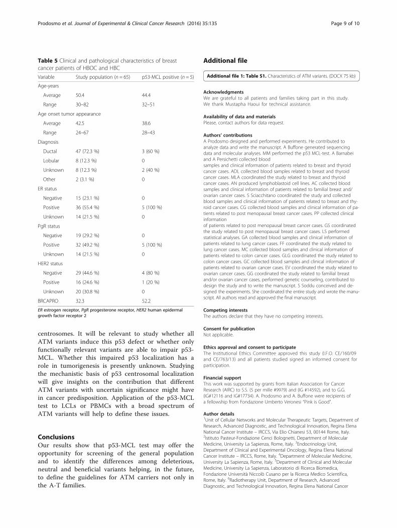
Table 5 Clinical and pathological characteristics of breastcancer patients of HBOC and HBC
Variable Study population (n = 65) p53-MCL positive (n = 5)
Age-years
Average 50.4 44.4
Range 30–82 32–51
Age onset tumor appearance
Average 42.5 38.6
Range 24–67 28–43
Diagnosis
Ductal 47 (72.3 %) 3 (60 %)
Lobular 8 (12.3 %) 0
Unknown 8 (12.3 %) 2 (40 %)
Other 2 (3.1 %) 0
ER status
Negative 15 (23.1 %) 0
Positive 36 (55.4 %) 5 (100 %)
Unknown 14 (21.5 %) 0
PgR status
Negative 19 (29.2 %) 0
Positive 32 (49.2 %) 5 (100 %)
Unknown 14 (21.5 %) 0
HER2 status
Negative 29 (44.6 %) 4 (80 %)
Positive 16 (24.6 %) 1 (20 %)
Unknown 20 (30.8 %) 0
BRCAPRO 32.3 52.2
ER estrogen receptor, PgR progesterone receptor, HER2 human epidermalgrowth factor receptor 2
Prodosmo et al. Journal of Experimental & Clinical Cancer Research (2016) 35:135 Page 9 of 10

Institute – IRCCS, Rome, Italy. 7Medical Physics Unit, Department of Research,Advanced Diagnostic, and Technological Innovation, Regina Elena NationalCancer Institute – IRCCS, Rome, Italy. 8Toracic Surgery Unit, Department ofClinical and Experimental Oncology, Regina Elena National Cancer Institute –IRCCS, Rome, Italy. 9Hepato-pancreato-biliary Surgery Unit, Department ofClinical and Experimental Oncology, Regina Elena National Cancer Institute –IRCCS, Rome, Italy. 10Gynecological Oncology Unit, Department of Clinicaland Experimental Oncology, Regina Elena National Cancer Institute – IRCCS,Rome, Italy. 11Department of Experimental Medicine, Sapienza University ofRome, Policlinico Umberto I, Viale Regina Elena, 32400161 Rome, Italy.
Received: 28 July 2016 Accepted: 23 August 2016
References1. Gatti RA, Boder E, Vinters HV, Sparkes RS, Norman A, Lange K. Ataxia-
telangiectasia: an interdisciplinary approach to pathogenesis. Medicine(Baltimore). 1991;70:99–117.
2. Su Y, Swift M. Mortality rates among carriers of ataxia-telangiectasia mutantalleles. Ann Intern Med. 2000;133:770–8.
3. Uziel T, Savitsky K, Platzer M, Ziv Y, Helbitz T, Nehls M, et al. Genomicorganization of the ATM gene. Genomics. 1996;33:317–20.
4. Matsuoka S, Ballif BA, Smogorzewska A, McDonald 3rd ER, Hurov KE, Luo J,et al. ATM and ATR substrate analysis reveals extensive protein networksresponsive to DNA damage. Science. 2007;316:1160–6.
5. Campbell C, Mitui M, Eng L, Coutinho G, Thorstenson Y, Gatti RA. ATMmutations on distinct SNP and STR haplotypes in ataxia-telangiectasiapatients of differing ethnicities reveal ancestral founder effects. Hum Mutat.2003;21:80–5.
6. Mitui M, Campbell C, Coutinho G, Sun X, Lai CH, Thorstenson Y, et al.Independent mutational events are rare in the ATM gene: haplotypeprescreening enhances mutation detection rate. Hum Mutat. 2003;22:43–50.
7. Concannon P, Gatti RA. Diversity of ATM gene mutations detected inpatients with ataxia-telangiectasia. Hum Mutat. 1997;10:100–7.
8. Roberts NJ, Jiao Y, Yu J, Kopelovich L, Petersen GM, Bondy ML, et al. ATMmutations in patients with hereditary pancreatic cancer. Cancer Discov.2012;2:41–6.
9. Swift M, Reitnauer PJ, Morrell D, Chase CL. Breast and other cancers infamilies with ataxia-telangiectasia. N Engl J Med. 1987;316:1289–94.
10. Renwick A, Thompson D, Seal S, Kelly P, Chagtai T, Ahmed M, et al. ATMmutations that cause ataxia-telangiectasia are breast cancer susceptibilityalleles. Nat Genet. 2006;38:873–5.
11. Maxwell KN, Wubbenhorst B, D’Andrea K, Garman B, Long JM, Powers J, etal. Prevalence of mutations in a panel of breast cancer susceptibility genesin BRCA1/2-negative patients with early-onset breast cancer. Genet Med.2015;17:630–8.
12. Gatti RA, Tward A, Concannon P. Cancer risk in ATM heterozygotes: a modelof phenotypic and mechanistic differences between missense andtruncating mutations. Mol Genet Metab. 1999;68:419–23.
13. Shen L, Yin ZH, Wan Y, Zhang Y, Li K, Zhou BS. Association between ATMpolymorphisms and cancer risk: a meta-analysis. Mol Biol Rep. 2012;39:5719–25.
14. Prodosmo A, De Amicis A, Nisticò C, Gabriele M, Di Rocco G, MonteonofrioL, et al. p53 centrosomal localization diagnoses ataxia-telangiectasiahomozygotes and heterozygotes. J Clin Invest. 2013;123:1335–42.
15. Meier M, den Boer ML, Hall AG, Irving JA, Passier M, Minto L, et al. Relationbetween genetic variants of the ataxia telangiectasia-mutated (ATM) gene,drug resistance, clinical outcome and predisposition to childhood T-lineageacute lymphoblastic leukaemia. Leukemia. 2005;19:1887–95.
16. Stredrick DL, Garcia-Closas M, Pineda MA, Bhatti P, Alexander BH, DoodyMM, et al. The ATM missense mutation p.Ser49Cys (c.146C>G) and the riskof breast cancer. Hum Mutat. 2006;27:538–44.
17. Giannini G, Capalbo C, Ristori E, Ricevuto E, Sidoni T, Buffone A, et al. NovelBRCA1 and BRCA2 germline mutations and assessment of mutationspectrum and prevalence in Italian breast and/or ovarian cancer families.Breast Cancer Res Treat. 2006;100:83–91.
18. Berry DA, Iversen Jr ES, Gudbjartsson DF, Hiller EH, Garber JE, Peshkin BN, etal. BRCAPRO validation, sensitivity of genetic testing of BRCA1/BRCA2, andprevalence of other breast cancer susceptibility genes. J Clin Oncol. 2002;20:2701–12.
19. Adzhubei IA, Schmidt S, Peshkin L, Ramensky VE, Gerasimova A, Bork P, etal. A method and server for predicting damaging missense mutations. NatMethods. 2010;7:248–9.
20. Biosoftware Alamut v2.3. http://www.interactive-biosoftware.com21. Walsh T, Casadei S, Lee MK, Pennil CC, Nord AS, Thornton AM, et al.
Mutations in 12 genes for inherited ovarian, fallopian tube, and peritonealcarcinoma identified by massively parallel sequencing. Proc Natl Acad SciU S A. 2011;108:18032–7.
22. Apostolou P, Fostira F. Hereditary breast cancer: the era of newsusceptibility genes. Biomed Res Int. 2013;2013:747318.
23. Miki Y, Swensen J, Shattuck-Eidens D, Futreal PA, Harshman K, Tavtigian S, etal. A strong candidate for the breast and ovarian cancer susceptibility geneBRCA1. Science. 1994;266:66–71.
24. Wooster R, Bignell G, Lancaster J, Swift S, Seal S, Mangion J, et al.Identification of the breast cancer susceptibility gene BRCA2. Nature. 1995;378:789–92.
25. Wu J, Lu LY, Yu X. The role of BRCA1 in DNA damage response. Protein Cell.2010;1:117–23.
26. Palma M, Ristori E, Ricevuto E, Giannini G, Gulino A. BRCA1 and BRCA2: thegenetic testing and the current management options for mutation carriers.Crit Rev Oncol Hematol. 2006;57:1–23.
27. Thorstenson YR, Shen P, Tusher VG, Wayne TL, Davis RW, Chu G, et al.Global analysis of ATM polymorphism reveals significant functionalconstraint. Am J Hum Genet. 2001;69:396–412.
28. Concannon P, Haile RW, Børresen-Dale AL, Rosenstein BS, Gatti RA, TeraokaSN, et al. Variants in the ATM gene associated with a reduced risk ofcontralateral breast cancer. Cancer Res. 2008;68:6486–91.
29. Mao C, Chung VC, He BF, Luo RC, Tang JL. Association between ATM5557G > A polymorphism and breast cancer risk: a meta-analysis. Mol BiolRep. 2012;39:1113–8.
30. Sandoval N, Platzer M, Rosenthal A, Dörk T, Bendix R, Skawran B, et al.Characterization of ATM gene mutations in 66 ataxia telangiectasia families.Hum Mol Genet. 1999;8:69–79.
31. Telatar M, Teraoka S, Wang Z, Chun HH, Liang T, Castellvi-Bel S, et al. Ataxia-telangiectasia: identification and detection of founder-effect mutations inthe ATM gene in ethnic populations. Am J Hum Genet. 1998;62:86–97.
32. Angèle S, Falconer A, Edwards SM, Dörk T, Bremer M, Moullan N, et al. ATMpolymorphisms as risk factors for prostate cancer development. Br J Cancer.2004;91:783–7.
33. Zhang S, Hemmerich P, Grosse F. Centrosomal localization of DNA damagecheckpoint proteins. J Cell Biochem. 2007;101:451–65.
34. Brunet J, Gutiérrez-Enríquez S, Torres A, Bérez V, Sanjosé S, Galceran J, et al.ATM germline mutations in Spanish early-onset breast cancer patientsnegative for BRCA1/BRCA2 mutations. Clin Genet. 2008;73:465–73.
35. Thompson D, Duedal S, Kirner J, McGuffog L, Last J, Reiman A, et al. Cancerrisks and mortality in heterozygous ATM mutation carriers. J Natl CancerInst. 2005;97:813–22.
36. Tavtigian SV, Oefner PJ, Babikyan D, Hartmann A, Healey S, Le Calvez-KelmF, et al. Rare, evolutionarily unlikely missense substitutions in ATM conferincreased risk of breast cancer. Am J Hum Genet. 2009;85:427–46.
• We accept pre-submission inquiries
• Our selector tool helps you to find the most relevant journal
• We provide round the clock customer support
• Convenient online submission
• Thorough peer review
• Inclusion in PubMed and all major indexing services
• Maximum visibility for your research
Submit your manuscript atwww.biomedcentral.com/submit
Submit your next manuscript to BioMed Central and we will help you at every step:
Prodosmo et al. Journal of Experimental & Clinical Cancer Research (2016) 35:135 Page 10 of 10