research open access evaluation of obstructed defecation
TRANSCRIPT

RESEARCH Open Access
Evaluation of obstructed defecationsyndrome (ODS) using magnetic resonancedefecography (MRD)Arshed Hussain Parry* and Abdul Haseeb Wani
Abstract
Background: Obstructed defecation syndrome is associated with varying combinations of a host of ano-rectalabnormalities, and no physical examination can demonstrate these abnormalities. The present study was aimed toevaluate the spectrum of various pelvic floor abnormalities in obstructed defecation syndrome (ODS).
Results: Of the total 302 patients imaged with age range of 18–72 years (mean age 54 years), 218 were females,and 84 were males. Ano-rectal junction descent was the commonest abnormality observed in 273 (90.3%) patientsfollowed by rectocele (232) (76.8%), rectal intussusception (93) (30.7%), and cystocele (92) (30.4%). Cervical descentwas observed in 78 (35.7%) of female patients. Spastic perineum was seen in 27 (8.9%) patients.
Conclusion: MRD serves as single stop shop for demonstrating and grading a gamut of pelvic organ abnormalitiesunderpinning ODS which in turn helps in choosing the best treatment plan for the patient.
Keywords: Obstructed defecation syndrome, Magnetic resonance defecography, Pelvic floor dysfunction, Spasticperineum syndrome, Rectocele
BackgroundConstipation constitutes a major health concern glo-bally especially among the aging population. Ten per-cent of Indians above the age of 50 years are found tohave constipation. In the USA, constipation leads to2.5 million physician visits per year [1]. A uniformand consistent definition for constipation has beenelusive, and a slew of attempts have been made to ar-rive at a comprehensive definition of constipation thatwould encompass all the myriad symptoms and mani-festations of constipation. Obstructed defecation syn-drome (ODS) constitutes an important subset ofpatients of constipation. ODS has been defined byNICE (National Institute for health and Clinical Ex-cellence) guidelines as inability to completely evacuateor expel fecal bolus in the presence of urge todefecate [2, 3]. Repeated unsuccessful attempts at
defecation, sense of incomplete fecal evacuation, andexcessive straining at toilet pan adversely affecting thequality of life typifies this subset of constipatedpatients. These patients usually resort to digital ma-neuvers to attain rectal evacuation [3, 4]. ODS is usu-ally associated with varying combinations of a host ofano-rectal abnormalities, and no physical examinationcan demonstrate these abnormalities. Dynamic MRIimaging referred to as MRD is a single stop shop todemonstrate various pelvic floor and ano-rectalabnormalities underpinning ODS. This capability ofMRD to evaluate defecation process dynamically helpsin demonstration of various ano-rectal and pelvicfloor abnormalities and thus allows colorectal sur-geons to plan a comprehensive treatment for thesepatients [4, 5]. This study was undertaken to evaluateODS with MRD. The objective of this study was todemonstrate various pelvic floor and ano-rectal ab-normalities associated with ODS.
© The Author(s). 2020 Open Access This article is licensed under a Creative Commons Attribution 4.0 International License,which permits use, sharing, adaptation, distribution and reproduction in any medium or format, as long as you giveappropriate credit to the original author(s) and the source, provide a link to the Creative Commons licence, and indicate ifchanges were made. The images or other third party material in this article are included in the article's Creative Commonslicence, unless indicated otherwise in a credit line to the material. If material is not included in the article's Creative Commonslicence and your intended use is not permitted by statutory regulation or exceeds the permitted use, you will need to obtainpermission directly from the copyright holder. To view a copy of this licence, visit http://creativecommons.org/licenses/by/4.0/.
* Correspondence: [email protected] of Radiodiagnosis, Sher-i-Kashmir Institute of Medical Sciences,Srinagar, Jammu and Kashmir 190011, India
Egyptian Journal of Radiologyand Nuclear Medicine
Parry and Wani Egyptian Journal of Radiology and Nuclear Medicine (2020) 51:78 https://doi.org/10.1186/s43055-020-00197-z
RETRACTED ARTIC
LE

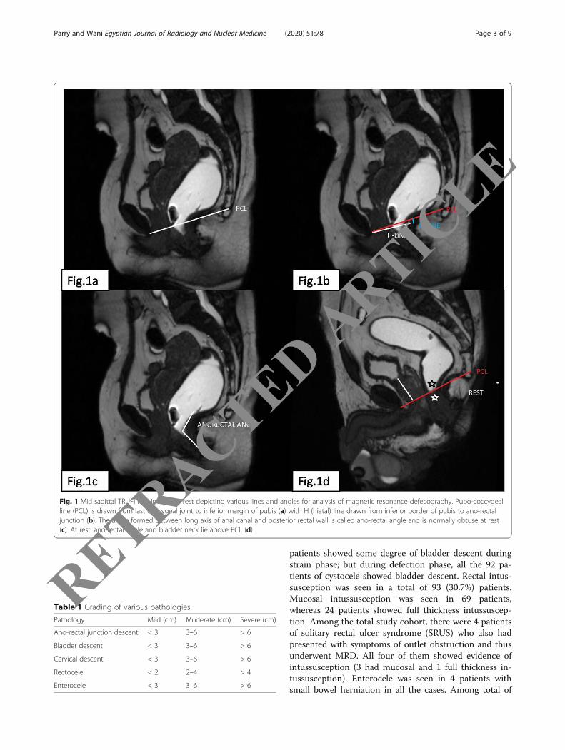
MethodsThis was a prospective study. Patients fulfilling the clin-ical criteria for ODS as laid down in NICE guidelineswere referred to our department for MRD by the colo-rectal division of surgery department. A total of 302 pa-tients were evaluated over a period of 3 years fromDecember 2016 to January 2020. The study was per-formed on 1.5 Tesla superconducting magnetic reson-ance imager (Magnetom Avanto, Siemens MedicalSystem) using standard pelvic coil. All the patients weresubjected to preliminary sigmoidoscopy or colonoscopyto rule out any organic cause of constipation like rectalor colonic neoplasm. Patients were thoroughly explainedthe procedure to ensure their cooperation. A writtenconsent was obtained in each case. Two hundred andfifty millilitre of ultrasound jelly was instilled into therectum using a rectal tube after putting the patient inleft lateral position on the MRI table. Ultrasound jellywas chosen because of its ready availability and its highT2 contrast. Diapers were given to the patients to allowthem to defecate on the MRI gantry. This ensures clean-liness of the gantry table and helps patients to saveblushes and avoid unnecessary embarrassment. The im-aging protocol consisted of preliminary T2 weightedaxial and sagittal sequences {repetition time (TR)/echotime (TE) 2880ms/89 ms; slice thickness of 3 mm; fieldof view 200 mm} to study the anatomy. Following thisdynamic imaging was performed using TRUFI (True fastimaging with steady state free precession) sequence hav-ing a repetition time (TR) of 45.6 ms, echo time (TE) of1.3 ms, slice thickness of 3 mm, and field of view 340mm in sagittal plane during rest, squeeze, strain, anddefecation (drain out) phases. Defecation or drain outphase was run for a sufficient time (approximately 1 to2 min).The images were analyzed on an Apple work sta-tion by two radiologists possessing 9 and 10 years of ex-perience respectively in abdominal radiology. Theinterpreting radiologists were blind to the clinical historyof patients. MR defecography images were analyzed inmid-sagittal plane in cine mode using standard sagittalanatomical planes. Pubo-coccygeal line (PCL) was drawnfrom the inferior margin of pubis to the last coccygealarticulation (Fig. 1a). H (hiatal) line was drawn from theinferior margin of pubic symphysis to the posterior wallof ano-rectal junction (Fig. 1b). H line corresponds tothe pelvic or levator hiatus. M line was drawn perpen-dicular to PCL line from the posterior end of H line(Fig. 1b). The PCL line defines the level of pelvic floor,and the abnormal descent of pelvic structures is diag-nosed when a structure descends below PCL duringstraining or defecation. The ano-rectal angle is the anglemeasured between central axis of anal canal and poster-ior border of distal part of rectum. Ano-rectal angle isformed by the stretch of pubo-rectalis sling on the
posterior ano-rectal junction (Fig. 1c). The position ofano-rectal junction, cervix, and bladder neck was studiedin all the phases. Presence and degree of bladder, cer-vical, and ano-rectal junction descent below PCL werestudied. Presence and degree of intussusception, recto-cele, and enterocele were evaluated. Ano-rectal junctiondescent defined as abnormal descent of ano-rectal junc-tion below pubo-coccygeal line is graded into mild (< 3cm), moderate (3–6 cm), and severe (> 6 cm). Rectoceleis defined as abnormal protrusion of the rectal wall be-yond the expected rectal contour. It is graded into mild(< 2 cm), moderate (2–4 cm), and severe (> 4 cm). Ab-normal caudal descent of bladder and cervix belowpubo-coccygeal line is also graded into mild, moderate,and severe. Abnormal caudal descent of various pelvicstructures is graded as per the standard classificationgiven in Table 1. Invagination of rectal wall into itslumen is called rectal intussusception and is classifiedinto mucosal intussusception or full thickness intussus-ception. When rectal intussusception extends outsideanal verge, it is referred to as rectal prolapse. Enteroceleis defined as caudal displacement of small bowel loopsinto the recto-vesical or recto-vaginal space. Various pel-vic floor abnormalities were noted down. Defecationphase of MR defecography was compared with all theother three phases of defecation (i.e., rest, strain, andsqueeze) combined together.All patients included in this research gave written in-
formed consent to publish the data contained within thisstudy. The datasets used and/or analyzed during thecurrent study are available from the corresponding au-thor on reasonable request.
ResultsA total of 302 patients fulfilling the clinical criteria forODS were studied with a mean age of 54 years (range18–72 years). With regards to gender, 218 were females,and remaining 84 were males. Ano-rectal junction des-cent was commonest abnormality seen in 273 (90.3%)patients with 132 (48 %) showing mild descent, 71 (26%)showing moderate descent, and remaining 70 (25.6%)showing severe descent. During maximal strain, only 101patients showed ano-rectal junction descent, whereasdefecation phase identified another 172 (63%) patientswith ano-rectal junction descent. Anterior rectocele wasseen in 232 (76.8%) patients with mild rectocele seen in192 patients, moderate rectocele seen in 27 patients, andsevere rectocele seen in 13 patients. Anterior rectocelewas seen during strain phase in 151 patients, whereasdefecation phase identified another 81(34.9%) patientswith rectocele taking the total to 232 (76.8%). Cystocelewas seen in 92 (30.4%) patients with 71 patients showingmild cystocele, 17 showing moderate cystocele, andremaining 4 patients showing severe cystocele. Only 16
Parry and Wani Egyptian Journal of Radiology and Nuclear Medicine (2020) 51:78 Page 2 of 9
RETRACTED ARTIC
LE
patients showed some degree of bladder descent duringstrain phase; but during defection phase, all the 92 pa-tients of cystocele showed bladder descent. Rectal intus-susception was seen in a total of 93 (30.7%) patients.Mucosal intussusception was seen in 69 patients,whereas 24 patients showed full thickness intussuscep-tion. Among the total study cohort, there were 4 patientsof solitary rectal ulcer syndrome (SRUS) who also hadpresented with symptoms of outlet obstruction and thusunderwent MRD. All four of them showed evidence ofintussusception (3 had mucosal and 1 full thickness in-tussusception). Enterocele was seen in 4 patients withsmall bowel herniation in all the cases. Among total of
Fig. 1 Mid sagittal TRUFI MRI images at rest depicting various lines and angles for analysis of magnetic resonance defecography. Pubo-coccygealline (PCL) is drawn from last coccygeal joint to inferior margin of pubis (a) with H (hiatal) line drawn from inferior border of pubis to ano-rectaljunction (b). The angle formed between long axis of anal canal and posterior rectal wall is called ano-rectal angle and is normally obtuse at rest(c). At rest, ano-rectal angle and bladder neck lie above PCL (d)
Table 1 Grading of various pathologies
Pathology Mild (cm) Moderate (cm) Severe (cm)
Ano-rectal junction descent < 3 3–6 > 6
Bladder descent < 3 3–6 > 6
Cervical descent < 3 3–6 > 6
Rectocele < 2 2–4 > 4
Enterocele < 3 3–6 > 6
Parry and Wani Egyptian Journal of Radiology and Nuclear Medicine (2020) 51:78 Page 3 of 9
RETRACTED ARTIC
LE
218 females, cervical descent was seen in 78 (35.7%) pa-tients. A comparison between strain and drain outphases revealed that cervical descent was seen in only 32(41%) patients during maximum strain; whereas in drainout phase, all the 78 patients showed descent. Spasticperineum syndrome was seen in 27 (8.9%) patients. Theentire gamut of pelvic floor abnormalities is enumeratedin Table 2.
DiscussionPelvic floor dysfunction is characterized by bladder,bowel, or sexual dysfunction with a variable combinationof pelvic organ prolapse. It affects multiparous womenmore commonly than men. Obstetric damage to pelvic
Table 2 Various pelvic floor abnormalities observed onmagnetic resonance defecography
Abnormality Total Mild Moderate Severe
Ano-rectal junction descent 273 (90.3%) 132 71 70
Rectocele 232 (76.8%) 192 27 13
Rectal intussusception 93 (30.7%)
Enterocele 4
Cervical descent 78 (35.7%)✦ 63 11 4
Spastic perineum 27 (8.9%)
Cystocele 92 (30.4%) 71 17 4
✦35.7% of female patients
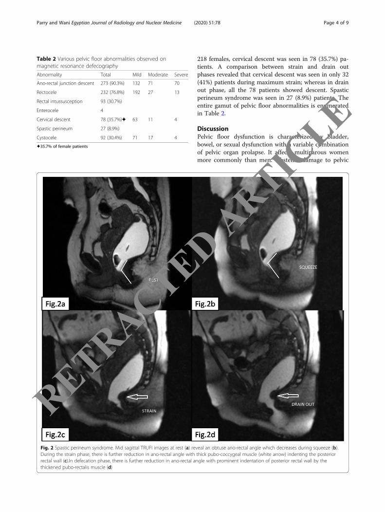
Fig. 2 Spastic perineum syndrome. Mid sagittal TRUFI images at rest (a) reveal an obtuse ano-rectal angle which decreases during squeeze (b).During the strain phase, there is further reduction in ano-rectal angle with thick pubo-coccygeal muscle (white arrow) indenting the posteriorrectal wall (c).In defecation phase, there is further reduction in ano-rectal angle with prominent indentation of posterior rectal wall by thethickened pubo-rectalis muscle (d)
Parry and Wani Egyptian Journal of Radiology and Nuclear Medicine (2020) 51:78 Page 4 of 9
RETRACTED ARTIC
LE
floor structures like ilio-coccygeus muscle, pubo-coccygeus muscle, anal sphincter, endopelvic fascia, andpudendal nerve is believed to cause pelvic floor dysfunc-tion in multiparous women. Obstructed defecation syn-drome (ODS) constitutes a unique set of chronicallyconstipated patients who fail to completely evacuatetheir rectum. These patients resort to excessive strainingand digital maneuvering of rectum to attain completerectal evacuation. ODS can result either from a func-tional abnormality or organic ano-rectal abnormality.Patients with functional abnormality can be treated withbio feedback therapy or psychotherapy, whereas those
with an organic ano-rectal disorder respond to surgicalcorrection [5]. The diagnostic armamentarium chieflyconsists of fluoroscopic defecography and magnetic res-onance defecography (MRD) [5–7]. MRD has the cap-ability of demonstrating the various pelvic floorabnormalities with great accuracy. MRD serves as a onestop shop for studying the normal pelvic anatomy andthe complete range of pelvic floor abnormalities. MRDlacks radiation exposure. MRD can be performed in sit-ting position using open configuration MRI or in supineposition using closed configuration magnet [7]. MRDperformed in supine position yields comparable results
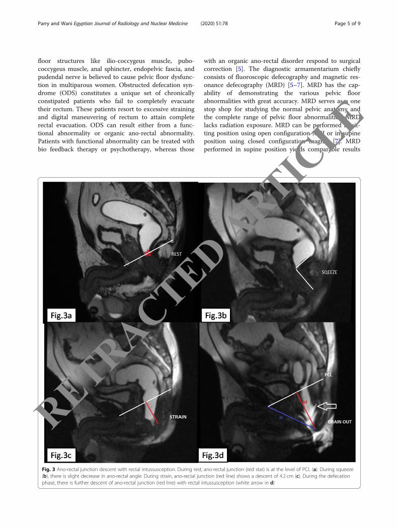
Fig. 3 Ano-rectal junction descent with rectal intussusception. During rest, ano-rectal junction (red star) is at the level of PCL (a). During squeeze(b), there is slight decrease in ano-rectal angle. During strain, ano-rectal junction (red line) shows a descent of 4.2 cm (c). During the defecationphase, there is further descent of ano-rectal junction (red line) with rectal intussusception (white arrow in d)
Parry and Wani Egyptian Journal of Radiology and Nuclear Medicine (2020) 51:78 Page 5 of 9
RETRACTED ARTIC
LE
to that performed in sitting position for the reason thatthe straining forces applied during defecation are of suf-ficient magnitude to elicit the various pathologies [8, 9].Pelvic floor is divided into three compartments: anter-
ior compartment comprises of bladder and urethra, mid-dle compartment comprises of uterus and vagina, andthe posterior compartment is comprised of ano-rectalcanal [9, 10]. However, all the three compartments workin unison, and combined disorders of pelvic floor arecommon and should be assessed simultaneously. Normalano-rectal angle measures between 108° and 127° [11,12]. During normal defecation, the pubo-rectalis slingrelaxes leading to widening of the ano-rectal angle by
15–20° so that the rectum and anal canal are aligned ina straight line to allow expulsion of fecal matter [13, 14].Failure of widening of ano-rectal angle during defecationwith persistence of acute ano-rectal angle forms thebasis for the diagnosis of spastic perineum syndrome(SPS) (Fig. 2) (Video 1). This disorder is also called asparadoxical pubo-rectalis syndrome (PPS). It resultsfrom failure of pubo-rectalis muscle to relax duringdefecation. In fact, there is paradoxical contraction ofthis muscle during defecation which prevents opening ofano-rectal angle during defecation with consequent fail-ure of evacuation of feces. Thickening of pubo-rectalismuscle has been reported previously in literature in PPS
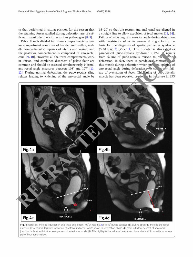
Fig. 4 Rectocele. There is reduction in ano-rectal angle from 109° at rest (Fig.4a) to 92° during squeeze (b). During strain (c), there is ano-rectaljunction descent (red star) with formation of anterior rectocele (white arrow). In defecation phase (d), there is further descent of ano-rectaljunction (> 6 cm) with further enlargement of anterior rectocele (d). This highlights the value of defecation phase which elicits or adds to variouspelvic floor abnormalities
Parry and Wani Egyptian Journal of Radiology and Nuclear Medicine (2020) 51:78 Page 6 of 9
RETRACTED ARTIC
LE

patients [12]. However, Liu et.al in their study concludedthat though mean thickness of the pubo-rectalis musclewas more in patients with PPS than in patients withoutPPS, but the difference between groups was not statisti-cally significant [15]. However, they reported a signifi-cant difference in apparent diffusion co-efficient (ADC)values of pubo-rectalis muscle between patients withPPS and patients without PPS which points to the factthat alteration in muscle microstructure might be theunderlying mechanism for PPS [15]. Ano-rectal junctiondescent was the commonest abnormality encounteredwith 273 (90.3%) patients demonstrating various gradesof ano-rectal junction descent (Fig. 3). Descent of ano-rectal junction can occur in isolation but frequently des-cent of the anterior, and middle compartment structuresare also seen in association with it. This is frequently as-sociated with feeling of incomplete evacuation resultingin further increase in straining during defecation andconsequent neuropathic injury that may result in incon-tinence [12]. Anterior rectocele was second commonestabnormality observed in 232 (76.8%) (Fig. 4). Factorsthat increase the likelihood of developing a rectocele in-clude birth trauma, hysterectomy, chronically increasedintra-abdominal pressure, and increased age. Rectocelesassume clinical relevance when symptoms develop asthey are responsible for obstructed defecation whichusually requires vaginal or perineal digitations to attainrectal emptying [12]. Post defecation retention of jellywithin rectocele fairly correlates with patient symptomsand is an important abnormality which usually necessi-tates digitization (Fig. 5b). Rectal intussusception is
classified into mucosal intussusception or full thicknessintussusception (Fig. 3d) (Video 2). This causes obstruc-tion to the passage of feces. MR defecography isadvantageous in discriminating between mucosal intus-susception and full-thickness intussusception and is rele-vant in treatment planning. Mucosal intussusception canbe treated with transanal excision of the redundant orprolapsing mucosa, whereas a rectopexy might be re-quired for full-thickness intussusception [12]. Enterocele,defined as caudal displacement of small bowel loops intothe recto-vesical or recto-vaginal space, occurs morecommonly in patients who have undergone hysterec-tomy owing to disruption of pubo-cervical and recto-vaginal portions of supporting endopelvic fascia. Entero-celes are more clearly demonstrable towards the end ofdefecation process because a fully loaded rectum doesnot allow sufficient space for descent of small bowel intopelvis [12, 16, 17]. It is vital to detect enterocele becauseit forms a contraindication for stapled transanal rectalresection (STARR) due to the potential danger to theherniated small bowel during this surgery [11, 18]. Ab-normal caudal descent of bladder and cervix belowpubo-coccygeal line is also graded into mild, moderate,and severe (Fig. 5a). Abnormal pelvic floor descent grad-ing can be easily remembered by the rule of 3 with des-cent of an organ below PCL by ≤ 3 cm mild descent, 3–6 cm moderate descent, and > 6 cm severe descent [8,12, 13]. Defecation phase puts the maximum downwardforce on pelvic floor which helps in demonstration of ahigher number of pelvic organ descents when comparedto strain phase [15, 19]. Ano-rectal junction descent was
Fig. 5 Descent of all the compartments. Terminal drain out (defecation phase) of same patient as in Fig. 4 reveals descent of bladder neck (redline) and cervix (blue line) (a). Same patient also shows severe ano-rectal junction decent (red line) with retention of jelly in anterior rectocele(white arrow) (b). This picture highlights the role of running the defecation phase imaging for a sufficient time to demonstrate thefull abnormality
Parry and Wani Egyptian Journal of Radiology and Nuclear Medicine (2020) 51:78 Page 7 of 9
RETRACTED ARTIC
LE
visible in 101 (36.9%) patients on strain phase which in-creased to 273 in defecation phase. Thus, defecationphase clearly has higher detection rate for ano-rectaljunction descent [20]. Similarly, bladder and cervicaldescent were seen in 16 and 32 patients during strainphase and in 92 and 78 patients respectively duringdefecation or drain out phase. Defecation phase alsoidentified an additional number 81 (34.9%) rectoceleswhen compared to strain phase. None of the patientsshowed intussusceptions during strain, and all the 93 pa-tients of intussusception were identified duringdefecation phase. Also, we noted that the maximumdepth or degree of an abnormality was visible duringdefecation phase (Fig. 4c, d). So clearly, the diagnosticyield of defecation phase is best among all the phases ofdefecation and this attests to the fact that defecationphase is the single most important phase to elicit the fullrange of pelvic floor abnormalities and must be includedin magnetic resonance defecography (Video 3 and Video4). This comes at a slightly higher cost of providing thepatient with waterproof diaper and having to explain thepatient to defecate on MRI table which might be littleembarrassing to many patients.
ConclusionA vast range of pelvic floor abnormalities existing invarious combinations in ODS patients can be demon-strated and graded using MRD which in turn helps inchoosing the best treatment plan for the patient.Defecation phase is the single most important phase ofMRD and has the highest diagnostic yield and must beincluded in all MRD studies.
Supplementary informationSupplementary information accompanies this paper at https://doi.org/10.1186/s43055-020-00197-z.
Additional file 1: Video 1. Patient of spastic perineum syndromeduring defecation phase shows abnormal acute ano-rectal angle withmarkedly thick pubo-rectalis muscle indenting posterior rectal wall.
Additional file 2: Video 2. Mid sagittal cine loop TRUFI duringdefecation phase reveals severe ano-rectal junction descent with forma-tion of full thickness rectal intussusception.
Additional file 3: Video 3. During strain phase the vector of forceseems to be directed anteriorly (rather than downwards) with resultantanterior rectocele formation and ano-rectal junction descent.
Additional file 4: Video 4. Cine loop TRUFI during defecation phase ofthe same patient as in video 3 shows enlargement of rectocele withdescent of all the three (bladder, cervix and rectum) compartments.Towards the end of defecation there is retention of jelly within therectocele.
AbbreviationsMRD: Magnetic resonance defecography; MRI: Magnetic resonance imaging;ODS: Obstructed defecation syndrome; SPS: Spastic perineum syndrome;PCL: Pubo-coccygeal line; H line: Hiatal line; NICE: National Institute for healthand Clinical Excellence; TRUFI: True fast imaging with steady state freeprecession
AcknowledgementsNone.
Authors’ contributionsPA and WA performed, analyzed, and interpreted the magnetic resonancedefecography images. Both the authors were involved in manuscriptpreparation and literature research. Both the authors have read andapproved the manuscript.
FundingNo funding was required for this study as it was the part of evaluation as perthe institutional protocol. The patients paid themselves the nominal fee forthe procedure.
Availability of data and materialsAll the data and materials were obtained from patients registered in ourhospital.
Ethics approval and consent to participateThis study was duly approved by the Institutional Ethical Committee (IEC) ofSher-i-Kashmir Institute of Medical Sciences (SKIMS) under the No. SIMS 037/IEC-SKIMS/2016-45. No animal participants were used in this study. Informedverbal consent was obtained from all the patients included in the study.
Consent for publicationNone
Competing interestsWe declare that we have no (financial and non-financial) competinginterests.
Received: 12 March 2020 Accepted: 5 May 2020
References1. Thapar RB, Patankar RV, Kamat RD, Thapar RR, Chemburkar V (2015 Jan) MR
defecography for obstructed defecation syndrome. The Indian journal ofradiology & imaging 25(1):25
2. Longstreth GF, Thompson WG, Chey WD, Houghton LA, Mearin F, Spiller RC(2006) Functional bowel disorders. Gastroenterology. 130(5):1480–1491
3. Mohamed F. O, Ahmed FA. Role of MR Defecography in the assessment ofobstructed defecation syndrome. The Medical Journal of Cairo University2018;86(March):927-931.
4. Bamboriya R, Jaipal U, Jakhar S (2020) A descriptive study of MRdefecography for evaluation of obstructed defecation syndrome.International Journal of Medical and Biomedical Studies 31:4(1)
5. Lembo A, Camilleri M (2003) Chronic constipation. N Engl J Med 349:1360–13686. Garcia del Salto L, de Miguel CJ, Aguilera del Hoyo LF et al (2014) MR
imaging-based assessment of the female pelvic floor. Radiographics. 34(5):1417–1439
7. Schreyer AG, Paetzel C, Furst A et al (2012) Dynamic magnetic resonancedefecography in 10 asymptomatic volunteers. World J Gastroenterol 18(46):6836–6842
8. Law YM, Fielding JR (2008) MRI of pelvic floor dysfunction: review. AJR Am JRoentgenol 191(6 Suppl):S45–S53
9. Faccioli N, Comai A, Mainardi P, Perandini S, Farah M, Pozzi-Mucelli R (2010)Defecography: a practical approach. Diagn Interv Radiol 16:209–216
10. Roos JE, Weishaupt D, Wildermuth S et al (2002) Experience of 4 years withopen MR defecography: pictorial review of anorectal anatomy and disease.Radiographics. 22(4):817–832
11. Pannu HK, Kaufman HS, Cundiff GW et al (2000) Dynamic MR imaging of pelvicorgan prolapse: spectrum of abnormalities. Radiographics. 20(6):1567–1582
12. Colaiacomo MC, Masselli G, Polettini E et al (2009) Dynamic MR imaging ofthe pelvic floor: a pictorial review. Radiographics. 29(3):e35
13. Boyadzhyan L, Raman SS, Raz S (2008) Role of static and dynamic MRimaging in surgical pelvic floor dysfunction. Radiographics. 28(4):949–967
14. Elshazly WG, El Nekady AA, Hassan H (2010) Role of dynamic magneticresonance imaging in management of obstructed defecation case series. IntJ Surg 8:274–282
Parry and Wani Egyptian Journal of Radiology and Nuclear Medicine (2020) 51:78 Page 8 of 9
RETRACTED ARTIC
LE
15. Liu G, Cui Z, Dai Y, Yao Q, Xu J, Wu G (2017) Paradoxical puborectalissyndrome on diffusion-weighted imaging: a retrospective study of 72 cases.Sci Rep 7(1):1–6
16. Woodfield CA, Hampton BS, Sung V, Brody JM (2009) Magnetic resonance imagingof pelvic organ prolapse: comparing pubococcygeal and midpubic lines withclinical staging. Int Urogynecol J Pelvic Floor Dysfunct 20(6):695–701
17. Alt CD, Brocker KA, Lenz F, Sohn C, Kauczor HU, Hallscheidt P (2014) MRIfindings before and after prolapse surgery. Acta Radiol 55(4):495–504
18. McNevin MS (2010) Overview of pelvic floor disorders. Surg Clin N Am 90:195–205
19. Fielding JR (2002) Practical MR imaging of female pelvic floor weakness.Radiographics. 22(2):295–304
20. DeLancey JO (1994) The anatomy of the pelvic floor. Curr Opin ObstetGynecol 6(4):313–316
Publisher’s NoteSpringer Nature remains neutral with regard to jurisdictional claims inpublished maps and institutional affiliations.
Parry and Wani Egyptian Journal of Radiology and Nuclear Medicine (2020) 51:78 Page 9 of 9
RETRACTED ARTIC
LE