research open access mechanical stretch up-regulates the b ... · cardial fibroblast. fibroblast...
TRANSCRIPT
RESEARCH Open Access
Mechanical stretch up-regulates the B-typenatriuretic peptide system in human cardiacfibroblasts: a possible defense againsttransforming growth factor-β mediated fibrosisChris J Watson1†, Dermot Phelan1†, Maojia Xu1, Patrick Collier1, Roisin Neary1, Albert Smolenski1, Mark Ledwidge2,Kenneth McDonald2 and John Baugh1*
Abstract
Background: Mechanical overload of the heart is associated with excessive deposition of extracellular matrixproteins and the development of cardiac fibrosis. This can result in reduced ventricular compliance, diastolicdysfunction, and heart failure. Extracellular matrix synthesis is regulated primarily by cardiac fibroblasts, morespecifically, the active myofibroblast. The influence of mechanical stretch on human cardiac fibroblasts’ response topro-fibrotic stimuli, such as transforming growth factor beta (TGFβ), is unknown as is the impact of stretch on B-type natriuretic peptide (BNP) and natriuretic peptide receptor A (NPRA) expression. BNP, acting via NPRA, has beenshown to play a role in modulation of cardiac fibrosis.
Methods and results: The effect of cyclical mechanical stretch on TGFβ induction of myofibroblast differentiationin primary human cardiac fibroblasts and whether differences in response to stretch were associated with changesin the natriuretic peptide system were investigated. Cyclical mechanical stretch attenuated the effectiveness ofTGFβ in inducing myofibroblast differentiation. This finding was associated with a novel observation thatmechanical stretch can increase BNP and NPRA expression in human cardiac fibroblasts, which could haveimportant implications in modulating myocardial fibrosis. Exogenous BNP treatment further reduced the potency ofTGFβ on mechanically stretched fibroblasts.
Conclusion: We postulate that stretch induced up-regulation of the natriuretic peptide system may contribute tothe observed reduction in myofibroblast differentiation.
Keywords: Mechanical stretch, BNP, Natriuretic peptide receptor A, Transforming growth factor beta, Myofibroblast,Alpha smooth muscle actin
BackgroundHypertensive heart disease describes a phase of remodel-ing which occurs in the myocardium when exposed tosustained elevation in arterial blood pressure or hyper-trophic substances associated with the hypertension syn-drome. The two most notable features of hypertensiveheart disease are myocyte hypertrophy and reactive
fibrosis. The relationship between reactive fibrosis andhypertension is well described [1-3].In physiological terms, the effect of this fibrosis is a re-
duction in compliance of the myocardium and diastolicdysfunction. This premise is based on two principles.Firstly, the addition of fibrillar collagen to normal tissueresults in reduced compliance of that tissue, and sec-ondly, regression of such fibrosis improves complianceand reduces cardiac stiffness [4-8]. The synthesis anddegradation of fibrillar collagen is regulated by the myo-cardial fibroblast. Fibroblast differentiation to the moreactive myofibroblast form is a hallmark of cardiac
* Correspondence: [email protected]†Equal contributors1School of Medicine & Medical Science, The Conway Institute ofBiomolecular and Biomedical Research, University College Dublin, Dublin,IrelandFull list of author information is available at the end of the article
© 2012 Watson et al.; licensee BioMed Central Ltd. This is an Open Access article distributed under the terms of the CreativeCommons Attribution License (http://creativecommons.org/licenses/by/2.0), which permits unrestricted use, distribution, andreproduction in any medium, provided the original work is properly cited.
Watson et al. Fibrogenesis & Tissue Repair 2012, 5:9http://www.fibrogenesis.com/content/5/1/9
fibrosis, and is associated with increased collagen pro-duction, enhanced proliferative and migratory potential,and is associated with increased expression of alpha-smooth muscle actin (ASMA) [9]. Although myofibro-blast differentiation is an essential process required fornormal wound healing, prolonged injury or perhaps lossof regulation can result in pathological fibrosis.Modulation of pro-fibrotic signals within the myocar-
dium is necessary to regulate normal wound healing pro-cesses. One possible regulator may be B-type natriureticpeptide (BNP). BNP is an endogenous hormone which isknown to be secreted by cardiac myocytes in response tomyocardial stretch and overload and is an important regu-lator of blood volume homeostasis through its diuretic,natriuretic, and vasodilating actions and by inhibitingrenin and aldosterone [10,11]. More recent data suggestthat natriuretic peptides also appear to play a major rolein modulation of both cardiac and renal fibrosis [12-14].The aim of this study was to investigate the combined
effects of mechanical stretch and the pro-fibrotic cytokinetransforming growth factor beta (TGFβ) on myofibroblastdifferentiation, and the impact of BNP in this process.
ResultsThe impact of mechanical stretch on human primary car-diac fibroblast cells response to recombinant TGFβ wasinvestigated. These experiments were carried out on flex-ible six well culture plates coated with 2 μg/cm2 humanfibronectin, as described in the Methods section. Cells at aconfluency of approximately 70% were exposed to cyclicequibiaxial Heart Simulation strain (1 Hz, maximumelongation 10%) for 72 h. Control plates were exposed tothe same conditions without being stretched. Theseexperiments were carried out either in the presence or ab-sence of 10 ng/mL recombinant TGFβ.
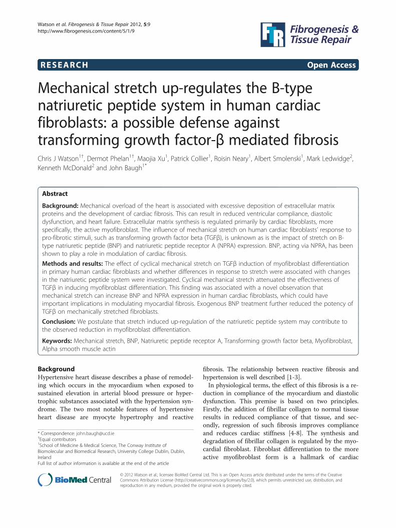
TGFβ-induced ASMA and collagen 1 is reduced onmechanically stretched cardiac fibroblast cellsThe combined effects of mechanical stretch and TGFβtreatment on human primary cardiac fibroblast expres-sion of the differentiation marker ASMA and the pro-fibrotic markers collagen 1 and 3 were investigated.Quantitative real-time PCR analysis revealed that TGFβinduction of ASMA gene expression was significantlyreduced on stretched cells. TGFβ treatment for 72 h onnon-stretched cells up-regulated ASMA by over 15-fold,whereas TGFβ stimulated ASMA expression was onlytwo-fold in stretched cells, P< 0.001 (Figure 1A). Asimilar response was detected for collagen 1, whereTGFβ induction of collagen 1 gene expression wasreduced from approximately four-fold to two-fold whencells were mechanically stretched compared to non-stretched cells, P< 0.001 (Figure 1A). Interestingly, un-like ASMA and collagen1, TGFβ induction of collagen 3
expression was significantly enhanced when cells weremechanically stretched, P< 0.05. Due to the magnitudeof the suppressive effects of stretch on TGFβ up-regulation of ASMA, this observation was verified at theprotein level by Western blotting, where TGFβ inducedASMA was significantly diminished in stretched cells,P< 0.001 (Figure 1B).
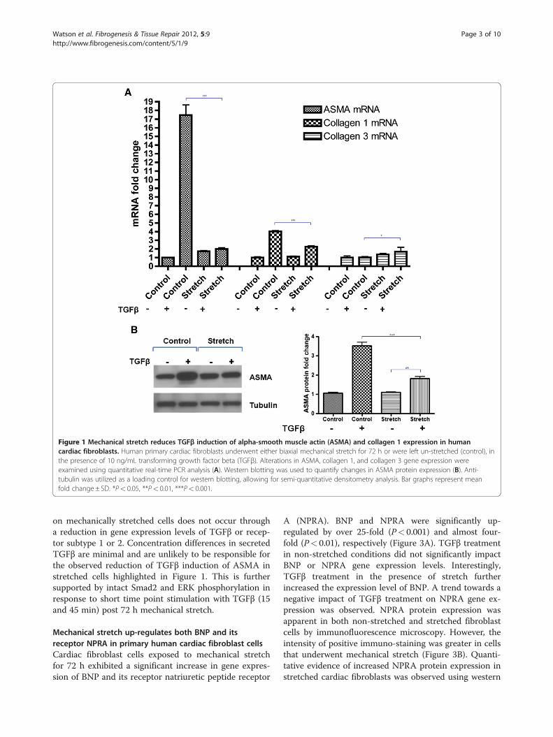
Diminished responses to exogenous TGFβ on mechaniallystretched cardiac fibroblast cells are not due to decreasedbasal expression levels of TGFβThe impact of cyclical mechanical stretch on basal andTGFβ stimulated expression of TGFβ, and its receptorsubtypes 1 and 2 (TGFβ-R1; TGFβ-R2) were investigated.Human cardiac fibroblast cells underwent mechanicalstretch for 72 h in the presence or absence of 10 ng/mLTGFβ. Quantitative gene expression analysis revealed thatmechanical stretch did not alter the levels of TGFβ recep-tors (Figure 2A). However, mechanical stretch did impactthe basal expression levels of TGFβ. A significant 2.5-foldincrease in gene expression was observed (P< 0.05)(Figure 2A). Further evidence that the diminished re-sponse to exogenous TGFβ on mechanically stretchedcells was not due to reduced basal expression of TGFβ, orits receptors, is shown in Figure 2B. A reduction in Smad2phosphorylation was not observed under stretched condi-tions. Of note, treatment with TGFβ in stretched cellsresulted in a 10-fold increase in basal expression of TGFβgene expression (P< 0.01), Figure 2C, but had no impacton expression of its receptors (data not shown). The im-pact of mechanical stretch and recombinant TGFβ treat-ment on HCF production of TGFβ protein productionwas investigated. Both total and active TGFβ1 was quanti-fied in the supernatant of HCF using ELISA based meth-ods (Figure 2D). The relatively large increase in TGFβmRNA expression in cells exposed to mechanical stretchand recombinant TGFβ stimulation was not reflected atthe protein level. The highest concentration of secretedTGFβ protein (both latent and active) was in un-stretchedcardiac fibroblast cells stimulated with recombinant TGFβtreatment for 72 h. The impact of stretch alone did nothave a significant impact on TGFβ secretion (although adownward trend was apparent). However, the ability of re-combinant TGFβ to stimulate endogenous TGFβ produc-tion was reduced when the cells were mechanicallystretched. Interestingly however, it is important to note thatthe detectable levels of secreted TGFβ were <1 pg/mL,thus the physiological relevance of these small changeswithin the experimental cell culture environment is un-known, especially under conditions of recombinant TGFβtreatment were cells are exposed to 10,000x thisconcentration.Collectively, these data suggest that the observed re-
duction in TGFβ-mediated myofibroblast differentiation
Watson et al. Fibrogenesis & Tissue Repair 2012, 5:9 Page 2 of 10http://www.fibrogenesis.com/content/5/1/9
on mechanically stretched cells does not occur througha reduction in gene expression levels of TGFβ or recep-tor subtype 1 or 2. Concentration differences in secretedTGFβ are minimal and are unlikely to be responsible forthe observed reduction of TGFβ induction of ASMA instretched cells highlighted in Figure 1. This is furthersupported by intact Smad2 and ERK phosphorylation inresponse to short time point stimulation with TGFβ (15and 45 min) post 72 h mechanical stretch.
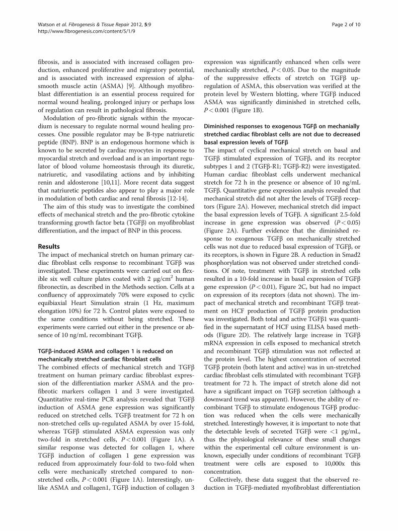
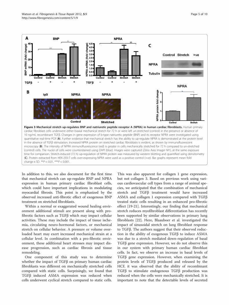
Mechanical stretch up-regulates both BNP and itsreceptor NPRA in primary human cardiac fibroblast cellsCardiac fibroblast cells exposed to mechanical stretchfor 72 h exhibited a significant increase in gene expres-sion of BNP and its receptor natriuretic peptide receptor
A (NPRA). BNP and NPRA were significantly up-regulated by over 25-fold (P< 0.001) and almost four-fold (P< 0.01), respectively (Figure 3A). TGFβ treatmentin non-stretched conditions did not significantly impactBNP or NPRA gene expression levels. Interestingly,TGFβ treatment in the presence of stretch furtherincreased the expression level of BNP. A trend towards anegative impact of TGFβ treatment on NPRA gene ex-pression was observed. NPRA protein expression wasapparent in both non-stretched and stretched fibroblastcells by immunofluorescence microscopy. However, theintensity of positive immuno-staining was greater in cellsthat underwent mechanical stretch (Figure 3B). Quanti-tative evidence of increased NPRA protein expression instretched cardiac fibroblasts was observed using western
Figure 1 Mechanical stretch reduces TGFβ induction of alpha-smooth muscle actin (ASMA) and collagen 1 expression in humancardiac fibroblasts. Human primary cardiac fibroblasts underwent either biaxial mechanical stretch for 72 h or were left un-stretched (control), inthe presence of 10 ng/mL transforming growth factor beta (TGFβ). Alterations in ASMA, collagen 1, and collagen 3 gene expression wereexamined using quantitative real-time PCR analysis (A). Western blotting was used to quantify changes in ASMA protein expression (B). Anti-tubulin was utilized as a loading control for western blotting, allowing for semi-quantitative densitometry analysis. Bar graphs represent meanfold change± SD. *P< 0.05, **P< 0.01, ***P< 0.001.
Watson et al. Fibrogenesis & Tissue Repair 2012, 5:9 Page 3 of 10http://www.fibrogenesis.com/content/5/1/9
blotting. NPRA protein expression was up-regulatedmore than two-fold following 72 h of mechanicalstretching (P< 0.01) (Figure 3C). Cell lysates derivedfrom transient over-expression of NPRA served as apositive control.
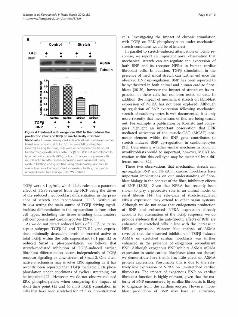
Exogenous BNP further reduces the pro-fibrotic effects ofTGFβ on stretched primary human cardiac fibroblast cellsThe reduced potency of TGFβ to induce myofibroblastdifferentiation on stretched cardiac fibroblasts was fur-ther investigated in the context of stretch induced BNPand NPRA expression. The impact of recombinant BNPtreatment in combination with TGFβ on stretched cellswas explored. It was found that the anti-fibrotic effects
of exogenous BNP, as measured by ASMA protein re-pression, were only apparent when cardiac fibroblastswere mechanically stretched for 72 h (Figure 4).
DiscussionTGFβ is a powerful mediator of adverse cardiac remod-eling and fibrosis primarily via Smad-dependent induc-tion of collagen expression as well as differentiation offibroblasts to the myofibroblast phenotype which exhi-bits increased secretory, migratory, and proliferativeproperties and express the contractile protein ASMA[15-18]. The current study presents a novel observationthat the pro-fibrotic effects of TGFβ are reduced onmechanically stretched human primary cardiac fibroblasts.
Figure 2 Mechanical stretch does not reduce basal TGFβ expression in human cardiac fibroblast cells. Human primary cardiac fibroblastcells underwent either biaxial mechanical stretch for 72 h or were left un-stretched (control), in the presence or absence of 10 ng/mLtransforming growth factor beta (TGFβ). Analysis of stretch induced basal gene expression changes of TGFβ, TGFβ receptor type-1 (TGFβ-R1), andTGFβ receptor type-2 (TGFβ-R2) by quantitative real-time PCR are highlighted (A). Presented gene expression fold changes in Figure A areindependent of recombinant TGFβ treatment and represent the impact of mechanical stretch alone. Western blot analysis of the impact of 72 hof mechanical stretch on TGFβ induced phosphorylation of Smad2 and ERK protein expression (B). Quantitative real-time PCR analysis of theeffect of exogenous TGFβ treatment on basal TGFβ expression (C). The impact of mechanical stretch and recombinant TGFβ treatment on cardiacfibroblast production and secretion of total and active TGFβ protein was assessed using ELISA-based methods (D). Bar graphs represent meanfold change± SD. *P< 0.05, **P< 0.01.
Watson et al. Fibrogenesis & Tissue Repair 2012, 5:9 Page 4 of 10http://www.fibrogenesis.com/content/5/1/9
In addition to this, we also document for the first timethat mechanical stretch can up-regulate BNP and NPRAexpression in human primary cardiac fibroblast cells,which could have important implications in modulatingmyocardial fibrosis. This point is emphasized by theobserved increased anti-fibrotic effect of exogenous BNPtreatment on stretched fibroblasts.Within a normal or exaggerated wound healing envir-
onment additional stimuli are present along with pro-fibrotic factors such as TGFβ which may impact cellularactivities. These may include the impact of tissue ische-mia, circulating neuro-hormonal factors, or mechanicalstretch on cellular behavior. A pressure or volume over-loaded heart may exert increased mechanical strain at acellular level. In combination with a pro-fibrotic envir-onment, these additional heart stresses may impact dis-ease progression, such as cardiac fibrosis and tissueremodeling.One component of this study was to determine
whether the impact of TGFβ on primary human cardiacfibroblasts was different on mechanically stretched cellscompared with static cells. Surprisingly, we found thatTGFβ induced ASMA expression was reduced whencells underwent cyclical stretch compared to static cells.
This was also apparent for collagen 1 gene expression,but not collagen 3. Based on previous work using vari-ous cardiovascular cell types from a range of animal spe-cies, we anticipated that the combination of mechanicalstretch and TGFβ treatment would have increasedASMA and collagen 1 expression compared with TGFβtreated static cells resulting in an enhanced pro-fibroticeffect [19-21]. Interestingly, our finding that mechanicalstretch reduces myofibroblast differentiation has recentlybeen supported by similar observations in primary lungfibroblasts [22]. Here, Blaauboer et al. investigated theimpact of sinusoidal stretch on lung fibroblast responseto TGFβ. The authors suggest that their observed reduc-tion in the ability of exogenous TGFβ to induce ASMAwas due to a stretch mediated down-regulation of basalTGFβ gene expression. However, we do not observe thisin our system with primary human cardiac fibroblastcells. In fact, we observe an increase in basal levels ofTGFβ gene expression. However, when examining theprotein levels of TGFβ produced and released by theHCF, it was observed that the ability of recombinantTGFβ to stimulate endogenous TGFβ production wasreduced when the cells were mechanically stretched. It isimportant to note that the detectable levels of secreted
Figure 3 Mechanical stretch up-regulates BNP and natriuretic peptide receptor A (NPRA) in human cardiac fibroblasts. Human primarycardiac fibroblast cells underwent either biaxial mechanical stretch for 72 h or were left un-stretched (control) in the presence or absence of10 ng/mL recombinant TGFβ. Changes in gene expression of b-type natriuretic peptide (BNP) and its receptor NPRA were investigated usingquantitative real-time PCR (A). Further evidence that mechanical stretch has the ability to up-regulate NPRA is demonstrated at the protein levelin the absence of TGFβ stimulation. Increased NPRA protein on stretched cardiac fibroblasts is evident, as shown by immunofluorescentmicroscopy (B). The intensity of NPRA immunofluorescence (red) is greater in cells mechanically stretched for 72 h compared to un-stretched(control) cells. The nuclei of cells were counterstained using DAPI (blue). Images were captured (Zeiss Axio Imager M1), at the same exposuretime for comparison. Stretch-induced (72 h) up-regulation of NPRA protein was measured by western blotting and quantified using densitometry(C). Protein extracted from HEK-293-T cells over-expressing NPRA were used as a positive control (+ve). Bar graphs represent mean foldchange± SD. **P< 0.01, ***P< 0.001.
Watson et al. Fibrogenesis & Tissue Repair 2012, 5:9 Page 5 of 10http://www.fibrogenesis.com/content/5/1/9
TGFβ were <1 pg/mL, which likely rules out a paracrineeffect of TGFβ released from the HCF being the driverof the reduced myofibroblast differentiation in the pres-ence of stretch and recombinant TGFβ. Within anin vivo setting the main source of TGFβ driving myofi-broblast differentiation in the myocardium is from othercell types, including the tissue invading inflammatorycell component and cardiomyocytes [23-26].As we do not detect reduced levels of TGFβ, or its re-
ceptor subtypes TGFβ-R1 and TGFβ-R2 gene expres-sion, minimally detectable levels of secreted active ortotal TGFβ within the cells supernatant (<1 pg/mL) orreduced Smad 2 phosphorylation, we believe thatstretch-mediated inhibition of TGFβ-induced cardiacfibroblast differentiation occurs independently of TGFβreceptor signaling or downstream of Smad 2. One alter-native mechanism may involve ERK signaling as it hasrecently been reported that TGFβ mediated ERK phos-phorylation under conditions of cyclical stretching canbe impaired [27]. However, we do not observe reducedERK phosphorylation when comparing the impact ofshort time point (15 and 45 min) TGFβ stimulation incells that have been stretched for 72 h vs. non-stretched
cells. Investigating the impact of chronic stimulationwith TGFβ on ERK phosphorylation under mechanicalstretch conditions would be of interest.In parallel to stretch-induced attenuation of TGFβ re-
sponse, we report an important novel observation thatmechanical stretch can up-regulate the expression ofboth BNP and its receptor NPRA in human cardiacfibroblast cells. In addition, TGFβ stimulation in thepresence of mechanical stretch can further enhance theobserved BNP up-regulation. BNP has been reported tobe synthesized in both animal and human cardiac fibro-blasts [28-30], however the impact of stretch on its ex-pression in these cells has not been noted to date. Inaddition, the impact of mechanical stretch on fibroblastexpression of NPRA has not been explored. Althoughup-regulation of BNP expression following mechanicalstretch of cardiomyocytes is well-documented, it is onlymore recently that mechanisms of this are being teasedout. For example, a publication by Koivisto and collea-gues highlight an important observation that ERKmediated activation of the muscle-CAT (MCAT) pro-moter element within the BNP gene contributes tostretch induced BNP up-regulation in cardiomyocytes[31]. Determining whether similar mechanisms occur inmyofibroblasts would be important, however, MCAT ac-tivation within this cell type may be mediated by a dif-ferent means [32].These two observations that mechanical stretch can
up-regulate BNP and NPRA in cardiac fibroblasts haveimportant implications on our understanding of fibro-blast biology in the context of the fibro-inhibitory effectsof BNP [13,28]. Given that NPRA has recently beenshown to play a protective role in an animal model ofrenal fibrosis [14] the relevance of stretch-regulatedNPRA expression may extend to other organ systems.Although we do not show that endogenous productionof BNP and enhanced NPRA expression directlyaccounts for attenuation of the TGFβ response, we doprovide evidence that the anti-fibrotic effects of BNP areenhanced in stretched cells in line with the increase inNPRA expression. Western blot analysis of ASMArevealed that the observed inhibition of TGFβ-inducedASMA on stretched cardiac fibroblasts was furtherenhanced in the presence of exogenous recombinantBNP. Although exogenous BNP inhibits ASMA mRNAexpression in static cardiac fibroblasts (data not shown)we demonstrate here that it has little effect on ASMAprotein expression. Presumably this is due to the rela-tively low expression of NPRA on un-stretched cardiacfibroblasts. The impact of exogenous BNP on cardiacfibroblast function is highly relevant, given that the ma-jority of BNP encountered by cardiac fibroblasts is likelyto originate from the cardiomyocytes. However, fibro-blast production of BNP may have an autocrine/
Figure 4 Treatment with exogenous BNP further reduces thepro-fibrotic effects of TGFβ on mechanically stretchedfibroblasts. Human primary cardiac fibroblast cells underwent eitherbiaxial mechanical stretch for 72 h or were left un-stretched(control). During this time, cells were either exposed to 10 ng/mLtransforming growth factor beta (TGFβ) or 1,000 nM recombinant b-type natriuretic peptide (BNP), or both. Changes in alpha-smoothmuscle actin (ASMA) protein expression were measured usingwestern blotting and quantified using densitometry. Anti-tubulinwas utilized as a loading control for western blotting. Bar graphsrepresent mean fold change± SD. ***P< 0.001.
Watson et al. Fibrogenesis & Tissue Repair 2012, 5:9 Page 6 of 10http://www.fibrogenesis.com/content/5/1/9
paracrine effect and provide regulation at a local level.This could be particularly important in scar tissue, werefibroblast cells are isolated from neighboring myocyteswithin a sea of collagen.Further work is required to delineate the precise
mechanisms behind our findings, including the investi-gation of additional experimental time points, concen-tration of agonists used, and degree of mechanicalstretch applied. Under normal physiological conditions,cardiac fibroblasts are exposed to cyclical mechanicalstretch with each heart beat, at a frequency of approxi-mately 1 Hz. Given the different cellular responsesobserved in this study when cells are exposed to cyclicalmechanical stretch compared with static cells, additionalstudies expanding these observations in an enhancedpathological setting are warranted. In particular, it maybe worth assessing the impact of increasing the degreeof mechanical strain beyond 10% elongation or varyingthe level of TGFβ stimulation on the findings reportedherein. Application of continuous cyclical mechanicalstretch for durations beyond 72 h may provide a moreaccurate representation of hypertensive heart diseasewhich is a chronic condition that exposes the myocar-dium to long periods of strain.On this note, it must be highlighted that this study uti-
lized one primary cell line from a healthy donor, andtherefore to investigate these findings further one wouldneed to take a more comprehensive approach using mul-tiple primary cells lines from various donors spanningthe age and cardiovascular health spectrum. It is possiblethat cardiac fibroblasts derived from a disease myocar-dium may behave differently, for example, the pathwaycontrolling stretch up-regulation of BNP or NPRA maynot be functional in diseased fibroblasts.An additional benefit of using multiple primary cell
lines from various donors would be to determinewhether our observed findings are applicable to an arrayof phenotypically diverse fibroblast cells across thespectrum of myofibroblast differentiation. Extensivework carried out by Gabbiani and colleagues has high-lighted an important intermediate phenotype between afibroblast and a myofibroblast, termed the proto-myofibroblast [21,33]. The evolution of a fibroblast to aproto-myofibroblast is signified by the presence of betaand gamma actin stress fibers, with the development ofcytoplasmic ASMA fibers only appearing in the fully dif-ferentiated myofibroblast [21,33]. The primary cardiacfibroblasts used in this study exhibited basal mRNA andprotein expression of the contractile protein ASMA,however, ASMA stress fibers were not detected in con-trol cells by immunocytochemistry (data not shown). Itis therefore likely that these primary cells are on thecontinuum between a phenotypic fibroblast and a myofi-broblast, and therefore may be more appropriately
referred to as ‘proto-myofibroblasts’. With this in mind,the observed responses to mechanical stimuli hereinmay only be relevant to proto-myofibroblasts. This couldalso account for some of the conflicting findings withcurrent literature as previously mentioned [19-21].Cellular interpretation of a mechanical stimulus is
likely to be influenced by the surrounding extracellularmatrix, thus investigating the importance of integrin sig-naling during stretched cardiac fibroblast’s response toTGFβ and BNP should be considered [29,34,35]. For ex-ample, it is possible that the cellular responses weobserved may be specific to cardiac fibroblasts interact-ing with extracellular matrix proteins containing RGD(Arg-Gly-Asp) domains, such as fibronectin and collagenIV [29,36]. Interestingly, NPRA has been demonstratedto interact with RGD-binding integrin receptors therebyenhancing BNP activation of cGMP in human cardiacfibroblasts [29,36].
ConclusionIn summary, our data expand on the complex relation-ship between mechanical strain and cell/matrix interac-tions and human cardiac fibroblast physiology. Wedemonstrate that cell deformation via cyclical biaxialstrain attenuates the effectiveness of TGFβ in inducingmyofibroblast differentiation while simultaneously indu-cing BNP and NPRA production in human cardiac fibro-blast cells. We confirm the inhibitory properties of BNPon TGFβ regulation of the myofibroblast differentiationmarker ASMA and show that exogenous BNP treatmentof stretched fibroblasts further reduces the potency ofTGFβ. We postulate that the stretch-induced up-regulation of the natriuretic peptide system could ac-count for these findings. These findings may describe anovel protective negative feedback mechanism for car-diac fibroblasts exposed to multiple pro-fibrotic stimuliand would encourage future work to directly link thisobservation, possibly through the use of siRNA experi-ments or NPRA receptor antagonists.
MethodsCell culturePrimary human cardiac fibroblast cells from the adultventricle (HCF) were purchased from ScienCell ResearchLaboratories. Primary cells were derived from a singlefemale donor aged 20 years. Until required for experi-ments, cells were cultured and maintained in Dulbecco’smodified eagles medium (DMEM) (Gibco), supplemen-ted with 10% fetal bovine serum (Gibco) and penicillin-streptomycin antibiotics (Gibco) in a 5% CO2 humidifiedincubator kept at 37 °C. All experiments involving car-diac fibroblasts were carried out under serum-freeconditions.
Watson et al. Fibrogenesis & Tissue Repair 2012, 5:9 Page 7 of 10http://www.fibrogenesis.com/content/5/1/9
TransfectionHEK-293-T cells were used to generate a positive controlfor natriuretic peptide receptor A (NPRA; also known asguanylyl cyclase A, GCA) expression. Cells were pur-chased from American Type Culture Collection (ATCC)and were cultured in DMEM supplemented in the samemanner as for HCF cells. HEK-293-T cells were transientlytransfected in six well plates for 48 h with 1 μg/well of apcDNA3_NPRA expression construct (kind gift from DrMichaela Kuhn, University of Wurzburg, Germany) orempty vector using FuGENE (Roche) as recommended bythe manufacturer’s instructions.
TreatmentsWhere indicated, HCF cells were treated with 10 ng/mLhuman recombinant TGFβ1 (R&D Systems) for 72 h.For analysis of Smad phosphorylation, cells were treatedwith TGFβ1 for 15 or 45 min. Human recombinant BNPwas purchased from American Peptide Company Inc.HCF cells were treated with recombinant BNP at a con-centration of 1000nM for 72 h. BNP was added to themedium three times a day as previously described [13].
Mechanical stretchAll mechanical stretch experiments were carried out onBioFlex six well culture plates (Dunn LabortechnikGmbH) coated with 2 μg/cm2 human fibronectin(Sigma). Cells were seeded and grown to a confluency ofapproximately 70% on the fibronectin-coated BioFlexculture plates prior to transfer onto the loading stationof the FX-4000 T mechanical stretch machine (FlexcellInternational Corporation), and exposed to cyclic equi-biaxial Heart Simulation strain (1 Hz, maximum elong-ation 10%) for 72 h. Control plates were exposed to thesame conditions without being stretched.
Quantitative real-time PCR (QPCR)RNA isolation from cells was achieved using NucleoSpinRNA II Kit (Macherey-Nagel). First strand cDNA syn-thesis was carried out using SuperScript II RT (Invitro-gen). QPCR primers were designed so that one of eachprimer pair was exon/exon boundary spanning to ensureonly mature mRNA was amplified. The sequences of thegene-specific primers used are as follows; BNP, 5'-ACCGCAAAATGGTCCTCTAC-3′ (forward), 5′- CGCCTCAGCACTTTGCAG-3′ (reverse); NPR1, 5′-CGCAAAGGCCGAGTTATCTA-3′ (forward), 5′-AACGTAGTCCTCCCCACACA-3′ (reverse); ASMA, 5′-CGTTACTACTGCTGAGCGTGA-3′ (forward), 5′-AACGTTCATTTCCGATGGTG-3′ (reverse); collagen 1 α1 (COL1A1), 5′-GAACGCGTGTCATCCCTTGT-3′ (forward), 5′-GAACGAGGTAGTCTTTCAGCAACA-3′ (reverse); collagen 3α1 (COL3A1), 5′- AACACGCAAGGCTGTGAGACT-3′ (forward), 5′- GAACGAGGTAGTCTTTCAGCAAC
A-3′ (reverse); TGFβ, 5′-CGACTCGCCAGAGTGGTTA-3′ (forward), 5′-GAACCCGTTGATGTCCACTT-3′(reverse); TGFβ receptor type-1 (TGFβR1), 5′-ATTGCTGGACCAGTGTGCTT-3′ (forward), 5′-AAACCTGAGCCAGAACCTGA-3′ (reverse); TGFβ receptor type-2(TGFβR2), 5′-AGTCGGATGTGGAAATGGAG-3′ (for-ward), 5′-GCTCATGCAGGATTTCTGGT-3′ (reverse).QPCR reactions were normalized by amplifying thesame cDNA with GAPDH primers, 5′-ACAGTCAGCCGCATCTTCTT-3′ (forward), 5′-ACGACCAAATCCGTTGACTC-3′ (reverse).QPCR was performed using Platinum SYBR Green
qPCR SuperMix-UDG (Invitrogen). Amplification anddetection were carried out using Mx3000P System (Stra-tagene). The PCR cycling program consisted of 40 three-step cycles of 15 s/95 °C, 30 s/TA, and 30 s/72 °C. Eachsample was amplified in duplicate. In order to confirmsignal specificity, a melting program was carried outafter the PCR cycles were completed. The samples werequantified by comparison with a standard calibrationcurve created at the same time and the data was normal-ized by an internal control (GAPDH).
Western blot analysisWhole cell protein lysates were generated using RIPALysis Buffer (Millipore), containing a protease inhibitorcocktail (Roche). Protein concentrations were deter-mined using the BCA Protein Assay Kit (Pierce). A totalof 10–50 μg of whole cell lysates were denatured,reduced, and resolved on SDS-polyacrylamide gels bySDS-PAGE before transfer onto 0.45 μm pore sizeImmobilon-P polyvinylidene fluoride (PVDF) mem-branes (Millipore).Membranes were incubated with blocking buffer (TBS,
0.25% Tween-20, 0.1% serum from species that second-ary antibody was raised in, and 10% fat-free skimmedmilk) for 1 h at room temperature. Membranes weresubsequently probed overnight with either anti-NPRA(FabGennix Inc. International), anti-ASMA (Sigma),anti-phospho Smad2 (Cell Signaling Technologies), anti-total Smad2 (Cell Signaling Technologies), or anti-phospho ERK (Cell Signaling Technologies). Detectionof the specific binding of the primary antibody wasachieved using HRP-conjugated secondary antibodies,followed by signal detection with Immobilon Westernchemiluminescent HRP substrate (Millipore) accordingto the manufacturer’s instructions. Anti-beta tubulin(Sigma) was used to verify equal loading.
ImmunoassayBoth total and active forms of TGFβ1 released by HCFwere quantified in cell supernatants. An acid activationstep was carried out for assessment of total TGFβ1. Thisinvolved incubating the sample in 1 N HCl for 10 min at
Watson et al. Fibrogenesis & Tissue Repair 2012, 5:9 Page 8 of 10http://www.fibrogenesis.com/content/5/1/9

room temperature, followed by neutralization with 1.2 NNaOH/0.5 M Hepes. This step was excluded whenassaying for active TGFβ1 within the cell culture super-natant. Following sample preparation, TGFβ1 was quan-tified using the human active TGFβ1 ultra-sensitiveimmunoassay with electrochemiluminescence detectionas instructed by the manufacturer (Meso Scale Discov-ery). This assay exhibits a large dynamic range suitablefor quantifying pg/mL concentrations in the superna-tants of the cultured cardiac fibroblasts.
Immunofluorescence microscopyHCF cells adhered to BioFlex wells were fixed in 4% par-aformaldhyde for 20 min prior to being immuno-stainedwith either anti-NPRA (1:25) or immunoglobulin controlat the same protein concentration, for 1 h at roomtemperature. Fluorescent detection of NPRA wasachieved using Alexa Fluor 568 conjugated secondaryantibody (Invitrogen), followed by nuclear staining with4'-6-diamidino-2-phenylindole (DAPI). The siliconewells containing the immuno-stained cells were subse-quently excised, mounted onto microscope slides, andvisualized using fluorescent microscopy (Zeiss AxioImager M1). Images were acquired at the same exposuretime for comparison.
Statistical analysisComparisons between the control and stretched groupswere made using independent t-test or ANOVA (Tukeypost-hoc analysis), where appropriate, with P values ≤0.05considered statistically significant. All statistical calcula-tions were performed using Graph Pad prism Software(Version 4, San Diego, CA, USA).
AbbreviationsASMA: Alpha-smooth muscle actin; BNP: B-type natriuretic peptide;ERK: Extracellular-signal-regulated kinases; HCF: Primary human adult cardiacfibroblast cells; NPRA: Natriuretic peptide receptor A; QPCR: Quantitative real-time PCR; TGFβ: Transforming growth factor beta; TGFβ-R: Transforminggrowth factor beta receptor.
Competing interestsThe authors declare that they have no competing interests.
AcknowledgementsThe Authors wish to thank Dr Michaela Kuhn (Institute of Physiology,University of Würzburg, Germany) for the kind gift of the pcDNA3_GCAexpression construct. The authors would also like to thank the HealthResearch Board of Ireland (grant number RP/2007/313 to JB and KM) forsupporting this work.
Author details1School of Medicine & Medical Science, The Conway Institute ofBiomolecular and Biomedical Research, University College Dublin, Dublin,Ireland. 2Heart Failure Unit, St Vincent’s University Hospital Healthcare Group,Elm Park, Dublin, Ireland.
Authors’ contributionsCW, DP, MX, PC, and RN carried out the experiments and performed thedata analysis. CW, DP, AS, ML, KM, and JB designed the experiments,
interpreted the data, and wrote the final manuscript. All authors read andapproved the final manuscript.
Received: 1 March 2012 Accepted: 7 July 2012Published: 7 July 2012
References1. Ciulla M, Paliotti R, Hess DB, Tjahja E, Campbell SE, Magrini F, Weber KT:
Echocardiographic patterns of myocardial fibrosis in hypertensivepatients: endomyocardial biopsy versus ultrasonic tissuecharacterization. J Am Soc Echocardiogr 1997, 10:657–664.
2. Rossi MA: Pathologic fibrosis and connective tissue matrix in leftventricular hypertrophy due to chronic arterial hypertension in humans.J Hypertens 1998, 16:1031–1041.
3. Querejeta R, Varo N, Lopez B, Larman M, Artinano E, Etayo JC, MartinezUbago JL, Gutierrez-Stampa M, Emparanza JI, Gil MJ, Monreal I, Mindan JP,Diez J: Serum carboxy-terminal propeptide of procollagen type I is amarker of myocardial fibrosis in hypertensive heart disease. Circulation2000, 101:1729–1735.
4. Diez J, Querejeta R, Lopez B, Gonzalez A, Larman M, Martinez Ubago JL:Losartan-dependent regression of myocardial fibrosis is associated withreduction of left ventricular chamber stiffness in hypertensive patients.Circulation 2002, 105:2512–2517.
5. Brilla CG, Funck RC, Rupp H: Lisinopril-mediated regression of myocardialfibrosis in patients with hypertensive heart disease. Circulation 2000,102:1388–1393.
6. Weber KT, Brilla CG, Janicki JS: Myocardial fibrosis: functional significanceand regulatory factors. Cardiovasc Res 1993, 27:341–348.
7. Ciulla MM, Paliotti R, Esposito A, Diez J, Lopez B, Dahlof B, Nicholls MG,Smith RD, Gilles L, Magrini F, Zanchetti A: Different effects ofantihypertensive therapies based on losartan or atenolol on ultrasoundand biochemical markers of myocardial fibrosis: results of a randomizedtrial. Circulation 2004, 110:552–557.
8. Brilla CG, Rupp H, Maisch B: Effects of ACE inhibition versus non-ACEinhibitor antihypertensive treatment on myocardial fibrosis in patientswith arterial hypertension. Retrospective analysis of 120 patients withleft ventricular endomyocardial biopsies. Herz 2003, 28:744–753.
9. van den Borne SW, Diez J, Blankesteijn WM, Verjans J, Hofstra L, Narula J:Myocardial remodeling after infarction: the role of myofibroblasts. NatRev Cardiol 2010, 7:30–37.
10. Nishikimi T, Maeda N, Matsuoka H: The role of natriuretic peptides incardioprotection. Cardiovasc Res 2006, 69:318–328.
11. Blaauw E, van Nieuwenhoven FA, Willemsen P, Delhaas T, Prinzen FW,Snoeckx LH, van Bilsen M, van der Vusse GJ: Stretch-induced hypertrophyof isolated adult rabbit cardiomyocytes. Am J Physiol Heart Circ Physiol2010, 299:H780–H787.
12. Christoffersen TE, Aplin M, Strom CC, Sheikh SP, Skott O, Busk PK, Haunso S,Nielsen LB: Increased natriuretic peptide receptor A and C geneexpression in rats with pressure-overload cardiac hypertrophy. Am JPhysiol Heart Circ Physiol 2006, 290:H1635–H1641.
13. Kapoun AM, Liang F, O’Young G, Damm DL, Quon D, White RT, Munson K,Lam A, Schreiner GF, Protter AA: B-type natriuretic peptide exerts broadfunctional opposition to transforming growth factor-beta in primaryhuman cardiac fibroblasts: fibrosis, myofibroblast conversion,proliferation, and inflammation. Circ Res 2004, 94:453–461.
14. Nishikimi T, Inaba-Iemura C, Ishimura K, Tadokoro K, Koshikawa S, Ishikawa K,Akimoto K, Hattori Y, Kasai K, Minamino N, Maeda N, Matsuoka H:Natriuretic peptide/natriuretic peptide receptor-A (NPR-A) system hasinhibitory effects in renal fibrosis in mice. Regul Pept 2009, 154:44–53.
15. Lijnen P, Petrov V: Transforming growth factor-beta 1-induced collagenproduction in cultures of cardiac fibroblasts is the result of theappearance of myofibroblasts. Methods Find Exp Clin Pharmacol 2002,24:333–344.
16. Lijnen PJ, Petrov VV, Fagard RH: Induction of cardiac fibrosis bytransforming growth factor-beta(1). Mol Genet Metab 2000, 71:418–435.
17. Bujak M, Frangogiannis NG: The role of TGF-beta signaling in myocardialinfarction and cardiac remodeling. Cardiovasc Res 2007, 74:184–195.
18. Vaughan MB, Howard EW, Tomasek JJ: Transforming growth factor-beta1promotes the morphological and functional differentiation of themyofibroblast. Exp Cell Res 2000, 257:180–189.
Watson et al. Fibrogenesis & Tissue Repair 2012, 5:9 Page 9 of 10http://www.fibrogenesis.com/content/5/1/9
19. Merryman WD, Lukoff HD, Long RA, Engelmayr GC Jr, Hopkins RA, Sacks MS:Synergistic effects of cyclic tension and transforming growth factor-beta1 on the aortic valve myofibroblast. Cardiovasc Pathol 2007,16:268–276.
20. Sadoshima J, Jahn L, Takahashi T, Kulik TJ, Izumo S: Molecularcharacterization of the stretch-induced adaptation of cultured cardiaccells. An in vitro model of load-induced cardiac hypertrophy. J Biol Chem1992, 267:10551–10560.
21. Tomasek JJ, Gabbiani G, Hinz B, Chaponnier C, Brown RA: Myofibroblastsand mechano-regulation of connective tissue remodelling. Nat Rev MolCell Biol 2002, 3:349–363.
22. Blaauboer ME, Smit TH, Hanemaaijer R, Stoop R, Everts V: Cyclic mechanicalstretch reduces myofibroblast differentiation of primary lung fibroblasts.BiochemBiophys Res Commun 2011, 404:23–27.
23. Westermann D, Lindner D, Kasner M, Zietsch C, Savvatis K, Escher F, vonSchlippenbach J, Skurk C, Steendijk P, Riad A, Poller W, Schultheiss HP,Tschope C: Cardiac inflammation contributes to changes in theextracellular matrix in patients with heart failure and normal ejectionfraction. Circ Heart Fail 2011, 4:44–52.
24. Kuwahara F, Kai H, Tokuda K, Takeya M, Takeshita A, Egashira K, Imaizumi T:Hypertensive myocardial fibrosis and diastolic dysfunction: anothermodel of inflammation? Hypertension 2004, 43:739–745.
25. Takahashi N, Calderone A, Izzo NJ Jr, Maki TM, Marsh JD, Colucci WS:Hypertrophic stimuli induce transforming growth factor-beta 1expression in rat ventricular myocytes. J Clin Invest 1994, 94:1470–1476.
26. Shiota N, Rysa J, Kovanen PT, Ruskoaho H, Kokkonen JO, Lindstedt KA: Arole for cardiac mast cells in the pathogenesis of hypertensive heartdisease. J Hypertens 2003, 21:1935–1944.
27. Syedain ZH, Tranquillo RT: TGF-beta1 diminishes collagen productionduring long-term cyclic stretching of engineered connective tissue:implication of decreased ERK signaling. J Biomech 2011, 44:848–855.
28. Tsuruda T, Boerrigter G, Huntley BK, Noser JA, Cataliotti A, Costello-Boerrigter LC, Chen HH, Burnett JC Jr: Brain natriuretic Peptide isproduced in cardiac fibroblasts and induces matrix metalloproteinases.Circ Res 2002, 91:1127–1134.
29. Huntley BK, Sandberg SM, Noser JA, Cataliotti A, Redfield MM, Matsuda Y,Burnett JC Jr: BNP-induced activation of cGMP in human cardiacfibroblasts: interactions with fibronectin and natriuretic peptidereceptors. J Cell Physiol 2006, 209:943–949.
30. Jarvis MD, Rademaker MT, Ellmers LJ, Currie MJ, McKenzie JL, Palmer BR,Frampton CM, Richards AM, Cameron VA: Comparison of infarct-derivedand control ovine cardiac myofibroblasts in culture: response tocytokines and natriuretic peptide receptor expression profiles. Am JPhysiol Heart Circ Physiol 2006, 291:H1952–H1958.
31. Koivisto E, Karkkola L, Majalahti T, Aro J, Tokola H, Kerkela R, Ruskoaho H: M-CAT element mediates mechanical stretch-activated transcription of B-type natriuretic peptide via ERK activation. Can J Physiol Pharmacol 2011,89:539–550.
32. Gan Q, Yoshida T, Li J, Owens GK: Smooth muscle cells andmyofibroblasts use distinct transcriptional mechanisms for smoothmuscle alpha-actin expression. Circ Res 2007, 101:883–892.
33. Gabbiani G: The myofibroblast in wound healing and fibrocontractivediseases. J Pathol 2003, 200:500–503.
34. Dalla Costa AP, Clemente CF, Carvalho HF, Carvalheira JB, Nadruz W Jr,Franchini KG: FAK mediates the activation of cardiac fibroblasts inducedby mechanical stress through regulation of the mTOR complex.Cardiovasc Res 2010, 86:421–431.
35. Atance J, Yost MJ, Carver W: Influence of the extracellular matrix on theregulation of cardiac fibroblast behavior by mechanical stretch. J CellPhysiol 2004, 200:377–386.
36. Huntley BK, Ichiki T, Sangaralingham SJ, Chen HH, Burnett JC Jr: B-typenatriuretic peptide and extracellular matrix protein interactions inhuman cardiac fibroblasts. J Cell Physiol 2010, 225:251–255.
doi:10.1186/1755-1536-5-9Cite this article as: Watson et al.: Mechanical stretch up-regulates the B-type natriuretic peptide system in human cardiac fibroblasts: a possibledefense against transforming growth factor-β mediated fibrosis.Fibrogenesis & Tissue Repair 2012 5:9.
Submit your next manuscript to BioMed Centraland take full advantage of:
• Convenient online submission
• Thorough peer review
• No space constraints or color figure charges
• Immediate publication on acceptance
• Inclusion in PubMed, CAS, Scopus and Google Scholar
• Research which is freely available for redistribution
Submit your manuscript at www.biomedcentral.com/submit
Watson et al. Fibrogenesis & Tissue Repair 2012, 5:9 Page 10 of 10http://www.fibrogenesis.com/content/5/1/9