research open access neuroprotection by the histone deacetylase
TRANSCRIPT

RESEARCH Open Access
Neuroprotection by the histone deacetylaseinhibitor trichostatin A in a model oflipopolysaccharide-sensitised neonatalhypoxic-ischaemic brain injuryBobbi Fleiss1,5*, Marie KL Nilsson2, Klas Blomgren3,4 and Carina Mallard1
Abstract
Background: Perinatal brain injury is complex and may be associated with both inflammation and hypoxia-ischaemia(HI). In adult inflammatory brain injury models, therapies to increase acetylation are efficacious in reducinginflammation and cerebral injury. Our aim in the present study was to examine the neuropathological andfunctional effects of the histone deacetylase inhibitor (HDACi) trichostatin A (TSA) in a model of neonatallipopolysaccharide (LPS)-sensitised HI. We hypothesised that, by decreasing inflammation, TSA would improveinjury and behavioural outcome. Furthermore, TSA’s effects on oligodendrocyte development, which isacetylation-dependent, were investigated.
Methods: On postnatal day 8 (P8), male and female mice were exposed to LPS together with or without TSA. OnP9 (14 hours after LPS), mice were exposed to HI (50 minutes at 10% O2). Neuropathology was assessed at 24hours, 5 days and 27 days post-LPS/HI via immunohistochemistry and/or Western blot analysis for markers of greymatter (microtubule-associated protein 2), white matter (myelin basic protein) and cell death (activated caspase-3).Effects of TSA on LPS or LPS/HI-induced inflammation (cytokines and microglia number) were assessed byLuminex assay and immunohistochemistry. Expression of acetylation-dependent oligodendrocyte maturationalcorepressors was assessed with quantitative PCR 6 hours after LPS and at 24 hours and 27 days post-LPS/HI.Animal behaviour was monitored with the open-field and trace fear-conditioning paradigms at 25 days post-LPS/HI to identify functional implications of changes in neuropathology associated with TSA treatment.
Results: TSA induced increased Ac-H4 in females only after LPS exposure. Also only in females, TSA reduced greymatter and white matter injury at 5 days post-LPS/HI. Treatment altered animal behaviour in the open field andimproved learning in the fear-conditioning test in females compared with LPS/HI-only females at 25 days post-HI.None of the inflammatory mechanisms assessed that are known to mediate neuroprotection by HDACi in adultscorrelated with improved outcome in TSA-treated neonatal females. Oligodendrocyte maturation was not differentbetween the LPS-only and LPS + TSA-treated mice before or after exposure to HI.
Conclusions: Hyperacetylation with TSA is neuroprotective in the female neonatal mouse following LPS/HI andcorrelates with improved learning long-term. TSA appears to exert neuroprotection via mechanisms unique to the
* Correspondence: [email protected] Center, Department of Neuroscience and Physiology, SahlgrenskaAcademy, University of Gothenburg, Box 432, Gothenburg 405 30, SwedenFull list of author information is available at the end of the article
© 2012 Fleiss et al.; licensee BioMed Central Ltd. This is an Open Access article distributed under the terms of the CreativeCommons Attribution License (http://creativecommons.org/licenses/by/2.0), which permits unrestricted use, distribution, andreproduction in any medium, provided the original work is properly cited.
Fleiss et al. Journal of Neuroinflammation 2012, 9:70 JOURNAL OF NEUROINFLAMMATIONhttp://www.jneuroinflammation.com/content/9/1/70

neonate. Deciphering the effects of age, sex and inflammatory sensitisation in the cerebral response to HDACi iskey to furthering the potential of hyperacetylation as a viable neuroprotectant. TSA did not impair oligodendrocytematuration, which increases the possible clinical relevance of this strategy.
Keywords: Neonatal, Histone deacetylase, Lipopolysaccharide, Trichostatin A, Hypoxia-ischaemia
BackgroundPerinatal brain injury has a complex aetiology thatcan involve inflammation in conjunction with hypox-ia-ischaemia (HI). Furthermore, experimental studieshave shown that inflammation sensitises the neonatalbrain to HI injury, possibly by increasing levels of proin-flammatory cytokines [1,2]. Brain injury in the newborncauses considerable mortality and long-term neurologicalsequelae, and treatment and prevention options are limited.Acetylation of histones is recognised as an important
posttranslational modulation of gene expression, includ-ing inflammatory genes. Histone deacetylase inhibitor(HDACi) treatment results in an accumulation of acety-lated proteins, which has been shown to either increasegene expression by reducing chromatin compaction orreduce gene activation via increases in repressor tran-scription [3,4]. HDACis reduce expression of proinflam-matory-associated molecules such as p53 and NFκB andinduce heat shock proteins (HSPs) in sterile adult inflam-matory models [5-7]. Also, HDACis decrease lipopoly-saccharide (LPS)-induced inflammatory response in vitroby reducing inflammatory cell recruitment [8], and theyalso decrease cytokine expression [9]. Epigenetic regula-tion, including HDAC class I/II activity, is required fornormal brain development, including acquisition of sexu-ally dimorphic brain structure [10] and the proliferationand differentiation of oligodendrocytes [11,12].Across brain injury models, HDACis categorised by a
zinc finger domain, and predominantly inhibiting class I/IIHDACs, have been shown to be neuroprotective in adultanimals [5,13-15]. As mechanisms of cell death, andthereby the efficacy of neuroprotectants, can differ betweenadults and neonates [16,17], and because neuroprotectantscan disrupt normal developmental processes [18], it is im-portant to investigate potential neuroprotective drugs inimmature animals. To date, very little information is avail-able on HDACis as neuroprotectants in immature animals.HDACi treatment following an excitotoxic lesion to theventral hippocampus in neonatal rats reduced hypersensi-tivity to apomorphine and deterioration of associativelearning [19]. However, researchers in a small neuro-protective study who used valproic acid (VPA) inneonatal rats following HI demonstrated only limitedefficacy of VPA and did not examine long-termneuropathological or behavioural follow-up [20]. Thepossible beneficial effects of HDACis on perinatal in-flammation-induced HI brain injury are unknown.
In this study, we used a well-characterised neonatalanimal model of LPS-sensitized HI (LPS/HI) brain injury[1,21,22] to investigate the neuroprotective efficacy of aclass I/II HDACi, trichostatin A (TSA). This animalmodel mimics aspects of brain injury in the humannewborn, including the sensitising effects of inflamma-tion to HI injury [23-25]. As sex is now a well-recognisedfactor in perinatal brain injury mechanisms [26-28], weassessed outcome after LPS/HI and the effects of TSAtreatment in males and females separately. Our hypoth-esis was that TSA would reduce the sensitising effects ofLPS on HI brain injury and improve functional outcomesfollowing neonatal LPS/HI via a reduced inflammatoryresponse. We therefore examined TSA effects on whiteand grey matter injury volume and apoptosis and soughtto identify the mechanisms of the neuroprotection byassessing cytokine and chemokine production andmicroglia activation. To test the hypothesis that neonatalTSA treatment provides long-term beneficial effects, wealso assessed brain injury and monitored behaviouraloutcomes in young adults. Finally, as HDACi activity iscritical for oligodendrocyte maturation [12], we soughtto determine if there are effects on the program of whitematter development following neonatal TSA treatment.
MethodsAnimalsC57BL/6 time-mated pregnant mice or dams withpostnatal day (P) 7 pups were supplied by Charles RiverLaboratories International, Sulzfeld, Germany, and main-tained at Experimental Biomedicine, University of Goth-enburg, Sweden, under specific pathogen-free conditionswith a 12-hour light-dark cycle. Standard laboratorychow (B&K, Solna, Sweden) and drinking water wereavailable ad libitum. All experiments were approved (No.374-2009) and conducted within the guidelines of theGothenburg Animal Ethics Committee. All drugs usedwere from Sigma-Aldrich (St Louis, MO, USA) unlessotherwise stated. The time points of animal treatmentand tissue collection are summarised in Figure 1.
Histone deacetylase dose-response trialOn P8, mice were randomly selected to receive intraperi-toneal LPS (O55:B5; 0.3 mg/kg in 0.9% NaCl) and vehicle(10% dimethyl sulphoxide (DMSO) in 0.9% saline) orLPS and either 1, 5 or 10 mg/kg TSA in 10% DMSO(n = 8 to 14 per group) or 100 mg/kg VPA in saline
Fleiss et al. Journal of Neuroinflammation 2012, 9:70 Page 2 of 15http://www.jneuroinflammation.com/content/9/1/70
(n=14). As VPA at this relatively low dose [5,29] induceddeath in pups (data not shown), only TSA was used in fur-ther studies. A total of 10 litters were used for the TSAdose-response trial, and one male and one female pupwere randomly allocated to each treatment group fromeach litter where possible. Before injection rectal tem-perature was measured while pups remained in the homecage nest, then they were weighed. At 14 hours after treat-ment, temperatures and weights were measured again andpups were deeply anaesthetised and perfused intracardiallywith ice-cold saline, and their brains were removed. Thehippocampus and cortex were dissected from the deepgrey matter rapidly on ice and were snap-frozen togetherin liquid nitrogen for subsequent protein analysis.
Lipopolysaccharide-sensitised brain injury modelAt P8, between 18:00 and 21:00 hours, mice were alter-nately allocated to receive intraperitoneal LPS (O55:B5;0.3 mg/kg in 0.9% NaCl) and vehicle (10% DMSO in 0.9%NaCl) or LPS and 1 mg/kg TSA (in 10% DMSO). On P9,between 06:00 and 08:00 hours, mice were anaesthetisedwith isoflurane (5% induction, 1.5% maintenance) in a mix-ture of nitrous oxide and oxygen (1:1). The left commoncarotid artery was ligated, and mice were returned to thehome cage to recover for 1 hour. The duration of anaesthe-sia was <5 minutes. After recovery, mice were placed for50 minutes in a humidified incubator maintained at 36°Cperfused with 10.00± 0.01% oxygen in nitrogen [1]. Wetimed the experiment so that hypoxia began at 14 hoursafter LPS with or without TSA was administered (theevening before at P8), as it is known that this leads toexacerbation of HI injury [1]. Following exposure to hyp-oxia, pups were returned to the home cage undisturbed for24 hours (n=7 females per group), 5 days (n=12 malesand females per group) or until weaning at P21 and behav-ioural testing on P35 (n=11 females per group). On P14,
animals were deeply anaesthetised and perfused with for-malin. Their brains were fixed for a further 24 hours atroom temperature and, following embedding in paraffin,sectioned at 5 μm for immunohistochemical assessment ofinjury. On P10, or at P37 following behavioural testing,animals were deeply anaesthetised, perfused intracardiallywith saline, and the hippocampus, cortex and subcorticalwhite matter were snap-frozen. Alternatively, at P10 orP37, intracardial perfusion with saline was followed by for-malin, and the brains fixed for a further 24 hours at 4°Cfollowed by sucrose cryoprotection and sectioning at25 μm for immunohistochemical staining.
Tissue preparation for protein analysisFrozen brain tissues were homogenised first in PBSusing only a handheld homogeniser, and a small aliquotwas separately stored for later gene analysis (see below).The remainder was sonicated in ice-cold homogenisa-tion buffer (2 mM ethylenediaminetetraacetic acid and1% protease inhibitor cocktail (Sigma-Aldrich) in 0.05 MTris-buffered saline (TBS), and stored at -20°C. The nu-clear (P1) fraction was obtained by centrifugation at800 × g for 10 minutes and resuspending the pellet in0.1 M TBS. Cytosolic proteins were collected by centri-fuging the supernatant minus P1 at 9,200 × g for 15 min.The supernatant (S2) was decanted and protein concen-tration for P1 and S2 were determined via a BCA assay.
ImmunoblottingImmunoblotting was performed as previously described[30]. Samples were mixed with sample buffer and heatedat 70°C for 10 minutes before 10 μg of each was loadedand run on a 4% to 12% reducing gel (Invitrogen, Carls-bad, CA, USA), and transferred to nitrocellulose mem-branes (Bio-Rad Laboratories, Inc, Hercules, CA, USA).Membranes were blocked with TBS-Tween (TBST)
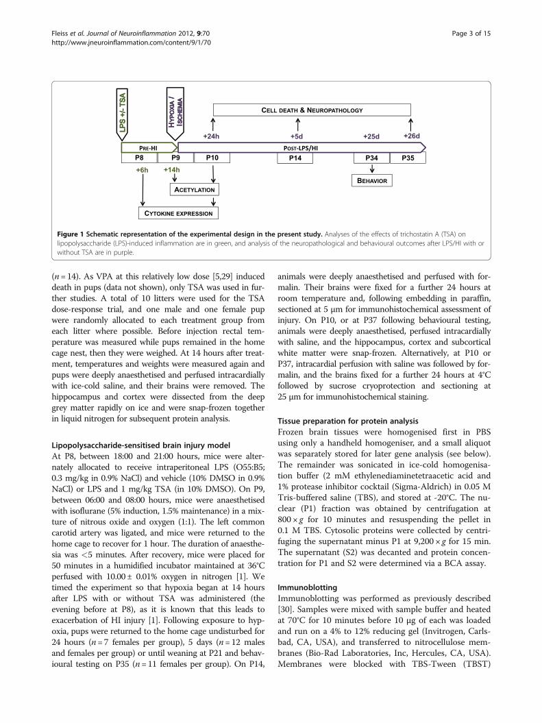
Figure 1 Schematic representation of the experimental design in the present study. Analyses of the effects of trichostatin A (TSA) onlipopolysaccharide (LPS)-induced inflammation are in green, and analysis of the neuropathological and behavioural outcomes after LPS/HI with orwithout TSA are in purple.
Fleiss et al. Journal of Neuroinflammation 2012, 9:70 Page 3 of 15http://www.jneuroinflammation.com/content/9/1/70

buffer (30 mM/L Tris-HCl (pH 7.4), 100 mM/L NaCland 0.1% Tween) containing 5% fat-free milk powder for60 minutes at room temperature. After being washed inTBST, membranes were incubated for 60 minutes atroom temperature with primary antibodies under theconditions listed in Additional file 1: Table S1. Mem-branes were washed again, and were incubated with theappropriate peroxidase-labelled secondary antibody(0.25 μg/ml; Vector Laboratories, Burlingame, CA, USA)for 60 minutes. Immunoreactive bands were visualisedusing the SuperSignal Western Dura substrate (PierceBiotechnology, Rockford, IL, USA) and a LAS 1000-cooled CCD camera (Fujifilm, Tokyo, Japan) and quanti-fied using Image Gauge software (Fujifilm).
Cytokine/chemokine assayCytokines and chemokines were measured in whole-brainhomogenate supernatants from (1) P8 mice killed 6 hoursafter intraperitoneal treatment with LPS and vehicle(n=15) or LPS and TSA (1 mg/kg; n=16) and (2) P10mice killed 24 hours after LPS/HI and vehicle (n=10) orLPS/HI and TSA (n=10). Levels of IL-1β, KC/chemokine(C-X-C motif) ligand 1 (CXCL1), monocyte chemotacticprotein-1 (MCP-1)/chemokine (C-C motif) ligand 2(CCL2), IL-2, IL-3, IL-4, IL-6, IL-9, IL-10, IL-17, macro-phage inflammatory protein 1α (MIP-1α)/CCL3, MIP-1β/CCL4, granulocyte colony-stimulating factor (G-CSF) andTNFα were simultaneously measured using the Bio-PlexMultiplex Cytokine Assay (Bio-Rad Laboratories). Theresults were normalised to the amount of protein per wellas determined using a Bio-Rad DC protein assay.
Quantitative real-time PCRA fraction of whole-hemisphere homogenate removedbefore the addition of homogenisation buffer duringpreparation for protein analysis was used for RNAextraction. A QIAGEN mini extraction kit (Valencia,CA, USA) was used according to the manufacturer’sinstructions, and the quality and concentration of RNAwere verified by spectrophotometry. Reverse transcrip-tion was performed in duplicate on 500 pg of RNA usinga QIAGEN kit as per the manufacturer’s instructions, in-cluding DNase treatment. Real-time quantitative PCRwas set up using SYBR Green Supermix (Bio-Rad La-boratories) for 40 cycles of a three-step procedure, in-cluding 15-second denaturation at 96°C, 30-secondannealing at 55°C and a 30-second extension at 72°C. Tocorrelate the threshold cycle to copy number, a standardcurve was generated from serial dilutions of a samplewith high target gene expression, as ascertained by pilotanalysis. A reference gene panel (TATAA Biocenter AB,Göteborg, Sweden) was run on a randomly selected sub-set of samples from each time point to select the mostappropriate reference gene. At 6 and 14 hours after LPS
with or without TSA glyceraldehyde 3-phosphate de-hydrogenase (GAPDH) and hypoxanthine phosphoribo-syltransferase 1 (HPRT1) expression was used. Insamples collected 24 hours after LPS/HI GAPDH wasused, and for samples collected 25 days after LPS/HI β-glucuronidase (GUSB) was chosen for standardisation.The specific ratio of the gene of interest to the referencegene, or the geometric average of the ratio of the tworeference genes was used in analyses.
Immunohistochemical stainingImmunohistochemistry was performed as described pre-viously for fixed paraffin-embedded [30] and fixed free-floating sections. Briefly, where appropriate, sectionswere deparaffinised, rehydrated through decreasing con-centrations of ethanol and antigen retrieval performedby boiling in citric acid buffer (0.01 M, pH 6.0). En-dogenous peroxidase activity was blocked (3% hydrogenperoxide in 0.1 M PBS), as was nonspecific binding (3%serum in 0.1 M PBS), and sections were incubated for24 to 72 hours with primary antibodies under the condi-tions listed in Additional file 1: Table S1. Following thor-ough washing, sections were incubated with theappropriate secondary antibodies (1:250; Vector Labora-tories) for 60 minutes. Visualisation of immunoreactivitywas achieved using VECTASTAIN ABC Elite reagentwith 0.5 mg/ml 3,3′-diaminobenzidine enhanced with15 mg/ml ammonium nickel sulphate, as well as with0.01 mg/ml β-glucose oxidase. Sections were dehydratedin graded ethanol and xylene and coverslipped withmounting medium. Sections stained with antiactivecaspase-3 were also counterstained with acid fuchsin tovisualise pyknotic cells before coverslipping.
Brain injury evaluationAll evaluations were conducted by an experimenterunaware of the treatment group. Grey and white matterchanges were measured in 5-μm-thick serial sectionsevery 375 μm through the brain (n = 5 or 6 levels),stained for microtubule-associated protein 2 (MAP-2)and myelin basic protein (MBP), respectively. UsingMicro Image Analysis software (Olympus, Tokyo, Japan)we measured area by manually tracing around theareas of the lateral ventricle, MBP-immunopositive sub-cortical white matter or the areas of each hemispheredisplaying MAP-2 immunopositive staining (uninjured)and immunonegative staining (infarct). The white mattervolume, hemispheric volume, lateral ventricle volume,tissue loss and infarct volume were calculated accordingto the Cavalieri’s principle using the following formula:V = SA × P × T, where V is total volume, SA is the sumof the areas measured, P is the inverse of the samplingfraction and T is the section thickness, as previouslydescribed [1].
Fleiss et al. Journal of Neuroinflammation 2012, 9:70 Page 4 of 15http://www.jneuroinflammation.com/content/9/1/70

Cell countingThe numbers of ionized calcium-binding adaptormolecule 1 (Iba-1)-positive cells in the whole hemisphereand oligodendrocyte transcription factor 2 (Olig2)-positivecells in the corpus callosum plus the external capsulewere determined. All counts were performed by aninvestigator blinded to treatment group using stereologi-cal methods (grid sizes 750× 750 μm and 100× 100 μm,respectively) in three to five serial sections spanningapproximately bregma -1 to -2.5 (Stereo Investigator ver-sion 7; MicroBrightField, Inc, Williston, VT, USA).Numbers of activated caspase-3-and pyknotic-positivecells were assessed in the cortex, hippocampal CA1,dentate gyrus, thalamus and caudate putamen in two orthree sections per pup. All cells within a given regionwere counted and expressed as number of cells persquare millimetre.
Behavioural testingAll testing and training was conducted by an observerblind to the treatment group in a sound attenuatedroom, under low lighting and during the dark-phase ofthe circadian cycle. Mice were tested at P35, prior to theonset of regular oestrous [31].
Open-field testingMice (n= 11 per group) were placed into the centre ofan unfamiliar 44 × 44-cm dark grey-coloured Plexiglasopen-field arena with clean cage bedding (changed be-tween animals) covering the floor, and their behaviourwas recorded for 15 minutes with the examiner outsidethe testing room. Four arenas were run in parallel. Nine-teen behavioural variables were extracted from the track-ing software (Bioserve Viewer II; Biobserve, St. Augustin,Germany) and are displayed in Table 1. The variableswere summarised into 3-minute bins and 15-minutetotals for analyses.
Fear conditioningA schematic of the testing procedure is shown in Add-itional file 2: Figure S1. On day 1, mice (n= 15 to 19 pergroup) were placed in a 39 × 9.5 × 16.5-cm automatic re-flex conditioner box (7530; Ugo Basile Srl, Comerio,Italy) adapted for fear conditioning and baseline freezingscored for 2 minutes. ‘Freezing’ was defined as the cessa-tion of all movement except for that required for breath-ing, and this was scored every 10 seconds during testing,later adjusted to percentage of time spent freezing. Afterthe mice spent 2 minutes in the testing box, a combinedvisible and ultraviolet light, and tone (80 db, 670-Hzsquare sound wave; neutral conditioned stimulus) werepresented for 20 seconds, followed 2 seconds later by ascrambled foot shock (0.5 mA; aversive unconditionedstimulus) lasting 2 seconds. Mice were left in the
chamber following the foot shock for 30 seconds to allowextinction of any association between the aversive stimu-lus (foot shock) and the context of the testing apparatus.The mice were removed from the testing box and
returned to their home cages, and the apparatus cleanedwith 70% alcohol. Twenty-four hours later the mice werereturned to the testing box for 2 minutes and freezingwas scored (pretone score) to assess any learned rela-tionship between the testing box and the conditioningstimulus. After this 2-minute pretone period, the toneand light were presented for 30 seconds and freezingwas scored in the following 2 minutes (posttone score).Increased posttone freezing relative to pretone scoreindicated a learned relationship between the condition-ing stimulus and the aversive stimulus, thus a type ofPavlovian learning.
Statistical analysisData are presented as means ± SEM. The effects of TSAtreatment on outcome measures were assessed for malesand females separately. We used a Student’s t-test whenwe compared two groups of normally distributed dataand a Mann-Whitney U test when data were nonnormal.We used one-way analysis of variance (ANOVA) with aStudent’s t-test post hoc to compare the effects of TSAon groups of three or more. We used two-way ANOVAwith a Student’s t-test post hoc when comparing hemi-spheres after HI. For ANOVA, TSA treatment was thewithin-subject variable and hemisphere was the between-subjects variable. P< 0.05 was accepted as statisticallysignificant.
Table 1 Variables evaluated in the open field experimentand compared within the multivariate analyses
Total arena Centre zonea
Average velocity (cm/second) Velocity (cm/second)
Track length (cm) Track length (cm)
Activity (%) (velocity of >0.5 pixels/secondor occurrences of head stretches/bobs andtail moves)
Activity (%)
Ambulation (accelerations from stationaryto <0.5 pixels/second)
Ambulation
Speed moved in field (0 to 1 cm/minute) Visits
Speed moved in field (1 to 4 cm/minute) Visit latencies
Speed moved in field (4 to 10 cm/minute) Durations
Head bobs
Head stretches
Tail moves
Number of zone crossings
Number of ratedzone crossings
aThe centre zone was defined as lying 5 cm or more from the arena edge.
Fleiss et al. Journal of Neuroinflammation 2012, 9:70 Page 5 of 15http://www.jneuroinflammation.com/content/9/1/70

Open-field data were analysed using multivariate ana-lyses with principal component analysis followed by partialleast squares discriminant analysis (PLS-DA). The resultsof PLS-DA are presented in an illustrative score plot whichcan be seen as a projection wherein individuals close toeach other in the score plot have similar characteristics.The preprocessing of data consisted of unit variance scal-ing and mean-centring. The Simca-P + version 11 softwareprogram (Umetrics AB, Umeå, Sweden) was used for thecalculations. Selected time curves of open-field data basedon the output of a loading plot were analysed using two-way ANOVA. In total, 20 mice were run in the open-fieldexperiment. After visual inspection of track files, four (twofrom each treatment group) had to be discarded becauseof discontinuous tracking of the animals during recording.
ResultsTrichostatin A dose-response trial followinglipopolysaccharide exposureUsing Western blot analysis, we demonstrated that TSAdose-dependently increased H4 acetylation in the brainat 14 hours after LPS exposure (Figure 2) (P< 0.001 byone-way ANOVA), as previously reported [32]. We alsomonitored indices of pup health, that is, pup weightgain, rectal temperature and the presence of righting re-flex. TSA dose-dependently decreased weight gain andrectal temperature (P< 0.05 by one-way ANOVA) (Fig-ures 3A and 3B). Also, pups in the 10-mg group dis-played a loss of righting reflex (data not shown), butthere was no significant difference in mortality up to 14hours after treatment (Additional file 3: Table S2).
Figure 2 Trichostatin A (TSA) dose-dependently affects temperature and weight gain and increases histone acetylation in female, butnot in male, neonatal mice. Data shown were gathered at 14 hours after lipopolysaccharide (LPS) treatment with or without TSA. (A) Meanrectal temperature (n = 6 to 10 pups). (B) Mean change in body weight (n = 6 to 10 pups). (C) Mean acetylated histone-4 expression (Ac-H4)normalised to reference protein histone-2B (H2B) (each n = 14). *P<0.05 treatment effect by one-way analysis of variance (ANOVA), #P<0.05 bypost hoc t-test. (D) and (E) Sex-specific Western blot analysis of Ac-H4 (molecular weight 10 kDa) and H2B (molecular weight 15 kDa) showingthe effects of LPS (L) and LPS + TSA treatment (+T) in females and males (D) and mean normalised acetylation for female and male mice at 14hours after intraperitoneal injection (all n = 7). * P<0.05 by Student’s t-test.
Fleiss et al. Journal of Neuroinflammation 2012, 9:70 Page 6 of 15http://www.jneuroinflammation.com/content/9/1/70

Post hoc analysis revealed that H4 acetylation was sig-nificantly increased by 1 mg/kg TSA (P< 0.05 by t-test).As this dose caused the least change in weight gain andbody temperature and has been shown to be
neuroprotective when given prior to an excitotoxic le-sion [33], 1 mg/kg was chosen for all further studies.Additional analysis revealed that the increase in acetyl-
ation caused by TSA was significant for female, but notmale, pups at 1 mg/kg (Student’s t-test, P< 0.05) (Fig-ures 3D and 3E) and 5 mg/kg (data not shown), but thatweight changes (Additional file 3: Table S2) andtemperature (data not shown) were not different be-tween the sexes. In this model, exposure of the pups toLPS did not cause hypoacetylation compared to saline-treated pups (data not shown), as reported in models ofmore severe inflammation [34,35].
Trichostatin A induces sustained acetylation in theneonatal brain after lipopolysaccharide-sensitisedhypoxia-ischaemiaIncreased acetylation is lost 24 hours after withdrawal ofHDACis from cultured oligodendrocytes [11] and 24hours after HDACi treatment in a middle cerebral arteryocclusion model in adults [6]. On the basis of the dataderived from the dose-response trial, which showed sex-specific effects of TSA after LPS treatment, we alsosought to determine if there were persistent effects ofTSA on the epigenome in the female neonatal brainafter LPS/HI. At 24 hours after LPS/HI, we observedpersistently increased acetylation of histone-3 in bothhemispheres of LPS/HI + TSA female mice compared toLPS/HI-only mice (all n= 7, P< 0.05 by two-wayANOVA) (Figures 3A and 3C). Furthermore, TSA treat-ment persistently increased histone-4 acetylation at 24hours after HI in females (all n= 7, P< 0.05 by two-wayANOVA), an effect localised to the ipsilateral hemi-sphere (P< 0.05 by post hoc t-test) (Figure 4A,B).
Trichostatin A protects grey and white matter at 5-dayfollow-up but does not reduce acute cell death regulatorsSeveral studies in which adult models of cerebral ischae-mia were used have shown neuroprotection followingTSA treatment [5,29,35]. To investigate TSA’s effects onshort-term neonatal brain injury, tissue loss in cerebralgrey and subcortical white matter was assessed 5 daysafter LPS/HI. Grey and white matter tissue loss wasreduced in TSA-treated female pups compared to LPS/HI-exposed females without TSA treatment (both n= 12,P< 0.05 by Student’s t-test) (Figure 4). TSA treatmentdid not protect the male brain from grey or white mattertissue loss following LPS/HI (both n= 12, P> 0.05 byStudent’s t-test). There was no loss of MAP-2 staining inthe contralateral hemisphere, indicating lack of injury.The lateral ventricular volume in the ipsilateral hemi-sphere was increased by approximately 200% comparedto the contralateral hemisphere, regardless of TSA treat-ment and in both sexes (female LPS/HI = 185 ± 29% vsfemale LPS + TSA/HI= 232± 58%, male LPS/HI = 210±
Figure 3 Trichostatin A (TSA) persistently increases histoneacetylation at 24 hours after injury. (A) Western blots ofacetylated histone-4(Ac-H4), acetylated histone-3 (Ac-H3, molecularweight 17 kDa) and reference protein H2B in femalelipopolysaccharide-sensitised hypoxiaischaemia (LPS/HI) mice (redbars) and LPS + TSA/HI mice (green bars) 24 hours after injury. (B)and (C) Mean normalised acetylation data are shown for Ac-H4 (B)and Ac-H3 (C) (all n = 7). Data are means ± SEM. *P<0.05 treatmenteffect by one-way ANOVA, #P<0.05 by post hoc t-test.
Fleiss et al. Journal of Neuroinflammation 2012, 9:70 Page 7 of 15http://www.jneuroinflammation.com/content/9/1/70

16% vs male LPS + TSA/HI = 198 ± 43%; P> 0.05 byStudent’s t-test). Volume of tissue loss in LPS/HI ani-mals without TSA treatment was not different betweenfemales and males (females = 8.95 ± 2.29 mm3 (n = 12),males = 6.48 ± 1.80 mm3 (n = 12); P> 0.05 by Student’st-test). There were no differences in mortality or bodyweight between groups with or without 1 mg/kg TSAat 5 days after LPS/HI (Additional file 3: Table S2).Reduced caspase-3 activation and upregulation of
heat shock cognate 70 (HSC70) and the LPS-bindingprotein gelsolin [29,32] have been implicated in neuro-protection by HDACis in adult animals. As such, wealso assessed gelsolin expression immediately prior toHI, as well as the number of activated caspase-3-immu-nopositive cells and protein expression of HSC7024 hours after LPS/HI. Neither caspase-3-immunoposi-tive cell number nor protein expression of HSP70 orgelsolin was altered by TSA treatment (Additional file4: Figure S2; Additional file 5: Table S3). Also, the re-gional distribution of cell death (as identified by pykno-tic cells or caspase-3-positive cells) was not affected byTSA treatment in females at 24 hours after LPS/HI(Additional file 5: Table S3).
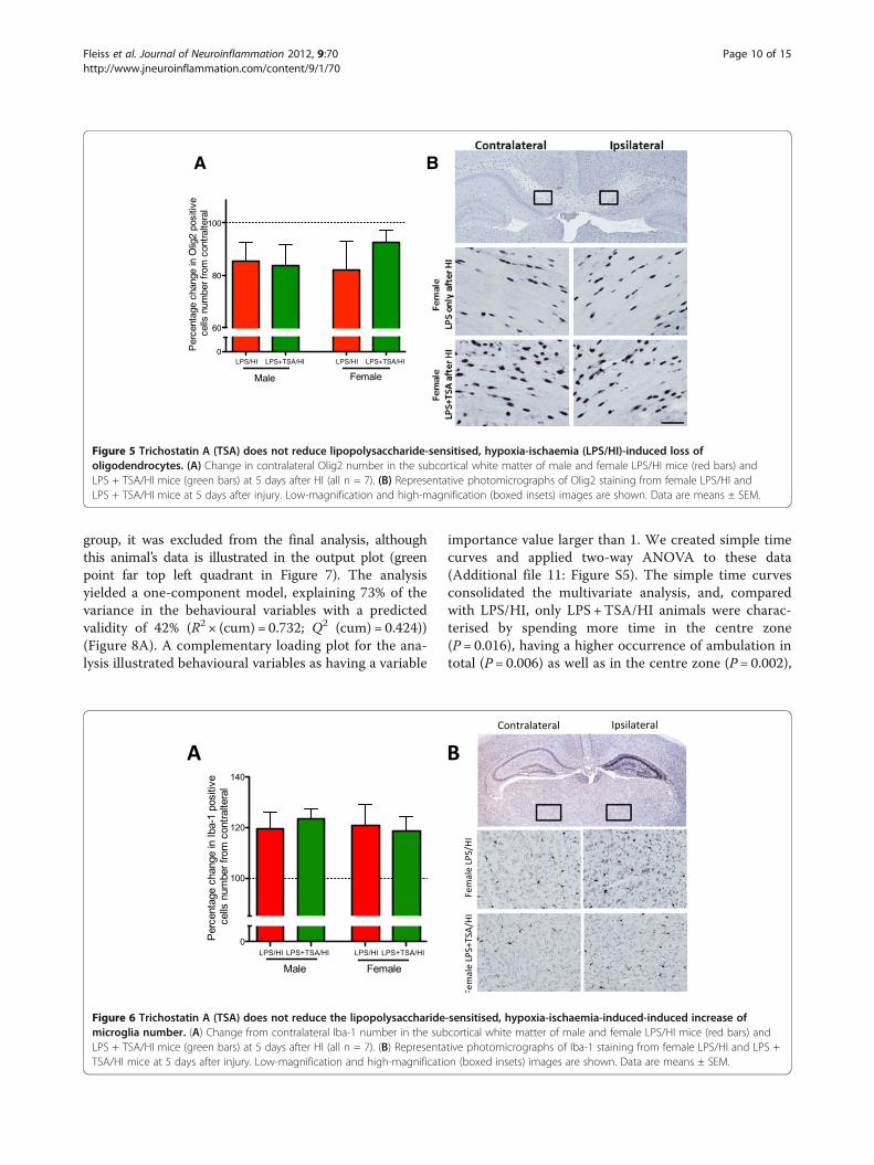
Trichostatin A treatment did not affect oligodendrocytenumber following lipopolysaccharide-sensitised hypoxia-ischaemiaOligodendrocytes in the neonatal mouse brain are vul-nerable to HI injury [36], and we sought to determinewhether any loss induced by LPS/HI was prevented byTSA. We counted the number of Olig2-immunopositivecells, a marker of oligodendrocytes, at all stages of devel-opment. The number of Olig2-immunopositive cells wasdecreased by approximately 15% in the ipsilateral com-pared to the contralateral subcortical white matter inboth treatment groups and in both males and females(all n= 7, P> 0.05 by Student’s t-test) (Figure 5).
Oligodendrocyte maturation/differentiation factors areunaltered by trichostatin A treatmentNormal oligodendrocyte differentiation and maturationrely on HDAC activity to reduce expression of ID2, ID4and HES5, corepressors of myelin gene transcription[37]. As a preliminary investigation of the safety of TSAtreatment in the immature brain, we assessed theseHDAC-dependent oligodendrocyte maturational factorsand markers of oligodendrocyte maturation (platelet-derived growth factor α (PGDFRα) and MBP). Therewas no effect of TSA on expression of any of the mar-kers assessed at either 6 hours after LPS with or withoutTSA treatment (Additional file 6: Table S4) or at 24hours or 35 days post-LPS/HI in TSA-treated females(Additional file 7: Table S5; Additional file 8: Table S6).
Trichostatin A did not affect lipopolysaccharide-sensitised,hypoxia-ischaemia-induced microglia numberHDACis reportedly confer neuroprotection by causingmicroglial apoptosis [6,15], and we therefore sought todetermine TSA’s effects on microglia number followingLPS/HI. Microglia number assessed by Iba-1 stainingwas increased by approximately 20% in the ipsilateralhemisphere compared to the contralateral hemisphereafter LPS/HI. This increase in cell number was localisedmainly to the hippocampus but was higher across theentire hemisphere. Microglia number was increased inmice of both sexes, and the increase in the number ofmicroglia was independent of TSA treatment (n= 6 to 8,P> 0.05 by Student’s t-test) (Figure 5).
Cytokine/chemokine production in response tolipopolysaccharide exposure was effected by trichostatinA treatment and sexIncreased proinflammatory cytokine expression is sug-gested to be one mechanism by which LPS sensitises theneonatal brain to HI injury [1,2]. The ability of HDACis toreduce cytokine production in vitro and in vivo in adults issuggested to mediate their neuroprotective effects [38,39].As such, we sought to investigate if there was any effect ofTSA on LPS-induced cytokine expression before HI inmales or females in this study (Additional file 9: FigureS3A). Prior to HI in males, TSA treatment increased MIP-1β expression (all n=6 to 8, P< 0.05 by Student’s t-test).G-CSF was higher in both males and females treated withTSA compared to those treated with LPS only (P< 0.05,n= 6 to 10 by Student’s t-test).Cytokine expression is dependent at least in part on
the total expression of, as well as on the ratio of, phos-phorylated and nonphosphorylated forms of the NFκB-binding protein IκB [40]. As reduced cytokine expressionis observed in conjunction with reduced NFκB activationfollowing HDACi treatment [38], we measured IκB totalexpression and phosphorylation. In agreement with ourobservations of only limited effects of TSA treatment oncytokine/chemokine expression, the overall expression ofIκB and IκB phosphorylation was unaltered by TSA inmales and females (n= 7, P> 0.05 by Student’s t-test)(Additional file 9: Figures S3B and S3C).
Trichostatin A treatment modulates cytokine expressionafter lipopolysaccharide-sensitised hypoxia-ischaemiaThe extent of brain injury can be altered by the type andlevels of cytokine exposure [41], and as such we investi-gated TSA’s effects on cytokine expression after LPS/HI infemale pups. At 24 hours after LPS/HI, all inflammatorymodulators within the range of the assay were increasedin the ipsilateral (injured) hemisphere compared to thecontralateral (noninjured) hemisphere of both LPS/HI andLPS/HI+TSA pups (all P< 0.01 by two-way ANOVA)
Fleiss et al. Journal of Neuroinflammation 2012, 9:70 Page 8 of 15http://www.jneuroinflammation.com/content/9/1/70

(Additional file 10: Figure S4). TSA treatment reduced IL-4 levels in the LPS/HI ipsilateral hemisphere comparedwith ipsilateral levels following LPS/HI without TSA treat-ment (P< 0.05, by two-way ANOVA). TSA treatment didnot affect expression of any of the other cytokines mea-sured in the ipsilateral and contralateral hemispheres.
Trichostatin A treatment improved learning in earlyadulthood following neonatal lipopolysaccharide-sensitised hypoxia-ischaemia but did not providelong-term neuroprotectionSeveral studies, including this one, have found short-termneuroprotective effects of HDACis following cerebral ischae-mia. Long-term follow-up, however, including both neuro-pathological and functional assessment, is lacking. To
evaluate long-term TSA effects on neuropathology and be-haviour and learning following LPS/HI, we examined braininjury and utilised the open field and trace fear-conditioningparadigms. As locomotor activity and exploratory behaviourand learning in the fear-conditioning test is reduced follow-ing neonatal HI [42,43], these behavioural tests wereselected to examine long-term effects of TSA followingLPS/HI. As TSA, in the short-term part of this study,increased only acetylation and provided neuroprotection infemales, studies at P35 were performed in females only.Multivariate analysis was used to analyse the open-field
data. A single extreme outlier was identified. As multivari-ate analyses are sensitive to extreme outliers and to pre-vent the analysis’s becoming focused on the differencebetween the one identified outlier and the rest of the
Figure 4 Trichostatin A (TSA) treatment reduces white and grey matter injury in female, but not male, neonates at 5 days afterlipopolysaccharide-sensitised hypoxia-ischaemia (LPS/HI). (A) Grey matter tissue loss (%) in male and female LPS/HI and LPS + TSA/HI miceat 5 days after HI (all n = 12). (B) and (D) Total volume of grey matter tissue loss (B) and subcortical white matter loss (D) in male and femaleLPS/HI and LPS + TSA/HI at 5 days after HI. (C) and (E) Representative images of microtubule-associated protein 2 (MAP-2)-stained wholehemispheres (C) and myelin basic protein (MBP)-stained subcortical white matter (E) in female LPS/HI and female LPS + TSA/HI mice at 5 daysafter HI. Data are means ± SEM. *P< 0.05 by Student’s t-test.
Fleiss et al. Journal of Neuroinflammation 2012, 9:70 Page 9 of 15http://www.jneuroinflammation.com/content/9/1/70
group, it was excluded from the final analysis, althoughthis animal’s data is illustrated in the output plot (greenpoint far top left quadrant in Figure 7). The analysisyielded a one-component model, explaining 73% of thevariance in the behavioural variables with a predictedvalidity of 42% (R2 × (cum) = 0.732; Q2 (cum) = 0.424))(Figure 8A). A complementary loading plot for the ana-lysis illustrated behavioural variables as having a variable
importance value larger than 1. We created simple timecurves and applied two-way ANOVA to these data(Additional file 11: Figure S5). The simple time curvesconsolidated the multivariate analysis, and, comparedwith LPS/HI, only LPS +TSA/HI animals were charac-terised by spending more time in the centre zone(P = 0.016), having a higher occurrence of ambulation intotal (P= 0.006) as well as in the centre zone (P = 0.002),
FemaleMale
Per
cent
age
chan
ge in
Olig
2 po
sitiv
e c
ells
num
ber
from
con
tral
tera
l
A B
LPS/HI LPS+TSA/HI LPS/HI LPS+TSA/HI 0
60
80
100
Figure 5 Trichostatin A (TSA) does not reduce lipopolysaccharide-sensitised, hypoxia-ischaemia (LPS/HI)-induced loss ofoligodendrocytes. (A) Change in contralateral Olig2 number in the subcortical white matter of male and female LPS/HI mice (red bars) andLPS + TSA/HI mice (green bars) at 5 days after HI (all n = 7). (B) Representative photomicrographs of Olig2 staining from female LPS/HI andLPS + TSA/HI mice at 5 days after injury. Low-magnification and high-magnification (boxed insets) images are shown. Data are means ± SEM.
Figure 6 Trichostatin A (TSA) does not reduce the lipopolysaccharide-sensitised, hypoxia-ischaemia-induced-induced increase ofmicroglia number. (A) Change from contralateral Iba-1 number in the subcortical white matter of male and female LPS/HI mice (red bars) andLPS + TSA/HI mice (green bars) at 5 days after HI (all n = 7). (B) Representative photomicrographs of Iba-1 staining from female LPS/HI and LPS +TSA/HI mice at 5 days after injury. Low-magnification and high-magnification (boxed insets) images are shown. Data are means ± SEM.
Fleiss et al. Journal of Neuroinflammation 2012, 9:70 Page 10 of 15http://www.jneuroinflammation.com/content/9/1/70

showing longer distance moved in the centre (P=0.006),and demonstrating more head stretches (P=0.039) andtail moves in the centre (P= 0.006).
During fear conditioning, female LPS/HI and LPS/HI +TSA adult mice responded identically to the testingapparatus on the day of training, with less than 10% ofthe time spent immobile before conditioning (light/tone andshock; data not shown). Time spent immobile in responseto the training box before conditioning on day 2 was alsoidentical for both groups. Posttone on day 2 two-wayANOVA indicated significantly increased freezing in re-sponse to the light and tone, indicative of a learned relation-ship between these conditioning stimuli and the foot shock(Figure 8B). Post hoc analysis indicated that only animalstreated with TSA had learnt this association (P=0.002 byStudent’s t-test), whereas animals in the LPS/HI (no-TSA)group had not learnt it (P=0.856 by Student’s t-test).Following behavioural analysis, gross neuropatho-
logical score and white matter volume analysis wereundertaken. At P35, there was no difference in bodyweight, injury severity or volume of MBP between LPS/HI only- and LPS/HI + TSA-treated females (Figures 8Aand 8B and Additional file 12: Figure S6).
DiscussionA body of evidence indicates that HDACis are efficaciousneuroprotectants across injury models leading to neu-roinflammation in adult rodents [44]. Our present studyis the first to demonstrate that TSA is neuroprotective in
Figure 7 Neonatal trichostatin A (TSA) treatment altered open-field behaviours and improved learning in young adulthood afterlipopolysaccharide-sensitised hypoxia-ischaemia (LPS/HI). (A) Representative trace recordings from LPS/HI- and LPS + TSA/HI-treated mice.(B) Output from the multivariate partial least squares discriminant analysis (PLS-DA) of open-field data illustrating significant differences betweenLPS/HI- and LPS + TSA/HI-treated mice in young adulthood. Each point represents the cumulative value for all behavioural variables for oneindividual, and red circles represent female LPS/HI. Green squares represent female LPS + TSA/HI. The y-axis is for visualisation purposes only andshould not be overinterpreted. The statistics are described further in the Materials and methods section. (C) Trace fear-conditioning dataillustrating time spent immobile (frozen) pretone and after exposure to the fear-conditioned stimulus (light and tone) on day 2 (n = 15 to 19).Data are means ± SEM. *P<0.05 by post hoc Student’s t-test.
Figure 8 TSA treatment did not reduce gross brain injury inyoung adulthood after neonatal LPS sensitized HI. In femaleLPS/HI and LPS+TSA/HI mice at P35, A) representative MAP-2stainedcoronal brain sections, and data for B) body weight at P35 and C)macroscopic brain injury score (all, n=15-18). Mean ± SEM.
Fleiss et al. Journal of Neuroinflammation 2012, 9:70 Page 11 of 15http://www.jneuroinflammation.com/content/9/1/70

a neonatal LPS-sensitised HI model and specifically thatit is protective in a female cohort of animals at any age.TSA at this dose does not affect weight gain or temperatureand does not appear to impair the regulatory mechanismgoverning oligodendrocyte maturation. We also demon-strate that TSA treatment in the neonate can contribute tolong-term changes in behaviour following LPS/HI. In thefemale neonates in this study, the mechanism underpinningneuroprotection by the HDACi TSA was not any of thosecommonly reported in adult cerebral studies, that is, de-crease in cytokine levels, microglial number, caspase-3 orincreased HSC70 and gelsolin.
Trichostatin A is neuroprotective in females only afterneonatal lipopolysaccharide-sensitised hypoxia-ischaemiaOur data are in general agreement with those reported pre-viously that HDACis are neuroprotective in models of cere-bral injury in adult rodents [5,32,35]. Our data also agreeswith a short report of neuroprotection following repetitiveVPA treatment, a known HDACi, after HI in neonatal rat[20]. Building from this observation, we have demonstratedefficacy with only a single treatment of HDACi when admi-nistered prior to injury. TSA treatment did not protectmales from LPS/HI injury in this study or lead to histonehyperacetylation, a key indicator of HDACi activity. Undernormal conditions, histone acetylation is higher in malemice than in females from E18 through early postnatal life[45]. Thus a reduced efficacy of TSA compared with femalesmay represent a lower level of available unacetylated lysineresidues upon which to act in males. Also, under normalgrowth conditions, there are regional sex-specific differencesin glial density [46], and the immunoreactivity of glia to LPSdiffers between the sexes [47-49]. These glial effects,together with possible (as yet undescribed) sex differences inthe metabolism of TSA and/or blood-brain barrier (BBB) re-sponse to LPS or after injury, may have influenced the sex-dependent neuroprotective ability of TSA. Furthermore, sexdifferences in neonatal stress hormone responsiveness havebeen described previously [50]. Another possibility is thatsex differences in the nuclear receptor of such hormonesmay have influenced the sex dependent outcome.Interestingly, despite not significantly altering histone
acetylation in males, TSA had a sex-specific effect on LPS-induced cytokine production before HI: higher MIP-1βand G-CSF. These effects likely represent direct or indirecteffects of nonhistone HDACi targets that are known to in-clude STAT (signal transducer and activator of transcrip-tion), p53, FOXO (Forkhead box O), cell cycle, apoptosis-related genes and RNA processing and stability [51]. Theseeffects may also mediate in part the neuroprotective effectsof TSA treatment in females. Also, significant redistribu-tion of epigenetic markers can occur in the absence ofchanges in total expression [52], and we cannot discountany effect of locus-specific changes in acetylation in males.
Trichostatin A mediates improved neuropathology laterthan 24 hours after lipopolysaccharide-sensitisedhypoxia-ischaemia injuryTSA treatment failed to reduce cell death 24 hours afterLPS/HI, despite improving outcome at 5 days post-HI. Thisfinding is in contrast to reports on reduced cell death inadult HI models at early and late time points postinjury[5,29,32]. Persistent increases in histone-3 and histone-4acetylation at 24 hours post-LPS/HI suggest that the singledose of TSA is capable of continuing to alter gene expres-sion and improve cell fate at least up to 24 hours post-HI.We were unable to determine any TSA-dependentmechanisms responsible for improvements in neuropathol-ogy between 24 hours and 5 days post-HI. Speculatively,the delayed neuroprotection may be mediated in part byacetylation-dependent increases in glial trophic factor pro-duction [53], reduced changes in BBB integrity post-HI [54]or increased proliferation and/or neurogenesis [55].
Neuroprotective mechanisms of action for trichostatin Adiffer between the adult and neonateIn contrast to adult models of cerebral injury treated withHDACi, improved neuropathogical outcome in the neonatedid not correlate with decreased microglial number [6,15]or NFκB-mediated reductions in cytokine expression[38,39,56]. Although we did not investigate microglial apop-tosis directly in the present study, we found no TSA-dependent decrease in total microglial number or differ-ences in caspase-3 activity or expression in total corticalextracts from females before or after HI (personal observa-tion, B Fleiss & C Mallard). In agreement with this observa-tion, we have previously been unable to demonstrate adirect relationship between microglial number and neuro-protection following neonatal HI in mice [1,57].Previous work suggests two reasons why cytokines may
not be reduced by HDACi treatment in this LPS-sensitisedHI model, as occurs in HI- or inflammation-only models.First, coapplication of TSA and LPS in vitro causes bidirec-tional changes in chemokine and/or cytokine production[15,58,59], and, second, HDACis differentially alter gene ex-pression from activated (LPS-treated) and unstimulatedmacrophages [8]. To completely exclude that TSA does notimprove outcome by altering cytokine expression, however,more extensive temporal analyses post-LPS/HI are necessary.In addition to cytokine expression and microglia number,
we investigated expression of the LPS-binding proteingelsolin and HSC 70, additional mechanisms by whichHDACis are reported to mediate neuroprotection in adultcerebral injury models [6,29,32,38,60]. These targets werenot altered by TSA treatment in neonatal females, and assuch we suggest that they do not mediate neuroprotectionin the female LPS-sensitised neonatal brain. This discre-pancy in the mechanism of action between previous stud-ies and ours may be related to several factors, including
Fleiss et al. Journal of Neuroinflammation 2012, 9:70 Page 12 of 15http://www.jneuroinflammation.com/content/9/1/70

the use of LPS to sensitise the brain to HI. As mentionedabove, exposure to LPS in conjunction with HDACis leadsto substantial differences in the profile of inflammatoryand cell death genes activated compared to LPS or HDACialone [8]. In addition, neuroprotection with HDACis inadult models is associated with decreased caspase and p53and increased Akt. LPS sensitisation is associated with aninversion of the neuroprotective changes in these import-ant cell death regulators [5,6,13,29], possibly antagonisingthe efficacy of TSA in this model. Also possibly modifyingthe response of the neonate to HDACi treatment is thatacetylation of genes, including those affecting cell survival,such as caspase-3 and HSPs, is greater in neonates [59-61]. As such, saturation of acetylation at targets mediatingneuroprotection in adults may reduce the ability of TSAto act via these mechanisms in the neonatal brain.
Long-term behaviours were altered by neonataltrichostatin A treatment prior to lipopolysaccharide-sensitised hypoxia-ischaemiaFunctional follow-up in adult rodent models of cerebral in-jury treated with HDACis consistently correlate withreduced neurological injury severity with improved behav-ioural outcome [5,13]. However, it has been reported thatwhen using a HDACi to treat lesions of the ventral hippo-campus in neonates, there is a discrepancy between neuro-pathology and behaviour in adulthood [19]. In this study,though injury progressed to the same extent over time,hypersensitivity to apomorphine and deterioration of asso-ciative learning, but not anxiety, were reduced by HDACitreatment. Similarly, we found that although TSA did notprovide long-term neuroprotection, small changes acrossmultiple indices were observed in the open-field paradigmand learning was improved in the fear-conditioning test.Also, in the present study, we assessed only gross neuro-pathology in the adult animals. It is possible that thesemethods were not sensitive enough to identify subtleimprovements in neuropathology due to TSA treatmentthat may underpin altered behaviour in the open field andimproved learning.
Trichostatin A did not effect myelin corepressorexpressionDrugs that reduce excitotoxicity and cell death can havedeleterious effects on the developing brain [18,62]. Acritical concern in considering the safety of HDACis as aneurotherapy is that, during development, decreasingacetylation facilitates maturation of oligodendrocyteprecursor cells to myelin-producing oligodendrocytes[12]. In the short or long term after LPS sensitised HI,however, TSA appears to have no effect on the expres-sion of myelin corepressors or the balance of immatureto mature oligodendrocytes. Although not conclusive,this finding suggests that the maturation and/or function
of Olig2-positive cells are not disrupted by this neuro-protective dosage of TSA.
ConclusionThis study provides evidence that the HDACi TSA is anefficacious neuroprotectant in females in a neonatalmodel of LPS-sensitised HI. The sex dependency of TSAas a neuroprotectant is an addition to the accumulatingevidence that treatment and patient characteristics needto be more carefully considered and that a one-size-fits-all approach is futile in the search for efficacious neuro-protective strategies [26,63]. Contrary to our original hy-pothesis, TSA-dependent neuroprotection does notappear to be related to a reduction in LPS- or LPS/HI-induced inflammation. Although we were unable to de-termine the underlying neuroprotective mechanisms,our study demonstrates that, in neonatal mice, epigenet-ics can be modified to protect the developing brain with-out causing gross abnormalities in white matterdevelopment. Further studies, including post-injury andrepetitive treatment regimens possibly involving add-itional types of HDACis, will elucidate whether HDACishave a role in future clinically applicable neurotherapies.
Additional files
Additional file 1: Table S1. List of antibodies used in the study.
Additional file 2: Figure S1. Schematic representation of the trace fearconditioning testing procedure.
Additional file 3: Table S2. Pup characteristics before and after treatmentand/or HI.
Additional file 4: Figure S2. TSA has no effect on the amount of cell death24 h after LPS sensitized HI or induce HSP-70 expression. A) Activated caspase-3 and cresyl violet-stained sections from female LPS+TSA/HI treated mouse,showing injury in areas assessed for levels of cell death (see Table 4). HSP-70expression; B) Western blot of HSC-70 (MW 70 kDa) and reference proteinactin (MW 40 kDa) and C) mean HSP-70 expression normalized to actin forfemale LPS/HI and LPS+TSA/HI (all n=7).
Additional file 5: Table S3. Cell death in the ipsilateral hemisphere 24 hafter LPS sensitized HI in females.
Additional file 6: Table S4. Oligodendrocyte differentiation/maturationfactor expression 6 h after LPS+/− TSA.
Additional file 7: Table S5. Oligodendrocyte differentiation/maturationfactor expression 24 h after LPS sensitized HI in females.
Additional file 8: Table S6. Oligodendrocyte differentiation/maturationfactor expression 35 d after LPS sensitized HI in females.
Additional file 9: Figure S3. After LPS+/− TSA (before HI) cytokineexpression was different dependent on treatment and sex. Cytokineexpression adjusted to mg/ml protein per well, LPS only, red; LPS + TSA,green. Mean± SEM, all n = 6-10 . , Φ, interaction effect and } sex effect(P< 0.05) 2 way ANOVA.
Additional file 10: Figure S4. After LPS sensitized HI cytokine expressionwas increased in the ipsilateral hemisphere irrespective of TSA treatment.Cytokine expression adjusted to mg/ml protein per well. Mean±SEM, alln=6-8. *, P< 0.05 treatment effect in Student’s t-test.
Additional file 11: Figure S5. Time curves for behavioural variablesindicated from the multivariate analysis to have a strong treatment effect.Shown are group mean±SEM for 3- minute blocks of time, n=7-8, *, P< 0.05in a two-way ANOVA.
Fleiss et al. Journal of Neuroinflammation 2012, 9:70 Page 13 of 15http://www.jneuroinflammation.com/content/9/1/70

Additional file 12: Figure S6. Mean total MBP expression normalizedto actin for female LPS/HI (red) and LPS + TSA/HI (green) showingcontralateral (C) and ipsilateral (I) hemispheres (all n = 7), mean ± SEM.
AbbreviationsIκB: Inhibitor of κB; IL: Interleukin; kDa: Kilodalton; NFκB: Nuclear factor κB;PBS: Phosphate-buffered saline; PCR: Polymerase chain reaction.
Competing interestsThe authors declare they have no competing interests.
Authors’ contributionsCM and BF conceived and designed the experiments. BF performed theexperiments. BF, CM and MN analysed and interpreted the data. BF and CMdrafted the article. BF, CM, KB and MN revised the article critically forimportant intellectual content. All authors read and approved the finalmanuscript.
AcknowledgementsWe gratefully acknowledge the expert technical assistance and advice ofAnna-Lena Leverin and the contribution to data interpretation and draftingof Michelle Porritt. This work was supported by grants from the SwedishMedical Research Council (VR 2009-2630 to CM), Wilhelm and MartinaLundgren (vet2-41/2010 to BF), government grant to researcher in PublicHealth Service at the Sahlgrenska University Hospital (ALFGBG-142881 toCM), European Commission FP6 (Neobrain, 2006-036534, to CM), EuropeanUnion grant FP7 (Neurobid, HEALTH-F2-2009-241778, to CM), the Leducqfoundation (DSRR_P34404 to CM), Åhlén stiftelse (to CM) and FrimurareBarnhusfonden (to CM). The funders had no role in the study design, datacollection and analysis, decision to publish or preparation of themanuscript.
Author details1Perinatal Center, Department of Neuroscience and Physiology, SahlgrenskaAcademy, University of Gothenburg, Box 432, Gothenburg 405 30, Sweden.2Institute of Neuroscience and Physiology, University of Gothenburg, Box432, Gothenburg 405 30, Sweden. 3Center for Brain Repair and Rehabilitation,Institute of Neuroscience and Physiology, University of Gothenburg, Box 432,Gothenburg 405 30, Sweden. 4Karolinska Institutet, Department of Women’sand Children’s Health, Karolinska University Hospital Q2:07, Stockholm SE 17176, Sweden. 5Inserm U676, Hôpital Robert Debré, 48 blvd Serurier,Paris F-75019, France.
Received: 19 October 2011 Accepted: 28 February 2012Published: 18 April 2012
References1. Wang X, Stridh L, Li W, Dean J, Elmgren A, Gan L, Eriksson K, Hagberg H,
Mallard C: Lipopolysaccharide sensitizes neonatal hypoxic-ischemic braininjury in a MyD88-dependent manner. J Immunol 2009, 183:7471–7477.
2. Dean JM, Wang X, Kaindl AM, Gressens P, Fleiss B, Hagberg H, Mallard C:Microglial MyD88 signaling regulates acute neuronal toxicity of LPS-stimulated microglia in vitro. Brain Behav Immun 2010, 24:776–783.
3. Mielnicki LM, Ying AM, Head KL, Asch HL, Asch BB: Epigenetic regulation ofgelsolin expression in human breast cancer cells. Exp Cell Res 1999,249:161–176.
4. Butler LM, Zhou X, Xu WS, Scher HI, Rifkind RA, Marks PA, Richon VM: Thehistone deacetylase inhibitor SAHA arrests cancer cell growth,up-regulates thioredoxin-binding protein-2, and down-regulatesthioredoxin. Proc Natl Acad Sci USA 2002, 99:11700–11705.
5. Kim HJ, Rowe M, Ren M, Hong JS, Chen PS, Chuang DM: Histonedeacetylase inhibitors exhibit anti-inflammatory and neuroprotectiveeffects in a rat permanent ischemic model of stroke: multiplemechanisms of action. J Pharmacol Exp Ther 2007, 321:892–901.
6. Shein NA, Grigoriadis N, Alexandrovich AG, Simeonidou C, LourbopoulosA, Polyzoidou E, Trembovler V, Mascagni P, Dinarello CA, Shohami E:Histone deacetylase inhibitor ITF2357 is neuroprotective, improvesfunctional recovery, and induces glial apoptosis followingexperimental traumatic brain injury. FASEB J 2009, 23:4266–4275.
7. Moreira JM, Scheipers P, Sørensen P: The histone deacetylase inhibitortrichostatin A modulates CD4+ T cell responses. BMC Cancer 2003, 3:30.
8. Brogdon JL, Xu Y, Szabo SJ, An S, Buxton F, Cohen D, Huang Q: Histonedeacetylase activities are required for innate immune cell control of Th1but not Th2 effector cell function. Blood 2007, 109:1123–1130.
9. Suh HS, Choi S, Khattar P, Choi N, Lee SC: Histone deacetylase inhibitorssuppress the expression of inflammatory and innate immune responsegenes in human microglia and astrocytes. J Neuroimmune Pharmacol2010, 5:521–532.
10. McCarthy MM, Auger AP, Bale TL, De Vries GJ, Dunn GA, Forger NG, MurrayEK, Nugent BM, Schwarz JM, Wilson ME: The epigenetics of sex differencesin the brain. J Neurosci 2009, 29:12815–12823.
11. Marin-Husstege M, Muggironi M, Liu A, Casaccia-Bonnefil P: Histonedeacetylase activity is necessary for oligodendrocyte lineageprogression. J Neurosci 2002, 22:10333–10345.
12. Ye F, Chen Y, Hoang T, Montgomery RL, Zhao XH, Bu H, Hu T, Taketo MM,van Es JH, Clevers H, Hsieh J, Bassel-Duby R, Olson EN, Lu QR: HDAC1 andHDAC2 regulate oligodendrocyte differentiation by disrupting theβ-catenin-TCF interaction. Nat Neurosci 2009, 12:829–838.
13. Sinn DI, Kim SJ, Chu K, Jung KH, Lee ST, Song EC, Kim JM, Park DK, Kun LeeS, Kim M, Roh JK: Valproic acid-mediated neuroprotection inintracerebral hemorrhage via histone deacetylase inhibition andtranscriptional activation. Neurobiol Dis 2007, 26:464–472.
14. Wu X, Chen PS, Dallas S, Wilson B, Block ML, Wang CC, Kinyamu H, Lu N,Gao X, Leng Y, Chuang DM, Zhang W, Lu RB, Hong JS: Histonedeacetylase inhibitors up-regulate astrocyte GDNF and BDNF genetranscription and protect dopaminergic neurons. Int JNeuropsychopharmacol 2008, 11:1123–1134.
15. Chen PS, Wang CC, Bortner CD, Peng GS, Wu X, Pang H, Lu RB, Gean PW,Chuang DM, Hong JS: Valproic acid and other histone deacetylaseinhibitors induce microglial apoptosis and attenuate lipopolysaccharide-induced dopaminergic neurotoxicity. Neuroscience 2007, 149:203–212.
16. Zhu C, Wang X, Xu F, Bahr BA, Shibata M, Uchiyama Y, Hagberg H, Blomgren K:The influence of age on apoptotic and other mechanisms of cell deathafter cerebral hypoxia-ischemia. Cell Death Differ 2005, 12:162–176.
17. Cheng Y, Gidday JM, Yan Q, Shah AR, Holtzman DM: Marked age-dependent neuroprotection by brain-derived neurotrophic factor againstneonatal hypoxic-ischemic brain injury. Ann Neurol 1997, 41:521–529.
18. Dou H, Ellison B, Bradley J, Kasiyanov A, Poluektova LY, Xiong H, MaggirwarS, Dewhurst S, Gelbard HA, Gendelman HE: Neuroprotective mechanismsof lithium in murine human immunodeficiency virus-1 encephalitis.J Neurosci 2005, 25:8375–8385.
19. Sandner G, Host L, Angst MJ, Guiberteau T, Guignard B, Zwiller J: The HDACinhibitor phenylbutyrate reverses effects of neonatal ventralhippocampal lesion in rats. Front Psychiatry 2011, 1:153.
20. Kabakus N, Ay I, Aysun S, Söylemezoglu F, Ozcan A, Celasun B: Protectiveeffects of valproic acid against hypoxic-ischemic brain injury in neonatalrats. J Child Neurol 2005, 20:582–587.
21. Eklind S, Mallard C, Leverin AL, Gilland E, Blomgren K, Mattsby-Baltzer I,Hagberg H: Bacterial endotoxin sensitizes the immature brain tohypoxic-ischaemic injury. Eur J Neurosci 2001, 13:1101–1106.
22. Wang X, Svedin P, Nie C, Lapatto R, Zhu C, Gustavsson M, Sandberg M,Karlsson JO, Romero R, Hagberg H, Mallard C: N-acetylcysteine reduceslipopolysaccharide-sensitized hypoxic-ischemic brain injury. Ann Neurol2007, 61:263–271.
23. Volpe JJ: Brain injury in premature infants: a complex amalgam ofdestructive and developmental disturbances. Lancet Neurol 2009, 8:110–124.
24. Hagberg H, Mallard C: Effect of inflammation on central nervous systemdevelopment and vulnerability. Curr Opin Neurol 2005, 18:117–123.
25. Dammann O, Leviton A: Maternal intrauterine infection, cytokines, andbrain damage in the preterm newborn. Pediatr Res 1997, 42:1–8.
26. Renolleau S, Fau S, Goyenvalle C, Charriaut-Marlangue C: Sex,neuroprotection, and neonatal ischemia. Dev Med Child Neurol 2007,49:477–478.
27. Golomb MR, Fullerton HJ, Nowak-Gottl U, Deveber G, International PediatricStroke Study Group: Male predominance in childhood ischemic stroke:findings from the International Pediatric Stroke Study. Stroke 2009, 40:52–57.
28. Nijboer CH, Kavelaars A, van Bel F, Heijnen CJ, Groenendaal F: Gender-dependent pathways of hypoxia-ischemia-induced cell death andneuroprotection in the immature P3 rat. Dev Neurosci 2007, 29:385–392.
Fleiss et al. Journal of Neuroinflammation 2012, 9:70 Page 14 of 15http://www.jneuroinflammation.com/content/9/1/70

29. Ren M, Leng Y, Jeong M, Leeds PR, Chuang DM: Valproic acid reducesbrain damage induced by transient focal cerebral ischemia in rats:potential roles of histone deacetylase inhibition and heat shock proteininduction. J Neurochem 2004, 89:1358–1367.
30. Svedin P, Hagberg H, Savman K, Zhu C, Mallard C: Matrixmetalloproteinase-9 gene knock-out protects the immature brain aftercerebral hypoxia-ischemia. J Neurosci 2007, 27:1511–1518.
31. Mayer C, Acosta-Martinez M, Dubois SL, Wolfe A, Radovick S, Boehm U,Levine JE: Timing and completion of puberty in female mice depend onestrogen receptor α-signaling in kisspeptin neurons. Proc Natl Acad SciUSA 2010, 107:22693–22698.
32. Yildirim F, Gertz K, Kronenberg G, Harms C, Fink KB, Meisel A, Endres M:Inhibition of histone deacetylation protects wildtype but not gelsolin-deficient mice from ischemic brain injury. Exp Neurol 2008, 210:531–542.
33. Chia Ghee Sng J, Taniura H, Yoneda Y: Inhibition of histone deacetylationby trichostatin A intensifies the transcriptions of neuronal c-fos and c-jungenes after kainate stimulation. Neurosci Lett 2005, 386:150–155.
34. Shang Y, Jiang YX, Ding ZJ, Shen AL, Xu SP, Yuan SY, Yao SL: Valproic acidattenuates the multiple-organ dysfunction in a rat model of septicshock. Chin Med J (Engl) 2010, 123:2682–2687.
35. Faraco G, Pancani T, Formentini L, Mascagni P, Fossati G, Leoni F, Moroni F,Chiarugi A: Pharmacological inhibition of histone deacetylases bysuberoylanilide hydroxamic acid specifically alters gene expression andreduces ischemic injury in the mouse brain. Mol Pharmacol 2006,70:1876–1884.
36. Rothstein RP, Levison SW: Gray matter oligodendrocyte progenitors andneurons die caspase-3 mediated deaths subsequent to mild perinatalhypoxic/ischemic insults. Dev Neurosci 2005, 27:149–159.
37. Liu J, Casaccia P: Epigenetic regulation of oligodendrocyte identity. TrendsNeurosci 2010, 33:193–201.
38. Choi Y, Park SK, Kim HM, Kang JS, Yoon YD, Han SB, Han JW, Yang JS, HanG: Histone deacetylase inhibitor KBH-A42 inhibits cytokine production inRAW 264.7 macrophage cells and in vivo endotoxemia model. Exp MolMed 2008, 40:574–581.
39. Leoni F, Zaliani A, Bertolini G, Porro G, Pagani P, Pozzi P, Donà G, Fossati G,Sozzani S, Azam T, Bufler P, Fantuzzi G, Goncharov I, Kim SH, Pomerantz BJ,Reznikov LL, Siegmund B, Dinarello CA, Mascagni P: The antitumor histonedeacetylase inhibitor suberoylanilide hydroxamic acid exhibitsantiinflammatory properties via suppression of cytokines. Proc Natl AcadSci USA 2002, 99:2995–3000.
40. Chen LF, Greene WC: Shaping the nuclear action of NF-κB. Nat Rev MolCell Biol 2004, 5:392–401.
41. Mesplès B, Plaisant F, Fontaine RH, Gressens P: Pathophysiology ofneonatal brain lesions: lessons from animal models of excitotoxicity. ActaPaediatr 2005, 94:185–190.
42. Carty ML, Wixey JA, Kesby J, Reinebrant HE, Colditz PB, Gobe G, Buller KM:Long-term losses of amygdala corticotropin-releasing factor neurons areassociated with behavioural outcomes following neonatalhypoxia-ischemia. Behav Brain Res 2010, 208:609–618.
43. Järlestedt K, Atkins AL, Hagberg H, Pekna M, Mallard C: Trace fearconditioning detects hypoxic-ischemic brain injury in neonatal mice. DevNeurosci 2011, 33:222–230.
44. Gibson CL, Murphy SP: Benefits of histone deacetylase inhibitors for acutebrain injury: a systematic review of animal studies. J Neurochem 2010,115:806–813.
45. Tsai HW, Grant PA, Rissman EF: Sex differences in histone modifications inthe neonatal mouse brain. Epigenetics 2009, 4:47–53.
46. Mouton PR, Long JM, Lei DL, Howard V, Jucker M, Calhoun ME, Ingram DK:Age and gender effects on microglia and astrocyte numbers in brains ofmice. Brain Res 2002, 956:30–35.
47. Marriott I, Bost KL, Huet-Hudson YM: Sexual dimorphism in expression ofreceptors for bacterial lipopolysaccharides in murine macrophages: apossible mechanism for gender-based differences in endotoxic shocksusceptibility. J Reprod Immunol 2006, 71:12–27.
48. Santos-Galindo M, Acaz-Fonseca E, Bellini MJ, Garcia-Segura LM: Sexdifferences in the inflammatory response of primary astrocytes tolipopolysaccharide. Biol Sex Differ 2011, 2:7.
49. Sorge RE, LaCroix-Fralish ML, Tuttle AH, Sotocinal SG, Austin JS, Ritchie J,Chanda ML, Graham AC, Topham L, Beggs S, Salter MW, Mogil JS: Spinal cordToll-like receptor 4 mediates inflammatory and neuropathic
hypersensitivity in male but not female mice. J Neurosci 2011, 31:15450–15454.
50. Davis M, Emory E: Sex differences in neonatal stress reactivity. Child Dev1995, 66:14–27.
51. Spange S, Wagner T, Heinzel T, Krämer OH: Acetylation of non-histoneproteins modulates cellular signalling at multiple levels. Int J Biochem CellBiol 2009, 41:185–198.
52. Tariq M, Saze H, Probst AV, Lichota J, Habu Y, Paszkowski J: Erasure of CpGmethylation in Arabidopsis alters patterns of histone H3 methylation inheterochromatin. Proc Natl Acad Sci USA 2003, 100:8823–8827.
53. Huang Y, Doherty JJ, Dingledine R: Altered histone acetylation atglutamate receptor 2 and brain-derived neurotrophic factor genes is anearly event triggered by status epilepticus. J Neurosci 2002, 22:8422–8428.
54. Wang Z, Leng Y, Tsai LK, Leeds P, Chuang DM: Valproic acid attenuatesblood-brain barrier disruption in a rat model of transient focal cerebralischemia: the roles of HDAC and MMP-9 inhibition. J Cereb Blood FlowMetab 2011, 31:52–57.
55. Kim HJ, Leeds P, Chuang DM: The HDAC inhibitor, sodium butyrate,stimulates neurogenesis in the ischemic brain. J Neurochem 2009,110:1226–1240.
56. Chakravortty D, Koide N, Kato Y, Sugiyama T, Mu MM, Yoshida T, Yokochi T:The inhibitory action of butyrate on lipopolysaccharide-induced nitricoxide production in RAW 264.7 murine macrophage cells. J Endotoxin Res2000, 6:243–247.
57. Doverhag C, Hedtjärn M, Poirier F, Mallard C, Hagberg H, Karlsson A,Sävman K: Galectin-3 contributes to neonatal hypoxic-ischemic braininjury. Neurobiol Dis 2010, 38:36–46.
58. Perlman JM: White matter injury in the preterm infant: an importantdetermination of abnormal neurodevelopment outcome. Early Hum Dev1998, 53:99–120.
59. Liu AY, Lin Z, Choi HS, Sorhage F, Li B: Attenuated induction of heat shockgene expression in aging diploid fibroblasts. J Biol Chem 1989, 264:12037–12045.
60. Yakovlev A, Khafizova M, Abdullaev Z, Loukinov D, Kondratyev A: Epigeneticregulation of caspase-3 gene expression in rat brain development. Gene2010, 450:103–108.
61. Espinoza CR, Feeney AJ: The extent of histone acetylation correlates withthe differential rearrangement frequency of individual VH genes in pro-Bcells. J Immunol 2005, 175:6668–6675.
62. Davenport CM, Sevastou IG, Hooper C, Pocock JM: Inhibiting p53 pathwaysin microglia attenuates microglial-evoked neurotoxicity followingexposure to Alzheimer peptides. J Neurochem 2010, 112:552–563.
63. Giza CC, Mink RB, Madikians A: Pediatric traumatic brain injury: not justlittle adults. Curr Opin Crit Care 2007, 13:143–152.
doi:10.1186/1742-2094-9-70Cite this article as: Fleiss et al.: Neuroprotection by the histonedeacetylase inhibitor trichostatin A in a model of lipopolysaccharide-sensitised neonatal hypoxic-ischaemic brain injury. Journal ofNeuroinflammation 2012 9:70.
Submit your next manuscript to BioMed Centraland take full advantage of:
• Convenient online submission
• Thorough peer review
• No space constraints or color figure charges
• Immediate publication on acceptance
• Inclusion in PubMed, CAS, Scopus and Google Scholar
• Research which is freely available for redistribution
Submit your manuscript at www.biomedcentral.com/submit
Fleiss et al. Journal of Neuroinflammation 2012, 9:70 Page 15 of 15http://www.jneuroinflammation.com/content/9/1/70