research report retinal projection to the dorsal raphe
TRANSCRIPT

Brain Research 895 (2001) 139–145www.elsevier.com/ locate /bres
Research report
Retinal projection to the dorsal raphe nucleus in the Chilean degus(Octodon degus)
* ˇKatherine V. Fite , Skirmantas JanusonisNeuroscience and Behavior Program, Tobin Hall, University of Massachusetts, Amherst, MA 01003, USA
Accepted 26 December 2000
Abstract
A substantial projection from the retina to the dorsal raphe nucleus (DRN) has been demonstrated in the Chilean degus, adiurnal /crepuscular hystricomorph rodent. Following intraocular injection of cholera toxin subunit B (CTB), immunocytochemicallylabeled CTB-positive axons and terminals were observed in all major retinorecipient nuclei as well as in the DRN and periaqueductal gray(PAG) of the mesencephalon. Two streams of optic axons to the DRN were observed: one descending from the optic tract at the level ofthe pretectum and anterior superior colliculus, the other emerging as a small fascicle at the anterior pole of the inferior colliculus anddescending bilaterally through the PAG. Contralateral retinal afferents in the DRN appeared to terminate primarily in the dorsomedial andlateral subdivisions of the DRN, and a less extensive ipsilateral component also was observed. Axonal arborizations were characterized byshort branches and multiple varicosities, both in the DRN and in the PAG. The extent and density of DRN retinal afferents were not asextensive as previously observed in Mongolian gerbils using identical techniques, but the retinal-DRN projection is considerably larger indegus than in rats. The functional significance of the retinal-DRN pathway remains to be determined, although a variety of evidenceindicates that light may directly affect the activity of neurons and serotonin levels in the DRN. 2001 Elsevier Science B.V. All rightsreserved.
Theme: Sensory systems
Topic: Subcortical visual pathways
Keywords: Subcortical visual system; Non-imaging forming visual system; Environmental light stimulation; Periaqueductal gray; Serotonin
1. Introduction indicates that light stimulation can directly affect neuronalactivity in the DRN [11,16], and also that DRN serotonin
The vast majority of serotonergic (5-HT) neurons that levels can be altered by light independently of circadianproject to the forebrain are located in the dorsal raphe factors [2].nucleus (DRN) of the ventral mesencephalon [15,37]. At present, little is known about the neural circuitryDespite the wide range of targets in the neocortex, whereby photic stimulation may directly influence thestriatum, limbic system, and diencephalon innervated by serotonergic nuclei that are involved in arousal, affective5-HT projections from the DRN, little information is and/or emotional states via their ascending projectioncurrently available with regard to the localization and systems. One possible route is a direct optic pathway to thedensity of afferent terminals in the DRN. DRN efferents DRN that has been described in several species: cat [10],also innervate a number of structures in the central visual rat [9,18,34], and Mongolian gerbil [9]; this projectionsystem, including the lateral geniculate nuclear complex, remains a relatively unexplored component of mammaliansuperior colliculus, and visual cortex. Some evidence central visual pathways. The distribution of retinal affer-
ents to the DRN and the retinal ganglion cells from whichthis pathway originates have been described recently in*Corresponding author. Tel.: 11-413-545-0351; fax: 11-413-545-Mongolian gerbils and laboratory rats [9]. The retinal-0996.
E-mail address: [email protected] (K.V. Fite). DRN projection is more extensive in gerbils than in rats,
0006-8993/01/$ – see front matter 2001 Elsevier Science B.V. All rights reserved.PI I : S0006-8993( 01 )02061-3

ˇ140 K.V. Fite, S. Janusonis / Brain Research 895 (2001) 139 –145
which may be related to the relative importance of vision and incubated in 4.5% normal rabbit serum (NRS, Vector),in these two species; gerbils are active primarily during 2.5% bovine serum albumin (BSA, Sigma) and 0.4%daylight hours, while rats are predominantly nocturnal. Triton X-100 (TX) in PBS overnight at 48C. Sections wereAlso, the gerbil retina contains a substantially larger then rinsed two times (5 min each) in PBS and incubatedpopulation of cone photoreceptors [14] than rats, and in a goat anti-CTB IgG (1:2700, List Biological Labs) ingerbils have a well-defined visual streak in the superior PBS containing 2% NRS, 2.5% BSA and 2% TX in PBSretina [25]. for 4 days at 48C. Sections were rinsed four times (15 min
In the present study, the retinal-DRN projection was each) in PBS; incubated in 2% NRS and 2.5% BSA in PBSinvestigated in the Chilean degus, a highly visual South for 10 min; then incubated in biotinylated rabbit anti-goatAmerican hystricomorph rodent that is diurnal in its IgG antibody (Vector, diluted 1:200) with 2% NRS, 2.5%natural environment and shows ‘robust’ responses to both BSA and 1% TX in PBS for 1.5 h. Sections were rinsedphotic and non-photic circadian zeitgebers [13]. Results four times (15 min each) in PBS and again incubated inindicate that the projection pathway is quite well-de- 2% NRS, 2.5% BSA in PBS for 10 min. Sections wereveloped in degus, but neither the density nor distribution of incubated in a 1:100 ABC (ABC Elite; Vector) solution inretinal terminals in the degus DRN is as extensive as found PBS for 1 h, rinsed four times in PBS (15 min each), thenpreviously in Mongolian gerbils [9]. rinsed twice (5 min each) in 0.05 M Tris buffer (TB; pH
7.4), incubated in 0.5% CoCl in TB for 10 min, then2
rinsed in TB for 2 min followed by two rinses in PBS (52. Materials and methods min each). Sections were then preincubated in 3,39-
diaminobenzidine (DAB; 0.05%) in PBS for 5 min andAdult female Octodon degus (170–220 g) were anes- reacted for 3 min by adding 0.01% H O to the DAB2 2
thetized with an intraperitoneal injection (mixture of solution. Sections were then rinsed five times (1 min each)ketamine (55 mg/kg), xylazine (0.5 mg/kg) and acep- in PBS, mounted on chromium-subbed slides, allowed toromazine (1 mg/kg)). All procedures were approved by air dry, cleared with Hemo-De, and coverslipped withthe University of Massachusetts Institutional Animal Care Permount.and Utilization Committee. Following a topical application Sections containing CTB-labeled axons and terminalsof corneal anesthetic (0.5% proparacaine hydrochloride), were charted serially using both bright- and darkfieldthe needle of a 10-ml Hamilton microsyringe was inserted microscopy with reference to a standard rat atlas [29]. Theinto the posterior chamber just behind the corneal margin, borders of the DRN were determined using alternate brainand 6–8 ml of 2% (w/v) solution of cholera toxin subunit sections from one of the CTB-injected degus processedB (CTB, low salt, List Biological Labs) dissolved in 2% immunocytochemically for serotonin. The CTB-immuno-dimethyl sulfoxide (to facilitate CTB uptake) was slowly cytochemical protocol (described above) was modified toinjected into the eye, and the needle left in place for 10 demonstrate DRN serotonergic neurons as follows: themin to minimize leakage of tracer from the eye. Sub- goat anti-CTB antibody was replaced with a rabbit anti-5-sequently, the needle was withdrawn, the injection site HT antibody (Protos Biotech Corp., New York) at awashed immediately with saline, and antibiotic ointment dilution of 1:1500, the rabbit anti-goat IgG antibody was(Bacitracin) applied topically to the site. All intraocular replaced with goat anti-rabbit IgG antibody (Vector)injections were unilateral. diluted 1:200, and the normal rabbit serum was replaced
Animals were allowed to survive for 6–7 days post- with normal goat serum (Vector).injection, and were anesthetized for transcardial perfusion.The descending aorta was clamped, the heart injected with1 ml of heparin (5000 USP U/ml), and perfusion with 3. Resultssaline was followed with 400 ml of chilled 4% paraformal-dehyde in phosphate buffer (PB, pH 7.2). The brain was All major retinorecipient nuclei contained denselysubsequently removed, post-fixed in the same fixative labeled, CTB-positive axons and terminals. In addition,overnight at 48C, and immersed in 30% sucrose in PB sparse retinal afferents were observed in a variety of otherovernight at 48C. Serial, coronal sections throughout the sites, including the lateral posterior nucleus, periventricularthalamus and mesencephalon were cut on a freezing gray, periaqueductal gray (PAG), and parabrachial nucleus.microtome at 40 mm thickness. Retinal axons appeared to innervate the DRN via at least
The CTB immunocytochemistry (ICC) protocol was two descending streams. At the level of the pretectum andbased on that described previously by Angelucci et al. [1] the anterior pole of the contralateral superior colliculus, a(see also Ref. [9]). Sections were rinsed four times (5 min stream of CTB-positive axons emerged from the optic tracteach) in 0.1 M phosphate-buffered saline (PBS, pH 7.4); near the olivary pretectal nucleus and descended mediallyincubated in 0.3% H O in PBS for 20 min; rinsed in PBS through the commissure of the superior colliculus to enter2 2
three times (5 min each); incubated in 0.1 M glycine in the PAG (Fig. 1A). Some CTB-positive axons in the PAGPBS for 30 min; rinsed three times (5 min each) in PBS; contained short branches with conspicuous varicosities,
ˇK.V. Fite, S. Janusonis / Brain Research 895 (2001) 139 –145 141
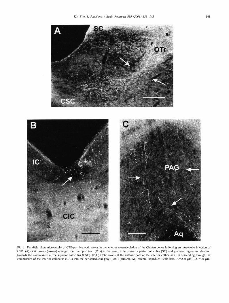
Fig. 1. Darkfield photomicrographs of CTB-positive optic axons in the anterior mesencephalon of the Chilean degus following an intraocular injection ofCTB. (A) Optic axons (arrows) emerge from the optic tract (OTr) at the level of the rostral superior colliculus (SC) and pretectal region and descendtowards the commissure of the superior colliculus (CSC). (B,C) Optic axons at the anterior pole of the inferior colliculus (IC) descending through thecommissure of the inferior colliculus (CIC) into the periaqueductal gray (PAG) (arrows). Aq, cerebral aqueduct. Scale bars: A5250 mm; B,C550 mm.

ˇ142 K.V. Fite, S. Janusonis / Brain Research 895 (2001) 139 –145
some of which entered the ependymal layer lining the ized by short branches with multiple varicosities, oftenaqueduct, particularly in the region immediately dorsal to occurring in clusters (Fig. 4).the rostral pole of the DRN. In addition, a second streamof CTB-labeled axons was observed at the anterior pole ofthe inferior colliculus; these axons descended bilaterally 4. Discussionthrough the lateral PAG into the caudal regions of the DRN(Fig. 1B,C). The direct retinal pathway observed in the Chilean
CTB-positive axonal arborizations in the contralateral degus appears similar to that reported in several otherDRN were observed primarily in the dorsomedial and rodents, including gerbils and rats [9,18,34]. However,dorsolateral portions of the nucleus (Figs. 2 and 3), with a both the density of optic terminals and the size of the DRNsmaller number of ipsilateral, retinal afferents in the retinorecipient zone were less extensive in degus thandorsolateral DRN near the aqueduct. CTB-positive axons previously observed in gerbils using the same techniquesand terminals also occurred in the lateral and ventrolateral [9]. In degus, the retinal-DRN afferent projection isPAG, extending from the anterior pole of the superior primarily contralateral, with a smaller ipsilateral com-colliculus to the posterior pole of the DRN. In both the ponent; whereas in gerbils, the retinal-DRN projection isDRN and PAG, CTB-positive optic axons were character- more bilaterally symmetrical. The location and greatest
density of retinal terminals in the DRN of all three speciessuggest that this optic pathway is well-placed to influenceserotonergic neurons, particularly in the lateral cell groups,many of which project to the superior colliculus and/orLGN [17,26,38] and/or to the visual cortex [39].
Previous studies have shown that serotonergic afferentsfrom the raphe nuclei can modulate neuronal activity at theinitial stages of visual processing and transmission in thebrain. The effects of the serotonergic projection to thedorsal LGN are primarily inhibitory and tonic in nature[19,23,28]. Serotonin also can inhibit retinotectal transmis-sion in the superior colliculus by acting on presynaptic5-HT receptors that are located on retinal terminals1B
[27,28]. In the visual cortex, serotonin also inhibits theinduction of long-term potentiation, presumably by actingthrough 5-HT and 5-HT receptors [5,6,8,12].1A 2
The retinal-DRN pathway may be associated with whathas been described as a ‘non-image-forming’ subsystem ofretinal afferents that includes the retino-hypothalamicprojection to the suprachiasmatic nucleus, as well as sparseretinal afferents to the lateral habenular nucleus [31],lateral hypothalamic area [4,22,40], paraventricular nuclei[40], medial amygdala and peri-amygdaloid area [4,7],piriform cortex, and the olfactory tubercle [4,24,40]. All ofthese retinal afferents are characterized by fine-caliberaxons with multiple varicosities that arborize in regionsthat lie more medially than those typically associated withthe classically defined ‘image-forming’ and visuomotorpathways. This non-image-forming subset of retinal affer-ents may be involved in the photic regulation of arousal,neuroendocrine and circadian functions, and they appear toencode the temporal rather than the spatial characteristicsof light stimulation [3,4].
Although the functional significance of the retinal-DRNFig. 2. (A) Schematic coronal section showing the location and extent of pathway remains unknown at present, previous studiesCTB-positive optic axons (shaded area) in the dorsal raphe nucleus have shown that DRN neurons may respond to visual(DRN) following an intraocular injection of CTB into the left eye. (B) stimulation [11,16,32,35]. Also, the concentration of ex-Photomicrograph of serotonin-positive neurons used to determine the
tracellular DRN serotonin appears to parallel neuronalboundaries and extent of the dorsal raphe nucleus. Aq, cerebral aqueduct;activity in the DRN [30], and oscillations of serotonin inmlf, medial longitudinal fasciculus; PAG, periaqueductal gray; SC,
superior colliculus. Scale bars: A5500 mm; B5250 mm. the pineal gland and in serum show high levels of

ˇK.V. Fite, S. Janusonis / Brain Research 895 (2001) 139 –145 143
Fig. 3. Dorsomedial region of the contralateral dorsal raphe nucleus. (A) Serotonin-immunoreactive neurons. (B) CTB-positive optic axons. Aq, cerebralaqueduct. Scale bars5100 mm.
serotonin during the day and low levels at night. Seasonal seasonal depression is thought to involve central serotoner-variations in serotonin metabolism have been reported as gic mechanisms and metabolism [33,36]. Ultimately, thewell [20,21], and the effectiveness of light therapy for retinal-DRN pathway may prove to be an important
Fig. 4. CTB-positive optic axons in the lateral DRN showing multiple branches and varicosities. Scale bar550 mm.

ˇ144 K.V. Fite, S. Janusonis / Brain Research 895 (2001) 139 –145
[15] G. Halliday, A. Harding, G. Paxinos, Serotonin and tachykininafferent channel whereby the intensity, total flux, and/orsystems, in: G. Paxinos (Ed.), The Rat Nervous System, 2ndphase characteristics of environmental light stimulationEdition, Academic Press, San Diego, CA, 1995, pp. 929–973.
may influence serotonergic activity in the brain, particu- [16] J. Heym, M.E. Trulson, B.L. Jacobs, Raphe unit activity in freelylarly in more diurnal species that rely extensively on vision moving cats: effects of phasic auditory and visual stimuli, Brainfor adaptation and survival. Res. 232 (1981) 29–34.
[17] S. Janusonis, K.V. Fite, W. Foote, Topographic organization ofserotonergic dorsal raphe neurons projecting to the superior col-liculus in the Mongolian gerbil (Meriones unguiculatus), J. Comp.
Acknowledgements Neurol. 413 (1999) 341–355.[18] H. Kawano, K. Decker, S. Reuss, Is there a direct retina-raphe-
suprachiasmatic nucleus pathway in the rat?, Neurosci. Lett. 212We would like to express our great appreciation to Dr(1996) 143–146.Barbara Tate for providing the Octodon degus used in this
[19] Y. Kayama, S. Shimada, Y. Hishikawa, T. Ogawa, Effects ofstudy, and to Ms Lynn Bengston for her excellent technicalstimulating the dorsal raphe nucleus of the rat on neuronal activity
assistance. This research was supported by grants from the in the dorsal lateral geniculate nucleus, Brain Res. 489 (1989) 1–11.Whitehall Foundation and the University of Massachusetts- [20] J.L. Klompenhouwer, K. Fekkes, A.M. van Hulst, P. Moleman, L.Baystate Health Center Collaborative Research Program. Pepplinkhuizen, P.G. Mulder, Seasonal variations in binding of
3H-paroxetine to blood platelets in healthy volunteers: indicationsfor a gender difference, Biol. Psychiatry 6 (1990) 509–517.
[21] V. Lacoste, A. Wirz-Justice, Seasonal variation in normal subjects:References An update of variables current in depression research, in: N.E.
Rosenthal, M.C. Blehar (Eds.), Seasonal Affective Disorders andPhototherapy, Guilford, New York, 1989, pp. 167–230.[1] A. Angelucci, F. Clasca, M. Sur, Anterograde axonal tracing with
[22] R.K. Leak, R.Y. Moore, Identification of retinal ganglion cellsthe subunit B of cholera toxin: a highly sensitive immunohistoch-projecting to the lateral hypothalamic area of the rat, Brain Res. 770emical protocol for revealing fine axonal morphology in adult and(1997) 105–114.neonatal brains, J. Neurosci. Methods 65 (1996) 101–112.
[2] F.R. Cagampang, S. Yamazaki, Y. Otori, S.T. Inouye, Serotonin in [23] G.A. Marks, S.G. Speciale, K. Cobbey, H.P. Roffwarg, Serotonergicthe raphe nuclei: regulation by light and an endogenous pacemaker, inhibition of the dorsal lateral geniculate nucleus, Brain Res. 418Neuroreport 5 (1993) 49–52. (1987) 76–84.
[3] H.M. Cooper, G. Mick, M. Magnin, Retinal projection to mam- [24] G. Mick, H. Cooper, M. Magnin, Retinal projections to the olfactorymalian telencephalon, Brain Res. 477 (1989) 350–357. tubercle and basal telencephalon in primates, J. Comp. Neurol. 327
[4] H.M. Cooper, M. Herbin, E. Nevo, Visual system of a naturally (1993) 205–219.microopthalmic mammal: The blind mole rat, Spalax ehrenbergi, J. [25] J. Mitrofanis, B.L. Finlay, Developmental changes in the distribu-Comp. Neurol. 328 (1993) 313–350. tion of retinal catecholaminergic neurons in hamsters and gerbils, J.
[5] Y. Edagawa, H. Saito, K. Abe, 5-HT receptor-mediated inhibition Comp. Neurol. 292 (1990) 480–494.1A
of long-term potentiation in rat visual cortex, Eur. J. Pharmacol. 349 [26] R.R. Mize, L.H. Horner, Origin, distribution, and morphology of(1998) 221–224. serotonergic afferents to the cat superior colliculus: A light and
[6] Y. Edagawa, H. Saito, K. Abe, The serotonin 5-HT receptor- electron microscope immunocytochemistry study, Exp. Brain Res.2
phospholipase C system inhibits the induction of long-term potentia- 75 (1989) 83–98.tion in the rat visual cortex, Eur. J. Neurosci. 12 (2000) 1391–1396. [27] R.D. Mooney, M.Y. Shi, R.W. Rhoades, Modulation of retinotectal
[7] A.S. Elliott, M.L. Weiss, A.A. Nunez, Direct retinal communication transmission by presynaptic 5-HT receptors in the superior1B
with the peri-amygdaloid area, Neuroreport 6 (1995) 806–808. colliculus of the adult hamster, J. Neurophysiol. 72 (1994) 3–13.[8] J.M. Elliott, T.P. Flanigan, N.R. Newberry, T. Zetterstrom, R.A. [28] R.D. Mooney, X. Huang, M.Y. Shi, C.A. Bennet-Clarke, R.W.
Leslie, 5-HT receptor sub-types: Aspects of their regulation and Rhoades, Serotonin modulates retinotectal and corticotectal conver-function, Neurochem. Int. 25 (1994) 537–543. gence in the superior colliculus, Prog. Brain Res. 112 (1996) 57–69.
[9] K.V. Fite, S. Janusonis, W. Foote, L. Bengston, Retinal afferents to [29] G. Paxinos, D. Watson, The Rat Brain in Stereotaxic Coordinates,the dorsal raphe nucleus in rats and Mongolian gerbils, J. Comp. 4th Edition, Academic Press, San Diego, CA, 1998.Neurol. 414 (1999) 469–484. [30] C.M. Portas, R. McCarley, Behavioral state-related changes of
[10] W.E. Foote, E. Taber-Pierce, L. Edwards, Evidence for retinal extracellular serotonin concentration in the dorsal raphe nucleus: aprojection to the midbrain raphe of the cat, Brain Res. 156 (1978) microdialysis study in the freely moving cat, Brain Res. 648 (1994)135–140. 306–312.
[11] C.A. Fornal, B.L. Jacobs, Physiological and behavioral correlates of [31] T. Qu, D. Dong, K. Sugioka, T. Yamadori, Demonstration of directserotonergic single-unit activity, in: N.N. Osborne, M. Hamon input from the retina to the lateral habenular nucleus in the albino(Eds.), Neuronal Serotonin, Wiley, New York, 1988, pp. 305–345. rat, Brain Res. 779 (1996) 251–258.
[12] C.A. Fornal, W.J. Litto, C.W. Metzler, F. Marrosu, K. Tada, B.L. [32] K. Rasmussen, R.E. Strecker, B.L. Jacobs, Single unit response ofJacobs, Single-unit responses of serotonergic dorsal raphe neurons to noradrenergic, serotonergic and dopaminergic neurons in freely5-HT1A agonist and antagonist drug administration in behaving moving cats to simple sensory stimuli, Brain Res. 369 (1986)cats, J. Pharmacol. Exp. Ther. 207 (1994) 1345–1358. 336–340.
[13] N. Goel, T.M. Lee, L. Smale, Suprachiasmatic nucleus and the [33] N.E. Rosenthal, The mechanisms of action of light in the treatmentintergeniculate leaflet in the diurnal rodent Octodon degus: Retinal of seasonal affective disorder (SAD), in: M.F. Holick, E.G. Jungprojections and immunocytochemical characterization, Neuroscience (Eds.), Biologic Effects of Light, Walter de Gruyter, Berlin, 1996,92 (1999) 1491–1509. pp. 317–324.
[14] V.I. Govardovskii, P. Rohlich, A. Szel, T.V. Khokhlova, Cones in the [34] H. Shen, K. Semba, A direct retinal projection to the dorsal rapheretina of the Mongolian gerbil, Meriones unguiculatus: an immuno- nucleus in the rat, Brain Res. 635 (1994) 159–168.cytochemical and electrophysiological study, Vis. Res. 32 (1992) [35] K. Shima, H. Nakahama, M. Yamamoto, Firing properties of two19–27. types of nucleus raphe dorsalis neurons during the sleep-waking

ˇK.V. Fite, S. Janusonis / Brain Research 895 (2001) 139 –145 145
cycle and their responses to sensory stimuli, Brain Res. 399 (1986) tribution of cells projecting to visual system structures, J. Comp.317–326. Neurol. 336 (1993) 345–361.
[36] M. Terman, J.S. Terman, F.M. Quitkin, P.J. McGrath, J.W. Stewart, [39] B.D. Waterhouse, G.A. Mihaloff, J.C. Baack, D.J. Woodward,B. Rafferty, Light therapy for seasonal affective disorder. A review Topographical distribution of dorsal and median raphe neuronsof efficacy, Neuropsychopharmology 2 (1989) 1–22. projecting to motor, sensorimotor and visual cortical areas in the rat,
[37] R.P. Vertes, A PHA-L analysis of ascending projections of the dorsal J. Comp. Neurol. 249 (1986) 460–476.raphe nucleus in the rat, J. Comp. Neurol. 313 (1991) 643–668. [40] T.G. Youngstrom, M.L. Weiss, A.A. Nunez, Retinofugal projections
[38] B.D. Waterhouse, B. Border, L. Wahl, G.A. Mihailoff, Topographic to the hypothalamus, anterior thalamus and basal forebrain inorganization of rat locus coeruleus and dorsal raphe nuclei: Dis- hamsters, Brain Res. Bull. 26 (1991) 403–411.