researcharticle comparisonoftherelationshipbetween...
TRANSCRIPT
RESEARCH ARTICLE
Comparison of the Relationship betweenCerebral White Matter and Grey Matter inNormal Dogs and Dogs with LateralVentricular EnlargementMartin J. Schmidt1*, Steffi Laubner1, Malgorzata Kolecka1, Klaus Failing2,Andreas Moritz1, Martin Kramer1, Nele Ondreka1
1 Department of Veterinary Clinical Sciences, Clinic for Small Animals, Justus-Liebig-University-Giessen,Giessen, Germany, 2 Unit for Biomathematics and Data Processing, Faculty of Veterinary Medicine, JustusLiebig-University-Giessen, Giessen, Germany
AbstractLarge cerebral ventricles are a frequent finding in brains of dogs with brachycephalic skull
conformation, in comparison with mesaticephalic dogs. It remains unclear whether over-
sized ventricles represent a normal variant or a pathological condition in brachycephalic
dogs. There is a distinct relationship between white matter and grey matter in the cerebrum
of all eutherian mammals. The aim of this study was to determine if this physiological pro-
portion between white matter and grey matter of the forebrain still exists in brachycephalic
dogs with oversized ventricles. The relative cerebral grey matter, white matter and cerebro-
spinal fluid volume in dogs were determined based on magnetic-resonance-imaging data-
sets using graphical software. In an analysis of covariance (ANCOVA) using body mass as
the covariate, the adjusted means of the brain tissue volumes of two groups of dogs were
compared. Group 1 included 37 mesaticephalic dogs of different sizes with no apparent
changes in brain morphology, and subjectively normal ventricle size. Group 2 included 35
brachycephalic dogs in which subjectively enlarged cerebral ventricles were noted as an in-
cidental finding in their magnetic-resonance-imaging examination. Whereas no significant
different adjusted means of the grey matter could be determined, the group of brachyce-
phalic dogs had significantly larger adjusted means of lateral cerebral ventricles and signifi-
cantly less adjusted means of relative white matter volume. This indicates that
brachycephalic dogs with subjective ventriculomegaly have less white matter, as expected
based on their body weight and cerebral volume. Our study suggests that ventriculomegaly
in brachycephalic dogs is not a normal variant of ventricular volume. Based on the changes
in the relative proportion of WM and CSF volume, and the unchanged GM proportions in
dogs with ventriculomegaly, we rather suggest that distension of the lateral ventricles might
be the underlying cause of pressure related periventricular loss of white matter tissue, as
occurs in internal hydrocephalus.
PLOS ONE | DOI:10.1371/journal.pone.0124174 May 4, 2015 1 / 14
OPEN ACCESS
Citation: Schmidt MJ, Laubner S, Kolecka M, FailingK, Moritz A, Kramer M, et al. (2015) Comparison ofthe Relationship between Cerebral White Matter andGrey Matter in Normal Dogs and Dogs with LateralVentricular Enlargement. PLoS ONE 10(5):e0124174. doi:10.1371/journal.pone.0124174
Academic Editor: Annette Sterr, University ofSurrey, UNITED KINGDOM
Received: June 1, 2014
Accepted: March 13, 2015
Published: May 4, 2015
Copyright: © 2015 Schmidt et al. This is an openaccess article distributed under the terms of theCreative Commons Attribution License, which permitsunrestricted use, distribution, and reproduction in anymedium, provided the original author and source arecredited.
Data Availability Statement: All data are availablewithin the paper and its Supporting Information files.
Funding: The authors received no specific fundingfor this work.
Competing Interests: The authors have declaredthat no competing interests exist.
IntroductionDespite the high diversity of head conformation in dog breeds, differences in canine brain mor-phology are comparably small [1–3]. Due to the restricted longitudinal skull growth in brachy-cephalic dogs their brains show reduced longitudinal extension [2, 4]. As a result the externalmorphology resembles the juvenile state of canine brains, showing a wide and stocky appear-ance and ventrally orientated olfactory bulbs [2, 5]. Nevertheless, the general morphology ofthe brain including the cortical pattern of sulci and gyri is the same amongst dogs with differenthead conformations [1, 3]. However, a frequent finding in brachycephalic dog brains is rela-tively large lateral cerebral ventricles in comparison with mesaticephalic dogs [6–8]. It has beenwidely accepted that this increase in ventricular volume is not associated with clinical signs andthat most small-breed dogs normally have large lateral ventricles as a breed characteristic andare not truly hydrocephalic [9–12]. As most brachycephalic dogs are small toy-breeds it hasbeen suggested that ventricular size follows negative allometric growth principles [13]. Theterm “constitutional hydrocephalus” has been used to describe the common association oflarge ventricles with short stature in brachycephalic dogs [14]. On the other hand the terms“ventricular enlargement” and “ventriculomegaly” have been used to describe the large ventri-cles in these breeds, which would imply a pathological condition to some extent [11, 15, 16].
Several studies aiming to determine the physiological ventricular dimensions in dogs sug-gested that ventricular size can vary in individual dogs of the same breed and size [17, 18].However, the following morphological rule conflict with this suggestion: The expansion of thecerebral cortex is one of the most distinctive morphological features of the mammalian brain.Its functional organization is subject to defined spatial, electro-physiological and metabolicconstraints within a limited volume [19]. In the process of evolution this has led to brains witha defined relationship between white matter (WM) and grey matter (GM) mass [19].
As brain size has increased during evolution, the various parts of the brain have not simplyincreased proportionally. In brains of large animals the WMmass in the neocortex has in-creased disproportionately relative to GM [20–22]. This reflects changes in the diameter ofaxons, with larger brains having thicker axons and thicker myelin sheaths [23] and more cor-tico-cortical connections per neuron [20]. Both, the defined relationship between WM andGM and its defined increase with body size are universal in all mammalian species (eutheria)and can be expressed by allometric scaling laws [19, 20, 24, 25]. We would therefore expect acharacteristic relationship between WM and GMmass to exist in different dog breeds and thatWMwould also hyperscale with increasing body weight in dogs. If large ventricles would repre-sent a normal morphologic variation, a WM/GM relationship similar to dogs with normal ven-tricles should be preserved in dog brains with large ventricular volume. Aberrations from theWM/GM relationship would indicate that ventricular enlargement is associated with a reduc-tion of brain tissue as the sum of brain tissues and CSF is constant (Monroe-Kellie-doctrine).This hypothesis states that CSF and brain tissue (including blood vessels) create a state of vol-ume equilibrium. Any increase in volume of one of the cranial constituents must be compen-sated by a decrease in volume of another. In clinical analyses of brain images of dogs withsubjectively normal lateral ventricles and dogs with ventriculomegaly it was our observationthat the periventricular WM is reduced in individuals with larger ventricles, as occurs in hydro-cephalus. We therefore hypothesize that the relationship between WM and GM is decreased indogs with “oversized” ventricles.
Ethics statementThe MRI data were obtained for diagnostic purposes and retrospectively analyzed in this study.Therefore approval from the ethics committee of the Justus Liebig University and government
White Matter Loss in Dogs with Ventriculomegaly
PLOS ONE | DOI:10.1371/journal.pone.0124174 May 4, 2015 2 / 14

of the county Hessen (Regierungspräsidium) was not sought as it is the policy of the ethicscommittee not to subject retrospective studies of images stored in the archive to ethical review.The “Deutsches Tierschutzgesetz” (German Animal Protection Law) does not request a writtenwaiver for retrospective studies. MR-imaging data of five healthy Beagles were also taken froman experimental study of brain diffusion and perfusion in normal dogs. This study was carriedout in strict accordance with the recommendations in the Guidelines for Care and Use of Labo-ratory Animals of the German Animal Protection Law. The protocol was approved by theCommittee on the Ethics of Animal Experiments of the Justus Liebig University Giessen andRegierungspräsidium Hessen (Permit Number: V54-19c2015(I)Gi18/17 No. 78/2011).
Materials and Methods
AnimalsThe archive of MRI scans of the Justus Liebig University (JLU), Giessen, Germany, wassearched retrospectively for MR-imaging reports including the diagnoses “primary or idiopath-ic epilepsy”, “within normal limits” and “ventriculomegaly” or “enlarged ventricles”. The pres-ence of ventriculomegaly was based on the following criteria: The majority of dogs have verynarrow and slit-like horns of the lateral ventricles. In the finding of large ventricles/ventriculo-megaly, the interpreter subjectively noted a greater proportion of the intracranial volume occu-pied by the lateral ventricles. The closely spaced walls of the temporal horns and/or theolfactory recesses were separated by CSF in these brains and the lacking septum pellucidumcreated a large connection between the first and second ventricle (Fig 1) [26]. MRI reports foreach series were obtained by board certified radiologists at the JLU. These dog brain series wereassessed for suitability of inclusion in this study using the following criteria. None of the pa-tients was allowed to show evidence of space occupying lesions or other morphological alter-ations of the brain parenchyma. Transverse scans had to include the whole brain from thecribriform plate rostrally to the first cervical spinal cord segment caudally. Series with inade-quate image contrast and spatial resolution or incomplete transverse series were excluded fromthe study. The finding of “ventriculomegaly” or “enlarged ventricles” had to be judged as an in-cidental finding. “The breed, gender, age, and body weight of the dog at the time of scanning
Fig 1. Comparison of a canine brain with normal lateral cerebral ventricles (A) and enlarged ventricles (B).
doi:10.1371/journal.pone.0124174.g001
White Matter Loss in Dogs with Ventriculomegaly
PLOS ONE | DOI:10.1371/journal.pone.0124174 May 4, 2015 3 / 14
were recorded, and dogs that were between 1 and 6 years of age and up to 17 kg in bodyweightwere included.” Subjects were divided into the following groups: Group one included dogswhose brain and lateral ventricles were assessed as normal by the diagnosis of “within normallimits”. Group two included dogs in which a dilatation of the lateral ventricles was noted on in-terpretation of the images and ventriculomegaly or enlarged ventricles were recorded as anincidental finding.
Imaging techniqueImaging was performed using a 1 Tesla MRI scanner (Gyroscan Intera, Phillips, Hamburg, Ger-many). From the whole MR dataset T2-weighted transverse images of the head were chosen forimage segmentation. Images were obtained using T2-Turbospin echo sequences (TE: 120 ms,TR: 2900 ms). Slice thickness varied from 2–3 mm. The field of view measured 180 x 180 mm insmall dogs and 210 x 210 mm in large dogs. The matrix was 288 x 288 in small dogs and 384 x384 in large dogs leading to an in-plane pixel size between 0.625 x 0.625 mm and 0.54 x 0.54 mm.
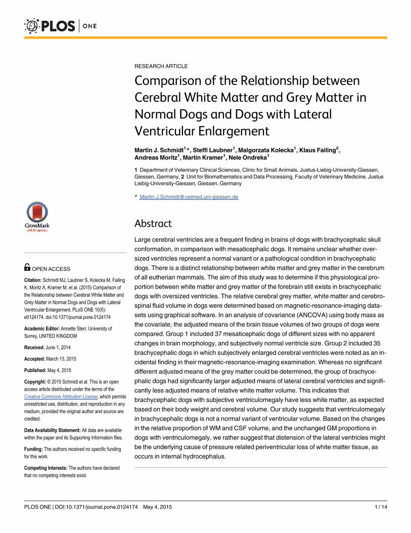
Morphometric proceduresThe mass of GM andWM in the brain was determined based on MRI datasets as described pre-viously [15, 27–30]. Study numbers were assigned to dogs so that the observer was blinded tothe signalment of the dog and its previous image interpretation. All segmentation procedureswere performed by one investigator. Image processing for volume rendering was achievedusing specialized graphical software (AMIRA, Mercury Computers Systems, Berlin, Germany),which allows manual image segmentation on a slice-by-slice basis. This program allows inter-active segmentation in all image planes (“four-viewer mode”). In this mode, three 2D viewerswith different reconstructed orientations and an additional 3D viewer are displayed in whichsegmentation can be simultaneously performed. Image segmentation in this context describesthe manual tracing of the WM and GM in each image based on their differential signal intensi-ty. All voxels corresponding to a single anatomical structure in the images are selected and as-signed to the same value in the mask. The final mask thus contains information about allselected anatomical structures and in combination with the original data and polygonal surfacereconstruction algorithms, allows the visualization of different structures in the images [29].Segmentation was performed manually in transverse orientation from individual slices (Fig 2).Only the GM andWM of the cerebrum (neocortex and paleocortex) including the basal gangliawere determined. The ammon`s horn (archicortex) was excluded because nine dogs were ex-amined due to seizures, which can lead to volume loss of this structure.
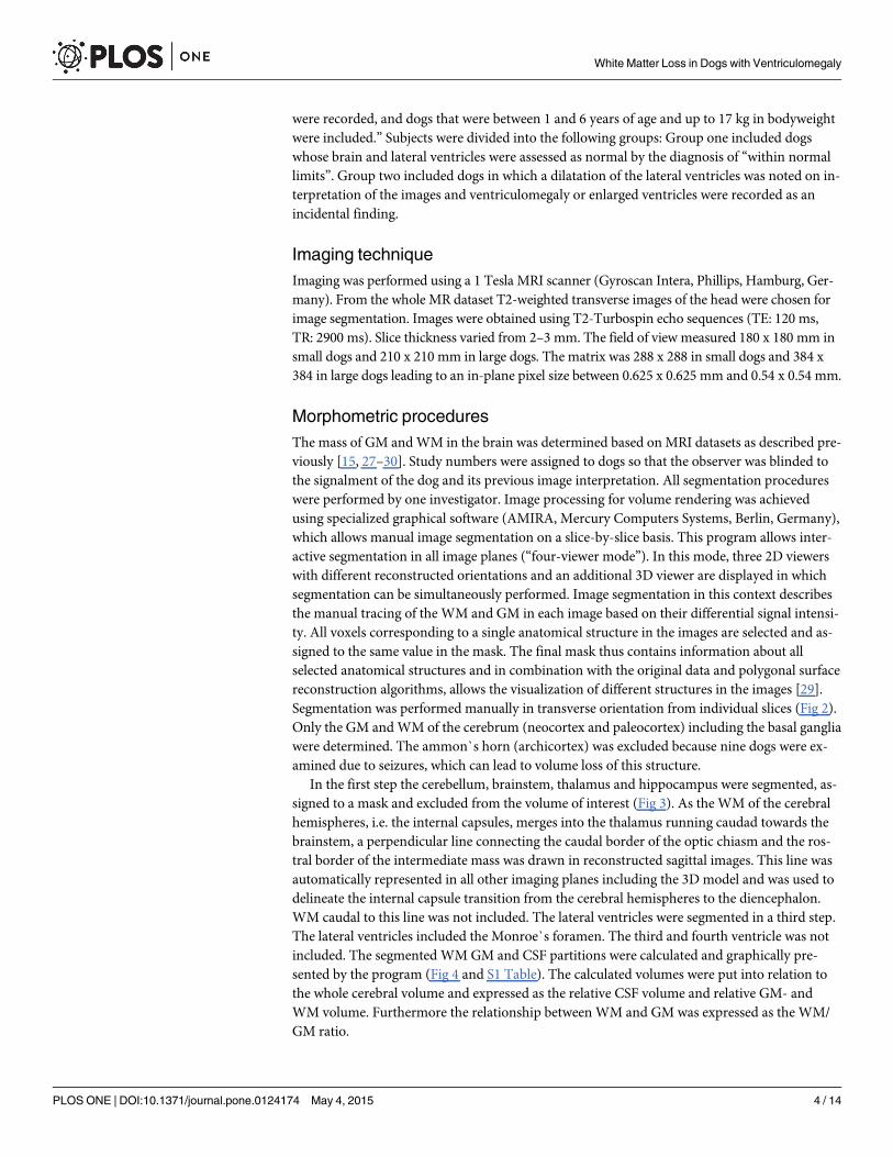
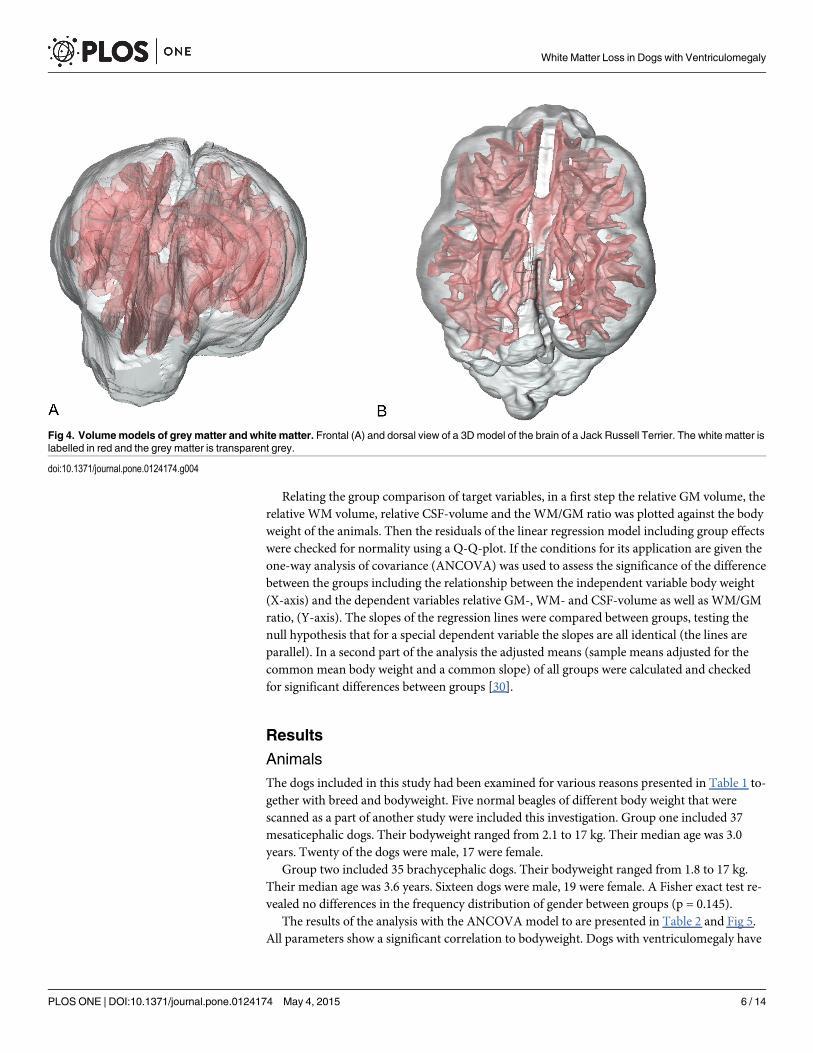
In the first step the cerebellum, brainstem, thalamus and hippocampus were segmented, as-signed to a mask and excluded from the volume of interest (Fig 3). As the WM of the cerebralhemispheres, i.e. the internal capsules, merges into the thalamus running caudad towards thebrainstem, a perpendicular line connecting the caudal border of the optic chiasm and the ros-tral border of the intermediate mass was drawn in reconstructed sagittal images. This line wasautomatically represented in all other imaging planes including the 3D model and was used todelineate the internal capsule transition from the cerebral hemispheres to the diencephalon.WM caudal to this line was not included. The lateral ventricles were segmented in a third step.The lateral ventricles included the Monroe`s foramen. The third and fourth ventricle was notincluded. The segmented WMGM and CSF partitions were calculated and graphically pre-sented by the program (Fig 4 and S1 Table). The calculated volumes were put into relation tothe whole cerebral volume and expressed as the relative CSF volume and relative GM- andWM volume. Furthermore the relationship between WM and GM was expressed as the WM/GM ratio.
White Matter Loss in Dogs with Ventriculomegaly
PLOS ONE | DOI:10.1371/journal.pone.0124174 May 4, 2015 4 / 14
Statistical analysisStatistical analysis was performed using the commercial statistical software package BMDP(BMDP Statistical Software, Inc., Los Angeles, USA). To test homogeneity of the groups the rel-ative frequency of male and female animals in the groups was compared using Fisher`sexact test.
Fig 2. Volume determination based on MRI-datasets. Image segmentation of white matter and grey matter using manual segmentation on a slice-by-slicebasis from transverse images. Each tissue of interest is labelled red and thereby assigned to a group (mask). All masks are then assembled and the tissuescan be depicted in volume form.
doi:10.1371/journal.pone.0124174.g002
Fig 3. Volume rendering of brain tissues of interest. 3D viewer mode of the graphical software AMIRA. The voxels of the tissue of interest (white matter/grey matter) of each slice have been assembled and are now displayed as a 3D model. Each tissue can be displayed solid or transparent. The localizer linessupport the segmentation process. As they are displayed in both the 2D images and the 3D model, the thalamus, medulla and cerebellum can be accuratelyseparated from the volume of interest.
doi:10.1371/journal.pone.0124174.g003
White Matter Loss in Dogs with Ventriculomegaly
PLOS ONE | DOI:10.1371/journal.pone.0124174 May 4, 2015 5 / 14
Relating the group comparison of target variables, in a first step the relative GM volume, therelative WM volume, relative CSF-volume and the WM/GM ratio was plotted against the bodyweight of the animals. Then the residuals of the linear regression model including group effectswere checked for normality using a Q-Q-plot. If the conditions for its application are given theone-way analysis of covariance (ANCOVA) was used to assess the significance of the differencebetween the groups including the relationship between the independent variable body weight(X-axis) and the dependent variables relative GM-, WM- and CSF-volume as well as WM/GMratio, (Y-axis). The slopes of the regression lines were compared between groups, testing thenull hypothesis that for a special dependent variable the slopes are all identical (the lines areparallel). In a second part of the analysis the adjusted means (sample means adjusted for thecommon mean body weight and a common slope) of all groups were calculated and checkedfor significant differences between groups [30].
Results
AnimalsThe dogs included in this study had been examined for various reasons presented in Table 1 to-gether with breed and bodyweight. Five normal beagles of different body weight that werescanned as a part of another study were included this investigation. Group one included 37mesaticephalic dogs. Their bodyweight ranged from 2.1 to 17 kg. Their median age was 3.0years. Twenty of the dogs were male, 17 were female.
Group two included 35 brachycephalic dogs. Their bodyweight ranged from 1.8 to 17 kg.Their median age was 3.6 years. Sixteen dogs were male, 19 were female. A Fisher exact test re-vealed no differences in the frequency distribution of gender between groups (p = 0.145).
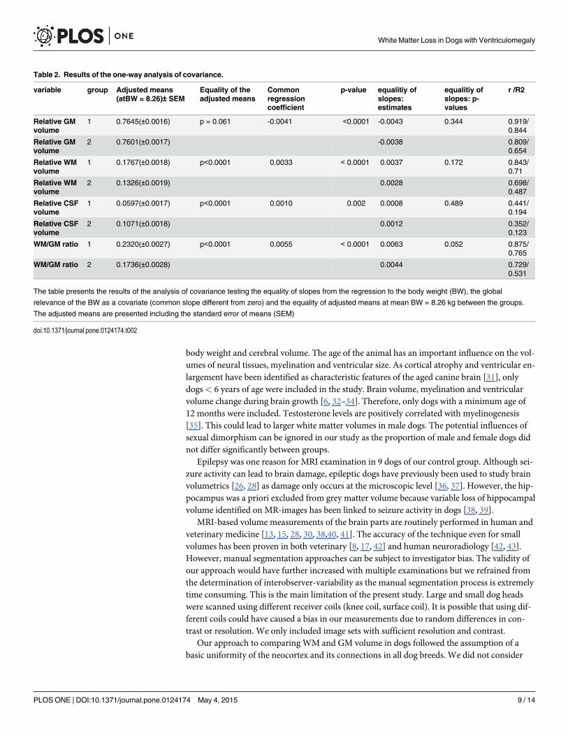
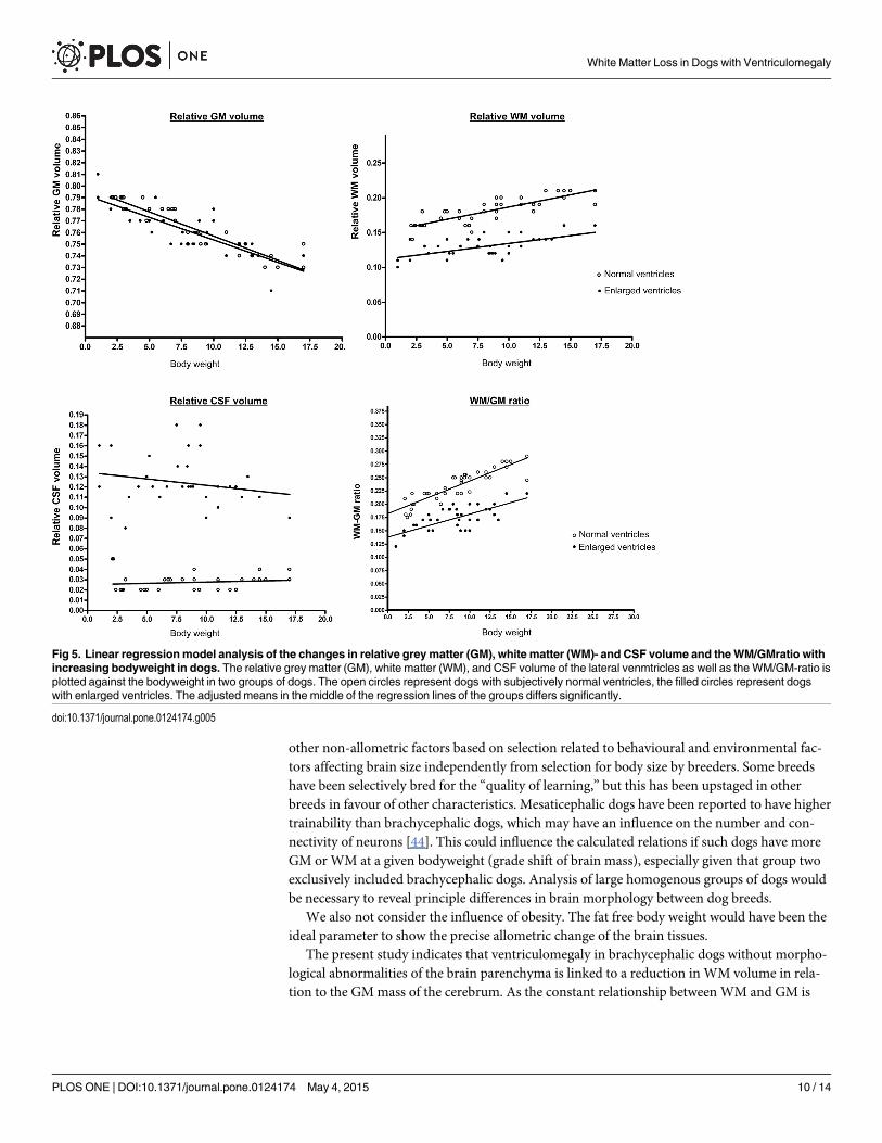
The results of the analysis with the ANCOVA model to are presented in Table 2 and Fig 5.All parameters show a significant correlation to bodyweight. Dogs with ventriculomegaly have
Fig 4. Volumemodels of grey matter and white matter. Frontal (A) and dorsal view of a 3D model of the brain of a Jack Russell Terrier. The white matter islabelled in red and the grey matter is transparent grey.
doi:10.1371/journal.pone.0124174.g004
White Matter Loss in Dogs with Ventriculomegaly
PLOS ONE | DOI:10.1371/journal.pone.0124174 May 4, 2015 6 / 14

Table 1. Breed, body weight and diagnosis of the dogs included in groups one and two.
Group1: Breed Bodyweight Indication for MRI/ Final diagnosis
1 Poodle 3 kg Optic neuritis
2 Wirehaired dachshund 4.5 kg Behavioral changes / aggression
3 Dachshund 2.1 kg Idiopathic epilepsy
4 Beagle 10 kg Study of brain perfusion
5 Beagle 11 kg Study of brain perfusion
6 Beagle 10 kg Study of brain perfusion
7 Beagle 9 kg Study of brain perfusion
8 Beagle 9.4 kg Study of brain perfusion
9 Mixed breed 3 kg Otitis media
10 Dachshund 2.2 kg Idiopathic epilepsy
11 Dachshund 2.8 kg Idiopathic epilepsy
12 Miniature schnauzer 6 kg Behavioral abnormality /aggression
13 West Highland White terrier 9 kg Otitis externa
14 Wirehaired dachshund 7 kg Otitis media
15 Mixed breed 3 kg Otitis media
16 Mixed breed 3.2 kg Retrobulbar abscess
17 Mixed breed 9.4 kg Intraorbital inflammation
18 Beagle 9.4 kg Retrobulbar tumor
19 Jack Russel terrier 5 kg Nasal tumor
20 Norfolk terrier 6.5 kg Dorsal dens angulation
21 Miniature pinscher 4.8 kg Nasal tumor
22 Jack Russel terrier 9 kg Masticatory myositis
23 Cocker spaniel 13 kg Trigeminal nerve neuritis
24 Wirehaired dachshund 8 kg Behavioral abnormality /aggression
25 Nova Scotia duck tolling retriever 17 kg Pain of undetermined origin
26 Schnauzer 14.5 Otitis media
27 Austrian hound 17 kg Rhinitis
28 Cocker Spaniel 12.5 kg Otitis media
29 Schnauzer 14 kg Nasopharyngeal mass
30 Beagle 14.5 Facial nerve paralysis
31 Mixed breed 10 kg Otitis media
32 Mixed breed 7 kg Idiopathic vestibular syndrome
33 Dachshund 2.4 kg Retropharyngeal abscess
34 Miniature Pinscher 6 kg Idiopathic epilepsy
35 Mixed breed 15 kg Trigeminal nerve neuritis
36 Poodle 2.9 kg Idiopathic epilepsy
37 Beagle 8.5 kg Retrobulbar abscess
Group 2 Breed Bodyweight Indication for MRI/ Final diagnosis
1 Chihuahua 1 kg Neck pain, atlanto-axial subluxation
2 Chihuahua 1 kg Neck pain, atlanto-axial subluxation
3 Chihuahua 2 kg Neck pain, dorsal dens angulation
4 Bolonka Zwetna 2 kg Atlantoaxial instability
5 Papillion 3.2 kg Atlantoaxial instability
6 Shih Tzu 5.2 kg Otitis media
7 Shih Tzu 5.5 kg Pain of undetermined origin
8 Pug dog 8.5 kg Masticatory myositis
9 Pug dog 13 kg Otitis media
(Continued)
White Matter Loss in Dogs with Ventriculomegaly
PLOS ONE | DOI:10.1371/journal.pone.0124174 May 4, 2015 7 / 14
a significantly higher adjusted means of the relative CSF volume. The WM/GMratio is signifi-cantly decreased in dogs with ventriculomegaly. Whereas the adjusted means of the relativeGM was not significantly different between groups the adjusted means of the relative WM vol-ume was significantly decreased in the groups with ventriculomegaly. Therefore, the modelpredicts that at the same bodyweight dogs with oversized ventricles have less WM than dogswith subjectively normal lateral ventricles.
DiscussionThe dimensions of the cerebral ventricular volume in dogs have been reported to vary [17, 18].In brachycephalic dogs in particular, the ventricles can be larger than expected. This has beenreferred to as ventriculomegaly to differentiate this condition from clinically relevant internalhydrocephalus. However, the designation of normal ventricles and enlarged ventricles remainpoorly defined and are usually assessed subjectively [26].
It is unclear whether ventriculomegaly should be interpreted as one variant of a spectrum ofventricular dimensions in dogs or rather as a pathological condition. It was our hypothesis thatthe larger ventricles in brachycephalic dogs are a pathological condition as indicated by con-current presence of WM loss due to ventricular distention as found in internal hydrocephalus.To investigate this hypothesis we examined the relative WM, GM, and CSF volume in dogswith different ventricular dimensions as well as their WM/GM ratio in proportion to their
Table 1. (Continued)
Group1: Breed Bodyweight Indication for MRI/ Final diagnosis
10 Pug dog 8.4 kg Compulsive obsessive behavior
11 Pug dog 8.7 kg Dorsal dens angulation
12 French Bulldog 8.5 kg Otitis media
13 French Bulldog 13 kg Otitis media
14 French Bulldog 13.5 kg Optic neuritis
15 French Bulldog 9 kg Deafness
16 French Bulldog 8,9 kg Neck pain—arachnoid cyst C2
17 French Bulldog 11 kg Otitis media/interna
18 French Bulldog 10 kg Retropharyngeal mass
19 French Bulldog 10 kg Otitis media
20 French Bulldog 12.5 kg Otitis media
21 Shih Tzu 6.7 kg Retrobulbar abscess
22 Shih Tzu 7.6 kg Optic neuritis
23 Tibet terrier 5 kg Seizures—cardiac syncopes
24 Yorkshire terrier 4.3 kg Pain of undetermined origin
25 CKCS 11 kg Retropharyngeal abscess
26 CKCS 7.5 kg Lymphoma trigeminal nerve (extrancranial)
27 CKCS 5 kg Breeding selection syringomyelia
28 CKCS 8 kg Breeding selection syringomyelia
29 CKCS 12 kg Breeding selection syringomyelia
30 CKCS 10 kg Breeding selection syringomyelia
31 CKCS 14.5 kg Otitis media
32 Yorkshire terrier 3.5 kg Pain of undetermined origin
33 Pekingese 6.1 kg Otitis media
34 Pekingese 7.6 kg Otitis media
35 English bulldog 17 kg Head bobbing
doi:10.1371/journal.pone.0124174.t001
White Matter Loss in Dogs with Ventriculomegaly
PLOS ONE | DOI:10.1371/journal.pone.0124174 May 4, 2015 8 / 14
body weight and cerebral volume. The age of the animal has an important influence on the vol-umes of neural tissues, myelination and ventricular size. As cortical atrophy and ventricular en-largement have been identified as characteristic features of the aged canine brain [31], onlydogs< 6 years of age were included in the study. Brain volume, myelination and ventricularvolume change during brain growth [6, 32–34]. Therefore, only dogs with a minimum age of12 months were included. Testosterone levels are positively correlated with myelinogenesis[35]. This could lead to larger white matter volumes in male dogs. The potential influences ofsexual dimorphism can be ignored in our study as the proportion of male and female dogs didnot differ significantly between groups.
Epilepsy was one reason for MRI examination in 9 dogs of our control group. Although sei-zure activity can lead to brain damage, epileptic dogs have previously been used to study brainvolumetrics [26, 28] as damage only occurs at the microscopic level [36, 37]. However, the hip-pocampus was a priori excluded from grey matter volume because variable loss of hippocampalvolume identified on MR-images has been linked to seizure activity in dogs [38, 39].
MRI-based volume measurements of the brain parts are routinely performed in human andveterinary medicine [13, 15, 28, 30, 38,40, 41]. The accuracy of the technique even for smallvolumes has been proven in both veterinary [8, 17, 42] and human neuroradiology [42, 43].However, manual segmentation approaches can be subject to investigator bias. The validity ofour approach would have further increased with multiple examinations but we refrained fromthe determination of interobserver-variability as the manual segmentation process is extremelytime consuming. This is the main limitation of the present study. Large and small dog headswere scanned using different receiver coils (knee coil, surface coil). It is possible that using dif-ferent coils could have caused a bias in our measurements due to random differences in con-trast or resolution. We only included image sets with sufficient resolution and contrast.
Our approach to comparing WM and GM volume in dogs followed the assumption of abasic uniformity of the neocortex and its connections in all dog breeds. We did not consider
Table 2. Results of the one-way analysis of covariance.
variable group Adjusted means(atBW = 8.26)± SEM
Equality of theadjusted means
Commonregressioncoefficient
p-value equalitiy ofslopes:estimates
equalitiy ofslopes: p-values
r /R2
Relative GMvolume
1 0.7645(±0.0016) p = 0.061 -0.0041 <0.0001 -0.0043 0.344 0.919/0.844
Relative GMvolume
2 0.7601(±0.0017) -0.0038 0.809/0.654
Relative WMvolume
1 0.1767(±0.0018) p<0.0001 0.0033 < 0.0001 0.0037 0.172 0.843/0.71
Relative WMvolume
2 0.1326(±0.0019) 0.0028 0.698/0.487
Relative CSFvolume
1 0.0597(±0.0017) p<0.0001 0.0010 0.002 0.0008 0.489 0.441/0.194
Relative CSFvolume
2 0.1071(±0.0018) 0.0012 0.352/0.123
WM/GM ratio 1 0.2320(±0.0027) p<0.0001 0.0055 < 0.0001 0.0063 0.052 0.875/0.765
WM/GM ratio 2 0.1736(±0.0028) 0.0044 0.729/0.531
The table presents the results of the analysis of covariance testing the equality of slopes from the regression to the body weight (BW), the global
relevance of the BW as a covariate (common slope different from zero) and the equality of adjusted means at mean BW = 8.26 kg between the groups.
The adjusted means are presented including the standard error of means (SEM)
doi:10.1371/journal.pone.0124174.t002
White Matter Loss in Dogs with Ventriculomegaly
PLOS ONE | DOI:10.1371/journal.pone.0124174 May 4, 2015 9 / 14
other non-allometric factors based on selection related to behavioural and environmental fac-tors affecting brain size independently from selection for body size by breeders. Some breedshave been selectively bred for the “quality of learning,” but this has been upstaged in otherbreeds in favour of other characteristics. Mesaticephalic dogs have been reported to have highertrainability than brachycephalic dogs, which may have an influence on the number and con-nectivity of neurons [44]. This could influence the calculated relations if such dogs have moreGM or WM at a given bodyweight (grade shift of brain mass), especially given that group twoexclusively included brachycephalic dogs. Analysis of large homogenous groups of dogs wouldbe necessary to reveal principle differences in brain morphology between dog breeds.
We also not consider the influence of obesity. The fat free body weight would have been theideal parameter to show the precise allometric change of the brain tissues.
The present study indicates that ventriculomegaly in brachycephalic dogs without morpho-logical abnormalities of the brain parenchyma is linked to a reduction in WM volume in rela-tion to the GMmass of the cerebrum. As the constant relationship between WM and GM is
Fig 5. Linear regressionmodel analysis of the changes in relative grey matter (GM), white matter (WM)- and CSF volume and theWM/GMratio withincreasing bodyweight in dogs. The relative grey matter (GM), white matter (WM), and CSF volume of the lateral venmtricles as well as theWM/GM-ratio isplotted against the bodyweight in two groups of dogs. The open circles represent dogs with subjectively normal ventricles, the filled circles represent dogswith enlarged ventricles. The adjusted means in the middle of the regression lines of the groups differs significantly.
doi:10.1371/journal.pone.0124174.g005
White Matter Loss in Dogs with Ventriculomegaly
PLOS ONE | DOI:10.1371/journal.pone.0124174 May 4, 2015 10 / 14
universal in all mammalian brains, the reduced WM in combination with it the larger ventri-cles in these dogs should not be considered a physiological condition.
The origin of the WM loss is not clear. A primary white matter disease and distension of theventricles “ex vacuo” would theoretically be possible. Leukencephalitis, leukodystrophies,hypomyelination and spongy degeneration must be considered in this context. However, suchdiseases would cause distinct MRI-findings beyond white matter atrophy [9]. Moreover, pri-mary white matter diseases would cause severe neurological signs, such as tremor, ataxia,which were not diagnosed in our patients.
Internal hydrocephalus can lead to progressive destruction of WM in dogs [45, 46] as a con-sequence of high intraventricular pressure. It is reasonable to expect that the degree of intra-ventricular pressure determines the speed and severity of white matter injury and therebyfunctional brain deficits. Moderately increased pressure or intermittently high intraventricularCSF pressure might play an important role in the pathogenesis of WM loss in dogs with ventri-culomegaly as proposed for normal pressure hydrocephalus (NPH) in humans [47]. It hasbeen suggested that in NPH intermittent CSF pressure waves can produce temporary phases ofischaemia in the periventricular white matter. The chronic cumulative effects of these ischae-mic events can produce WM atrophy that does not arise until late adulthood [47, 48]. Brainperfusion studies have revealed reduced regional cerebral blood flow in the periventricular re-gion in human patients with NPH, which has been attributed to disturbances in CSF flow [49].
Ventriculomegaly has also been attributed to disturbances in CSF dynamics in dogs bysome authors [15, 50]. Impaired CSF flow can be caused by obstruction of the foramen mag-num, as found in dogs with morphological changes in the cranio-cervical junction referred toas Chiari-like malformation [50, 51]. It has been proposed that impaired CSF flow can result insyringomyelia as well as ventricular dilatation [52, 53]. However, this has only been proposedfor the Cavalier King Charles spaniel and the Brussels Griffon and the prevalence of Chiari-likemalformation in other breeds with ventriculomegaly needs closer evaluation.
Human patients with enlarged ventricles in association with NPH show characteristic signsof neural function deficits. The cardinal symptoms of NPH are gait impairment, dementia andurinary incontinence (“Hakim triad”) [54]. However, it has also been proposed that the fullsymptom triad represents an advanced stage of the disease and that NPH can be diagnosed inthe presence of only two or even just one of the symptoms [54]. In contrast, the association ofsolitary ventriculomegaly with neurological deficits in dogs has been rejected [9–11, 13]. Thelack of clinical signs as a consequence of ventricular enlargement in dogs as opposed to humansmay initially be striking. However, control of locomotion is far more complex in humans andthe motor cortex and its connections play a substantial role even during undemanding steady-state walking [55]. The cerebral control of locomotion plays a minor role in dogs and diseasesaffecting the forebrain may have only minimal influence on locomotion [56]. Urinary inconti-nence was not reported in the dogs of this study either. Although the control of micturition in-volves cortical centres in carnivores, pontine micturition centres maintain function withoutcerebral control [56]. Signs of dementia are difficult to diagnose in dogs. In human medicine, adiagnosis of dementia will be considered based on the occurrence of distinct clinical signs. Themost important criteria are memory impairment, disturbed language function, inability tocarry out purposeful movement, and failure to recognize people [54]. Such brain functions can-not be assessed in dogs, at least not in a clinical setting, and the real status of canine cognitiveabilities cannot be determined with absolute certainty. On the other hand, it has been shownthat even severe ventricular dilation is compatible with normal physical and intellectual devel-opment in humans and that the ventricular volumes of hydrocephalic patients did not correlatewell with their intelligence quotient [57, 58].
White Matter Loss in Dogs with Ventriculomegaly
PLOS ONE | DOI:10.1371/journal.pone.0124174 May 4, 2015 11 / 14
The influence of ventriculomegaly on brain function in dogs is unclear. Detailed behaviouralstudies of the impact of WM loss on the full functional integration of the nervous system arenecessary to clarify whether ventriculomegaly might be an indication for CSF shunting proce-dures in dogs. If clinical or experimental data for cognitive impairment or intermittently highCSF pressure waves could be found, naturally occurring ventriculomegaly in dogs might be aninteresting animal model for human NPH.
ConclusionBrachycephalic dogs with ventriculomegaly can have reduced cerebral white matter. As a pre-determined relationship exists between WM and GMmass in the brain of mammals this aber-ration cannot be interpreted as a physiological condition.
Supporting InformationS1 Table. Calculated volumes of grey matter, white matter, lateral ventricles and the WM/GM ratio for each dog included into the study.(XLSX)
Author ContributionsConceived and designed the experiments: MJS NOM. Kolecka. Performed the experiments:NO SL M. Kolecka. Analyzed the data: KF MJS SL. Contributed reagents/materials/analysistools: MJS M. Kramer. Wrote the paper: MJS AMNO.
References1. Seiferle E (1966) Zur Topographie des Gehirns bei lang—und kurzköpfigen Hunderassen. Acta Anat
63: 346–362. PMID: 5918444
2. Roberts T, McGreevy P, Valenzuela M (2010) Human induced rotation and reorganization of the brainof domestic dogs. PLoS One 26: e11946. doi: 10.1371/journal.pone.0011946 PMID: 20668685
3. Oboussier H (1955) Behavior of cerebral sulcus formation in crossbreeding of extreme breed types ofdogs. Z Mensch Vererb Konstitutionsl 33:1–9. PMID: 13248058
4. Schmidt MJ, Amort K, Kramer M (2012) Postnatal development of the cerebral gyrification in the caninebrain. Vet Radiol & Ultrasound 53: 643–649.
5. Schmidt MJ, Neumann AC, Amort KH,Failin K, Kramer M (2011) Cephalometric measurements and de-termination of the general skull type of Cavalier King Charles Spaniels. Vet Radiol & Ultrasound 52:436–440.
6. Kii S, Uzuka Y, Taura Y, Nakaichi M, Takeuchi A, Inokuma H, et al. (1997) Magnetic resonance imagingof the lateral ventricles in beagle-type dogs. Vet Radiol & Ultrasound 38: 430–433.
7. Schröder H, Meyer-Lindenberg A, Nolte I (2006) Comparative examination of the lateral cerebral ventri-cles of different dog breeds using quantitative computed tomography. Berl Munch Tierarztl Wochenschr119: 506–511. PMID: 17172139
8. Vullo T, Korenman E, Manzo RP, Gomez DG, Deck MD, Cahill PT. (1997) Diagnosis of cerebral ventri-culomegaly in normal adult beagles using quantitative MRI. Vet Radiol & Ultrasound 38: 277–281.
9. Gavin PR, Bagley RS (2011). Practical Small Animal MRI Wiley Blackwell, Iowa USA. 64p
10. Schwarz T, Saunders J (2012) Veterinary Computed Tomography. Wiley Blackwell Iowa USA. 185 p
11. Lu D, Lamb CR, Pfeiffer DU, Targett MF (2003) Neurological signs and results of magnetic resonanceimaging in 40 cavalier King Charles spaniels with Chiari type 1-like malformations. Vet Rec 153: 60–63. PMID: 12885216
12. Driver CJ, Chandler K, Walmsley G, Shibab N, Volk HA (2013) The association between Chiari-likemalformation, ventriculomegaly and seizures in cavalier King Charles spaniels. Vet J 195: 235–237.doi: 10.1016/j.tvjl.2012.05.014 PMID: 22749114
13. Vite CH, Insko EK, Schotland HM, Panckeri K, Hendricks JC (1997) Quantification of cerebral ventricu-lar volume in English bulldogs. Vet Radiol & Ultrasound 38: 437–443.
White Matter Loss in Dogs with Ventriculomegaly
PLOS ONE | DOI:10.1371/journal.pone.0124174 May 4, 2015 12 / 14
14. Dexler H (1923) Über die konstitutionelle innere Hydrocephalie beim Hunde. Prag Tierärztl Arch 3:103–268.
15. Driver CJ, Rusbridge C, Cross HR, McGonnell I, Volk HA (2010) Relationship of brain parenchymawithin the caudal cranial fossa and ventricle size to syringomyelia in cavalier King Charles spaniels. JSmall Anim Pract 51: 382–386. doi: 10.1111/j.1748-5827.2010.00952.x PMID: 20536691
16. Saito M, Olby NJ, Spaulding K, Muñana K, Sharp NJ (2003) Relationship among basilar artery resis-tance index, degree of ventriculomegaly, and clinical signs in hydrocephalic dogs. Vet Radiol & Ultra-sound 44: 687–694.
17. De Haan CE, Kraft SL, Gavin PR, Wendling LR (1994) Normal variation in size of the lateral ventriclesof the Labrador Retriever dog as assessed by magnetic resonance imaging. Vet Radiol & Ultrasound35: 83–86.
18. Spaulding KA, Sharp NJH (1990) Ultrasonographic imaging of the lateral cerebral ventricles in the dog.Vet Radiol & Ultrasound 31: 59–64.
19. Zhang K, Sejnowski TJ (2000) A universal scaling law between gray matter and white matter of cerebralcortex. Proc Natl Acad Sci USA 97: 5621–5626. PMID: 10792049
20. Frahm HD, Stephan H, StephanM (1982) Comparison of brain structure volumes in Insectivora and Pri-mates. I. Neocortex. J Hirnforsch 23: 375–389. PMID: 7161477
21. Rilling JK, Insel TR (1999) Differential expansion of neural projection systems in primate brain evolu-tion. Neuroreport 10: 1453–1459. PMID: 10380962
22. Changizi MA (2001) Principles underlying mammalian neocortical scaling. Biol Cybern 84: 207–215.PMID: 11252638
23. Roth G, Dickie U (2005) Evolution of the brain and intelligence. Trends Cogn Sci. 9: 250–257. PMID:15866152
24. Purves D, Augustine GJ, Fitzpatrick D, Katz LC, LaMantia AS, McNamara JO, Williams SM, editors(2001) Neuroscience. 2nd edition. Sunderland (MA): Sinauer Associates.
25. Carrera I, Dennis R, Mellor DJ, Penderis J, Sullivan M (2009) Use of magnetic resonance imaging formorphometric analysis of the caudal cranial fossa in Cavalier King Charles Spaniels. Am J Vet Res 70:340–345. doi: 10.2460/ajvr.70.3.340 PMID: 19254145
26. Thomas B (2010) Hydrocephalus in dogs and cats. Vet Clin Small Anim 40: 143–159 doi: 10.1016/j.cvsm.2009.09.008 PMID: 19942061
27. Cross HR, Capello R, Rusbridge C (2009) Comparison of cerebral cranium volumes between CavalierKing Charles Spaniels with Chiari-like malformation, small breed dogs and Labradors. J Small AnimPract 50: 399–405. doi: 10.1111/j.1748-5827.2009.00799.x PMID: 19689667
28. Schmidt MJ, Biel M, Klumpp S, Schneider M, Kramer M (2009) Evaluation of the volumes of cranial cav-ities in Cavalier King Charles spaniels with Chiari- like malformation and other brachycephalic dogs asmeasured via computed tomography. Am J Vet Res. 70: 508–512. doi: 10.2460/ajvr.70.4.508 PMID:19335107
29. Shaw TA, McGonnell IM, Driver CJ, Rusbridge C, Volk HA (2012) Increase in cerebellar volume in Cav-alier King Charles Spaniels with Chiari-like malformation and its role in the development of syringomye-lia. PLoS One 7(4): e33660. doi: 10.1371/journal.pone.0033660 PMID: 22506005
30. Barton RA, Harvey PH (2000) Mosaic evolution of brain structure in mammals. Nature 29: 1055–1058.PMID: 11193258
31. SuMY, Head E, BrooksWM (1998) Magnetic resonance imaging of anatomic and vascular characteris-tics in a canine model of human aging. Neurobiol Ageing 19: 479–485. PMID: 9880050
32. Gross B, Garcia-Tapia D, Riedesel E, Ellinwood NM, Jens JK (2010) Normal canine brain maturation atmagnetic resonance imaging. Vet Radiol & Ultrasound 51: 361–373.
33. Arant BS, GoochWM (1982) Developmental changes in the mongrel canine brain during postnatal life.Early Hum Dev 7: 179–194. PMID: 7151729
34. Fox MW (1964) The postnatal growth of the canine brain and correlated anatomical and behaviouralchange during neuro-ontogenesis. Growth 28: 135–141. PMID: 14189833
35. Martini L, Melcangi RC (1991) Androgen metabolism in the brain. J Steroid BiochemMol Biol 39: 819–828. PMID: 1954172
36. Montgomery DL, Lee AC (1983) Brain damage in the epileptic Beagle dog. Vet Pathol 20: 160–169.PMID: 6836872
37. Morita T, Shimada A, Ohama E, Umemura T, Fukuda S (1999) Oligodendroglial vacuolar degenerationin the bilateral motor cortices and astrocytosis in epileptic Beagle dogs. J Vet Med Sci 61: 107–111.PMID: 10081746
White Matter Loss in Dogs with Ventriculomegaly
PLOS ONE | DOI:10.1371/journal.pone.0124174 May 4, 2015 13 / 14
38. Yamasaki H, Furuoka H, Takechi M, Itakura C (1991) Neuronal loss and gliosis in limbic system in anepileptic dog. Vet Pathol 28: 540–542. PMID: 1771745
39. Breitschwerdt EB, Breazile JE, Broadhurst JJ (1979) Clinical and electroencephalographic findings as-sociated with ten cases of suspected limbic epilepsy in the dog. J Am Anim Hosp Assoc 15: 37–50.
40. Milne ME, Anderson GA, Chow KE, O'Brien TJ, Moffat BA, Long SN (2013) Description of techniqueand lower reference limit for magnetic resonance imaging of hippocampal volumetry in dogs. Am J VetRes 74: 224–231. doi: 10.2460/ajvr.74.2.224 PMID: 23363346
41. Thames R, Robertson ID, Flegel T, Henke D, O'Brien DP, Coates JR, et al. (2010) Development of amorphometric Magnetic Resonance Image parameter suitable for distinguishing between normal dogsand dogs with cerebellar atrophy. Vet Radiol & Ultrasound 51: 246–253.
42. Rifa H, Bloch I, Hutchinson S, et al. (2000) Segmentation of the skull in MRI volumes using deformablemodel and taking the partial volume effect into account. Med Image Anal 4: 219–233. PMID: 11145310
43. Vullo T, Deo-Narine V, Stallmeyer MJ, Wiart J, Garnero L (1996) Quantitation of normal canine hippo-campus formation volume: correlation of MRI with gross histology. Magn Reson Imaging 14: 657–662.PMID: 8897370
44. HeltonWS (2009) Cephalic index and perceived dog trainability. Behav Proc 82: 355–358.
45. Wünschmann A, Oglesbee M (2001) Periventricular changes associated with spontaneous canine hy-drocephalus. Vet Pathol 38: 67–73. PMID: 11199166
46. Yamada H, Yokota A, Haratake J, Horie A (1996) Morphological study of experimental syringomyeliawith kaolin-induced hydrocephalus in a canine model. J Neurosurg 84: 999–1005. PMID: 8847595
47. Del Bigio MR (1993) Neuropathological changes caused by hydrocephalus. Acta Neuropathol 85:573–585. PMID: 8337936
48. Akai K, Uchigasaki S, Tanaka U, Komatsu A (1987) Normal pressure hydrocephalus: neuropathologi-cal study. Acta Pathol Jpn 37: 97–110. PMID: 3577765
49. Momjian S, Owler BK, Czosnyka Z, Czosnyka M, Pena A, Pickard JD (2004) Pattern of white matter re-gional cerebral blood flow and autoregulation in normal pressure hydrocephalus. Brain. 127: 965–972.PMID: 15033897
50. Rusbridge C, Knowler SP, Pieterse L Mc Fadyen A (2009) Chiari-like malformation in the Griffon Brux-ellosis. J Small Anim Pract 50: 386–393. doi: 10.1111/j.1748-5827.2009.00744.x PMID: 19689665
51. Rusbridge C, MacSweeney JRE, Davies SJChandler K, Fitzmaurice SN, Dennis R, Cappello R, Wheel-er SJ. (2000) Syringohydromyelia in Cavalier King Charles Spaniels. J Am Anim Hosp Assoc 36: 34–41. PMID: 10667404
52. Levine DN (2004) The pathogenesis of syringomyelia associated with lesions at the foramen magnum:a critical review of existing theories and proposal of a new hypothesis. J Neurol Sci 220: 3–21. PMID:15140600
53. Rusbridge C, Knower SP (2004) Inheritance of occipital bone hypoplasia (ChiariI malformation) in Cav-alier King Charles spaniels. J Vet Int Med 18:673 678.
54. Kiefer M, Unterberg A (2012) The differential diagnosis and treatment of normal-pressure hydrocepha-lus. Dtsch Arztebl Int 109: 15–25. doi: 10.3238/arztebl.2012.0015 PMID: 22282714
55. Miyai I, Tanabe HC, Sase I, Eda H, Oda I, Konishi I, Tsunazawa Y, Suzuki T, Yanagida T, Kubota K(2001) Cortical mapping of gait in humans: A near-infrared spectroscopic topography study. Neuro-image 141186–1192.
56. DeLahunta A, Glass E (2009).Veterinary neuroanatomy and clinical neurology. Saunders Elsevier,Missouri. 37p.
57. Lorber J (1983) Hydrocephalus in frühen Kindersalter. Edited by Stuttgart VD. Enke: 2–14.
58. Jackson PH, Lorber J (1984) Brain and ventricular volume in hydrocephalus. Z Kinderchir 39 (Suppl 2):91–93. PMID: 6524106
White Matter Loss in Dogs with Ventriculomegaly
PLOS ONE | DOI:10.1371/journal.pone.0124174 May 4, 2015 14 / 14