researchhoxb13 promotes androgen independent growth of ... · understand androgen-independent...
TRANSCRIPT
Kim et al. Molecular Cancer 2010, 9:124http://www.molecular-cancer.com/content/9/1/124
Open AccessR E S E A R C H
ResearchHOXB13 promotes androgen independent growth of LNCaP prostate cancer cells by the activation of E2F signalingYoung-Rang Kim†1, Kyung-Jin Oh†2, Ra-Young Park1, Nguyen Thi Xuan1, Taek-Won Kang2,5, Dong-Deuk Kwon2,5, Chan Choi3,5, Min Soo Kim4, Kwang Il Nam1,5, Kyu Youn Ahn1,5 and Chaeyong Jung*1,5
AbstractBackground: Androgen signaling plays a critical role in the development of prostate cancer and its progression. However, androgen-independent prostate cancer cells emerge after hormone ablation therapy, resulting in significant clinical problems. We have previously demonstrated that the HOXB13 homeodomain protein functions as a prostate cancer cell growth suppressor by inhibiting androgen-mediated signals. However, the role of the HOXB13 in androgen-independent growth of prostate cancer cells remains unexplained.
Results: In this report, we first demonstrated that HOXB13 was highly overexpressed in hormone-refractory tumors compared to tumors without prostate-specific antigen after initial treatment. Functionally, in an androgen-free environment minimal induction of HOXB13 in LNCaP prostate cancer cells, to the level of the normal prostate, markedly promoted cell proliferation while suppression inhibited cell proliferation. The HOXB13-mediated cell growth promotion in the absence of androgen, appears to be mainly accomplished through the activation of RB-E2F signaling by inhibiting the expression of the p21waf tumor suppressor. Indeed, forced expression of HOXB13 dramatically decreased expression of p21waf; this inhibition largely affected HOXB13-mediated promotion of E2F signaling.
Conclusions: Taken together, the results of this study demonstrated the presence of a novel pathway that helps understand androgen-independent survival of prostate cancer cells. These findings suggest that upregulation of HOXB13 is associated with an additive growth advantage of prostate cancer cells in the absence of or low androgen concentrations, by the regulation of p21-mediated E2F signaling.
BackgroundHox homeobox genes contain highly homologous home-odomains and are considered transcription factors thatregulate axial regional specification during embryonicdevelopment. Due to temporal and spatial colineality,Hox genes are expressed in a tissue-specific and fre-quently stage-related fashion. The Hox-13 paralog isespecially important to the development of accessory sex-ual organs, including the prostate gland [1-3]. Hoxb13 hasbeen shown to have limited expression at the caudalextent of the spinal cord, tail bud and urogenital sinus inan androgen-independent manner [4,5]. The expression
of HOXB13 is restricted but abundant in the adult humanprostate, representing almost one out of 2,000 transcripts[6]. However, the role of HOXB13 during the adult stageis mostly unknown. Mice homozygous for Hoxb13 loss-of-function mutations have shown overgrowth in allmajor structures derived from the tail bud [7] and malfor-mation of the ducts of the ventral prostate, includingcomplete loss of secretory proteins [8]. Hoxb13 mutantmice do not develop a prominent phenotypical changebut do have a swollen prostate in old mice, probably dueto the functional redundancy of other Hox-13 molecules.
Prostate cancer (PCa) is the most commonly diagnosedcancer in North American men; a significant number ofpatients manifest androgen resistant PCa, leading to afatal disease. Androgen receptor (AR) activity is essentialto the growth and progression of PCa at all phases of the
* Correspondence: [email protected] Department of Anatomy, Chonnam National University Medical School, Gwangju, Korea† Contributed equallyFull list of author information is available at the end of the article
© 2010 Kim et al; licensee BioMed Central Ltd. This is an Open Access article distributed under the terms of the Creative Commons At-tribution License (http://creativecommons.org/licenses/by/2.0), which permits unrestricted use, distribution, and reproduction in anymedium, provided the original work is properly cited.
Kim et al. Molecular Cancer 2010, 9:124http://www.molecular-cancer.com/content/9/1/124
Page 2 of 14
disease [9,10]. Therefore, androgen ablation therapy isthe frontline treatment for metastatic PCa; however, theclinical response is transient, resulting in the recurrenceof tumors. These androgen-independent (AI) tumorsparadoxically depend on the AR [11,12], suggesting thatAR-mediated signaling is required for the growth of can-cer cells, even when absent or present only in low concen-trations. However, the regulation of AR target genesunder androgen-deprived conditions is not fully under-stood. There is evidence to support a link between therole of HOXB13 and the malignant progression of PCa.
The highly prostate-specific HOXB13 is involved in theformation and maintenance of this hormone-dependentorgan [5,6,13,14]. In our previous studies, HOXB13expression was correlated with the AR in both culturedPCa cells and PCa xenograft models. All PCa cell linesretaining the AR expressed moderate to high levels ofHOXB13 whereas AR-negative cells had low to undetect-able amounts of HOXB13. However, the expression ofHOXB13 and AR was not mutually regulated [5,15].There is also close functional and mechanistic communi-cations between HOXB13 and AR signaling. HOXB13suppresses androgen-stimulated AR activity by physicalinteraction with the AR and subsequently inhibits thegrowth-regulating function of AR [15]. Due to the multi-focal nature of PCa, however, the pattern of expression ofHOXB13, during prostate tumorigenesis, has not beenwell defined [15,16]. These results suggest that the bal-ance between the HOXB13 and the AR (and/or hormoneresponse) may be important in the development and pro-gression of PCa cells. In this study, we investigated thepattern of expression of HOXB13 in androgen-dependent(AD) and AI tumors and the functional and mechanisticrole of HOXB13 in hormone-refractory PCa cells.
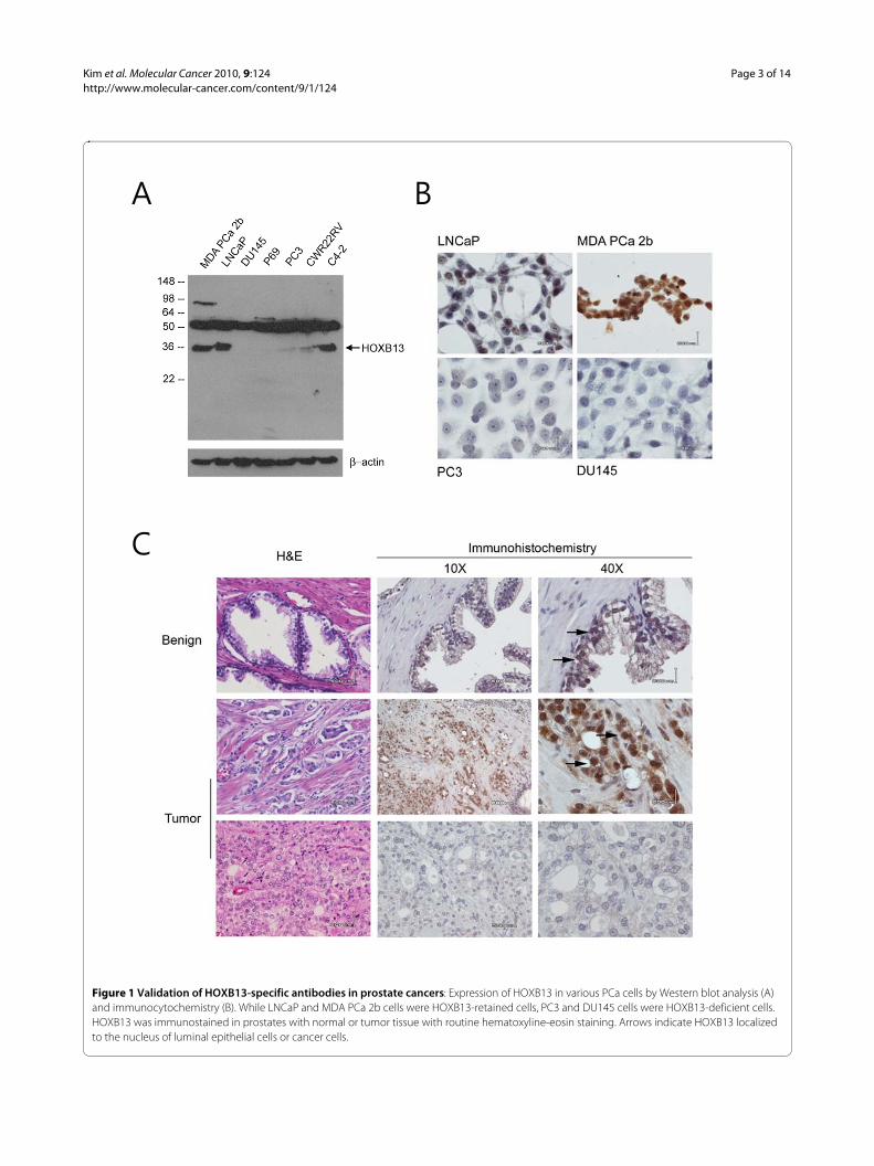
ResultsAnti-HOXB13 antibodiesTo study the tissue distribution and expression pattern ofHOXB13 protein in PCas, HOXB13-specific antibodiesare critical tool. To date, there are no anti-HOXB13 anti-bodies suitable for immunohistochemistry that can bepurchased in the commercial market. Therefore, anti-HOXB13 antibodies were developed and their specificityto HOXB13 was tested by Western blot analysis usingvarious PCa cell lines. As shown in Figure 1A, anti-HOXB13 antibodies had a band at 35kDa, which washighly expressed in the LNCaP, C42, and MDAPCa2bcells, with only minor to no expression in the PC3,DU145, P69, and CWR22RV cells. These results wereconsistent with the expression pattern of HOXB13 RNAas previously demonstrated [15]. These anti-HOXB13antibodies did not react with recombinant homologousHOXA13 or HOXD13 in transfection experiments (datanot shown). However, the antibodies nonspecifically
reacted to protein around 50kDa. To confirm that theantibodies could be used to localize HOXB13 in tumorcells, immunocytochemistry was performed in bothHOXB13 positive and negative cells (Figure 1B).HOXB13 was detected in the nuclear compartment ofHOXB13-positive LNCaP and MDAPCa2b cells, but notin the HOXB13-negative PC3 and DU145 cells; thesefindings suggested that antibodies did not react to thenative form of the 50 kDa protein observed in the West-ern blot. Next, to evaluate the tissue expression ofHOXB13 in PCas, immunohistochemistry was per-formed on formalin-fixed and paraffin-embedded PCas(Figure 1C). Similar to the results of prior mouse studies[4,5], moderate expression of HOXB13 was observed inbenign prostate luminal epithelium, which was confinedto the nuclear compartment (arrows in upper row). Inlocalized PCas, some ductal epithelial cells highlyexpressed HOXB13 (arrows in middle row) while someducts did not (lower row), probably due to the multifocaland heterogeneous nature of PCa.
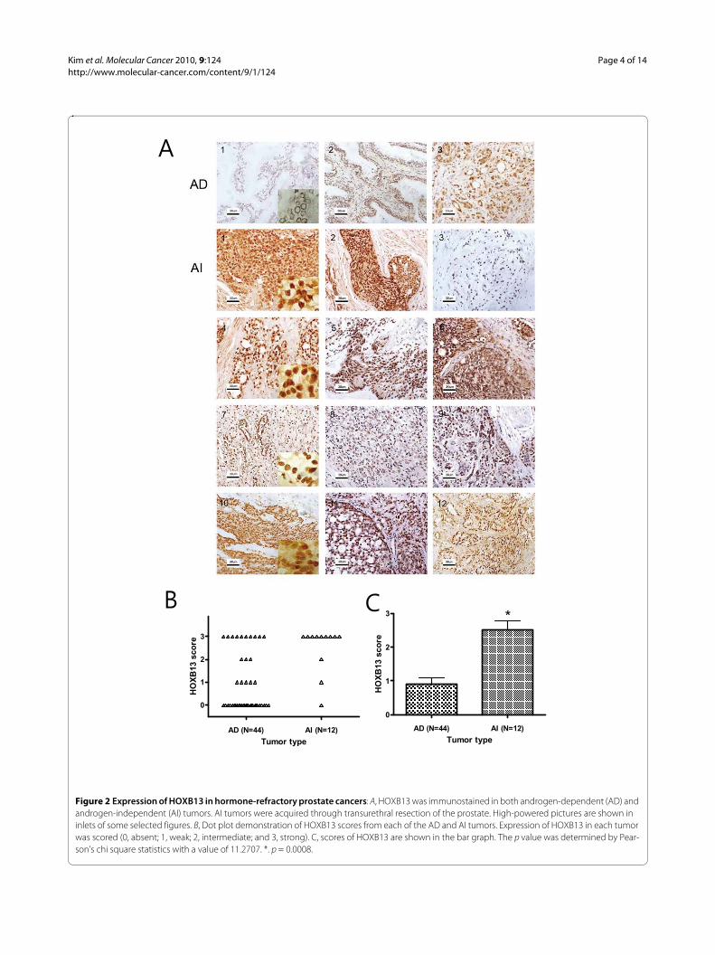
HOXB13 was overexpressed in androgen-refractory prostate cancerThe expression of HOXB13 was investigated in tumorspecimens from failed androgen ablation therapy andcompared to tumors without PSA recurrence. Fifty sixclinical samples of hormone-dependent (n = 44) and hor-mone-refractory (n = 12) PCas were successfully stained.Among AD tumors, 31 (70%) showed negative or weakstaining (score 0-1) and only 13 (30%) had moderate orstrong staining (score 2-3) for HOXB13. In Figure 2A,AD1 represents no HOXB13 expression while AD2-3represents moderate to high expression of HOXB13.Although there were some difficulties in the precise scor-ing of the HOXB13 expression due to the heterogeneouspopulation of PCa, expression of HOXB13 in the ADtumors were predominantly low. Of note was that the ADtumors with high Gleason scores tended to have highexpression of HOXB13 (data not shown). For the hor-mone-refractory tumors, however, 10 (83%) samplesshowed moderate to strong expression while only 2 (17%)were negative or weak. At least 50% of tumor cells over-expressed HOXB13 in the 10 samples. All 12 AI samplesare shown in Figure 2A AI 1-12. Figure 2B shows the dis-tribution of the HOXB13 expression scores in the andro-gen independent and androgen dependent tumors.Overall, hormone-refractory tumors showed significantlystronger expression of HOXB13 than androgen depen-dent tumors (Pearson's = 11.2707, p = 0.0008) (Figure2C), suggesting that HOXB13 expression is somehowaltered, or that high HOXB13 expressing cells areselected during androgen-independent progression ofPCa.
Kim et al. Molecular Cancer 2010, 9:124http://www.molecular-cancer.com/content/9/1/124
Page 3 of 14
Figure 1 Validation of HOXB13-specific antibodies in prostate cancers: Expression of HOXB13 in various PCa cells by Western blot analysis (A) and immunocytochemistry (B). While LNCaP and MDA PCa 2b cells were HOXB13-retained cells, PC3 and DU145 cells were HOXB13-deficient cells. HOXB13 was immunostained in prostates with normal or tumor tissue with routine hematoxyline-eosin staining. Arrows indicate HOXB13 localized to the nucleus of luminal epithelial cells or cancer cells.
Kim et al. Molecular Cancer 2010, 9:124http://www.molecular-cancer.com/content/9/1/124
Page 4 of 14
Figure 2 Expression of HOXB13 in hormone-refractory prostate cancers: A, HOXB13 was immunostained in both androgen-dependent (AD) and androgen-independent (AI) tumors. AI tumors were acquired through transurethral resection of the prostate. High-powered pictures are shown in inlets of some selected figures. B, Dot plot demonstration of HOXB13 scores from each of the AD and AI tumors. Expression of HOXB13 in each tumor was scored (0, absent; 1, weak; 2, intermediate; and 3, strong). C, scores of HOXB13 are shown in the bar graph. The p value was determined by Pear-son's chi square statistics with a value of 11.2707. *. p = 0.0008.
�
�
�������� �������
�
�
�
����������
����
��������
�������� �������
�
�
� �
����������
����
��������
�
Kim et al. Molecular Cancer 2010, 9:124http://www.molecular-cancer.com/content/9/1/124
Page 5 of 14
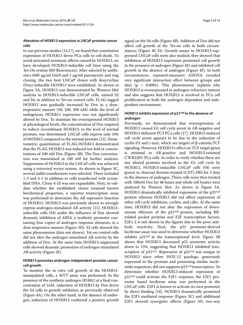
Alteration of HOXB13 expression in LNCaP prostate cancer cellsIn our previous studies [15,17], we found that constitutiveexpression of HOXB13 drove PCa cells to cell death. Toavoid unwanted xenotoxic effects caused by HOXB13, wehave developed HOXB13-inducible cell lines using theTet-On system (BD Biosciences). After selected by antibi-otics (600 μg/ml G418 and 1 μg/ml puromycin) and ringcloning, the two best LNCaP clones with doxycycline(Dox)-inducible HOXB13 were established. As shown inFigure 3A, HOXB13 was demonstrated by Western blotanalysis in HOXB13-inducible LNCaP cells, named S2and S4, in addition to Tet-on control cells. FLAG-taggedHOXB13 was gradually increased by Dox in a dose-responsive manner (50, 100, 200 nM) while the level ofendogenous HOXB13 expression was not significantlyaltered by Dox. To maintain the overexpressed HOXB13at physiological levels, the concentration of Dox requiredto induce recombinant HOXB13, to the level of normalprostate, was determined. LNCaP cells express only 10%of HOXB13 compared to the normal prostate [15]. Densi-tometric quantitation of FLAG-HOXB13 demonstratedthat the FLAG-HOXB13 was induced ten-fold at concen-trations of 100 nM Dox (Figure 3B). The Dox concentra-tion was maintained at 100 nM for further analyses.Suppression of HOXB13 in the LNCaP cells was achievedusing a retroviral vector system. As shown in Figure 3C,several stable transfectants were selected. These included1-3 and 4-1 in addition to cells transfected with scram-bled DNA. Clone 4-1S was not expandable. Next, to vali-date whether the established clones retained knownbiochemical properties, a reporter transcription assaywas performed to determine the AR repressive functionof HOXB13. HOXB13 was previously shown to stronglyrepress androgen-stimulated AR activity [15]. HOXB13-inducible cells (S4) under the influence of Dox showeddramatic inhibition of ARE4, a synthetic promoter con-taining four copies of androgen response elements, in adose responsive manner (Figure 3D). S2 cells showed thesame phenomenon (data not shown). Tet-on control cellsdid not alter the androgen-stimulated AR activity by theaddition of Dox. At the same time HOXB13-suppressedcells showed dramatic promotion of androgen-stimulatedAR activity (Figure 3E).
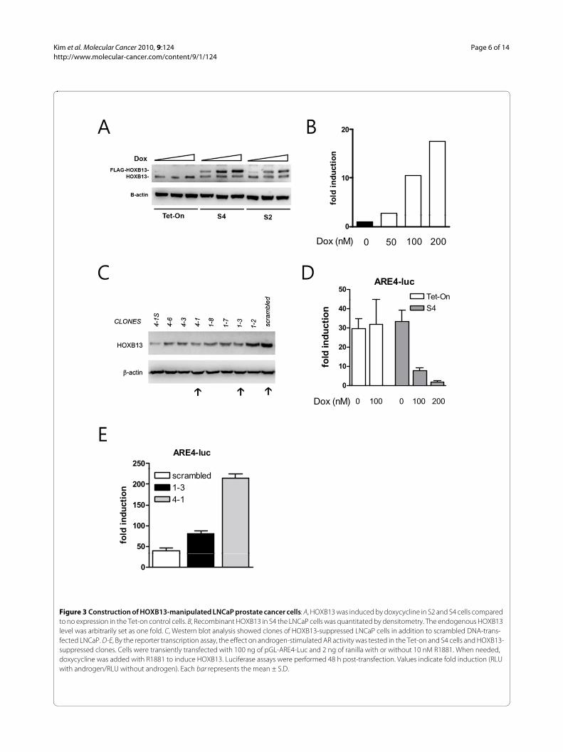
HOXB13 promotes androgen-independent prostate cancer cell growthTo monitor the in vitro cell growth of the HOXB13-manipulated cells, a MTT assay was performed. In thepresence of the synthetic androgen (R1881) at a final con-centration of 1nM, induction of HOXB13 by Dox drovethe S4 cells to growth inhibition as previously observed(Figure 4A). On the other hand, in the absence of andro-gen, induction of HOXB13 conferred a positive growth
signal on the S4 cells (Figure 4B). Addition of Dox did notaffect cell growth of the Tet-on cells in both circum-stances (Figure 4C-D). Growth assays in HOXB13-sup-pressed LNCaP cells were also studied; they showed thatinhibition of HOXB13 expression promoted cell growthin the presence of androgen (Figure 4E) and inhibited cellgrowth in the absence of androgen (Figure 4F). In bothcircumstances, repeated-measures ANOVA revealedvery significant interaction effect between groups anddays (p < 0.0001). This phenomenon explains whyHOXB13 is overexpressed in androgen-refractory tumorsand also suggests that HOXB13 is involved in PCa cellproliferation in both the androgen dependent and inde-pendent environment.
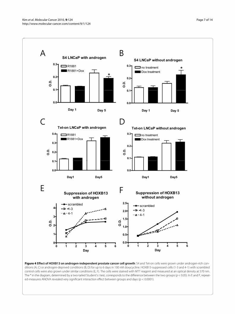
HOXB13 inhibits expression of p21waf in the absence of androgenPreviously, we demonstrated that overexpression ofHOXB13 caused G1 cell cycle arrest in AR-negative andHOXB13-deficient PC3 PCa cells [17]. HOXB13-inducedcell cycle arrest appears to be due to the reduction ofcyclin D1 and c-myc, which are targets of β-catenin/TCFsignaling. However, HOXB13's effect on TCF target genesis minimal in AR-positive and HOXB13-retainedCWR22RV PCa cells. In order to verify whether there areany altered proteins involved in the G1 cell cycle byHOXB13, HOXB13-inducible LNCaP cells (S4) weregrown in charcoal dextran-treated (CDT)-FBS for 3 daysin the absence of androgen. These cells were then treatedwith 100nM Dox for 48 hours and whole cell lysates wereanalyzed by Western blot. As shown in Figure 5A,HOXB13 dramatically inhibited expression of the p21waf
protein whereas HOXB13 did not affect expression ofother cell cycle inhibitors, cyclins, and cdks. At the sametime, HOXB13 did not alter the expression of down-stream effectors of the p21waf protein, including RB-related pocket proteins and E2F transcription factors.E2F1-2 is not shown in the figures due to the poor anti-body reactivity. Next, the p21 promoter-derivedluciferase assay was used to determine whether HOXB13inhibits p21waf at the transcriptional level. Figure 5Bshows that HOXB13 decreased p21 promoter activitydown to 15%, suggesting that HOXB13 inhibited tran-scription of p21waf. Repression of p21waf was unique toHOXB13 since other HOX-13 paralogs, generouslyexpressed in the prostate and possessing similar nucle-otide sequences, did not suppress p21waf transcription. Todetermine whether HOXB13-induced repression ofp21waf could activate the E2F1 response, the E2F1 pro-moter based luciferase assay was performed in theLNCaP cells. E2F1 is known to activate its own promoterby direct binding [18]. HOXB13 dramatically promotedthe E2F1-mediated response (Figure 5C) and additionalE2F1 showed synergistic effects (Figure 5D, two-way
Kim et al. Molecular Cancer 2010, 9:124http://www.molecular-cancer.com/content/9/1/124
Page 6 of 14
Figure 3 Construction of HOXB13-manipulated LNCaP prostate cancer cells: A, HOXB13 was induced by doxycycline in S2 and S4 cells compared to no expression in the Tet-on control cells. B, Recombinant HOXB13 in S4 the LNCaP cells was quantitated by densitometry. The endogenous HOXB13 level was arbitrarily set as one fold. C, Western blot analysis showed clones of HOXB13-suppressed LNCaP cells in addition to scrambled DNA-trans-fected LNCaP. D-E, By the reporter transcription assay, the effect on androgen-stimulated AR activity was tested in the Tet-on and S4 cells and HOXB13-suppressed clones. Cells were transiently transfected with 100 ng of pGL-ARE4-Luc and 2 ng of ranilla with or without 10 nM R1881. When needed, doxycycline was added with R1881 to induce HOXB13. Luciferase assays were performed 48 h post-transfection. Values indicate fold induction (RLU with androgen/RLU without androgen). Each bar represents the mean ± S.D.
20
10
20
fold
in
du
cti
on
0
Dox (nM) 0 50 100 200
50Tet-On
ARE4-luc
0
10
20
30
40 S4
fold
in
du
cti
on
ARE4-luc
250
0
Dox (nM) 0 100 0 200100
50
100
150
200 1-3scrambled
4-1
fold
in
du
cti
on
0
Kim et al. Molecular Cancer 2010, 9:124http://www.molecular-cancer.com/content/9/1/124
Page 7 of 14
Figure 4 Effect of HOXB13 on androgen-independent prostate cancer cell growth: S4 and Tet-on cells were grown under androgen-rich con-ditions (A, C) or androgen-deprived conditions (B, D) for up to 6 days in 100 nM doxycycline. HOXB13-suppressed cells (1-3 and 4-1) with scrambled control cells were also grown under similar conditions (E, F). The cells were stained with MTT reagent and measured at an optical density at 570 nm. The * in the diagram, determined by a two-tailed Student's t test, corresponds to the difference between the two groups (p < 0.05). In E and F, repeat-ed-measures ANOVA revealed very significant interaction effect between groups and days (p < 0.0001).
S4 LNCaP with androgen
0 1
0.2
0.3R1881R1881+Dox
*
O.D
.
S4 LNCaP without androgen
0 1
0.2
0.3
*no treatmentDox treatment
O.D
.
0.0
0.1
Day 1 Day 5
0.0
0.1
Day 1 Day 5
Tet-on LNCaP with androgen
0 1
0.2
0.3
0.4
R1881+DoxR1881
O.D
.
Tet-on LNCaP without androgen
0.1
0.2
0.3 no treatmentDox treatment
O.D
.
0.0
0.1
Day1 Day5
0.0
Day1 Day5
Suppression of HOXB13ith t d
Suppression of HOXB13without androgen
1.0
1.5
2.0
2.5
4-1
scrambled1-3
O.D
.
with androgen
2
3
4
4-1
scrambled1-3
O.D
.
0 1 2 3 4 5 60.0
0.5
Day
0 1 2 3 4 5 60
1
Day
Kim et al. Molecular Cancer 2010, 9:124http://www.molecular-cancer.com/content/9/1/124
Page 8 of 14
Figure 5 Effect of HOXB13 on the p21-RB-E2F signaling pathway: A, S4 cells were grown under hormone-deprived conditions for 3 days and in-duced for HOXB13 with 100nM doxycycline. Extracted lysates were evaluated for Western blot analysis. LNCaP cells were grown under hormone-de-prived condition for 3 days. B, Cells were transfected with p21-luc and pFLAG-HOXB13, pCDNA-HOXA13, or pCMV-HOXD13. C, Cells were transfected with pE2F1-luc and pFLAG-HOXB13. D, Cells were transfected with pE2F1-luc, pFLAG-HOXB13, and pCDNA-E2F1. Corresponding control vectors were used to match each DNA. After 48 hours, the cells were evaluated by luciferase assays. Each bar represents the mean ± S.D.
p21-luc; LNCaP
0.6
0.7
0.8
0.9
0.0
0.1
0.2
0.3
0.4
0.5
HOXB13
HOXA13
HOXD13
+-+-
+
RL
U
HOXD13 +-
pE2F1-luc; LNCaPpE2F1 luc; LNCaP
0.02
0.03
0.04
0.05
0.06
0.07
RL
U
LNCaP; pE2F1-luc
20000
30000
40000
50000
60000
70000
RL
U-b
gal
0.00
0.01
HOXB13 +-0
10000
HOXB13
E2F1
_ _+ +
_ _ + +
Kim et al. Molecular Cancer 2010, 9:124http://www.molecular-cancer.com/content/9/1/124
Page 9 of 14
ANOVA: F = 12.44, p < 0.01). These results suggest thatHOXB13 is involved in the activation of the oncogenicE2F signal by downregulation of p21waf expression.
Promotion of the E2F signal by HOXB13 was mainly due to p21waf repressionSince HOXB13's effect on E2F did not necessarily comefrom the regulation of the expression of p21waf, we testedwhether HOXB13 affects E2F response elements by wayof p21waf. First, we tested whether HOXB13's effect onthe E2F1 promoter was mediated through the E2F1 bind-ing sites. By using three copies of simple E2F1 responseelements, HOXB13 and E2F1also showed additive effectsof the E2F1 response (Figure 6A), suggesting that E2F1promotion by HOXB13 was mediated by the E2F1response elements. Next, we studied HCT116 colon can-cer cells with either wild type or deleted p21waf. Both cellswere transfected with pE2F1-luc and HOXB13. WhileHCT116 (p21+/+) showed a dramatic increase in E2Factivity, in a dose responsive manner (up to 14 foldincrease), HCT116 (p21-/-) cells showed minimal pro-moting effects (up to 4 fold increase) (Figure 6B). In addi-tion, increase of p21 gradually decreased the E2F activitystimulated by HOXB13 in a dose responsive manner inthe HCT116 (p21+/+) cells (Figure 6C). At maximumexpression of p21, however, the activity did not com-pletely return to basal levels. These results suggest thatthere is a minor p21-independent activation of E2F1 byHOXB13. To verify the alteration of the E2F1 target genesby HOXB13, Western blot and RT-PCR analyses wereperformed (Figure 6D). HOXB13 up-regulated expres-sion of the p107 pocket protein, which is a target of E2F-mediated transactivation [19]. Since E2F1 protein expres-sion is known to be barely detectable in cells grown underhormone-deprived conditions [20], RT-PCR analysisshowed that HOXB13 stimulated expression of E2F1.These results suggest that HOXB13's promoting effect onE2F signaling mainly comes from a p21-dependent path-way.
DiscussionHOXB13 is a highly specific prostate homeobox proteinand is involved in the formation of this hormone-domi-nant organ [5,7,8,15]. Functionally, Hoxb13 loss-of-func-tion mice show swollen prostate glands [8]. Involvementof HOXB13 during carcinogenesis of human tissues hasbeen reported by many groups [21-24]. Many reportshave published findings supporting a link between andseveral solid tumors, including breast cancer, cervicalcancer, ovarian cancer, and skin cancers [16,24-29]. Inbreast cancer, HOXB13 was found to be overexpressed inhormone-refractory tumors compared to hormone-responsive tumors, further suggesting that HOXB13might be a useful prognostic marker for hormone-refrac-
tory breast cancers [26]. In addition, the forced expres-sion of HOXB13 promoted the growth of MCF10Aestrogen receptor (ER)-negative breast cells along withincreased migration and invasion. In this report, we havedemonstrated that HOXB13 was also overexpressed inhormone-refractory prostate tumors; its expression pro-vided a positive growth signal in the absence of androgen.Our previous reports showed no expression of HOXB13in only PC3 and DU145 cells [15]. While these cells aredescribed as androgen-independent prostate cancer cells,they are generally considered as 'non-prostate-like cancercells' mainly due the deficiency of androgen receptors(AR). In addition, there was positive correlation betweenHOXB13 and AR with consequent functional counterac-tion between these two proteins, suggesting thatHOXB13 functions as a PCa cell growth suppressor in thepresence of androgen [15]. HOXB13 antagonizes thefunction of the androgen-stimulated AR signal by dis-rupting coactivator formation. Although the prognosticvalue of the HOXB13, for the progression of PCa,requires further study, it is surprising that the function ofHOXB13 is paradoxically opposite, depending on the sta-tus of androgen. In fact, tumor suppressor NKX3.1homeodomain protein is overexpressed in metastaticprostate cancer [30]. In ovarian cancer, HOXB13 pro-motes cancer cell proliferation and progression [28].Taken together, the balance between HOXB13 and theAR is important for the modulation of growth in PCacells. In other words, HOXB13 expression must be dereg-ulated for the survival of androgen-independent tumorswhile its expression is highly maintained in tumors withhigh androgen responsiveness.
Some HOX proteins are involved in the progression ofPCa. Both HOXC6 and HOXC8 have been shown to beoverexpressed in more advanced metastatic and recur-rent PCa [31,32]. Despite the high expression of HOXB13in the normal prostate, alteration of HOXB13 expressionduring the transformation process remains controversial.Some studies suggest overexpression of HOXB13 in pros-tate tumors [13,33], while others reported no such change[15,34]. Using total RNA we did not observe a differentialexpression of HOXB13 between the benign and malig-nant tumors; this was likely due to the multifocal natureof PCa [15]. In addition, immunohistochemical stainingdid not demonstrate alteration of HOXB13 during thetumorigenic process (unpublished data). Suppression ordown-regulation of HOXB13 has been observed in othertumors, including colorectal cancer and skin tumors [22-24,35], although the level of HOXB13 protein was notdetectable in normal skin. It is believed that the expres-sion pattern and role of HOXB13 can be significantly dif-ferent in breast cancers and PCa based on the followingobservations: 1) Breast tissues are HOXB13-negative asreported by many groups [6,14-16]. Accordingly, all cul-

Kim et al. Molecular Cancer 2010, 9:124http://www.molecular-cancer.com/content/9/1/124
Page 10 of 14
tured breast cancer cells, including several MCF7,MCF10a, T47D, and MDA 231, showed no expression oronly minimally expressed HOXB13 [16,36]. 2) WhileHOXB13 and estrogen signals can be mutually regulatedin breast cancer [21], there is no mutual regulation ofHOXB13 and the androgen receptor in PCa [15]. 3)HOXB13 promotes growth of MCF10A cells, ER-negative
normal breast cells [26] but HOXB13 inhibits AR-nega-tive PC3 PCa cells by regulating β-catenin/TCF signals[17]. Therefore, the role of HOXB13 is variable depend-ing on the cell type itself and the cell environment.
This study demonstrated that HOXB13 was overex-pressed in androgen-refractory prostate tumors and wasinvolved in providing a positive growth signal to PCa cells
Figure 6 Effect of HOXB13 on p21-dependent E2F1-mediated transactivation: A, LNCaP cells were grown under hormone-deprived conditions for 3 days and transfected with pE2F1(3X)-luc, pFLAG-HOXB13, and pCDNA-E2F1. B, Both HCT116 (p21+/+) and HCT116 (p21-/-) cells were transfected with pE2F1-luc and an increased amount of pFLAG-HOXB13. C, HCT116 (p21+/+) cells were transfected with pE2F1-luc, pFLAG-HOXB13, and an in-creased amount of pFLAG-p21. Corresponding control vectors were used to match each DNA. After 48 hours, the cells were evaluated for luciferase assays. Each bar represents the mean ± S.D. D, S4 cells were treated with Dox to induce HOXB13 followed by Western blot and RT-PCR analyses.
� �
�
�����
�����
�����
�����
�����
�����
������
���
� � �������
����� ����� ��������
���
��������
���
���
���
���
���
���
������
�� �����������
�� �����������
���
�
��� �� ����������
�
����
����
����
����
����
����
����
������
����
� �� �� � � �
����!"��
Kim et al. Molecular Cancer 2010, 9:124http://www.molecular-cancer.com/content/9/1/124
Page 11 of 14
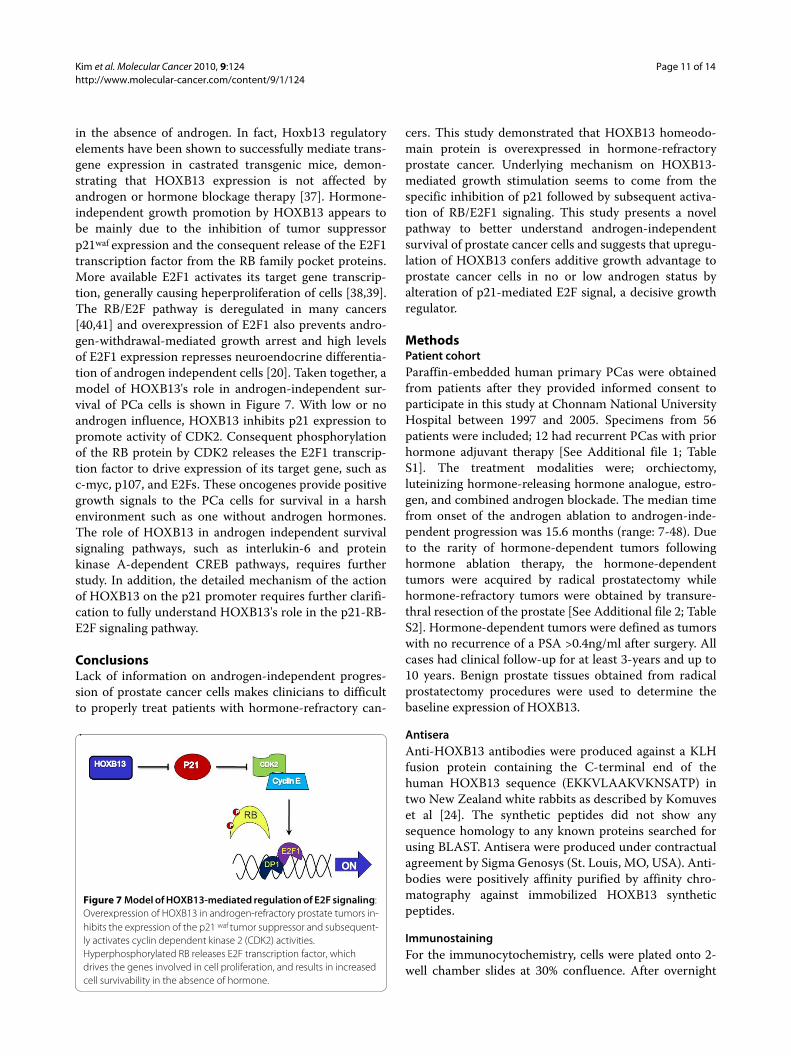
in the absence of androgen. In fact, Hoxb13 regulatoryelements have been shown to successfully mediate trans-gene expression in castrated transgenic mice, demon-strating that HOXB13 expression is not affected byandrogen or hormone blockage therapy [37]. Hormone-independent growth promotion by HOXB13 appears tobe mainly due to the inhibition of tumor suppressorp21waf expression and the consequent release of the E2F1transcription factor from the RB family pocket proteins.More available E2F1 activates its target gene transcrip-tion, generally causing heperproliferation of cells [38,39].The RB/E2F pathway is deregulated in many cancers[40,41] and overexpression of E2F1 also prevents andro-gen-withdrawal-mediated growth arrest and high levelsof E2F1 expression represses neuroendocrine differentia-tion of androgen independent cells [20]. Taken together, amodel of HOXB13's role in androgen-independent sur-vival of PCa cells is shown in Figure 7. With low or noandrogen influence, HOXB13 inhibits p21 expression topromote activity of CDK2. Consequent phosphorylationof the RB protein by CDK2 releases the E2F1 transcrip-tion factor to drive expression of its target gene, such asc-myc, p107, and E2Fs. These oncogenes provide positivegrowth signals to the PCa cells for survival in a harshenvironment such as one without androgen hormones.The role of HOXB13 in androgen independent survivalsignaling pathways, such as interlukin-6 and proteinkinase A-dependent CREB pathways, requires furtherstudy. In addition, the detailed mechanism of the actionof HOXB13 on the p21 promoter requires further clarifi-cation to fully understand HOXB13's role in the p21-RB-E2F signaling pathway.
ConclusionsLack of information on androgen-independent progres-sion of prostate cancer cells makes clinicians to difficultto properly treat patients with hormone-refractory can-
cers. This study demonstrated that HOXB13 homeodo-main protein is overexpressed in hormone-refractoryprostate cancer. Underlying mechanism on HOXB13-mediated growth stimulation seems to come from thespecific inhibition of p21 followed by subsequent activa-tion of RB/E2F1 signaling. This study presents a novelpathway to better understand androgen-independentsurvival of prostate cancer cells and suggests that upregu-lation of HOXB13 confers additive growth advantage toprostate cancer cells in no or low androgen status byalteration of p21-mediated E2F signal, a decisive growthregulator.
MethodsPatient cohortParaffin-embedded human primary PCas were obtainedfrom patients after they provided informed consent toparticipate in this study at Chonnam National UniversityHospital between 1997 and 2005. Specimens from 56patients were included; 12 had recurrent PCas with priorhormone adjuvant therapy [See Additional file 1; TableS1]. The treatment modalities were; orchiectomy,luteinizing hormone-releasing hormone analogue, estro-gen, and combined androgen blockade. The median timefrom onset of the androgen ablation to androgen-inde-pendent progression was 15.6 months (range: 7-48). Dueto the rarity of hormone-dependent tumors followinghormone ablation therapy, the hormone-dependenttumors were acquired by radical prostatectomy whilehormone-refractory tumors were obtained by transure-thral resection of the prostate [See Additional file 2; TableS2]. Hormone-dependent tumors were defined as tumorswith no recurrence of a PSA >0.4ng/ml after surgery. Allcases had clinical follow-up for at least 3-years and up to10 years. Benign prostate tissues obtained from radicalprostatectomy procedures were used to determine thebaseline expression of HOXB13.
AntiseraAnti-HOXB13 antibodies were produced against a KLHfusion protein containing the C-terminal end of thehuman HOXB13 sequence (EKKVLAAKVKNSATP) intwo New Zealand white rabbits as described by Komuveset al [24]. The synthetic peptides did not show anysequence homology to any known proteins searched forusing BLAST. Antisera were produced under contractualagreement by Sigma Genosys (St. Louis, MO, USA). Anti-bodies were positively affinity purified by affinity chro-matography against immobilized HOXB13 syntheticpeptides.
ImmunostainingFor the immunocytochemistry, cells were plated onto 2-well chamber slides at 30% confluence. After overnight
Figure 7 Model of HOXB13-mediated regulation of E2F signaling: Overexpression of HOXB13 in androgen-refractory prostate tumors in-hibits the expression of the p21 waf tumor suppressor and subsequent-ly activates cyclin dependent kinase 2 (CDK2) activities. Hyperphosphorylated RB releases E2F transcription factor, which drives the genes involved in cell proliferation, and results in increased cell survivability in the absence of hormone.

Kim et al. Molecular Cancer 2010, 9:124http://www.molecular-cancer.com/content/9/1/124
Page 12 of 14
incubation, the cells were fixed with methanol/acetonesolution for 15 minutes. Then, the cells were treated with0.2% Triton X100 in PBS for 10 minutes. Anti-HOXB13antibodies in 2% BSA/PBS were added to the slides for 1hour. After washing, the slides were incubated with don-key anti-rabbit IgG (Fab fragment) conjugated to peroxi-dase (Jackson Laboratories). Colorimetric signals weredetected using diaminobenzidine (DAB). Sections werecounterstained with hematoxylin for microscopic evalua-tion. Immunohistochemistry was performed as describedpreviously [24]. Tissues were deparaffinized followed bymicrowave antigen retrieval in citrate buffer. Endogenousperoxidase activity was destroyed by the treating tissuesections with 3% H2O2. After nonspecific reactivity wassequentially blocked by an Avidin-Biotin BlockingReagent and 10% normal goat serum, the tissues wereincubated with anti-HOXB13 antibodies and then withdonkey anti-rabbit IgG (Fab fragment) conjugated to per-oxidase (Jackson Laboratories). Colorimetric signals weredetected using DAB. Sections were counterstained withhematoxylin for microscopic evaluation. Anti-androgenreceptor antibodies (Upstate) were used to confirm tissueintegrity. For the negative control slide, non-immune rab-bit IgG was used. The immunostained slides were evalu-ated by two different investigators, including apathologist, blinded to the patient's clinical features. Theintensity of the HOXB13 staining was classified into oneof four grades (0, absent; 1, weak; 2, intermediate; and 3,strong). Tumors with less than 5% HOXB13 expressionwere considered negative regardless of the intensitydescribed [42]. This approach has been previously deter-mined to be reliable and reproducible by several groups[43-45].
Cell cultureHuman PCa cell lines, LNCaP, PC3, and DU145, wereroutinely cultured in RPMI media (Invitrogen) supple-mented with 5% FBS at 37°C in an atmosphere containing5% CO2. MDA PCa 2b cells were grown in BRFF-HPC1medium (Athena Environmental Sciences, Inc., Balti-more, MD, USA) with 20% FBS. Wild type HCT116(p21+/+) and HCT116 (p21-/-) were kindly provided byDr. Bert Vogelstein and have been well described previ-ously [46]. All cultures were fed with fresh medium every3-4 days.
Plasmids and reagentsThe pFLAG-HOXB13 and pGL-ARE4-Luc have beenpreviously described [17]. The p21-luc, incorporating a2.4 kb genomic fragment from the p21 promoter waskindly provided by Dr. Vogelstein. The pFLAG-p21 vec-tor was constructed by PCR cloning. The pCMV-E2F1was kindly provided by Dr. Hong-Wu Chen (UC Davis).The pE2F1-luc was provided by Dr. Chihuei Wang (Kaoh-
siung Medical University, Taiwan). pCMV-Hoxa13 andpCDNA-Hoxd13 were constructed from pMiw-Hoxa13and pBK-Hoxd13, respectively, both of which were ofmouse origin and provided by Dr. Atsushi Kuroiwa(Nagoya University, Japan). Antibodies to p16, p18, p21,p27, CDK2, CDK4, CDK6, CDK8, Cyclin D1, D2, D3, E,p107, RB, p130, E2F1-5, DP1-2, the androgen receptorand β-actin were from Santa Cruz Biotechnology. Anti-FLAG M5 antibodies and doxycycline were from Sigma.pTet-on, pTRE-luc, and pTRE-puro were from BD Biosci-ences. Synthetic testosterone, R1881, was from NEN LifeScience (Boston, MA, USA) and used at a final concen-tration of 10 nM. Both charcoal dextran-treated (CDT)FBS and tetracycline-free FBS were from Invitrogen.
Transient transfectionsApproximately 1 × 10⁵ cells were plated in a 24-well plate16 hours before transfection. To determine the hormoneeffects, cells were grown under 5% charcoal dextran-treated (CDT) FBS for three days before the transfection.Transfections were carried out using Lipofectamine 2000(Invitrogen) with 0.1 μg of reporter, 0.1 μg of test plasmid,and 2 ng of ranilla as described by the manufacturer'sprotocol. Six hours after transfection, the cells werewashed and fed with medium containing 5% CDT-FBS. Ifneeded, the cells were treated with either 10 nM R1881synthetic androgen or ethanol. After 36 hours, the cellswere washed with PBS, lysed with 100 μl of passive lysisbuffer, and assayed for luciferase activity as relative lightunits (RLU) using a dual luciferase assay system (Pro-mega). Transfection experiments were performed in trip-licate and the results are reported as mean ± S.D.
Stable transfectionsUsing the Tet-On system (BD Biosciences), the LNCaPPCa cells were transfected with Tet-on and a single clonewas obtained by ring cloning and G418 selection (600 μg/ml; Invitrogen). Inducibility of the resulting clones wastested by transient transfection using pTRE-luc in thepresence of doxycycline (Sigma). A second transfectionwas performed with pTRE-puro expressing FLAG-taggedHOXB13, followed by ring cloning and puromycin selec-tion (1 μg/ml). The two best clones in terms of growingability and protein inducibility were selected, namely S2and S4. The Tet-on transfected cells were used as controlcells. To suppress the HOXB13 in the LNCaP cells, shorthairpin oligonucleotides were generated from either the5' or 3' end of the HOXB13 [17]. The DNAs were ligatedinto the H1 RNA gene promoter-based vector, pSU-PER.retro (DNA Engine, Seattle, WA, USA). The result-ing vector was infected into PA316 packaging cells toproduce retroviral supernatants. After infection of theLNCaP cells, viral plaques were isolated by a ring cloneand selected by 1 μg/ml puromycin.
Kim et al. Molecular Cancer 2010, 9:124http://www.molecular-cancer.com/content/9/1/124
Page 13 of 14
Western Blotting AssayCells were grown to 50% confluence in P60 culture dishescontaining 5% FBS-RPMI media. If necessary, the cellswere grown in RPMI media containing either 5% tet-freeFBS or CDT-FBS. The cells were then lysed in proteinextraction buffer (1x TBS, 1% NP-40, 0.5% sodium deoxy-cholate, 0.1% SDS and protease inhibitors) followed byneedle sonication to break up the ribonucleosome.Twenty μg of total cell lysates were loaded onto 10% Bis-Tris gel (Invitrogen) and separated using a Biorad elec-troporation system. After the proteins were transferred toa PVDF membrane, the primary antibodies were applied,followed by incubation with horse peroxidase-conjugatedsecondary antibodies. The blots were developed using theECL detection system (Pierce).
RT-PCRRNA extraction from the cells was performed as previ-ously described [47]. RT-PCR was performed to verify theexpression of HOXB13, E2F1, and β-actin. The primersequences for each gene were as follows: HOXB13fw, 5'-ccccactgagtttgccttctatc-3'; HOXB13rv, 5'-gcctcttgtccttg-gtgatgaac-3'; E2F1fw, 5'-aggccgccatccaggaaaag-3'; E2F1rv,5'-ggatgccctcaacgacgttg -3'; β-actinfw, 5'-gcaccacaccttcta-caatgagc-3'; β-actinrv, 5'-tagcacagcctggatagcaacg-3'.
Growth AssaysThe MTT in vitro proliferation assay was performed aspreviously described [17]. Briefly, the cells were grown inRPMI media containing 5% CDT-FBS for three days andthen plated on 24-well plates at 30% confluence. The nextday, if needed, the cells were treated with 100nM doxycy-cline and/or 1nM R1881 and grown for up to 7 days.Then, the cells were stained with 100 μl of 5 mg/ml MTTsolution and incubated for 2 hours at 37°C. The reactionwas stopped by adding 400 μl of extraction buffer (50%formamide and 10% SDS, pH 4.7). After overnight incu-bation at 37°C, the absorbance at 570 nm was measuredusing a microplate reader with SOFTmax PRO software(Molecular Devices, Sunnyvale, CA, USA). Densitomet-ric values were analyzed with the Student's t test, usingGraphPad Prism software (San Diego, CA, USA).
List of Abbreviations UsedPCa: prostate cancer; AR: androgen receptor; AI: andro-gen-independent; AD: androgen-dependent; Dox: doxy-cyline; CDT: charcoal dextran-treated; ER: estrogenreceptor; DAB: diaminobenzidine; RLU: relative lightunits; MTT: 3-(4,5-dimethylthiazol-2-yl)-2,5-diphe-nyltetrazolium bromide.
Additional material
Competing interestsThe authors declare that they have no competing interests.
Authors' contributionsYRK performed experiments and data analysis; KJO provided patient samplesand evaluated data; RYP and NTXN performed experiments; DDK and TWKevaluated patient information; CC, KIN and KYA contributed to immunohis-tochemical scoring; MSK provided statistical assistance; CJ performed experi-ments, data analysis and wrote the manuscript. All authors read and approvedthe final manuscript.
AcknowledgementsThis study was supported by grants from the National R&D Program for Cancer Control, Ministry for Health, Welfare and Family Affairs, Republic of Korea (#0720120), the Korea Research Foundation Grant funded by the Korean Gov-ernment (MOEHRD, Basic Research Promotion Fund) (KRF-2007-313-E00353), and the Korea Science & Engineering Foundation through the Medical Research Center for Gene Regulation (R13-2002-013-04002-0) at Chonnam National University.
Author Details1Department of Anatomy, Chonnam National University Medical School, Gwangju, Korea, 2Department of Urology, Chonnam National University Medical School, Gwangju, Korea, 3Department of Pathology, Chonnam National University Medical School, Gwangju, Korea, 4Department of Statistics, Chonnam National University, Gwangju, Korea and 5Research Institute of Medical Sciences, Chonnam National University, Gwangju, Korea
References1. Podlasek CA, Clemens JQ, Bushman W: Hoxa-13 gene mutation results in
abnormal seminal vesicle and prostate development. J Urol 1999, 161:1655-1661.
2. Podlasek CA, Duboule D, Bushman W: Male accessory sex organ morphogenesis is altered by loss of function of Hoxd-13. Dev Dyn 1997, 208:454-465.
3. Podlasek CA, Seo RM, Clemens JQ, Ma L, Maas RL, Bushman W: Hoxa-10 deficient male mice exhibit abnormal development of the accessory sex organs. Dev Dyn 1999, 214:1-12.
4. Zeltser L, Desplan C, Heintz N: Hoxb-13: a new Hox gene in a distant region of the HOXB cluster maintains colinearity. Development 1996, 122:2475-2484.
5. Sreenath T, Orosz A, Fujita K, Bieberich CJ: Androgen-independent expression of hoxb-13 in the mouse prostate. Prostate 1999, 41:203-207.
6. Hood L, Heath JR, Phelps ME, Lin B: Systems biology and new technologies enable predictive and preventative medicine. Science 2004, 306:640-643.
7. Economides KD, Zeltser L, Capecchi MR: Hoxb13 mutations cause overgrowth of caudal spinal cordand tail vertebrae. Dev Biol 2003, 256:317-33.
8. Economides KD, Capecchi MR: Hoxb13 is required for normal differentiation and secretory function of the ventral prostate. Development 2003, 130:2061-2069.
9. Golias C, Iliadis I, Peschos D, Charalabopoulos K: Amplification and co-regulators of androgen receptor gene in prostate cancer. Exp Oncol 2009, 31:3-8.
10. Nieto M, Finn S, Loda M, Hahn WC: Prostate cancer: Re-focusing on androgen receptor signaling. Int J Biochem Cell Biol 2007, 39:1562-1568.
11. Kokontis J, Takakura K, Hay N, Liao S: Increased androgen receptor activity and altered c-myc expression in prostate cancer cells after long-term androgen deprivation. Cancer Res 1994, 54:1566-1573.
12. Linja MJ, Savinainen KJ, Saramaki OR, Tammela TL, Vessella RL, Visakorpi T: Amplification and overexpression of androgen receptor gene in hormone-refractory prostate cancer. Cancer Res 2001, 61:3550-3555.
Additional file 1 Hormone-refractory prostate cancer patients cohort.
Additional file 2 Hormone-dependent prostate cancer patients.
Received: 24 February 2010 Accepted: 27 May 2010 Published: 27 May 2010This article is available from: http://www.molecular-cancer.com/content/9/1/124© 2010 Kim et al; licensee BioMed Central Ltd. This is an Open Access article distributed under the terms of the Creative Commons Attribution License (http://creativecommons.org/licenses/by/2.0), which permits unrestricted use, distribution, and reproduction in any medium, provided the original work is properly cited.Molecular Cancer 2010, 9:124
Kim et al. Molecular Cancer 2010, 9:124http://www.molecular-cancer.com/content/9/1/124
Page 14 of 14
13. Feber A, Clark J, Goodwin G, Dodson AR, Smith PH, Fletcher A, Edwards S, Flohr P, Falconer A, et al.: Amplification and overexpression of E2F3 in human bladder cancer. Exp Cell Res 2004, 293:144-153.
14. Takahashi Y, Hamada J, Murakawa K, Takada M, Tada M, Nogami I, Hayashi N, Nakamori S, Monden M, Miyamoto M, et al.: Expression profiles of 39 HOX genes in normal human adult organs and anaplastic thyroid cancer cell lines by quantitative real-time RT-PCR system. Exp Cell Res 2004, 293:144-153.
15. Jung C, Kim RS, Zhang HJ, Lee SJ, Jeng MH: HOXB13 induces growth suppression of prostate cancer cells as a repressor of hormone-activated androgen receptor signaling. Cancer Res 2004, 64:9185-9192.
16. Edwards S, Campbell C, Flohr P, Shipley J, Giddings I, Te-Poele R, Dodson Y, Foster C, Clark J, Jhavar S, et al.: Expression analysis onto microarrays of randomly selected cDNA clones highlights HOXB13 as a marker of human prostate cancer. Br J Cancer 2005, 92:376-381.
17. Jung C, Kim RS, Lee SJ, Wang C, Jeng MH: HOXB13 Homeodomain Protein Suppresses the Growth of Prostate Cancer Cells by the Negative Regulation of T-Cell Factor 4. Cancer Res 2004, 64:3046-3051.
18. Bracken AP, Ciro M, Cocito A, Helin K: E2F target genes: unraveling the biology. Trends Biochem Sci 2004, 29:409-417.
19. Hurford RK Jr, Cobrinik D, Lee MH, Dyson N: pRB and p107/p130 are required for the regulated expression of different sets of E2F responsive genes. Genes Dev 1997, 11:1447-1463.
20. Libertini SJ, Tepper CG, Guadalupe M, Lu Y, Asmuth DM, Mudryj M: E2F1 expression in LNCaP prostate cancer cells deregulates androgen dependent growth, suppresses differentiation, and enhances apoptosis. Prostate 2006, 66:70-81.
21. Rodriguez BA, Cheng AS, Yan PS, Potter D, Agosto-Perez FJ, Shapiro CL, Huang TH: Epigenetic repression of the estrogen-regulated Homeobox B13 gene in breast cancer. Carcinogenesis 2008, 29:1459-1465.
22. Hoek K, Rimm DL, Williams KR, Zhao H, Ariyan S, Lin A, Kluger HM, Berger AJ, Cheng E, Trombetta ES, et al.: Expression profiling reveals novel pathways in the transformation of melanocytes to melanomas. Cancer Res 2004, 64:5270-5282.
23. Jung C, Kim RS, Zhang H, Lee SJ, Sheng H, Loehrer PJ, Gardner TA, Jeng MH, Kao C: HOXB13 is downregulated in colorectal cancer to confer TCF4-mediated transactivation. Br J Cancer 2005, 92:2233-2239.
24. Komuves LG, Ma XK, Stelnicki E, Rozenfeld S, Oda Y, Largman C: HOXB13 homeodomain protein is cytoplasmic throughout fetal skin development. Dev Dyn 2003, 227:192-202.
25. Zhao Y, Yamashita T, Ishikawa M: Regulation of tumor invasion by HOXB13 gene overexpressed in human endometrial cancer. Oncol Rep 2005, 13:721-726.
26. Ma XJ, Wang Z, Ryan PD, Isakoff SJ, Barmettler A, Fuller A, Muir B, Mohapatra G, Salunga R, Tuggle JT, et al.: A two-gene expression ratio predicts clinical outcome in breast cancer patients treated with tamoxifen. Cancer Cell 2004, 5:607-616.
27. Reid JF, Lusa L, De Cecco L, Coradini D, Veneroni S, Daidone MG, Gariboldi M, Pierotti MA: Limits of predictive models using microarray data for breast cancer clinical treatment outcome. J Natl Cancer Inst 2005, 97:927-930.
28. Miao J, Wang Z, Provencher H, Muir B, Dahiya S, Carney E, Leong CO, Sgroi DC, Orsulic S: HOXB13 promotes ovarian cancer progression. Proc Natl Acad Sci USA 2007, 104:17093-17098.
29. Okuda H, Toyota M, Ishida W, Furihata M, Tsuchiya M, Kamada M, Tokino T, Shuin T: Epigenetic inactivation of the candidate tumor suppressor gene HOXB13 in human renal cell carcinoma. Oncogene 2006, 25:1733-1742.
30. Bowen C, Bubendorf L, Voeller HJ, Slack R, Willi N, Sauter G, Gasser TC, Koivisto P, Lack EE, Kononen J, et al.: Loss of NKX3.1 expression in human prostate cancers correlates with tumor progression. Cancer Res 2000, 60:6111-6115.
31. Singh D, Febbo PG, Ross K, Jackson DG, Manola J, Ladd C, Tamayo P, Renshaw AA, D'Amico AV, Richie JP, et al.: Gene expression correlates of clinical prostate cancer behavior. Cancer Cell 2002, 1:203-209.
32. Waltregny D, Alami Y, Clausse N, de Leval J, Castronovo V: Overexpression of the homeobox gene HOXC8 in human prostate cancer correlates with loss of tumor differentiation. Prostate 2002, 50:162-169.
33. Johnson PH, Walker RP, Jones SW, Stephens K, Meurer J, Zajchowski DA, Luke MM, Eeckman F, Tan Y, Wong L, et al.: Multiplex gene expression analysis for high-throughput drug discovery: screening and analysis of
compounds affecting genes overexpressed in cancer cells. Mol Cancer Ther 2002, 1:1293-1304.
34. Fromont G, Chene L, Latil A, Bieche I, Vidaud M, Vallancien G, Mangin P, Fournier G, Validire P, Cussenot O: Molecular profiling of benign prostatic hyperplasia using a large scale real-time reverse transcriptase-polymerase chain reaction approach. J Urol 2004, 172:1382-1385.
35. Birkenkamp-Demtroder K, Olesen SH, Sorensen FB, Laurberg S, Laiho P, Aaltonen LA, Orntoft TF: Differential gene expression in colon cancer of the caecum versus the sigmoid and rectosigmoid. Gut 2005, 54:374-384.
36. Clark J, Edwards S, John M, Flohr P, Gordon T, Maillard K, Giddings I, Brown C, Bagherzadeh A, Campbell C, et al.: Identification of amplified and expressed genes in breast cancer by comparative hybridization onto microarrays of randomly selected cDNA clones. Genes Chromosomes Cancer 2002, 34:104-114.
37. McMullin RP, Mutton LN, Bieberich CJ: Hoxb13 regulatory elements mediate transgene expression during prostate organogenesis and carcinogenesis. Dev Dyn 2009, 238:664-672.
38. Adams PD, Kaelin WG Jr: The cellular effects of E2F overexpression. Curr Top Microbiol Immunol 1996, 208:79-93.
39. Johnson DG, Schwarz JK, Cress WD, Nevins JR: Expression of transcription factor E2F1 induces quiescent cells to enter S phase. Nature 1993, 365:349-352.
40. Hanahan D, Weinberg RA: The hallmarks of cancer. Cell 2000, 100:57-70.41. Sherr CJ: Cancer cell cycles. Science 1996, 274:1672-1677.42. Foster CS, Falconer A, Dodson AR, Norman AR, Dennis N, Fletcher A,
Southgate C, Dowe A, Dearnaley D, Jhavar S, et al.: Transcription factor E2F3 overexpressed in prostate cancer independently predicts clinical outcome. Oncogene 2004, 23:5871-5879.
43. Dhanasekaran SM, Barrette TR, Ghosh D, Shah R, Varambally S, Kurachi K, Pienta KJ, Rubin MA, Chinnaiyan AM: Delineation of prognostic biomarkers in prostate cancer. Nature 2001, 412:822-826.
44. Rhodes DR, Sanda MG, Otte AP, Chinnaiyan AM, Rubin MA: Multiplex biomarker approach for determining risk of prostate-specific antigen-defined recurrence of prostate cancer. J Natl Cancer Inst 2003, 95:661-668.
45. Rubin MA, Zhou M, Dhanasekaran SM, Varambally S, Barrette TR, Sanda MG, Pienta KJ, Ghosh D, Chinnaiyan AM: alpha-Methylacyl coenzyme A racemase as a tissue biomarker for prostate cancer. Jama 2002, 287:1662-1670.
46. Waldman T, Kinzler KW, Vogelstein B: p21 is necessary for the p53-mediated G1 arrest in human cancer cells. Cancer Res 1995, 55:5187-5190.
47. Jung C, Ou YC, Yeung F, Frierson HF Jr, Kao C: Osteocalcin is incompletely spliced in non-osseous tissues. Gene 2001, 271:143-150.
doi: 10.1186/1476-4598-9-124Cite this article as: Kim et al., HOXB13 promotes androgen independent growth of LNCaP prostate cancer cells by the activation of E2F signaling Molecular Cancer 2010, 9:124