respiration: ventilation, circulation, &...
TRANSCRIPT
1
© Copyright 2012, John P. Fisher, All Rights Reserved
Respiration: Ventilation, Circulation, & Transport
Adapted From:
Textbook Of Medical Physiology, 11th Ed. Arthur C. Guyton, John E. Hall
Chapters 37, 38, 39, & 40
John P. Fisher
© Copyright 2012, John P. Fisher, All Rights Reserved
Overview of Respiration Introduction
• Respiration provides oxygen to tissues for the metabolism of fats, carbohydrates, and proteins, while removing the waste carbon dioxide that results from these metabolic reactions
• Respiration can be divided into four functions • Pulmonary ventilation - the inflow and outflow of air between the atmosphere
and lung alveoli • Gas diffusion in the lung - the transport of oxygen and carbon dioxide between
the alveoli and the blood • Gas diffusion in the tissues - the transport of oxygen and carbon dioxide
between the blood and tissues • Regulation of ventilation - the control systems that regulate these mechanisms
2
© Copyright 2012, John P. Fisher, All Rights Reserved
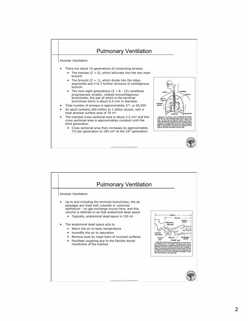
Pulmonary Ventilation Alveolar Ventilation
• There are about 16 generations of conducting airways • The trachea (Z = 0), which bifurcate into the two main
bronchi • The bronchi (Z = 1), which divide into the lobar,
segmental and 4 to 5 further divisions of cartilaginous bronchi
• The next eight generations (Z = 8 - 15) constitute progressively smaller, ciliated noncartilaginous bronchioles, the last of which is the terminal bronchiole which is about 0.5 mm in diameter
• Total number of airways is approximately 216, or 65,000 • An adult contains 300 million to 1 billion alveoli, with a
total alveolar surface area of 70 m2 • The tracheal cross sectional area is about 2.5 cm2 and this
cross sectional area is approximatley constant until the third generation • Cross sectional area then increases by approximately
7/5 per generation to 180 cm2 at the 16th generation
© Copyright 2012, John P. Fisher, All Rights Reserved
Pulmonary Ventilation Alveolar Ventilation
• Up to and including the terminal bronchioles, the air passages are lined with cuboidal or columnar epithelium - no gas exchange occurs here, and this volume is referred to as that anatomical dead space • Typically, anatomical dead space is 150 ml
• The anatomical dead space acts to • Warm the air to body temperature • Humidify the air to saturation • Remove dust by nasal hairs of mucosal surfaces • Facilitate coughing due to the flexible dorsal
membrane of the trachea
3
© Copyright 2012, John P. Fisher, All Rights Reserved
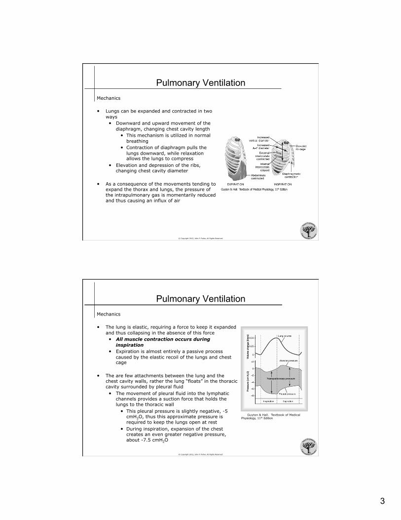
Pulmonary Ventilation Mechanics
• Lungs can be expanded and contracted in two ways • Downward and upward movement of the
diaphragm, changing chest cavity length • This mechanism is utilized in normal
breathing • Contraction of diaphragm pulls the
lungs downward, while relaxation allows the lungs to compress
• Elevation and depression of the ribs, changing chest cavity diameter
• As a consequence of the movements tending to expand the thorax and lungs, the pressure of the intrapulmonary gas is momentarily reduced and thus causing an influx of air
Guyton & Hall. Textbook of Medical Physiology, 11th Edition
© Copyright 2012, John P. Fisher, All Rights Reserved
Pulmonary Ventilation Mechanics
• The lung is elastic, requiring a force to keep it expanded and thus collapsing in the absence of this force • All muscle contraction occurs during
inspiration • Expiration is almost entirely a passive process
caused by the elastic recoil of the lungs and chest cage
• The are few attachments between the lung and the chest cavity walls, rather the lung “floats” in the thoracic cavity surrounded by pleural fluid • The movement of pleural fluid into the lymphatic
channels provides a suction force that holds the lungs to the thoracic wall
• This pleural pressure is slightly negative, -5 cmH2O, thus this approximate pressure is required to keep the lungs open at rest
• During inspiration, expansion of the chest creates an even greater negative pressure, about -7.5 cmH2O
Guyton & Hall. Textbook of Medical Physiology, 11th Edition
4
© Copyright 2012, John P. Fisher, All Rights Reserved
Pulmonary Ventilation Mechanics
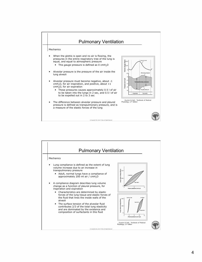
• When the glottis is open and no air is flowing, the pressures in the entire respiratory tree of the lung is equal, and equal to atmospheric pressure • This gauge pressure is defined as 0 cmH2O
• Alveolar pressure is the pressure of the air inside the lung alveoli
• Alveolar pressure must become negative, about -1 cmH2O, for air inspiration, and positive, about +1 cmH2O, for air expiration • These pressures causes approximately 0.5 l of air
to be taken into the lungs in 2 sec, and 0.5 l of air to be expelled out in 2 to 3 sec
• The difference between alveolar pressure and pleural pressure is defined as transpulmonary pressure, and is a measure of the elastic forces of the lung
Guyton & Hall. Textbook of Medical Physiology, 11th Edition
© Copyright 2012, John P. Fisher, All Rights Reserved
Pulmonary Ventilation Mechanics
• Lung compliance is defined as the extent of lung volume increase due to an increase in transpulmonary pressure • Adult, normal lungs have a compliance of
approximately 200 ml air / cmH2O
• A compliance diagram describes lung volume change as a function of pleural pressure, for inspiration and expiration • Characteristics are determined by elastic
forces of the lung tissue and elastic forces of the fluid that lines the inside walls of the alveoli
• The surface tension of the alveolar fluid contributes 2/3 of the total lung elasticity and are dominated by the existence and composition of surfactants in this fluid
Guyton & Hall. Textbook of Medical Physiology, 11th Edition
5
© Copyright 2012, John P. Fisher, All Rights Reserved
Pulmonary Ventilation Mechanics
• Similar to the surface tension in raindrops or meniscus, the surface tension created by the alveolar fluid provides a contracting force in the alveoli - a surface tension induced elastic force
• Surfactants, typically amphiphilic molecules, reduce water surface tension • In the lung, surfactants are release by type II alveolar epithelial cells • The released surfactant contains phospholipids, proteins, and ions - specifically
dipalmitoylphosphatidylcholine, surfactant apoproteins, and calcium ions • A portion of the surfactant dissolves, while the remainder spreads over the
alveolar fluid, reducing surface tension up to 90%, from 50 dynes/cm (alveolar fluid) to between 5 and 30 dynes/cm (alveolar fluid with surfactant)
• In an occluded alveoli, the amount of pressure generated from the collapsing alveoli may be estimated by: • Pressure = 2 x surface tension / alveolar radius • Thus, the decrease is surface tension provided by surfactants plays a key role in
keeping alveoli open • Also, note that smaller alveoli observe significantly higher pressures
© Copyright 2012, John P. Fisher, All Rights Reserved
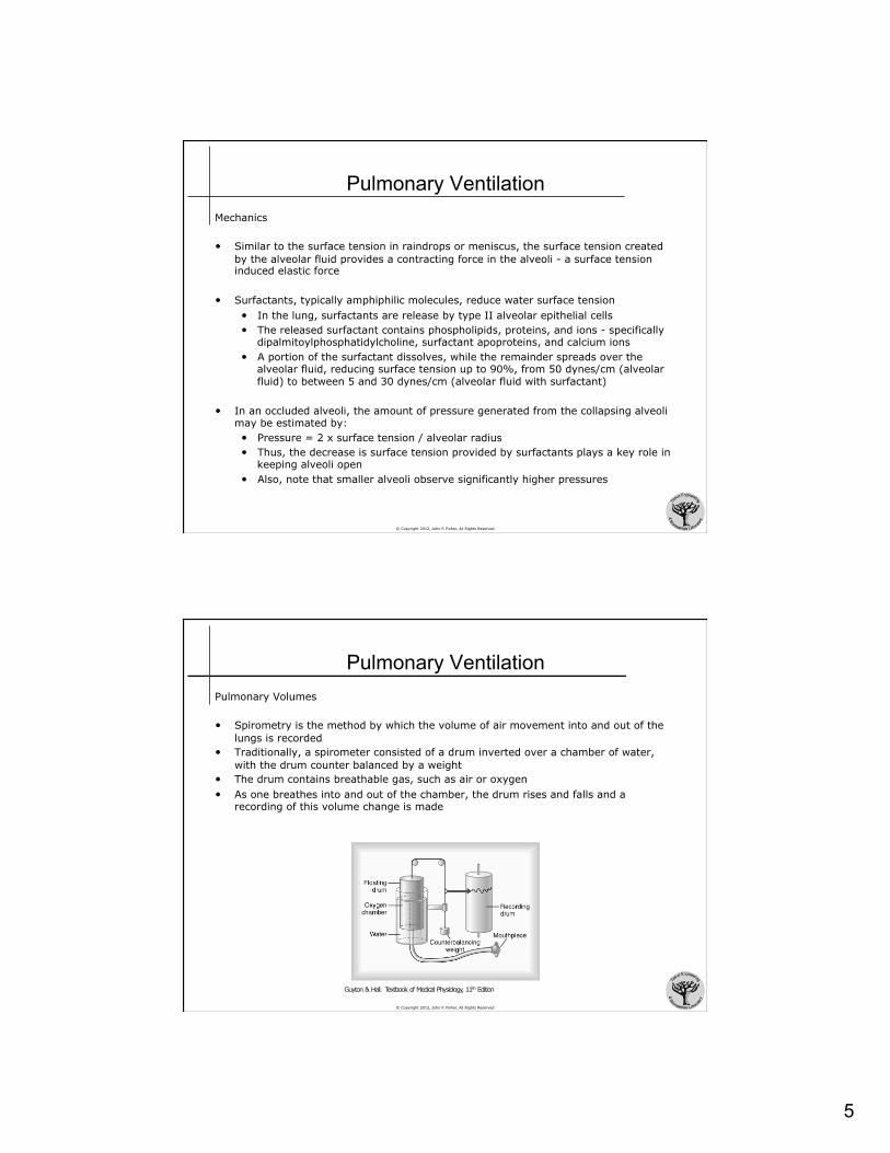
Pulmonary Ventilation Pulmonary Volumes
• Spirometry is the method by which the volume of air movement into and out of the lungs is recorded
• Traditionally, a spirometer consisted of a drum inverted over a chamber of water, with the drum counter balanced by a weight
• The drum contains breathable gas, such as air or oxygen • As one breathes into and out of the chamber, the drum rises and falls and a
recording of this volume change is made
Guyton & Hall. Textbook of Medical Physiology, 11th Edition
6
© Copyright 2012, John P. Fisher, All Rights Reserved
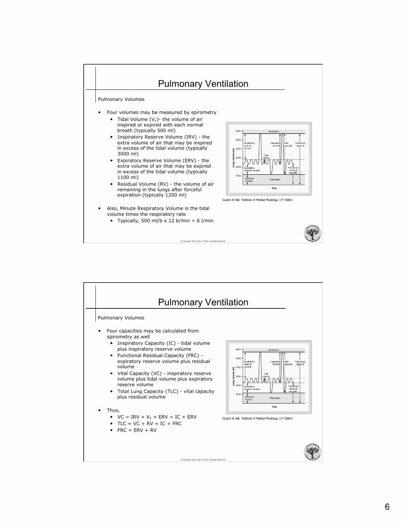
Pulmonary Ventilation Pulmonary Volumes
• Four volumes may be measured by spirometry • Tidal Volume (VT)- the volume of air
inspired or expired with each normal breath (typically 500 ml)
• Inspiratory Reserve Volume (IRV) - the extra volume of air that may be inspired in excess of the tidal volume (typically 3000 ml)
• Expiratory Reserve Volume (ERV) - the extra volume of air that may be expired in excess of the tidal volume (typically 1100 ml)
• Residual Volume (RV) - the volume of air remaining in the lungs after forceful expiration (typically 1200 ml)
• Also, Minute Respiratory Volume is the tidal volume times the respiratory rate • Typically, 500 ml/b x 12 b/min = 6 l/min
Guyton & Hall. Textbook of Medical Physiology, 11th Edition
© Copyright 2012, John P. Fisher, All Rights Reserved
Pulmonary Ventilation Pulmonary Volumes
• Four capacities may be calculated from spirometry as well • Inspiratory Capacity (IC) - tidal volume
plus inspiratory reserve volume • Functional Residual Capacity (FRC) -
expiratory reserve volume plus residual volume
• Vital Capacity (VC) - inspiratory reserve volume plus tidal volume plus expiratory reserve volume
• Total Lung Capacity (TLC) - vital capacity plus residual volume
• Thus, • VC = IRV + VT + ERV = IC + ERV • TLC = VC + RV = IC + FRC • FRC = ERV + RV
Guyton & Hall. Textbook of Medical Physiology, 11th Edition
7
© Copyright 2012, John P. Fisher, All Rights Reserved
Pulmonary Ventilation Pulmonary Volumes
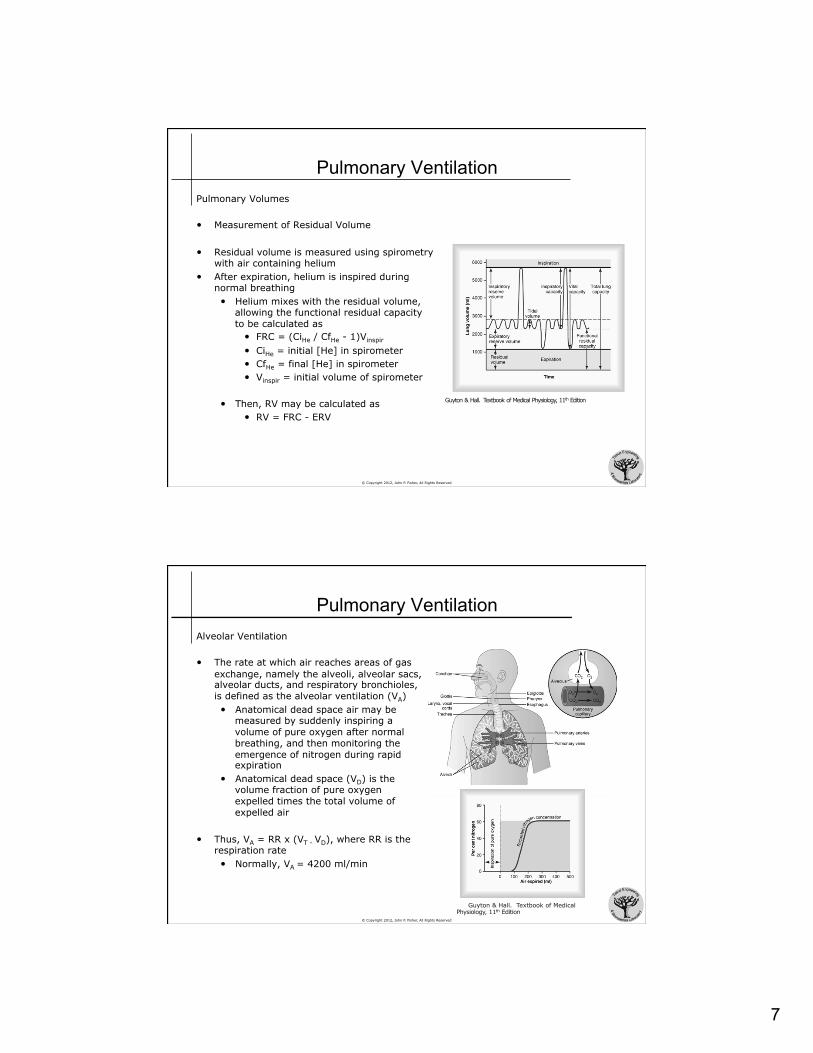
• Measurement of Residual Volume
• Residual volume is measured using spirometry with air containing helium
• After expiration, helium is inspired during normal breathing • Helium mixes with the residual volume,
allowing the functional residual capacity to be calculated as
• FRC = (CiHe / CfHe - 1)Vinspir • CiHe = initial [He] in spirometer
• CfHe = final [He] in spirometer
• Vinspir = initial volume of spirometer
• Then, RV may be calculated as
• RV = FRC - ERV
Guyton & Hall. Textbook of Medical Physiology, 11th Edition
© Copyright 2012, John P. Fisher, All Rights Reserved
Pulmonary Ventilation Alveolar Ventilation
• The rate at which air reaches areas of gas exchange, namely the alveoli, alveolar sacs, alveolar ducts, and respiratory bronchioles, is defined as the alveolar ventilation (VA) • Anatomical dead space air may be
measured by suddenly inspiring a volume of pure oxygen after normal breathing, and then monitoring the emergence of nitrogen during rapid expiration
• Anatomical dead space (VD) is the volume fraction of pure oxygen expelled times the total volume of expelled air
• Thus, VA = RR x (VT - VD), where RR is the respiration rate • Normally, VA = 4200 ml/min
Guyton & Hall. Textbook of Medical Physiology, 11th Edition
8
© Copyright 2012, John P. Fisher, All Rights Reserved
Pulmonary Ventilation Alveolar Ventilation
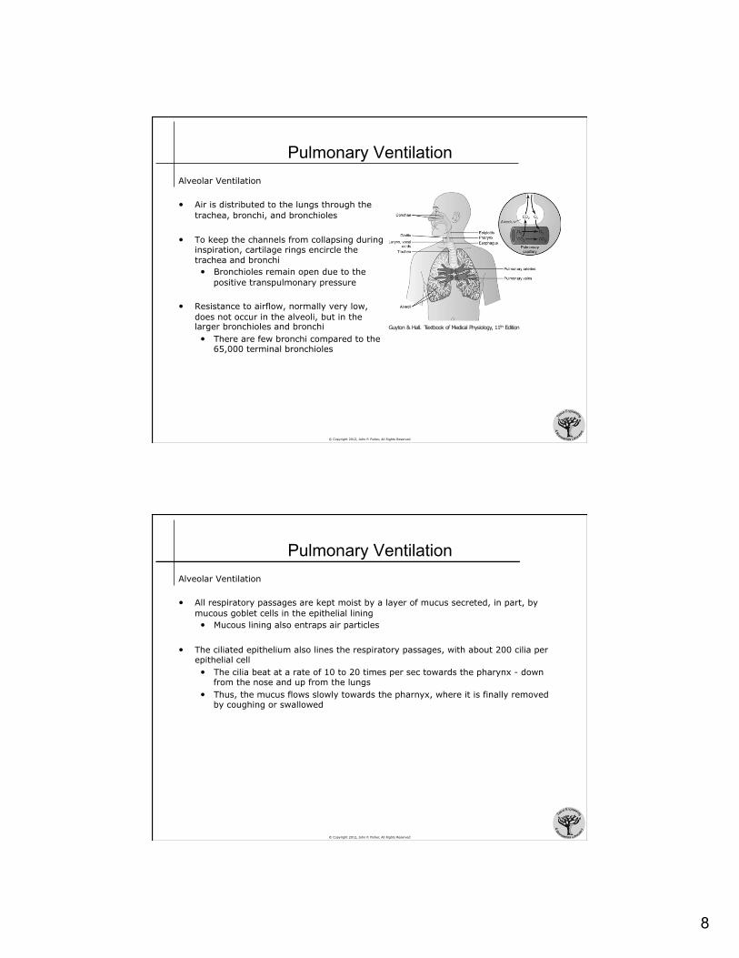
• Air is distributed to the lungs through the trachea, bronchi, and bronchioles
• To keep the channels from collapsing during inspiration, cartilage rings encircle the trachea and bronchi • Bronchioles remain open due to the
positive transpulmonary pressure
• Resistance to airflow, normally very low, does not occur in the alveoli, but in the larger bronchioles and bronchi • There are few bronchi compared to the
65,000 terminal bronchioles
Guyton & Hall. Textbook of Medical Physiology, 11th Edition
© Copyright 2012, John P. Fisher, All Rights Reserved
Pulmonary Ventilation Alveolar Ventilation
• All respiratory passages are kept moist by a layer of mucus secreted, in part, by mucous goblet cells in the epithelial lining • Mucous lining also entraps air particles
• The ciliated epithelium also lines the respiratory passages, with about 200 cilia per epithelial cell • The cilia beat at a rate of 10 to 20 times per sec towards the pharynx - down
from the nose and up from the lungs • Thus, the mucus flows slowly towards the pharnyx, where it is finally removed
by coughing or swallowed
9
© Copyright 2012, John P. Fisher, All Rights Reserved
Pulmonary Circulation Physiology
• The pulmonary artery extends 5 cm beyond the apex of the right ventricle, and then divides to supply blood to the two lungs • Pulmonary artery walls are thin, 1/3 that of the aorta • Pulmonary arterial branches are short • Most pulmonary vessels have diameters larger than those in the systemic
system • Thus, pulmonary arteries have a high compliance, allowing the system to
accommodate the stroke volume output of the right ventricle
• Lung tissue is nourished by small bronchial arteries originating from the systemic system and emptying into the pulmonary veins
© Copyright 2012, John P. Fisher, All Rights Reserved
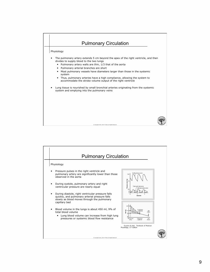
Pulmonary Circulation Physiology
• Pressure pulses in the right ventricle and pulmonary artery are significantly lower than those observed in the aorta
• During systole, pulmonary artery and right ventricular pressure are nearly equal
• During diastole, right ventricular pressure falls quickly, and pulmonary arterial pressure falls slowly as blood moves through the pulmonary capillary bed
• Blood volume in the lungs is about 450 ml, 9% of total blood volume • Lung blood volume can increase from high lung
pressures or systemic blood flow resistance
Guyton & Hall. Textbook of Medical Physiology, 11th Edition
10
© Copyright 2012, John P. Fisher, All Rights Reserved
Pulmonary Circulation Blood Distribution
• Under most conditions, pulmonary vessels act as passive, distensible tubes that enlarge with increasing pressure and narrow with decreasing pressure
• It is critical that blood flows to the regions of the lung where alveoli are most oxygenated • Normal concentration of oxygen in the air of the alveoli is 73 mmHg (PO2) • When concentrations falls below this level, the adjacent blood vessels constrict
• This response is in opposition to that observed in the systemic system, where blood vessels would be expected to dilate is response to low oxygen
• This constrictions prevents blood flow to the low oxygenated areas, thus increasing blood flow to the high oxygenated areas
• Constriction is thought to occur by the release of a vasoconstrictor signaling molecule, although this molecule has not yet been identified
© Copyright 2012, John P. Fisher, All Rights Reserved
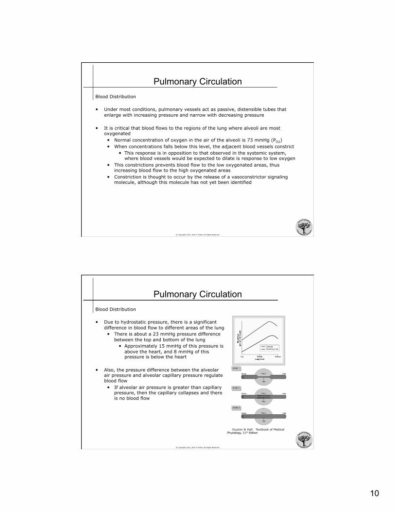
Pulmonary Circulation Blood Distribution
• Due to hydrostatic pressure, there is a significant difference in blood flow to different areas of the lung • There is about a 23 mmHg pressure difference
between the top and bottom of the lung • Approximately 15 mmHg of this pressure is
above the heart, and 8 mmHg of this pressure is below the heart
• Also, the pressure difference between the alveolar air pressure and alveolar capillary pressure regulate blood flow • If alveolar air pressure is greater than capillary
pressure, then the capillary collapses and there is no blood flow
Guyton & Hall. Textbook of Medical Physiology, 11th Edition
11
© Copyright 2012, John P. Fisher, All Rights Reserved
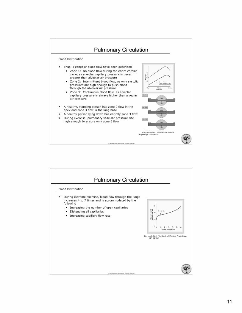
Pulmonary Circulation Blood Distribution
• Thus, 3 zones of blood flow have been described • Zone 1: No blood flow during the entire cardiac
cycle, as alveolar capillary pressure is never greater than alveolar air pressure
• Zone 2: Intermittent blood flow, as only systolic pressures are high enough to push blood through the alveolar air pressure
• Zone 3: Continuous blood flow, as alveolar capillary pressure is always higher than alveolar air pressure
• A healthy, standing person has zone 2 flow in the apex and zone 3 flow in the lung base
• A healthy person lying down has entirely zone 3 flow • During exercise, pulmonary vascular pressure rise
high enough to ensure only zone 3 flow
Guyton & Hall. Textbook of Medical Physiology, 11th Edition
© Copyright 2012, John P. Fisher, All Rights Reserved
Pulmonary Circulation Blood Distribution
• During extreme exercise, blood flow through the lungs increases 4 to 7 times and is accommodated by the following • Increasing the number of open capillaries • Distending all capillaries • Increasing capillary flow rate
Guyton & Hall. Textbook of Medical Physiology, 11th Edition
12
© Copyright 2012, John P. Fisher, All Rights Reserved
Pulmonary Circulation Blood Distribution
• Notes about pulmonary capillary dynamics
• Capillaries in the lung are so dense that they almost touch one another side by side, leading to blood flow in the alveolar walls to appear as nearly a sheet rather than through individual capillaries
• Pulmonary capillary pressure has been estimated at 7 mmHg, between left atrial pressure (2 mmHg) and pulmonary arterial pressure (15 mmHg)
• Blood flow rate through the pulmonary capillaries is typically 0.8 sec, but can be as little as 0.3 sec
© Copyright 2012, John P. Fisher, All Rights Reserved
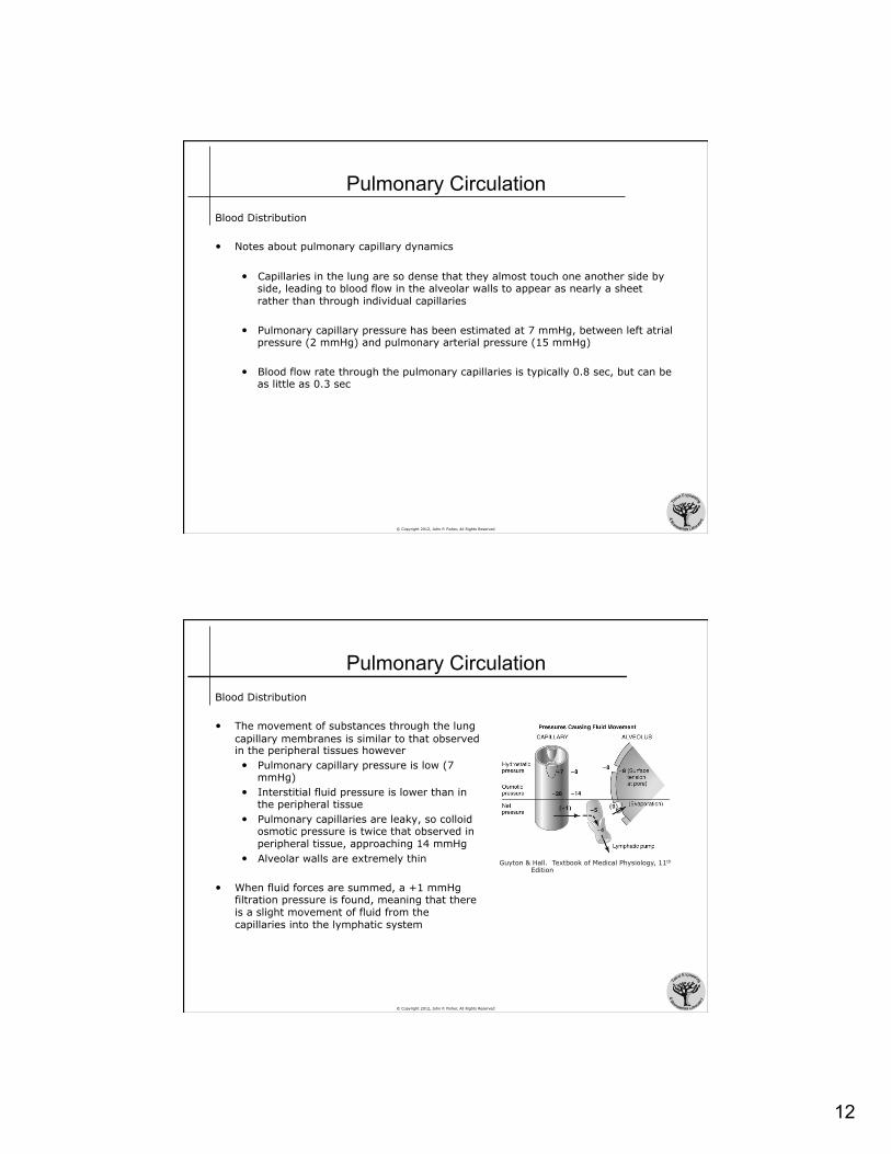
Pulmonary Circulation Blood Distribution
• The movement of substances through the lung capillary membranes is similar to that observed in the peripheral tissues however • Pulmonary capillary pressure is low (7
mmHg) • Interstitial fluid pressure is lower than in
the peripheral tissue • Pulmonary capillaries are leaky, so colloid
osmotic pressure is twice that observed in peripheral tissue, approaching 14 mmHg
• Alveolar walls are extremely thin
• When fluid forces are summed, a +1 mmHg filtration pressure is found, meaning that there is a slight movement of fluid from the capillaries into the lymphatic system
Guyton & Hall. Textbook of Medical Physiology, 11th Edition
13
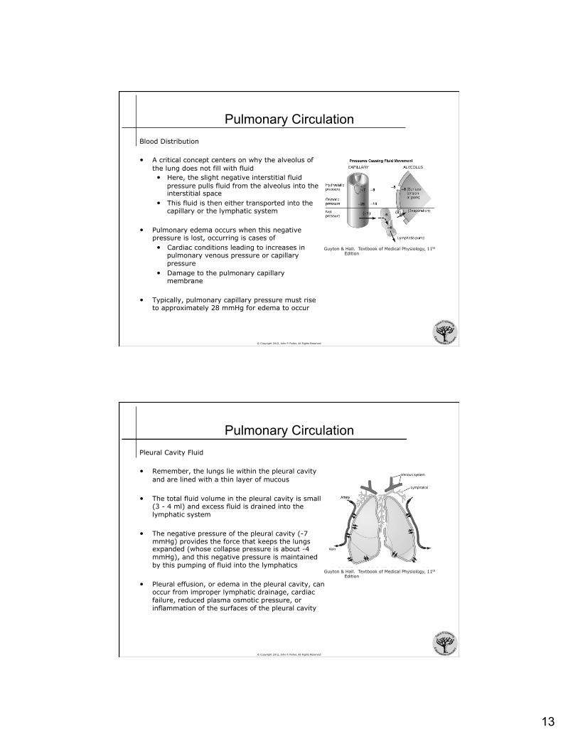
© Copyright 2012, John P. Fisher, All Rights Reserved
Pulmonary Circulation Blood Distribution
• A critical concept centers on why the alveolus of the lung does not fill with fluid • Here, the slight negative interstitial fluid
pressure pulls fluid from the alveolus into the interstitial space
• This fluid is then either transported into the capillary or the lymphatic system
• Pulmonary edema occurs when this negative pressure is lost, occurring is cases of • Cardiac conditions leading to increases in
pulmonary venous pressure or capillary pressure
• Damage to the pulmonary capillary membrane
• Typically, pulmonary capillary pressure must rise to approximately 28 mmHg for edema to occur
Guyton & Hall. Textbook of Medical Physiology, 11th Edition
© Copyright 2012, John P. Fisher, All Rights Reserved
Pulmonary Circulation Pleural Cavity Fluid
• Remember, the lungs lie within the pleural cavity and are lined with a thin layer of mucous
• The total fluid volume in the pleural cavity is small (3 - 4 ml) and excess fluid is drained into the lymphatic system
• The negative pressure of the pleural cavity (-7 mmHg) provides the force that keeps the lungs expanded (whose collapse pressure is about -4 mmHg), and this negative pressure is maintained by this pumping of fluid into the lymphatics
• Pleural effusion, or edema in the pleural cavity, can occur from improper lymphatic drainage, cardiac failure, reduced plasma osmotic pressure, or inflammation of the surfaces of the pleural cavity
Guyton & Hall. Textbook of Medical Physiology, 11th Edition
14
© Copyright 2012, John P. Fisher, All Rights Reserved
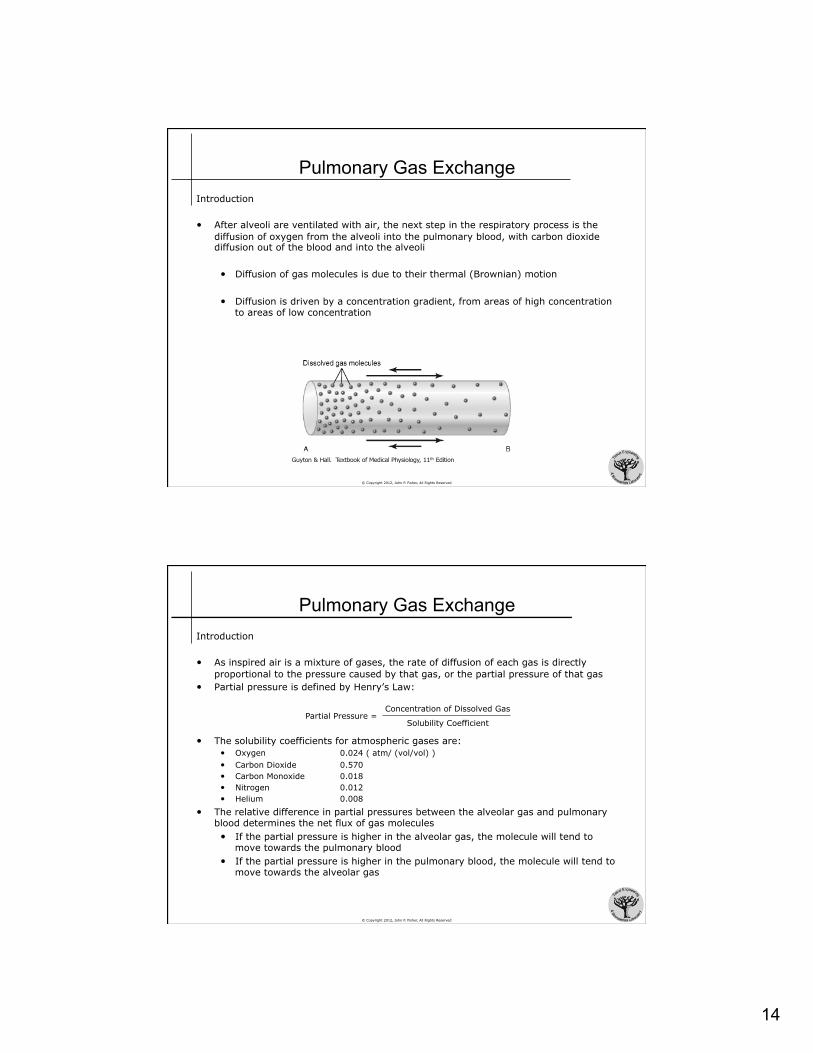
Pulmonary Gas Exchange Introduction
• After alveoli are ventilated with air, the next step in the respiratory process is the diffusion of oxygen from the alveoli into the pulmonary blood, with carbon dioxide diffusion out of the blood and into the alveoli
• Diffusion of gas molecules is due to their thermal (Brownian) motion
• Diffusion is driven by a concentration gradient, from areas of high concentration to areas of low concentration
Guyton & Hall. Textbook of Medical Physiology, 11th Edition
© Copyright 2012, John P. Fisher, All Rights Reserved
Pulmonary Gas Exchange Introduction
• As inspired air is a mixture of gases, the rate of diffusion of each gas is directly proportional to the pressure caused by that gas, or the partial pressure of that gas
• Partial pressure is defined by Henry’s Law:
• The solubility coefficients for atmospheric gases are: • Oxygen 0.024 ( atm/ (vol/vol) ) • Carbon Dioxide 0.570 • Carbon Monoxide 0.018 • Nitrogen 0.012 • Helium 0.008
• The relative difference in partial pressures between the alveolar gas and pulmonary blood determines the net flux of gas molecules • If the partial pressure is higher in the alveolar gas, the molecule will tend to
move towards the pulmonary blood • If the partial pressure is higher in the pulmonary blood, the molecule will tend to
move towards the alveolar gas
Partial Pressure = Concentration of Dissolved Gas
Solubility Coefficient

15
© Copyright 2012, John P. Fisher, All Rights Reserved
Pulmonary Gas Exchange Introduction
• As air is inspired, water immediately evaporates from the surfaces of the airways and humidifies the air • Water evaporation is also driven by differences in water partial pressure in the
atmospheric air and inspired air
• The partial pressure exerted by water is called the vapor pressure • At physiological conditions, the vapor pressure of water is 47 mmHg
• Thus, once air is inspired, its is humidified to relative saturation to 47 mmHg
© Copyright 2012, John P. Fisher, All Rights Reserved
Pulmonary Gas Exchange Introduction
• The rate of diffusion may be described by
• where D = diffusion rate ΔP = partial pressure difference A = cross sectional area S = gas solubility d = distance MW = molecule’s molecular weight
• Here, the diffusion coefficient (D) of the gas is proportional to S/(MW)1/2
• Diffusion coefficients for some gases in body fluids are • Oxygen 1.00 (D/D O2) • Carbon Dioxide 20.30 • Carbon Monoxide 0.81
• Nitrogen 0.53 • Helium 0.95
MWdSAPD Δ
∝
16
© Copyright 2012, John P. Fisher, All Rights Reserved
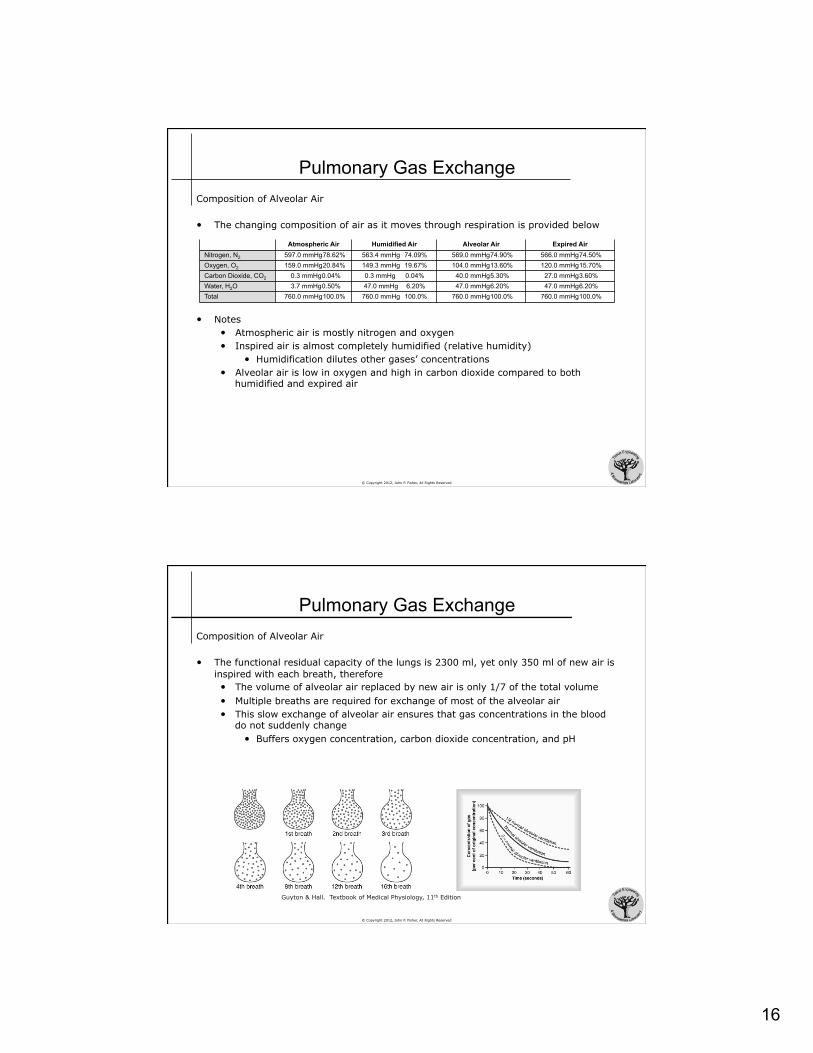
Pulmonary Gas Exchange Composition of Alveolar Air
• The changing composition of air as it moves through respiration is provided below
• Notes • Atmospheric air is mostly nitrogen and oxygen • Inspired air is almost completely humidified (relative humidity)
• Humidification dilutes other gases’ concentrations • Alveolar air is low in oxygen and high in carbon dioxide compared to both
humidified and expired air
Atmospheric Air Humidified Air Alveolar Air Expired Air Nitrogen, N2 597.0 mmHg 78.62% 563.4 mmHg 74.09% 569.0 mmHg 74.90% 566.0 mmHg 74.50% Oxygen, O2 159.0 mmHg 20.84% 149.3 mmHg 19.67% 104.0 mmHg 13.60% 120.0 mmHg 15.70% Carbon Dioxide, CO2 0.3 mmHg 0.04% 0.3 mmHg 0.04% 40.0 mmHg 5.30% 27.0 mmHg 3.60% Water, H2O 3.7 mmHg 0.50% 47.0 mmHg 6.20% 47.0 mmHg 6.20% 47.0 mmHg 6.20% Total 760.0 mmHg 100.0% 760.0 mmHg 100.0% 760.0 mmHg 100.0% 760.0 mmHg 100.0%
© Copyright 2012, John P. Fisher, All Rights Reserved
Pulmonary Gas Exchange Composition of Alveolar Air
• The functional residual capacity of the lungs is 2300 ml, yet only 350 ml of new air is inspired with each breath, therefore • The volume of alveolar air replaced by new air is only 1/7 of the total volume • Multiple breaths are required for exchange of most of the alveolar air • This slow exchange of alveolar air ensures that gas concentrations in the blood
do not suddenly change • Buffers oxygen concentration, carbon dioxide concentration, and pH
Guyton & Hall. Textbook of Medical Physiology, 11th Edition
17
© Copyright 2012, John P. Fisher, All Rights Reserved
Pulmonary Gas Exchange Composition of Alveolar Air
• Oxygen concentration in the alveoli is determined by • Rate of entry of oxygen by ventilation • Rate of absorption of oxygen into the blood
• Normal respiration sees an alveolar ventilation of 4.2 l/min, an alveolar oxygen partial pressure of 104 mmHg, and oxygen consumption of 250 ml/min • Increased oxygen consumptions requires a
significant increase in ventilation to maintain oxygen partial pressure
• Normal respiration and ventilation also sees an alveolar carbon dioxide partial pressure of 40 mmHg and carbon dioxide excretion of 200 ml/min • Again, increased carbon dioxide excretion
requires a significant increase in ventilation Guyton & Hall. Textbook of Medical Physiology,
11th Edition
© Copyright 2012, John P. Fisher, All Rights Reserved
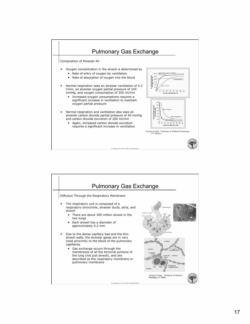
Pulmonary Gas Exchange Diffusion Through the Respiratory Membrane
• The respiratory unit is composed of a respiratory bronchiole, alveolar ducts, atria, and alveoli • There are about 300 million alveoli in the
two lungs • Each alveoli has a diameter of
approximately 0.2 mm
• Due to the dense capillary bed and the thin alveoli walls, the alveolar gases are in very close proximity to the blood of the pulmonary capillaries • Gas exchange occurs through the
membranes of all the terminal portions of the lung (not just alveoli), and are described as the respiratory membrane or pulmonary membrane
Guyton & Hall. Textbook of Medical Physiology, 11th Edition
18
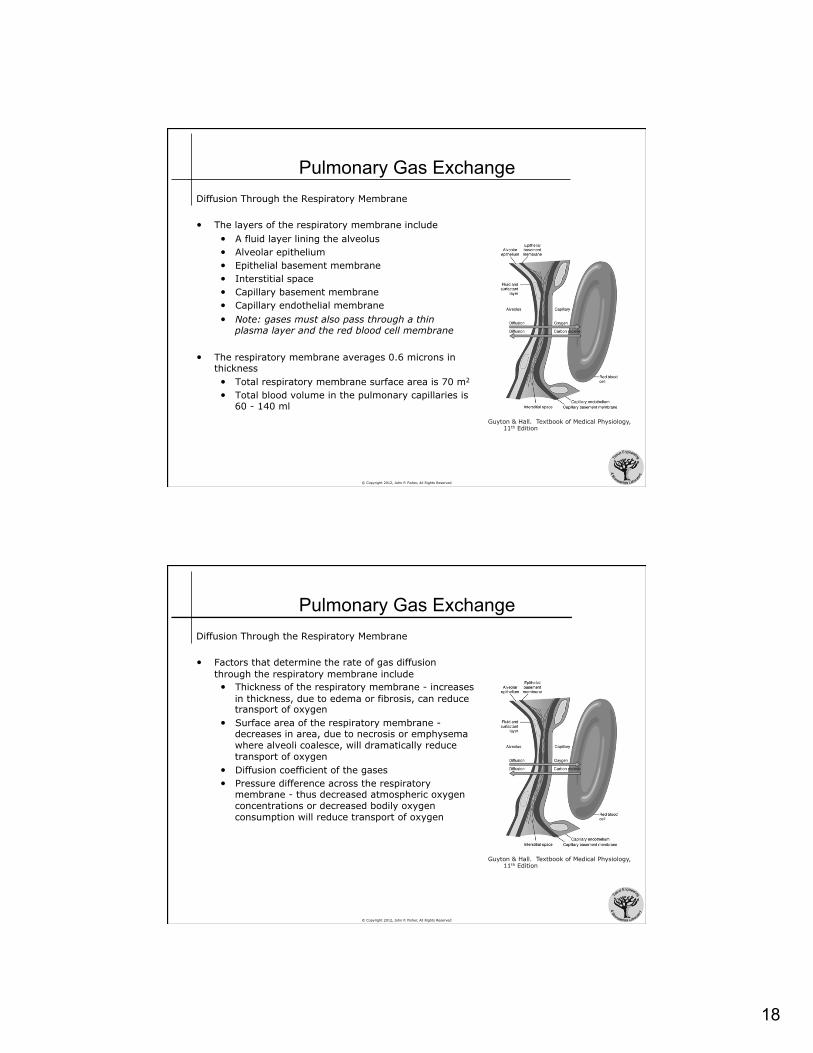
© Copyright 2012, John P. Fisher, All Rights Reserved
Pulmonary Gas Exchange Diffusion Through the Respiratory Membrane
• The layers of the respiratory membrane include • A fluid layer lining the alveolus • Alveolar epithelium • Epithelial basement membrane • Interstitial space • Capillary basement membrane • Capillary endothelial membrane • Note: gases must also pass through a thin
plasma layer and the red blood cell membrane
• The respiratory membrane averages 0.6 microns in thickness • Total respiratory membrane surface area is 70 m2
• Total blood volume in the pulmonary capillaries is 60 - 140 ml
Guyton & Hall. Textbook of Medical Physiology, 11th Edition
© Copyright 2012, John P. Fisher, All Rights Reserved
Pulmonary Gas Exchange Diffusion Through the Respiratory Membrane
• Factors that determine the rate of gas diffusion through the respiratory membrane include • Thickness of the respiratory membrane - increases
in thickness, due to edema or fibrosis, can reduce transport of oxygen
• Surface area of the respiratory membrane - decreases in area, due to necrosis or emphysema where alveoli coalesce, will dramatically reduce transport of oxygen
• Diffusion coefficient of the gases • Pressure difference across the respiratory
membrane - thus decreased atmospheric oxygen concentrations or decreased bodily oxygen consumption will reduce transport of oxygen
Guyton & Hall. Textbook of Medical Physiology, 11th Edition
19
© Copyright 2012, John P. Fisher, All Rights Reserved
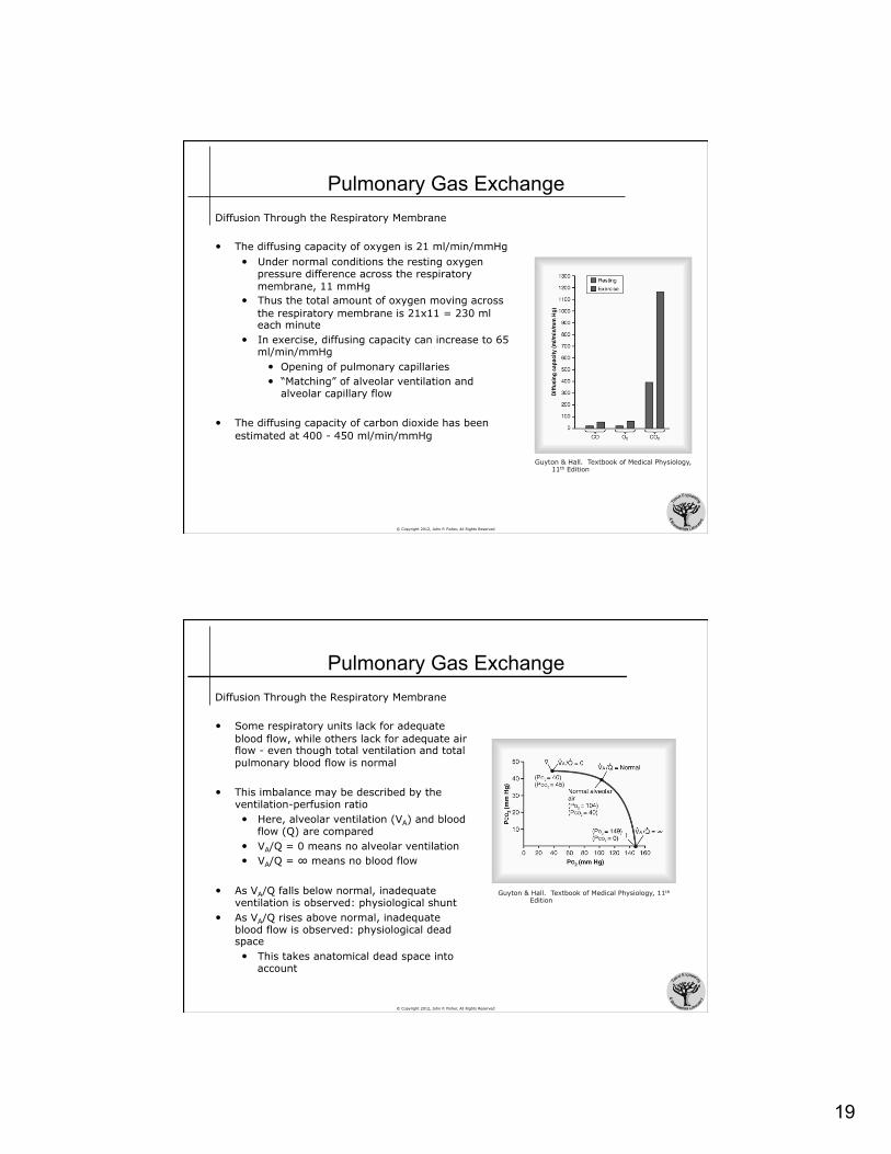
Pulmonary Gas Exchange Diffusion Through the Respiratory Membrane
• The diffusing capacity of oxygen is 21 ml/min/mmHg • Under normal conditions the resting oxygen
pressure difference across the respiratory membrane, 11 mmHg
• Thus the total amount of oxygen moving across the respiratory membrane is 21x11 = 230 ml each minute
• In exercise, diffusing capacity can increase to 65 ml/min/mmHg
• Opening of pulmonary capillaries • “Matching” of alveolar ventilation and
alveolar capillary flow
• The diffusing capacity of carbon dioxide has been estimated at 400 - 450 ml/min/mmHg
Guyton & Hall. Textbook of Medical Physiology, 11th Edition
© Copyright 2012, John P. Fisher, All Rights Reserved
Pulmonary Gas Exchange Diffusion Through the Respiratory Membrane
• Some respiratory units lack for adequate blood flow, while others lack for adequate air flow - even though total ventilation and total pulmonary blood flow is normal
• This imbalance may be described by the ventilation-perfusion ratio • Here, alveolar ventilation (VA) and blood
flow (Q) are compared • VA/Q = 0 means no alveolar ventilation • VA/Q = ∞ means no blood flow
• As VA/Q falls below normal, inadequate ventilation is observed: physiological shunt
• As VA/Q rises above normal, inadequate blood flow is observed: physiological dead space • This takes anatomical dead space into
account
Guyton & Hall. Textbook of Medical Physiology, 11th Edition
20
© Copyright 2012, John P. Fisher, All Rights Reserved
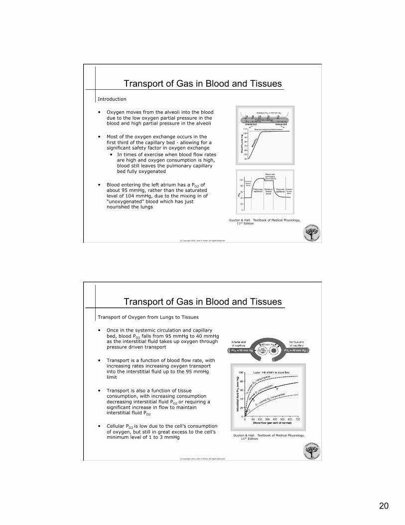
Transport of Gas in Blood and Tissues Introduction
• Oxygen moves from the alveoli into the blood due to the low oxygen partial pressure in the blood and high partial pressure in the alveoli
• Most of the oxygen exchange occurs in the first third of the capillary bed - allowing for a significant safety factor in oxygen exchange • In times of exercise when blood flow rates
are high and oxygen consumption is high, blood still leaves the pulmonary capillary bed fully oxygenated
• Blood entering the left atrium has a PO2 of about 95 mmHg, rather than the saturated level of 104 mmHg, due to the mixing in of “unoxygenated” blood which has just nourished the lungs
Guyton & Hall. Textbook of Medical Physiology, 11th Edition
© Copyright 2012, John P. Fisher, All Rights Reserved
Transport of Gas in Blood and Tissues Transport of Oxygen from Lungs to Tissues
• Once in the systemic circulation and capillary bed, blood PO2 falls from 95 mmHg to 40 mmHg as the interstitial fluid takes up oxygen through pressure driven transport
• Transport is a function of blood flow rate, with increasing rates increasing oxygen transport into the interstitial fluid up to the 95 mmHg limit
• Transport is also a function of tissue consumption, with increasing consumption decreasing interstitial fluid PO2 or requiring a significant increase in flow to maintain interstitial fluid PO2
• Cellular PO2 is low due to the cell’s consumption of oxygen, but still in great excess to the cell’s minimum level of 1 to 3 mmHg Guyton & Hall. Textbook of Medical Physiology,
11th Edition
21
© Copyright 2012, John P. Fisher, All Rights Reserved
Transport of Gas in Blood and Tissues Transport of Oxygen from Lungs to Tissues
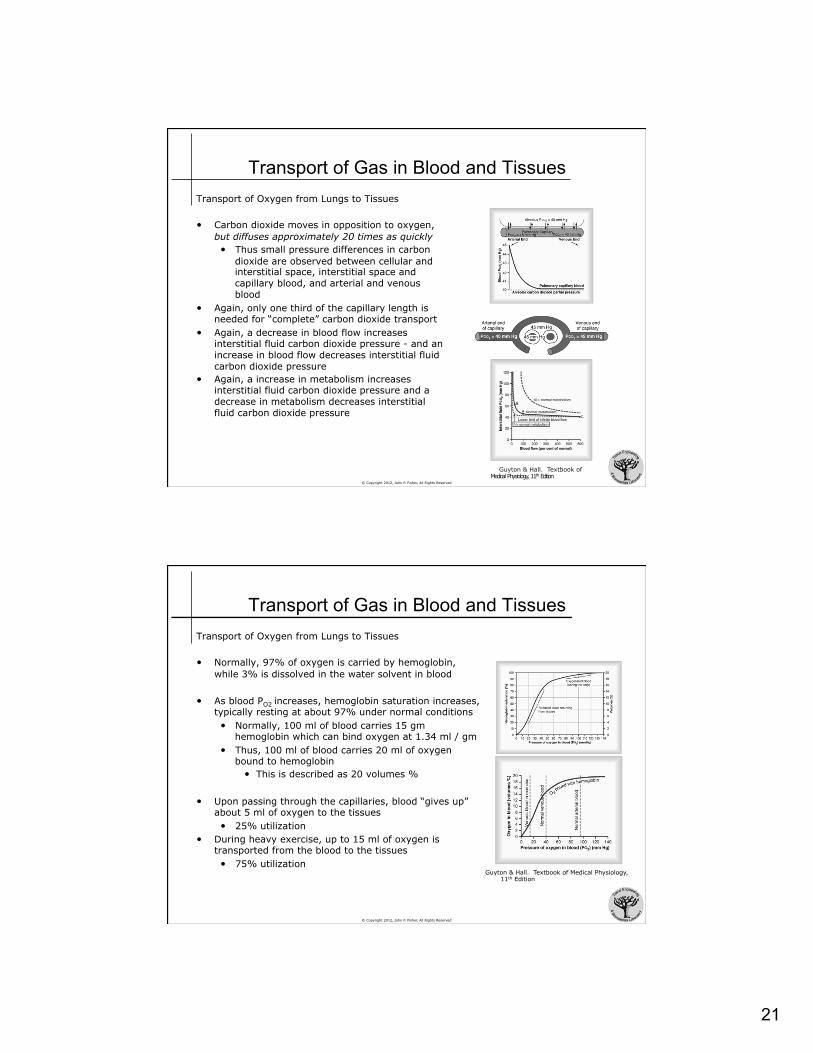
• Carbon dioxide moves in opposition to oxygen, but diffuses approximately 20 times as quickly • Thus small pressure differences in carbon
dioxide are observed between cellular and interstitial space, interstitial space and capillary blood, and arterial and venous blood
• Again, only one third of the capillary length is needed for “complete” carbon dioxide transport
• Again, a decrease in blood flow increases interstitial fluid carbon dioxide pressure - and an increase in blood flow decreases interstitial fluid carbon dioxide pressure
• Again, a increase in metabolism increases interstitial fluid carbon dioxide pressure and a decrease in metabolism decreases interstitial fluid carbon dioxide pressure
Guyton & Hall. Textbook of Medical Physiology, 11th Edition
© Copyright 2012, John P. Fisher, All Rights Reserved
Transport of Gas in Blood and Tissues Transport of Oxygen from Lungs to Tissues
• Normally, 97% of oxygen is carried by hemoglobin, while 3% is dissolved in the water solvent in blood
• As blood PO2 increases, hemoglobin saturation increases, typically resting at about 97% under normal conditions • Normally, 100 ml of blood carries 15 gm
hemoglobin which can bind oxygen at 1.34 ml / gm • Thus, 100 ml of blood carries 20 ml of oxygen
bound to hemoglobin • This is described as 20 volumes %
• Upon passing through the capillaries, blood “gives up” about 5 ml of oxygen to the tissues • 25% utilization
• During heavy exercise, up to 15 ml of oxygen is transported from the blood to the tissues • 75% utilization
Guyton & Hall. Textbook of Medical Physiology, 11th Edition
22
© Copyright 2012, John P. Fisher, All Rights Reserved
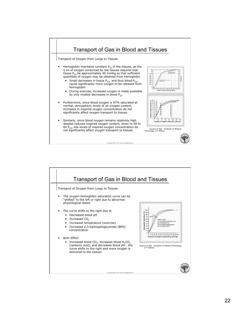
Transport of Gas in Blood and Tissues Transport of Oxygen from Lungs to Tissues
• Hemoglobin maintains constant PO2 in the tissues, as the 5 ml of oxygen consumed by the tissues requires that tissue PO2 be approximately 40 mmHg so that sufficient quantities of oxygen may be obtained from hemoglobin • Small decreases in tissue PO2, and thus blood PO2,
cause significantly more oxygen to be released from hemoglobin
• During exercise, increased oxygen is made available by only modest decreases in blood PO2
• Furthermore, since blood oxygen is 97% saturated at normal, atmospheric levels of air oxygen content, increases in inspired oxygen concentration do not significantly affect oxygen transport to tissues
• Similarly, since blood oxygen remains relatively high despite reduced inspired oxygen content, down to 50 to 60 PO2, low levels of inspired oxygen concentration do not significantly affect oxygen transport to tissues Guyton & Hall. Textbook of Medical
Physiology, 11th Edition
© Copyright 2012, John P. Fisher, All Rights Reserved
Transport of Gas in Blood and Tissues Transport of Oxygen from Lungs to Tissues
• The oxygen-hemoglobin saturation curve can be “shifted” to the left or right due to abnormal physiological states
• The curve shifts to the right due to • Decreased blood pH • Increased CO2
• Increased temperature (exercise) • Increased 2,3-biphosphoglycerate (BPG)
concentration
• Bohr Effect • Increased blood CO2, increases blood H2CO3
(carbonic acid), and decreases blood pH - the curve shifts to the right and more oxygen is delivered to the tissues
Guyton & Hall. Textbook of Medical Physiology, 11th Edition
23
© Copyright 2012, John P. Fisher, All Rights Reserved
Transport of Gas in Blood and Tissues Transport of Oxygen from Lungs to Tissues
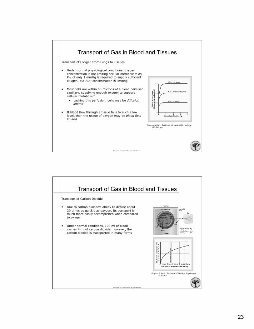
• Under normal physiological conditions, oxygen concentration is not limiting cellular metabolism as PO2 of only 1 mmHg is required to supply sufficient oxygen, but ADP concentration is limiting
• Most cells are within 50 microns of a blood perfused capillary, supplying enough oxygen to support cellular metabolism • Lacking this perfusion, cells may be diffusion
limited
• If blood flow through a tissue falls to such a low level, then the usage of oxygen may be blood flow limited
Guyton & Hall. Textbook of Medical Physiology, 11th Edition
© Copyright 2012, John P. Fisher, All Rights Reserved
Transport of Gas in Blood and Tissues Transport of Carbon Dioxide
• Due to carbon dioxide’s ability to diffuse about 20 times as quickly as oxygen, its transport is much more easily accomplished when compared to oxygen
• Under normal conditions, 100 ml of blood carries 4 ml of carbon dioxide, however, the carbon dioxide is transported in many forms
Guyton & Hall. Textbook of Medical Physiology, 11th Edition
24
© Copyright 2012, John P. Fisher, All Rights Reserved
Transport of Gas in Blood and Tissues Transport of Carbon Dioxide
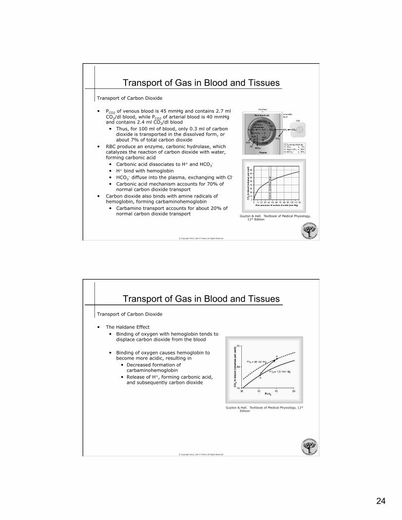
• PCO2 of venous blood is 45 mmHg and contains 2.7 ml CO2/dl blood, while PCO2 of arterial blood is 40 mmHg and contains 2.4 ml CO2/dl blood • Thus, for 100 ml of blood, only 0.3 ml of carbon
dioxide is transported in the dissolved form, or about 7% of total carbon dioxide
• RBC produce an enzyme, carbonic hydrolase, which catalyzes the reaction of carbon dioxide with water, forming carbonic acid • Carbonic acid dissociates to H+ and HCO3
-
• H+ bind with hemoglobin • HCO3
- diffuse into the plasma, exchanging with Cl-
• Carbonic acid mechanism accounts for 70% of normal carbon dioxide transport
• Carbon dioxide also binds with amine radicals of hemoglobin, forming carbaminohemoglobin • Carbamino transport accounts for about 20% of
normal carbon dioxide transport Guyton & Hall. Textbook of Medical Physiology, 11th Edition
© Copyright 2012, John P. Fisher, All Rights Reserved
Transport of Gas in Blood and Tissues Transport of Carbon Dioxide
• The Haldane Effect • Binding of oxygen with hemoglobin tends to
displace carbon dioxide from the blood
• Binding of oxygen causes hemoglobin to become more acidic, resulting in
• Decreased formation of carbaminohemoglobin
• Release of H+, forming carbonic acid, and subsequently carbon dioxide
Guyton & Hall. Textbook of Medical Physiology, 11th Edition