respiratory distress of unknown etiology in a transplant
TRANSCRIPT
Autopsy and Case Reports. ISSN 2236-1960. Copyright © 2017. This is an Open Access article distributed under the terms of the Creative Commons Attribution Non-Commercial License, which permits unrestricted non-commercial use, distribution, and reproduction in any medium provided the article is properly cited.
a St. John Hospital and Medical Center, Department of Pathology. Detroit, MI, USA.
Respiratory distress of unknown etiology in a transplant recipient: think toxoplasmosis!
Ameer Hamzaa, Ian Jacob Andersona, Basim Al-Khafajia
How to cite: Hamza A, Anderson IJ, Al-Khafaji B. Respiratory distress of unknown etiology in a transplant recipient: think toxoplasmosis!. Autops Case Rep [Internet]. 2017;7(4):37-41. http://dx.doi.org/10.4322/acr.2017.038
Article / Autopsy Case Report
ABSTRACT
Disseminated toxoplasmosis is a life-threatening disease in immunocompromised individuals. Infection is contracted from handling contaminated soil, cat litter, or through the consumption of contaminated water or food. It is the third most common lethal foodborne infection in the United States. In transplant patients, most cases occur as a result of reactivation of a latent infection resulting from immunosuppression. We present a case of disseminated toxoplasmosis diagnosed at the time of autopsy. This case emphasizes the importance of maintaining a high index of clinical suspicion and active disease surveillance in this era of sophisticated diagnostic testing.
Keywords: Autopsy; Allografts; Immunosuppression; Opportunistic infections; Kidney transplantation; Toxoplasmosis.
INTRODUCTION
Toxoplasmosis is caused by the protozoan parasite Toxoplasma gondii. It is a zoonotic disease with worldwide distribution and an opportunistic pathogen primarily infecting immunocompromised individuals. Global prevalence is 10-80%,1 but clinically significant disease is uncommon. In the United States, approximately 225,000 cases of toxoplasmosis are reported annually, resulting in 750 deaths, which makes T. gondii the third most common cause of lethal foodborne infection in the United States.2
In organ transplant recipients, toxoplasmosis is most commonly a result of reactivation of latent infection; however, donor-transmitted infection or de novo infection are known to occur. In these patients, the most common presentations are retinochoroiditis, cerebritis, pneumonitis, myocarditis, and disseminated disease. We describe the autopsy findings in a renal transplant recipient with disseminated toxoplasmosis diagnosed at autopsy.
CASE REPORT
A 49-year-old Caucasian male with a past medical history of end stage renal disease secondary to diabetes mellitus, status post deceased-donor kidney allograft and on immunosuppression with prednisone, tacrolimus and mycophenolate mofetil; hypertension, obesity, peripheral vascular disease, and anemia presented to the emergency department 6 weeks post-transplantation procedure with dehiscence of his right lower quadrant surgical wound. Upon admission, the patient was noted to be acidotic, hypovolemic, hyperkalemic, and in respiratory distress, necessitating intubation. He was urgently taken to the operating room for debridement and closure of the wound which he tolerated well; however, during the course of his hospital stay he exhibited altered mental status with continued respiratory distress. Despite receiving high flow supplemental oxygen, oxygen saturations were continually in the low 90% range. He also exhibited marked leukocytosis with a white blood cel l count of 26,400/mm3
Respiratory distress of unknown etiology in a transplant recipient: think toxoplasmosis!
38 Autops Case Rep (São Paulo). 2017;7(4):37-41
(normal range [NR]: 4000-11000/mm3). Blood and urine cultures yielded no growth. CMV serology by PCR was negative with quantitative CMV DNA less than 250 CPY/ml (NR: < 250 CPY/ml). Toxoplasma serology was not ordered and was not done as a pre-transplant work up. His arterial partial oxygen pressure became critically low at 35.4 mmHg (NR: 80-100 mmHg) and he was intubated, however, there was no improvement of the respiratory status, and the patient experienced cardiorespiratory arrest. Resuscitative efforts were unsuccessful and the patient was pronounced
deceased with a postmortem examination requested and subsequently performed.
AUTOPSY FINDINGS
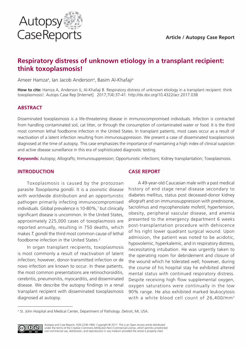
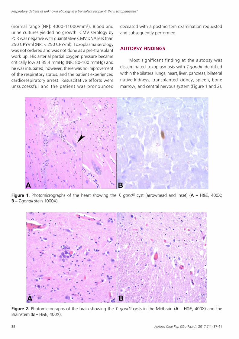
Most significant finding at the autopsy was
disseminated toxoplasmosis with T.gondii identified
within the bilateral lungs, heart, liver, pancreas, bilateral
native kidneys, transplanted kidney, spleen, bone
marrow, and central nervous system (Figure 1 and 2).
Figure 1. Photomicrographs of the heart showing the T. gondii cyst (arrowhead and inset) (A – H&E, 400X; B – T.gondii stain 1000X).
Figure 2. Photomicrographs of the brain showing the T. gondii cysts in the Midbrain (A – H&E, 400X) and the Brainstem (B – H&E, 400X).
Hamza A, Anderson IJ, Al-Khafaji B
39Autops Case Rep (São Paulo). 2017;7(4):37-41
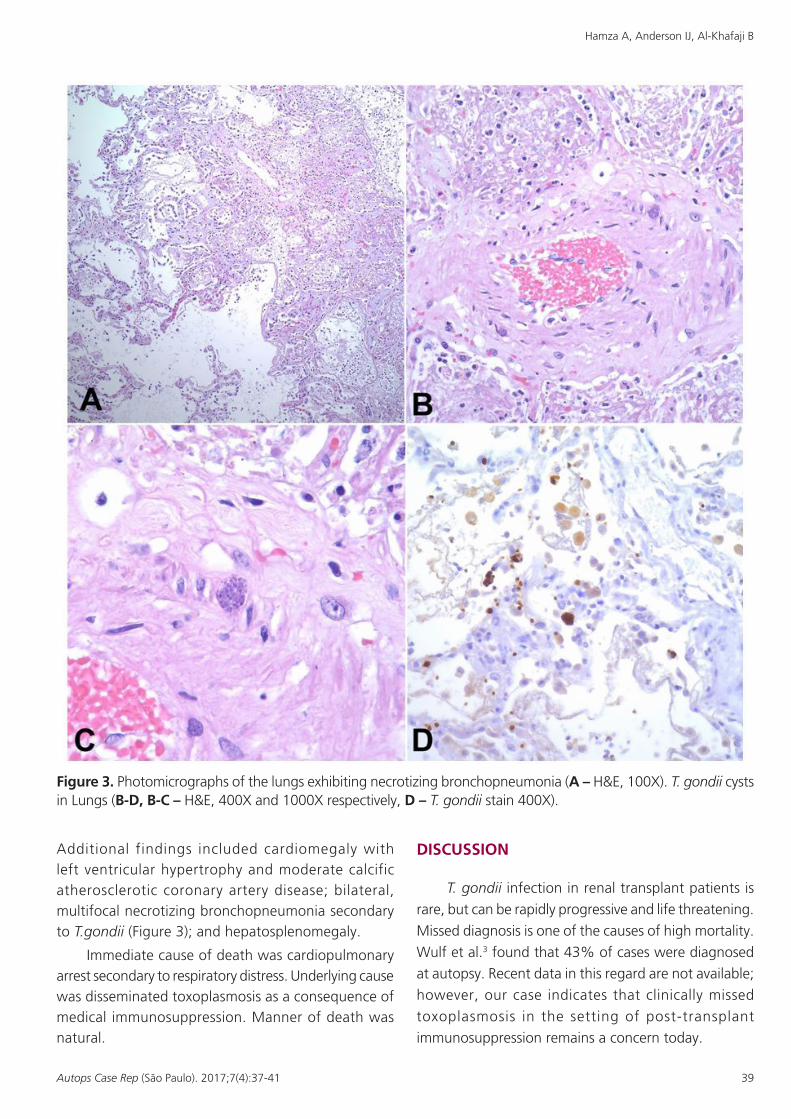
Figure 3. Photomicrographs of the lungs exhibiting necrotizing bronchopneumonia (A – H&E, 100X). T. gondii cysts in Lungs (B-D, B-C – H&E, 400X and 1000X respectively, D – T. gondii stain 400X).
Additional findings included cardiomegaly with left ventricular hypertrophy and moderate calcific atherosclerotic coronary artery disease; bilateral, multifocal necrotizing bronchopneumonia secondary to T.gondii (Figure 3); and hepatosplenomegaly.
Immediate cause of death was cardiopulmonary arrest secondary to respiratory distress. Underlying cause was disseminated toxoplasmosis as a consequence of medical immunosuppression. Manner of death was natural.
DISCUSSION
T. gondii infection in renal transplant patients is
rare, but can be rapidly progressive and life threatening.
Missed diagnosis is one of the causes of high mortality.
Wulf et al.3 found that 43% of cases were diagnosed
at autopsy. Recent data in this regard are not available;
however, our case indicates that clinically missed
toxoplasmosis in the setting of post-transplant
immunosuppression remains a concern today.
Respiratory distress of unknown etiology in a transplant recipient: think toxoplasmosis!
40 Autops Case Rep (São Paulo). 2017;7(4):37-41
T. gondii has two distinct life cycles. The sexual cycle occurs only in cats which are the definitive host. The asexual cycle occurs in other mammals, including humans and some avian species. In humans, infection occurs by ingestion of oocysts from handling contaminated soil or cat l itter or through the consumption of contaminated water or undercooked meat of infected animals. Our patient, in fact, did own a cat.
Clinical presentation is nonspecific. Fever occurs frequently, followed by flu like symptoms, lymphadenopathy, visual changes, pneumonia, and neurologic manifestations such as headache, seizures, altered mental status, and focal neurologic deficits. Our patient exhibited altered mental status and severe respiratory distress which correlated with the autopsy findings of bilateral, multifocal necrotizing bronchopneumonia and central nervous system involvement.
Diagnosis is confirmed by serology, tissue biopsy, or PCR assay for the presence of T. gondii DNA. Serology is of limited value in the setting of reactivation of a latent infection. Moreover, interpretation of serologic tests in immunocompromised patients is not straightforward and absence of specific antibodies does not exclude active disease. The duration for which T. gondii can be detected in blood is not clear. Additionally, T. gondii is only present intermittently in cerebrospinal fluid for unknown reasons,4 which limits the use of PCR assay. However, it is found to be useful in acute primary infection.3 Tissue biopsy is not only an invasive procedure, but is also limited by sampling artifact. Maintaining a high index of clinical suspicion is therefore the cornerstone of proper diagnosis. Toxoplasmosis should be high on the list of differential diagnoses in a post-transplant patient with respiratory distress and culture negative sepsis.
Imaging studies are valuable in cerebral and pulmonary toxoplasmosis. MRI is superior to CT scan in cerebral toxoplasmosis. Toxoplasma lesions on MRI appear as high-signal abnormalities on T2-weighted studies and demonstrate a rim of enhancement surrounding the edema on T1-weighted, contrast-enhanced images. CT scans show multiple bilateral cerebral lesions. Chest radiographs in toxoplasma pneumonia usually demonstrate interstitial infiltrate. In our patient, a CT of the head showed
asymmetric decreased attenuation in left frontal
lobe white matter suggestive of microangiopathy or
encephalomalacia. No acute intracranial abnormality or
evidence pointing to T. gondii was noted. Chest x-ray
showed mild to moderate prominence of the pulmonary
vessels, interstitial infiltrates, and changes consistent
with atelectasis.
For more prevalent infections, such as HIV, HBV
and HCV there is general agreement with respect
to pre-transplant screening guidelines. Regarding
screening for other infectious agents, including
T. gondii, each transplant center follows different
protocols. Many transplant centers screen both
donors and recipients for anti-Toxoplasma antibody,
especially in cases of heart transplant.5 Screening of
donors and recipients for anti-Toxoplasma antibody
is not routinely performed for noncardiac transplants,
but is part of the screening panel at some transplant
centers.5 Seropositivity is not a contraindication to
transplantation but allows for administration of
appropriate prophylaxis.5 Our transplant center does
not perform toxoplasma screening routinely.
Moreover, trimethoprim-sulfamethoxazole
prophylaxis against Pneumocystis jiroveci is thought
to be effective in preventing toxoplasmosis.6 For these
reasons routine pre-transplant screening for T. gondii is not recommended in kidney transplant donors
and recipients, especially in low prevalence areas.
Toxoplasma serology was neither performed on
our patient nor the donor. The patient was initially
started on trimethoprim-sulfamethoxazole prophylaxis;
however, it was discontinued due to resultant
hyperkalemia.
Pyrimethamine is the mainstay of treatment.
The most effective available therapeutic combination is
pyrimethamine plus sulfadiazine or trisulfapyrimidines.
Unfortunately, our patient expired before treatment
could be initiated.
This case emphasizes the importance of
maintaining a high index of clinical suspicion and
active disease surveillance in this era of sophisticated
diagnostic testing.
The manuscript is in accordance with St.
John Hospital and Medical Center review board’s
recommendations.
Hamza A, Anderson IJ, Al-Khafaji B
41Autops Case Rep (São Paulo). 2017;7(4):37-41
REFERENCES
1. Pappas G, Roussos N, Falagas ME. Toxoplasmosis snapshots: global status of Toxoplasma gondii seroprevalence and implications for pregnancy and congenital toxoplasmosis. Int J Parasitol. 2009;39(12):1385-94. PMid:19433092. http://dx.doi.org/10.1016/j.ijpara.2009.04.003.
2. Hökelek M. Toxoplasmosis. 2017 [cited 2017 Oct 11]. Available from: http://emedicine.medscape.com/article/229969-overview
3. Wulf MW, van Crevel R, Portier R, et al. Toxoplasmosis after renal transplantation: implications of a missed diagnosis. J Cl in Microbiol. 2005;43(7):3544-7. PMid:16000502. http://dx.doi.org/10.1128/JCM.43.7.3544-3547.2005.
4. Schoondermark-van de Ven E, Galama J, Kraaijeveld C, van Druten J, Meuwissen J, Melchers W. Value of the polymerase chain reaction for the detection of Toxoplasma gondii in cerebrospinal fluid of patients with AIDS. Clin Infect Dis. 1993;16(5):661-6. PMid:8507757. http://dx.doi.org/10.1093/clind/16.5.661.
5. Fischer SA, Lu K. Screening of donor and recipient in solid organ transplantation. Am J Transplant. 2013;13(Suppl 4):9-21. PMid:23464994. http://dx.doi.org/10.1111/ajt.12094.
6. Gourishankar S, Doucette K, Fenton J, Purych D, Kowalewska-Grochowska K, Preiksaitis J. The use of donor and recipient screening for toxoplasma in the era of universal trimethoprim sulfamethoxazole prophylaxis. Transplant. 2008;85(7):980-5. PMid:18408578. http://dx.doi.org/10.1097/TP.0b013e318169bebd.
Author contributions: All authors contributed significantly and are in agreement with the content of the manuscript.
Conflict of interest: None
Financial support: None
Submitted on: October 23rd 2017 Accepted on: November 1st, 2017
Correspondence Ameer Hamza Department of Pathology - St John Hospital and Medical Center 22101 Moross Road – Detroit/MI – USA 48236 Phone: +1 (313) 613-7511 / Fax: +1 (313) 343-8318 [email protected]