response to cassava brown streak disease …koima and orek; ijpr, 1(3): 1-14, 2018; article...
TRANSCRIPT
_____________________________________________________________________________________________________ *Corresponding author: Email: [email protected];
International Journal of Pathogen Research
1(3): 1-14, 2018; Article no.IJPR.45272
Response to Cassava Brown Streak Disease Infections in Local and Improved Cassava
Genotypes under Field and Greenhouse Assays in Lower Eastern Kenya
I. N. Koima1 and C. O. Orek1*
1Department of Agricultural Sciences, School of Agriculture and Veterinary Sciences, South Eastern
Kenya University, P.O. BOX 170-90200, Kitui, Kenya.
Authors’ contributions
This work was carried out in collaboration between both authors. Both authors read and approved the final manuscript.
Article Information
DOI: 10.9734/IJPR/2018/45272
Editor(s): (1) Dr. M. Sasikala, Associate Professor, Department of Pharmaceutical Analysis, Karpagam College of Pharmacy,
Coimbatore, India. Reviewers:
(1) Baba Sani Wudil, Bayero University, Nigeria. (2) Adetoro Najimu Adeniyi, International Institute of Tropical Agriculture, Nigeria.
Complete Peer review History: http://www.sciencedomain.org/review-history/27357
Received 11 September 2018 Accepted 18 November 2018
Published 22 November 2018
ABSTRACT
Cassava brown streak disease (CBSD) is caused by two cassava brown streak viruses (CBSVs) transmitted by whiteflies (Bemisia tabaci). CBSD significantly inhibits cassava production in Kenya through losses of up to 100% in farmer-preferred but susceptible varieties. As a management strategy, the present study evaluated the effect of CBSD on two local varieties (Thika-5 & Serere) and 15 improved genotypes in lower Eastern Kenya. Between October 2016 and June 2017, the genotypes were infected with CBSVs through whitefly transmission under field experiment at SEKU research farm (1.31ºS, 37.75ºE) and chip-bud grafting at KALRO-Katumani (1.35ºS, 37.14ºS) greenhouse conditions. RCBD and CRD experimental designs were respectively applied in field and greenhouse assays. CBSD symptoms were quantified through disease incidence (DIC) and severity (DSY) every 3 months for the field experiment and weekly for greenhouse assay. At harvest, storage root necrosis (SRN) was scored and non-necrotic roots weighed as marketable root yield (MRY). Molecular diagnostics was accomplished through duplex RT-PCR. Results revealed significantly (P≤0.01) higher foliar field DIC (81- 100%) and SRN (2.3 – 5.0) recorded in
Original Research Article

Koima and Orek; IJPR, 1(3): 1-14, 2018; Article no.IJPR.45272
2
Thika-5 and Serere compared to all the improved genotypes that were foliarly asymptomatic (0% DIC and mean SRN of 1.0). Concomitantly and substantially lower (P≤0.01) MRY (1.99 – 2.16 t/ha) were bulked by Thika-5 and Serere compared to 10 improved genotypes that bulked 5.81 – 9.21 t/ha MRY. Upon chip-bud graft infection, Thika-5 and Serere showed higher DIC of 81 – 90% compared to four improved genotypes with 20 - 35% DIC. Correlations between MRY, DIC and SRN were inverse and significant (P≤0.01). RT-PCR detected pre-dominantly CBSV. In conclusion, the natural whitefly-based transmission of CBSVs was ineffective compared to chip-bud grafting. The inverse correlations between CBSD symptoms and yield corroborated the deleterious impact of CBSD on cassava production. The ten improved, high yielding and asymptomatic genotypes identified in the current study could potentially be used to confer resistance against CBSD into farmer-preferred but often sensitive varieties.
Keywords: Cassava brown streak disease; cassava brown streak viruses; chip bud grafting;
whiteflies; landraces; improved genotypes. 1. INTRODUCTION Cassava (Manihot esculenta Crantz) is the most important source of calories in the tropics after rice and maize [1] and forms a major part of the diet for nearly a billion people in approximately 105 countries mostly in sub-Saharan Africa (SSA), Asia, the pacific and South America [2,3]. Cassava is now produced in 40 of the 53 countries of SSA, accounting for 61% of global production [4]. In Kenya, approximately 60, 30 and 10% cassava production occur in western, coastal and eastern regions respectively [5,6]. The crop is grown by small, poor households for subsistence and forms an important source of food security and poverty alleviation [7]. Cassava consumption in Kenya includes roasted tubers, boiled fresh roots, processed into flour for porridge, cakes, bread and dried or fried chips or crisps. Despite this potential, Kenya’s annual cassava fresh root production is estimated at 662,405 tonnes, against an annual demand of 1, 204, 800 metric tonnes of fresh roots according to 2014 data by FAO [8]. The low production level is attributed to many biotic factors of which viral diseases such as cassava brown streak disease (CBSD) are of major threat causing up to 100% economic loss on the susceptible local cultivars [9,10]. Indeed CBSD is a serious constraint of cassava crop production for farmers and growers throughout East Africa [11]. CBSD is caused by two distinct virus species; cassava brown streak virus (CBSV) and Ugandan cassava brown streak virus (UCBSV) of the family Potyviridae and genus Ipomovirus [12-14]. Both CBSV and UCBSV are often referred as CBSVs. The CBSVs are transmitted by whiteflies (Bemisia tabaci) and CBSD spread through infected stakes inadvertently used for vegetative propagation [15,11,16]. CBSD-
infected cassava plant exhibit yellow leaf chlorosis on secondary and tertiary veins on older leaves, brown lesions on mature stems and on severe cases shoot dieback of younger green stems [17,18]. Further, the brown necrotic lesions developed on the storage roots render the roots unpalatable and unmarketable [19,5]. All these can result in 70 – 100% yield losses especially on susceptible varieties [20,10]. In Kenya, CBSD incidences of up to 93% have been reported in western region [5], more than 95% at the coast [21] and 53% in lower eastern areas [22]. A weight loss of produced storage roots of up to 70% has been linked to CBSD in Kenya [23]. Due to its devastating impact on cassava production as reviewed above, strategies to combat or control CBSD has been proposed and imposed [24]. These include farmer sensitization, rouging infected plants, field sanitation, strict quarantine, virus-free or clean planting materials and cultivation of resistant or tolerant genotypes [19]. Breeding for CBSD resistant cassava genotypes is, however, the most sustainable or long term approach. Indeed CBSD is often more severe on susceptible than resistant genotypes [25]. However, few resistant or tolerant cassava varieties currently exist [26, 11]. Further, breeding for CBSD resistant cassava is still nascent or lags in Kenya. To bridge this gap, the present study evaluated the response of cassava genotypes (previously bred for CBSD resistance) to CBSVs infections under field and greenhouse conditions in lower eastern Kenya.
2. MATERIALS AND METHODS
2.1 Cassava Genotypes Fifteen improved cassava genotypes and two local (Thika-5 & Serere) susceptible controls

Koima and Orek; IJPR, 1(3): 1-14, 2018; Article no.IJPR.45272
3
(Table 1) assessed in the present study were sourced from Kenya Agricultural and Livestock Research Organization (KALRO) – Kandara research station located at 0°59' South and 37 04' East at an altitude of 1548 meters above sea level [27]. Two CBSD resistant parents (Kisimbani & Sepinde) were sourced from Zanzibar and planted in crossing blocks with four (990005, Thika-5, 990183 & 990127) local genotypes (Table 1). Seedling establishment was done at KALRO Kandara and the genotypes showing CBSD resistance selected for further multi-locational screening.
2.2 Field Experiment Field experiment was conducted between October 2016 and June 2017 at the South Eastern Kenya University (SEKU) research farm (1.3076° S, 37.7545° E). Experimental design was randomized complete block design with plot size of 13 m by 7 m first ploughed with inter-plot space of 1 m and a 2 m space between the blocks. Eight cuttings (each ~20 cm) from each of the selected genotypes (Table 1) were then randomly planted in each of the four plots at depths of 15 cm and spacing of 1 m by 1 m between plants [28]. Plants from two local varieties (Serere & Thika-5) that were used as susceptible controls as well “CBSVs spreaders” planted in spreader rows were obtained from fields that had 100% CBSD incidences and severities of 4.5 – 5.0 [10]. Disease incidence (DIC), calculated as the proportion of cassava
plants in a plot expressing CBSD symptoms and severity (DSY), scored as degree of CBSD infection on an individual plant, were used to quantify CBSD [5,29] at 3, 6 and 9 MAP. The DSY was visually scored on a scale of 1-5 where 1- no apparent symptoms and 5-defoliation with stem lesions and pronounced dieback [30-32]. At 9 MAP, four plants per block were destructively harvested. Cross sections were made on the roots [33] to score severity of storage root necrosis (SRN) based on a 1-5 scale where 1- no apparent necrosis and 5- root necrotic and severe constriction [31,32]. Non necrotic roots (score of 1.0) were counted as marketable storage roots (MSR) and their fresh weight weighed as marketable root yield (MRY) data. The MRY was converted into tonnes per hectare (t/ha) as described by Masinde and colleagues [34]. 2.3 Greenhouse Assay Concurrent with field trials, greenhouse assay was laid in a complete randomized design at KALRO-Katumani (1º 35'S and 37º 14'E). Eighteen 4-L pots for each genotype (Table 1) were first filled with soil: manure mixture (1:1) and a single cutting (10 cm) planted per pot. The pots were then irrigated to field capacity once per day until sprouting then twice per week. Eight weeks after planting, nine potted plants from each genotype were inoculated with CBSVs from CBSD-infected local variety Serere through previously described chip bud grafting [17,35].
Table 1. List of cassava genotypes assessed for response to CBSD in the present study
# Code Genotypes Parents Parents Comments 1 KB-275 Kiboko 275 Thika-5 X Sepinde Thika-5 Susceptible local genotype 2 TK-279 Thika 279 990183 X 990127 990183 Resistant local genotype 3 TK-272 Thika 272 990127 X 990005 990127 Resistant local genotype 4 KB-281 Kiboko 281 990183 X Thika-5 990005 Resistant local genotype 5 KB-277 Kiboko 277 Thika-5 X Sepinde Kisimbani Resistant genotype from
Zanzibar 6 TK-280 Thika 280 990183 X 990127 Sepinde Resistant genotype from
Zanzibar 7 KB-271 Kiboko 271 990127 X 990005 8 TK-273 Thika 273 990127 X 990005 9 KB-274 Kiboko 274 990127 X 990005 10 TK-289 Thika 289 990183 X Kisimbani 11 KB-297 Kiboko 297 990183 X Kisimbani 12 KB-300 Kiboko 300 990183 X Kisimbani 13 KB-295 Kiboko 295 990183 X Kisimbani 14 TK-278 Thika 278 990183 X 990127 15 KB-276 Kiboko 276 Thika-5 X Sepinde 16 LCV1 Serere Local landrace 17 LCV2 Thika-5 Local landrace
Koima and Orek; IJPR, 1(3): 1-14, 2018; Article no.IJPR.45272
4
The remaining nine non-grafted plants acted as controls. Two weeks after bud graft insertion, the parafilm wrapping was removed and success or failure of graft union assessed and recorded [17]. One week after grafting (WAG), development of CBSD foliar symptoms (through DIC & DSY) was visually monitored once per week [17] and the experiment terminated at 8 WAG.
2.4 Molecular Diagnostics Cassava leaves were randomly sampled at 9 MAP (from field experiment) and at 8 WAG (from greenhouse assays) for RNA isolation, cDNA synthesis and molecular detection of CBSVs through Reverse transcriptase Polymerase chain reaction (RT-PCR). Leaves from three plants per genotype were pooled, total RNA extracted using modified CTAB-based pine tree method [36,37] and cDNA synthesised from the RNA using Bio-Rad’s iScript cDNA Synthesis Kit following manufacturer’s instructions. Prior to RT-PCR, concentration and integrity of RNA samples were confirmed on NanoDrop ND-1000 and 1% agarose electrophoresis. Primers to detect both CBSV and UCBSV in a sample through duplex RT-PCR were CBSVF2: 5'-GGRCCATACATYAARTGGTT-3'; CBSVR7: 5'-CCCTTTGCAAARCTRAAATARC-3' and CBSVR7: 5'-CCATTRTCTYTCCAMADCTTC-3' [38]. RT-PCR reaction and cycling conditions were adopted from Maruthi and colleagues [39].
2.5 Data Analysis Data on DIC, DSY, SRN, MSR and MRY were subjected to analysis of variance (ANOVA) using SAS software version 9.0. Group of means were separated by Duncan’s Multiple Range Test (DMRT) (P≤0.05). Two-tailed Pearson correlation coefficient (r) was used to determine correlation between parameters. For molecular diagnostics, RT-PCR products (amplicons) were separated on 1.5% agarose gel electrophoresis.
3. RESULTS AND DISCUSSION
3.1 Genotypic Response to CBSD Infections under Field Experiment
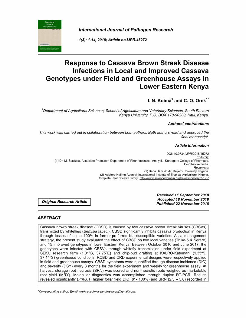
Foliar symptoms typical of CBSD were observed as early as 3 MAP in two (Serere & Thika-5) local susceptible landraces (Fig. 1a & 1b) compared to improved genotypes that showed no symptoms under field trials (Fig. 1c).These symptoms included yellow vein banding, expressed mainly on the lower, older leaves and chlorosis which
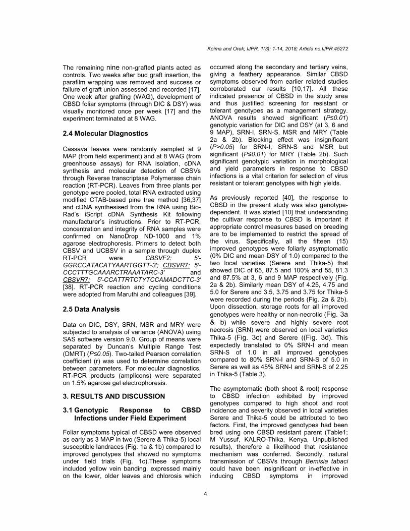
occurred along the secondary and tertiary veins, giving a feathery appearance. Similar CBSD symptoms observed from earlier related studies corroborated our results [10,17]. All these indicated presence of CBSD in the study area and thus justified screening for resistant or tolerant genotypes as a management strategy. ANOVA results showed significant (P≤0.01) genotypic variation for DIC and DSY (at 3, 6 and 9 MAP), SRN-I, SRN-S, MSR and MRY (Table 2a & 2b). Blocking effect was insignificant (P>0.05) for SRN-I, SRN-S and MSR but significant (P≤0.01) for MRY (Table 2b). Such significant genotypic variation in morphological and yield parameters in response to CBSD infections is a vital criterion for selection of virus resistant or tolerant genotypes with high yields. As previously reported [40], the response to CBSD in the present study was also genotype-dependent. It was stated [10] that understanding the cultivar response to CBSD is important if appropriate control measures based on breeding are to be implemented to restrict the spread of the virus. Specifically, all the fifteen (15) improved genotypes were foliarly asymptomatic (0% DIC and mean DSY of 1.0) compared to the two local varieties (Serere and Thika-5) that showed DIC of 65, 87.5 and 100% and 55, 81.3 and 87.5% at 3, 6 and 9 MAP respectively (Fig. 2a & 2b). Similarly mean DSY of 4.25, 4.75 and 5.0 for Serere and 3.5, 3.75 and 3.75 for Thika-5 were recorded during the periods (Fig. 2a & 2b). Upon dissection, storage roots for all improved
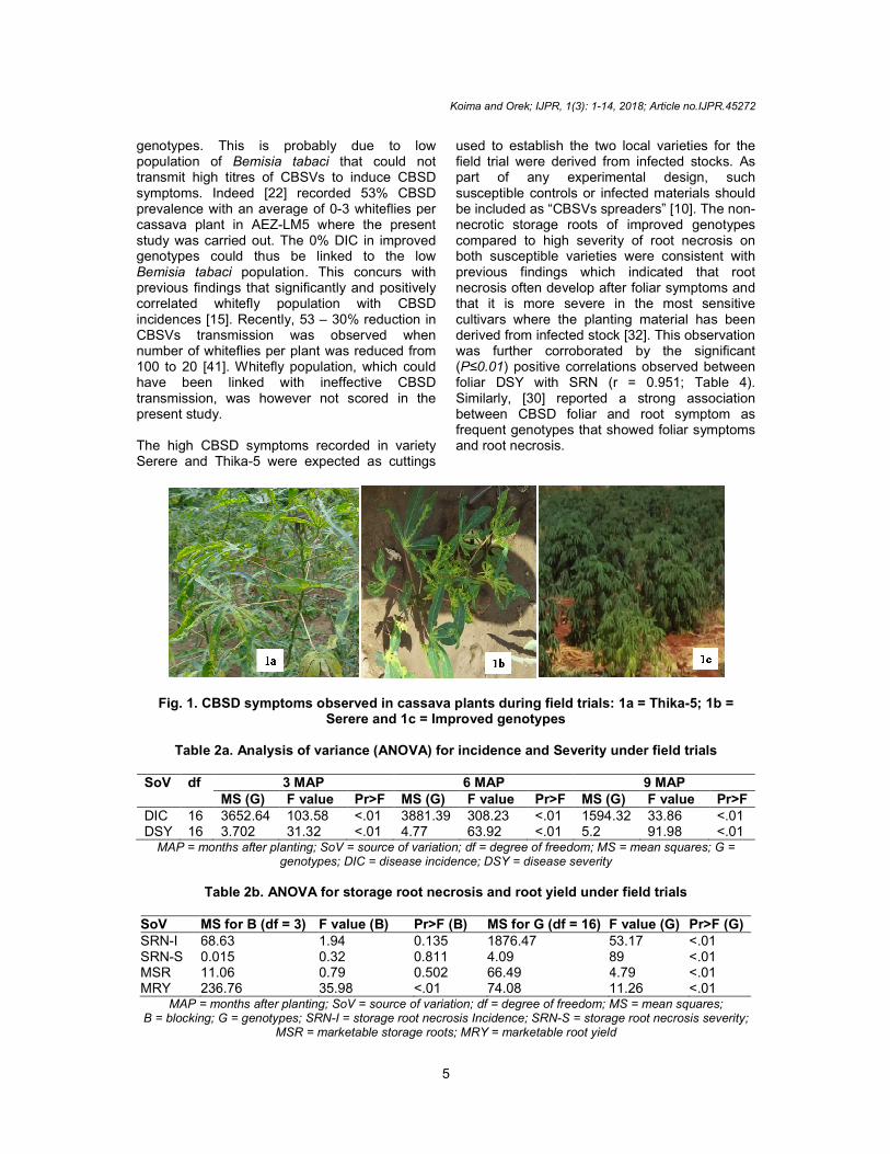
genotypes were healthy or non-necrotic (Fig. 3a & b) while severe and highly severe root necrosis (SRN) were observed on local varieties
Thika-5 (Fig. 3c) and Serere ((Fig. 3d). This expectedly translated to 0% SRN-I and mean SRN-S of 1.0 in all improved genotypes compared to 80% SRN-I and SRN-S of 5.0 in Serere as well as 45% SRN-I and SRN-S of 2.25 in Thika-5 (Table 3). The asymptomatic (both shoot & root) response to CBSD infection exhibited by improved genotypes compared to high shoot and root incidence and severity observed in local varieties Serere and Thika-5 could be attributed to two factors. First, the improved genotypes had been bred using one CBSD resistant parent (Table1; M Yussuf, KALRO-Thika, Kenya, Unpublished results), therefore a likelihood that resistance mechanism was conferred. Secondly, natural transmission of CBSVs through Bemisia tabaci could have been insignificant or in-effective in inducing CBSD symptoms in improved
genotypes. This is probably due to low population of Bemisia tabaci thattransmit high titres of CBSVs to induce CBSD symptoms. Indeed [22] recorded 53% CBSD prevalence with an average of 0-3 whiteflies per cassava plant in AEZ-LM5 where the present study was carried out. The 0% DIC in improved genotypes could thus be linked to the low Bemisia tabaci population. This concurs with previous findings that significantly and positively correlated whitefly population with CBSD incidences [15]. Recently, 53 – 30% reduction in CBSVs transmission was observed when number of whiteflies per plant was reduced from 100 to 20 [41]. Whitefly population, which could have been linked with ineffective CBSD transmission, was however not scored in the present study. The high CBSD symptoms recorded in variety Serere and Thika-5 were expected as cuttings
Fig. 1. CBSD symptoms observed in cassava plants during field trials: 1a = ThikaSerere and 1c = Improved
Table 2a. Analysis of variance (ANOVA) for incidence and Severity under field trials
SoV df 3 MAP MS (G) F value
DIC 16 3652.64 103.58 DSY 16 3.702 31.32
MAP = months after planting; SoV = source of variation; dfgenotypes; DIC = disease incidence; DSY = disease severity
Table 2b. ANOVA for storage root necrosis and root yield under field trials
SoV MS for B (df = 3) F value (B)SRN-I 68.63 1.94 SRN-S 0.015 0.32 MSR 11.06 0.79 MRY 236.76 35.98
MAP = months after planting; SoV = source of variation; dfB = blocking; G = genotypes; SRN-I = storage root necrosis Incidence; SRN
MSR = marketable storage roots; MRY = marketable root yield
Koima and Orek; IJPR, 1(3): 1-14, 2018; Article no.
5
genotypes. This is probably due to low that could not
transmit high titres of CBSVs to induce CBSD symptoms. Indeed [22] recorded 53% CBSD
3 whiteflies per LM5 where the present
study was carried out. The 0% DIC in improved linked to the low
population. This concurs with previous findings that significantly and positively correlated whitefly population with CBSD
30% reduction in CBSVs transmission was observed when
teflies per plant was reduced from 100 to 20 [41]. Whitefly population, which could have been linked with ineffective CBSD transmission, was however not scored in the
The high CBSD symptoms recorded in variety 5 were expected as cuttings
used to establish the two local varieties for the field trial were derived from infected stocks. As part of any experimental design, such susceptible controls or infected materials should be included as “CBSVs spreaders” [10]. The nonnecrotic storage roots of improved genotypes compared to high severity of root necrosis on both susceptible varieties were consistent with previous findings which indicated that root necrosis often develop after foliar symptoms and that it is more severe in the most sensitive cultivars where the planting material has been derived from infected stock [32]. This observation was further corroborated by the significant (P≤0.01) positive correlations observed between foliar DSY with SRN (r = 0.951; Table 4). Similarly, [30] reported a strong association between CBSD foliar and root symptom as frequent genotypes that showed foliar symptoms and root necrosis.
CBSD symptoms observed in cassava plants during field trials: 1a = Thika
erere and 1c = Improved genotypes
Analysis of variance (ANOVA) for incidence and Severity under field trials
6 MAP 9 MAPPr>F MS (G) F value Pr>F MS (G) F value<.01 3881.39 308.23 <.01 1594.32 33.86<.01 4.77 63.92 <.01 5.2 91.98
MAP = months after planting; SoV = source of variation; df = degree of freedom; MS = mean squares; G = genotypes; DIC = disease incidence; DSY = disease severity
ANOVA for storage root necrosis and root yield under field trials
F value (B) Pr>F (B) MS for G (df = 16) F value (G)
0.135 1876.47 53.17 0.811 4.09 89 0.502 66.49 4.79
35.98 <.01 74.08 11.26 MAP = months after planting; SoV = source of variation; df = degree of freedom; MS = mean squares;
I = storage root necrosis Incidence; SRN-S = storage root necrosis severity; MSR = marketable storage roots; MRY = marketable root yield
; Article no.IJPR.45272
used to establish the two local varieties for the field trial were derived from infected stocks. As part of any experimental design, such susceptible controls or infected materials should
rs” [10]. The non-necrotic storage roots of improved genotypes compared to high severity of root necrosis on both susceptible varieties were consistent with previous findings which indicated that root necrosis often develop after foliar symptoms and
t is more severe in the most sensitive cultivars where the planting material has been derived from infected stock [32]. This observation was further corroborated by the significant
) positive correlations observed between 951; Table 4).
strong association between CBSD foliar and root symptom as frequent genotypes that showed foliar symptoms
CBSD symptoms observed in cassava plants during field trials: 1a = Thika-5; 1b =
Analysis of variance (ANOVA) for incidence and Severity under field trials
9 MAP F value Pr>F 33.86 <.01 91.98 <.01
= degree of freedom; MS = mean squares; G =
ANOVA for storage root necrosis and root yield under field trials
F value (G) Pr>F (G) <.01 <.01 <.01 <.01
= degree of freedom; MS = mean squares; S = storage root necrosis severity;
Fig. 2a. Mean percent disease incidence (DIC) under field experimentMAP = Months after planting, CBSD
Fig. 2b. Mean disease severity (DSY) under field experimentMAP = Months after planting, CBSD
Fig. 3. Storage root necrosis (SRN) from fieldTubers exhibiting CBSD-associated root necrosis: Fig. 3a & b = non
genotypes; Fig. 3c = severe necrosis on variety Thika
Koima and Orek; IJPR, 1(3): 1-14, 2018; Article no.
6
Mean percent disease incidence (DIC) under field experiment
MAP = Months after planting, CBSD-I = CBSD Incidence; KB = Kiboko; TK = Thika
Mean disease severity (DSY) under field experiment
MAP = Months after planting, CBSD-S = CBSD Severity; KB = Kiboko; TK = Thika
Storage root necrosis (SRN) from field-harvested root tubers
associated root necrosis: Fig. 3a & b = non-necrotic tubers fr= severe necrosis on variety Thika-5; Fig. 3d = high severity of necrosis of variety
Serere
; Article no.IJPR.45272
Mean percent disease incidence (DIC) under field experiment I = CBSD Incidence; KB = Kiboko; TK = Thika
S = CBSD Severity; KB = Kiboko; TK = Thika
harvested root tubers necrotic tubers from improved
5; Fig. 3d = high severity of necrosis of variety
Koima and Orek; IJPR, 1(3): 1-14, 2018; Article no.IJPR.45272
7
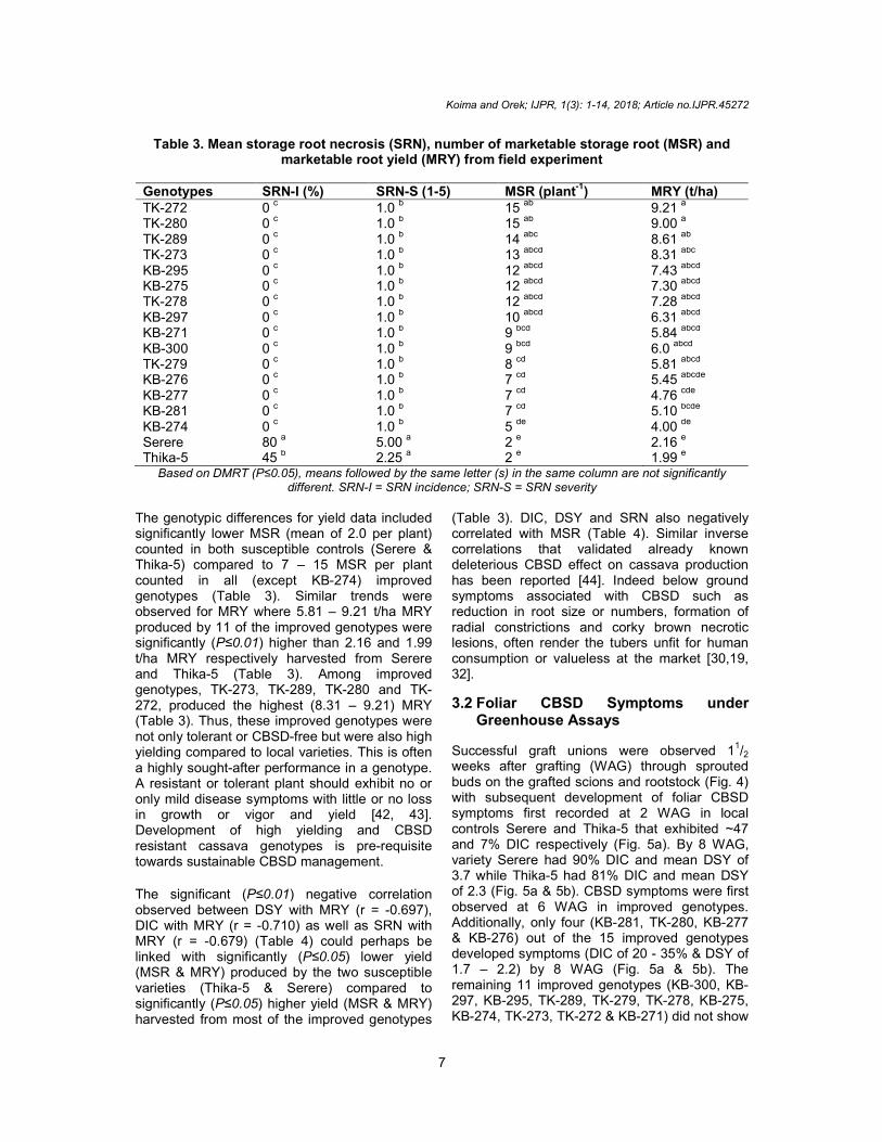
Table 3. Mean storage root necrosis (SRN), number of marketable storage root (MSR) and marketable root yield (MRY) from field experiment
Genotypes SRN-I (%) SRN-S (1-5) MSR (plant
-1) MRY (t/ha)
TK-272 0 c 1.0
b 15
ab 9.21
a
TK-280 0 c 1.0 b 15 ab 9.00 a TK-289 0
c 1.0
b 14
abc 8.61
ab
TK-273 0 c 1.0 b 13 abcd 8.31 abc KB-295 0
c 1.0
b 12
abcd 7.43
abcd
KB-275 0 c 1.0
b 12
abcd 7.30
abcd
TK-278 0 c 1.0 b 12 abcd 7.28 abcd KB-297 0
c 1.0
b 10
abcd 6.31
abcd
KB-271 0 c 1.0 b 9 bcd 5.84 abcd KB-300 0
c 1.0
b 9
bcd 6.0
abcd
TK-279 0 c 1.0
b 8
cd 5.81
abcd
KB-276 0 c 1.0 b 7 cd 5.45 abcde KB-277 0
c 1.0
b 7
cd 4.76
cde
KB-281 0 c 1.0 b 7 cd 5.10 bcde KB-274 0
c 1.0
b 5
de 4.00
de
Serere 80 a 5.00
a 2
e 2.16
e
Thika-5 45 b 2.25 a 2 e 1.99 e Based on DMRT (P≤0.05), means followed by the same letter (s) in the same column are not significantly
different. SRN-I = SRN incidence; SRN-S = SRN severity The genotypic differences for yield data included significantly lower MSR (mean of 2.0 per plant) counted in both susceptible controls (Serere & Thika-5) compared to 7 – 15 MSR per plant counted in all (except KB-274) improved genotypes (Table 3). Similar trends were observed for MRY where 5.81 – 9.21 t/ha MRY produced by 11 of the improved genotypes were significantly (P≤0.01) higher than 2.16 and 1.99 t/ha MRY respectively harvested from Serere and Thika-5 (Table 3). Among improved genotypes, TK-273, TK-289, TK-280 and TK-272, produced the highest (8.31 – 9.21) MRY (Table 3). Thus, these improved genotypes were not only tolerant or CBSD-free but were also high yielding compared to local varieties. This is often a highly sought-after performance in a genotype. A resistant or tolerant plant should exhibit no or only mild disease symptoms with little or no loss in growth or vigor and yield [42, 43]. Development of high yielding and CBSD resistant cassava genotypes is pre-requisite towards sustainable CBSD management.
The significant (P≤0.01) negative correlation observed between DSY with MRY (r = -0.697), DIC with MRY (r = -0.710) as well as SRN with MRY (r = -0.679) (Table 4) could perhaps be linked with significantly (P≤0.05) lower yield (MSR & MRY) produced by the two susceptible varieties (Thika-5 & Serere) compared to significantly (P≤0.05) higher yield (MSR & MRY) harvested from most of the improved genotypes
(Table 3). DIC, DSY and SRN also negatively correlated with MSR (Table 4). Similar inverse correlations that validated already known deleterious CBSD effect on cassava production has been reported [44]. Indeed below ground symptoms associated with CBSD such as reduction in root size or numbers, formation of radial constrictions and corky brown necrotic lesions, often render the tubers unfit for human consumption or valueless at the market [30,19, 32].
3.2 Foliar CBSD Symptoms under Greenhouse Assays
Successful graft unions were observed 11/2
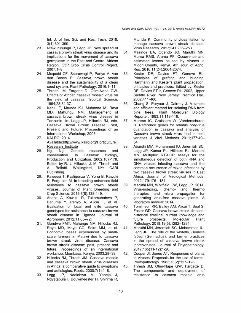
weeks after grafting (WAG) through sprouted buds on the grafted scions and rootstock (Fig. 4) with subsequent development of foliar CBSD symptoms first recorded at 2 WAG in local controls Serere and Thika-5 that exhibited ~47 and 7% DIC respectively (Fig. 5a). By 8 WAG, variety Serere had 90% DIC and mean DSY of 3.7 while Thika-5 had 81% DIC and mean DSY of 2.3 (Fig. 5a & 5b). CBSD symptoms were first observed at 6 WAG in improved genotypes. Additionally, only four (KB-281, TK-280, KB-277 & KB-276) out of the 15 improved genotypes developed symptoms (DIC of 20 - 35% & DSY of 1.7 – 2.2) by 8 WAG (Fig. 5a & 5b). The remaining 11 improved genotypes (KB-300, KB-297, KB-295, TK-289, TK-279, TK-278, KB-275, KB-274, TK-273, TK-272 & KB-271) did not show
CBSD symptoms at the end of 8 WAG (Fig. 5a & 5b). A number of implications were deducedabove results. First, through sprouting of the buds and subsequent pronounced CBSD symptom development, the chiptechnique effectively transmitted CBSVs. Previous successful transmission of CBSVs through chip bud grafting resulted in 70 CBSD incidence [17,45]. Secondly, the period taken for symptoms to develop or show and severity in symptom expression was genotype-dependent. For instance, symptoms developed as early as 2 WAG in local varieties Serere and Thikacompared to delayed symptoms observed at 6 WAG in some improved genotypes. variations in time taken for symptoms to develop
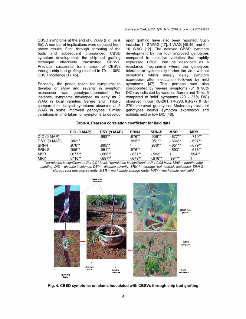
Table 4. Pearson correlation coefficient for field data
DIC (9 MAP) DIC (9 MAP) 1 DSY (9 MAP) .992** SRN-I .976** SRN-S .906** MSR -.677** MRY -.710**
**correlation is significant at P ≤ 0.01 level; *correlation is significant at P ≤ 0.05 level; MAP = months after planting; DIC = disease incidence; DSY = disease severity; SRN
storage root necrosis severity; MSR = marketable storage roots; MRY
Fig. 4. CBSD symptoms on plants inoculated with CBSVs through chip bud grafting
Koima and Orek; IJPR, 1(3): 1-14, 2018; Article no.
8
oms at the end of 8 WAG (Fig. 5a & were deduced from
results. First, through sprouting of the buds and subsequent pronounced CBSD symptom development, the chip-bud grafting technique effectively transmitted CBSVs.
us successful transmission of CBSVs through chip bud grafting resulted in 70 – 100%
for symptoms to develop or show and severity in symptom
dependent. For instance, symptoms developed as early as 2 WAG in local varieties Serere and Thika-5 compared to delayed symptoms observed at 6 WAG in some improved genotypes. Similar
taken for symptoms to develop
upon grafting have also been reportedincludes 1 - 2 WAG [17], 4 WAG [45,10 WAG [12]. The delayed CBSD symptom development by the four improved genotypes compared to sensitive varieties that rapidly expressed CBSD, can be described as a resistance mechanism where the genotypes tolerates or systemically harbor the virus without symptoms which merely delay symptom expression after inoculation followed by mild symptoms [47]. This perhaps was also corroborated by 'severe' symptoms (81 & 90% DIC) as indicated by varieties Serere and Thikacompared to 'mild' symptoms (20 observed in four (KB-281, TK-280, KB276) improved genotypes. Moderately resistant genotypes delays symptom expression and exhibits mild or low DIC [48].
Pearson correlation coefficient for field data
DSY (9 MAP) SRN-I SRN-S MSR .992** .976** .906** -.677** 1 .995** .951** -.666** .995** 1 .976** -.651** .951** .976** 1 -.593* -.666** -.651** -.593* 1 -.697** -.679** -.616** .994**
≤ 0.01 level; *correlation is significant at P ≤ 0.05 level; MAP = months after planting; DIC = disease incidence; DSY = disease severity; SRN-I = storage root necrosis incidence; SRN
storage root necrosis severity; MSR = marketable storage roots; MRY = marketable root yield
CBSD symptoms on plants inoculated with CBSVs through chip bud grafting
; Article no.IJPR.45272
been reported. Such G [17], 4 WAG [45,46] and 6 –
10 WAG [12]. The delayed CBSD symptom development by the four improved genotypes
ties that rapidly can be described as a
resistance mechanism where the genotypes the virus without
symptoms which merely delay symptom expression after inoculation followed by mild
s was also corroborated by 'severe' symptoms (81 & 90% DIC) as indicated by varieties Serere and Thika-5 compared to 'mild' symptoms (20 - 35% DIC)
280, KB-277 & KB-276) improved genotypes. Moderately resistant
symptom expression and
MRY -.710** -.697** -.679** -.616** .994** 1
≤ 0.01 level; *correlation is significant at P ≤ 0.05 level; MAP = months after I = storage root necrosis incidence; SRN-S =
= marketable root yield
CBSD symptoms on plants inoculated with CBSVs through chip bud grafting

Thirdly, unlike field trials where all improved genotypes showed no CBSD symptoms, the chip bud grafting technique produced a 20 in four (KB-281, TK-280, KB-277 & KBimproved genotypes. This perhaps elucidated variation in efficiency of CBSVs transmission between the two methods. Indeed transmission or inoculation of viruses in cassava through grafting has been reported to be more efficient both in the field and greenhouse conditions compared to vector-borne (whitefly) transmission [45,17,49]. Thus transmission of CBSVs through whiteflies in the current field experiment was considered insignificant.
3.3 Molecular Diagnostics
RT-PCR detected CBSV (345 bp) in two susceptible controls (Thika-5 & Serere) and one improved genotype (KB-276) while the remaining fourteen improved genotypes were all CBSVunder field trials (Fig. 6a). Under chipgrafting (at 8 WAG), the two susceptible controls (Thika-5 & Serere) and five improved genotypes (KB-276, KB-281, TK-280, KB-277 & KBwere positive for CBSV (345 bp) while the remaining eleven improved genotypes (KBKB-297, KB-295, TK-289, TK-279, TK272, KB-275, KB-274 & KB-273) were negative for CBSV (Fig. 6b). In either experiments (greenhouse and field), no UCBSV (441 bp) were detected (Fig. 6a & 6b).
Fig. 5a. Disease incidence (DIC) of genotypes inoculated
Wag = weeks after grafting;
Koima and Orek; IJPR, 1(3): 1-14, 2018; Article no.
9
Thirdly, unlike field trials where all improved genotypes showed no CBSD symptoms, the chip bud grafting technique produced a 20 – 35% DIC
277 & KB-276) perhaps elucidated
s transmission between the two methods. Indeed transmission or inoculation of viruses in cassava through grafting has been reported to be more efficient both in the field and greenhouse conditions
whitefly) transmission CBSVs through
whiteflies in the current field experiment was
PCR detected CBSV (345 bp) in two 5 & Serere) and one
ile the remaining fourteen improved genotypes were all CBSV-free under field trials (Fig. 6a). Under chip-bud grafting (at 8 WAG), the two susceptible controls
5 & Serere) and five improved genotypes 277 & KB-271)
sitive for CBSV (345 bp) while the remaining eleven improved genotypes (KB-300,
279, TK-278, KB-273) were negative
for CBSV (Fig. 6b). In either experiments (greenhouse and field), no UCBSV (441 bp) were
Molecular detection of CBSV in five improved genotypes further provides evidence on effectiveness of transmission of CBSV through chip-bud grafting. Although visually asymptomatic under field trial (Fig. 2a & 2b), improved genotype KB-276 was positive for CBSV (Fig. 6a). SimilarlyKB-271 showed no CBSD symptoms upon grafting (Fig. 5a & 5b) presence of CBSV was detectedPCR (Fig. 6b). The two genotypes exhibited CBSV latency, a situation where some infected plants may the virus but remain visually symptomless or where some varieties express symptoms in roots rather than on leaves [23,9]. It is, therefore, prerequisite to carry out molecular diagnostics in addition to visual monitoring of CBSD symptoms in cassava. Although the duplex RTto detect both CBSV and UCBSV in a sample, only CBSV was detected from both field greenhouse leaf samples. Thisassociated with differential interaction between CBSV and UCBSV. CBSV is a more aggressive and virulent CBSD viral pathogen inducing more rapid and severe CBSD symptoms compared to UCBSV that produces milder folia[50,51]. Additionally, tolerant cassava varieties have been infected with only CBSV, but free of UCBSV, suggesting their resistance UCBSV [52]. Indeed CBSV isolates have been reported
Disease incidence (DIC) of genotypes inoculated with CBSVs through Chip
grafting Wag = weeks after grafting; DIC = Disease or CBSD incidence
; Article no.IJPR.45272
Molecular detection of CBSV in five improved genotypes further provides evidence on
of transmission of CBSV bud grafting. Although visually
asymptomatic under field trial (Fig. 2a & 2b), 276 was positive for
Similarly genotype 271 showed no CBSD symptoms
upon grafting (Fig. 5a & 5b) but was detected through RT-
The two genotypes exhibited CBSV latency, a situation where some infected plants may harbor the virus but remain visually symptomless or where some varieties express symptoms in roots
9]. It is, therefore, pre-requisite to carry out molecular diagnostics in addition to visual monitoring of CBSD symptoms in cassava. Although the duplex RT-PCR was to detect both CBSV and UCBSV in a sample,
from both field and This could be
interaction between CBSV and UCBSV. CBSV is a more aggressive and virulent CBSD viral pathogen inducing more rapid and severe CBSD symptoms compared to
ces milder foliar symptoms tolerant cassava varieties
with only CBSV, but free of UCBSV, suggesting their resistance UCBSV [52]. Indeed CBSV isolates have been reported
with CBSVs through Chip-bud
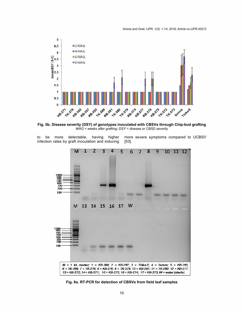
Fig. 5b. Disease severity (DSY) of genotypes inoculated with CBSVs through ChipWAG = weeks after grafting
to be more detectable, having higher infection rates by graft inoculation and inducing
Fig. 6a. RT-PCR for detection of CBSVs from field leaf samples
Koima and Orek; IJPR, 1(3): 1-14, 2018; Article no.
10
Disease severity (DSY) of genotypes inoculated with CBSVs through Chip
weeks after grafting; DSY = disease or CBSD severity
to be more detectable, having higher infection rates by graft inoculation and inducing
more severe symptoms compared to UCBSV [53].
PCR for detection of CBSVs from field leaf samples
; Article no.IJPR.45272
Disease severity (DSY) of genotypes inoculated with CBSVs through Chip-bud grafting
more severe symptoms compared to UCBSV

Fig. 6b. RT-PCR for detection of CBSVs from chip 4. CONCLUSION Conclusively, of the 17 genotypes screened, the present study indicated that, two local varieties (Thika-5 & Serere) and five improved genotypes (KB-276, KB-281, TK-280, KB-277 and KBexhibited CBSD symptoms while ten improved genotypes (KB-300, KB-297, KB-TK-279, TK-278, TK-272, KB-275, KBTK-273) were asymptomatic. The ten genotypes were not only CBSD-free, but also produced significantly higher marketable root yield compared to susceptible local varieties. As suggested by [30], such genotypes can potentially be considered as parental breeding stocks for CBSD resistance breeding. genotypes should however be subjected to further molecular characterisation to identify underlying tolerance or resistance mechanisms.
ACKNOWLEDGEMENT The authors acknowledge Dr. Evans Nyaboga of the University of Nairobi, department of
Koima and Orek; IJPR, 1(3): 1-14, 2018; Article no.
11
PCR for detection of CBSVs from chip-bud grafted samples
Conclusively, of the 17 genotypes screened, the present study indicated that, two local varieties
5 & Serere) and five improved genotypes 277 and KB-271)
exhibited CBSD symptoms while ten improved -295, TK-289,
275, KB-274 and 273) were asymptomatic. The ten genotypes
free, but also produced significantly higher marketable root yield compared to susceptible local varieties. As
such genotypes can parental breeding
stocks for CBSD resistance breeding. The genotypes should however be subjected to further molecular characterisation to identify underlying tolerance or resistance mechanisms.
The authors acknowledge Dr. Evans Nyaboga of the University of Nairobi, department of
Biochemistry who assisted with molecular diagnostics; Dr. Benard Mware of International Institute of Tropical Agriculture (IITA) at Kenya who assisted with chip-bud grafting and Mr. Migwa Yussuf of Kenya Agricultural and Livestock Research Organization (KALRO-Katumani) who provided cassava cuttings for the study.
COMPETING INTERESTS Authors have declared that no interests exist.
REFERENCES 1. Food and Agricultural Organization (FAO)
of the United Nations, 2002. Partnership formed to improve cassava, staple food for 600 million people. Available:http://www.fao.org/english/newsroom/news/2002/10541-en.html
2. Food and Agriculture Organization (FAO) of the United Nations, 2008. Cassava for
; Article no.IJPR.45272
bud grafted samples
Biochemistry who assisted with molecular diagnostics; Dr. Benard Mware of International
Tropical Agriculture (IITA) at ILRI-bud grafting and
Migwa Yussuf of Kenya Agricultural and Livestock Research Organization –Katumani
Katumani) who provided cassava
Authors have declared that no competing
Food and Agricultural Organization (FAO) of the United Nations, 2002. Partnership formed to improve cassava, staple food for
http://www.fao.org/english/newsren.html
Food and Agriculture Organization (FAO) of the United Nations, 2008. Cassava for

Koima and Orek; IJPR, 1(3): 1-14, 2018; Article no.IJPR.45272
12
food and energy security: Investing in cassava research and development could boost yields and Industrial uses. Available:http://www.fao.org/newsroom/en/news/2008/1000899/index.html
3. Latif S, Müller J. Cassava-how to explore the all-sufficient. International Journal for Rural Development. 2014;30–31.
4. Spencer, DSC, Ezedinma C, 2017. Cassava cultivation in sub-Saharan Africa. Available:http://dx.doi.org/10.19103/AS.2016.0014.06 Burleigh Dodds Science Publishing Limited. All rights reserved, 1 – 26
5. Mware B, Narla R, Mata R, Olubayo F, Songa J, Kyamanyua S, Ateka EM. Efficiency of cassava brown streak virus transmission by two whitefly species in coastal Kenya. Journal of Genetic Molecular Virology. 2009;1:040-045.
6. Wangari MF. Potential toxic levels of cyanide in cassava (Manihot esculenta Crantz) grown in some parts of Kenya. Master’s Thesis, 2013, Kenyatta University, Kenya; 2013.
7. Irungu J. Prevalence and Co-infection of cassava with Cassava Mosaic Geminivirus and Cassava Brown Streak Virus in popular cultivars in Western Kenya. International Journal of Pest Management. 2011;46:211-217.
8. Karuga J. Turning cassava into gold: Small producer taps into big demand in Kenya; 2016. Available:https://nextbillion.net/turning-cassava-into-gold-small-producer-taps-into-big-demand-in-kenya/
9. Alicai T, Omongo CA, Maruthi MN, Hillocks RJ, Baguma Y, Kawuki R, et al. Re-emergence of cassava brown streak disease in Uganda. Plant Diseases. 2007; 91:24-29.
10. Kaweesi T, Kawuki R, Kyaligonza V, Baguma Y, Tusiime G, Ferguson M. Field evaluation of selected cassava genotypes for cassava brown streak disease based on symptom expression and virus load. Virology Journal. 2014;11:2–14.
11. Legg JP, Jeremiah SC, Obiero HM, Maruthi MN, Ndyetabula I, Okao-Okuja G et al. Comparing the regional epidemiology of the cassava mosaic and cassava brown streak virus pandemics in Africa. Virus Research. 2011;159:161–170.
12. Winter S, Koerbler M, Stein B, Pietruszka A, Martina P, Butgereitt A. The analysis of cassava brown streak viruses reveals the
presence of distinct virus species causing cassava brown streak disease in East Africa. Journal of General Virology. 2010; 91:1365.
13. Monger WA, Alicai T, Ndunguru J, Kinyua ZM, Potts M, Reeder RH, et al. The complete genome sequence of the Tanzanian strain of Cassava brown streak virus and comparison with the Ugandan strain sequence. Arch Virology. 2010;155: 429–433.
14. Mbanzibwa DR, Tian YP, Tugume AK, Patil BL, Yadav JS, Bagewadi B, et al, Evolution of cassava brown streak disease associated viruses. Journal of General Virology. 2011;92:974–987.
15. Maruthi MN, Hillocks RJ, Mtunda K, Raya M, Muhama M, Kiozia H, et al. Transmission of Cassava brown streak virus by Bemisia tabaci (Gennadius). Journal of Phytopathology. 2005;153:307-312.
16. Alabi OJ, Kumar PL, Naidu RA. Cassava mosaic disease: A curse to food security in Sub-Saharan Africa. Online APSnet Features. 2011;1–17.
17. Wagaba H, Beyene G, Trembley C, Alicai T, Fauquet CM, Taylor NJ. Efficient transmission of Cassava Brown Streak disease viral pathogens by chip bud grafting. BMC Research Notes. 2013;6: 516.
18. Aloyce RC. Development and evaluation of efficient diagnostic tools for cassava mosaic and cassava brown streak diseases. MSc. Thesis; 2013. Available:http://wiredspace.wits.ac.za/handle/10539/14018
19. Hillocks RJ, Jennings DL. Cassava brown streak disease: A review of present knowledge and research needs. International Journal Pest Management. 2003;49:225–234.
20. Hillocks RJ, Raya MD, Mtunda K, Kiozia H. Effects of brown streak virus disease on yield and quality of cassava in Tanzania. Journal of Phytopathology. 2001;149:389-394.
21. Ngure GK. The occurrence and integrated management of Cassava brown streak disease in Coastal Kenya. MSc thesis; 2012.
22. Koima IN, Orek CO, Nguluu SN. Distribution of cassava mosaic and cassava brown streak diseases in agro-ecological zones of lower Eastern Kenya.
Koima and Orek; IJPR, 1(3): 1-14, 2018; Article no.IJPR.45272
13
Int. J. of Inn. Sci. and Res. Tech. 2018; 3(1):391-399.
23. Ntawuruhunga P, Legg JP. New spread of cassava brown streak virus disease and its implications for the movement of cassava germplasm in the East and Central African Region. C3P Crop Crisis Control Project. 2007;1–8.
24. Mcquaid CF, Sseruwagi P, Pariyo A, van den Bosch F. Cassava brown streak disease and the sustainability of a clean seed system. Plant Pathology. 2016;1–11.
25. Thresh JM, Fargette D, Otim-Nape GW. Effects of African cassava mosaic virus on the yield of cassava. Tropical Science. 1994;28:34-37.
26. Kanju E, Mtunda KJ, Muhanna M, Raya MD, Mahungu NM. Management of cassava brown streak virus disease in Tanzania. In: Legg JP, Hillocks RJ, eds. Cassava Brown Streak Disease: Past, Present and Future. Proceedings of an International Workshop; 2003
27. KALRO; 2014. Available:http://www.kalro.org/Horticulture_Research_Institute
28. Ng, Ng. Genetic resources and conservation. In Cassava: Biology, Production and Utilization. 2002;167-178. Edited by R. J. Hillocks, J. M. Thresh and A. Bellotti. Wallingford, NY: CABI Publishing.
29. Kaweesi T, Kyaligonza V, Yona B, Kawuki R, Ferguson M. In-breeding enhances field resistance to cassava brown streak viruses. Journal of Plant Breeding and Crop Science. 2016;8(8):138-149.
30. Abaca A, Kawuki R, Tukamuhabwa P, Baguma Y, Pariyo A, Alicai T, et al. Evaluation of local and elite cassava genotypes for resistance to cassava brown streak disease in Uganda. Journal of Agronomy. 2012;11:65–72.
31. Gondwe FMT, Mahungu NM, Hillocks RJ, Raya MD, Moyo CC, Soko MM, et al. Economic losses experienced by small-scale farmers in Malawi due to cassava brown streak virus disease. Cassava brown streak disease: past, present and future. Proceedings of an international workshop, Mombasa, Kenya. 2003;28–38.
32. Hillocks RJ, Thresh JM. Cassava mosaic and cassava brown streak virus diseases in Africa: a comparative guide to symptoms and aetiologies. Roots. 2000;7(1):1–8.
33. Legg JP, Ndalahwa M, Yabeja J, Ndyetabula I, Bouwmeester H, Shirima R,
Mtunda K. Community phytosanitation to manage cassava brown streak disease. Virus Research. 2017;241:236–253.
34. Masinde EA, Ogendo JO, Maruthi MN, Mulwa RMS, Arama PF. Occurrence and estimated losses caused by viruses in Migori County, Kenya. Afr. Jour. of Agric. Res. 2016;11(24):2064-2074.
35. Kester DE, Davies FT, Geneve RL. Principles of grafting and budding. Hartmann and Kester's plant propagation: principles and practices. Edited by: Kester DE, Davies FTJr, Geneve RL. 2002, Upper Saddle River, New Jersey: Prentice Hall. 2002;411-460.
36. Chang S, Puryear J, Cairney J. A simple and efficient method for isolating RNA from pine trees. Plant Molecular Biology Reporter. 1993;11:113-116.
37. Moreno IC, Gruissem W, Vanderschuren H. Reference genes for reliable potyvirus quantitation in cassava and analysis of Cassava brown streak virus load in host varieties. J. Virol. Methods. 2011;177:49–54.
38. Abarshi MM, Mohammed IU, Jeremiah SC, Legg JP, Kumar PL, Hillocks RJ, Maruthi MN. Multiplex RT-PCR assays for the simultaneous detection of both RNA and DNA viruses infecting cassava and the common occurrence of mixed infections by two cassava brown streak viruses in East Africa. Journal of Virological Methods. 2012;179:176 –184.
39. Maruthi MN, Whitfield CW, Legg JP. 2014. Virus-indexing, chemo- and thermo therapies, and micro propagation for generating virus-free cassava plants. A laboratory manual; 2014.
40. Tomlinson KR, Bailey AM, Alicai T, Seal S, Foster GD. Cassava brown streak disease: historical timeline, current knowledge and future prospects. Molecular Plant Pathology. 2018;19(5):1282–1294.
41. Maruthi MN, Jeremiah SC, Mohammed IU, Legg JP. The role of the whitefly, Bemisia tabaci (Gennadius), and farmer practices in the spread of cassava brown streak Ipomoviruses. Journal of Phytopathology. 2017;165(11-12):1-20.
42. Cooper JI, Jones AT. Responses of plants to viruses: Proposals for the use of terms. Phytopathology. 1983;73(2):127–128.
43. Thresh JM, Otim-Nape GW, Fargette D. The components and deployment of resistance to cassava mosaic virus
Koima and Orek; IJPR, 1(3): 1-14, 2018; Article no.IJPR.45272
14
disease. Inter Pest Man Rev. 2008;3:209-224.
44. Koros JC, Runo SM, Yusuf M, Orek CO. Screening selected cassava cultivars for resistance against cassava viruses and cassava green mites under advanced yield trials in Kenya. IOSR-JBB. 2018;4(5):37-52.
45. Rwegasira GM, Chrissie MER. Efficiency of non-vector methods of cassava brown streak virus transmission to susceptible cassava plants. African Journal of Food, Agriculture, Nutrition and Development. 2015;15(4).
46. Anjanappa RB, Mehta D, Maruthi MN, Kanju E, Gruissem W, Vanderschuren H. Characterization of Brown Streak Virus-Resistant Cassava. MPMI. 2016;29(7):527 – 534.
47. Lima JAA, Silva AKFD, Aragao MDL, Ferreira NRDA, Teofilo EM. Simple and multiple resistances to viruses in cowpea genotypes. Pesquisa Agropecuaria Brasileira. 2011;46:1432-1438.
48. Nath S, Aktar S, Hazra P. Biochemical factors for TLCV tolerance in tomato genotypes. Journal of Crop and Weed. 2009;5(1):152-156.
49. Houngue JA, Zandjanakou TM, Ngallec HB, Pitad JS, Cacaïa GHT, Ngatate SE, et al. Evaluation of resistance to cassava mosaic disease in selected African cassava cultivars using combined
molecular and greenhouse grafting tools. Physiological and Molecular Plant Pathology; 2018. Available:https://doi.org/10.1016/j.pmpp.2018.07.003
50. Ogwok E, Ilyas M, Alicai T, Rey MEC, Taylor NJ. Comparative analysis of virus-derived small RNAs within cassava (Manihot esculenta Crantz) infected with cassava brown streak viruses. Virus Research. 2016;215:1–11.
51. Ndunguru J, Sseruwagi P, Tairo F, Stomeo F, Maina S, Djinkeng A, et al. Analyses of twelve new whole genome sequences of cassava brown streak viruses and ugandan cassava brown streak viruses from East Africa: Diversity, Supercomputing and Evidence for Further Speciation. PLoS ONE. 2015; 10(10):1–18.
52. Alicai T, Ndunguru J, Sseruwagi P, Tairo F, Okao-Okuja G, Nanvubya R, et al. Cassava brown streak virus has a rapidly evolving genome: implications for virus speciation, variability, and diagnosis and host resistance. Scientific Reports; 2016. DOI: 10.1038/srep36164
53. Mohammed IU, Abarshi MM, Muli B, Hillocks RJ, Maruthi MN. The symptom and genetic diversity of cassava brown streak viruses infecting cassava in East Africa. Advances in Virology. 2012; 795697. DOI: 10.1155/2012/795697
© 2018 Koima and Orek; This is an Open Access article distributed under the terms of the Creative Commons Attribution License (http://creativecommons.org/licenses/by/4.0), which permits unrestricted use, distribution, and reproduction in any medium, provided the original work is properly cited. Peer-review history:
The peer review history for this paper can be accessed here: http://www.sciencedomain.org/review-history/27357