restriction fragment length polymorphismstudies show...
TRANSCRIPT
Restriction Fragment Length Polymorphism Studies Show Consistent Lossof Chromosome 3p Alleles in Small Cell Lung Cancer Patients' TumorsBruce E. Johnson, A. Y. Sakaguchi,* Adi F. Gazdar, John D. Minna, David Burch, Angus Marshall,* and Susan L. Naylor*National Cancer Institute-Navy Medical Oncology Branch, National Cancer Institute and Naval Hospital and the Departmentof Pathology, Naval Hospital, Bethesda, Maryland 20814; and *The Department of Cellular and Structural Biology,The University of Texas Health Sciences Center at San Antonio, San Antonio, Texas 78284
Abstract
Previous karyotypic analysis of human small cell lung cancercell lines has demonstrated a consistent deletion of a portion ofthe short arm of chromosome 3(pl4-23). DNAprepared fromtumors and normal tissues obtained from 24 small cell lungcancer and two extrapulmonary small cell cancer patients washybridized to four probes that detect restriction fragmentlength polymorphisms within chromosome region 3p14-21. Ofthe 25 patients who were heterozygous for at least one markerin this region in the DNAfrom normal tissue, 23 (92%) showedan unequivocal loss of heterozygosity in the DNAfrom theirtumor tissue. From these studies we conclude that loss of al-leles from the short arm of chromosome 3 is a consistent find-ing in unselected small cell lung cancer patients' tumor DNA.
Introduction
The deletion of chromosomal regions in specific cancers wasinitially identified by karyotype analysis of childhood tumors.Karyotype studies and biochemical analysis of esterase D infamilial retinoblastoma tumors and normal tissues identifiedchromosome 13q14 as the critical region for deletion in thiscancer (1-5). Subsequent DNArestriction fragment lengthpolymorphism studies using probes that map at or near 1 3q 14in nonfamilial cases demonstrated deletion of genetic materialin sporadic cases as well (6, 7). In a subset of Wilms' tumorpatients with accompanying aniridia, a deletion of chromo-some 1 1p13 was identified cytogenetically (8, 9). Additionalstudies using 1 lp restriction fragment length polymorphismprobes in sporadic Wilms' tumor patients whose tumors ap-pear karyotypically normal have also demonstrated deletion ofgenetic material in the area of 1 p1 3 in tumor compared totheir normal tissue DNA(10-12). Therefore, in both of theserare childhood tumors, karyotypic analysis of familial or con-genital cases has identified regions of chromosomes that havealso been shown to be deleted in sporadic cases by restrictionfragment length polymorphism studies of normal and tumortissue DNA.
In contrast to these rare childhood tumors, small cell lungcancer is a commonadult tumor with more than 30,000 casesper year in the United States (13). Small cell lung cancer is one
Address reprint requests to Dr. Naylor, Department of Cellular andStructural Biology, University of Texas Health Sciences Center, SanAntonio, TX 78284.
Received for publication 11 August 1987 and in revised form 28December 1987.
The Journal of Clinical Investigation, Inc.Volume 82, August 1988, 502-507
of the few adult solid tumors where a consistent chromosomalabnormality has been described. A deletion involving chro-mosomebands 3p14-23 has been identified by shortest overlapanalysis of cytogenetic data from small cell lung cancer celllines and tumors (14-17). Most of this cytogenetic analysis wasperformed on small cell lung cancer cell lines. Tumor cell linesare established from a minority of our small cell lung cancerpatients and these patients have a shortened survival com-pared with patients who do not have cell lines established (18).Therefore, the tumor cell lines may not be typical of all smallcell lung cancer patients' tumors but may be more likely to beestablished from patients with aggressive tumors leading to ashortened patient survival. In addition, cell culture conditionsmay select for a nonrepresentative subpopulation of tumorcells. Therefore we considered it important to study chromo-some 3p in tumors taken directly from patients before cellculture conditions.
While cytogenetic studies targeted the 3p region, the aneu-ploidy of the tumor cells and multiple other chromosomalchanges left open the possibility that the 3p region was relo-cated in the tumor cell genome and was not actually deleted.Wehave recently shown using DNAprobes that detect restric-tion fragment length polymorphisms within 3pl4-23 that theDNAmarkers localized to the short arm of chromosome 3 losetheir heterozygosity in DNAprepared from seven small celllung cancer patients' tumors and two cell lines (19) suggestingactual loss of DNAfrom the 3p region. However, this smallsample size precluded extrapolating this data to the full rangeof small cell lung cancer patients. In this report, we extend ouranalysis by 19 additional patients representing a more diversespectrum of the disease. In addition, established tumor celllines are available for study from some of these small cellcancer patients (20). This now allows us to investigate thisdeletion in a series of 26 tumor samples obtained directly from24 unselected small cell lung cancer patients and 2 extrapul-monary small cell cancer patients in a variety of clinical situa-tions. In the present study four probes that detect restrictionfragment length polymorphisms within the previously de-scribed deletion of chromosome 3p14-23 were used to investi-gate allele loss from DNAprepared from tumors obtaineddirectly from patients before cell culture conditions.
Methods
Tumors and normal tissues were obtained from 24 patients with histo-logically documented small cell lung cancer and 2 with extrapulmon-ary small cell cancer treated on institutional review board approvedprotocols at the NCI-Navy Medical Oncology Branch. The patients'clinical characteristics and course were reviewed and the age, sex, stageof disease, history of chemotherapy treatment, response to chemother-apy, and survival were recorded. The patients were staged as limited,extensive stage disease or extrapulmonary and the response to chemo-
502 Johnson, Sakaguchi, Gazdar, Minna, Burch, Marshall, and Naylor
therapy was defined as partial or complete as previously described (21).Survival was measured from the initiation of chemotherapy treatment.
Tumor and normal tissues procured at postmortem examinationswere grossly identified, collected, and reviewed by the authors, includ-ing an anatomic pathologist. Normal tissue was obtained by seriallysectioning an uninvolved organ with subsequent histological docu-mentation of lack of tumor involvement. Tumor cell lines were estab-lished using techniques previously described (20).
High molecular weight DNAwas prepared from tumors and nor-mal tissue using finely minced tissue by the method of Hieter (22)while DNAwas prepared from cell lines as previously described (18). 5jgg of DNAwas digested with appropriate restriction endonucleases,electrophoresed in 0.8% agarose, transferred to nylon filters and hy-bridized to radiolabeled DNA fragments. The eight chromosome 3probes used in this study are listed in Table I.
Results
Tumor and normal tissue was available for study from 26small cell cancer patients treated at the NCI-Navy MedicalOncology Branch from 1983 to 1986 (Table II). The restrictionfragment length polymorphism data on patients 1-7 has beenpreviously reported and the same numbering system is used inthis report (19). This series of small cell cancer patients had amedian age of 56 (range 35-75). There were 18 males and 8females; 9 limited stage, 15 extensive stage patients, and 2patients with extrapulmonary small cell cancer. Patient 8 pre-sented with extrapulmonary small cell cancer in his prostatewith multiple metastatic sites and his postmortem examina-tion showed no evidence of a primary lung tumor. Patientnumber 18 had extrapulmonary small cell cancer in his cervi-cal lymph node with a normal chest radiograph and fiberopticbronchoscopic examination. Tumor tissue from 20 patientswas obtained after combination chemotherapy treatmentwhile six were obtained before treatment. Of the 21 patientsevaluable for response, 13 patients had a complete response, 6had a partial response, and 2 had no response. The mediansurvival of these 26 patients was 12 mo, range 0-67 mo.
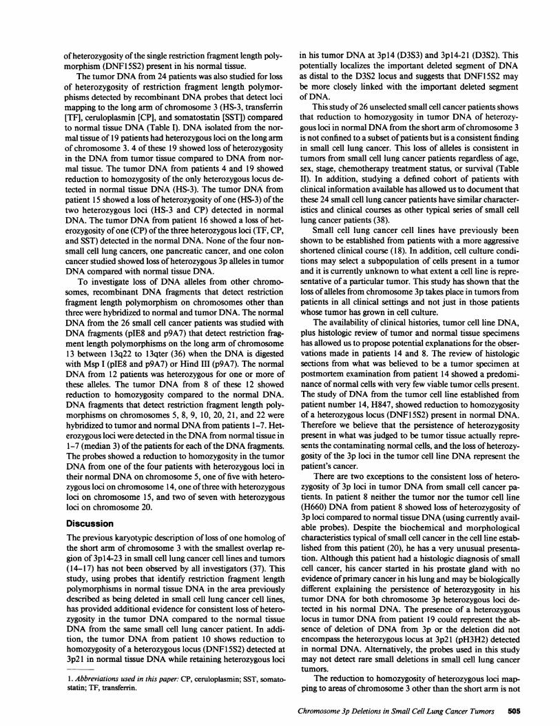
25 of the 26 patients had heterozygous loci detected in theDNAprepared from their normal tissue with at least one of thefour probes (pMSl-37, p 12-32, pH3H2, and pH3E4) identify-ing restriction fragment length polymorphisms in the region of3p14-21 (Table III). 22 of these 25 tumor DNAs(88%) showedreduction to homozygosity of one or more heterozygous locidetected in normal tissue DNAwithin 3pI4-21 (Table I). Fig.1 depicts examples of loss of heterozygous alleles in the DNAfrom two small cell lung cancer patients' tumor tissue com-
Table I. Chromosome 3 Probes Detecting Restriction FragmentLength Polymorphisms
Restriction ChromosomalProbes endonuclease Locus localization References
3p ProbespMSI-37 Msp I D3S3 3p14.2 (23-25)p12-32 Msp I D3S2 3p(14.2-21) (24, 26, 27)pH3H2 Hind III DNF15S2 3p21 (28-31)pH3E4 Hind III DNF15S2 3p21 (29-31)
3q ProbesHS-3 Hind III D3S1 3q12 (32)TF Eco RI 3q21 (33)CP Pst I 3q25 (34)SST Bam HI and 3q28 (35)
Eco Ri
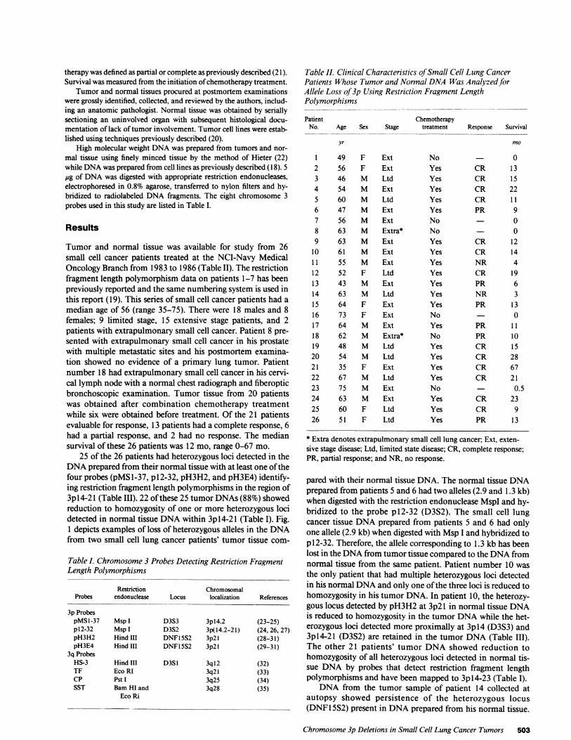
Table II. Clinical Characteristics of Small Cell Lung CancerPatients Whose Tumor and Normal DNAWasAnalyzed forAllele Loss of 3p Using Restriction Fragment LengthPolymorphisms
Patient ChemotherapyNo. Age Sex Stage treatment Response Survival
yr mO
1 49 F Ext No 02 56 F Ext Yes CR 133 46 M Ltd Yes CR 154 54 M Ext Yes CR 225 60 M Ltd Yes CR 116 47 M Ext Yes PR 97 56 M Ext No 08 63 M Extra* No 09 63 M Ext Yes CR 12
10 61 M Ext Yes CR 1411 55 M Ext Yes NR 412 52 F Ltd Yes CR 1913 43 M Ext Yes PR 614 63 M Ltd Yes NR 315 64 F Ext Yes PR 1316 73 F Ext No 017 64 M Ext Yes PR 1118 62 M Extra* No PR 1019 48 M Ltd Yes CR 1520 54 M Ltd Yes CR 2821 35 F Ext Yes CR 6722 67 M Ltd Yes CR 2123 75 M Ext No 0.524 63 M Ext Yes CR 2325 60 F Ltd Yes CR 926 51 F Ltd Yes PR 13
* Extra denotes extrapulmonary small cell lung cancer; Ext, exten-sive stage disease; Ltd, limited state disease; CR, complete response;PR, partial response; and NR, no response.
pared with their normal tissue DNA. The normal tissue DNAprepared from patients 5 and 6 had two alleles (2.9 and 1.3 kb)when digested with the restriction endonuclease MspI and hy-bridized to the probe p12-32 (D3S2). The small cell lungcancer tissue DNAprepared from patients 5 and 6 had onlyone allele (2.9 kb) when digested with Msp I and hybridized top 12-32. Therefore, the allele corresponding to 1.3 kb has beenlost in the DNAfrom tumor tissue compared to the DNAfromnormal tissue from the same patient. Patient number 10 wasthe only patient that had multiple heterozygous loci detectedin his normal DNAand only one of the three loci is reduced tohomozygosity in his tumor DNA. In patient 10, the heterozy-gous locus detected by pH3H2 at 3p2 1 in normal tissue DNAis reduced to homozygosity in the tumor DNAwhile the het-erozygous loci detected more proximally at 3p14 (D3S3) and3p14-21 (D3S2) are retained in the tumor DNA(Table III).The other 21 patients' tumor DNAshowed reduction tohomozygosity of all heterozygous loci detected in normal tis-sue DNAby probes that detect restriction fragment lengthpolymorphisms and have been mapped to 3p14-23 (Table I).
DNA from the tumor sample of patient 14 collected atautopsy showed persistence of the heterozygous locus(DNF15S2) present in DNAprepared from his normal tissue.
Chromosome 3p Deletions in Small Cell Lung Cancer Tumors 503
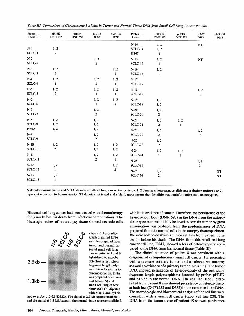
Table III. Comparison of Chromosome 3 Alleles in Tumor and Normal Tissue DNAfrom Small Cell Lung Cancer Patients
Probes ... pH3H2 pH3E4 p12-32 pMSl-37 Probes. pH3H2 pH3E4 p12-32 pMSl-37Locus ... DNF15S2 DNF15S2 D3S2 D3S3 Locus ... DNF15S2 DNFI5S2 D3S2 D3S3
N-14 1,2 NTN-i 1,2 SCLC-14 1,2SCLC-1 2 H847 1N-2 1,2 N-15 1,2 NTSCLC-2 2 SCLC-15 1
N-3 1,2 1,2 N-16 1,2SCLC-3 2 1 SCLC-16 1N-4 1,2 1,2 1,2 N-17SCLC-4 1 2 1 SCLC-17N-5 1,2 1,2 1,2 N-18 1,2SCLC-5 2 1 1 SCLC-18 1N-6 1,2 1,2 N-19 1,2SCLC-6 1 2 SCLC-19 1,2N-7 1,2 N-20 1,2SCLC-7 2 SCLC-20 2
N-8 1,2 1,2 N-21 1,2 1,2SCLC-8 1,2 1,2 SCLC-21 2 1H660 1,2 1,2 N-22 1,2 1,2N-9 1,2 SCLC-22 2 2SCLC-9 2 N-23 1,2N-10 1,2 1,2 1, 2 SCLC-23 2SCLC-10 2 1,2 1,2 N-24 1,2 1,2N-ll 1,2 1,2 SCLC-24 1 1SCLC- 1 2 1 N-25 1,2N-12 1,2 1,2 SCLC-25 2SCLC-12 1 2 N-26 1,2 NTN-13 1,2 SCLC-26 2 NTSCLC-13 1
N denotes normal tissue and SCLCdenotes small cell lung cancer tumor tissue. 1, 2 denotes a heterozygous allele and a single number (I or 2)represent reduction to homozygosity. NT denotes not tested and a blank space means that the allele was noninformative (not heterozygous).
His small cell lung cancer had been treated with chemotherapyfor 3 mo before his death from infectious complications. Thehistologic review of his autopsy tissue showed necrotic cells
p Figure 1. Autoradio-CJ () graph of paired DNA
4) 4 rp (y samples prepared from4 Cv v Cv tumor and normal tis-
sue of small cell lungcancer patients 5 and 6hybridized to a probe
2.9kb - detecting a restrictionfragment length poly-
*:: i. .'"'.'''.'3morphism localizing tochromosome 3p. DNAwas prepared from nor-
1 .3kb- * > * mal tissue (N) andsmall cell lung cancertissue (SCLC), digestedwith Msp I, and hybrid-
ized to probe p12-32 (D3S2). The signal at 2.9 kb represents allele 1and the signal at 1.3 kilobases in the normal tissue represents allele 2.
with little evidence of cancer. Therefore, the persistence of theheterozygous locus (DNF1 5S2) in the DNAfrom the autopsytissue specimen we initially believed to contain tumor by grossexamination was probably from the predominance of DNAprepared from the normal cells in the autopsy tissue specimen.Wewere able to establish a tumor cell line from patient num-ber 14 before his death. The DNA from this small cell lungcancer cell line, H847, showed a loss of heterozygosity com-pared to the DNAfrom his normal tissue (Table III).
The clinical situation of patient 8 was consistent with adiagnosis of extrapulmonary small cell cancer. He presentedwith a prostate primary tumor and a subsequent autopsyshowed no evidence of a primary tumor in his lung. The tumorDNAshowed persistence of heterozygosity of the restrictionfragment length polymorphisms detected by probes pH3H2and p 12-32 in the normal DNA. The cell line, H660, estab-lished from patient 8 also showed persistence of heterozygosityat both loci (DNF I5S2 and D3S2) in the tumor cell line DNA.The morphologic and biochemical analysis of the cell line wereconsistent with a small cell cancer tumor cell line (20). TheDNAfrom the tumor tissue of patient 19 showed persistence
504 Johnson, Sakaguchi, Gazdar, Minna, Burch, Marshall, and Naylor
of heterozygosity of the single restriction fragment length poly-morphism (DNF1 5S2) present in his normal tissue.
The tumor DNAfrom 24 patients was also studied for lossof heterozygosity of restriction fragment length polymor-phisms detected by recombinant DNAprobes that detect locimapping to the long arm of chromosome 3 (HS-3, transferrin[TF], ceruloplasmin [CP], and somatostatin [SST]) comparedto normal tissue DNA(Table I). DNAisolated from the nor-mal tissue of 19 patients had heterozygous loci on the long armof chromosome 3. 4 of these 19 showed loss of heterozygosityin the DNAfrom tumor tissue compared to DNAfrom nor-mal tissue. The tumor DNAfrom patients 4 and 19 showedreduction to homozygosity of the only heterozygous locus de-tected in normal tissue DNA(HS-3). The tumor DNAfrompatient 15 showed a loss of heterozygosity of one (HS-3) of thetwo heterozygous loci (HS-3 and CP) detected in normalDNA. The tumor DNAfrom patient 16 showed a loss of het-erozygosity of one (CP) of the three heterozygous loci (TF, CP,and SST) detected in the normal DNA. None of the four non-small cell lung cancers, one pancreatic cancer, and one coloncancer studied showed loss of heterozygous 3p alleles in tumorDNAcompared with normal tissue DNA.
To investigate loss of DNA alleles from other chromo-somes, recombinant DNA fragments that detect restrictionfragment length polymorphism on chromosomes other thanthree were hybridized to normal and tumor DNA. The normalDNAfrom the 26 small cell cancer patients was studied withDNAfragments (pIE8 and p9A7) that detect restriction frag-ment length polymorphisms on the long arm of chromosome13 between 13q22 to 13qter (36) when the DNAis digestedwith Msp I (pIE8 and p9A7) or Hind III (p9A7). The normalDNAfrom 12 patients was heterozygous for one or more ofthese alleles. The tumor DNA from 8 of these 12 showedreduction to homozygosity compared to the normal DNA.DNAfragments that detect restriction fragment length poly-morphisms on chromosomes 5, 8, 9, 10, 20, 21, and 22 werehybridized to tumor and normal DNAfrom patients 1-7. Het-erozygous loci were detected in the DNAfrom normal tissue in1-7 (median 3) of the patients for each of the DNAfragments.The probes showed a reduction to homozygosity in the tumorDNAfrom one of the four patients with heterozygous loci intheir normal DNAon chromosome 5, one of five with hetero-zygous loci on chromosome 14, one of three with heterozygousloci on chromosome 15, and two of seven with heterozygousloci on chromosome 20.
DiscussionThe previous karyotypic description of loss of one homolog ofthe short arm of chromosome 3 with the smallest overlap re-gion of 3p14-23 in small cell lung cancer cell lines and tumors(14-17) has not been observed by all investigators (37). Thisstudy, using probes that identify restriction fragment lengthpolymorphisms in normal tissue DNAin the area previouslydescribed as being deleted in small cell lung cancer cell lines,has provided additional evidence for consistent loss of hetero-zygosity in the tumor DNAcompared to the normal tissueDNAfrom the same small cell lung cancer patient. In addi-tion, the tumor DNA from patient 10 shows reduction tohomozygosity of a heterozygous locus (DNF1 5S2) detected at3p2 1 in normal tissue DNAwhile retaining heterozygous loci
1. Abbreviations used in this paper: CP, ceruloplasmin; SST, somato-statin; TF, transferrin.
in his tumor DNAat 3p14 (D3S3) and 3pl4-21 (D3S2). Thispotentially localizes the important deleted segment of DNAas distal to the D3S2 locus and suggests that DNF1S2maybe more closely linked with the important deleted segmentof DNA.
This study of 26 unselected small cell cancer patients showsthat reduction to homozygosity in tumor DNAof heterozy-gous loci in normal DNAfrom the short arm of chromosome 3is not confined to a subset of patients but is a consistent findingin small cell lung cancer. This loss of alleles is consistent intumors from small cell lung cancer patients regardless of age,sex, stage, chemotherapy treatment status, or survival (TableII). In addition, studying a defined cohort of patients withclinical information available has allowed us to document thatthese 24 small cell lung cancer patients have similar character-istics and clinical courses as other typical series of small celllung cancer patients (38).
Small cell lung cancer cell lines have previously beenshown to be established from patients with a more aggressiveshortened clinical course (18). In addition, cell culture condi-tions may select a subpopulation of cells present in a tumorand it is currently unknown to what extent a cell line is repre-sentative of a particular tumor. This study has shown that theloss of alleles from chromosome 3p takes place in tumors frompatients in all clinical settings and not just in those patientswhose tumor has grown in cell culture.
The availability of clinical histories, tumor cell line DNA,plus histologic review of tumor and normal tissue specimenshas allowed us to propose potential explanations for the obser-vations made in patients 14 and 8. The review of histologicsections from what was believed to be a tumor specimen atpostmortem examination from patient 14 showed a predomi-nance of normal cells with very few viable tumor cells present.The study of DNAfrom the tumor cell line established frompatient number 14, H847, showed reduction to homozygosityof a heterozygous locus (DNF1 5S2) present in normal DNA.Therefore we believe that the persistence of heterozygositypresent in what was judged to be tumor tissue actually repre-sents the contaminating normal cells, and the loss of heterozy-gosity of the 3p loci in the tumor cell line DNArepresent thepatient's cancer.
There are two exceptions to the consistent loss of hetero-zygosity of 3p loci in tumor DNAfrom small cell cancer pa-tients. In patient 8 neither the tumor nor the tumor cell line(H660) DNAfrom patient 8 showed loss of heterozygosity of3p loci compared to normal tissue DNA(using currently avail-able probes). Despite the biochemical and morphologicalcharacteristics typical of small cell cancer in the cell line estab-lished from this patient (20), he has a very unusual presenta-tion. Although this patient had a histologic diagnosis of smallcell cancer, his cancer started in his prostate gland with noevidence of primary cancer in his lung and may be biologicallydifferent explaining the persistence of heterozygosity in histumor DNAfor both chromosome 3p heterozygous loci de-tected in his normal DNA. The presence of a heterozygouslocus in tumor DNAfrom patient 19 could represent the ab-sence of deletion of DNA from 3p or the deletion did notencompass the heterozygous locus at 3p21 (pH3H2) detectedin normal DNA. Alternatively, the probes used in this studymay not detect rare small deletions in small cell lung cancertumors.
The reduction to homozygosity of heterozygous loci map-ping to areas of chromosome 3 other than the short arm is not
Chromosome 3p Deletions in Small Cell Lung Cancer Tumors 505
a commonevent in small cell lung cancer tumor DNA. 4 of 19small cell cancer tumor DNAsshowed a reduction to homozy-gosity of one heterozygous locus mapping to 3q compared tonormal tissue DNAand two of these four still had other het-erozygous loci mapping to 3q present in their tumor DNA.Therefore the loss of chromosome 3 alleles in small cell lungcancer appears to typically involve the short arm of chromo-some 3 because heterozygous loci on the long arm of chromo-some 3 are retained in 17 of 19 (89%) small cell lung cancerpatients' tumor DNA. This is consistent with the previouscytogenetic (14, 15) and restriction fragment length polymor-phism studies (19) suggesting the most commonmechanism ofloss of genetic information from chromosome 3p is an intersi-titial or terminal deletion.
The long arm of chromosome 13 was the only other chro-mosomal area studied where the loss of heterozygosity of re-striction fragment length polymorphisms commonly occurs intumor DNA. Of the 12 patients who had heterozygous locidetected by DNAfragments that map to 13q22 to 13qter, 8showed reduction to homozygosity. This is of interest becausethis same locus has been reported to be deleted in retinoblas-toma tumor DNAby restriction fragment length polymor-phism studies and is near the Rb locus at 13q14 (1-7). Addi-tional studies are needed to confirm this initial observationand to study the Rb locus more directly in small cell lungcancer. None of the DNA from six other adult carcinomasstudied showed deletion of any 3p alleles.
In tumors other than small cell lung cancer where deletionsof genetic material may be important in the generation ofmalignancy, the identification of specific chromosomal dele-tions in retinoblastoma and Wilms' tumors was initiallyprompted by the cytogenetic analysis of normal lymphocytesand tumors in familial cases and congenital syndromes respec-tively (1-5, 8, 9). The identification of families or congenitalsyndromes associated with small cell lung cancer is very rare(13). In contrast to retinoblastoma and Wilms' tumors whereinheritance appears to play a significant role in the develop-ment of the tumor (39), nearly all small cell lung cancers areassociated with cigarette smoking (13). The chromosomal de-letion in small cell lung cancer, therefore, was initially identi-fied in karyotypic analysis of sporadic cases of small cell lungcancer cell lines (14-17).
The identification of DNAdeletions in familial and spo-radic cases of retinoblastoma and Wilms' tumor DNApro-vides evidence for recessive genes playing a potential role inthe pathogenesis of these tumors. The evidence for the exis-tence of such recessive genes has been suggested by somatic cellhybrid studies between malignant and normal cells where ma-lignancy was suppressed (40) and the occurrence of multipleretinoblastomas in familial cases (39). Additional evidence forrecessive genes playing a potential role in the development ofhuman malignancy was provided by a normal chromosome 11suppressing tumor formation in athymic nude mice of aWilms' tumor cell line with a lIp deletion (41).
The identification of a consistent loss of heterozygosity at3p loci in small cell lung cancer tumors using restriction frag-ment polymorphism studies provides additional support forthis chromosomal region to be important in the generation ofsmall cell lung cancer. It is of interest that the chromosome 3pregion is implicated in renal cell carcinoma as well. Zbar et al.described the loss of 3p alleles in sporadic renal cell carcinomausing restriction fragment length polymorphism studies (42).Similar to the findings in our small cell lung cancer patients'
tumor DNA, all heterozygous alleles in normal DNAdetectedat the DNF15S2 locus were reduced to homozygosity in therenal cell carcinoma DNA. In addition, Dr. Zbar identified asingle patient where the heterozygous alleles detected in DNAfrom normal tissue at DNF15S2 were reduced to homozygos-ity in the tumor DNA, while the heterozygous alleles detectedat D3S2 were retained in the tumor DNAsimilar to patient 1Opresented here. If the same recessive gene located on 3p isinvolved in the etiology of small cell lung cancer and renal cellcarcinoma as has been proposed by Zbar (42), the gene appearsto be more closely linked with DNF15S2 on 3p21 than withD3S2 or D3S3 on 3pl4.
Abnormalities of chromosome 3 have also been identifiedin familial renal cell carcinoma. A t(3;8) (p14.2;q24. 1) consti-tutional reciprocal translocation (27, 43) and a 3; 11 transloca-tion limited to the tumor cells (44) have been described infamilial studies of renal cell carcinoma. The D3S2 probe hy-bridizes to the der (8) chromosome in the familial t(3;8) con-stitutional reciprocal translocation (27), suggesting the break-point is proximal to the D3S2 locus. In contrast to these famil-ial studies suggesting the important region on chromosome 3 isproximal to D3S2, the studies of small cell lung cancer DNAand sporadic renal cell carcinoma DNAwould suggest thatDNF15S2 is more closely linked to a potential recessive cancergene, which is more distal on chromosome 3p than D3S2.Potential reasons for the different chromosome 3 regions in-volvement in the familial cases of the 3;8 translocation in renalcell carcinoma versus small cell lung cancer and sporadic renalcell carcinoma include a different gene on chromosome 3causing the familial renal cell carcinoma than small cell lungcancer and sporadic renal cell carcinoma, a submicroscopicdeletion accompanies the chromosomal translocation in thet(3;8) familial renal cell carcinoma, or the constitutionaltranslocation of chromosome 3 is not the etiologic event in thegeneration of the familial renal cell carcinoma but allows adeletion of DNAdistal to the breakpoint on chromosome 3pto subsequently occur. Cloning of cDNAs in this region willprovide reagents to experimentally test this relationship. Ad-ditionally, further investigation is needed to determine if dele-tion of chromosome 3p may be helpful in the clinical diagnosisof small cell lung cancer.
AcknowledgmentsSupported in part by NIH CA-44764, Mather Foundation Z004, andCouncil for Tobacco Research Center 1620.
References
1. Strong, L. C., V. M. Riccardi, R. E. Ferrell, and R. S. Sparkes.1981. Familial retinoblastoma and chromosome 13 deletion transmit-ted via an insertional translocation. Science (Wash. DC). 213:1501-1503.
2. Sparkes, R. S., A. L. Murphree, R. W. Lingua, M. C. Sparkes,L. L. Field, S. J. Funderburk, and W. F. Benedict. 1983. Gene forhereditary retinoblastoma assigned to human chromosome 13 by link-age to esterase D. Science (Wash. DC). 219:971-973.
3. Benedict, W. F., A. L. Murphree, A. Banerjee, C. A. Spina, M. C.Sparkes, and R. S. Sparkes. 1983. Patient with 13 chromosome dele-tion: evidence that the retinoblastoma gene is a recessive cancer gene.Science(Wash. DC). 219:973-975.
4. Sparkes, R. S., M. C. Sparkes, M. G. Wilson, J. W. Towner, W.Benedict, A. L. Murphree, and J. J. Yunis. 1980. Regional assignmentof genes for human esterase D and retinoblastoma to chromosomeband 13q 14. Science (Wash. DC). 208:1042-1044.
506 Johnson, Sakaguchi, Gazdar, Minna, Burch, Marshall, and Naylor
5. Yunis, J. J., and N. Ramsey. 1978. Retinoblastoma and subbanddeletion of chromosome 13. Am. J. Dis. Child. 132:161-163.
6. Cavenee, W. K., T. P. Dryja, R. A. Phillips, W. F. Benedict, R.Godbout, B. L. Gallie, A. L. Murphree, L. C. Strong, and R. L. White.1983. Expression of recessive alleles by chromosomal mechanisms inretinoblastoma. Nature (Lond.). 305:779-784.
7. Dryja T. P., W. Cavenee, R. White, J. M. Rapaport, R. Petersen,D. M. Albert, and G. A. P. Bruns. 1984. Homozygosity of chromo-some 13 in retinoblastoma. N. Engl. J. Med. 310:550-553.
8. Riccardi, V. M., E. Sujansky, A. C. Smith, and U. Francke. 1978.Chromosomal imbalance in the Aniridia-Wilms' tumor association:lI p interstitial deletion. Pediatrics. 61:604-609.
9. Miller, R. W., J. F. Fraumeni, and M. D. Manning. 1964. Associ-ation of Wilms' tumor with aniridia, hemihypertrophy and other con-genital malformations. N. Engl. J. Med. 270:922-927.
10. Orkin, S. H., D. S. Goldman, and S. E. Sallan. 1984. Develop-ment of homozygosity for chromosome lp markers in Wilms' tu-mour. Nature (Lond.). 309:172-174.
11. Reeve, A. E., P. J. Housiaux, R. J. M. Gardner, W. E. Chew-ings, R. M. Grindley, and L. J. Millow. 1984. Loss of a Harvey rasallele in sporadic Wilms' tumour. Nature (Lond.). 309:174-176.
12. Fearon, E. R., B. Vogelstein, and A. P. Feinberg. 1984. Somaticdeletion and duplication of genes on chromosome 11 in Wilms' tu-mours. Nature (Lond.). 309:176-178.
13. Minna, J. D., G. A. Higgins, and E. J. Glatstein. 1985. Cancerof the Lung. In Cancer: Principles and Practice of Oncology. V. T.DeVita, S. Hellman, and S. A. Rosenberg, editors. J. B. Lippincott Co.,Philadelphia. 507-597.
14. Whang-Peng, J., P. A. Bunn, Jr., C. S. Kao-Shan, E. C. Lee,D. N. Carney, A. Gazdar, and J. D. Minna. 1982. A nonrandomchromosomal abnormality, del 3p(14-23), in human small cell lungcancer (SCLC). Cancer Genet. Cytogenet. 6:119-134.
15. Whang-Peng, J., C. S. Kao-Shan, E. C. Lee, P. A. Bunn, D. N.Carney, A. F. Gazdar, and J. D. Minna. 1981. Specific chromosomedefect associated with human small-cell lung cancer: Deletion 3p(14-23). Science (Wash. DC). 215:181-182.
16. Falor, W. H., R. Ward-Skinner, and S. Wegryn. 1985. A 3pdeletion in small cell lung carcinoma. Cancer Genet. Cytogenet.16:175-177.
17. Yunis, J. J. 1983. The chromosomal basis of human neoplasia.Science (Wash. DC). 221:227-236.
18. Johnson, B. E., D. C. Ihde, R. W. Makuch, A. F. Gazdar, D. N.Carney, H. Oie, E. Russell, M. M. Nau, and J. D. Minna. 1987. mycfamily oncogene amplification in tumor cell lines established fromsmall cell lung cancer patients and its relationship to clinical status andcourse. J. Clin. Invest. 79:1629-1634.
19. Naylor, S. L., B. E. Johnson, J. D. Minna, and A. Y. Sakaguchi.1987. Loss of heterozygosity of chromosome 3p markers in small celllung cancer. Nature (Lond.). 329:451-454.
20. Carney, D. N., A. F. Gazdar, G. Bepler, J. G. Guccion, P. J.Marangos, T. W. Moody, M. H. Zweig, and J. D. Minna. 1985. Estab-lishment and identification of small cell lung cancer cell lines havingclassic and variant feature. Cancer Res. 45:2913-2923.
21. Bunn, P. A., Jr., A. S. Lichter, R. W. Makuch, M. H. Cohen,S. R. Veach, M. J. Matthews, A. Johnston Anderson, M. Edison, E.Glatstein, and J. D. Minna. 1987. Chemotherapy alone or chemother-apy with chest radiation therapy in limited stage small cell lung cancer.Ann. Intern. Med. 106:655-662.
22. Heiter, P. A., G. F. Hollis, S. J. Korsmeyer, T. A. Waldman,and P. Leder. 1981. Clustered arrangement of immunoglobulinlambda constant region genes in man. Nature (Lond.). 294:536-540.
23. de Martinville, B., M. Shafer, R. White, and U. Francke. 1984.Assignment of RFLP loci D3S3, D2054, and Dl 5S I to chromosomes3p, 20, and 15, respectively. Cytogenet. Cell Genet. 37:531-532.
24. Barker, D., M. Schafer, and R. White. 1984. Restriction sitescontaining CpGshow a higher frequency of polymorphism in humanDNA. Cell. 36:131-138.
25. Gerber, M. J., Y. E. Miller, H. A. Drabkin, and C. H. Scoggins.
1986. Regional assignment of the polymorphic probe D3S3 to 3p14 bymolecular hybridization. Cytogenet. Cell Genet. 42:72-74.
26. Naylor, S. L., A. Y. Sakaguchi, D. Barker, R. White, and T. B.Shows. 1984. DNApolymorphic loci mapped to human chromosomes3, 5, 9, 11, 17, 18, and 22. Proc. NatL. Acad. Sci. USA. 81:2447-2451.
27. Harris, P., C. C. Morton, P. Guglielmi, F. Li, K. Kelly, and S. A.Latt. 1986. Mapping by chromosome sorting of several gene probes,including c-myc, to the derivative chromosomes of a 3;8 translocationassociated with familial renal cancer. Cytometry. 7:589-594.
28. Harper, M. E., and G. F. Saunders. 1981. Localization of singlecopy DNAsequences on G-banded human chromosomes by in situhybridization. Chromosoma. 83:431-439.
29. Goode, M. E., P. vanTuinen, D. H. Ledbetter, and S. P. Daiger.1986. The anonymous polymorphic DNAclone DlS1 previouslymapped to human chromosome Ip36 by in situ hybridization, is fromchromosome 3 and is duplicated on chromosome 1. Am. J. Hum.Genet. 38:437-446.
30. Carritt, B., H. M. Welch, and N. J. Parry-Jones. 1986. Se-quences homologous to the human DlS1 locus present on humanchromosome 3. Am. J. Hum. Genet. 38:428-436.
31. Donlon, T. A., and R. E. Magenis. 1984. Localization of thecloned segment of lambda Ch4A-H3 to chromosome 1 band p36:acomfirmation of locus D1SI. Cytogenet. Cell Genet. 37:454-455.
32. Naylor, S. L., A. Y. Sakaguchi, J. F. Gusella, D. Housman, andT. B. Shows. 1982. Mapping of an arbitrary restriction polymorphism(ARP2) to human chromosome 3. Cytogenet. Cell Genet. 32:302.
33. Yang, F., J. B. Lum, J. R. McGill, C. M. Moore, S. L. Naylor,P. H. van Bragt, W. D. Baldwin, and B. H. Bowman. 1984. Humantransferrin: cDNA characterization and chromosomal localization.Proc. Natl. Acad. Sci. USA. 81:2752-2756.
34. Yang, F., S. L. Naylor, J. B. Lum, S. Cutshaw, J. L. McCombs,K. H. Naberhaus, J. R. McGill, G. S. Adrian, C. M. Moore, D. R.Barnett, and B. H. Bowman. 1986. Characterization, mapping, andexpression of the human ceruloplasmin gene. Proc. Natl. Acad. Sci.USA. 83:3257-3261.
35. Naylor S. L., A. Y. Sakaguchi, L. P. Shen, G. I. Bell, W. J.Rutter, and T. B. Shows. 1983. Polymorphic human somatostatin geneis located on chromosome 3. Proc. Natl. Acad. Sci. USA. 80:2686-2689.
36. Cavenee, W., R. Leach, T. Mohandas, P. Pearson, and R.White. 1984. Isolation and regional localization of DNA segmentsrevealing polymorphic loci from human chromosome 13. Am. J.Hum. Genet. 36:10-24.
37. Wurster-Hill, D. H., L. A. Cannizzaro, 0. S. Pettengill, G. D.Sorenson, C. C. Cate, and L. H. Maurer. 1984. Cytogenetics of smallcell carcinoma of the lung. Cancer Genet. and Cytogenet. 13:303-330.
38. Morstyn, G., D. C. Ihde, A. S. Lichter, P. A. Bunn, D. N.Carney, E. Glatstein, and J. D. Minna. 1984. Small cell lung cancer1973-1983: early progress and recent obstacles. Int. J. Radiat. Oncol.Biol. Phys. 10:515-539.
39. Knudson, A. G. 1975. The genetics of childhood cancer.Cancer. 35:1022-1026.
40. Harris, H., 0. J. Miller, G. Klein, P. Worst, and T. Tachibana.1969. Suppression of malignancy by cell fusion. Nature (Lond.).223:363-368.
41. Weissman, B. E., P. J. Saxon, S. R. Pasquale, G. R. Jones, A. G.Geiser, and E. J. Stanbridge. 1987. Introduction of a normal chromo-some I 1 into a Wilms' tumor cell line controls its tumorigenic expres-sion. Science (Wash. DC). 236:175-180.
42. Zbar, B., H. Brauch, C. Talmadge, and M. Linehan. 1987. Lossof alleles of loci on the short arm of chromosome 3 in renal cellcarcinoma. Nature (Lond.). 327:721-724.
43. Cohen, A. J., F. P. Li, S. Berg, D. J. Marchetto, S. Tsai, S. C.Jacobs, and R. S. Brown. 1979. Hereditary renal-cell carcinoma asso-ciated with a chromosomal translocation. N. Engl. J. Med. 301:592-595.
44. Pathak, S., L. C. Strong, R. E. Ferrell, and A. Trindade. 1982.Familial renal cell carcinoma with a 3; 11 chromosome translocationlimited to tumor cells. Science (Wash. DC). 217:939-941.
Chromosome 3p Deletions in Small Cell Lung Cancer Tumors 507