restrictive right ventricular physiology after tetralogy of fallot...
TRANSCRIPT

LUND UNIVERSITY
PO Box 117221 00 Lund+46 46-222 00 00
Restrictive right ventricular physiology after Tetralogy of Fallot repair is associatedwith fibrosis of the right ventricular outflow tract visualized on cardiac magneticresonance imaging.
Munkhammar, Peter; Carlsson, Marcus; Arheden, Håkan; Pesonen, Erkki
Published in:European Heart Journal-Cardiovascular Imaging
DOI:10.1093/ehjci/jet009
Published: 2013-01-01
Link to publication
Citation for published version (APA):Munkhammar, P., Carlsson, M., Arheden, H., & Pesonen, E. (2013). Restrictive right ventricular physiology afterTetralogy of Fallot repair is associated with fibrosis of the right ventricular outflow tract visualized on cardiacmagnetic resonance imaging. European Heart Journal-Cardiovascular Imaging, 14(10), 978-985. DOI:10.1093/ehjci/jet009
General rightsCopyright and moral rights for the publications made accessible in the public portal are retained by the authorsand/or other copyright owners and it is a condition of accessing publications that users recognise and abide by thelegal requirements associated with these rights.
• Users may download and print one copy of any publication from the public portal for the purpose of privatestudy or research. • You may not further distribute the material or use it for any profit-making activity or commercial gain • You may freely distribute the URL identifying the publication in the public portal

Take down policyIf you believe that this document breaches copyright please contact us providing details, and we will removeaccess to the work immediately and investigate your claim.
Download date: 16. Jul. 2018

1
Restrictive right ventricular physiology after tetralogy of Fallot repair is associated with
fibrosis of the right ventricular outflow tract visualized on cardiac magnetic resonance
imaging
Peter Munkhammar, MD*
Marcus Carlsson, MD, PhD †
Håkan Arheden, MD, PhD†
Erkki Pesonen, MD, PhD*
* Dep of Pediatric Cardiology and † Dep of Clinical Physiology, Skåne University Hospital,
Lund University, Sweden
Running title: Restrictive physiology and fibrosis in Fallot children
Address for correspondence: Peter Munkhammar, Dep of Pediatric Cardiology, Skane
University Hospital, SE-221 85 Lund, Sweden
Email [email protected]

2
ABSTRACT
Aims: To determine whether restrictive physiology seen in tetralogy of Fallot (TOF) patients
can be explained by fibrosis of the right ventricular (RV) outflow tract.The aetiology for
restrictive RV physiology after TOF repair is not known.
Methods and Results: TOF patients (n=31, 13 girls, 10.2 years ± 2.8) were included 9.2±2.9
years after total correction and examined with CMR and Doppler-echocardiography. Cine,
flow and late gadolinium contrast enhanced (LGE) CMR were performed to quantify RV
volumes, pulmonary flow and regurgitation (PR) and fibrosis. Healthy children (n=12) were
investigated with CMR of pulmonary flow. Forward flow during atrial contraction above
mean+2SD of healthy subjects was set as a marker of restrictive physiology. Four patients
were excluded due to suboptimal LGE-CMR. Fisher´s exact test was used to determine the
association between restrictive physiology and fibrosis.16 patients showed fibrosis in the
RVOT on LGE-CMR and CMR showed restrictive physiology in 14 of them. In the 11
patients without fibrosis in the RVOT, 1 showed restrictive physiology. The odds ratio for
RVOT fibrosis in patients with restrictive RV physiology was 70.0 (CI 5.6-882.7, p<0.001).
Transannular patch repair did not differ between the groups (p=0.37). The degree of RVOT
fibrosis correlated positively with PR (r2=0.38, p<0.001) and RV volumes (r2=0.51 for EDV
and r2=0.47 for ESV, p<0.001).
Conclusion: There is a strong association between restrictive RV physiology detected on
CMR and fibrosis of the RVOT in children after TOF repair.

3
INTRODUCTION
Mid-and long-term out-come after Tetralogy of Fallot (TOF) repair are nowadays excellent
and life quality among those repaired is generally good 1-4. Age at repair has gradually
decreased since the early era of TOF surgery without increase in mortality or need for re-
intervention 5-6. Furthermore, surgical techniques have improved with the ability to reduce the
surgical trauma of the right ventricular outflow tract (RVOT) and minimize postoperative
pulmonary regurgitation (PR) 7. In some patients the diastolic properties of the RV wall allow
less compliance to diastolic filling. This RV diastolic dysfunction after TOF repair has been
named restrictive RV physiology and is reflected by the occurrence of an end-diastolic
forward flow in the pulmonary artery (PA) detected by Doppler echocardiography 8.
Restrictive RV physiology in the setting of significant PR has been shown to be of positive
prognostic value at mid term follow-up 9 but the pathophysiological explanation to restrictive
physiology is so far unknown.
Cardiac magnetic resonance imaging (CMR) permits three dimensional visualization of the
RV throughout the cardiac cycle enabling functional assessment with high accuracy.
Furthermore, phase velocity encoded CMR enables flow quantification of the PR with less
observer dependency compared to Doppler echocardiography and CMR can also be used to
detect restrictive physiology 10-12. Thus, CMR has become a valuable tool in the assessment of
patients operated for TOF 13-14. Late gadolinium contrast enhancement (LGE) CMR has been
used to localize and quantify myocardial infarction and this technique has in recent years been
shown to be able to visualize fibrosis of the RV in patients with congenital heart disease
(CHD) 15-18. However, no prospective study has used LGE-CMR in children with TOF to
investigate the link to restrictive physiology.
Therefore, the aim of this study was to determine if restrictive physiology seen
in children after TOF repair can be associated with fibrosis of the RVOT. Furthermore, we
4
aimed to investigate if there are differences in RV size and function in patients with fibrosis in
the RVOT compared to patients without RVOT fibrosis at mid-term follow up.
METHODS
Subjects:
Patients who had undergone TOF repair, with measurable PR at Doppler echocardiography
were included. Patients with residual pulmonary stenosis pressure gradient >25 mmHg on
Doppler echocardiography, associated atrioventricular septum defect, double outlet right
ventricle of Fallot type, pulmonary atresia with ventricular septum defect (VSD) or TOF with
absent pulmonary valve were excluded as well as patients who had undergone pulmonary
valve replacement (PVR). Thirty-one patients (13 girls, mean age at investigation 10.2±2.8
(range 3-16) years, were prospectively included at 9.2±2.9 (range 2-16) years after surgical
correction. Medium age at repair was 9.5±8.6 months (range 3 weeks to 1 year and 11
months).The study groups included patients surgically corrected < 6 months of age (n=11)
and > 6 months of age (n=20).
The local institutional ethics committee approved the study, and parents gave informed
consent to the children’s participation in the study. CMR and echocardiography examinations
were performed within 2 days.
Echocardiography:
Transthoracic echocardiography was performed by one observer (PM) using a GE Vingmed
Vivid Five system with FPA 3.5, 5 and 10 MHz transducers. Echocardiographic
measurements of restrictive RV physiology were performed as previously described 19. In
short, restrictive RV physiology was defined as forward pulmonary flow in late diastole
present throughout the respiratory cycle (Figure 1).

5
CMR
The CMR protocol and parameters are listed in the Appendix. CMR was performed on a 1.5 T
Philips Intera CV (n= 27) and a 1.5 T Siemens Magnetom Vision (n=4). Cine, pulmonary
flow velocity mapping and LGE CMR were acquired. Cine images were acquired in the
RVOT plane, oblique transverse plane and/or the left ventricular short axis plane in order to
determine the RV end-diastolic volume (EDV) and end-systolic volume (ESV) as well as the
RV ejection fraction (EF). Flow velocity mapping CMR was performed during free breathing
perpendicular to the plane of the flow in the pulmonary artery to determine the regurgitant
volume and regurgitant fraction (RF).
Restrictive physiology using CMR
Forward flow during atrial contraction was used to detect restrictive physiology. In normal
subjects there is slight forward flow during late diastole in flow quantification using CMR,
caused by the movement of the pulmonary valve during atrioventricular
displacement20.Therefore, forward flow as a percentage of net forward flow during the cardiac
cycle (Figure 1) was calculated in 12 healthy children (15±3 years) with normal CMR
referred for screening of arrythmogenic right-ventricular cardiomyopathy because of family
history of this disease. A threshold of mean + 2 SD of the percentage forward flow during
atrial contraction in healthy subjects was set as normal and values above this as restrictive
physiology. Percentage forward flow in TOF-patients was defined as a forward flow in the PA
during atrial contraction divided by the net (effective) flow (forward minus backward flow)
(Figure 1). In a separate analysis, patients were classified as restrictive and non-restrictive
physiology using visual assessment where any peak of forward flow prior to the systolic
forward flow in the PA was considered as restrictive physiology 10, 12.

6
LGE-CMR was obtained for fibrosis visualization in the same plane as for cine CMR. Images
were acquired 10-20 minutes after intravenous administration of Gadolinium (Gd)-based
contrast agent (Gd-DOTA or Gd-DTPA, 0.2 mmol/kg body weight).
Image analysis
Echocardiography images were analysed without knowledge of CMR findings and vice versa.
The echocardiography images were evaluated by vendor provided software in the scanner.
CMR data were analysed using the software Segment v1.8 (http://segment.heiberg.se). The
endocardial contours were drawn on cine CMR to calculate RVEDV, RVESV and RVEF 21.
The right atrial (RA) size in end-systole was delineated in cine CMR to provide an indication
of RV diastolic pressures. The pulmonary artery was segmented on flow CMR to quantify
pulmonary regurgitant volume as the backward volume in diastole 22 and RF in percent of
forward flow. LGE-CMR was visually scored by two experienced observers (MC and HA) in
consensus. The RV was divided into 3 short axis and 3 transversal regions giving a total of 9
segments as previously described 22. Each segment was given a score from 0-4, no fibrosis
was given the score 0, 1-25% segmental fibrosis 1, 26-50% 2, 51-75 % 3 and 76-100% 4.
Fibrosis had to be present in two different imaging planes to be considered a true finding.
Percent fibrosis of the RV was calculated as (total score/36)x100%.
Statistical analysis:
All statistical analyses were performed with Graphpad Prism v 5.01. All continuous variables
were expressed as mean ± SD. The Mann-Whitney test was used to compare continuous
variables in the restrictive versus non-restrictive RV physiology groups and fibrotic versus
non-fibrotic groups. Fisher´s exact test was used when comparing non-continuous variables
between the groups and the odds ratio (OR) with confidence interval (CI) was calculated.
Pearson´s correlation analysis was performed between the degree of RVOT fibrosis and

7
RVEDV, RVESV, RVEF and regurgitant volume. Results with a p value <0.05 were
considered significant.
RESULTS
Restrictive RV physiology
Percentage end-diastolic forward flow of total flow was 1.10±0.71% in healthy subjects and
therefore the upper normal limit was 2.5%. 16 patients had end-diastolic forward flow ≥2.5%
and was considered to have restrictive physiology using CMR. The diastolic forward flow as
percentage of stroke volume in all patient was 4.7±6.3%, in patients with restrictive
physiology 9.1±5.4% and in patients with non-restrictive physiology -0.5±2.9%. Most
patients with restrictive physiology (n=14) demonstrated negative PA flow at the onset of the
distinct end-diastolic flow wave as exemplified in Figure 1C. On Doppler echocardiography
15 patients were found to be restrictive. Ten of these were restrictive also on CMR. On
Doppler echocardiography 16 patients were non-restrictive. Ten of these were non-restrictive
on CMR. The kappa value between CMR and Doppler echocardiography for restrictive
physiology was 0.29, the odds ratio was not significant (OR 3.3, CI 0.8-14.6, p=0.16).
Restrictive RV physiology and RV myocardial fibrosis
Four patients were excluded from the fibrotic vs. non-fibrotic analysis due to inadequate or
incomplete LGE-CMR acquisition. Fibrosis of the RVOT on LGE-CMR (Figures 2 and 3)
was found in 16 out of 27 (59%) investigated patients. There was a strong association
between RVOT fibrosis on LGE-CMR and restrictive RV physiology on CMR (OR 70, CI
5.6-882.7, p<0.001), Table 1. Fourteen of the 16 patients with RVOT fibrosis showed
restrictive RV physiology and only one of the 11 patients without RVOT fibrosis showed
restrictive RV physiology. Fifteen of the patients with fibrosis had restrictive physiology
when using visual assessment and five of the non-fibrotic patients had non- restrictive

8
physiology. The odds ratio for visual assessment of restrictive physiology and fibrosis was
12.5 (CI 1.2-130.7, p=0.03).
However, if using Doppler Echocardiography only 10 patients of the sixteen with fibrosis
showed restrictive physiology. Three of the patients without fibrosis showed restrictive
physiology on Echocardiography (OR 4.4, CI 0.8-24.0, p=0.12).
Fibrosis at the location of the VSD patch in the basal part of the septum was seen in 87% (22
out of 27 patients). Furthermore minimal fibrosis could be seen at the inferoseptal RV
insertion point to the LV in 3 patients, in apical trabeculation in 1 patient and in the RV free
wall in 1 patient.
Table 2 shows the clinical data of patients with and without RVOT fibrosis. Of note, the
incidence of transannular patch (TAP) repair did not differ between the groups (p=0.37).
There was a positive correlation between degree of RV fibrosis and RVEF (p<0.01), RVEDV,
RVESV and regurgitant volume (p<0.001 for all), Figure 4. RA size was higher in the group
with fibrosis (p=0.046) indicating higher RV diastolic pressure (Table 2).
Patients with restrictive physiology had larger RVEDV/BSA (159±49ml/m2, p=0.003),
RVESV/BSA (83±34 ml/m2, p=0.008) and higher RF (45±9%, p=0.003) compared to patients
with non-restrictive physiology on CMR (111±29 ml/m2, 52±18 ml/m2, 23±19 %), Table 3.
DISCUSSION
This study is the first to show that there is a strong association between restrictive RV
physiology and fibrosis of the RVOT in children after TOF repair with residual postoperative
PR. RVOT fibrosis correlated positively to RV volumes and PR but did not relate to TAP
repair.
Fibrosis and restrictive physiology

9
Fibrosis of the RVOT was associated with restrictive physiology assessed by CMR and the
link may be that the fibrosis decreases the RV compliance. In an RV with low compliance
atrial systole will pump against a stiff RV resulting in forward pulmonary flow in ventricular
diastole. Only 1 out of 15 patients with restrictive physiology showed no sign of fibrosis in
the RV. Fibrosis was mainly found in the anterior free wall of the RVOT. Furthermore,
fibrosis in the region of the VSD repair was seen in most patients and in the inferoseptal RV
insertion points or in RV trabeculaes in a minority of patients. Previous studies in adults have
found RVOT fibrosis in 71-99% after primary repair of TOF 17, 23 in line with our findings.
Extensive RV fibrosis (≥75th percentile) has earlier been associated with restrictive RV
physiology in adults 17. In our study in children, we found a clear association with restrictive
RV physiology even when including milder degrees of RVOT fibrosis. This difference may
be attributed to the younger population in our material as previously proposed 17. In our
population patients with fibrosis also had larger RA size indicating higher RA pressure and
therefore higher RV diastolic pressure. Restrictive physiology was originally described as
patients with end-diastolic forward flow through the pulmonary valve during atrial contraction
due to a stiff and non-compliant hypertrophied RV wall. Hence, RV restrictive physiology in
the early and midterm postoperative period protects the RV from dilatation and minimizes the
effects of PR and this may be the reason that postoperative PR is well tolerated initially after
TOF repair 24. In our patient population, at 9.2±2.9 years after repair, we found end-diastolic
forward flow through the pulmonary valve in patients with fibrosis of the RVOT coupled to
higher PR and larger RV volumes. Furthermore, we found a positive correlation with the
degree of fibrosis and RV volumes in line with previous studies 17, 23.This indicates that the
protective nature of restrictive RV physiology early after TOF repair previously described 19,
25-26 may attenuate with time 14, 17, 27-28 and fibrosis development may represent the substrate
for the secondary form of restrictive RV physiology suggested by Lee et al12. Therefore, the

10
etiology of the restrictive physiology in our patient population may be different to the
“classical” description. Lee et al divided the patient groups according to restrictive physiology
and RV volumes, and found that forward end-diastolic flow was present in both large and
small RVs but was associated with better physical exercise tolerance only in conjunction with
small RVs 12. This could explain why some previous studies have found restrictive physiology
to be a positive prognostic factor 12, 24 and other studies have shown that restrictive physiology
is coupled to reduced RVEF 29 and low quality of life 30.
We found slightly higher end-diastolic forward flow in restrictive patients (9% of SV)
compared to earlier studies (6%) 11-12 and this is probably explained by different definitions of
restrictive physiology between the studies. We used 2.5% end-diastolic forward flow as cut-
off for restrictive physiology based on the forward flow seen in healthy children due to
longitudinal AV-plane movement during atrial contraction 20.
The association of restrictive physiology and RV fibrosis in our patient population does not
necessarily mean that RV fibrosis is the cause of restrictive physiology. However, our
findings show that restrictive physiology found at follow up in patients with large RV
volumes and RF is strongly associated with myocardial fibrosis in the RVOT.
The agreement between CMR and Doppler-echocardiography was fair (kappa value 0.29)
which is similar to the findings of Lee et al (kappa value 0.35 calculated from the published
results). A possible explanation for this difference is that CMR averages several cardiac and
respiratory cycles and can therefore be viewed as more inclusive for restrictive physiology 12.
CMR has been proposed to be more accurate compared to Doppler in the detection of
restrictive physiology in dilated pulmonary arteries lacking laminar flow patterns 10.
Cause of fibrosis
11
The cause of RVOT fibrosis is not clear, but several mechanisms have been proposed, such as
the long-term effects of preoperative hypoxemia 17, 31. Dilatation of the RV caused by PR has
been postulated to be one cause of progressive fibrosis 17 and we have showed a positive
correlation between the degree of RVOT fibrosis and PR as well as RV dilatation (Figure 4).
However, cause and effect could be reverse with RV fibrosis causing RV dilatation and
thereby worsening of the PR. Wald et al. showed that the fibrosis is associated with dys-
/akinesia, aneurysmatic dilatation of the RVOT and conduction delay 22. PR volume is mainly
determined by the size of the pulmonary valvular orifice which may become enlarged in
RVOT fibrosis as the pulmonary annulus is connected to the RVOT myocardium 32-33.
One possible mechanism for RVOT fibrosis may be that the muscular resection of the
infundibulum at repair imposes damages to the microvasculature causing fibrosis
development and thereby contributes to restrictive RV physiology 28 . All patients in our study
were repaired via transatrial or transpulmonary approach with only minimal ventricular
incisions made in patients with TAP with the aim of preserving the RV infundibulum 34.
Interestingly, in the study by Wald et al. fibrosis was found extending to the anterior RV free
wall and neighboring segments where surgery had not taken place 22 and this was also found
in our material. Therefore, it is possible that fibrosis caused by RV volume overload may co-
exist with the surgically related substrates for fibrosis in RVOT creating RV dysfunction.
There was no difference in fibrosis development or restrictive RV physiology between the
early and late repaired group in the present study and this may be attributed to an over-all
earlier repair age compared to previous studies 17, 23. There were no differences in cross-clamp
times at repair between the fibrotic and the non-fibrotic groups. This indicates that prolonged
myocardial hypoperfusion during surgery is not a cause for fibrosis. Consequently, neither
long-term effects of hypoxemia, TAP nor ventriculotomies are likely to be the cause of the
RVOT fibrosis in children after TOF repair.

12
Interestingly, we did not find any LGE in four patients with TAP. In our material the patch
material used for TAP consisted of the pericardium and the size was quite small (5-10 mm
including the supravalvular and subvalvular parts) which may explain why CMR “missed” to
detect LGE in this region.
Limitations
The lack of fibrosis in the RVOT on LGE-CMR does not exclude the existence of a more
diffusely spread fibrosis throughout the entire RV wall not detected. LGE-CMR lacks spatial
resolution to detect very small areas of fibrosis and the use of an inversion-recovery pulse
does not permit the detection of evenly distributed diffuse intercellular fibrosis 35. Further
studies using high-resolution T1-mapping to quantify the extracellular volume fraction 36-37
may provide information on the relationship of increased amount of diffuse fibrosis and
restrictive physiology. In this study, fibrosis detection was performed by two experienced
observers in consensus with visual scoring. Better image quality with higher resolution and
better signal-to-noise ratio would be needed for a semi-automatic quantification as used in left
ventricular infarct quantification 38.The number of patients in the study is fairly limited, which
may explain why we did not find a statistically significant relationship between restrictive
physiology on Doppler-echocardiography and fibrosis on LGE-CMR and the comparisons of
cross-clamp times.
Conclusion
To our knowledge this study is the first showing an association between restrictive RV
physiology and RVOT fibrosis visualized on LGE-CMR in children repaired for TOF. The
cause for fibrosis still remains unclear and needs to be further addressed in future studies.

13
Acknowledgements: Annica Maxedius is greatly appreciated for help with patient
administration. This study was supported by a grant from the Swedish Heart Lung foundation

14
REFERENCES:
1. Hamada H, Terai M, Jibiki T, Nakamura T, Gatzoulis MA, Niwa K. Influence of
early repair of tetralogy of fallot without an outflow patch on late arrhythmias and sudden
death: a 27-year follow-up study following a uniform surgical approach. Cardiol Young
2002;12(4):345-51.
2. Hickey EJ, Veldtman G, Bradley TJ, Gengsakul A, Manlhiot C, Williams WG,
et al. Late risk of outcomes for adults with repaired tetralogy of Fallot from an inception
cohort spanning four decades. Eur J Cardiothorac Surg 2009;35(1):156-64; discussion 164.
3. Pokorski RJ. Long-term survival after repair of tetralogy of Fallot. J Insur Med
2000;32(2):89-92.
4. Murphy JG, Gersh BJ, Mair DD, Fuster V, McGoon MD, Ilstrup DM, et al.
Long-term outcome in patients undergoing surgical repair of tetralogy of Fallot. N Engl J Med
1993;329(9):593-9.
5. Tamesberger MI, Lechner E, Mair R, Hofer A, Sames-Dolzer E, Tulzer G. Early
primary repair of tetralogy of fallot in neonates and infants less than four months of age. Ann
Thorac Surg 2008;86(6):1928-35.
6. Vohra HA, Adamson L, Haw MP. Is early primary repair for correction of
tetralogy of Fallot comparable to surgery after 6 months of age? Interact Cardiovasc Thorac
Surg 2008;7(4):698-701.
7. Owen AR, Gatzoulis MA. Tetralogy of Fallot: Late outcome after repair and
surgical implications. Semin Thorac Cardiovasc Surg Pediatr Card Surg Annu 2000;3:216-
226.
15
8. Norgard G, Gatzoulis MA, Josen M, Cullen S, Redington AN. Does restrictive
right ventricular physiology in the early postoperative period predict subsequent right
ventricular restriction after repair of tetralogy of Fallot? Heart 1998;79(5):481-4.
9. Norgard G, Gatzoulis MA, Moraes F, Lincoln C, Shore DF, Shinebourne EA, et
al. Relationship between type of outflow tract repair and postoperative right ventricular
diastolic physiology in tetralogy of Fallot. Implications for long-term outcome. Circulation
1996;94(12):3276-80.
10. Helbing WA, Niezen RA, Le Cessie S, van der Geest RJ, Ottenkamp J, de Roos
A. Right ventricular diastolic function in children with pulmonary regurgitation after repair of
tetralogy of Fallot: volumetric evaluation by magnetic resonance velocity mapping. J Am Coll
Cardiol 1996;28(7):1827-35.
11. van den Berg J, Wielopolski PA, Meijboom FJ, Witsenburg M, Bogers AJ,
Pattynama PM, et al. Diastolic function in repaired tetralogy of Fallot at rest and during
stress: assessment with MR imaging. Radiology 2007;243(1):212-9.
12. Lee W, Yoo SJ, Roche SL, Kantor P, van Arsdell G, Park EA, et al.
Determinants and functional impact of restrictive physiology after repair of tetralogy of
Fallot: New insights from magnetic resonance imaging. Int J Cardiol 2012.
13. Weber OM, Higgins CB. MR evaluation of cardiovascular physiology in
congenital heart disease: flow and function. J Cardiovasc Magn Reson 2006;8(4):607-17.
14. Bouzas B, Kilner PJ, Gatzoulis MA. Pulmonary regurgitation: not a benign
lesion. Eur Heart J 2005;26(5):433-9.
15. Prakash A, Powell AJ, Krishnamurthy R, Geva T. Magnetic resonance imaging
evaluation of myocardial perfusion and viability in congenital and acquired pediatric heart
disease. Am J Cardiol 2004;93(5):657-61.
16
16. Harris MA, Johnson TR, Weinberg PM, Fogel MA. Delayed-enhancement
cardiovascular magnetic resonance identifies fibrous tissue in children after surgery for
congenital heart disease. J Thorac Cardiovasc Surg 2007;133(3):676-81.
17. Babu-Narayan SV, Kilner PJ, Li W, Moon JC, Goktekin O, Davlouros PA, et al.
Ventricular fibrosis suggested by cardiovascular magnetic resonance in adults with repaired
tetralogy of fallot and its relationship to adverse markers of clinical outcome. Circulation
2006;113(3):405-13.
18. Muzzarelli S, Ordovas KG, Cannavale G, Meadows AK, Higgins CB. Tetralogy
of Fallot: impact of the excursion of the interventricular septum on left ventricular systolic
function and fibrosis after surgical repair. Radiology 2011;259(2):375-83.
19. Gatzoulis MA, Norgard G, Redington AN. Biventricular long axis function after
repair of tetralogy of Fallot. Pediatr Cardiol 1998;19(2):128-32.
20. Kozerke S, Schwitter J, Pedersen EM, Boesiger P. Aortic and mitral
regurgitation: quantification using moving slice velocity mapping. J Magn Reson Imaging
2001;14(2):106-12.
21. Carlsson M, Ugander M, Heiberg E, Arheden H. The quantitative relationship
between longitudinal and radial function in left, right, and total heart pumping in humans. Am
J Physiol Heart Circ Physiol 2007;293(1):H636-44.
22. Wald RM, Haber I, Wald R, Valente AM, Powell AJ, Geva T. Effects of
regional dysfunction and late gadolinium enhancement on global right ventricular function
and exercise capacity in patients with repaired tetralogy of Fallot. Circulation
2009;119(10):1370-7.
23. Oosterhof T, Mulder BJ, Vliegen HW, de Roos A. Corrected tetralogy of Fallot:
delayed enhancement in right ventricular outflow tract. Radiology 2005;237(3):868-71.

17
24. Gatzoulis MA, Till JA, Somerville J, Redington AN. Mechanoelectrical
interaction in tetralogy of Fallot. QRS prolongation relates to right ventricular size and
predicts malignant ventricular arrhythmias and sudden death. Circulation 1995;92(2):231-7.
25. Gatzoulis MA, Balaji S, Webber SA, Siu SC, Hokanson JS, Poile C, et al. Risk
factors for arrhythmia and sudden cardiac death late after repair of tetralogy of Fallot: a
multicentre study. Lancet 2000;356(9234):975-81.
26. Munkhammar P, Cullen S, Jogi P, de Leval M, Elliott M, Norgard G. Early age
at repair prevents restrictive right ventricular (RV) physiology after surgery for tetralogy of
Fallot (TOF): diastolic RV function after TOF repair in infancy. J Am Coll Cardiol
1998;32(4):1083-7.
27. Geva T, Sandweiss BM, Gauvreau K, Lock JE, Powell AJ. Factors associated
with impaired clinical status in long-term survivors of tetralogy of Fallot repair evaluated by
magnetic resonance imaging. J Am Coll Cardiol 2004;43(6):1068-74.
28. Uebing A, Gibson DG, Babu-Narayan SV, Diller GP, Dimopoulos K, Goktekin
O, et al. Right ventricular mechanics and QRS duration in patients with repaired tetralogy of
Fallot: implications of infundibular disease. Circulation 2007;116(14):1532-9.
29. Sun AM, AlHabshan F, Cheung M, Bronzetti G, Redington AN, Benson LN, et
al. Delayed onset of tricuspid valve flow in repaired tetralogy of Fallot: an additional
mechanism of diastolic dysfunction and interventricular dyssynchrony. J Cardiovasc Magn
Reson 2011;13:43.
30. Lu JC, Cotts TB, Agarwal PP, Attili AK, Dorfman AL. Relation of right
ventricular dilation, age of repair, and restrictive right ventricular physiology with patient-
reported quality of life in adolescents and adults with repaired tetralogy of fallot. Am J
Cardiol 2010;106(12):1798-802.

18
31. Reddy S, Osorio JC, Duque AM, Kaufman BD, Phillips AB, Chen JM, et al.
Failure of right ventricular adaptation in children with tetralogy of Fallot. Circulation
2006;114(1 Suppl):I37-42.
32. Gross L, Kugel MA. Topographic Anatomy and Histology of the Valves in the
Human Heart. Am J Pathol 1931;7(5):445-474 7.
33. Bartelings MM, Bogers AJ, Galantowicz ME, Gittenberger-De Groot AC.
Anatomy of the aortic and pulmonary roots. Semin Thorac Cardiovasc Surg Pediatr Card
Surg Annu 1998;1:157-164.
34. Morales DL, Zafar F, Fraser CD, Jr. Tetralogy of Fallot repair: the Right
Ventricle Infundibulum Sparing (RVIS) strategy. Semin Thorac Cardiovasc Surg Pediatr
Card Surg Annu 2009:54-8.
35. Broberg CS, Chugh SS, Conklin C, Sahn DJ, Jerosch-Herold M. Quantification
of diffuse myocardial fibrosis and its association with myocardial dysfunction in congenital
heart disease. Circ Cardiovasc Imaging;3(6):727-34.
36. Arheden H, Saeed M, Higgins CB, Gao DW, Bremerich J, Wyttenbach R, et al.
Measurement of the distribution volume of gadopentetate dimeglumine at echo-planar MR
imaging to quantify myocardial infarction: comparison with 99mTc-DTPA autoradiography
in rats. Radiology 1999;211(3):698-708.
37. Ugander M, Oki AJ, Hsu LY, Kellman P, Greiser A, Aletras AH, et al.
Extracellular volume imaging by magnetic resonance imaging provides insights into overt and
sub-clinical myocardial pathology. Eur Heart J 2012.
38. Heiberg E, Ugander M, Engblom H, Gotberg M, Olivecrona GK, Erlinge D, et
al. Automated quantification of myocardial infarction from MR images by accounting for
partial volume effects: animal, phantom, and human study. Radiology 2008;246(2):581-8.

19
TABLES
Table 1. Relationship between restrictive RV physiology detected on CMR and RVOT
fibrosis detected on LGE-CMR
Patients with
fibrosis
(n=16)
Patients with no
fibrosis
(n=11)
Restrictive RV physiology
detected on CMR (n=15)
14 1 P<0.001 OR= 70.0 (CI 5.6-882.7)
Non-restrictive RV physiology
detected on CMR (n=12)
2
10

20
Table 2. Patient characteristics in patients with and without fibrosis.
All
patients
(n= 27)
Patients
with fibrosis
in RVOT
(n=16)
Patients
without
fibrosis
in RVOT
(n=11)
p values
Age (years) 10.2±2.6 10.5±2.5 9.8±2.8 0.73
Follow up time (years) 9.2±2.7 9.3±2.7 9.0±2.7 0.98
BSA (m2) 1.2±0.3 1.2±0.3 1.1±0.3 0.69
Females (%) 48 50 45
Age at repair (years) 0.8±0.8 0.9±0. 9 0.7±0.4 0.29
repair < 6 months (%) 37 31 44
TAP (%) 52 56 36 0.37
RVEDV ml/m2 134 ± 50 158 ± 47 100±31 0.002
RVESV ml/m2 67±33 83±33 44±17 0.002
RV EF % 52±7 49±8 56±5 0.18
RF, ml 29±24 36±23 17±22 0.02
RF, ml/BSA 23±16 30±14 12±17 0.02
RF, % 33±19 40±16 22±19 0.02
RA ml/m2 46±12 50±12 40±12 0.046
Transatrial approach (%) 100 100 100 1.0
Cross-clamp time (min) 49±17 49±10 49±25 0.32
Hospital stay (days) 13±6 14±7 12±5 0.50
Need for PVR surgery on follow up 41 56 18 0.07

21
Table 3. Patient characteristics in patients with and without restrictive physiology detected on
CMR.
All
patients
(n= 31)
Patients
with
restrictive
physiology
(n=16)
Patients
without
restrictive
physiology
(n=15)
p values
RVEDV/BSA ml/m2 136±47 159±49 111±29 0.003
RVESV/BSA ml/m2 68±31 83±34 52±18 0.008
RVSV/BSA ml/m2 68±18 76±19 59±13 0.005
RVEF 52±7 49±8 54±6 0.07
RF (ml) 29±22 39±21 18±20 0.005
RF/BSA (ml/m2) 25±16 33±11 16±16 0.003
RF (%) 35±18 45±9 23±19 0.003

22
Figure 1: Doppler echocardiography (top) and CMR (bottom) examples of restrictive RV
physiology (A and C) and non-restrictive RV physiology (B and D) in patients with
pulmonary regurgitation (PR).The red arrows points at the end-diastolic forward flow signal
in the pulmonary artery which is used as a marker for restrictive RV physiology.
23
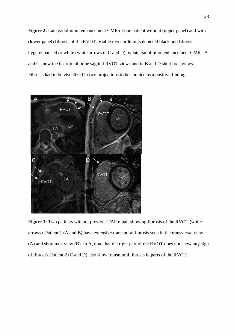
Figure 2: Late gadolinium enhancement CMR of one patient without (upper panel) and with
(lower panel) fibrosis of the RVOT. Viable myocardium is depicted black and fibrosis
hyperenhanced or white (white arrows in C and D) by late gadolinium enhancement CMR . A
and C show the heart in oblique sagittal RVOT views and in B and D short axis views.
Fibrosis had to be visualized in two projections to be counted as a positive finding.
Figure 3: Two patients without previous TAP repair showing fibrosis of the RVOT (white
arrows). Patient 1 (A and B) have extensive transmural fibrosis seen in the transversal view
(A) and short axis view (B). In A, note that the right part of the RVOT does not show any sign
of fibrosis. Patient 2 (C and D) also show transmural fibrosis in parts of the RVOT.

24
Figure 4. Correlation between the degree of fibrosis and A right ventricular ejection fraction
(RVEF), B right ventricular end-diastolic volume (RVEDV) indexed to BSA, C right
ventricular end-systolic volume (RVESV) indexed to BSA and D pulmonary regurgitant
volume.
25
APPENDIX . MR sequence parameters.
Cine:
Philips: A steady-state free-precession sequence with retrospective ECG triggering was used
with acquired temporal resolution of typically 47 ms reconstructed to 25 ms, echo time 1.4
ms, flip angle 60˚and 8 mm slice thickness. Siemens: A gradient-echo sequence with
prospective ECG triggering was used with typically 15 phases per cardiac cycle, with
acquired temporal resolution of 100 ms reconstructed using echo sharing reconstructed to
every 50 ms, echo time 4.8 ms, flip angle 30˚ and 10 mm slice thickness. Breath-hold times
were typically 15 seconds.
Flow:
Philips: A fast field echo velocity encoded sequence with retrospective ECG triggering was
used with repetition time 10 ms, echo time 5 ms, flip angle 15˚, slice thickness 6 mm, 35
phases, number of acquisitions 1, no parallel imaging and a velocity encoding gradient
(VENC) of 200 cm/s.
Siemens: Imaging parameters were the same as above except that the images were obtained
with prospective ECG triggering. Velocity information was acquired over two heartbeats to
quantify the flow during the end of diastole.
The flow sequences were non-segmented without echo sharing with an acquired temporal
resolution of 20 ms for Philips and 35 ms for Siemens.
Late gadolinium enhancement
Philips: An inversion-recovery balanced turbo field echo sequence with slice thickness, 8
mm; field of view, 340 mm; matrix, 126 x 256; repetition time, 3.14 ms; echo time, 1.58 ms
was used.

26
Siemens: An inversion-recovery turbo fast low-angle shot sequence with slice thickness, 10
mm; field of view, 380 mm; matrix, 126 x 256; flip angle, 25º; repetition time, 100 ms; echo
time, 4.8 ms was used.
The inversion time was manually adjusted to null the signal from the non-fibrotic
myocardium and images were acquired at end-diastole.