results of a graft harvesting study: comparing a
TRANSCRIPT

P#1
Abstract Title: Results of a Graft Harvesting Study: Comparing a Rotational Excision Ring Dermatome with Two Standard, Oscillating-Blade-Dermatomes
Author and Co-authors:
Michel H.E. Hermans, MD Hermans Consulting, LLC., Doral, FL
Objective:
Upon completion of the lecture, attendees should be better prepared to: • Describe identified drawbacks with current dermatomes with regard to harvesting skin graft. • Understand how design changes may influence clinical results.
Abstract:
Introduction: The tradition pneumatic or electric dermatome uses an oscillating blade for taking split skin graft. The proper use of such a dermatome requires a specific skill set since the angle of the dermatome, the location of the graft site, the thickness setting of the device and the presence of bony prominences all have an influence on the consistency and the thickness of the graft. In addition, since the dermatome is pushed away from the surgeon, the actual graft is difficult to see while being harvested and adjusting the thickness while taking a graft is very difficult because of the location of the depth-adjustment dial on the side of the device. A new pneumatic dermatome (test device) is designed around a high-speed rotating excision ring. The design aims at making it easier to harvest a graft, particularly also over contoured areas as well as generating a graft with a more consistent thickness. The dermatome has a window which allows for immediate view of graft quality and thickness and since the depth adjustment dial is located on top of the device easy control and quick adjustment of the graft thickness is possible without having to stop the procedure. It is available in a 2-inch and 4-inch version. Methods: A GLP animal study was designed to test the device for safety and efficacy. From the back of each of four female Yorkshire pigs six split thickness skin grafts were harvested in a randomized manner (N=24). 12 Grafts were harvested with two different standard dermatomes and 12 with the 4-inch version of the test device. The wounds measured 61 cm2 on average. Dermatome thickness setting and post wounding topical treatment was standardized amongst all wounds. Wound healing of the animals was followed for 28 days after which the animals were euthanized for histological evaluation of the (now healed) donor sites. Results: On visual observation, no differences existed between the different donor sites with regard to the amount of erythema, edema and granulation tissue formation. All wounds had reached complete re-epithelialization on post-operative day 12. The overall thickness of the split skin graft, measured with a calibrated microscope at three locations of each graft was 0.017 mm on average for the test dermatome (range, 0.008-0.032, SD: 0.006). For the “standard” dermatomes the average thickness was 0.017 mm (range: 0.005-0.035, SD: 0.008). With regards to overall usability, test device and comparators were all considered easy to sterilize, assemble and disassemble and they scored similar on overall ease of use. The test device scored 100% positive on questions on consistency of depth/graft thickness, however, whereas the control

devices received a negative answer on this question. On maneuverability, the test device score “good” in 100% and the comparators scored “fair” in 100%. The test device also had a better score (less bleeding) than the comparator dermatomes with regard to post op bleeding. There were no adverse experiences. Conclusion: A new dermatome with a rotating excisional ring is designed to make graft harvesting easier with regard to handling of the device and obtaining better consistency of graft thickness. In a comparative test of grafting procedures on pigs trends show that the objectives of the design have been fulfilled. The number of wounds in this study were too small to reach statistical significance: superiority of the new rotating-knife dermatome will have to be confirmed in larger scale, comparative clinical trials in humans.
Disclosure: Michel Hermans – Consultant: Exsurco

P#2
Abstract Title:
Excision of Partial Thickness Burns in Pigs: A Study Comparing a Standard Hand Dermatome to a Dermatome with a High-speed Rotating Excision Ring
Author and Co-authors:
Michel H.E. Hermans, MD Hermans Consulting, LLC., Doral, FL
Objective:
Upon completion of the lecture, attendees should be better prepared to: • Describe new dermatome designed to improve on current dermatome design. • Understand how design changes have positive impact on accuracy of excision of necrosis.
Abstract:
Introduction: Excision of necrosis in (deep) partial and full thickness burns is a standard procedure that allows for rapid spontaneous re-epithelialization or prepares the wound for grafting. The Weck dermatome (standard device) has long been among the standard equipment for this type of excision: the dermatome has a fixed number of depth gauges which have to be chosen and applied over the knife prior to the excision taking place. This may result in having to stop the excision procedure and change the gauge and excision levels that are (initially) not deep enough or too deep. A new pneumatic dermatome (test device), designed for both excision of necrosis and (split skin) grafting, has a high-speed rotating excision ring. The dermatome design is aimed at making it easier to use, particularly with regard to maneuverability and over contoured areas. It cuts through tissue quickly and efficiently compared to conventional methods. The rotational excision ring is also expected to allow for more consistent thickness of the excised tissue, graft donor area or necrosis. The dermatome has a window which allows for immediate view of the excised tissues. A depth adjustment dial is located on top of the device which makes it easy to control and quickly adjust the depth level of the excision without having to stop the surgical intervention. The dermatome in available in 2-inch and 4-inch wide versions. Methods: A GLP animal study was designed to test the device for safety and efficacy. On the back of each of four female Yorkshire pigs 6 deep partial thickness burns of 4 cm in diameter were created, using a validated method with water-heated aluminum blocks. Post-operative treatment was with a triple antibiotic ointment and non-adherent dressings. On post-operative day three, all wounds were randomized to be excised with the standard device (N=12) or the 2-inch test device (N=12), until punctate bleeding of the wound bed was observed. The excised layers of necrosis were evaluated, particularly with regard to thickness, measured at three standardized location for each piece of tissue. After the excision, wounds were observed daily and dressed with a non-adherent pad and an adhesive bandage. 28 days after excision animals were euthanized. Wounds were excised and evaluated. Results: On visual observation, no differences existed between the different wounds with regard to the amount of erythema, edema and granulation tissue formation. On

average, re-epithelialization was slightly faster and more extensive for the test device excised wounds than for the wounds excised with control, both visually and histologically. The test tissues (excised layers) were felt to be similar in depth/thickness to the surgical setting while the tissue excised with the control were appreciably deeper than the surgical setting of the device. This was confirmed by histological measurements with a calibrated microscope: the thickness of the areas, excised with the test device was 0.021 mm on average (range: 0.008-0.033, SD: 0.007). The thickness of the areas excised with the standard dermatome was 0.058 mm on average (range: 0.020-0.089, SD: 0.015). Accuracy (staying with intended area to be excised) also scored better for the test device, as did maneuverability. Test and comparators were all considered easy to sterilize but the test device was found to be easier to assemble and disassemble. There were no adverse events in this study. Conclusion: In a small study on partial thickness burns in pigs a dermatome with an excisional ring outperformed a standard hand dermatome with regard to use of use, maneuverability, and accuracy of the area to be excised and consistency of the thickness of the excised tissues. The number of burns in this study were small: larger studies in humans are necessary to confirm the strong trends seen in this study.
Disclosure: Michel Hermans – Preparation trial: Exsurco

P#3
Abstract Title: Bromelain Based Debridement: Review of Preclinical Data
Author and Co-authors:
Yaron Shoham, MD; Yuval Krieger, MD; Eldad Silberstein, MD; Alexander Bogdanov-Berezovsky, MD Soroka University Medical Center, Be’er Sheva, Israel
Objective:
Upon completion of the lecture, attendees should be better prepared to: ▪ Describe the characteristics of bromelain based debridement based on preclinical trial results and identify possible advantages over current surgical/non-surgical techniques of eschar removal.
Abstract:
Background: Bromelain Based Debridement (BBD) for deep burns is approved for use in the European Union where more than 2000 patients have already been treated, and is currently an investigational product in the US in a multicenter randomized controlled phase III trial. The objective of this presentation is to review the preclinical trial results in view of clinical trial results that lead to the approval of BBD in Europe and to the FDA IND phase III clinical trial in the US. Methods: In five preclinical studies with BBD several animal burn models were used: A guinea-pig chemical burn (Sulfur-Mustard) model, a porcine contact comb burn model, a porcine radiant heat model, and a porcine circumferential contact burn induced compartment syndrome (BICS) model. The burns were treated with a 4 hour BBD application versus control treatments. In four of the studies punch biopsies were performed in order to study the efficacy of eschar removal and selectivity to burn eschar versus control treatments. In the BICS model efficacy of non-surgical escharotomy was tested by continuously measuring compartment pressure levels compared to controls that later underwent surgical escharotomy. Results: Biopsies demonstrated that BBD completely removed the eschar within 4 hours (116 biopsies, BBD vs. control, p<0.001) with selectivity to non-viable tissues, unharming the viable dermis of donor sites (150 biopsies, no difference between BBD and control) or intact skin (72 biopsies, no difference between BBD and control). In the BICS model BBD lowered compartment pressures to below 30mmHg within 30 minutes (BBD vs. control, p<0.0001), with the added benefit of early eschar removal. Additionally, BBD lead to a significant preservation of zone of stasis within 48 hours (BBD vs. control, p=0.05) and a significant increase in the incidences of complete re-epithelialization within 11-15 days (BBD vs. control, p<0.02). Conclusions: Preclinical data confirm the efficacy and selectivity of BBD as a non-surgical eschar removal agent and an efficient solution for treating BICS in circumferential burns. These data are in line with clinical results.

Funding: This project is funded in whole or in part with Federal funds from the Office of the Assistant Secretary for Preparedness and Response, Biomedical Advanced Research and Development Authority, under Contract No. HHSO100201500035C References and Resources: Singer AJ, McClain SA, Taira BR, Rooney J, Steinhauff N, Rosenberg L. Rapid and selective enzymatic debridement of porcine comb burns with bromelain-derived Debrase: acute-phase preservation of noninjured tissue and zone of stasis. J Burn Care Res 2010;31(March–April (2)):304–9. Rosenberg L, Krieger Y, Silberstein E, Arnon O, Sinelnikov IA, Bogdanov-Berezovsky A, et al. Selectivity of a bromelain based enzymatic debridement agent: a porcine study. Burns 2012;38:1035–40. Singer AJ, Taira BR, Anderson R, McClain SA, Rosenberg L. The effects of rapid enzymatic debridement of deep partial thickness burns with Debrase on wound reepithelialization in swine. J Burn Care Res 2010;31(September–October(5):795–802. Krieger Y, Rosenberg L, Lapid O, Glesinger R, Bogdanov-Berezovsky A, Silberstein E, et al. Escharotomy using an enzymatic debridement agent for treating experimental burn-induced compartment syndrome in an animal model. J Trauma 2005;58 June (6):1259–64. Eldad A, Weinberg A, Breiterman S, Chaouat M, Palanker D, Ben-Bassat H. Early nonsurgical removal of chemically injured tissue enhances wound healing in partial thickness burns. Burns 1998;24:166–72.
Disclosure:
Yaron Shoham – MediWound Ltd: Director of Medical Affairs - Salary Yuval Krieger – No Relevant Financial Relationships to Disclose Eldad Silberstein – No Relevant Financial Relationships to Disclose Alexander Bogdanov-Berezovsky – No Relevant Financial Relationships to Disclose

P#4
Abstract Title:
A Correlation of Frozen Section Diagnosis and Final Diagnosis in Patients with Suspected Stevens Johnson Syndrome/Toxic Epidermal Necrolysis (SJS/TENS)
Author and Co-authors:
William C. Lineaweaver, MD1; James Neill, MD2; Barbara Proctor, MD3 1Joseph M. Still Burn and Reconstructive Center at Merit Health Central Hospital, Jackson, MS 2University of Mississippi Medical Center, Jackson, MS 3Merit Health Central Hospital, Jackson, MS
Objective:
Upon completion of the lecture, attendees should be better prepared to: ▪ Examine the accuracy of initial frozen section readings of SJS/TENS biopsies to permanent section readings to evaluate the accuracy of the earlier examination. ▪ Discuss the importance of rapidly assessing SJS/TENS in affected patients.
Abstract:
Introduction: SJS-TENS is a severe mucocutaneous disease often apparently related to drug reactions. The disorder includes extensive skin blistering, epidermal loss, mucosal ulcerations, and corneal lesions. Mortality and reach 30%. Early diagnosis is important for discontinuing culprit drugs, initiation of supportive care, and consideration of specific therapy. Histopathologic examination shows characteristic apoptotic keratinocytes and subepidermid blistering. We have routinely evaluated suspected SJS/TENS patients with prompt punch skin biopsies assessed initially by frozen section. Final diagnosis was obtained from permanent sections. We have compared frozen section readings to permanent section readings to evaluate the accuracy of the earlier examination. Methods: 43 consecutive patients admitted with SJS/TENS were subjected to skin punch biopsies. Initial frozen section diagnosis from these specimens were compared to final diagnoses from permanent sections. Results: 9 frozen sections were read as positive for SJS/TENS. 8(88%) were confirmed by permanent sections. 34 frozen sections were read as negative for SJS/TENS. 30(88%) were confirmed by permanent section. Conclusions: Frozen section evaluations were 88% correct for both identification and exclusion of SJS/TENS. Early diagnosis is important for reasons noted above. Early excision is also important for re-directing therapy to correct diagnoses and avoiding specific SJS/TENS therapies (e.g., steroids, IgG) that have their own complications. We continue to use and refine our frozen section analyses to rapidly assess these

complicated patients. At this time, we regard our frozen sections as sufficiently reliable to guide initial therapeutic decisions.
Disclosure: William Lineaweaver – No Relevant Financial Relationships to Disclose James Neill – No Relevant Financial Relationships to Disclose Barbara Proctor – No Relevant Financial Relationships to Disclose

P#5
Abstract Title: MRI Compatibility of Silver Based Wound Dressings
Author and Co-authors:
Heather M. Powell, PhD1; Steffen Sammet, MD, PhD2; Jason Overocker, PA-C3; Beretta Craft-Coffman, PA-C4; Cristina M. Acevedo, PhD5; Martin Cowan, PhD5; J. Kevin Bailey, MD1 1The Ohio State University, Columbus, OH 2University of Chicago Medical Center, Chicago, IL 3Saint Alphonsus Medical Group, Nampa, ID 4Burn and Reconstructive Centers of America, Augusta, GA 5Milliken Healthcare, Spartanburg, SC
Objective:
Upon completion of the lecture, attendees should be better prepared to: ▪ Describe testing protocols for evaluating the MRI safety and compatibility of silver-based wound dressings. ▪ Demonstrate the MRI safety and compatibility of a subset of silver-based wound dressings.
Abstract:
Introduction: The use of dressings containing long-acting silver has become routine in the care of burns and wounds. As these dressings become more popular, it becomes progressively more likely that patients benefitting from this class of dressings will require magnetic resonance imaging (MRI). Burn dressing changes and wound care can be painful with over 75% of burn centers using premedication with opioids for dressing changes. Thus the need to remove dressings simply for MR imaging only adds to the patient’s pain and can contribute to opioid dependency. The goal of this study was to establish a protocol for examining MRI safety and compatibility silver-based dressings and subsequently perform this analysis on multiple forms of silver-based dressings. Methods: To determine if silver wound dressings are safe for MR imaging and thus can remain on during scanning, magnetically induced displacement force and torque were assessed following the American Society for Testing and Materials International (ASTM) Standards F2052-15 and F2213-06, respectively. To assess magnetically induced heating and image distortion, hind limbs from euthanized pigs were studied in a clinical high field 3T MRI scanner. A full-thickness excisional wound was created and dressings applied to the wound first in a dry application with no additional fixation or dressings and scanned in the 3T MRI scanner. Directly after scanning in their dry form, dressings were soaked in saline + 5% bovine serum albumin (BSA) and re-scanned. Finally, one silver-based dressing was investigated in its “in use” form, where the dressing was soaked in the saline-BSA and applied using a custom-assembled patch technique. The test dressing was affixed to the excised area using surgical skin staples, covered with absorbent gauze and both layers wrapped with an elastic dressing. A series of six standard MRI sequences (Survey, T1-weighted SE, T1-weighted IR TSE, T2-weighted TSE, DUAL TSE, and FLAIR) were used and run concurrently. Temperature was assessed using fluoroptic probes affixed on the
porcine surface for the control sections (blank scans) and placed between the dressing and porcine surface with one probe at the center of the wound and one at the periphery for the experimental sections. Temperature at the start and end of each MRI sequence was recorded with change in temperature reported. Results: Six different silver dressings with various forms were tested along with two non-silver dressings. In the ASTM deflection and torsion tests, none of the dressings exhibited any deflection or torsion. In the heating and image distortion tests, all dressings in all six MRI scans no dressing induced heating greater than the safety threshold of +2°C. The average heating was less than 0.5°C in all cases and on average between 0-0.2°C. Additionally, the dressings, both in their dry and hydrated forms, caused no image distortion in any of the six MR scans performed. Finally, to more closely mimic the dressing assembly utilized clinically, MR scans were performed on a silver-based, hydrated dressing which was affixed to the excisional wound using surgical staples and wrapped with gauze followed by an elastic dressing. No image distortion or heating was observed under or adjacent to the dressing in this format. Conclusions: Evaluation of MRI safety and compatibility following ASTM guidelines for deflection and torque and using a porcine limb model for revealed no concerns for safety or issues with image distortion in any of the silver-containing wound dressings tested thus it would be acceptable to leave these dressings intact during MR imaging. It may prove important that similar tests are completed with similar products, so as to ensure widespread benefit from other members of this class of dressings. The ability to leave dressings in place during imaging will provide a significant benefit to patient care by reducing pain when removing the dressings and subsequently will lead to a decreased use of narcotics for treatment of anxiety and pain.
Disclosure:
Heather M. Powell – No Relevant Financial Relationships to Disclose Steffen Sammet – No Relevant Financial Relationships to Disclose Jason Overocker – No Relevant Financial Relationships to Disclose Beretta Craft-Coffman – No Relevant Financial Relationships to Disclose Cristina Acevedo – No Relevant Financial Relationships to Disclose Martin Cowan – No Relevant Financial Relationships to Disclose J. Kevin Bailey – Research Funding
P#6
Abstract Title: Effect of Early Cessation of Pressure Garment Therapy on Structural Properties of Burn Scars
Author and Co-authors:
Heather M. Powell, PhD1; Danielle DeBruler, MS1; Jacob Zbinden, BS1; Molly Baumann, MS1; Britani Blackstone, PhD1; Dorothy Supp, PhD2; J. Kevin Bailey, MD1
1The Ohio State University, Columbus, OH 2Shriners Hospitals for Children, Cincinnati, OH
Objective:
Upon completion of the lecture, attendees should be better prepared to: ▪ Examine the relationship between scar maturation and pressure garment therapy. ▪ Demonstrate the need for scar maturation prior to cessation of pressure garment therapy.
Abstract:
Introduction: Pressure garment therapy is commonly used to treat hypertrophic scarring following burn injury, resulting in improvements in scar appearance, reduced scar height and erythema, and reduced contractures. However, the garments have been reported to be uncomfortable, and this contributes to decreased patient compliance. There is a wide variation in the suggested amount of time before stopping therapy, ranging from as little as 4 months to as long as 2 years. The goal of this study was to determine if 4 months of wear time is sufficient to maintain the benefits of pressure therapy as a possible way to improve patient compliance. Methods: To have complete control over subject compliance, a large animal model, known for its excessive scarring, was utilized. Full thickness burn wounds (1 x 1 in) were created on the dorsum of female Red Duroc pigs. Burns were excised and split-thickness autograft applied (1:1.5 mesh). Pressure garments were applied within 1 week after grafting and maintained at 20 + 2 mmHg. Control scars did not receive any pressure. Seventeen weeks after grafting, pressure was removed from half of the pressure treated scars while pressure was maintained in the other half for an additional 12 weeks. This resulted in 3 treatment groups: the continuous pressure group which received pressure for a total of 29 weeks, the pressure release group which received pressure for 17 weeks, then pressure was removed for an additional 12 weeks, and the control group that did not receive pressure (n=8 scars per treatment group). Several scar properties were monitored over time including contraction, scar height, surface roughness, and biomechanics. Results: Pressure garment therapy significantly reduced contraction, scar height, and roughness compared to controls at 17 weeks after grafting. When pressure was removed, the pressure release scars contracted and increased in height and surface roughness while the continuous pressure scars continued to have less contraction, surface roughness, and height compared to controls. Pressure treatment tended to result in softer scars at 17 weeks after grafting compared to controls, but after pressure was removed the scars became harder. By 29 weeks after grafting, the continuous pressure group was significantly softer compared to both controls and the

pressure release group. Conclusions: Although pressure garment therapy can reduce scar height, surface roughness, and contraction and improve biomechanical properties of scars, the therapy must be continued for a period of time greater than 4 months to maintain these observed benefits.
Disclosure:
Heather M. Powell – No Relevant Financial Relationships to Disclose Danielle DeBruler – No Relevant Financial Relationships to Disclose Jacob Zbinden – No Relevant Financial Relationships to Disclose Molly Baumann – No Relevant Financial Relationships to Disclose Britani Blackstone – No Relevant Financial Relationships to Disclose Dorothy Supp – No Relevant Financial Relationships to Disclose J. Kevin Bailey – No Relevant Financial Relationships to Disclose
P#7
Abstract Title: Effect of Daily Wear Time on Outcomes of Pressure Garment Therapy
Author and Co-authors:
Heather M. Powell, PhD1; Heather M. Powell, PhD1; Molly Baumann, MS1; Danielle DeBruler, MS1; Jacob Zbinden, BS1; Britani Blackstone, PhD1; Dorothy Supp, PhD2; J. Kevin Bailey, MD1
1The Ohio State University, Columbus, OH
2Shriners Hospitals for Children, Cincinnati, OH
Objective:
Upon completion of the lecture, attendees should be better prepared to: ▪ Describe the clinical impact of reduced duration of daily wear of pressure garment therapy.
Abstract:
Introduction: Pressure garments have been shown to reduce erythema, scar height, and reduce contractures when applied following burn injury. As garments have been reported to cause skin irritation, tenderness, and blistering and patients are instructed to wear garments 23 hours out of the day, patient compliance has been a major challenge associated with pressure garment therapy. If patients were able to wear the garments for less time during the day, it is possible that skin irritation would be reduced and patient compliance would improve. The goal of this study was to examine the efficacy of pressure garment therapy when applied for reduced daily durations of wear. Methods: To tightly control for subject compliance, this study was performed in a porcine model known for excessive scarring post burn injury. Full thickness burn wounds (1 x 1 in) were created on Red Duroc pigs (8 burns per pig). Burns were then excised and autografted with split-thickness autograft (1:1.5 mesh). Custom adjustable pressure garments were applied 1 week after grafting and maintained at 20 + 2 mmHg. Garments were applied for 8, 16, or 24 hours a day for 15 weeks; control scars did not receive any pressure treatment (n=16 wounds per group). Characteristics of the scars were monitored over time, including contraction, scar height, erythema, surface roughness, and biomechanics. Results: By 16 weeks after grafting, wounds that received pressure 24 hours a day had significantly reduced contraction compared to all other groups. Wounds that received pressure 16 and 8 hours a day contracted more than the 24 hour group, but were still significantly less contracted than control scars. Reduced wear times slightly improved surface roughness and scar height compared to controls, but a greater improvement was seen in the 24 hour group. There was no difference in erythema between pressure treated scars and control scars. Applying pressure 8 and 16 hours a day improved some biomechanical properties compared to controls, such as elastic deformation, ultimate deformation, and stiffness, but was unable to improve others such as elasticity and viscoelastic deformation. However, applying pressure 24 hours a day improved all measured biomechanical properties over controls.

Conclusions: Although applying pressure therapy for shorter times during the day did improve some scar characteristics over controls, in order to achieve significant improvements in all aspects of scar properties garments need to be applied 23-24 hours a day.
Disclosure:
Heather M. Powell – No Relevant Financial Relationships to Disclose Molly Baumann – No Relevant Financial Relationships to Disclose Danielle DeBruler – No Relevant Financial Relationships to Disclose Jacob Zbinden – No Relevant Financial Relationships to Disclose Britani Blackstone – No Relevant Financial Relationships to Disclose Dorothy Supp – No Relevant Financial Relationships to Disclose J. Kevin Bailey – Research Funding

P#8
Abstract Title: Avoiding Misplacement of Feeding Tubes in the Trachea using Feeding Tube with Balloon and Pulse Oximetry
Author and Co-authors:
Sabry Gabriel, MD Navident Health Medical Center, Mercer University School of Medicine, Macon, GA
Objective:
Upon completion of the lecture, attendees should be better prepared to: ▪ Avoid misplacement of feeding tubes in the trachea and lung and prevent pneumothorax. ▪ Use pulse-oximetry to recognize misplacement in the trachea of a feeding tube with balloon before causing injury. ▪ Increase the likelyhood of distal migration of a feeding tube by using a new feeding tube with distal end balloon.
Abstract:
Background: Enteral feeding is essential for severe trauma, burn and head injury patients who are unable to swallow or consume an oral diet. Enteral feeding tubes are associated with rare but serious complications. Feeding tube misplacement in the lung, although rare (2%), is associated with high mortality rate (50%). Over the last decade gastric feeding became widely accepted among clinicians because the preferred post-pyloric feeding frequently resulted in delayed initiation of feeding with poor total calorie intake, delayed recovery and healing. An ideal feeding tube should minimize tracheal misplacement, allow early gastric feeding with high potential for post-pyloric migration without extra skills or costly procedures. The Gabriel feeding tube with balloon was developed with support from the DOD (W81XWH-09-2-0097) to accomplish these goals. Materials and method: The feeding tube has a 3 ml balloon at its distal end. The tube wall is very thin, flexible but does not occlude by kinking as it is enforced with a spiral wire. The feeding tube kit contains a numbing gel, applicator for numbing gel, lubricant, syringe, skin adhesive and securing tape. All components are essential for the procedure and add only few grams to the tube's kit. The tube is inserted through topically anesthetized nostril. At the 35 cm depth mark, mid-esophagus, the balloon is inflated. If pulse oximetry does not drop, a confirmation of esophageal placement rather than lung or tracheal placement is established within few seconds. Feeding tube is further advanced to the 70 cm mark and the stiffening stylet is pulled out 40 cm and an additional 40 cm of the tube is advanced through the nose to the 110 cm mark. The tube's distal end balloon remains inflated, and the tube

is secured at the nose. Enteral feeding can begin immediately with head of bed elevated 30 degrees once gastric placement is confirmed by x-ray. The coiled feeding tube in the stomach will not occlude by kinking and it provides slack that allows tube to advance distally by the effect of natural peristalsis on the bolus-sized balloon. Results: Most feeding tubes advanced post pyloric to the duodenum or jejunum within 24 hours. Gastric feeding was initiated in all patients within one hour. There was no misplacement in the trachea or lung and no pneumothorax. Two tubes were occluded, one by Nexium and another by Flomax. Both medicines are capsules that contain granules. In general an alternative non-granulated medicine should be used in any feeding tube. Conclusion: The Gabriel feeding tube with balloon provides means for early enteral feeding. The feeding tube balloon and pulse oximetry were used to minimize the risk of misplacement in the lung and pneumothorax. Most of the feeding tubes advanced to the small bowel, reducing the risk of gastro esophageal reflux related aspiration pneumonias. Tube placement does not require costly fluroscopy or endoscopy. Any nurse who can place a nasogastric tube can place this feeding tube without need for extra skills or special training.
Disclosure: Sabry Gabriel – Founder: Syncro Medical Innovations

P#9
Abstract Title: Use of Dehydrated Amnion Chorion Membranes in Facial Burns and its Association with Pigment Dyscrasias
Author and Co-authors:
Tracee Short, MD Baton Rouge General Burn Unit, Baton Rouge, LA
Objective:
Upon completion of the lecture, attendees should be better prepared to: ▪ Discuss how amniotic membrane can be used in burn wounds ▪ Evaluate the photographic outcomes of burns treated with antibiotic ointment vs dehydrated human amniotic chorionic membrane ▪ Discuss whether the current practice of facial burn management should be reevaluated.
Abstract:
Facial burns are a frequent complication of scald and flame burns. They often heal quickly and well but in the interim cause significant swelling. Facial burns can be complicated by facial tightness, pigment dyscrasias and scarring. The use of amnion for the management of burn wounds dates back as far as 1913 by Sabela. Numerous studies have linked the use of amnion to reduction in pain. Our goal was to develop a standard of care in optimizing the care of our facial burn patients. In deciding what to look at whether return to baseline pigment was quicker than our standard of care of antibiotic ointment. All patients with significant facial burns were treated with debridement and application of dehydrated human amnion chorion membranes. The wounds were dressed in adaptic and outer dressings changed daily. Patients underwent photodocumentation per unit protocol and were followed in the outpatient clinic. Evaluation of the pictures indicated that the pigment return was more even and faster to return than standard of care. The incidence of pseudobarbae folliculitis was less. The incidence of complaints of tightness in the perioral region was unchanged. The results of this improvement project indicate that the long held conviction of superficial facial burns don’t need much attention may be a dogma of the past. Each patient is now screened for the need for allografting to decrease the edema, improve pigmentation and potentially minimize stricture formation. References and Resources Leon-Villapalos, Jorge, Marc G. Jeschke, and David N. Herndon. "Topical management of facial burns." Burns 34.7 (2008): 903-911. Kesting, Marco Rainer, et al. "The role of allogenic amniotic membrane in burn treatment." Journal of burn care & research 29.6 (2008): 907-916. Bose, Bcnn. "Burn wound dressing with human amniotic membrane." Annals of the Royal College of Surgeons of England 61.6 (1979): 444.

Disclosure: Tracee Short – No Relevant Financial Relationships to Disclose

P#10
Abstract Title: Can There be an Ideal Dressing that Gives a Two-for-One? Use of a Polylactic Acid Polymer for Donor Sites and Deep Dermal Burn Wounds
Author and Co-authors:
Tracee Short, MD Baton Rouge General Burn Unit, Baton Rouge, LA
Objective:
Upon completion of the lecture, attendees should be better prepared to: ▪ Discuss option available for the management of donor site. ▪ Describe potential advantages of an alloplastic skin substitute vs xenografts.
Abstract:
Partial thickness burn wounds have always had a plethora of treatment options. The history of burn has included use of animal skin, human cadaveric skin, and most recently the introduction of fish skin. Another non-xenograft made its way into the algorithm of burn wound care, alloplastic skin substitute. Our institution was trying to determine if there was a role for polylactic acid polymer as a replacement to our standard of care, a safetac, silicone lined silver impregnated foam dressing or the cryopreserved porcine xenograft. Published research indicated that the alloplastic substitute has decreased pain, minimized wound care and was capable of use on deep dermal wounds. Change is difficult to implement with a team of surgeons. As a measure to unify our standard of care we utilized the alloplastic substitute on donor sites and partial thickness wounds with a deep dermal component. In efforts to develop an improvement in quality we changed our existing practice of donor site management to utilizing the alloplastic skin substitute. In this time period we also investigated the use of the alloplastic substitute on deep dermal wounds instead of our traditionally used safetac silicone lined silver impregnated dressing. Patients were photodocumented and observations recorded per protocol. These patients and comments were recorded until follow-up ceased per usual. Our preliminary results indicate that the use of the alloplastic skin substitute were preferred by the three independent surgeons. Multiple team members felt the mobility post-operatively was improved and patient complaint was decreased. There were no documented infections in the skin substitute wounds and the use of the substitute on wounds not considered candidates for foam dressings allowed for minimal wound care in patients that otherwise would have had daily wound care. At the conclusion of our quality improvement period we have continued the use of the polylactic acid skin substitute and will continue to study its outcomes.
Disclosure: Tracee Short – No Relevant Financial Relationships to Disclose

P#11
Abstract Title: Management of Challenging Cases Using a Hyaluronic Acid Wound Device
Author and Co-authors:
Ariel M. Aballey, MD West Penn Hospital Burn Center, Pittsburgh, PA
Objective:
Upon completion of the lecture, attendees should be better prepared to: • Discuss hyaluronic acid wound devices to assist closing certain types of challenging wounds. • Describe the benefits of a hyaluronic acid wound device for closure of certain wounds.
Abstract:
Introduction: In clinical practice, is not uncommon to encounter wound beds that lack the necessary characteristics to support the viability of a split-thickness skin graft. In these cases, skin graft loss and other wound complications are common. In addition, some patients are not good surgical candidates. A bi-layered, flexible product made of a modified molecule of hyaluronic acid (a glycosaminoglycan present in the extracellular matrix) and a semi-permeable silicone membrane has been proposed as a possible viable therapy for wounds that are difficult to close. In our practice, we have started evaluating this wound device, to determine if these challenging full thickness wounds can be managed more effectively. Methods: Several patients with different challenging wounds underwent application of the wound device after debridement either in the office or operating room. The device was fenestrated with a scalpel previous to application. It was secured in place with absorbable sutures and covered with a non-adhering dressing. The wound device was inspected once or twice a week to check for adherence to the wound. Results: All the patients responded positively to the use of the wound device. No adverse reactions were seen. The time necessary to closure of wounds varied among the different patients. Conclusions: The use of a hyaluronic acid wound device to stimulate the formation of a dermal template in cases where wound closure can’t be achieved, seems like a valid option for challenging cases. References and Resources: Hyaluronic acid derivatives and their healing effect on burns, epithelial surgical wounds, and chronic wounds: a systematic review and meta-analysis of randomized controlled trials. Voigt J1, Driver VR. Wound Repair Regen. 2012 May-Jun;20(3):317-31
Disclosure: Ariel M. Aballay – Speaker: Medline

P#12
Abstract Title: Fasciotomy Wound Preparation for Staged Split Thickness Skin Graft using Fascial Perforation Technique
Author and Co-authors:
Peter Yen, PA-C; Madhawi Mitwalli, MD; James Hwang, MD University of Alabama at Birmingham Burn Center, Birmingham, AL
Objective:
Upon completion of the lecture, attendees should be better prepared to: • Recognize a surgical technique to improve wound bed preparation for staged autografting.
Abstract:
Introduction: Complex wounds and large fasciotomy sites, represent a significant health burden. The standard method of addressing edematous extremities with these type of wounds is autogenous split-thickness skin grafts (STSGs). The decision for a delayed primary closure is often superseded by the urgent need for wound closure, as the subsequent result of delayed closure is, poor anatomical and functional outcome as well as the development of complications such as, bleeding, infection and sepsis. Extensive soft tissue defects often require multistep approach before grafting can be performed. Generally, the technique of applying STSGs directly to the fascia, is often met with limited success due to shearing with movement of the underlying muscle, and variable vascularity of the fascia. In contrast to a well vascularized granulated wound bed, the probability of graft success is much less over poorly vascularized fascia, hence the need for wound bed preparation. Negative pressure wound therapy (NPWT) emerged over the past decade, proving excellent results in enhancing wound-bed preparation, by improving blood supply and promoting granulation tissue, with positive effect on graft take-rate and the average time for wound bed preparation using NPWT reported to be 4-6 weeks. We developed a simple surgical technique of fascial perforation in conjunction with NPWT. A technique which exposes the well vascularized muscle deep to the overlying fascia through perforations, accelerating wound preparation for Split thickness skin grafting. Method: 23-year-old male who had a motor vehicle collision, developed a left thigh compartment syndrome subsequent to intramedullary nailing of femur fracture. A complex wound of approximately 250 cm2 of total body surface area resulted after anterolateral fasciotomy. Despite multiple surgical debridement, complications developed, therefore, the decision for staged STSGs was made. After an initial debridement and excision of necrotic tissue, the wound-bed was prepped by fascial perforation (July 12). Using the bovie, 3 cm linear superficial perforations were made on the fascial plane, each line spaced 2 cm alongside the other. Once the fascia was adequately perforated, and without damaging the underlying muscle, a NPWT on sponge dressing was initiated with maximum recommended setting of 125 mmhg, maintained for 19 consecutive days and changed every 4-5 days. The wound-bed was ready, granulated and vascularized for grafting 3 weeks later (Aug 3). STSGs was successfully applied with 1:1.5 meshed and NPWT initiated for four days. On the fourth day, the graft revealed 100% take, no signs of infection and the wound was dressed in an antibiotic dressing.
Discussion: Based on the difficulties and complications encountered with delayed closure of complex soft tissue wounds, such as, infection, bleeding, besides the poor outcome in limb function, the fascial perforation technique is a simple way to provide direct blood supply to the wound-bed, for acceleration in granulation, and grafting subsequently. In comparison, to the approximate average time of 4-6 weeks’ treatment with NPWT for wound-bed preparation, we expect the time needed for granulation with this technique to be less than two weeks. Nevertheless, due to the limitation of clinic appointments and operative times availability, the frequency of assessment was not attained precisely. Further analyses with more subjects and frequent wound assessment needed.
Disclosure:
Peter Yen – No Relevant Financial Relationships to Disclose Madhawi Mitwalli – No Relevant Financial Relationships to Disclose James Hwang – No Relevant Financial Relationships to Disclose
P#13
Abstract Title:
A Phase III Open-label, Controlled, Randomized, Multicenter Study Evaluating the Efficacy and Safety of StrataGraft Skin Tissue in Promoting Autologous Skin Tissue Regeneration of Complex Skin Defects Due to Thermal Burns that Contain Intact Dermal Elements and for which Excision and Autografts are Clinically Indicated
Author and Co-authors:
Lynn Allen-Hoffmann, PhD1; Mary Lokuta, PhD1; Allen Comer, PhD 1; Peggy Rooney, PhD 1; Barbara Matthews, MD2; David Ng, PhD 3; David Morris, PhD 4; Kristine Lee, PhD 5 1Stratatech, a Mallinckrodt Company, Madison, WI 2BioDirect, Inc., Silver Spring, MD 3ResearchPoint Global, Austin, TX 4WebbWrites, Durham, NC 5University of Wisconsin-Madison, Madison, WI
Objective:
Meeting attendees should become familiar with the design of a phase III study being conducted to assess the efficacy and safety of a single application of StrataGraft skin tissue in the treatment of complex skin defects due to thermal burns containing intact dermal elements and for which surgical excision and autografting are clinically indicated.
Abstract:
Introduction: Every year in the United States, approximately 45,000 patients experience burns that require hospitalization, and ~10 to 20% require surgical intervention. Autografting, the standard of care for serious burns, is the surgical harvest of a sheet of healthy skin from an uninjured site on the patient and transplant to the wound after excision. It results in an iatrogenic donor site wound that requires medical management of pain, possibility of infection, scarring etc. StrataGraft skin tissue is a living, full-thickness human skin substitute that is being developed to reduce or eliminate the need for autograft in the treatment of complex skin defects due to thermal burns. In the STRATA2011 clinical study, 27 of 28 per-protocol subjects had complete wound closure of treatment sites at 3 months, and no subjects required autografting by day 28. No evidence of DNA from cells of StrataGraft skin tissue was seen after 3 months in all tested patients. No safety signal associated with StrataGraft skin tissue has been seen. Methods: A phase III open-label, controlled, randomized, study is being conducted at 12 to 15 ABA-verified burn centers across the United States. Targeted enrollment is up to 70 adult subjects with thermal burns containing intact dermal elements and for which surgical excision and autograft placement are clinically indicated. Each subject will have one burn area treated with StrataGraft skin tissue and a second wound area of comparable depth treated autograft. The two co-primary endpoints for the study are:
• The difference in the percent area of the StrataGraft treatment site and control autograft treatment site that is autografted by 3 months

• The proportion of subjects achieving durable wound closure of the StrataGraft treatment site at 3 months without autograft placement
Ranked secondary endpoints include:
• Difference between the StrataGraft and autograft donor sites in the average pain intensity through day 14 based on the Faces Pain Rating Scale
• Difference between the StrataGraft and autograft donor site cosmesis at 3 months based on observer Patient and Observer Scar Assessment (POSAS) total score
• Difference between the StrataGraft and autograft treatment site cosmesis at 12 months based on observer POSAS total score
Results: Patient enrollment and assessment for this study is ongoing. Additional clinical sites are being identified and qualified to facilitate completion of the study. Data from the ongoing phase III study will be available after completion of patent enrollment and followup. References and Resources: Clinicaltrials.gov NCT03005106
Disclosure:
Lynn Allen-Hoffmann – Senior Vice President, Regenerative Medicine, Mallinckrodt Pharmaceuticals Allen Comer – Salary: Mallinckrodt Pharmaceuticals Mary Lokuta - Salary: Mallinckrodt Pharmaceuticals Peggy Rooney - Salary: Mallinckrodt Pharmaceuticals Barbara Matthews – No Relevant Financial Relationships to Disclose David Ng – No Relevant Financial Relationships to Disclose David Morris – No Relevant Financial Relationships to Disclose Kristine Lee – No Relevant Financial Relationships to Disclose

P#14
Abstract Title: Use of Aerosolized Hypertonic Saline in Inhalation Injury – Novel Idea or Sound Science?
Author and Co-authors:
Russell E. Graham, BSRC, RRT, CPFT, RCP, FAARC; Todd F. Huzar, MD Memorial Hermann - Texas Medical Center, Dunn Burn Center, Houston, TX
Objective:
Upon completion of the lecture, attendees should be better prepared to: • Discuss the effect of adequate humidity and caliber of the airways in mechanical ventilation of inhalation injury. • Examine the processes hypothesized to be responsible for successful treatment of these patients. • Incorporate these findings into clinical practice.
Abstract:
Background: We have previously reported findings on the role of adequate humidity when utilizing High Frequency Percussive Ventilation (HFPV) in the treatment of inhalation injury (INHI) (1). Despite these advances in our group’s clinical practice, we have continued to face challenges in the successful treatment of severe inhalation injury. We are reporting on the successful treatment of three recent cases with aerosolized hypertonic saline solution. We recently treated three severe inhalation injuries in rapid succession. Two were placed on HFPV, while the third was placed on APRV. In all three instances, the inhalation injury was judged to be among the most severe ever seen by our group. All of them exhibited an increase in physiologic dead space and/or shunt that severely impacted arterial blood gas values with Pa02/Fi02 ratios of less than 100. Our standard protocol involves treatment with aerosolized bronchodilators, heparin, and n-acetylcysteine per previously reported successful findings currently in literature (2). After several days of treatment via this regimen, bronchoscopic findings were essentially unchanged with near airway obstruction, and the patients remained too critical for surgical treatment of their burns. In light of this, we made a clinical decision to stop the n-acetylcysteine and substitute aerosolized hypertonic saline solution based on a growing body of evidence that supports its’ use (3,4,5). Method: A comprehensive review of the literature, using Pub Med and MEDLINE, revealed a lack of discussion for using aerosolized hypertonic saline in patients with INHI. Results: While there appears to be more than adequate evidence supporting the use of aerosolized unfractionated heparin to prevent formation of fibrin casts, the link to combination use with n-acetylcysteine is less than compelling. In fact, there is no current supportive evidence for use of aerosolized n-acetylcysteine in cases other than Cystic Fibrosis (5). The growing body of evidence supporting aerosolized hypertonic saline indicates that there may be sustained hydration of airway secretions based on intermittent aerosol administration. In our clinical observation of these three initial patients, we noted rapid and consistent improvement in airway caliber, resolution of the processes surrounding their inhalation injury, and they were all successfully liberated from mechanical ventilation.

Conclusion: While we are not suggesting that aerosolized hypertonic saline is in itself a newfound panacea for treatment of inhalation injury, we did note improvement with its use. Certainly further study is warranted. We have amended our treatment protocols to include the hypertonic saline, and are now excluding n-acetylcysteine. We look forward to reporting additional findings in the future.
(1) Graham R et al. Humidification of High Frequency Percussive Ventilation: Problem Solved? (Abstract/Poster, 2016 ABA Annual Meeting)
(2) Miller AC et al. Influence of nebulized unfractionated heparin and N-acetylcysteine in acute lung injury after smoke inhalation injury. J Burn Care Res. 2009 Mar-Apr;30(2):249-56
(3) Randell S et al. Effective Mucus Clearance Is Essential for Respiratory Health, Am J Resp Cell Mol Bio (35), 2006
(4) Gould NS et al. Hypertonic saline increases lung epithelial lining fluid glutathione and thiocyanate: two protective CFTR-dependent thiols against oxidative injury, Respiratory Research 2010, 11:119
(5) Donaldson SH et al. Mucus Clearance and Lung Function in Cystic Fibrosis with Hypertonic Saline, N Engl J Med 2006; 354: 241-50
Disclosure: Russell E. Graham – No Relevant Financial Relationships to Disclose Todd F. Huzar – No Relevant Financial Relationships to Disclose

P#15
Abstract Title: A Blinded Comparison of Lubricants to Facilitate Split Thickness Skin Graft Harvest in a Porcine Skin Model
Author and Co-authors:
Allison R. Beckett, MD, Resident; Alicia C. Lintner, CRNP; Ronald M. Brooks, MD; Scott B. Patterson, DO; Mike L. Roberts, RN; Steven A. Kahn, MD Arnold Luterman Regional Burn Center at University of South Alabama Medical Center, Mobile, AL
Objective:
Upon completion of the lecture, attendees should be better prepared to: • Discuss the multiple options that exist when using lubricants to facilitate skin graft harvest • Recognize that glycerin had the worst performance while a water based surgical lubricant had the best performance in a porcine skin model.
Abstract:
Introduction: Multiple different skin lubricants have been utilized to facilitate the harvest of split thickness skin grafts. Lubricants are generally selected according to provider and institutional preference, as no single “ideal” lubricant has been objectively established. Many providers use poloxamer 188, but there has been a recent national shortage, prompting a search for a suitable substitute. The purpose of this study is to test some commonly used lubricants in the search of a substitute to poloxamer 188 at our institution. Methods: Four experienced skin graft harvesters (30 years of combined skin grafting experience) were selected to participate in the study. Using blocks of refrigerated “butcher shop” porcine skin and subcutaneous fat, five lubricating solutions were tested, including poloxamer 188, mineral oil, glycerin, normal saline and a novel lubricating solution (120 g sterile bacteriostatic water based surgical lubricant diluted to 200 cc total with sterile water), and also a dry control. The study was conducted in two rounds, in which each participant harvested four grafts of porcine skin using with a dermatome set at 0.014”, blindly testing the five solutions in random order during each round and assigning a score based on friction and ease of use of each lubricant (1-5 Likert Scale, 1=poor, 5=excellent), for a total of 20 data points per solution. Tests were also controlled based on number of passes per blade. Data was pooled and means were compared with ANOVA and a Tukey post-test. Results: Mean scores for each of the solutions were as follows: dry control= 1.1±0.1; glycerin= 2.62± 1.02, saline= 3.88 ±0.81, mineral oil= 3.75 ± 1.00, novel water based lubricant solution= 4.63 ± 0.71, and poloxamer 188= 3.88 ± 0.81. All solutions were superior to dry control (p<0.01). Glycerin was noted to have statistically lower scores than all of the other solutions (p<0.01). The novel water based surgical lubricant solution had significantly higher mean scores than both glycerin (p<0.01) and mineral oil (p<0.05). Conclusions: In a porcine skin model, the novel water based surgical lubricant solution had the best performance. It was statistically superior to glycerin and mineral oil. Glycerin had the worst performance with statistically lower scores than all other

solutions. Saline performed surprisingly better than expected, but this may have been related to the inherently greasy nature of the butcher shop porcine skin, creating some limitations and decreasing the fidelity of the model. In a search for the “ideal” lubricant, other models should be further studied. References and Resources: Total Burn Care Lippincott, Williams & Wilkins 351 West Camden Street Baltimore, MD 21201 United States of America
Disclosure:
Allison R. Beckett– No Relevant Financial Relationships to Disclose Alicia C. Lintner– No Relevant Financial Relationships to Disclose Ronald M. Brooks– No Relevant Financial Relationships to Disclose Scott B. Patterson– No Relevant Financial Relationships to Disclose Mike L. Roberts– No Relevant Financial Relationships to Disclose Steven A. Kahn– No Relevant Financial Relationships to Disclose

P#16
Abstract Title: A Multidisciplinary Approach to Managing a Critically Ill Patient with Recurrent Levamisole-Induced Vasculitis
Author and Co-authors:
Ashley Y. Williams, MD, Resident; Steven A. Kahn, MD Arnold Luterman Regional Burn Center at University of South Alabama Medical Center, Mobile, AL
Objective:
Upon completion of the lecture, attendees should be better prepared to: • Describe the relationship between cocaine use and the development of LIV. • Discuss the typical presentation, diagnosis, and management of patients with LIV. • Recognize that LIV has the potential to recur and become life-threatening.
Abstract:
Introduction: Levamisole is an increasingly popular adulterant of cocaine that has been shown to cause a vast number of side effects, most notably disseminated vasculitis and agranulocytosis. Patients frequently present with a characteristic rash described as tender, erythematous, retiform purpura with necrosis and bullae that has potential to evolve into large open wounds. The treatment ranges from supportive therapy with the cessation of levamisole to excision and split-thickness skin grafting. This case presentation is the first report in the literature of a single patient who developed levamisole-induced vasculitis (LIV) 3 times. Methods: The following is a case report involving a critically ill patient with 3 admissions to a Level I Burn Center over a 4 year period for levamisole-induced vasculitis, precipitated each time by a cocaine binge. Results: A 50 year old female with a past medical history significant for polysubstance abuse, Hepatitis C, cirrhosis, and two prior admissions for levamisole-induced vasculitis that required ICU admissions and grafting presented for a third time with painful, necrotic lesions consistent with LIV. Her lesion encompassed 10% total body surface area involving bilateral upper and lower extremities, face, and ears. With each subsequent hospitalization, she developed an increasing severity of illness. During this hospitalization, she developed hemolytic anemia and pancytopenia. She also developed anti-C, CW, and K antibodies, complicating the ability to find compatible blood for transfusion. Her hematocrit decreased to 12.4 despite transfusions of matching blood that was so rare it had to be brought in from another state. She became critically ill and developed mental status changes. She ultimately required IVIG, steroids, and plasma exchange as a last ditch effort to prevent further hemolysis and facilitate safe transfusion of additional blood. After she recovered, she underwent excision and grafting. Her care required a collaborative effort from multiple disciplines including Burn Surgery, Hematology/Oncology, Nephrology, Nutrition, Physical Therapy and Occupational Therapy. Despite the nearly fatal progression of LIV and its associating complicating factors, the patient recovered to the point of discharge with home wound care. Conclusions: Though documented cases of levamisole-induced vasculitis are rare, recent reports have described the presentation, diagnosis and management of this

disease process. This case highlights the potentially-fatal complications associated with LIV and the need for prompt recognition and multidisciplinary management of the critically ill patient with levamisole-induced vasculitis. References and Resources: 1. Ching JA, Smith DJ Jr. Levamisole-induced necrosis of skin, soft tissue, and bone: case report and review of literature. J Burn Care Res. 2012 Jan-Feb. 33(1):e1-5 2. Farmer RW, Malhotra PS, Mays MP, Egger ME, Smith JW, Jortani SA, Spiller H, Bosse GM, Callen JP, Franklin GA. Necrotizing peripheral vasculitis/vasculopathy following the use of cocaine laced with levamisole. J Burn Care Res. 2012 Jan-Feb. 33(1):e6-e11 3. Lentz CW. Drug abuse continues to inflict its harm "skin deep". J Burn Care Res. 2017 May-Jun; 38(3): e638-e646
Disclosure: Ashley Y. Williams– No Relevant Financial Relationships to Disclose Steven A. Kahn– No Relevant Financial Relationships to Disclose
P#17
Abstract Title:
A Safety Pathway Comparing the Use of Dehydrated Human Amniotic/Chorion versus Amniotic Allografts in Facial Burns, Is there a Practice Preference?
Author and Co-authors:
Salomon Puyana, MD, Resident; Adel Elkbuli, MD, MPH; Brenda Benson, RN, CEN, TCRN; Orlando Morejon, MD; Rizal Lim, MD; Mark McKenney, MD, MBA; Harris Mir, MD Kendall Regional Medical Center, Miami, FL
Objective:
Upon completion of the lecture, attendees should be better prepared to: • Describe the use of amniotic/chorion versus amniotic allografts as accepted skin substitutes to treat adult facial burns. • Examine and identify safety measures such as complications and necessity for re-operations when using amniotic/ chorion versus amniotic membrane allografts when treating adult facial burns • Demonstrate outcomes differences and consider practices preference when using amniotic/chorion versus amniotic membrane allografts in treating adult facial burns.
Abstract:
Background: Amniotic membrane is a novel, non-immunogenic, anti-inflammatory and anti-bacterial skin substitute that creates a matrix for cellular migration and proliferation. More than 226 growth factor cytokines and chemokines are involved in the regulation of wound healing and inflammation using amniotic membrane. The highest concentrations of growth factors tend to derive from the chorion layer with a notable exception of epidermal growth factor. It has a great potential in improving burn wound treatment. There is no enough substantial scientific evidence comparing outcomes and practice preferences associated with the use of amniotic/ chorion versus amniotic membrane allografts in treating facial burns. Our study purpose was to compare the safety and impact on practice preferences of amniotic/ chorion to amniotic membrane allografts in adult facial burns. Methods: We retrospectively reviewed prospectively collected data from years 2015-2017 utilizing our hospital Burn Registry. We compared the safety of amniotic/chorion membrane vs amniotic allografts in patients >15 years old with facial burns. Study outcomes included number of surgical re-operations, number of complications, primarily hypertrophic scars, pigmentation and reoperations, and healing at 2 weeks. Demographic variables and burn TBSA were also compared. Paired sample t-test and Chi Squared analysis were used with significance defined as p<0.05. Results: Our study had a total of 77 patients. The age range was from 16 to 88 years with the mean of 40.6 years. The percent total body surface area burn ranged between 1% to 57% and was not statistically significant in both groups. Complications were significantly lower in the group receiving amniotic/chorion membrane allografts (22.8%) versus the amniotic membrane group (52.5%) (p = 0.009). Patients in the group receiving amniotic/chorion allografts needed significantly less re-operations (10.9%) compared to the group receiving amniotic allografts (28.9%) (p = 0.03).

Amniotic/Chorionic
membrane
Amniotic
Membrane
p-value
Total # of Patients 39 38
Age (years) mean 41.4 39.8 ns
Burn TBSA 8.3 10.9 ns
Re-operations 10.9% 28.9% 0.03
Complications 22.8% 52.5% 0.009
% Healing at 2
weeks
94.8% 94.7% ns
Conclusion: This report is the first research work to examine safety and practice preference when comparing the use of amniotic/ chorion versus amniotic allografts in adult patients with facial burns. Amniotic/ chorion allografts showed to be significantly safer than amniotic only allografts in treating burn adult patients. There was a significantly lower number of complications including hypertrophic scars, pigmentations, and decreased number re-operations with the use of amniotic/chorion versus amniotic membrane allografts in the treatment of adult facial burns. Our study outcome could significantly impact practices related to the use of skin substitutes in treating adult facial burns, associated costs and overall quality of care. Further research is needed to reveal other factors associated with the use of Amniotic/ chorion vs amniotic membrane allografts in facial burns such as comorbidities, nutritional status/support and poly-trauma which may significantly alter outcomes.
Disclosure:
Salomon Puyana – No Relevant Financial Relationships to Disclose Adel Elkbuli – No Relevant Financial Relationships to Disclose Brenda Benson – No Relevant Financial Relationships to Disclose Orlando Morejon – No Relevant Financial Relationships to Disclose Rizal Lim – No Relevant Financial Relationships to Disclose Mark McKenney – No Relevant Financial Relationships to Disclose Harris Mir – No Relevant Financial Relationships to Disclose
P#18
Abstract Title: The Relationship Between Frailty and the Subjective Decision to Conduct a Goals of Care Discussion with Burned Elders
Author and Co-authors:
Tarik D. Madni, MD, PGY-41; Jonathan Imran, MD,1; Audra Clark, MD1; Paul A. Nakonezny, PhD1; Brett A. Arnoldo, MD1; Herbert A. Phelan, MD1; Steven E. Wolf, MD1; Joseph Bellal, MD2; M. Jane Mohler, RN, MSN, MPH, PhD2 1University of Texas Southwestern Medical Center, Parkland Regional Burn Center, Dallas, TX 2University of Arizona, Tucson, AZ
Objective:
Upon completion of the lecture, attendees should be better prepared to: ▪ Recognize there is wide variation in the actual implementation of the practice of a Goals of Care discussion nationally. ▪ Realize a frail physical appearance, whether consciously or unconsciously, influences the decision to conduct a GoC discussion.
Abstract:
Introduction: Best practices are to conduct an early discussion of goals of care (GoC) after serious injury in the elderly. GoC discussions are inconsistently performed in our burn unit, however. We hypothesized that a frail physical appearance was a subjective factor in the decision to conduct a documented GoC discussion for burned elders in our care. Method: We performed a retrospective review of all geriatric burn survivors aged ≥65 years at our ABA-verified Level 1 Burn Center between 4/02/2009 and 12/30/2014. Exclusion criteria were: <5% TBSA and inpatient status <2 midnights. Demographic information included age, gender, mechanism, %TBSA burned, revised Baux Score (rBaux), patient/physician racial discordance, documented GoC discussion, length of stay (LOS), and disposition. One reviewer retrospectively assigned frailty scores to all patients ranging from 1 (very fit) to 7 (severely frail) using the Canadian Study of Health and Aging Criteria. Ordinal logistic regression was performed to investigate the effects of the aforementioned covariates on frailty. Results: The cohort consisted of N=126 patients (67% male, 33% female). Demographics included (mean±SD): age =75.5± 7.7 yrs; %TBSA=11.9%±7.2; rBaux=87.8±10.2; hospital LOS (days)=14.9±13.7; ICU LOS (days)=6.2±12; frailty score=4.1±1.1. The major mechanism of burn injury was flame (78%). There was a 38% racial discordance between the patient and attending physician. Overall, 72% of geriatric survivors had a favorable discharge disposition. GoC discussions occurred in 31 (25%) of patients. Multiple ordinal logistic regression demonstrated that documented goals of care discussion (OR 3.42; 95% CI: 1.54-7.60) and an unfavorable discharge disposition (OR 9.01; 95% CI: 3.91-20.78) were associated with greater predicted odds of receiving a higher ordered frailty score. Conclusion: Documented GoC discussion and having an unfavorable discharge status are associated with a greater likelihood of receiving a higher frailty score.

References and Resources: Hodgman EI, Joseph B, Mohler J, et al. Creation of a decision aid for goal setting after geriatric burns: a study from the prognostic assessment of life and limitations after trauma in the elderly [PALLIATE] consortium. J Trauma Acute Care Surg. 2016;81(1):168-72. Partain NS, Subramanian M, Hodgman EI, et al. Characterizing End-of-Life Care after Geriatric Burns at a Verified Level I Burn Center. J Palliat Med. 2016. Rockwood K, Song X, MacKnight C, et al. A global clinical measure of fitness and frailty in elderly people. Cmaj. 2005;173(5):489-95. Joseph B, Pandit V, Rhee P, et al. Predicting hospital discharge disposition in geriatric trauma patients: is frailty the answer? J Trauma Acute Care Surg. 2014;76(1):196-200.
Disclosure:
Tarik Madni – No Relevant Financial Relationships to Disclose Jonathan Imran – No Relevant Financial Relationships to Disclose Audra Clark – No Relevant Financial Relationships to Disclose Paul A. Nakonezny – No Relevant Financial Relationships to Disclose Brett Arnoldo – No Relevant Financial Relationships to Disclose Herb Phelan – No Relevant Financial Relationships to Disclose Steven Wolf – No Relevant Financial Relationships to Disclose Joseph Bellal – No Relevant Financial Relationships to Disclose M. Jane Mohler – No Relevant Financial Relationships to Disclose

P#19
Abstract Title: Single-Stage Composite Skin Reconstruction Using a Dermal Regeneration Template: A Case Report
Author and Co-authors:
Boonyapa Purt, DMD, Resident; Daniel P. True, DMD, Resident; John L. Fletcher, MD, Resident; Rodney K. Chan, MD United States Army Institute of Surgical Research Burn Center, Ft. Sam Houston, TX
Objective:
Upon completion of the lecture, attendees should be better prepared to: • Examine and review the results and efficacy of single-stage bilayer reconstruction using a dermal regeneration template.
Abstract:
Introduction: Soft tissue loss associated with burn injuries is a common and clinically challenging problem that has led to the development of dermal replacement matrices to augment and improve the regeneration of the dermis. Historically, these matrices are applied in a two-stage fashion that provides time for the dermal matrix, which is inherently avascular, to incorporate and vascularize prior to being covered with an overlying split-thickness skin graft (STSG). While this technique has been largely reliable, it obligates the patient to a 3-5 week treatment course that requires a minimum of two operations, prolonged hospitalization or frequent outpatient visits, and, commonly, an extended period with a negative pressure wound dressing (NPWD). Consequently, if a STSG could be successfully applied to a dermal replacement matrix in a single-operation, substantial reductions in hospital stays, outpatient visits, operative encounters, and healthcare expenditures could be realized. Our successes with porcine models of single-stage bilayer reconstruction (SSBR) and the anecdotal experiences of other surgeons led to our selective use of SSBR with a dermal regeneration template (DRT) in this case report. Methods: An 18 year old male who presented ten years after his initial burn injury with multiple post-burn function-limiting contractures underwent three separate procedures using SSBR. The corresponding inpatient and outpatient records were used to evaluate the integration of the STSG and final skin quality. Operative technique was the same for all three operations and included the debridement to a healthy wound bed, application of a mono-layer, meshed DRT, and immediate application of a 0.012 inch meshed STSG. The patient received post-operative NPWDs after all three operations to aid in immobilization and compression of the graft/matrix/wound interface. Clinical assessments of the graft occurred in conjunction with regularly scheduled follow-up visits. Results: Over the course of his treatments with SSBR, there was no noted significant graft loss. There were no major complications associated with the reconstructions. Clinical assessments and photographs taken at scheduled follow-up appointments demonstrate successful graft take in all three instances. Discussion/Conclusion: Single-Stage Bilayer Reconstruction using a Dermal Regeneration Template is useful to obtain soft tissue coverage in well-selected adult patients. This single stage technique obviates the need for this patient to undergo

multiple operations, and decreases the overall length of treatment and frequent outpatient visits, especially in a patient who may require a multitude of reconstructive surgeries. SSBR shows promise in reducing healthcare utilization, improving patient comfort and limiting healthcare expenditures while still providing the benefits of augmented dermal regeneration. However, to better define the utility of this practice, further controlled studies are required. References and Resources: Pham C., Greenwood J., Cleland H., Woodruff P., and Maddern G. Bioengineered skin substitutes for the management of burns: a systematic review. Burns 2007; 33: 946-957. Burd A and Wong P. S. One-Stage Integra Reconstruction in Head and Neck Defects. Journal of Plastic, Reconstructive & Aesthetic Surgery 2010; 63: 404-09.
Disclosure: Boonyapa Purt – No Relevant Financial Relationships to Disclose Daniel True – No Relevant Financial Relationships to Disclose John Fletcher – No Relevant Financial Relationships to Disclose Rodney Chan – No Relevant Financial Relationships to Disclose

P#20
Abstract Title: Inner-city Pediatric Burns: A Retrospective Analysis of Burn Intensive Care Unit Admissions at a Large Public Community Hospital
Author and Co-authors:
Thais Polanco, MD, Resident; Anant Dinesh, MD; Arthur Cooper, MD; Alexius Ramcharan, MD; Ryan Engdalh, MD Harlem Hospital Center, Columbia University Medical Center, New York, NY
Objective:
Upon completion of the lecture, attendees should be better prepared to: • Recognize the socioeconomic barriers in inner city hospitals and implications it has on patient care/pediatric burn admissions. • Discuss circumstances/resources in such unique hospitals can impart better national campaigns for patient education/resources in such communities.
Abstract:
Introduction: Approximately 250,000 children aged 0–17 years old require medical attention for burns each year (Burn Foundation, 2016). Knowing how to prevent and treat the leading causes of burns in children is a step toward decreasing this number. Few if any burn characterization papers have reviewed patients living in inner cities who often face challenges in receiving health care due to various socioeconomic issues. The aim of this study is to understand the circumstances, pre-hospital and hospital care, mechanisms of burns and management challenges in an exclusively inner-city population at a large public community hospital Methods: A retrospective chart review was preformed of all electronic medical records of patients aged 0-17 years old admitted to our Burn Intensive Care Unit from 2006 to 2017. A total of 177 pediatric patients were identified and enrolled for analysis. Patient demographics, mechanism, pre-hospital and hospital care of sustained injuries were analyzed. Results: The majority of patients who sustained burns were toddlers (58%). The average total body surface area burnt was 9%. Almost all the injuries (95%) occurred at home, specifically in the kitchen (64%) followed by the bathroom (15%). Scald burns accounted for the majority (76%) of all burns, with common mechanisms including spillage of hot liquids (68%) and hot oil (10%). A significant seasonal correlation was observed with 40% burns obtained during winter months. Interestingly, 78% of patients did not receive appropriate first aid measures prior to arrival to the emergency department. Conclusions: Inner-city burns are a major public health issue in the pediatric population, with scald burns being the leading cause. In our inner city pediatric burn population, most injuries occurred at home and the majority lacked proper pre-hospital first aid care. Pediatric burn admissions at inner-city hospitals may have reasons for admission that differ from other communities. A better insight of the mechanisms and circumstances of burns can play a significant role in defining preventive measures. Campaigns focusing on safety measures to practice at home and proper first aid measures can effect a positive change in the education, actions, and attitude practiced by caregivers.

Disclosure:
Thais Polanco – No Relevant Financial Relationships to Disclose Anant Dinesh – No Relevant Financial Relationships to Disclose Arthur Cooper – No Relevant Financial Relationships to Disclose Alexius Ramcharan – No Relevant Financial Relationships to Disclose Ryan Engdalh – No Relevant Financial Relationships to Disclose

P#21
Abstract Title:
Evaluating the Use of Oxandrolone as the Standard of Care against Patient Outcomes at University Medical Center/Texas Tech Health Sciences Center
Author and Co-authors:
Logan A. Dobbe, MS-III; Cody Clapp, MS-III; John Griswold, MD Texas Tech University Health Sciences Center, University Medical Center, Timothy J. Harnar Burn Center, Lubbock, TX
Objective:
Upon completion of the lecture, attendees should be better prepared to: • Recognize data on the use of oxandrolone in burn care for the typical patient population remains limited. New, multi-center, prospective studies are needed to clearly evaluate its efficacy as a component of modern therapy. • Identify nutrition management as an important but often overlooked component of burn care. Thorough monitoring may improve patient outcomes and reduce costs.
Abstract:
Background: Major burns are a high-risk trauma whose prognosis should be improved. UMC/TTUHSC Department of Surgery is known for having far improved burn patient outcomes in comparison to the national average, as recognized by the American Burn Association (2016). Some of this success has been attributed to our incorporation of oxandrolone as a standard of care for patients treated by our burn unit. Existing literature shows that oxandrolone indeed improves patient outcomes but does so with limited effect magnitude. Published data on oxandrolone usage in burn units remains scarce, and repeated calls on for the ABA to incorporate its usage into the national repository on burn patients and encourage multicenter studies on oxandrolone’s efficacy have remained unanswered for several years. Its role in burn care protocols remains hotly contested. While previous studies attributed the improved outcomes to oxandrolone, a number of confounding nutrition and metabolic factors may have also contributed. A majority of the research evaluating oxandrolone has assessed the treatment within pediatric populations. The state of current literature on oxandrolone revolves around serial publications performing metadata analysis of the aforementioned research. While these publications and a number of conference proceedings have called for additional large scale studies on oxandrolone to be done, the status quo has prevailed. This case series review and broader analysis of recent clinical data from UMC seeks to address these lapses within the literature. Design: UMC incorporates oxandrolone ordering alongside aggressive nutrition management as standard of care. The original goal of this research was to summarize that clinical data with empirical comparison against national averages. It was discovered that over the past five years, a very small number of patients did not receive oxandrolone due to logistical complications. A longitudinal regression analysis model was constructed to evaluate correlates between clinical outcomes (acute length of stay, discharge weight/BMI, number of operations required) and clinical interventions (whether or not oxandrolone was used in therapy alongside aggressive nutritional management), while controlling for variabilities among the patient population (age, presence of inhalation injury, 2nd degree TBSA, 3rd degree TBSA, admit weight/BMI). Three case reports are also presented with an emphasis on close tracking of nutrition

parameters over the course of acute therapy to demonstrate the exemplary clinical performance of the UMC burn unit, of which oxandrolone use is a cornerstone. Findings: The preliminary longitudinal regression analysis model did not identify significant correlates between the use of oxandrolone and the aforementioned clinical outcomes. However, the model demonstrated a clear association between the length of stay and a downward trend in patient weight (p<0.001), which can be expressed with a simple formula: WeightDay = 0.8339 + 1.0039 (WeightAdmit) – 0.3201 (Day) The very small sample size of the control group (n=11, vs. oxandrolone-treated n=50) is a severe limitation. The model is being reconfigured to accept data with a higher resolution (stratification between 2nd vs. 3rd degree burns, transthyretin/prealbumin inflammation) to adjust for variability in burn severity between the treatment and control group. These findings, alongside three case reports, will be available at the 2017 Southern Burn Conference. Conclusions: Oxandrolone has promising implications for improving patient outcomes and lowering healthcare costs among the burn community. The state of the literature in this area is unsatisfactory. New, multicenter prospective cohort studies are needed to evaluate the efficacy of oxandrolone in the adult population of burn patients as a component of the modern advances in burn care. References and Resources: -American Burn Association. (2016). Ameriburn.org. Retrieved 8 August 2016, from http://www.ameriburn.org/verification_verifiedcenters.php -Barton, N. (1997). Oxandrin (oxandrolone tablets, USP) CIII - Clinical Update. Iselin: BTG Pharmaceuticals. -Chang, D., DeSanti, L., & Demling, R. (1998). ANTICATABOLIC AND ANABOLIC STRATEGIES IN CRITICAL ILLNESS. Shock, 10(3), 155-160. http://dx.doi.org/10.1097/00024382-199809000-00001 -Demling, R. & DeSanti, L. (1997). Oxandrolone, an Anabolic Steroid, Significantly Increases the Rate of Weight Gain in the Recovery Phase after Major Burns. The Journal Of Trauma: Injury, Infection, And Critical Care, 43(1), 47-51. http://dx.doi.org/10.1097/00005373-199707000-00012 -Demling, R. & DeSanti, L. (1998). Closure of the "non-healing wound" corresponds with correction of weight loss using the anabolic agent oxandrolone. (1998). Ostomy Wound Management, 44(10), 58-62. -Demling, R. & DeSanti, L. (2001). The rate of restoration of body weight after burn injury, using the anabolic agent oxandrolone, is not age dependent. Burns, 27(1), 46-51. http://dx.doi.org/10.1016/s0305-4179(00)00064-4 -Demling, R. & Orgill, D. (2000). The anticatabolic and wound healing effects of the testosterone analog oxandrolone after severe burn injury. Journal Of Critical Care, 15(1), 12-17. http://dx.doi.org/10.1053/jcrc.2000.0150012 -Demling, R. (1999). Comparison of the anabolic effects and complications of human growth hormone and the testosterone analog, oxandrolone, after severe burn injury. Burns, 25(3), 215-221. http://dx.doi.org/10.1016/s0305-4179(98)00159-4 -Demling, R. (2001). Involuntary Weight Loss, Wound Healing, and Optimal Nutritional Intervention. Journal Of The American Medical Directors Association, 2(4), H2-H4. http://dx.doi.org/10.1016/s1525-8610(01)80002-5 -Hart, D., Wolf, S., Ramzy, P., Chinkes, D., Beauford, R., & Ferrando, A. et al. (2001). Anabolic Effects of Oxandrolone After Severe Burn. Annals Of Surgery, 233(4), 556-564. http://dx.doi.org/10.1097/00000658-200104000-00012 -Herndon, D. N., & Tompkins, R. G. (2004, June 5). Support of the metabolic response to burn injury. The Lancet, 363(9424), 1895-1902. doi:10.1016/s0140-6736(04)16360-5 http://dx.doi.org/10.1097/01.ta.0000108636.63225.63

Disclosure:
Logan Dobbe– No Relevant Financial Relationships to Disclose Cody Clapp– No Relevant Financial Relationships to Disclose John Griswold – No Relevant Financial Relationships to Disclose

P#22
Abstract Title: Universal Suicide Screening in a Burn Center: Initial Findings A Case Report and Recommendations
Author and Co-authors:
Kimberly Roaten, PhD, CRC1; Christine Lane, RN, CCRN2 1University of Texas Southwestern Medical Center, Dallas, TX 2Parkland Regional Burn Center, Dallas, TX
Objective:
Upon completion of the lecture, attendees should be better prepared to: ▪ Describe the importance of screening for suicide risk in burn patients. ▪ Discuss the prevalence of suicide risk in burn and trauma patients. ▪ Describe the key components of implementing a universal suicide screening program in burn units.
Abstract:
Introduction: Suicide is the tenth leading cause of death in the United States with over 44,000 deaths in 2015. It is estimated that nearly half a million individuals were treated for self-inflicted injuries in emergency departments during the same time period, of which more than 1,600 were due to burn injuries. Additionally, suicide rates have risen by nearly 25% during the past 15 years and have risen most rapidly since 2006. Individuals who die by suicide are more likely to present for non-behavioral healthcare in the months prior to death, and are most likely to receive treatment in the emergency department or from primary care providers who often do not assess suicide risk. Self-inflicted burn injuries are relatively rare, but the available data suggests that patients with self-inflicted burn injuries are much more likely to have a history of mental health issues and pre-morbid alcohol use problems. There is no data available about the prevalence of suicide risk in patients admitted to burn centers, and little is known about trauma patients in general. Methods: The Columbia Suicide Severity Rating Scale (patients >18) or the Ask Suicide-Screening Questions (patients 12-17) was completed by nursing staff during all encounters in the hospital beginning in February, 2015. Data regarding suicide risk prevalence was collected from consecutive trauma and burn encounters over 19 months. Results: The administration of the suicide screening is completed by nursing staff and responses are entered into the electronic health record (EHR). A clinical decision support system is embedded in the EHR and results in prompts for appropriate safety and clinical interventions based on the screening responses. At-risk patients are further assessed by behavioral health providers and receive appropriate interventions as well as outpatient mental healthcare information. Prevalence data on suicide risk levels are provided for trauma and burn patient encounters. Of 11,190 trauma patients screened, 4.6% endorsed at least one item during the screening process. Of 1,573 burn patients screened, 4.1% endorsed at least one item during the screening process.

Case report: Patient J was a 17-year-old Hispanic woman who presented as a level 1 trauma activation following a motor vehicle collision in which she sustained 5% TBSA burns to her hands, eventually requiring multiple digit amputations. There was a fatality at the scene of the MVC. Patient J did not have any previous psychiatric history and was very involved in school activities including the National Honor Society, varsity soccer and cross-country, and social activities. The suicide screening was not completed initially in the trauma hall due to the patient’s level of sedation. She was subsequently admitted to the burn service where she was screened by nursing staff. Patient J endorsed 4 of 5 items on the standardized suicide screening instrument for adolescents. She was immediately placed under 1:1 observation and suicide precautions until a full risk assessment could be completed. During the assessment Patient J acknowledged that she had been feeling very depressed for at least 3 months and had made a suicide attempt in the past. She had not disclosed her distress to anyone prior to the screening. She met full criteria for a major depressive episode and received both medication and individual psychotherapy during her hospitalization. Conclusions: Given the observed rates of positive screening results, it is feasible to implement a universal suicide screening program in a busy regional burn center. This study provides prevalence data that will allow for estimation of resources needed to implement universal suicide screening programs in other settings. It is recommended that suicide screening programs be standardized and implemented with a clinical decision support system in place in order to enhance patient safety. Suicide screening in burn centers will lead to the identification of at-risk patients in a particularly vulnerable population who might otherwise go undetected. References and Resources: 1. Centers for Disease Control and Prevention. Fatal Injury Data. 2017; Web based Injury Statistics Query and Reporting System. Available at: http://www.cdc.gov/injury/wisqars/ :/ Accessed June 1, 2017. 2. American Burn Association. 2016 National Burn Repository: Report of data from 2006-2015. Chicago, IL 2016. 3. Curtin SC, Warner M, Hedegaard H. Increase in Suicide in the United States, 1999-2014. NCHS data brief. Apr 2016(241):1-8. 4. Ahmedani BK, Simon GE, Stewart C, et al. Health care contacts in the year before suicide death. Journal of General Internal Medicine,. Jun 2014;29(6):870-877. 5. Claassen CA, Larkin GL. Occult suicidality in an emergency department population. British Journal of Psychiatry. Apr 2005;186:352-353. 6. Da Cruz D, Pearson A, Saini P, et al. Emergency department contact prior to suicide in mental health patients. Emergency Medicine Journal. Jun 2011;28(6):467-471. 7. Gairin I, House A, Owens D. Attendance at the accident and emergency department in the year before suicide: retrospective study. The British Journal of Psychiatry. Jul 2003;183:28-33. 8. Luoma JB, Martin CE, Pearson JL. Contact with mental health and primary care providers before suicide: a review of the evidence. American Journal of Psychiatry. Jun 2002;159(6):909-916. 9. Hahn AP, Jochai D, Caufield-Noll CP, et al. Self-inflicted burns: a systematic review of the literature. Journal of burn care & research : official publication of the American Burn Association. Jan-Feb 2014;35(1):102-119. 10. Rietschel CH, Reese JB, Hahn AP, Fauerbach JA. Clinical and psychiatric characteristics of self-inflicted burn patients in the United States: comparison with a nonintentional burn group. Journal of burn

care & research : official publication of the American Burn Association. May-Jun 2015;36(3):381-386. 11. Posner K, Brown GK, Stanley B, et al. The Columbia-Suicide Severity Rating Scale: initial validity and internal consistency findings from three multisite studies with adolescents and adults. The American journal of psychiatry. Dec 2011;168(12):1266-1277. 12. Horowitz LM, Bridge JA, Teach SJ, et al. Ask Suicide-Screening Questions (ASQ): a brief instrument for the pediatric emergency department. Archives of pediatrics & adolescent medicine. Dec 2012;166(12):1170-1176.
Disclosure: Kristen Roaten – No Relevant Financial Relationships to Disclose Christine Lane – No Relevant Financial Relationships to Disclose

P#23
Abstract Title: Evaluating the Benefit of a Holistic Retreat for Young Women with Disfiguring Injuries
Author and Co-authors:
Lesia Cartelli, AA1; Terry Hewitt, BA1; Cara Lucke, BA2; Michael Murphy, EdD2 1Angel Faces, Encinitas, CA 2Massachusetts General Hospital, Boston, MA
Objective:
Upon completion of the lecture, attendees should be better prepared to: • Examine whether this weeklong holistic retreat for young women with disfiguring conditions positively impacts overall quality of life. • Discuss the results of these measures to assess whether components of the program could be enhanced to better serve these young women.
Abstract:
Introduction: For the past 13 years young women with significant disfigurements due to burns or other trauma have participated in weeklong holistic healing retreats. For the past several years the retreat has used validated measures to assess its impact. This poster presents a summary of findings to date. Methods: In 2009, 2010, and 2013 to 2016, participants were asked to complete questionnaires on the first and last days of the retreat. The measures used for at least 3 years included the Rosenberg Self-Esteem Scale, Future Scale, CDQL (Children’s Dermatology Quality of Life Index), CDI (Children’s Depression Inventory), SAS-A (Social Anxiety Scale for Adolescents), and SDQ (Strengths and Difficulties Questionnaire). For this poster we focused only on participants with both a pre- and post-test score for any given measure. We used t-test for pairs to assess the difference between pre- and post-scores using SPSS V24. Results: In the 6 years of data collection, 77 unique individuals attended the retreat, 22 of them for multiple years. Of the unique individuals, 86% completed at least one questionnaire during their first year at the retreat. Participants were all female, ranging in age from 11-19. The Rosenberg was used in all six years, and as shown in Table 1, had the largest number of respondents (N=65). The mean self-esteem score was 21.0 at the start of the retreat and 23.2 at the end, a statistically significant (p<.01) increase of 2.2 points. The Future Scale also showed a significant (p < .001) improvement of 3.1 points. On the CDQL lower scores reflect a more positive state, so the significant 3.6 point decrease suggested improved quality of life. The decrease of 9.7 points on the SAS-A indicated a significant decrease in social anxiety, and the decrease of 2 points on the CDI indicated a significant decrease in depression. The very small change of .5 points on the SDQ did not reach statistical significance. Conclusions: Data from the current study showed that participation in this retreat was associated with significant increases in self-esteem, hopefulness, and quality of life and significant decreases in depression and social anxiety. In addition to providing evidence for the overall effectiveness of this retreat, this study also suggested that it may be possible to use these measures to sharpen the specific effectiveness of the program with regard to discrete areas of functioning.

Table: Mean scores on outcome measures at the beginning and end of the retreat
*p<.05, **p<.01, ***p<.001 References and Resources: http://www.angelfaces.com
Pre-retreat score (T1) Mean (SD)
Post-retreat Score (T2) Mean (SD)
Change T2-T1
ROSENBERG (N=65) 21.0 (5.5) 23.2 (5.2) 2.2*** FUTURE (N=62) 50.7 (7.9) 53.8 (8.4) 3.1*** CDQL (N=43) 7.6 (4.9) 4.1 (4.4) 3.6*** SDQ (N=33) 25.7 (5.8) 25.1 (5.1) .5
SAS-A (N=31) 64.0 (16.8) 54.3 (15.1) 9.7***
CDI (N=29) 6.9 (6.2) 4.9 (6.0) 2.0**
Disclosure:
Lesia Cartelli – Founder/CEO: Angel Faces Terry Hewitt – Salary: Angel Faces Cara Lucke – No Relevant Financial Relationships to Disclose Michael Murphy – No Relevant Financial Relationships to Disclose

P#24
Abstract Title: Burn Survivors' Journeys: An Anthology of Challenges, Strength, and Triumph
Author and Co-authors:
Amy Barrera-Kovach, MSW, LCSW; Michelle Sierpina, PhD University of Texas Medical Branch, Blocker Burn Unit, Galveston, TX
Objective:
Upon completion of the lecture, attendees should be better prepared to: • Develop an effective process for capturing first person narratives from patients and families that can serve to inspire hope and convey actual stories of burn survivors' lives of meaning and success in the years after a traumatic burn. • Incorporate life story interviewing and patient/survivor narrative intervention that will improve coping skills for patients and families and instill positive, hopeful expectations of their future after a severe burn. • Utilize such tools as this anthology and other first person narratives to transform attitudes toward burn survivors among patients, their families, and community members who interact with them.
Abstract:
Burn Survivors’ Journeys: Real Stories of Challenges, Strength, and Triumph is an anthology containing short stories of 20 previously treated burn survivors’ journeys of recovery. Provided free to burn survivors on the Blocker Burn Unit (BBU), it will share real stories of individuals from different social and demographic backgrounds. Who better to share their stories than others who have walked a similar path? (Janssen 2013) A collaboration with Oshner Lifelong Learning Institute’s (OLLI) experienced volunteer writers guiding the storytelling of burn survivors. This book will communicate stories of burn survivors’ recovery by reflecting on their struggle, vulnerability, gratitude, and healing. Burn care professionals can use this book to partner with burn patients in their healing process, which continues long after being discharge. Telling one’s story and sharing the meaning found in life also strengthens the human bond (J Scott Janssen 2013). Patients will read this collection of stories from burn survivors from different socio-economic, gender, ethnic, and racial backgrounds. By reading other burn survivors’ stories, it can help validate their feelings by identifying with one or more of these stories. Reading and seeing pictures of actual burn survivors will be inspiring, encouraging new burn survivors that they can triumph and meet the challenges of what lies ahead and began their road to recovery. References and Resources: American Burn Association. (2011). Burn Incidence Fact Sheet, 2011. Retrieved June 22, 2011 from http://www.ameriburn.org/resources_factsheet.php Anzarut, A., Chen, M., Shankowsky, H., & Tredget, E., (2005). Quality-of-life and outcome predictors following massive burn injury. Plastic and Reconstructive Surgery 116 (3) 791-797. Atkinson, R. (1995). The gift of stories: Practical and spiritual applications of autobiography lifestories, and personal mythmaking. Westport, CT: Bergan and Garvey.

Badger, K & Royes, D. (2011). What’s in a story? A text analysis of survivors’ web-posted narratives. Badger, K., & Royse, D. (2010). Adult burn survivors’ views of peer support: A qualitative study. Social Work in Health Care 49 299-313. Bessell, A. & Moss, T. (2007). Evaluating the effectiveness of psychosocial intervention with visible differences: A systematic review of the empirical literature. Jnl of Body Image 4 227-238. Chang, F. & Herzog, B. (1975) A follow-up of physical and psychological disability. Burn Morbidity 183 (1) 34-37. Klinge, K., Chamberlain, D., Redden, M., & King, L. (2009). Psychological adjustments made by postburn injury patients: an integrative literature review. Jnl of Advanced Nursing 2274-2292. Pennebaker, J.W. (2000). Telling stories: The health benefits of narrative. Literature and Medicine, 19(1), 3–18. Pennebaker, J.W. (1997). Writing about emotional experiences as a therapeutic process. Psychological Sciences, 8(3), 162–166. Pennebaker, J.W., & Chung, C.K. (2011). Expressive writing: Connections to physical and mental health. In H.S. Friedman (ed.), Oxford Handbook of Health Psychology. New York, NY: Oxford University Press. Retrieved from http://homepage.psy.utexas.edu/HomePage/Faculty?Pennebaker/Reprints/Pennebaker%http:// 26Chung_FriedmanChapter.pdf Pennebaker, J.W., & Seagal, J.D. (1999). Forming a story: The health benefits of narrative. Journal of Clinical Psychology, 55(10), 1243–1254 Sierpina, M. & Cole, T. R., co-producers (2004). Bringing stories to life: Lifestory group facilitator training video. (Available from OLLI at UTMB, Galveston, Texas). Sproul, J., Malloy, S., & Abriam-Yago, K. (2009) Perceived sources of support of adult burn survivors. Journal of Burn Care & Research 975-982 UTMB Quarterly Summer/Fall 2000 Wallis, H. Renneberg, B., Ripper, S. Germann, G., Wind, G., & Jeter, A. (2006) Emotional distress and psychosocial resources in patients recovering from sever burn injury. Journal of burn care & Research 27 (5) 734-741. Wisely, J. Hoyle, E. Tarrier, N. & Edwards, J. (2207) Where to start? Attempting to meet the psychological needs of burn patients. Burns 33 736-746.
Disclosure: Amy Barrera-Kovach – No Relevant Financial Relationships to Disclose Michelle Sierpina – No Relevant Financial Relationships to Disclose

P#25
Abstract Title: Interdisciplinary Approach to Burn Care: Bridging the Gap
Author and Co-authors:
Felicia Sanders, COTA/L; Tana Lieb, PT, DPT; Ashton Reynolds, PT, DPT; SaNovia Robinson, OTR/L; Sharon Marbury, MS, CCC-SLP University of Alabama Burn Center, Birmingham, AL
Objective:
Upon completion of the lecture, attendees should be better prepared to: • Establish a community outreach project between UAB acute care and local home health agencies to develop a method for achieving optimal outcomes with burn injury patients upon returning home.
Abstract:
Introduction: The purpose of this project is to establish a community outreach project program between UAB acute care and local home health agencies aimed at developing a method for achieving optimal outcomes with burn injury patients upon returning home. Through an interdisciplinary approach of OT, PT, and SLP we will provide in-services, hands-on training, and develop a networking system in order to meet the needs of these patients. Background: Patients with burn injuries that return to clinic are being re-admitted for contractures. There are multiple home health agencies being utilized to provide therapy services for patients with burns after they discharge from the hospital. At this time, there is no formalized training, education, or collaboration between acute care therapy and these agencies regarding the treatments of patients with burns. There needs to be a line of communication between the acute care and home health therapist to allow for consistent follow through of care that could potentially decrease the numbers of returning contractures and possible readmission. We propose an active collaboration between UAB acute care therapy and local home health agencies, by providing burn education to improve consistency of care, patient compliance, and better outcomes in patients with burns. Significance: • Consistency of care has been proven to be most effective • Increased knowledge of burn care provides an outlet for better treatment methods • Networking therapists improves communication across the continuum • Increase knowledge of burn stages and healing process yields early treatment intervention to improve overall functional outcome Method: Consult Home Health agencies to set up in-services, perform hands on education, create useful handouts, collect feedback, and establish a networking system among therapists. The acute care OT, PT, and SLP will establish a consistent plan of action in order to educate the home health therapists. Results: • Improved knowledge of the clinicians providing burn care in the community • Decrease in number of patients returning to clinic or hospital re-admittance with contractures

• Improvement in functional outcomes • Increase in return to community activities Conclusion: We anticipate this project will be used to bridge the communication gap between acute care therapy and home health agencies, thus building therapist confidence in providing effective treatments and enhancing patient outcomes. References and Resources: “What's Home Health Care?",medicare.gov; Local Home Health Care Agencies (Birmingham, AL); UAB Burn Russell Clinic; UAB Acute Care Burn Handbook; "Factors Influencing Psychological, Social and Health Outcomes after Major Burn Injuries in Adults: Cohort Study Protocol", APTA
Disclosure:
Felicia Sanders – No Relevant Financial Relationships to Disclose Tana Lieb – No Relevant Financial Relationships to Disclose Ashton Reynolds – No Relevant Financial Relationships to Disclose SaNovia Robinson – No Relevant Financial Relationships to Disclose Sharon Marbury – No Relevant Financial Relationships to Disclose

P#26
Abstract Title: Can Mature Facial Scars Benefit From a Silicone Lined Compression Face Mask?
Author and Co-authors:
Jill Comstock, OTR; Rajiv Sood, MD Richard M. Fairbanks Burn Center at Eskenazi Health Hospital, Indianapolis, IN
Objective:
Upon completion of the lecture, attendees should be better prepared to: • Recognize the benefits of using a silicone lined compression face mask on hypertrophic scars. • Discuss the benefits of conservative treatment methods on mature facial scarring.
Abstract:
Introduction: Facial scars can have a long lasting and substantial impact on many aspects of a burn survivor’s life. Silicone and pressure therapy have become a first line of treatment in the prevention and treatment of hypertrophic scarring, with research supporting the early initiation of this modality for improved scar outcomes. Very little research exists that guides burn therapists in the conservative treatment of mature facial scarring. This case study examines the use of silicone lined face mask on mature hypertrophic facial scars and examines its efficacy using computer based scar assessment tools and patient reported outcome measures. Methods: Patient is a 51-year-old African American female who sustained flame burns to face in 2013 that required debridement and autografting and subsequent reconstructive surgeries. She presents nearly 3 years post burn injury with hypertrophic facial scars that are causing significant complaints of pain, itching, and discomfort. Subject was evaluated by a burn therapist and two scar sites were identified and measured for scar thickness and pliability using computer based tools. The Patient and Observer Scar Assessment Scale (POSAS) monitored pain, itch and patient satisfaction ratings. Patient was fit with a custom made, silicone bonded thermoplastic transparent face mask and received a standard compression elastic face mask with silicone gel sheet. She was instructed to wear thermoplastic mask full time during daytime hours and the elastic face mask with silicone at night. Subject returned once monthly for 6 months during which time mask adjustments were made, scars were assessed and wear time was recorded. Results: Over a 6-month period and with an average daily wear time of 6 hours, POSAS itch score decreased from 8/10 to a final rating of 3/10, and pain ratings decreased from 4/10 to 1/10. The most significant decline occurred after just one month of mask wear. Cheek pliability improved from a baseline reading of .09756 to a 6 month reading of .54, which translates into a 453% improvement. Chin pliability reading changed from .12618 to .2522, which is a 99 % improvement. Chin scar thickness measured in micrometers using ultrasonography showed 65% change from baseline score from 2078 to 727, while cheek scar thickness improved from 2273 to 1547, or 31% change. Patient overall satisfaction rating improved from 10/10 to 4/10, or a 60% improvement from baseline readings.

Conclusion: This case study demonstrates that mature hypertrophic facial scars can benefit from conservative treatment methods far beyond the first twelve months of scar formation. Results show that a silicone lined compression face mask effectively reduced pain and itch and improved scar thickness and pliability, which led to increased patient satisfaction and comfort. This treatment modality should be considered as an option to more expensive and invasive interventions for the treatment of mature scarring. Further research is needed to understand the dynamics of mature scars and how they respond to conservative scar management techniques. References and Resources: Nonsurgical Scar Management of the Face: Does Early Versus Late Intervention Affect Outcome? Parry, Ingrid MS, PT; Sen, Soman MD, FACS; Palmieri, Tina MD, FACS; Greenhalgh, David MD, FACS Silicone gel sheeting in scar therapy. Katz BE., Cutis.
Disclosure: Jill Comstock – No Relevant Financial Relationships to Disclose Rajiv Sood – Speaker: Vericel, Smith-Nephew

P#27
Abstract Title: Splint Modifications in the Correction of First Web Space Contractures
Author and Co-authors:
Laura N. Johnson, OTR/L; Ashley Evans, OTR Parkland Health and Hospital System, Regional Burn Center and Rehabilitation Unit, Dallas, TX
Objective:
Upon completion of the lecture, attendees should be better prepared to: • Identify benefits of alternative application techniques for web space splints. • Develop clinical reasoning skills to modify and adapt splint to achieve better custom fit through first web space. • Evaluate effectiveness of different application methods in the overall correction of scar band development in the rehabilitative phase of healing.
Abstract:
Introduction: Deep partial to full thickness burns of the hand often result in first web space contractures that alter grasp and limit overall functional use of the hand. First web space contractures, if not treated correctly and within a timely manner, can result in deformity, increased risk for skin maceration and breakdown, and scar formation. After a burn injury to the palmar or dorsal aspect of the hand, the following joints and their ranges of motion (ROM) are commonly restricted: first digit carpometacarpal (CMC) palmar and radial abduction, metacarpophalangeal (MP) flexion and extension, and interphalangeal (IP) flexion and extension, as well as second digit MP, proximal interphalangeal (PIP), and distal interphalangeal (DIP) flexion and extension. Splints are commonly used in burn rehabilitation to prevent contractures by facilitating tissue elongation while also providing compression to scars. Early identification, splint application, and patient compliance can decrease risk for potential contractures and scar formation. Traditionally, splinting of the first web space consisted of the first digit positioned in radial or palmar abduction depending on position of deformity, with IP joints of the first and second digits extended. Splint was anchored with straps over the IP joints of the first and second digits. Other documented variations include strapping from both palmar and dorsal aspects of the splint anchored to the radial styloid with a direct line of pull in relation to the fully extended second digit. Further modifications included additional straps at the wrist; however all of these options enabled the patient to pull into the position of contracture therefore, resulting in ineffective compression, poor tissue elongation, and decreased patient compliance. Modifications were made to the traditional splint with the application of a strap placed at a 45-degree angle pulling directly through the first web space. This allowed the splint to maintain proper position and increased direct compression along the length of the scar formation. Splint material depended on stage of wound healing. Thermoplastic splint material was used prior to full wound closure. Patients progressed to a silicone bonded thermoplastic splint material after full wound closure. Methods: Initial ROM measurements were taken prior to intervention to establish effectiveness of splint application. Appropriate splinting material was selected based on

wound healing. If the patient was in the proliferation stage of healing, thermoplastic material was selected. Silicone bonded thermoplastic material was used if in the rehabilitation stage of healing. Flexible measuring tape was used to measure the length of the splint material pattern from the most distal end of the second digit, down through the first web space, and to the most distal end of the first digit. The width of the rectangular pattern was determined by measuring half of the circumferential distance of the radial aspect of the second digit and the ulnar aspect of the first digit. Measurements were then traced onto splint material. Splint material was heated and molded to patient by applying slight stretch to the first web space allowing for total surface contact along the length of the developing scar band; excess material was removed after cooling to reduce bulk and allow for full ROM of uninvolved joints. Separate from the splint, a two-inch soft padded foam strap was circumferentially secured with Velcro on the radial aspect of the wrist to create the anchoring strap. Velcro on the radial aspect of the wrist facilitated easier application and removal by the patient. The length of the one-inch soft padded foam strap was measured directly over the splint material with a forty-five degree line of pull through the first web space to the base of the lunate dorsally and on the volar aspect of the lunate. The one-inch strap was sewn to the wrist strap to form one unit. Proper fit was ensured by applying the splint over the first web space then applying sewn strap. If IP joint contractures were present in the first or second digits, additional straps were applied directly over the IP joint(s). For increased compression through the first web space, the one-inch soft padded foam strap was replaced with a neoprene strap. Skin was monitored for signs of breakdown and modified as needed. Modifications were made as ROM increased until full first digit radial or palmar abduction was achieved. Measurements were taken prior to splint application, with modifications, and when patient discharged. Patients were issued a splint schedule dependent upon stages of wound healing and overall functional use of hand during activities of daily living (ADLs). Wearing schedules varied from one to six hours. Results: Modifying the line of pull with the first web space splint yielded increased first digit ROM. Silicone bonded thermoplastic splint material was noted to result in more supple and smooth scar appearance. Skin breakdown was not present due to decreased friction at the distal phalanx of first and second digits. Conclusion: Therapists can creatively treat and significantly effect a patient’s overall hand function by fabricating splints which better accommodate web space burns. Modifications to the first web space splint offered a more effective way to correct first web space contractures. Changing the line of pull directly over the web space provided an improved fit while providing effective compression and stretch along the entire length of the scar formation. The most significant improvements were seen with angle of pull modifications and the use of silicone bonded thermoplastic materials. Additionally, the silicone bonded material provided the most aesthetically pleasing and supple scar. In conclusion, modified strap placement, altered line of pull, and stage specific splint material selection, increased first digit ROM, overall functional use of the hand and increased independence with ADLs. Applicability of Research to Practice: Enhancing therapist’s knowledge on the typical presentation of scar formations, selecting proper splint materials, and modifying designs can improve outcomes for burn patients. Continued research considerations include different application methods for the same splint, using different splinting materials on different joints, modifying angles of pull on a variety of other splints, and other materials that can be utilized throughout different stages of wound healing.

Disclosure: Laura Jhnson – No Relevant Financial Relationships to Disclose Ashley Evans – No Relevant Financial Relationships to Disclose

P#28
Abstract Title: Comprehensive Clinical Assessment Tool for Axillary Burn Contractures
Author and Co-authors:
Lauren E. Benjamin, MOT, OTR/L; Kimberly Walker, OTR; Subhan Tabba; David J. Wainwright, MD John S. Dunn Burn Center at Memorial Hermann Texas Trauma Institute, Houston, TX
Objective:
Upon completion of the lecture, attendees should be better prepared to: • Identify an objective, comprehensive tool to standardize evaluation method while providing a measurable comparison of the same patient at different time intervals for shoulder contractures.
Abstract:
Introduction: Axillary burn contractures are one of the most common functional problems seen by the rehabilitation team and brought to the attention of reconstructive surgeons. Although there are tools to independently evaluate specific aspects of this problem (scars, range of motion, pain, function, etc.), there is not an established method of comprehensively assessing and documenting the status of burns of the axillary region during recovery. The purpose of this study was to design and implement a comprehensive assessment tool for the burn team to use as a resource during the rehabilitative phase of axillary burns. Methods: A thorough review of the orthopedic, burn and rehabilitation literature was conducted to determine the current standards and options for the assessment of burns to the axilla. The search terms “shoulder” and “axilla” were cross referenced with the terms “range of motion; burn; contractures; function; functional assessment; disability.” An independent search was performed with the terms “scar scale” and “burn.” The results were then evaluated, in the context of burns to the axilla, with the following factors in mind: What information was available; accepted standard norms and assessments; the methods which were most commonly used; which methods were the most user friendly; and how comprehensive was the evaluation. Results: Once the literature was reviewed, the authors assimilated the information to develop a comprehensive one page assessment tool with the following components: 1. Demographics and burn injury data 2. Range of Motion – the values published by the American Academy of Orthopedic Surgeons were used as they are the reference standard for the PT/OT, Hand and Upper Extremity societies 3. Scar characteristics – a modified Vancouver Scar Scale was incorporated as it is comprehensive and the most common reported evaluation method 4. Diagrams – for user reference and to provide the user a method of recording specific observations 5. Functional Assessment – combined a modified DASH questionnaire form with an author developed simple testing tool to use at the time of evaluation. 6. Final Assessment Section – for the user to make a final overall assessment and recommendations.
The reverse side of the printed form provides an option key for the parameters to be assessed. We created both a printed hard copy of the axillary burn evaluation form that can be used by the burn team as a standard for assessing burns and a writable pdf that can be added onto the electronic medical records of axillary burns contracture patients. Conclusion: The evaluation form includes a comprehensive assessment that has specific fields with respect to range of motion, classification of deformities/contractures, a diagram of the specific region affected, scar characteristics, functional tests and an outcome questionnaire. This is a comprehensive, all-inclusive form that can become a standard tool of management with this type of burn contracture.
Disclosure:
Lauren E. Benjamin – No Relevant Financial Relationships to Disclose Kimberly Walker – No Relevant Financial Relationships to Disclose David J. Wainwright – No Relevant Financial Relationships to Disclose Subhan Tabba – No Relevant Financial Relationships to Disclose

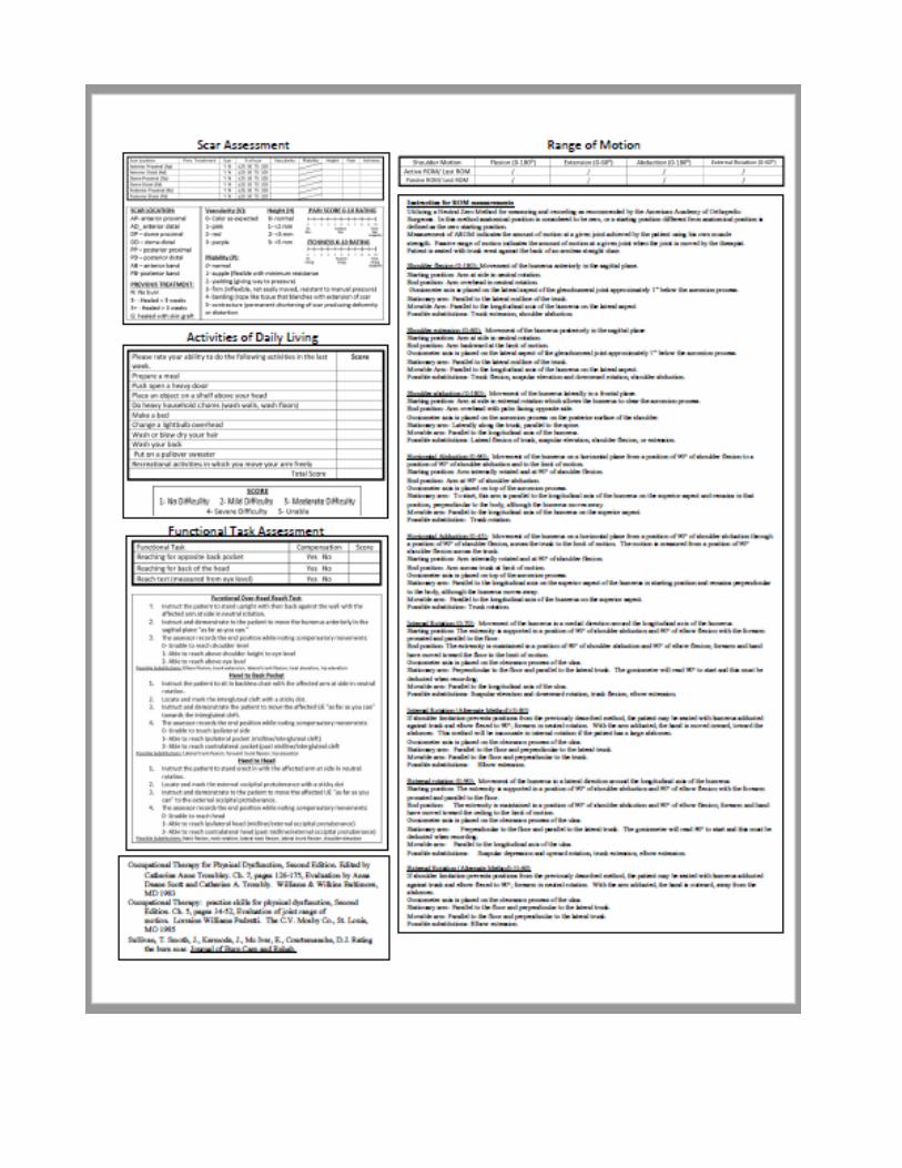

P#29
Abstract Title: Targeting Stakeholders to Reduce Phlebotomy in a Burn ICU
Author and Co-authors:
Clinton D. Leonard, MSN, NP Arnold Luterman Regional Burn Center at University of South Alabama, Mobile, AL
Objective:
Upon completion of the lecture, attendees should be better prepared to: • Identify the harm associated with daily lab draws. • Recognize the Choosing Wisely program as a potential aid for reducing unnecessary tests and interventions.
Abstract:
Introduction: Unnecessary phlebotomy represents a significant burden both in terms of hospital expenditures and patient discomfort. Routine labs (such as CBC and BMP) are estimated to cost $250 per patient per day and are often ordered by default rather than based on clinical necessity. These labs can lead to iatrogenic anemia. Patients lose roughly 50mLs of blood to phlebotomy each day changing a culture from defaulting to lab draws to only drawing labs when they are indicated is a difficult, long-term process that requires participation from multiple stakeholders, including physician leadership, bedside nurses, NPs, residents, and nursing management. These stakeholders all have different perspectives and priorities, and achieving buy-in means tailoring education to the needs of specific groups. The "Choosing Wisely" framework is a national initiative that can be used to drive this change at institutions interested in reducing unnecessary interventions. Methods: Education based on the "Choosing Wisely" framework was implemented at Vanderbilt as a collaborative effort among advanced practice providers in 6 ICUs and various subacute care areas. Monthly committee meetings were held to plan specific interventions, including disseminating information on common lab test and imaging patient charges, distributing fliers around target units, email correspondence and in-person education. Each area had a designated ambassador to act as liaison. In the Burn ICU we focused on CBC, BMP, pre-albumin, and CRP. Education was rolled out beginning Oct. 1, 2015 and lab draws were monitored on a unit-wide level. Results: Profound drop from 1.8 labs per day per patient to .53 labs per patient in initial month of study. Significant attenuation of effect seen in subsequent months as time from initial roll-out increased. Overall reduction in lab draws by 22.4% during study period, although the sample sizes are too low to achieve statistical significance Conclusions: Burn ICU presents challenges to implementing Choosing Wisely Varying patient load, differences in patient acuity, and prolonged operative courses must be controlled for further study with larger sample size is warranted to identify factors influencing lab draws for more targeted intervention: --APACHE-2 score --Baux Score Different practice patterns by attending physicians. Most important factor in reducing lab draws is achieving stakeholder buy-in and preventing complacence.

References and Resources: Choosingwsely.org
Disclosure: Clinton Leonard – No Relevant Financial Relationships to Disclose

P#30
Abstract Title: Improved Team Communication Through Nurse-Led Multidisciplinary Rounds
Author and Co-authors:
Lauren R. Baxley, BSN, RN, CCRN; Morgan Colson, BSN, RN University of Texas Medical Branch, Blocker Burn Unit, Galveston, TX
Objective:
Upon completion of the lecture, attendees should be better prepared to: • Identify ways nurse-led rounding can lead to improved communication between nurses and physicians. • Recognize the role of nurse-led rounding can play in better addressing patient and family concerns.
Abstract:
Introduction: Multidisciplinary rounds have been occurring bi-weekly in the burn unit of Arkansas Children’s Hospital for many years. Historically these rounds were led by the attending physician or surgical resident with variable input from nursing staff. Nurse-led rounding has previously been attempted in this burn unit, however this transitioned back to physician-led rounds. The voices of nursing staff can be beneficial to presenting valuable information in rounds pertinent to the patient’s plan of care. Previous studies have documented improvements in collaboration, decreased utilization of unnecessary catheters, decreased incidence of hospital acquired pressure ulcers and greater shared decision making through the use of nurse-led, multi-disciplinary rounds. Methods: The Burn Unit is a 10-bed critical care unit located within an American College of Surgeons Level I Trauma Center and teaching facility. The unit consists of a multidisciplinary team which meets and conducts rounds bi-weekly to discuss patients’ progress and plan of care. This team is composed of attending physician, resident, mid level practitioners, anesthesiologist, clinical pharmacologist, nurse manager, dietician, programs coordinator, occupational therapist, physical therapist, respiratory therapist, social worker, discharge planner, chaplain, and staff nurses. In 2016, it was felt there was a disconnect in communication between medical and nursing staff. In an attempt to improve teamwork, a nurse-led rounding presentation template was created in order for the patients’ information to be utilized both in nurse to nurse report and in bi weekly mutidisciplinary rounds. The patients’ primary nurse presented each patient systematically. All aspects of the patient’s care were discussed, including but not limited to: pain management, wound care plan, preventative care bundles, extubation criteria, and clinical changes over the last twenty four hours. Results: To assess success of this procedural change, surveys were administered prior to and one year after implementation of nurse- led rounding. Pre-survey results suggested that 80% of respondents believed that nurse led rounds could improve communication and collaboration between nurses and physicians. The post-implementation survey revealed that 100% of respondents felt that medical and nursing staff communication and collaboration during multi-disciplinary rounds were improved by nurse led rounds. Also of note, post-survey results showed a 25% increase in respondents who believe that patient and family concerns were “Always” addressed during multidisciplinary rounds. Nurse-led rounds have provided an opportune time for

all disciplines to have their opinions heard and collaborate in the care of the patient. Conclusions: System-based nurse-led rounding can enhance communication between nurses and physicians. This facilitates nursing staff to methodically review each body system when presenting the patient and provides an outlet to address concerns of the nursing staff, the patient, and the patients’ family. Further study into improvements in direct patient outcomes could solidify the importance of nurse led rounding to the overall care of the Burn Survivor. References and Resources: Fisher, K., Grosh, A., & Felty, V. (2016). Using nurse-led rounds to improve quality measures related to HAPUs. Nursing, 46(11), 63-68. doi:10.1097/01.NURSE.0000494657.67378.00 Der, Y. (2009). Multidisciplinary Rounds in Our ICU: Improved Collaboration and Patient Outcomes. Critical Care Nurse, 29, 83-84. doi:10.4037/ccn2009792 Fakih, M. G., Dueweke, C., Meisner, S., Berriel-Cass, D., Savoy-Moore, R., Brach, N., . . . Saravolatz, L. D. (2008). Effect of Nurse-Led Multidisciplinary Rounds on Reducing the Unnecessary Use of Urinary Catheterization in Hospitalized Patients. Infection Control and Hospital epidemiology, 29(9), 815-819. doi:10.1086/589584
Disclosure: Lauren R. Baxley – No Relevant Financial Relationships to Disclose Morgan Colson – No Relevant Financial Relationships to Disclose
Pre-Survey 20 participants
1-Never 2-Rarely 3-Sometimes 4-Always
Communication is open between nurses and physicians in this unit
0% 0% 85% 15%
Nurses and physicians participate in discussions to resolve disagreements
0% 10% 70% 20%
Nurses and physicians discuss patient and family concerns and considers them in the plan of care
5% 5% 40% 50%

Nurses and physicians cooperate in decision making
0% 5% 60% 35%
Nurses and physicians share information to verify the rationale for treatments or interventions implemented
0% 10% 50% 40%
Scale 1-4 Current practice of communication between nurses and physicians during rounds
5% 30% 45% 20%
Scale 1-4 Satisfaction with collaboration between physicians and nurses in discussing patient care and generating solutions
5% 30% 55% 10%
Yes No Scale 1-2 Do you feel that nurse led rounding will improve communication and collaboration between nurses and physicians?
80% 20% --------------------- ----------------------

Post Survey 8 Participants
1-Never 2-Rarely 3-Sometimes 4-Always
Communication is open between nurses and physicians in this unit
0% 0% 62.5% 37.5%
Nurses and physicians participate in discussions to resolve disagreements
0% 0% 100% 0%
Nurses and physicians discuss patient and family concerns and considers them in the plan of care
0% 0% 25% 75%
Nurses and physicians cooperate in decision making
0% 0% 87.5% 12.5%
Nurses and physicians share information to verify the rationale for treatments or interventions implemented
0% 12.5% 62.5% 25%
Scale 1-4 current practice of communication between nurses and physicians during rounds
0% 12.5% 62.5% 25%
Scale 1-4 satisfaction with collaboration between physicians and nurses in
0% 25% 75% 0%

discussing patient care and generating solutions Yes No Do you feel that nurse led rounding will improve communication and collaboration between nurses and physicians?
100% 0% --------------------- -----------------------

P#31
Abstract Title: Growing the Next Generation of Exceptional Nurses: Utilization of a Formal Preceptor Council
Author and Co-authors:
Martha L. Turner, BSN, RN; Catherine Gaudreault, BSN, RN University of Colorado Hospital, Burn Center, Aurora, CO
Objective:
Upon completion of the lecture, attendees should be better prepared to: • Identify three strategies for effectively supporting preceptors in an inpatient burn unit. • Identify two challenges of precepting in an inpatient burn unit.
Abstract:
Introduction: Preceptorship is a challenging role. It requires sound knowledge base, solid critical thinking skills, patience, attention to detail, and skills working with a variety of generations and personalities. In addition to the challenges inherent in the preceptor role, our Burn Center experienced significant change over the last two years including increasing our inpatient capacity to 19 beds from 9 beds, a change in medical directors, and a significant staff turnover. This resulted in a tremendous need for new preceptors; some of whom had less than a year of experience in our very complex healthcare system. This created an opportunity to provide support for this vital subset of our staff in order to ensure that they have the tools and support necessary to be successful preceptors in the Burn Center. Methods: To meet the changing needs of preceptors, we organized a team to provide leadership support to our preceptors through four preceptor council meetings throughout the year. All staff in a preceptor role were invited to these meetings. The meetings provided an opportunity to discuss successes and challenges of the preceptor role, as well as provide formal and informal support, recognition of the preceptors, and an opportunity to provide structured education topics specific to the preceptor role. Education included topics such as effective feedback. The challenges identified by the preceptors were prioritized and these were utilized to form the basis of future topics of discussion. Further, the preceptor council agreed to take on a project over the course of the year to develop a collection of critical thinking questions that could then be used as a teaching aid while precepting. Lastly, we used a portion of meeting time to discuss the "business" of precepting including items such as completing necessary documentation. We used a pre-post survey questionnaire to measure the effectiveness of our interventions. Results: A pre-post survey questionnaire was delivered to all preceptors to measure the effectiveness of our interventions. Following implementation of this preceptor council format, preceptors identified increased confidence in precepting, increased confidence in teaching critical thinking, felt they had adequate resources for precepting, and an increased sense of identification as a leader on the unit. Conclusion: Structured preceptor council meetings are an effective way to support preceptors in an inpatient burn unit.

References and Resources: Zilembo, M., & Monterosso, L. (2008). Nursing students’ perceptions of desirable leadership qualities in nurse preceptors: A descriptive survey. Contemporary Nurse, 27(2), 194-206. doi:10.5172/conu.2008.27.2.194 Goss, C. R. (2015). Systematic Review Building a Preceptor Support System. Journal for Nurses in Professional Development, 31(1). doi:10.1097/nnd.0000000000000117 Holland, C., & Ulrich, D. (2016). Critical thinking cards: An innovative teaching strategy to bridge classroom knowledge with clinical decision making. Teaching and Learning in Nursing, 11(3), 108-112. doi:10.1016/j.teln.2016.01.005
Disclosure: Martha Turner – No Relevant Financial Relationships to Disclose Catherine Gaudrealt – No Relevant Financial Relationships to Disclose

P#32
Abstract Title: Reducing Autograft Loss
Author and Co-authors:
Arnine Aurelien, RN; Marie Daniel, RN; Andrew Mesidor, NA Jackson Memorial Burn Center at University of Miami , Miami, FL
Objective:
Upon completion of the lecture, attendees should be better prepared to: • Discuss Burn Service Performance Improvement Project which is the prevention of auto-graft loss.
Abstract:
Burn injuries affect many lives every year. According to the World Health Organization, it is estimated that over 265,000 deaths occurs every year due to burn injuries (2016). Burn is defined as an injury to skin or other body tissues that can caused by fire, radiation, radioactivity, electricity, friction or contact with chemicals, (WHO, 2016). As registered nurses and burn technicians on a burn unit, we witness firsthand the trauma, pain, and life changing injuries cause by burns daily and many of these patients affected by burns requires skin grafts. Often many of these skin grafts result in rejection, where that patient must be readmitted in the hospital for treatment. The many skin grafts lost we have witness has led to the Burn Service Performance Improvement Project, which is the prevention of auto-graft loss. During multi-disciplinary rounding, it was discovered that the lack of using burn pads under graft sites to extremities, usage of common soap instead of 2% chlorohexidine to clean graft sites, and sub-therapeutic albumin levels in some patients may have contributed to many of our skin grafts losses. This discovery caused the burn service to implement new interventions, such as educating all staff on the problems found and solutions, checking pre-albumin level weekly, and weekly multi-disciplinary rounds for all burn patients to discuss progress. We have also implemented washing graft sites twice daily with chlorohexidine and water and applying Aloe-Vesta moisturizer after. New interventions implemented has led to only 1 graft loss out of 22 patients. With ongoing analysis of grafts and interventions our goal is to have a 100 percent success.
Disclosure:
Arnine Aurelien– No Relevant Financial Relationships to Disclose Marie Daniel– No Relevant Financial Relationships to Disclose Andrew Mesidor – No Relevant Financial Relationships to Disclose

P#33
Abstract Title: The Use of Cultured Epithelial Autografts and Spray Keratinocytes Provide Wound Coverage in Large TBSA Burns
Author and Co-authors:
David Roggy, RN; Rajiv Sood, MD Richard M. Fairbanks Burn Center at Eskenazi Health Hospital, Indianapolis, IN
Objective:
Upon completion of the lecture, attendees should be better prepared to: ▪ Describe the techniques used in this new process and recognize the value of combining the technologies into one treatment plan for large TBSA burns.
Abstract:
Introduction: Over the last two decades, a decrease in mortality rates of burn patients can be attributed to continued advancements in burn resuscitation, intensive care, trauma care and nutritional support and early excision and coverage of the burn wound. However, patients with burns >50% TBSA continue to pose specific challenges to the burn surgeon with regards to autologus coverage due to their lack of donor sites. Over the past twenty-seven years, our burn center has utilized cultured epithelial autografts to obtain coverage of these larger TBSA burns. In September of 2016, we changed our practice with large TBSA burns by incorporating the use of spray keratinocytes over widely meshed autografts to posterior surfaces along with CEA applied to anterior surfaces to accelerate wound coverage. We present a case report involving an eight male with a 95% full thickness injury that we utilized this new practice and obtained wound closure by post-burn day 117. Methods: The patient was enrolled in a FDA Clinical Trial evaluating the Compassionate Use of Spray Keratinocytes in Large TBSA burns. The study was IRB approved. A review of his medical record and case report forms was performed to obtain information on the progress of his care. Results: Our patient is an eight year old male who sustained a 95% full thickness flame injury caused by throwing gasoline on a fire. He had two areas available for donor sites, a small area of scalp and a small area to groin. The two donor sites together measured just over 300cm. He underwent five spray keratinocyte procedures treating ten areas and one CEA procedure. The spray keratinocyte treated areas all achieved 95% or greater wound closure between post-operative days 14 to 28. The CEA treated areas had 95% take on post-operative day 15. During his hospitalization, he experienced one early episode of mild sepsis but no other complications throughout his stay with wound closure on post-burn 117. Conclusion: This technique of using both spray keratinocytes and CEA to accelerate wound closure is our standard of care for large TBSA burns. By achieving wound closure early, the patient we present here experienced only one early episode of sepsis. We have treated four others with this same technique and have similar success. Although there is learning curve with the use of spray keratinocytes and CEA, the benefits to the patients are potential decrease in the incidence of sepsis, shorter

hospital stay, wound closure with less availability of donor sites, and potential earlier discharge from the acute setting to the rehabilitative setting.
Disclosure: David Roggy – No Relevant Financial Relationships to Disclose This is a FDA Clinical Trial evaluating the Compassionate Use of Spray Keratinocytes. Rajiv Sood – Speaker’s Bureau: Avita; Vericel

P#34
Abstract Title: The Application of Negative Pressure Wound Therapy to Promote Integration of Facial Grafts and Dermal Substitutes
Author and Co-authors:
Cassandra D. Rush, DPT, WCC; Audrey M. O’Neil, DPT; David Roggy, RN; Rajiv Sood, MD Richard M. Fairbanks Burn Center at Eskenazi Health Hospital, Indianapolis, IN
Objective:
Upon completion of the lecture, attendees should be better prepared to: ▪ Describe the application of a NPWT dressing to the face. ▪ Consider the use of ostomy products to more easily obtain a seal with NPWT dressings
Abstract:
INTRODUCTION: Negative pressure is an important modality in our burn center’s treatment algorithm of acute burns and reconstructive patients. Many studies have cited the potential benefits of NPWT but few look at the feasibility of application in locations such as the face and the care that is required to provide a successful outcome while maintaining important underlying structures. Here, we present our techniques for successful application and outcomes in the application of NPWT to the face. Methods: Patients requiring dermal substitute placement or autografting to the face were evaluated by the staff surgeon in the OR for potential placement of NPWT. When the burn surgeon determined the wound bed was deemed appropriate, the physical therapist was called to place the NPWT dressing with assistance from the OR staff and surgeons. Results: Due to the often complex nature of intricate grafting, specialty products were used to assist with the placement and maintenance of a seal. In areas of intact skin, skin glues and/or hydrocolloids were used. When the graft was bordered with continued burn or open wounds, products such as ostomy paste and strip adhesives were used. Vital structures such as the eyes, ear canal, nares, and mouth require more attention. Silicone sheeting coupled with bolsters or other forms of compression were used to decrease the chance of residue from the ostomy products migrating into these areas. Low pressure settings were used (<75mmHg). The dressings were changed per the manufactures’ time frames. There were no areas of unplanned graft or dermal substitute loss as a direct result of the NPWT dressing being placed and no adverse events. Conclusion: In our burn center, we have utilized NPWT to the face since 2007 without sequelae. With appropriate wound bed preparation, practitioners proficient with placement of a NPWT dressing to the face and close monitoring, NPWT is a safe and beneficial dressing to assist with the vascularization, immobilization, and splinting of skin grafts and dermal substitutes placed on the face.

Disclosure: Cassandra Rush – No Relevant Financial Relationships to Disclose Audrey O’Neil – No Relevant Financial Relationships to Disclose David Roggy – No Relevant Financial Relationships to Disclose Rajiv Sood – Speaker’s Bureau: Vericel; Avita

P#35
Abstract Title: Improving the Precepting Experience on a Burn ICU
Author and Co-authors:
Maria Gonzales, MSN, RN; Chelsea N. Bair, BSN, RN University of North Carolina Healthcare, North Carolina Jaycee Burn Center
Objective:
Upon completion of the lecture, attendees should be better prepared to: • Discuss a variety of tools to better build and execute a preceptorship program.
Abstract:
Introduction: Due to the complexity of burn injuries, few nurses have the knowledge, expertise, or skills needed to care for this unique patient population. This lack of experience requires every new staff nurse to undergo extensive training with an individual preceptor. Ensuring that all new nurses receive the same training can be challenging, particularly during times of staffing fluctuations. Despite best efforts, mismatching between preceptor and preceptee may occur based on personality, communication, and teaching/learning styles which can contribute to less than ideal training experiences for both parties. The setting for this project was a 21-bed burn ICU which underwent a recent influx of new hires. From May 2016 through Sep 2016, 19 nurses started precepting on the unit. More than half were new grads and only two had previous burn experience. With precepting lasting 12-20 weeks on the unit, preceptors felt overwhelmed and preceptees had less than ideal precepting experiences. Methods: Qualitative data was collected from two focus groups, one comprising 16 preceptors, and the other, 17 preceptees, to pinpoint main themes for improvement. Staff nurses were enlisted to improve the precepting experience based on information from the focus groups. Seven preceptors and two preceptees were invited to form a coalition with the Assistant Nurse Managers to identify actionable improvements. Results: Matching preceptees with preceptors. Previously preceptors had not been paired with preceptees using an established personality assessment tool, such as the Smalley Institute Personality Test. Even with mismatched personality types, knowledge of the preceptee personality may help the preceptor tailor an effective training approach. Preceptorship program support. Structured activities are a way of providing consistent, planned support to help the preceptorship program succeed. Activities should take place across three phases of the program: prior to starting, during, and maintenance. A welcome letter or a coffee meet/greet can help begin the relationship between preceptor and preceptee in a less rushed environment. As a preceptee advances to treating less stable patients, the preceptorship should be assessed by managers using feedback (e.g. surveys) from preceptors and preceptees. The maintenance phase proactively addresses potential loss of motivation that some preceptors may feel because of the extra duties.
Maintenance may include monthly meetings for preceptors to share experiences. Up-to-date training guidelines. Training guidelines should be treated as a living document owned by the preceptors and managers. It should therefore be updated in response to gaps in training and to new knowledge. Conclusion: Training many new nurses at the same time created a challenging situation for a 21-bed burn ICU. Feedback from both sides of the preceptorship program was key to developing effective solutions. These solutions included assessing personality when pairing preceptor and preceptee; having structured preceptorship program support; and maintaining up-to-date training guidelines. References and Resources: Hyrkas, K, & Shoemaker, M. (2007). Changes in the preceptor role: re-visiting preceptors’ perceptions of benefits, rewards, support and commitment to the role. Journal of Advanced Nursing, 60(5), 513-524. Personality - Test.pdf. (n.d.). Retrieved June 01, 2017, from http://smalley.cc/images/Personality-Test.pdf.
Disclosure: Maria Gonzales – No Relevant Financial Relationships to Disclose Chelsea Bair – No Relevant Financial Relationships to Disclose

P#36
Abstract Title: Burn Patient Post- Discharge Call Backs: A Process to Provide Excellent Provider-Patient Communication
Author and Co-authors:
Maryke V. Bard, PA-C; John Crow, MD Akron Children’s Hospital, Regional Burn Center, Akron, OH
Objective:
Upon completion of the lecture, attendees should be better prepared to: • Describe the process of integrating a post-discharge call back system. • Discuss patient satisfaction with post-discharge call back.
Abstract:
Introduction: Communication between healthcare professionals and patients is a key component to successful outcomes and overall positive patient experience. Burn patients not only have the burden of trying to cope with their injury and pain, but also the possibility of completing wound care post-discharge. During hospital admissions wound care, pain control and physiological changes are closely monitored by staff, upon discharge these needs are transitioned to the patient and/or caregivers. Method: Our center subjectively felt patients were struggling to monitor these needs and other issues, therefore a performance improvement project was designed where nursing staff would call the patient or caregiver 24-72 hours post hospital discharge. Prior to development of this process a literature review was completed and IRB approval obtained. A retrospective review of 50 discharged patients’ charts for telephone encounters post-discharge, but before their first visit, as well as complaints identified in their first outpatient visit history and physical interview was conducted. Of these 50 discharged patients, 18% had called before their first visit. The top three complaints noted were pain, dressing difficulties and itching. Other concerns identified were constipation, decreased energy, problems with filling prescriptions, difficulty eating, rash, splinting s, supply issues and swelling. Results: Using the information a script was developed to guide nursing staff as to what questions to ask patients or caregivers in the telephone interview. A calendar was placed on the huddle board to identify patients needing a phone call that day. Six nurses volunteered as champions of the project. Discharges, calendar information and phone calls made to the patients or caregivers were monitored. If a concern was escalated to a provider, the nurse responsible for that communication would also provide feedback to the patient. A template was created in the electronic health record so that the nurse could document the scripted phone call, patient response and feedback given. Conclusion: So far over 20 post-discharge phone calls have been made. No issues have been elevated to the provider level. We are now in the process of obtaining data from the patients regarding their perception about the phone call at their first outpatient visit. We believe this will provide an additional touchpoint for patient satisfaction and hopefully provide any needed intervention before patients’ first outpatient appointments.

Disclosure: Maryke Bard – No Relevant Financial Relationships to Disclose John Crow – No Relevant Financial Relationships to Disclose

P#37
Abstract Title: WITHDREW A Case Study: Self Immolation 30% TBSA Five Months Post Injury
Author and Co-authors:
Jill M. Krystofinski, CRNA, MSN Lehigh Valley Health Network, Allentown, PA
Objective:
Upon completion of the lecture, attendees should be better prepared to: • Demonstrate the multiple anesthetic approaches available to deliver anesthesia safely to the traumatized airway. • Explain the benefits of regional aesthesia indwelling catheters to decrease and/or eliminate mobility limitations due to contractures.
Abstract:
Objectives: Three different anesthetic approaches to facilitate multiple surgeries for severely contracted facial microstomia, bilateral entropian, anterior neck, bilateral axillary, elbow and hand contractures with anatomy not amenable to tracheostomy. Methods: 1) Multiple major facial reconstructive surgeries addressed with elective fiber optic intubation and protocol driven anesthetic. 2) Targeted reduction of opioid requirement by incorporation of right interscalene and left supraclavicular continuous indwelling catheters to reduce bilateral upper extremity contractures. 3) Repeated CO2 and Mosaic laser therapy to reduce scarring to face, neck, chest and upper extremities achieved with multimodal monitored anesthetic care to avoid repeated airway trauma. Results: Marked microstomia with severe neck contracture as a burn injury presented significant challenges to the safe delivery of anesthesia with anatomy not amenable to tracheostomy. Planned fiber optic intubation utilizing a dedicated anesthesia team allowed major reconstructive surgeries. Regional blockade of the brachial plexus allowed the team to reduce the need for general anesthesia and total opioid requirements. Robust team planning and patient engagement facilitated complex laser therapy with novel monitored anesthesia care. Conclusion: In April of 2015, after the death of her mother, a thirty seven year old African American female attempted suicide by self-immolation. Five months following her injury and treatment at another institution this patient was admitted to Lehigh Valley Health Network in Allentown, PA. Her treatment provided the historical expertise and team based planning to incorporate complex regional anesthesia and advanced airway management for her care. Her care helped build a regional pain management platform into our burn center care. March of 2017, she resides in a group home. She continues with her out patient CO2 laser therapy for further scar management efforts and currently volunteers at a local community center.

References and Resources: Cohen, NP, WN Levine, G. Marra, RG Pollock, EL Flatow, AR Brown, and LU Bigliani. "Indwelling Interscalene Catheter Anesthesia in the Surgical Management of Stiff Shoulder: A Report of 100 Consecutive Cases." J Shoulder Elbow Surgery. Medline, 09 Aug. 2000. Web. 1 May 2017.
Disclosure: Jill Krystofinski – No Relevant Financial Relationships to Disclose

P#38
Abstract Title: Mock Codes in the Burn Unit
Author and Co-authors:
Amy Van Cleave, BSN, RN, CCRN Parkland Memorial Hospital, Regional Burn Center, Dallas, TX
Objective:
Upon completion of the lecture, attendees should be better prepared to: • Implement mock codes in burn units.
Abstract:
Introduction: There is limited research on a nurse or nursing assistance’s confidence in responding to codes, especially in the burn community. Due to the potential for a burn patient to code during their hospital stay, it is important for burn staff to feel confident in responding to code situations. Furthermore, it is imperative that burn staff be able to intervene correctly, using proper CPR techniques. The purpose of implementing the Mock Code Scenarios in the Burn Unit was to assess the current knowledge gap of the burn staff in responding to a code situation, increase the staff’s confidence in responding to code situations, and to ensure proper return demonstration of correct CPR techniques. Method: For three months, monthly mock code scenarios were implemented in the burn unit, alternating between adult and pediatric cases. At the beginning of each scenario, the staff member was called to respond to the code, and then observed for unprompted interventions. After the scenario, a short debriefing occurred, correcting any erroneous interventions/CPR techniques. The staff member was then given the chance to repeat the scenario. Results: By the end of the third mock code scenario, the burn staff that participated showed an increase in confidence in responding to the scenarios and showing improved CPR skills. During the first month it was noted that all staff members needed to be corrected in at least one intervention related to responding to a code. However, no debriefing/correction was required during the third month. Conclusions: Implementing mock codes in the burn unit proved to be successful in increasing the confidence and skill of staff responding to a code. Providing periodic opportunities in which staff can practice responding to code situations is a good way to combat the fear that is often associated with codes as knowledge and skill are solidified through repetition. References and Resources: Herbers, M.D. & Heaser, J.A. (2016). Implementing an in situ mock code quality improvement program. American Journal of Critical Care (25)5.
Disclosure: Amy Van Cleave – No Relevant Financial Relationships to Disclose

P#39
Abstract Title: A Tale of Two Burn Centers: Clinical Nurse Specialist Roles
Author and Co-authors:
Michael G. Barba, MSN, RN1; Sarah K. Shingleton, MS, RN1; Maria Serio-Melvin, MSN, RN1; Sarah J. Murray, MSN, RN1; Cameron C. Bell, MS, RN2; Bonnie A. Jackson, MSN, RN1; Scott A. Phillips, MS, RN1; Elizabeth A. Mann-Salinas, PhD, RN, FCCM1 1United States Army Institute of Surgical Research, Ft. Sam Houston, TX 2University of Colorado Hospital, Aurora, CO
Objective:
Upon completion of the lecture, attendees should be better prepared to: ▪ Describe the NACNS Spheres of Influence model in which the CNS practices. ▪ Describe the various roles the CNS holds within the burn specialty.
Abstract:
Introduction: Care of burn patients is complex and requires a multidisciplinary, systems approach to provide effective care. The Clinical Nurse Specialist (CNS) is an advanced practice registered nurse who functions within three spheres of influence (figure 1), serves as expert practitioner and change agent, brings Evidence-Based Practice (EBP) to the bedside and conducts research. However, upon a literature review only one small study described CNS practice within the burn specialty. Methods: Burn centers from two urban medical centers collaborated to define CNS specialty practice within their American Burn Association (ABA) verified centers. CNSs described their roles during brainstorming sessions and how they function day to day and interact with the multidisciplinary team. Results: Various roles were identified among eight CNSs from two regional burn centers. Primary focus areas include intensive care (ICU), progressive care (PCU), wound care (WC), Computer Decision Support Systems (CDSS), Burn Program Manager (BPM), research, and burn clinic (BC). ICU and PCU CNSs are clinical experts who develop evidence-based programs such as preceptorship and delirium management. They serve as educators and mentors to staff and collaborate with patients and families. CNSs working in the BC and WC areas have similar roles and also serve as privileged providers. The BPM collaborates with the multidisciplinary team to ensure accreditation and ABA requirements are met and leads Patient Safety and Process Improvement projects. The CDSS CNS serves as a conduit between clinicians and engineers by developing and implementing innovative technologies that integrate scientific concepts with bedside practice. Finally, all CNSs within centers work closely with the research CNS, collaborate daily and participate in outreach. Conclusions: The CNS fulfills many roles within these two centers and adapts to the unpredictable burn center dynamic. CNSs serving as clinical experts in the burn specialty work within the nurse, patient, and system spheres to advance care across the continuum and improve outcomes by implementing EBP. Implications for Practice: The CNS benefits the multidisciplinary team; however,

little is known about CNS burn specialty practice or the level of coordination among CNSs from multiple centers. Currently there is neither a formal CNS list-serve nor Special Interest Group within the ABA to provide a conduit for sharing knowledge. Further research and collaboration among burn CNSs is warranted to further define the role within the specialty. References and Resources: http://www.nacns.org/docs/CNSCoreCompetenciesBroch.pdf Figure 1 follows.
Disclosure:
Michael G. Barba – No Relevant Financial Relationships to Disclose Sarah K. Shingleton – No Relevant Financial Relationships to Disclose Maria Serio-Melvin – No Relevant Financial Relationships to Disclose Sarah J. Murray – No Relevant Financial Relationships to Disclose Cameron C. Bell – No Relevant Financial Relationships to Disclose Bonnie A. Jackson – No Relevant Financial Relationships to Disclose Scott A. Phillips – No Relevant Financial Relationships to Disclose Elizabeth A. Mann-Salinas – No Relevant Financial Relationships to Disclose
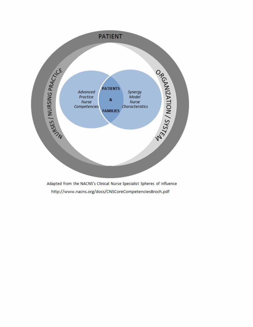

P#40
Abstract Title: Integrating Escalation of Care Training in Burn Center Annual Competencies: A Quality Improvement Initiative
Author and Co-authors:
Katherine L. Davis, RN; Tiffany Lorde, MSN, RN Evans-Haynes Burn Center at Virginia Commonwealth University Health, Richmond, VA
Objective:
Upon completion of the lecture, attendees should be better prepared to: ▪ Identify barriers and strengths of staff nurses' abilities to rescue patients experiencing unexpected changes in condition. ▪ Recognize the need to provide consistent education for all members of the health care team on usage of resources, teamwork, and open, effective communication, and its impact towards effective escalation of care. ▪ Recognize the importance of integrating safe communication strategies into the unit culture to maintain a culture of safety, and promote effective communication between team members.
Abstract:
INTRODUCTION: Escalation of care is the recognition and communication of patient deterioration aimed at patient rescue. Burn centers pose unique challenges related to escalation of care due to the multiple levels of service provided by various medical teams to patients across the lifespan. A root cause analysis was conducted on an adverse patient outcome that occurred in our burn center; the most significant contributing factor was improper escalation of care. At the time of the incident, no formal unit based escalation of care training was available. Therefore, the purpose of this quality improvement (QI) initiative was to identify barriers and facilitators to effective patient rescue, in order to develop a training program for nursing staff on effective escalation of care strategies. Method: A literature search was conducted using CINAHL complete, Pubmed/MEDLINE, and the Cochrane Library with keywords: escalation of care, barriers in escalating care, patient safety, rapid response teams, and failure to escalate. Articles were selected based on applicability to nursing practice and relevance to escalation of care training. Results: The literature search yielded 54 articles, out of which 13 were utilized. Barriers to effective escalation of care included: (a) ineffective communication between nursing staff and physicians; (b) hierarchical boundaries; (c) lack of knowledge and training on escalation protocols; (d) lack of clinical experience and confidence among staff. Evidence suggests nursing staff should be educated on escalation of care protocols, communication using SBAR technique, and resources available to aid in rescue attempts, including use of a rapid response team (RRT). Based on the literature results and increased time constraints for staff training, our burn center has integrated existing escalation of care training recommendations into our annual competency using an evolving case study approach.

Conclusions: Evidence suggests escalation of care training sessions should be included during new staff orientation, and twice throughout the year. Education should include availability of resources, escalation of care protocols, and communication techniques through use of case studies. Implementation of a training program on effective escalation of care strategies will be beneficial to all members of the healthcare team and can result in improved patient outcomes, improved communication between providers and nursing staff, and promote a culture of safety on the unit. Integrating escalation of care training into burn center specific annual competencies is an innovative, cost-effective, and timesaving strategy. References and Resources: Dingley, C., Daugherty, K. Derieg, M., & Rebecca Persing, R. (2008). Improving patient safety through provider communication strategy enhancements. Advances in Patient Safety: New Directions and Alternative Approaches, 3. Retrieved from http://www.ncbi.nlm.nih.gov/books/NBK43663/ Hamilton, P., Gemeinhardt, G., Mancuso, P., Sahlin, C., & Ivy, L. (2006). SBAR and nurse-physician communication: Pilot testing an educational intervention. Nursing Administration Quarterly, 30(3), 295-299. http://dx.doi.org/10.1097/00006216-200607000-00015 Johnston, M., Arora, S., King, D., Bouras, G., Almoudaris, A., Davis, R., & Darzi. A. (2015). A systematic review to identify the factors that affect failure to rescue and escalation of care in surgery. Surgery, 157(4), 752-763. doi:10.1016/j.surg.2014.10.017 Johnston, M., Arora, S., King, D., Stroman, L., & Darzi, A. (2014). Escalation of care and failure to rescue: A multicenter, multiprofessional qualitative study. Surgery, 155(6), 989-94. doi:10.1016/j.surg.2014.01.016 Mardon, R. E., Khanna, K., Sorra, J., Dyer, N., & Famolaro, T. (2010). Exploring relationships between hospital patient safety culture and adverse events. Journal of Patient Safety, 6(4), 226-232. doi: 10.1097/PTS.0b013e3181fd1a00 Rotella, J.A., Yu, W., Ferguson, J., & Jones, D. (2014). Factors influencing escalation of care by junior medical officers. Anesthesia & Intensive Care, 42(6), 723-729. Retrieved from http://www.aaic.net.au.proxy.library.vcu.edu Shapiro, S.E., Bailey V., Buick, M., Burke, K., Carroll, M., Christensen, S., . . . Vidyarthi, A. (2009). Implementing a conceptually based training program to increase nurses’ effectiveness in securing patient rescue. Journal for Nurses in Staff Development: JNSD: Official Journal of the National Nursing Staff Development Organization, 25(5), 236-41. doi: 10.1097/NND.0b013e3181bb34ad Shapiro, S. E., Donaldson, N. B., & Scott, M. (2010). Rapid response teams seen through the eyes of the nurse. AJN, American Journal of Nursing, 110(6), 28-34. doi: 10.1097/01.NAJ.0000377686.64479.84 Singer, S., Lin, S., Falwell, A., Gaba, D., & Baker, L. (2009). Relationship of safety climate and safety performance in hospitals. Health Services Research, 44(2p1), 399-421. doi: 10.1111/j.1475-6773.2008.00918.x Sutherland, K. (2013). Applying lewin’s change management theory to the implementation of bar-coded medication administration. Canadian Journal of Nursing Informatics, 8(1). Retrieved from http://cjni.net/journal/?p=2888 Wakeam, E, Hyder, J.A., Ashley, S.W., & Weissman, J.S. (2014). Barriers and strategies for effective patient rescue: A qualitative study of outliers. Joint Commission Journal on Quality and Patient Safety, 40(11), 503.

Williams, D., Newman, A., Jones, C., & Woodard, B. (2011). Nurses' perceptions of how rapid response teams affect the nurse, team, and system. Journal of Nursing Care. Quality, 26(3), 265-72. doi: 10.1097/NCQ.0b013e318209f135 Wiseman, B. & Kaprielian, V.S. (2016). Methods of Quality Improvement. Retrieved from Duke University School of Medicine website: http://patientsafetyed.duhs.duke.edu/module_a/methods.html
Disclosure: Katherine Davis – No Relevant Financial Relationships to Disclose Tiffany Lorde – No Relevant Financial Relationships to Disclose

P#41
Abstract Title: The Value of a Dedicated Multi-Disciplinary Team in the Outpatient Setting
Author and Co-authors:
Kari M. Gabehart, MSN, FNP; Jill Comstock, OT-R; Rajiv Sood, MD Richard M. Fairbanks Burn Center at Eskenazi Health Hospital, Indianapolis, IN
Objective:
Upon completion of the lecture, attendees should be better prepared to: ▪ Discuss the importance of a multidisciplinary team in the outpatient burn clinic. ▪ Recognize the importance of a centrally located burn outpatient clinic and its impact on patient outcomes and benefit.
Abstract:
Introduction: The American Burn Association verification requirement states that burn patients require outpatient burn follow- up. (pull standard and site) Within our Burn Center, we have a dedicated Outpatient Burn Clinic and multidisciplinary outpatient burn team that allows for consistency of care and follow through for our patients. Methods: Our Outpatient Burn clinic includes10 exam rooms and is operational 5 days a week. It is located within the Burn Center adjacent to the inpatient unit and has access to hydrotherapy, procedural areas and an outpatient gymnasium for burn rehabilitation. The outpatient team consists of burn surgeons, nurse practitioners, a registered nurse, physical and occupational therapists, central registrar, clinic scheduler, a care tech, and a medical assistant all of which are budgeted for full time hours. Burn surgeons have direct access to the burn clinic for preoperative and postoperative evaluations. The burn clinics location allows for enhanced continuity of care and opportunities for patients to meet the outpatient burn team prior to their discharge. Results: Since expanding the burn clinic to 10 rooms and our multidisciplinary outpatient team in 2013, our outpatient visit volumes have grown from 3400 visits in 2013 to 4600 visits in 2016. In addition to the increase in outpatient visits, we have increased the number of patients treated as strictly outpatients with no inpatient admission from 340 in 2014 to 435 in 2016. A member of the outpatient burn team participates in weekly inpatient burn rounds and conference to learn of inpatients and their course prior in preparation for discharge. Additionally, the outpatient nursing and therapy teams provide weekly updates to the inpatient team after discharge on any outlier in another facility to include medical, rehab and wound updates to the team. This allows conversation to occur with the entire team including our burn surgeons as issues or loop closure needs arise. Conclusions: The benefit of a dedicated on site multidisciplinary burn OP team incorporated into the burn center allows for immediate bidirectional communication; planning; increased patient safety; and continuity of coordination and transition of care. This outpatient clinic design allows for a larger team approach for the patient, as well as maintaining a consistent plan of care. Having the burn clinic located within the burn center allows for ease in transition of care, direct availability of medical leadership and

ease of returning as all services can be provided to the burn patient centrally which inpatient and families are already accustom.
Disclosure:
Kari Gabehart – No Relevant Financial Relationships to Disclose Jill Comstock – No Relevant Financial Relationships to Disclose Rajiv Sood – Speaker’s Bureau: Avita; Vericel

P#42
Abstract Title: Frostbite Prevention Outreach Program
Author and Co-authors:
Natalie A. Fitzgerald, BSN, RN; Lisa Hankee, OTR; Rajiv Sood, MD Richard M. Fairbanks Burn Center at Eskenazi Health Hospital, Indianapolis, IN David Roggy will present
Objective:
Upon completion of the lecture, attendees should be better prepared to: ▪ Identify unique resources to include in prevention education. ▪ Identify team methods to promote prevention within their community.
Abstract:
INTRODUCTION: In 2015, winter in Indiana was 4 degrees colder than normal and the 22nd coldest winter recorded in Indiana history. During this winter, our burn center saw the number of frostbite patients double from the years previous. While it was still a small percentage of the patients treated within the burn center it still caught the attention of the team and what we could do to attempt to decrease the incidence of frostbite within the community. Methods: The burn team developed a plan where items that would help protect individuals from getting frostbite, such as gloves, hats, and socks, would be collected and distributed throughout the community. We also focused on providing frostbite prevention education with these distributions of goods. The team started collections in 2016 by posting collection boxes and information within the burn unit and therapy department. Once the items were collected they were then tagged with frostbite prevention information. The decision was also made to utilize winter health fairs as a place to focus on frostbite prevention Results: In the fall of 2017, we expanded our outreach by identifying clinics, departments, and other groups outside our health system that could help to distribute items to those in need. Items collected went to our ED and various clinics with in the Eskenazi Health system. The departments and clinics would then distribute the items to patients and family members who needed them prior to discharge. One clinic in particular had a special need for the items that were collected as it was located in a day respite for the homeless community of Indianapolis. This clinic and day respite requested not only winter clothing items for their “friends” but also for education for the staff which interacted daily with individuals in the center and on the street. The education provided related to frostbite prevention and care as well as how to make referrals for care to our burn center. Conclusion: Our burn center is continuing this outreach as part of our prevention efforts. The collection process is available year round to ensure that there is an adequate supply to provide clinics and departments with items when they face individuals in need. Continued outreach education will also be provided. We encourage other burn centers in colder areas to not overlook this outreach opportunity and to assist those in need during the winter months.

Disclosure: Natalie Fitzgerald – No Relevant Financial Relationships to Disclose Lisa Hankee – No Relevant Financial Relationships to Disclose Rajiv Sood – Speaker’s Bureau: Vericel

P#43
Abstract Title: The Use of MediHoney for the Treatment of Facial Burns
Author and Co-authors:
Nicholas J. Ringfield, RN; Rachael Williams, MD; Juvonda Hodge, MD; Walter Ingram, MD Grady Health System Burn Center, Atlanta, GA
Objective:
Upon completion of the lecture, attendees should be better prepared to: ▪ Describe the pharmacologic properties of Medihoney along with clinical indications and protocols for usage. ▪ Increase awareness of the utility of Medihoney in treating facial burns.
Abstract:
INTRODUCTION: Burns to areas of cosmesis, such as the face, can result in high levels of patient anxiety. There is abundant literature from international burn units about the use of Medihoney in treating partial thickness burns of the face. Medihoney (Manuka Honey) comes from bee pollination of trees in New Zealand and Australia. Medihoney is inherently acidic lending broad spectrum antibacterial action and anti-fungal properties. Additionally, Medihoney is anti-inflammatory and a natural antioxidant. Here we describe five cases of facial burns that were treated with Medihoney. We further define our clinical indications and protocols for use. METHOD: This is a prospective observational study of the use of Medihoney in treating partial thickness burns of the face. We describe five cases in which Medihoney was used, our clinical indications, and define protocols for use. RESULTS: Our observational cohort included 5 patients with facial burn injuries; the most common mechanism of injury was flame. The average age of our patients was 60, and the average TBSA burn was 27%. All burns to the face were partial thickness in depth. The majority of our patients were male and Caucasian. MediHoney was applied daily to the face. While one patient was lost to follow up, 80% of our patients were seen in clinic two weeks post burn and noted to have 100% wound closure, good color matching, and no hypertrophic scarring. CONCLUSION: Medihoney is indeed an option for treating partial thickness facial burns. We found that the use of Medihoney resulted in 100% wound closure, good color matching, and no hypertrophic scarring. References and Resources: http://www.dermasciences.com/medihoney
Disclosure:
Nicholas Ringfield – No Relevant Financial Relationships to Disclose Rachael Williams – No Relevant Financial Relationships to Disclose Juvonda Hodge – No Relevant Financial Relationships to Disclose Walter Ingram – No Relevant Financial Relationships to Disclose


P#44
Abstract Title: The Struggle is Real: One Burn Unit's Journey to Hospital Acquired Infections (HAI) Reduction
Author and Co-authors:
Ginny Figel, MSN, RN; Celeste Adan, BSN, RN; Maria Chuma-Okere, BSN, RN Grady Health System Burn Center, Atlanta, GA
Objective: Upon completion of the lecture, attendees should be better prepared to: • Implement prevention and maintenance techniques to decrease CLABSI and CAUTI.
Abstract:
Hospital Acquired Infections (HAI) are a hot topic of debate in hospitals throughout the country. The burn patient is at an increased risk for these infections due to the nature of their burn injury. All centers follow the basics guidelines set forth by the accrediting bodies and prevention experts, but there is much variation in day to day practice. The purpose of this poster is to lay forth the best practices set forth by a 22 bed burn center that admits 685 patients annually. Over the past three years, data has been continually analyzed to determine room for improvement in our prevention and maintenance practices when it comes to the two major HAI, CLABSI and CAUTI. The burn center had 15 confirmed CLABSIs in 2015 and 13 CLABSIs confirmed in 2016. As of the first quarter of 2017 the center has had zero CLABSIs. The center developed a number of standardized improvement action plans to address the increased HAI. An important area of improvement determined by bedside staff was the inability to maintain an occlusive central venous line (CVL) dressing on certain ICU patients. The unit-based CLASBI committee set in order a standardized protocol for sterilely applying silver impregnated gauze on CVLs in place of an occlusive transparent dressing. If inserted through intact skin, a CHG impregnated disk is used on all centrally accessed devices, including central venous catheters, arterial lines, PICC lines, midlines and hemodialysis catheters. A staff RN was also designated to independently complete all CVL dressing changes following precise sterile procedure each week. The center was added to the hospital wide audit done every day to check that proper dressings are in place, appropriate time sensitive IV tubing is used, and the use of alcohol-containing caps are present on each access port. A hospital Six-Sigma project designed an easy-to-use pictorial guide for standardized central venous line dressing changes. This pictorial guide is laminated and used for each dressing change. The burn center’s CAUTIs have decreased from 5 in 2015 and 4 in 2016 to 1 in 2017. Measures implemented that have affected the CAUTI rate are: strict adherence to the CAUTI bundle, weekly audits of bedside care and documentation, and increased education. Through strong interdisciplinary teamwork the burn center removes indwelling catheters as soon as they are no longer indicated. The process for drawing a urine culture has been hardwired to all staff, physician and nurse. Buy-in from the multidisciplinary team has been integral to the constant quality improvement focus on the unit’s HAI. Our complex patient population demands continual reassessment of infection prevention practices. The unit has not reached zero harm events yet, but strives to implement best practices to provide the safest care for

our patient population. The team is hoping to foster an open dialogue of information and practice sharing to better the care of burn patients everywhere.
Disclosure: Ginny Figel – No Relevant Financial Relationships to Disclose Celeste Adan – No Relevant Financial Relationships to Disclose Maria Chuma-Okere – No Relevant Financial Relationships to Disclose

P#45
Abstract Title: Streamlining Infection Control Best Practices: Eliminating Invasive Fungal Infections in Burn Patients
Author and Co-authors:
Kelli F. Scott, BSN, RN; Nichole Johnson, BSN, RN; Rachel Williams, MD; Walter Ingram, MD; Juvonda Hodge, MD; Rita M. Gayed, PharmD; Elham Ghonim, PhD, CIC, MLS (ASCP) Grady Health System, Atlanta, GA
Objective:
Upon completion of the lecture, attendees should be better prepared to: • Identify risk factors and etiology of Mucormycosis. • Implement established enhanced infection prevention protocols.
Abstract:
The Burn Unit had a series of four patients with invasive fungal and mold infections since December 2016. Two of these invasive infections included rare cases of Zygomycies Mucormycosis. Through the BQIP initiative the unit has developed an Infection Control Sub-committee to perform a root cause analysis of the spread of these infections in the ICU. Currently, the unit uses strict contact precautions with all ICU burn patients to reduce rates of infection. The Sub-committee is seeking to be clear on other routes of transmission within the unit as well as other procedure areas. This Quality Improvement Initiative will include the review of four patients as well as any realized invasive infections moving forward. The current policy and procedure has been reviewed and changes have been implemented. Procedures for sterilization of wound care equipment and tools at bedside are being evaluated as well as new wound care processes. Other steps being taken are: disposable curtains, sterile water for wound care, inspection of the outside laundry facility, UVC disinfection technology, nurse to patient ratios, storage of wound dressing supplies, and a decontamination process for newly admitted patients. In conclusion our goal is to enhance best practice standards in order to eliminate invasive fungal infections in our burn patients.
Disclosure:
Kelli F. Scott – No Relevant Financial Relationships to Disclose Nichole Johnson – No Relevant Financial Relationships to Disclose Rachel Williams – No Relevant Financial Relationships to Disclose Walter Ingram – No Relevant Financial Relationships to Disclose Juvonda Hodge – No Relevant Financial Relationships to Disclose Rita M. Gayed – No Relevant Financial Relationships to Disclose Elham Ghonim – No Relevant Financial Relationships to Disclose
P#46
Abstract Title: Candida auris – Infection and Colonization in a Burn Patient, Treatment and Management Guidelines
Author and Co-authors:
David Roggy, RN; Rajiv Sood, MD Richard M. Fairbanks Burn Center at Eskenazi Health Hospital, Indianapolis, IN
Objective:
Upon completion of the lecture, attendees should be better prepared to: ▪ Discuss the initial treatment and recommended guidelines for C. Auris infections and colonization.
Abstract:
Introduction: Candida auris (C. auris) is a fungus that is emerging as a serious global health threat. We recently had a patient transferred from South Africa colonized with C. auris. Reports of C. auris causing severe illness in hospitalized patients with weakened immune systems have been documented since 2011.C. auris is one of the few species of Candida that can cause candidiasis. There reported cases of C. auris being resistant to all three major classes of antifungal drugs, and unlike other Candida species, C. auris can persist on surfaces and cross-contamination to other patients is highly likely. Methods: An online review was performed on Candida auris focusing on Centers for Disease Control (CDC) recommendations on management and treatment. Results: Upon identification of C. auris the CDC highly encourages notification of state and / or local public health authorities and the CDC. An Infectious Disease consult is recommended to help guide treatment. Most cases in the United States are susceptible to echinocandins, therefore, initial drug therapy should include one of the three echinocandin drugs with the appropriate dose listed in Table 1. Close monitoring and multiple follow-up cultures should be performed on patients receiving antifungal therapy as C. auris has the ability to develop resistance very quickly. Strict infection control and environmental measures must be taken immediately with isolation of the patient in a private room with Contact Precautions, terminal cleaning of rooms and work spaces, enforcement and monitoring of hand hygiene, and limited patient access. Following treatment with antifungals, the patient may remain colonized without any signs or symptoms of infection. With colonized patients in the outpatient setting, strict infection control and environmental measures should continue in the outpatient clinic. Conclusion: While C. auris is rare in the United States with only 77 reported cases. It is on the rise in Europe and Africa. Awareness of this fungus is necessary as misidentification increases the likeliness of drug resistance and complications for the patient. Strict infection control practices are necessary to contain the spread of this infection to other immune compromised patients and notification of your local / state health department as well as the CDC is highly encouraged to assist with treatment and management.

Disclosure: David Roggy – No Relevant Financial Relationships to Disclose Rajiv Sood – Speaker’s Bureau: Avita; Vericel

P#47
Abstract Title: Sweet Dressings: Honey Use in United States and Canadian Burn Centers
Author and Co-authors:
Charlotte J. Gadomski, BSN, RN; Sarah K. Shingleton, MS, RN; Alexandra J. Helms, BSN, RN; Leanna M. Thompson, BSN, RN; Jennifer M. Gurney, MD; Booker T. King, MD United States Army Institute of Surgical Research, Burn Center, Ft. Sam Houston, TX
Objective:
Upon completion of the lecture, attendees should be better prepared to: • Identify the types of wounds currently receiving honey dressings in US and Canadian burn centers. • Discuss the potential challenges of honey dressings to include product selection and availability. • Recognize that pain may be a challenge and consider alternative dressings if necessary.
Abstract:
Introduction: Burn care is constantly evolving and practice varies among burn centers. Honey is natural, non-toxic and has been used since ancient times to promote wound healing. Despite low to moderate quality evidence regarding improved wound healing and infection, per a 2015 Cochrane Review, honey dressings (HD) are making a resurgence. Our burn center recently began using HD in a gel formulation and experienced several challenges with application and dressing maintenance. As part of an evidence-based Performance Improvement project to improve HD implementation in our burn center, we contacted United States (US) and Canadian burn centers to learn how they are using and applying the product. Methods: The Burn Care Resource Directories for US (N=128) and Canadian (N=14) burn centers, available on the American Burn Association (ABA) website, were utilized. All centers were contacted via telephone. A ten-question interview was conducted with a nurse or alternate provider about HD use in their center. The questions included information on wound type and size, product type, past dressing successes/failures and the use of wound specialists. Results: Responses were received from 105 US and 10 Canadian burn centers with 67 being ABA verified. HD use for various wound types (burns=13, chronically open wounds=14, pressure ulcers=11, skin grafts [STSG]=2) was reported for 22 centers (19%). The most common formulation reported was gel in 16 centers (73%) while 5 centers reported absorbent polymer or alginate sheets (23%); 10 centers used a combination of products (45%). Of the 13 (11%) centers reporting HD use for acute or chronically open burns and STSG, 10 were ABA verified; 9 described use for facial burns and 4 for wounds >10% total body surface area. Intermittent reports of pain were described by 6 (46%) centers and 11 (85%) provided feedback regarding product successes and challenges. The Western Region had the highest use of HD (n=7, 32%) with the remaining from the Midwest, Northeast, Eastern Great Lakes and Canada. Certified wound nurses or specialists were part of the burn team in 59% of centers using HD (n=13/22).
Conclusions: Honey is currently being used in a small number of US and Canadian burn centers. Despite feedback that HD are challenging to use in that they are “sticky” and “drippy”, respondents reported good results, particularly with facial burns, and several said “we love it”. Most centers reported intermittent burning or stinging upon application for about 15 minutes. Overall, respondents from centers using HD were very positive and plan to continue using them in their practice. Implications for Practice: Wound dressing selection is important and should consider factors such as pain, ease of use and healing time. New product introduction is challenging and may be facilitated by sharing experiences with other burn centers. While there is no perfect dressing, HD appear to provide many of the elements for optimal wound healing. However, there is a paucity of high-quality evidence regarding the benefits of HD in burn wounds. Further research is warranted to establish if HD result in improved wound healing and reduced wound infection in burn patients.
Disclosure:
Charlotte J. Gadomski – No Relevant Financial Relationships to Disclose Sarah K. Shingleton – No Relevant Financial Relationships to Disclose Alexandra J. Helms – No Relevant Financial Relationships to Disclose Leanna M. Thompson – No Relevant Financial Relationships to Disclose Jennifer M. Gurney – No Relevant Financial Relationships to Disclose Booker T. King – No Relevant Financial Relationships to Disclose

P#48
Abstract Title: A Case Manager's Dilemma: Your Patient has Medicaid......
Author and Co-authors:
Julia Willis, BSN, RN; Rachael Williams, MD; Juvonda Hodge, MD; Walter Ingram, MD Grady Health System Burn Center, Atlanta, GA
Objective:
Upon completion of the lecture, attendees should be better prepared to: ▪ Examine services covered by Medicaid. ▪ Identify patients at risk for readmission and consequences of lack of rehab services.
Abstract:
There are over 69 million people in the United States who have some form of Medicaid. With the passage of the Affordable Care Act, some states expanded their Medicaid program to help close the coverage gap. Georgia and many other states in the Southeastern United States did not. The reimbursement levels for many services rendered as inpatient and outpatient are below the average rate in comparison to commercial plans. These services are crucial to decreasing length of stay, readmission rates and disability. As the ABA continues to monitor these rates in regards to quality of care and verification; insurance status is an important element. This presentation will look at various Medicaid programs administered by our state and neighboring states and look at services covered and the paucity of facilities that will accept patients with this insurance; i.e. home health, outpatient rehabilitation, inpatient rehabilitation, skilled nursing facilities, psychiatric care. There is a coverage gap and it will continue to widen unless significant changes occur for the underinsured. Should various metrics of quality care take insurance status into account?
Disclosure:
Julia Willis – No Relevant Financial Relationships to Disclose Rachael Williams – No Relevant Financial Relationships to Disclose Juvonda Hodge – No Relevant Financial Relationships to Disclose Walter Ingram – No Relevant Financial Relationships to Disclose
P#49
Abstract Title:
WITHDREW "They’re Afraid They’re Gonna Get It”: Parenting Children with Burn Injuries
Author and Co-authors:
Dana R. Dillard, MSW, PhD Candidate1; Stacey R. Kolomer, PhD, MSSW2 1Georgia Firefighters Burn Foundation, Atlanta, GA 2University of North Carolina, Wilmington, NC
Objective:
Upon completion of the lecture, attendees should be better prepared to: • Recognize the impact of social isolation on the whole family following a traumatic burn injury. • Describe the benefits of program design and implementation that addresses the needs of all family members following a traumatic burn injury. • Identify inclusive interventions related to trauma and consider how/where it fits within the context of the burn community.
Abstract:
Introduction: According to the World Health Organization (2016) burn injuries are a global public health concern that account for approximately 265,000 deaths every year. In 2016, hospitals in the United States with verified “burn centers” admitted 486,000 patients, including children between the ages of 1 to 15 years old comprising 30% of the total burn cases (American Burn Association [ABA], 2017a; ABA, 2017b). This number does not include patients treated in local emergency rooms, community clinics, or primary care offices (ABA, 2017a). The severe lasting impact of a burn injury is a persistent finding in the literature (Bakker, Maertens, Van Son, & Van Loey, 2013). Physical scarring and disfigurement are reminders of the trauma to the individual who sustained the injury, as well as others they encounter. As a result, people with visible burn scars experience decreased interactions with strangers, as well as friends/family, due to feelings of awkwardness and insecurity, fear of rejection, and exhaustion from managing invasive questions, remarks, and actions (Martin, Byrnes, McGarry, Rea, & Wood, 2017). Feeling ostracized to the point of social exclusion is a dehumanizing experience (Bastian & Haslam, 2010), which leads to social isolation and highlights the need for interventions promoting support and inclusion. Based upon the work of Goffman (1963) and Burke (2007, 2010) family members are not immune to similar consequences and likely develop their own associative identity or “courtesy stigma.” Currently, interventions for children with burn injuries largely focus on the child while consideration for the family appears incidental (Phillips & Rumsey, 2008). However, research on the adjustment of burn trauma in families suggests that parents/caregivers of children with burn injuries experience their own emotional response to the incident including, stress/anxiety/depression (Bakker et al., 2013), isolation (Gullick, Taggart, Johnston, & Ko, 2014; Öster, Hensing, Lojdstrorm, Sjoberg, & Willebrand, 2014), and guilt/blame (Ravindran, Rempel, & Ogilvie, 2013a, 2013b). When considering children with burn injuries as part of a larger family system, it seems reasonable to expect the impact of the burn trauma to extend beyond the patient, encompassing everyone within the context of the child’s environment; yet burn injury research within the framework of extended systems appears relatively untapped,

extending rich ground to cultivate. Qualitative methodologies exploring the impact of a burn injury on the family matured in the literature within the last several years, although interestingly none of the published work reviewed occurred within the United States. Therefore this exploratory study fills a gap by engaging the unique perspective of parents/caregivers of children with burn injuries and posits direct implications for essential program enhancement/development that benefit the family system. Methods: Parents/caregivers of children with burn injuries attending a newly developed family weekend program in its third year, offered by a southeastern burn foundation, participated in the study. Initially, the 35 year-old agency offered services exclusively to children with burn injuries by way of their flagship summer camp. The last decade is marked by an expansion of programs including a retreat for young adults with burn injuries and most recently the family weekend. Semi-structured interviews (N=11) guided by the research question, “What are the experiences of parents/caregivers of children with burn injuries?” occurred during the three day program. Only one parent attending the weekend program opted not to be interviewed. Participants’ self-identified as African American (n=4), Caucasian (n=5), and Latino (n=2). Ages ranged from 29 to 48 (M=38.36, SD=6.14). Interviews involved queries about the burn incident, from injury to present (M=7.12 years, SD=4.61), including support received, what they wish people understood about their experience, and ideas for programming. Interviews were recorded and transcribed. Data were analyzed using van Manen’s (1990) hermeneutic phenomenological approach. Results: Findings supported the unique experience of parents/caregivers of children with burn injuries coalescing into one theme—the never-ending trip from hell—conceptualized as, “I didn’t mean to get here—none of this is familiar and I can’t find anyone I know. What’s going on and when will it be over?” Mutual painful insights included parental guilt, social and geographical isolation, sensory experiences related to the acute incident and aftercare, unknown prognosis and medical treatment expectations, physical/emotional health problems, family members’ insensitivity, managing cruelty of others, marital strains, and sibling tensions. Families also reported positive outcomes: strength of informal support networks, empowerment through educating others, and creating new allies. Parents/caregivers of children with burn injuries emphasized the benefits associated with meeting other families who also endured a burn injury and the importance of a mutual support system. Conclusions: Programs must address the needs of all family members, not just the person with the physical injury. Findings from this research directly contributed to program planning within the burn foundation including, increased consideration of the burn injury’s impact on the family system, with focus on reducing feelings of isolation following the traumatic injury. As a result of these interviews, siblings are now included in the summer camp program, which has historically only served children with burn injuries, and the family weekend program is now offered in the spring and fall, as opposed to only once a year. In order to mitigate the impact of burn trauma, the systemic effect remains a critical primary consideration for research and interventions.
Disclosure: Dana R. Dillard – No Relevant Financial Relationships to Disclose Stacey R. Kolomer – No Relevant Financial Relationships to Disclose