ret rescues mitochondrial morphology and muscle ... · article ret rescues mitochondrial morphology...
TRANSCRIPT
Article
Ret rescues mitochondrial morphology and muscledegeneration of Drosophila Pink1 mutantsPontus Klein1, Anne Kathrin M€uller-Rischart2, Elisa Motori3,4,†, Cornelia Sch€onbauer5, Frank Schnorrer5,
Konstanze F Winklhofer2,3,6,7 & R€udiger Klein1,7,*
Abstract
Parkinson’s disease (PD)-associated Pink1 and Parkin proteins arebelieved to function in a common pathway controlling mitochon-drial clearance and trafficking. Glial cell line-derived neurotrophicfactor (GDNF) and its signaling receptor Ret are neuroprotective intoxin-based animal models of PD. However, the mechanism bywhich GDNF/Ret protects cells from degenerating remains unclear.We investigated whether the Drosophila homolog of Ret can rescuePink1 and park mutant phenotypes. We report that a signalingactive version of Ret (RetMEN2B) rescues muscle degeneration,disintegration of mitochondria and ATP content of Pink1 mutants.Interestingly, corresponding phenotypes of park mutants werenot rescued, suggesting that the phenotypes of Pink1 and parkmutants have partially different origins. In human neuroblastomacells, GDNF treatment rescues morphological defects of PINK1knockdown, without inducing mitophagy or Parkin recruitment.GDNF also rescues bioenergetic deficits of PINK knockdown cells.Furthermore, overexpression of RetMEN2B significantly improveselectron transport chain complex I function in Pink1 mutantDrosophila. These results provide a novel mechanism underlyingRet-mediated cell protection in a situation relevant for human PD.
Keywords Drosophila; neurodegeneration; neurotrophic factors; OXPHOS;
Parkinson’s disease
Subject Categories Molecular Biology of Disease; Neuroscience
DOI 10.1002/embj.201284290 | Received 20 December 2012 | Revised 8
November 2013 | Accepted 29 November 2013
Introduction
The etiology of Parkinson’s Disease (PD) is highly complex and
largely unknown, involving both environmental and genetic risk
factors. Mitochondrial dysfunction, oxidative stress and protein
aggregation are believed to be central events in the pathological
process, but their interconnection remains unclear (Schapira &
Jenner, 2011; Exner et al, 2012; McCoy & Cookson, 2012). The
first indications of a role for mitochondria came with the discovery
that the toxin 1-methyl-4-phenyl-1,2,3,4-tetrahydropyridine (MPTP)
causes Parkinsonism in humans and animal models (Burns et al,
1983; Langston et al, 1983). Its active metabolite, 1-methyl-4-phenyl-
pyridinium ion (MPP+), is selectively imported into dopaminergic
neurons via the dopamine transporter, and inhibits complex I of
the electron transport chain (ETC). Several other mitochondrial
toxins, including paraquat and rotenone, generating either mito-
chondrial reactive oxygen species (ROS) or specifically inhibiting
complex I, have been linked to PD in epidemiological studies and
animal models (de Lau & Breteler, 2006). Furthermore, patients
with sporadic PD can have decreased activity of complex I in brain
and other tissues (Schapira et al, 1989; Parker & Swerdlow, 1998),
or less complex I proteins in the substantia nigra (Mizuno et al,
1989).
Autosomal recessive PD-associated proteins Parkin, PINK1 and
DJ-1 (OMIM #600116, 605909, 606324) have been shown to have
functions related to mitochondrial integrity, (reviewed in Exner
et al, 2012; Martin et al, 2011). In three seminal studies, Pink1
mutant Drosophila displayed mitochondrial abnormalities and
muscle degeneration in a manner highly similar to park mutants,
and Parkin overexpression largely rescued the phenotypes of Pink1
mutants, but not vice versa, suggesting that the two proteins act in a
common linear pathway (Clark et al, 2006; Park et al, 2006; Yang
et al, 2006). Manipulation of the mitochondrial remodeling machinery
rescues some Pink1 and park mutant phenotypes in Drosophila and
in mammalian cell lines. However, while increasing fission rescues
the Drosophila phenotypes, shifting the fusion/fission balance in
the opposite direction rescues mammalian cell lines, but the under-
lying mechanisms are not fully understood (Deng et al, 2008; Poole
et al, 2008; Lutz et al, 2009). PINK1, a mitochondrial Ser/
Thr kinase, and Parkin, an E3 Ubiquitin ligase, were found to
1 Molecules – Signaling – Development, Max Planck Institute of Neurobiology, Martinsried, Germany2 German Center for Neurodegenerative Diseases (DZNE), Munich, Germany3 Neurobiochemistry, Adolf Butenandt Institute, Ludwig Maximilians University, Munich, Germany4 Department of Life Quality Studies - Alma Mater Studiorum, University of Bologna, Bologna, Italy5 Max Planck Institute of Biochemistry, Martinsried, Germany6 Molecular Cell Biology, Institute of Physiological Chemistry, Ruhr University Bochum, Bochum, Germany7 Munich Cluster for Systems Neurology (Synergy), Munich, Germany
*Corresponding author. Tel: +49 89 85783150; Fax: +49 89 85783152; E-mail: [email protected]†Present address: Department of Mitochondrial Biology, Max Planck Institute for Biology of Ageing, Cologne, Germany
ª 2014 The Authors. This is an open access article under the terms of the Creative Commons Attribution-NonCommercial-NoDerivs License,which permits use and distribution in any medium, provided the original work is properly cited, the use is non-commercial and no modifications or adaptations are made.
The EMBO Journal 1

regulate clearance of damaged mitochondria via mitophagy
(Geisler et al, 2010; Narendra et al, 2010; Vives-Bauza et al, 2010),
and microtubular transport (Weihofen et al, 2009; Wang et al,
2011). However, other studies have reported additional functions of
Parkin in the regulation of stress response proteins and mitochon-
drial biogenesis (Bouman et al, 2011; Shin et al, 2011), in promoting
NF-κB signaling (Henn et al, 2007; Muller-Rischart et al, 2013),
and in controlling cytochrome-c release (Berger et al, 2009). PINK1
also has additional functions, unrelated to recruiting Parkin, such as
regulating mitochondrial calcium buffering (Gandhi et al, 2009;
Sandebring et al, 2009; Heeman et al, 2011). Furthermore, PINK1
mutant mitochondria have decreased activity of complex I of the
ETC (Morais et al, 2009), and overexpression of a yeast substitute
for complex I rescued many of the functional impairments of Pink1
mutant flies (Vilain et al, 2012). Additional studies are required to
elucidate which of the functions reported for Parkin and PINK1 are
critical for causing Parkinson pathology.
The neurotrophic factor Glial cell line-derived neurotrophic
factor (GDNF) promotes the survival of dopamine neurons (Lin
et al, 1993) and protects nigral dopamine neurons from cell death in
rodent and primate toxin-models of PD such as 6-hydroxydopamine
(6-OHDA) and MPTP (Kearns & Gash, 1995; Sauer et al, 1995;
Tomac et al, 1995; Gash et al, 1996). Several clinical trials have
been performed with mixed outcomes, but ongoing research and
development aims at improving delivery methods of GDNF (Deierborg
et al, 2008). GDNF signals via the GPI-anchored co-receptor GFR-a1and the receptor tyrosine kinase Ret (Airaksinen & Saarma, 2002).
Endogenous Ret expression is required for long-term survival of a
fraction of nigral dopamine neurons in aged mice (Kramer et al,
2007). Conversely, mice that express a constitutively active Ret
receptor in dopamine neurons (RetMEN2B) show increased numbers
of dopamine neurons (Mijatovic et al, 2007). The mechanism by
which GDNF/Ret protects dopamine neurons from cell death is not
fully elucidated. We hypothesized that Ret-activated signaling path-
ways converge with functions of proteins associated with familial
PD. We recently reported that Ret and DJ-1 double loss-of-function in
aged mice exacerbates the neuron loss observed in Ret single
mutants (Aron et al, 2010). Here, we investigated whether Ret inter-
acts genetically with park and Pink1 in Drosophila. We found that
constitutively active RetMEN2B specifically rescues phenotypes of
Pink1 mutants, including muscle degeneration, mitochondrial
morphology and function, whereas parkmutants remained unaffected.
Moreover, Ret signaling rescued mitochondrial morphological and
functional defects of PINK1-deficient human SH-SY5Y cells, without
activating mitophagy. Mechanistically, Ret signaling restored the
activity of complex I of the ETC, which is reduced in Pink1, but not
park mutant flies. Thus our study indicates that Ret signaling can
specifically ameliorate Pink1 loss-of-function deficiencies that are
relevant to human Parkinson’s disease.
Results
Active Ret rescues Pink1 but not park mutantmuscle degeneration
To study whether Ret can modify Pink1 and park phenotypes, we
utilized the Drosophila indirect flight muscles (IFMs) as a model
system. Here, Pink1 and park mutants undergo significant muscle
degeneration, likely because of the high energy consumption of the
IFMs, and display enlarged mitochondria with broken cristae. Late
stage pupae display normal muscle morphology, but soon after eclo-
sion, the muscle tissue degenerates (Greene et al, 2003; Clark et al,
2006; Park et al, 2006). In 3- to 5-day-old Pink1 and park mutant
animals housed at 18°C, interrupted muscles were found, and one
or several of the six muscles displayed degenerated, highly irregular
myofibrils with abnormal sarcomere structure, hereafter referred to
as “degenerated” (Fig 1I and K) in approximately 65% of the
animals as compared to controls, which never displayed this pheno-
type (Fig 1A, B, E, F, L). To investigate whether Ret signaling could
modify muscle degeneration, we utilized the constitutively active ver-
sion, RetMEN2B, which has an activating point mutation in the kinase
domain (M955T) (Read et al, 2005). In an expression analysis of
endogenous Ret by reverse transcriptase PCR (RT-PCR), we detected
high levels of Ret mRNA in larvae and pupae, and lower levels in the
adult thorax and IFMs (Supplementary Fig S1). To achieve robust
overexpression of activated Ret specifically in muscles, we used the
UAS-GAL4 system and the Myocyte enhancer factor-2 (Mef2) GAL4
driver, which is active in all muscle tissues from the early embryo
throughout larval and pupal stages and in the adult fly. Mef2>
RetMEN2B overexpression caused lethality at 25°C, but at 18°C, viable
progeny eclosed with lower frequency. Surviving transgenic flies
displayed mild muscle abnormalities, including deposits of actin
dispersed over the muscle tissue, and some abnormally thick and
irregular myofibrils (Fig 1C, G, J). A recent RNAi screen for modifiers
of muscle development (Schnorrer et al, 2010) identified a large
number of lines with a highly reminiscent phenotypic class and
designated this “actin blobs”, we therefore refer to this by the same
term. When RetMEN2B was overexpressed in the background of Pink1
mutants, the majority of flies showed significantly improved muscle
morphology, with only 12% of flies displaying degenerated
myofibrils (Fig 1D and L). The frequency of flies with actin blobs
also decreased markedly compared to RetMEN2B expressing controls,
suggesting that Pink1 function may be required for this phenotype.
However, in contrast to Pink1 mutants, park mutants overexpressing
RetMEN2B showed no improvement as the frequency of degenerated
myofibrils remained unchanged (Fig 1H and L). Expression of the
RetMEN2B protein was examined by Western Blot of thorax homogen-
ates and levels were similar between the Pink1 and park mutants,
indicating that differences in transgene expression were not a likely
cause of the differential response (Fig 1M). To determine if Ret pro-
tein expression or Ret signaling was required for the phenotypic res-
cue, we overexpressed wild-type (WT) Ret using the same GAL4
driver. We found that RetWT was unable to modify the phenotype
probably because the putative Ret ligand was not present in the
IFMs at significant levels at this stage (Supplementary Fig S2).
Moreover, the effects of Ret on IFM morphology appeared rather
specific, since overexpression of a constitutively active fibroblast
growth factor receptor (FGFR), UAS-htlk, caused a dramatic change
in IFM fate (data not shown).
Rescue of Pink1 mutants is not developmental
The partial embryonic lethality and appearance of actin blobs by
Mef2>RetMEN2B overexpression indicated that high levels of Ret
signaling interfered with normal muscle development. Other receptor
The EMBO Journal ª 2014 The Authors
The EMBO Journal Ret signaling rescues Drosophila Pink1 mutants Pontus Klein et al
2
M
L
0
40
100
% o
f fl
ies
20
60
80
V
Mhc-GAL4/Tub-GAL80ts
+/Pink
1B9 ;U
AS-Ret
MEN2B
Pink1B9
Pink1B9 ;U
AS-Ret
MEN2B
+/Pink
1B9
park
1/25 ;U
AS-Ret
MEN2B
+/pa
rk1 ;U
AS-Ret
MEN2B
park
1/25
+/pa
rk1
n=5 9 5 20 5 14 13 15
Mef2-GAL4
Pink1B9 ;
U
AS-Ret
MEN2B
park
1/25 ;
UAS-R
etM
EN2B
W11
18
Ret
Tubulin
wild type
degenerated
Mhc-GAL4/Tub-GAL80ts
wild type
degenerated
actin blobs
0
40
100
% o
f flie
s
20
60
80
+/Pink
1B9 ;U
AS-Ret
MEN2B
Pink1B9
Pink1B9 ;U
AS-Ret
MEN2B
+/Pink
1B9
Mef2-GAL4
actin blobs °enerated
park
1/25 ;U
AS-Ret
MEN2B
+/pa
rk1 ;U
AS-Ret
MEN2B
park
1/25
+/pa
rk1
n=9 46 21 41 7 14 17 21
A B C D
wild type degeneratedactin blobs
I
E F G H
+/Pink1B9 Pink1B9 Pink1B9 ; RetMEN2B+/Pink1B9 ; RetMEN2B
park1/25 ; RetMEN2Bpark1/25+/park1 +/park1 ; RetMEN2B
KJ
N O P Q
R S T U
+/Pink1B9 Pink1B9 Pink1B9 ; RetMEN2B+/Pink1B9 ; RetMEN2B
park1/25 ; RetMEN2Bpark1/25+/park1 +/park1 ; RetMEN2B
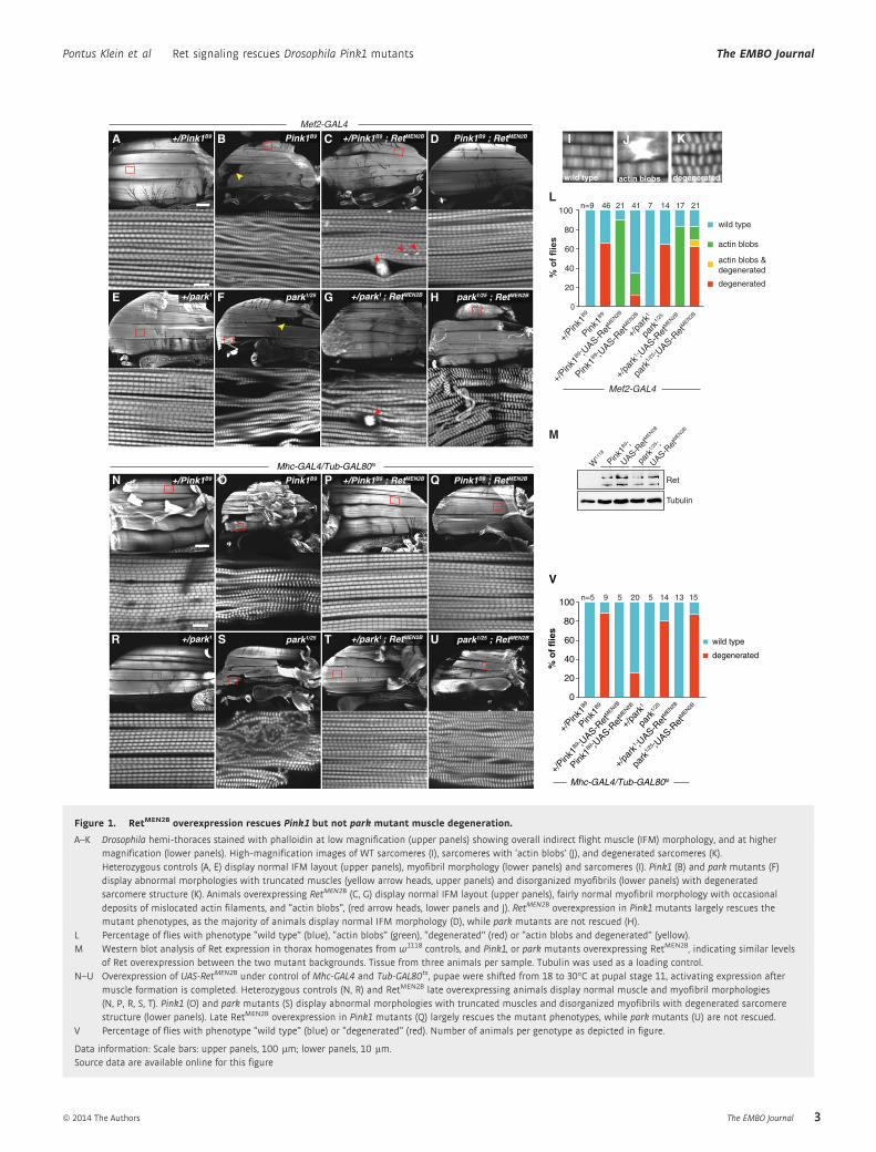
Figure 1. RetMEN2B overexpression rescues Pink1 but not park mutant muscle degeneration.
A–K Drosophila hemi-thoraces stained with phalloidin at low magnification (upper panels) showing overall indirect flight muscle (IFM) morphology, and at highermagnification (lower panels). High-magnification images of WT sarcomeres (I), sarcomeres with ‘actin blobs’ (J), and degenerated sarcomeres (K).Heterozygous controls (A, E) display normal IFM layout (upper panels), myofibril morphology (lower panels) and sarcomeres (I). Pink1 (B) and park mutants (F)display abnormal morphologies with truncated muscles (yellow arrow heads, upper panels) and disorganized myofibrils (lower panels) with degeneratedsarcomere structure (K). Animals overexpressing RetMEN2B (C, G) display normal IFM layout (upper panels), fairly normal myofibril morphology with occasionaldeposits of mislocated actin filaments, and “actin blobs”, (red arrow heads, lower panels and J). RetMEN2B overexpression in Pink1 mutants largely rescues themutant phenotypes, as the majority of animals display normal IFM morphology (D), while park mutants are not rescued (H).
L Percentage of flies with phenotype “wild type” (blue), “actin blobs” (green), “degenerated” (red) or “actin blobs and degenerated” (yellow).M Western blot analysis of Ret expression in thorax homogenates from w1118 controls, and Pink1, or park mutants overexpressing RetMEN2B, indicating similar levels
of Ret overexpression between the two mutant backgrounds. Tissue from three animals per sample. Tubulin was used as a loading control.N–U Overexpression of UAS-RetMEN2B under control of Mhc-GAL4 and Tub-GAL80ts, pupae were shifted from 18 to 30°C at pupal stage 11, activating expression after
muscle formation is completed. Heterozygous controls (N, R) and RetMEN2B late overexpressing animals display normal muscle and myofibril morphologies(N, P, R, S, T). Pink1 (O) and park mutants (S) display abnormal morphologies with truncated muscles and disorganized myofibrils with degenerated sarcomerestructure (lower panels). Late RetMEN2B overexpression in Pink1 mutants (Q) largely rescues the mutant phenotypes, while park mutants (U) are not rescued.
V Percentage of flies with phenotype “wild type” (blue) or “degenerated” (red). Number of animals per genotype as depicted in figure.
Data information: Scale bars: upper panels, 100 lm; lower panels, 10 lm.Source data are available online for this figure
ª 2014 The Authors The EMBO Journal
Pontus Klein et al Ret signaling rescues Drosophila Pink1 mutants The EMBO Journal
3

tyrosine kinases such as epidermal growth factor receptor (EGFR)
and FGFR are known to regulate embryonic myoblast specification
via Ras/Erk signaling (Carmena et al, 1998; Halfon et al, 2000), and
the insulin receptor controls muscle size (Demontis & Perrimon,
2009). Therefore, it is plausible that active RetMEN2B affects these, or
similar developmental processes. To verify that the rescue of the
Pink1 mutants is not a developmental interaction, we utilized the
GAL80ts system which permits transgene expression in a defined
time window regulated by temperature. To drive RetMEN2B expres-
sion, we chose the GAL4 driver, Myosin heavy chain (Mhc) GAL4,
which expresses only in differentiated muscles, not in myoblasts, in
difference to Mef-GAL4 and generates higher expression. Unlike
Mef2-GAL4, it causes complete lethality when driving RetMEN2B from
embryonic stages. Flies were crossed at 18°C (non-permissive
temperature), after which pupae were shifted to 30°C (permissive
temperature) at pharate adult stage P11 � 3 h (equivalent of 75 h
APF at 25°C) (Flybase FBdv:00005349), a time well after completion
of IFM development, but before the onset of apoptotic degeneration
in Pink1 and park mutants (Greene et al, 2003; Clark et al, 2006).
Analyses were again performed at 3–5 days post-eclosion. Using this
protocol, Pink1 and park mutants showed degenerated myofibrils
with a frequency of approximately 90% and 80% respectively as
compared to controls (Fig 1N, O, R, S, V), the higher penetrance being
likely due to the increased temperature. RetMEN2B-overexpressing
flies eclosed with Mendelian frequencies and displayed fully normal
muscle morphology, without the presence of actin blobs, confirming
the hypothesis that the lethality and actin blob phenotypes have
developmental origins (Fig 1P, T, V). When RetMEN2B was expressed
in Pink1mutants from this late pupal stage and onwards, it again lar-
gely rescued muscle degeneration, indicating that the rescue is not
due to a developmental interaction, but a direct protective effect of
Ret signaling on degenerating tissue (Fig 1Q and V). Interestingly,
park mutants were again not rescued using this expression protocol
(Fig 1U and V).
Ret signaling rescues mitochondrial morphology in flight muscles
One possibility is that RetMEN2B inhibits muscle degeneration without
directly targeting the primary cause of the Pink1 phenotype: mito-
chondrial impairments (Clark et al, 2006). To test this possibility,
we analyzed the ultrastructure of mitochondria using transmission
electron microscopy. IFMs from control flies showed regular
organization of myofibrils and densely packed mitochondria with
intact cristae (Fig 2A, E, L, M). Pink1 and park mutants displayed a
heterogeneous population of mitochondria with the majority having
significantly enlarged sizes and mild or severe disruption of their
cristae structure, when compared to control mitochondria (Fig 2B,
F, I-M). Mef2>RetMEN2B overexpression in control flies did not alter
normal mitochondria morphology (Fig 2C, G, L, M). However, in
Pink1 mutants, RetMEN2B overexpression significantly reduced the
fraction of severely impaired mitochondria and increased the
fraction of mitochondria with WT-like cristae structure (Fig 2D and L).
A B I L
M
J
KC D
E F
G H
Figure 2. RetMEN2B rescues mitochondrial cristae structure of Pink1 mutants.
A–K Transmission electron microscopy images of indirect flight muscles. Heterozygous controls (A, E) and animals overexpressing RetMEN2B (B, F) display normalmitochondria of similar size with highly dense cristae structure. Pink1 and park mutants have enlarged mitochondria with broken cristae (C, G). Phenotype canvary from mild to severe. High-power images of mitochondria are shown for the categories wild type (I), mild (J), severe phenotype (K). RetMEN2B overexpressionpartially restores mitochondrial size and cristae structure in Pink1 (D), but not park mutants (H). Scale bar, 2 lm.
L, M Percentages of mitochondria of the indicated categories, 500–800 mitochondria per animal, averages of 6 animals per genotype.
The EMBO Journal ª 2014 The Authors
The EMBO Journal Ret signaling rescues Drosophila Pink1 mutants Pontus Klein et al
4

In contrast, park mutants showed no improvement of structural
impairments when RetMEN2B was overexpressed (Fig 2H and M).
These results demonstrate that RetMEN2B can rescue mitochondrial
impairments of pink1 but not park mutants, suggesting that the
mitochondrial deficiencies of the two mutant strains have partially
different origins.
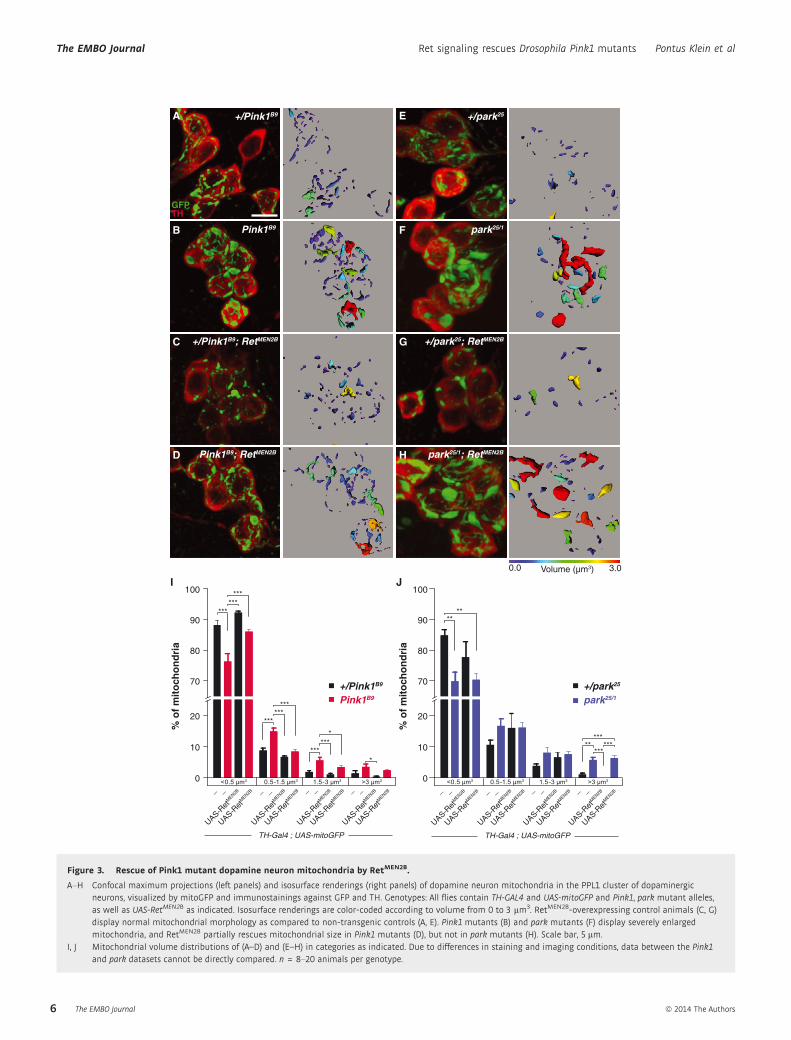
Ret rescues mitochondrial morphology in dopaminergic neurons
To address whether RetMEN2B also rescues the morphology of mito-
chondria in dopaminergic neurons, we overexpressed RetMEN2B
using TH-GAL4 together with the mitochondrial marker mitoGFP
(Pilling et al, 2006). Pink1 and park mutants displayed severely
enlarged mitochondria as compared to controls (Fig 3A, B, E, F, I, J).
RetMEN2B overexpression in a control background did not signifi-
cantly alter the normal mitochondrial background (Fig 3C, G, I, J).
However, when overexpressed in Pink1 mutants, mitochondrial size
was significantly rescued (Fig 3D and I). Quantification of mito-
chondrial volumes revealed that in the presence of RetMEN2B the
abundance of normal mitochondria was increased, while the frac-
tion of enlarged mitochondria decreased to levels similar to those of
control flies. Merely, the 4% largest mitochondria were not rescued.
In line with the analysis of mitochondria in muscle, mitochondrial
morphology in neurons of park mutants was not rescued by
RetMEN2B (Fig 3H and J).
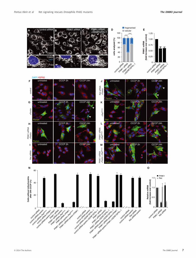
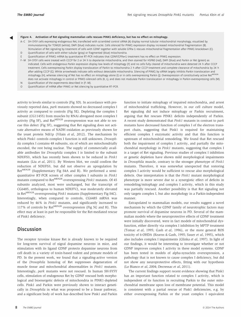
GDNF/Ret signaling rescues mitochondrial defects inmammalian cells
In order to assess whether signaling from endogenous Ret can also
rescue mitochondrial impairments caused by loss of PINK1 function,
we used the human dopaminergic neuroblastoma cell line SH-SY5Y,
which expresses endogenous Ret. Acute knock-down of PINK1 in
this cell line was previously shown to cause fragmentation of the
mitochondrial network (Lutz et al, 2009) (Fig 4A, B, D). Stimulation
of Ret by GDNF and soluble GFRa-1 rescued mitochondrial fragmen-
tation, demonstrating that endogenous mammalian Ret can rescue
mitochondrial impairments (Fig 4C and D). A semi-quantitative
RT-PCR analysis of PINK1 mRNA controlled that GDNF/GFRa-1stimulation did not upregulate PINK1 levels (Fig 4E).
Ret rescues mitochondrial morphology independently ofParkin-induced mitophagy
Although the data so far suggested that Ret rescues Pink1-deficient
mitochondria independently of Parkin, we cannot exclude that Ret
signaling activates Parkin translocation to mitochondria, thus pro-
moting their clearance through mitophagy. To test this hypothesis,
we treated SH-SY5Y cells overexpressing Parkin with carbonyl
cyanide m-chlorophenyl hydrazone (CCCP) to depolarizemitochondria.
CCCP treatment induced recruitment of Parkin to mitochondria
(detected 2 h after adding CCCP) followed by the removal of depo-
larized mitochondria in about 50% of Parkin-expressing SH-SY5Y
cells (monitored 24 h later) (Fig 4G and N). Parkin-induced
mitophagy required the presence of PINK1, as described previously
(Geisler et al, 2010; Narendra et al, 2010; Vives-Bauza et al, 2010),
but was not impaired in cells silenced for Ret expression (Fig 4H,
I, J, N, O). Moreover, the overexpression of constitutively active
RetMEN2A did not induce Parkin translocation or mitophagy under
any condition, including PINK1 knock-down with or without Par-
kin overexpression (Fig 4K, L, M, N). Similar results were obtained
when GDNF and soluble GFRa-1 was used to activate signaling via
endogenous Ret (Fig 4N). Furthermore, GDNF/GFRa-1 treatment
also rescued mitochondrial fragmentation induced by PINK1 silenc-
ing HeLa cells, a cell type which does not express endogenous
Parkin (Denison et al, 2003; Pawlyk et al, 2003), further indicating
that Ret signaling rescues PINK1 loss-of-function phenotypes inde-
pendently of Parkin (Supplementary Fig S3).
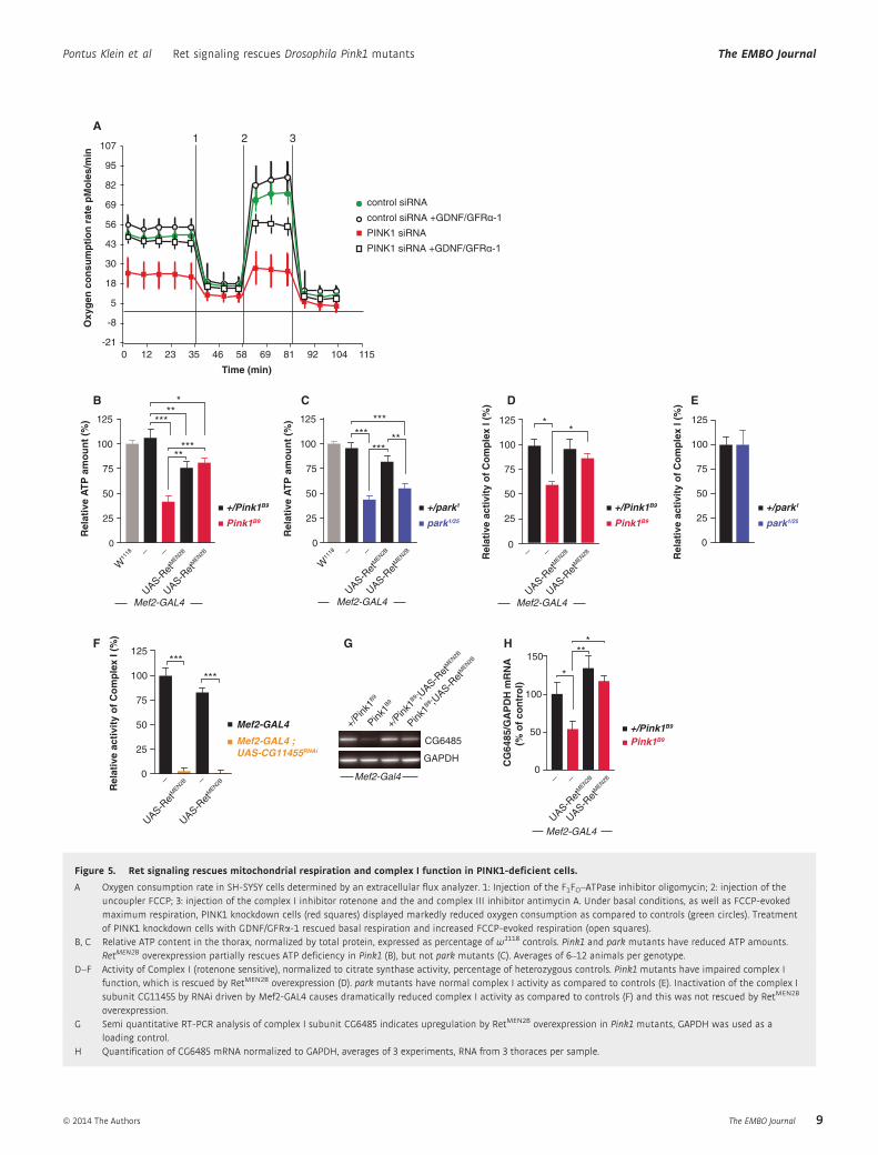
Ret signaling rescues impaired bioenergetics ofPink1-deficient cells
It has been reported previously that PINK1 deficiency impairs mito-
chondrial respiration (Gautier et al, 2008, 2012; Gandhi et al, 2009;
Lutz et al, 2009; Morais et al, 2009). We therefore investigated
whether activation of Ret signaling via GDNF/GFRa-1 treatment
could influence this phenotype. We measured mitochondrial func-
tion under basal and stress conditions in SH-SY5Y cells silenced for
PINK1 expression by using an extracellular oxygen flux analyzer. In
comparison to control siRNA-treated cells, PINK1-deficient cells
were characterized by a decreased oxygen consumption rate even
under basal conditions (Fig 5A). Moreover, the spare respiratory
capacity (difference between maximal and basal respiration) was
markedly reduced, indicating that the ability of PINK1-deficient cells
to respond to an increased energy demand under stress conditions
is severely impaired. Remarkably, GDNF/GFRa-1 treatment fully
rescued basal respiration and increased maximal respiration in
PINK1-deficient cells, indicating that the beneficial effect of
increased Ret signaling in PINK1-deficient models can be explained
by influencing the bioenergetic capacity of mitochondria rather than
mitophagy.
Complex I deficiency of Pink1 mutants rescued by Ret signaling
To investigate whether Ret signaling also rescued mitochondrial
functionality in Drosophila, we measured ATP content of thoracic
homogenates. As previously shown (Clark et al, 2006; Park et al,
2006; Yang et al, 2006; Vos et al, 2012), Pink1 and park mutants
showed reduced ATP content in the thorax to approximately 40% of
controls, including flies carrying the Mef2-GAL4 driver (Fig 5B and C).
Mef2 > RetMEN2B overexpression in control flies caused a slight
reduction in ATP as compared to controls, possibly as a result of
their mild muscle phenotype. In line with the rescue of myofibril and
mitochondrial structures, RetMEN2B overexpression largely rescued
ATP levels in Pink1 mutants, while ATP levels of park mutants did
not significantly improve (Fig 5B and C). To unravel the underlying
mechanism of the improved mitochondrial respiration, we
turned our attention to complex I of the ETC. Recent reports had
found that Pink1, in contrast to park mutants had decreased activity
of the ETC, and specifically of complex I function (Morais et al,
2009; Vilain et al, 2012). For these reasons, we measured complex I
activity in RetMEN2B-overexpressing Pink1 mutants, by monitoring
rotenone-sensitive NADH oxidation by spectrophotometry, normal-
ized to the activity of citrate synthase. As previously observed,
Pink1 mutants displayed markedly reduced complex I activity
(Fig 5D). Interestingly, RetMEN2B significantly increased complex I
ª 2014 The Authors The EMBO Journal
Pontus Klein et al Ret signaling rescues Drosophila Pink1 mutants The EMBO Journal
5
3.00.0 Volume (µm3)
Pink1B9
+/Pink1B9; RetMEN2B
Pink1B9; RetMEN2B
GFPTH
+/park25
park25/1; RetMEN2B
park25/1
+/park25; RetMEN2B
+/Pink1B9 E
F
G
H
A
B
C
D
0
10
20
70
80
90
100
0
10
20
70
80
90
100
% o
f m
ito
cho
nd
ria
% o
f m
ito
cho
nd
ria
UAS-Ret
MEN2B
TH-Gal4 ; UAS-mitoGFP
UAS-Ret
MEN2B
UAS-Ret
MEN2B
UAS-Ret
MEN2B
UAS-Ret
MEN2B
UAS-Ret
MEN2B
UAS-Ret
MEN2B
UAS-Ret
MEN2B
UAS-Ret
MEN2B
TH-Gal4 ; UAS-mitoGFP
UAS-Ret
MEN2B
UAS-Ret
MEN2B
UAS-Ret
MEN2B
UAS-Ret
MEN2B
UAS-Ret
MEN2B
UAS-Ret
MEN2B
UAS-Ret
MEN2B
JI
+/Pink1B9
Pink1B9
+/park25
park25/1
<0.5 μm3 0.5-1.5 μm3 >3 μm31.5-3 μm3 <0.5 μm3 0.5-1.5 μm3 >3 μm31.5-3 μm3
***
******
******
***
******
*
*
****
*****
******
_ _ _ _ _ _ _ _ _ _ _ _ _ _ _ _
Figure 3. Rescue of Pink1 mutant dopamine neuron mitochondria by RetMEN2B.
A–H Confocal maximum projections (left panels) and isosurface renderings (right panels) of dopamine neuron mitochondria in the PPL1 cluster of dopaminergicneurons, visualized by mitoGFP and immunostainings against GFP and TH. Genotypes: All flies contain TH-GAL4 and UAS-mitoGFP and Pink1, park mutant alleles,as well as UAS-RetMEN2B as indicated. Isosurface renderings are color-coded according to volume from 0 to 3 lm3. RetMEN2B-overexpressing control animals (C, G)display normal mitochondrial morphology as compared to non-transgenic controls (A, E). Pink1 mutants (B) and park mutants (F) display severely enlargedmitochondria, and RetMEN2B partially rescues mitochondrial size in Pink1 mutants (D), but not in park mutants (H). Scale bar, 5 lm.
I, J Mitochondrial volume distributions of (A–D) and (E–H) in categories as indicated. Due to differences in staining and imaging conditions, data between the Pink1and park datasets cannot be directly compared. n = 8–20 animals per genotype.
The EMBO Journal ª 2014 The Authors
The EMBO Journal Ret signaling rescues Drosophila Pink1 mutants Pontus Klein et al
6
CCCP 2h CCCP 24h
CCCP 2h CCCP 24h
CCCP 24h
CCCP 24h
CCCP 2h
CCCP 2h
0
20
40
60
cont
rol s
iRNA
cont
rol s
iRNA+p
arkin
cont
rol s
iRNA+G
DNF/GFRα
-1
cont
rol s
iRNA+p
arkin
+GDNF/G
FRα-1
PINK1
siRNA
PINK1
siRNA+p
arkin
PINK1
siRNA+G
DNF/GFRα
-1
PINK1
siRNA+p
arkin
+GDNF/G
FRα-1
PINK1
siRNA+p
arkin
+PIN
K1
PINK1
siRNA+p
arkin
+PIN
K1+GDNF/G
FRα-1
cont
rol s
iRNA
cont
rol s
iRNA+p
arkin
cont
rol s
iRNA+R
etM
EN2A
cont
rol s
iRNA+P
INK1
cont
rol s
iRNA+p
arkin
+PIN
K1
PINK1
siRNA
PINK1
siRNA+p
arkin
PINK1
siRNA+Ret
MEN2A
PINK1s
iRNA+P
INK1
PINK1
siRNA+p
arkin
+RetM
EN2A
PINK1
siRNA+p
arkin
+PIN
K1
Cel
ls w
ith
ou
t m
ito
cho
nd
ria
afte
r 24
h C
CC
P (
%)
N
Ret si
RNA
cont
rol s
iRNA
cont
rol s
iRNA+pa
rkin
Ret si
RNA+par
kin
0
1.0
Rel
ativ
e m
RN
A
(fol
d in
crea
se o
ver
con
tro
l)
0.5
1.5 RetPINK1
O
cont
rol s
iRNA
PINK1
siRNA
Ret si
RNA
CCCP 2h CCCP 24huntreated
cont
rol
+P
arki
n
DAPI HSP60
PIN
K1
siR
NA
+P
arki
n
CCCP 2h CCCP 24huntreated
untreated CCCP 24h
Parkin
Ret
siR
NA
untreated CCCP 24hParkin
F
G
H
I
CCCP 2h
CCCP 2h
Ret
siR
NA
+P
arki
n
Parkin
J untreated
PIN
K1
siR
NA
+
Par
kin
+R
etM
EN
2A
untreated
PIN
K1
siR
NA
+R
etM
EN
2A
untreated
Ret
untreated+
Ret
ME
N2A
Ret
Parkin
K
L
M
ED
PINK1
siRNA
PINK1
siRNA
+GDNF/G
FRα-1
tubularfragmented
cells
an
alyz
ed (
%)
******
cont
rol s
iRNA
0
60
100
120
40
80
20
0
1.00
PIN
K1
mR
NA
(f
old
incr
ease
ove
r co
ntr
ol)
0.50
1.25
0.75
0.25
PINK1
siRNA
PINK1
siRNA
+GDNF/G
FRα-1
cont
rol s
iRNA
B CA control siRNA PINK1 siRNA
TOM20
TOM20DAPI
PINK1 siRNA+GDNF/GFR -1 α
ª 2014 The Authors The EMBO Journal
Pontus Klein et al Ret signaling rescues Drosophila Pink1 mutants The EMBO Journal
7
activity to levels similar to controls (Fig 5D). In accordance with pre-
viously reported data, park mutants showed no decreased complex I
activity as compared to controls (Fig 5E). Depleting the complex I
subunit (CG11455) from muscles by RNAi abrogated most complex I
activity (Fig 5F), and RetMEN2B overexpression was not able to res-
cue this defect (Fig 5F), suggesting that Ret signaling does not acti-
vate alternative means of NADH oxidation as previously shown for
the yeast protein Ndi1p (Vilain et al, 2012). The mechanism by
which Pink1 controls complex I function is still unknown. Drosoph-
ila complex I contains 48 subunits, six of which are mitochondrially
encoded, the rest being nuclear. The supply of commercially avail-
able antibodies for Drosophila complex I is limited to the subunit
NDUFS3, which has recently been shown to be reduced in Pink1
mutants (Liu et al, 2011). By Western blot, we could confirm the
reduction of NDUFS3, but did not observe an upregulation by
RetMEN2B (Supplementary Fig S4A and B). We performed a semi-
quantitative RT-PCR screen of other complex I subunits in Pink1
mutants compared to RetMEN2B-overexpressing Pink1 mutants. Of 45
subunits analyzed, most were unchanged, but the transcript of
CG6485, orthologous to human NDUFV2, was moderately elevated
in RetMEN2B-overexpressing Pink1 mutants (Supplementary Fig S4C).
Interestingly, when compared to controls, CG6485 mRNA was
reduced by 46% in Pink1 mutants, and significantly increased to
117% of controls by RetMEN2B overexpression (Fig 5G and H). This
effect may at least in part be responsible for the Ret-mediated rescue
of Pink1 deficiency.
Discussion
The receptor tyrosine kinase Ret is already known to be required
for long-term survival of nigral dopamine neurons in mice, and
stimulation with its ligand GDNF protects dopamine neurons from
cell death in a variety of toxin-based rodent and primate models of
PD. In the present work, we found that a signaling-active version
of the Drosophila homolog of Ret suppresses degeneration of
muscle tissue and mitochondrial abnormalities in Pink1 mutants.
Interestingly, park mutants were not rescued. In human SH-SY5Y
cells, stimulation of endogenous Ret by GDNF rescued both morpho-
logical and bioenergetic defects of mitochondria in PINK1-depleted
cells. Pink1 and Parkin were previously shown to interact geneti-
cally in Drosophila in what was proposed to be a linear pathway,
and a significant body of work has described how Pink1 and Parkin
function to initiate mitophagy of impaired mitochondria, and arrest
of mitochondrial trafficking. However, in our cell culture model,
Ret signaling did not induce mitophagy or Parkin recruitment,
arguing that Ret rescues PINK1 deficits independently of Parkin.
A recent study demonstrated that Pink1 mutants in contrast to park
mutants have decreased function of complex I of the electron trans-
port chain, suggesting that Pink1 is required for maintaining
efficient complex I enzymatic activity and that this function is
upstream of mitochondrial remodeling. We found that Ret rescued
both the impairment of complex I activity, and partially the mito-
chondrial morphology in Pink1 mutants, suggesting that complex I
is a target of Ret signaling. Previous studies of complex I inhibition
or genetic depletion have shown mild morphological impairments
in Drosophila muscle, contrary to the stronger phenotype of Pink1
mutants. Therefore, it was somewhat unexpected that restoring
complex I activity would be sufficient to rescue also morphological
defects. One interpretation is that the Pink1 mutant morphological
phenotype is more severe due to a synergistic effect of deficits in
remodeling/mitophagy and complex I activity, which in this study
was partially rescued. Another possibility is that Ret signaling not
only targets complex I, but also morphology in a Parkin-independent
manner.
Extrapolated to mammalian models, our results suggest a novel
mechanism by which the GDNF family of neurotrophic factors may
promote survival of dopamine neurons in PD. Several of the mam-
malian models where the neuroprotective effects of GDNF treatment
were initially discovered, were in fact models of mitochondrial dys-
function, either directly via complex I inhibition by MPTP treatment
(Tomac et al, 1995; Gash et al, 1996), or the more general ROS
toxicity of 6-OHDA (Kearns & Gash, 1995; Sauer et al, 1995), which
also includes complex I impairments (Glinka et al, 1997). In light of
our findings, it would be interesting to investigate whether or not
GDNF improves complex I activity in these model systems. GDNF
has been tested in models of alpha-synuclein overexpression, a
pathology that is not known to cause complex I deficiency, but did
not show any neuroprotective effects, fitting with our hypothesis
(Lo Bianco et al, 2004; Decressac et al, 2011).
The current findings support recent evidence showing that Pink1
has an important function related to complex I activity, which is
independent of its function in recruiting Parkin to the outer mito-
chondrial membrane upon loss of membrane potential. This model
is consistent with a partial rescue of Pink1 deficiencies, e.g. by
either overexpressing Parkin or the yeast complex I equivalent
Figure 4. Activation of Ret signaling mammalian cells rescues PINK1 deficiency, but has no effect on mitophagy.
A–C SH-SY5Y cells expressing endogenous Ret, transfected with scrambled control siRNA (A) display normal tubular mitochondrial morphology, visualized byimmunostaining for TOM20 (white); DAPI (blue) indicates nuclei. Cells silenced for PINK1 expression display increased mitochondrial fragmentation (B).Stimulation of Ret signaling by treatment of cells with GDNF together with soluble GFRa-1 rescues mitochondrial fragmentation after PINK1 knockdown (C).
D Quantification of cells with either tubular (gray) or fragmented (blue) mitochondria.E Quantification of PINK1 mRNA by quantitative RT-PCR indicates that GDNF/GFRa-1 treatment has no effect on PINK1 expression.F–M SH-SY5Y cells were treated with CCCP for 2 or 24 h to depolarize mitochondria, and then stained for HSP60 (red), DAPI (blue) and Parkin or Ret (green) as
indicated. Cells with endogenous Parkin expression display low levels of mitophagy (F) and no cells fully cleared of mitochondria were detected 24 h after CCCPtreatment. Cells overexpressing Parkin display translocation of Parkin to mitochondria 2 h after CCCP treatment and complete clearance of mitochondria by 24 hafter adding CCCP (G). White arrowheads indicate cells without detectable mitochondria. Silencing of PINK1 by siRNA largely inhibits Parkin translocation andmitophagy (H), whereas silencing of Ret has no effect on mitophagy alone (I) or in cells overexpressing Parkin (J). Overexpression of constitutively active RetMEN2A
does not activate mitophagy in control or PINK1-silenced cells (K, L), and does not modulate Parkin translocation or mitophagy in Parkin-overexpressing cells (M).N Quantification of the experiments described in (F–M).O Quantification of mRNA after PINK1 or Ret silencing by quantitative RT-PCR.
◂
The EMBO Journal ª 2014 The Authors
The EMBO Journal Ret signaling rescues Drosophila Pink1 mutants Pontus Klein et al
8
0
0
G
Rel
ativ
e ac
tivi
ty o
f C
om
ple
x I (
%) D
**
0
25
100
50
75
125
Rel
ativ
e ac
tivi
ty o
f C
om
ple
x I (
%)
0
25
100
50
75
125
E
50
100
125**
25
75
***
***
Rel
ativ
e A
TP
am
ou
nt
(%)
Pink1B9
+/Pink1B9
0
50
100
125
25
75
W11
18
Mef2-GAL4UAS-R
etM
EN2B_
UAS-Ret
MEN2B_
park1/25
+/park1
Rel
ativ
e A
TP
am
ou
nt
(%)
***
CB
**
*
*****
***
Mef2-GAL4 ; UAS-CG11455RNAi
Mef2-GAL4
***
***
UAS-Ret
MEN2B
_ _
UAS-Ret
MEN2B
0
25
100
50
75
125
Rel
ativ
e ac
tivi
ty o
f C
om
ple
x I (
%)
Mef2-GAL4
*
UAS-Ret
MEN2B_ _
UAS-Ret
MEN2B
100
50
150
CG
6485
/GA
PD
H m
RN
A(%
of
con
tro
l)
F
CG6485
GAPDH
+/Pink
1B9 ;U
AS-Ret
MEN2B
Pink1B9
Pink1B9 ;U
AS-Ret
MEN2B
+/Pink
1B9
Mef2-Gal4
H
Pink1B9
+/Pink1B9
Pink1B9
+/Pink1B9
park1/25
+/park1
W11
18
Mef2-GAL4UAS-R
etM
EN2B_
UAS-Ret
MEN2B_
Mef2-GAL4UAS-R
etM
EN2B_
UAS-Ret
MEN2B_
***
control siRNA
control siRNA +GDNF/GFRα-1PINK1 siRNA
PINK1 siRNA +GDNF/GFRα-1
Oxy
gen
co
nsu
mp
tio
n r
ate
pM
ole
s/m
in
56
43
30
18
5
-8
-210 12 23 35 46 58 69 81 92 104 115
69
95
1071 2 3
82
Time (min)
A
Figure 5. Ret signaling rescues mitochondrial respiration and complex I function in PINK1-deficient cells.
A Oxygen consumption rate in SH-SY5Y cells determined by an extracellular flux analyzer. 1: Injection of the F1FO–ATPase inhibitor oligomycin; 2: injection of theuncoupler FCCP; 3: injection of the complex I inhibitor rotenone and the and complex III inhibitor antimycin A. Under basal conditions, as well as FCCP-evokedmaximum respiration, PINK1 knockdown cells (red squares) displayed markedly reduced oxygen consumption as compared to controls (green circles). Treatmentof PINK1 knockdown cells with GDNF/GFRa-1 rescued basal respiration and increased FCCP-evoked respiration (open squares).
B, C Relative ATP content in the thorax, normalized by total protein, expressed as percentage of w1118 controls. Pink1 and park mutants have reduced ATP amounts.RetMEN2B overexpression partially rescues ATP deficiency in Pink1 (B), but not park mutants (C). Averages of 6–12 animals per genotype.
D–F Activity of Complex I (rotenone sensitive), normalized to citrate synthase activity, percentage of heterozygous controls. Pink1 mutants have impaired complex Ifunction, which is rescued by RetMEN2B overexpression (D). park mutants have normal complex I activity as compared to controls (E). Inactivation of the complex Isubunit CG11455 by RNAi driven by Mef2-GAL4 causes dramatically reduced complex I activity as compared to controls (F) and this was not rescued by RetMEN2B
overexpression.G Semi quantitative RT-PCR analysis of complex I subunit CG6485 indicates upregulation by RetMEN2B overexpression in Pink1 mutants, GAPDH was used as a
loading control.H Quantification of CG6485 mRNA normalized to GAPDH, averages of 3 experiments, RNA from 3 thoraces per sample.
ª 2014 The Authors The EMBO Journal
Pontus Klein et al Ret signaling rescues Drosophila Pink1 mutants The EMBO Journal
9
NADH dehydrogenase, or, in the current work, RetMEN2B (Clark
et al, 2006; Park et al, 2006; Yang et al, 2006; Vilain et al, 2012). In
addition, our findings are consistent with a recent study showing
that Pink1-deficient flies but not Parkin-deficient flies can be rescued
by TRAP1, which also seems to have beneficial effects on complex I
activity (Zhang et al, 2013).
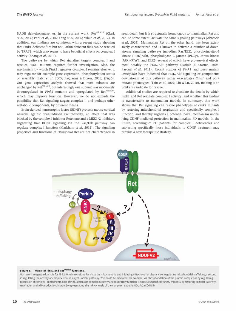
The pathways by which Ret signaling targets complex I and
rescues Pink1 mutants requires further investigation. Also, the
mechanism by which Pink1 regulates complex I remains elusive, it
may regulate for example gene expression, phosphorylation status
or assembly (Salvi et al, 2005; Pagliarini & Dixon, 2006) (Fig 6).
Our gene expression analysis showed that most subunits are
unchanged by RetMEN2B, but interestingly one subunit was moderately
downregulated in Pink1 mutants and upregulated by RetMEN2B,
which may improve function. However, we do not exclude the
possibility that Ret signaling targets complex I, and perhaps other
metabolic components, by different means.
Brain-derived neurotrophic factor (BDNF) protects mouse cortical
neurons against drug-induced excitotoxicity, an effect that was
blocked by the complex I inhibitor Rotenone and a MEK1/2 inhibitor,
suggesting that BDNF signaling via the Ras/Erk pathway can
regulate complex I function (Markham et al, 2012). The signaling
properties and functions of Drosophila Ret are not characterized in
great detail, but it is structurally homologous to mammalian Ret and
can, to some extent, activate the same signaling pathways (Abrescia
et al, 2005). Mammalian Ret on the other hand, has been exten-
sively characterized and is known to activate a number of down-
stream signaling pathways including Ras/ERK, phosphoinositol-3
kinase (PI3K)/Akt, phospholipase C-gamma (PLCγ), Janus kinase
(JAK)/STAT, and ERK5, several of which have pro-survival effects,
most notably the PI3K/Akt pathway (Sariola & Saarma, 2003;
Pascual et al, 2011). Recent studies of Pink1 and park mutant
Drosophila have indicated that PI3K/Akt signaling or components
downstream of this pathway rather exacerbates Pink1 and park
mutant phenotypes (Tain et al, 2009; Liu & Lu, 2010), making it an
unlikely candidate for rescue.
Additional studies are required to elucidate the details by which
Pink1 and Ret regulate complex I activity, and whether this finding
is transferrable to mammalian models. In summary, this work
shows that Ret signaling can rescue phenotypes of Pink1 mutants
by restoring mitochondrial respiration and specifically complex I
function, and thereby suggests a potential novel mechanism under-
lying GDNF-mediated protection in mammalian PD models. In the
future, screening of PD patients for complex I deficiencies and
subjecting specifically those individuals to GDNF treatment may
provide a new therapeutic strategy.
Parkin
NDUFV2
?
nucleus
IIIIII
PIN
K1
Ub ?
- mitophagy- trafficking
NADH
ATP
NAD+H+
H+
O2
V
H+
IV
Pi?
GDNF
GFR
α-1
Ret
Figure 6. Model of Pink1 and RetMEN2B functions.Our results suggest a dual role for Pink1: One in recruiting Parkin to the mitochondria and initiating mitochondrial clearance or regulating mitochondrial trafficking, a secondin regulating the activity of complex I via an as yet unclear pathway. This could be mediated, for example, via phosphorylation of the protein complex or by regulatingexpression of complex I components. Loss of Pink1 decreases complex I activity and respiratory function. Ret rescues specifically Pink1mutants, by restoring complex I activity,respiration and ATP production, in part by upregulating the mRNA levels of the complex I subunit NDUFV2 (CG6485).
The EMBO Journal ª 2014 The Authors
The EMBO Journal Ret signaling rescues Drosophila Pink1 mutants Pontus Klein et al
10
Materials and Methods
Fly strains and procedures
Mef2-GAL4;UAS-RetMEN2B is lethal at 25°C, therefore all crosses
were performed at 18°C. All analyses were performed with 2- to
5-day-old flies. In experiments with Mhc-GAL4;Tub-GAL80ts, pupae
were shifted from 18 to 30°C at pharate adult stages P11-P12
(Flybase FBdv:00005349) and analyzed at 3–4 days post eclosion.
park25 (Greene et al, 2003) was provided by Leo Pallanck, park1
(Cha et al, 2005) and Pink1B9 (Park et al, 2006) were provided by
Jongkyeong Chung, Pink1B9::Mef2-GAL4 (Tain et al, 2009) was
provided by Alex Whitworth, UAS-RetMEN2B (Read et al, 2005) was
provided by Ross Cagan, TH-GAL4 (Friggi-Grelin et al, 2003),
was provided by Hiromu Tanimoto, Mef2-GAL4 (Ranganayakulu
et al, 1996), Tub-GAL80ts (McGuire et al, 2003), and UAS-mitoGFP
(Pilling et al, 2006) were obtained from the Bloomington stock
center, UAS-CG11455RNAi (#12838) was obtained from Vienna
Drosophila RNAi Center. “+”-controls depict Pink1 and park WT
alleles from w1118 (Bloomington stock #5905). In For all histology
experiments, flies were genotyped by PCR to assure correct genotypes
and control for X-chromosome non-disjunction, for list of primers
see Supplementary information.
Myosin heavy chain – GAL4 flies
A 2.5 kb Mhc enhancer was amplified from genomic DNA using
primers FS124 (5′-tcaggtaccGGCCGCTCTAGAAATGATATGTG-3′)
and FS125 (5′-tcacgcggccgcATTATCCTTGCTTAAATTTCGTTTAG-
3′) and cloned with Asp718/NotI into a GAL4-containing
Casper-based P-element transformation vector. Transgenic flies
were generated using standard procedures. In contrast to the
formerly published GAL4 line (Schuster et al, 1996), which shows a
rather weak activity in embryos, larvae and adults, this new Mhc-
GAL4 line is very strong and very specifically expressed in differenti-
ated muscles from embryonic stages onwards (FS, unpublished).
Histology, transmission electron microscopy and analysis ofmitochondrial morphology
Hemi-thoraces were prepared as described previously (Schnorrer
et al, 2010), stained with Phalloidin-Alexa Fluor-568 (Molecular
Probes), and single plane images were acquired on an Olympus
FV1000 confocal scanning microscope. For transmission electron
microscopy, hemi-thoraces were fixed in 2.5% Glutaraldehyde, from
which semithin sections were prepared and stained with toluidine-
blue, subsequently ultrathin serial sections were prepared using a
Leica EM UC6 Ultramicrotome. Images at 5,000× magnification were
acquired using a JEOL JEM-1230 transmission electron microscope
at 80 kV, equipped with a Gatan Orius SC1000 digital Camera. Six
TEM Images per animal were acquired from randomly selected
regions of the indirect flight muscles. All mitochondria in these
images (500–800 per animal) were grouped into three categories,
based on the integrity of the cristae structure, with genotypes
blinded to the experimenter, using the ImageJ software (NIH).
Whole mount immunostaining of fly brains was performed according
to standard procedures. The following antibodies were used: rabbit
anti-tyrosine hydroxylase (ab152, Millipore; 1:200) and chick anti-
GFP (Abcam ab13970; 1:500). The PPL1 cluster was imaged using
an Olympus FV1000 confocal microscope with a 60× NA 1.3 objec-
tive with 4× zoom. 52 z-sections of 0.3 lm spacing were acquired
and deconvolved by the nearest neighbor algorithm using Metamor-
ph 7.5 (Molecular Devices). A volume corresponding to
26 × 26 × 15 lm was cropped, subjected to linear rescaling and
analyzed in Imaris x64 6.4.2 (Bitplane Scientific Software). Mito-
chondrial volume was measured by 3D isosurface rendering using a
fixed threshold.
Immunoblot analysis
Thoraces from three animals per sample were homogenized in
Triton-lysis buffer, protein concentration was determined using the
BCA method (BioRad), equal amounts of protein were separated
using SDS-PAGE and blotted according to standard procedures.
Antibodies used were: panRet (provided by C. Ibanez) and alpha-
Tubulin (clone DM1A, Sigma).
Cell culture, treatments and RNA Interference
SH-SY5Y (DSMZ number ACC 209) cells were cultivated as
described previously (Henn et al, 2005; Schlehe et al, 2008). For
acute stimulation of Ret, cells were incubated for 3–4 h with recom-
binant hGDNF (Shenandoah Biotechnology Inc.) and hGFRa-1 (R&D
Systems) at a final concentration of 100 ng/mL. PINK1 and Ret gene
silencing was performed with the following stealth siRNA oligos
(Invitrogen) using Lipofectamine RNAiMAX (Invitrogen): PINK1
human HSS127945 (SH-SY5Y), Ret human HSS109181.
Assessment of mitochondrial morphology
SH-SY5Y: Cells grown on 15-mm glass coverslips were fixed with
3.7% PFA in PBS for 10 min. Cells were permeabilized with 0.1%
Triton X-100 in PBS for 5 min and blocked with 5% BSA in PBS at
room temperature. Fixed cells were sequentially incubated with
primary antibody diluted in blocking solution (TOM20 pAb, overnight
at 4°C) and secondary antibody diluted in blocking buffer (goat anti
rabbit Alexa555- coniugated, 2 h at room temperature). Nuclei were
counterstained with DAPI. Coverslips were mounted on glass slides
and images were acquired with a Zeiss LSM710 confocal microscope
equipped with a 63× oil objective (NA 1.4). Cells displaying an
intact network of tubular mitochondria were classified as tubular.
When this network was disrupted and mitochondria appeared either
globular or rod-like they were classified as fragmented. The mito-
chondrial morphology of the cells was determined in a blinded man-
ner. Quantifications were based on 150 cells from at least 3
independent experiments.
Assessment of mitophagy
SH-SY5Y cells were plated on glass coverslips and reversely trans-
fected with siRNA and 24 h later with the indicated DNA
plasmid. Human GDNF and GFRalpha were added to the cells 24 h
after siRNA transfection and 3 h before CCCP treatment. The
next day, cells were treated with 10 lM carbonyl cyanide
ª 2014 The Authors The EMBO Journal
Pontus Klein et al Ret signaling rescues Drosophila Pink1 mutants The EMBO Journal
11
3-chlorophenylhydrazone (CCCP, Sigma) for 2 or 24 h. Recruitment
of parkin to mitochondria (after 2 h CCCP) and removal of mito-
chondria (after 24 h CCCP) was detected by indirect immunofluo-
rescence using a monoclonal anti-Parkin antibody (PRK8, Santa
Cruz Biotechnology) and a polyclonal antibody against HSP60
(Santa Cruz Biotechnology). Nuclei were stained by DAPI. Cells
were analysed by fluorescence microscopy using a Leica DMRB
microscope and confocal images were taken using a Zeiss LSM710
confocal microscope equipped with a 63× oil objective (NA 1.4).
Quantifications are based on three independent experiments. At
least 1,500 cells were analysed for each condition.
Real-time RT-PCR, cultured cells
Knock-down efficiency of PINK1 and Ret was evaluated by real-time
RT-PCR with the 7500 Fast Real Time System (Applied Biosystems)
as previosly described (Bouman et al, 2011). Statistical analysis
of RT-PCR data is based on at least four independent experiments
with triplicate samples. For list of primers, see Supplementary
information.
Measurement of mitochondrial oxygen consumption
The oxygen consumption rate was determined using a Seahorse XF
96 analyzer (Seahorse Biosciences). SH-SY5Y cells were reversely
transfected and plated in a XF 96 cell culture microplate. The next
day, fresh medium containing human GDNF/GFRa-1 was added to
the cells where indicated. The cells were incubated with low-glucose
(1 mM) medium overnight and the sensor cartridge was hydrated
overnight according to the manufacturers’ instructions. Measure-
ments were performed 48 h after transfection. The measured values
were normalized to protein levels. PINK1 knockdown did not induce
apoptosis under these conditions. The cells were washed using the
XF Prep Station three times with Seahorse Medium containing
10 mM galactose and 1 mM pyruvate. Mitochondrial function was
analyzed using the XF Cell Mito Stress Test Kit (Seahorse Biosciences)
and all measurements were carried out at 37°C. The following drugs
were diluted in Seahorse Medium and loaded on the sensor
cartridge: oligomycin (injection port A), carbonyl cyanide p-(triflu-
oromethoxy)phenylhydrazone (FCCP; injection port B), rotenone
and antimycin A (both injection port C). The drugs were diluted in
Seahorse Medium and loaded on the sensor cartridge. Measured
values were normalized to protein levels.
ATP measurement
Measurements of thoracic ATP were performed using a luciferase
assay as described previously (Park et al, 2006) with some modifi-
cations: Briefly, single thoraces from 3-day-old flies with heads and
wings removed were homogenized in 50 ll of extraction buffer
(100 mM Tris-HCl, 4 mM EDTA pH 7.8) with 6 M Guanidine-HCl
using a teflon-on-glass dounce homogenizer. The lysate was boiled
for 3 min and cleared by centrifugation at 20,000 g for 1 min. The
samples were diluted 1:100 in extraction buffer before analyzing
using the ATP determination kit (Invitrogen), according to the manu-
facturer’s instruction. Values were normalized to total protein
content, measured by absorbance at 280 nm using a NanoDrop
spectrophotometer. All measurements were performed in triplicate.
Enzymatic measurements
Activity of complex I (NADH: ubiquinone oxidoreductase) was
assessed by monitoring the oxidation of NADH as previously
described (Fischer et al, 1986). Briefly, thoraces from 20 animals
were homogenized in 250 mM sucrose, 10 mM Tris pH 7.4,
0.15 mM MgCl2, after which mitochondria were isolated as
described previously (Walker et al, 2006). Enzymatic activity of
complex I was assessed by NADH oxidation, monitored at
A340 nm as described (Bugiani et al, 2004), and rotenone insensi-
tive activity was subtracted. The activity of complex I was normal-
ized to Citrate Synthase activity, which was measured indirectly
by AcCoA-SH formation, as described (Ferguson & Williams,
1966).
RT-PCR, Drosophila complex I subunits
Thoraces were dissected and snap-frozen, homogenized in RLT
buffer (Qiagen) using a rotor-stator homogenizer. Total RNA was
prepared using the RNeasy mini kit according to instructions. Samples
were treated with DNase1 on-column for 15 min (RNase-free DNase
set, Qiagen). RT-PCR analysis was performed using the OneStep
RT-PCR kit (Qiagen) using 20 ng of template RNA and 35–40 cycles
of PCR amplification depending on signal strength of the primer
pair. Primers were designed using the primerBLAST tool (NCBI),
and when possible exon-junction spanning primers were used, for
list of primers, see Supplementary information. As some of the
analyzed transcripts are single-exon, control reactions omitting the
reverse transcriptase amplification step were performed to assure
that samples were free of contaminating genomic DNA, despite
DNase1 treatment.
Statistical analysis
Data represent mean � SEM. Statistical analysis was carried out
using analysis of variance (ANOVA) or Student’s t-test; *P ≤ 0.05;
**P ≤ 0.01; ***P ≤ 0.001.
Supplementary information for this article is available online:
http://emboj.embopress.org
AcknowledgementsWe would like to thank Marianne Braun and Ursula Weber for transmission
electron microcopy and Pilar Alcal�a for molecular biology assistance, the
Bloomington stock center for fly strains, Liviu Aron for discussions at early
stages of the project and Louise Gaitanos for critically reading the manuscript.
This work was in part supported by the Max-Planck Society, and grants from
the German Research Foundation (DFG), the European Union (MOLPARK) and
the European Research Council (TOPAG).
Author contributionsPK designed, performed and analyzed the majority of the experiments. CS and
FS contributed to the design of the fly genetics and analysis of muscle mor-
phology, and FS generated the Mhc-GAL4 line. EM and AKM-R designed, per-
formed and analyzed the SH-SY5Y experiments. KFW supervised the cell
culture work and contributed to the analysis of the fly data. RK supervised the
project, designed experiments and co-wrote the manuscript with PK.
The EMBO Journal ª 2014 The Authors
The EMBO Journal Ret signaling rescues Drosophila Pink1 mutants Pontus Klein et al
12
Conflict of interestThe authors declare that they have no conflict of interest.
References
Abrescia C, Sjostrand D, Kjaer S, Ibanez CF (2005) Drosophila RET contains an
active tyrosine kinase and elicits neurotrophic activities in mammalian
cells. FEBS Lett 579: 3789 – 3796
Airaksinen MS, Saarma M (2002) The GDNF family: signalling, biological
functions and therapeutic value. Nat Rev Neurosci 3: 383 – 394
Aron L, Klein P, Pham TT, Kramer ER, Wurst W, Klein R (2010) Pro-survival
role for Parkinsons associated gene DJ-1 revealed in trophically impaired
dopaminergic neurons. PLoS Biol 8: e1000349
Berger AK, Cortese GP, Amodeo KD, Weihofen A, Letai A, LaVoie MJ (2009)
Parkin selectively alters the intrinsic threshold for mitochondrial
cytochrome c release. Hum Mol Genet 18: 4317 – 4328
Bouman L, Schlierf A, Lutz AK, Shan J, Deinlein A, Kast J, Galehdar Z,
Palmisano V, Patenge N, Berg D, Gasser T, Augustin R, Trumbach D, Irrcher
I, Park DS, Wurst W, Kilberg MS, Tatzelt J, Winklhofer KF (2011) Parkin is
transcriptionally regulated by ATF4: evidence for an interconnection
between mitochondrial stress and ER stress. Cell Death Differ 18: 769 – 782
Bugiani M, Invernizzi F, Alberio S, Briem E, Lamantea E, Carrara F, Moroni I,
Farina L, Spada M, Donati MA, Uziel G, Zeviani M (2004) Clinical and
molecular findings in children with complex I deficiency. Biochim Biophys
Acta 1659: 136 – 147
Burns RS, Chiueh CC, Markey SP, Ebert MH, Jacobowitz DM, Kopin IJ (1983) A
primate model of parkinsonism: selective destruction of dopaminergic
neurons in the pars compacta of the substantia nigra by
N-methyl-4-phenyl-1,2,3,6-tetrahydropyridine. Proc Natl Acad Sci USA 80:
4546 – 4550
Carmena A, Gisselbrecht S, Harrison J, Jimenez F, Michelson AM (1998)
Combinatorial signaling codes for the progressive determination of cell
fates in the Drosophila embryonic mesoderm. Genes Dev 12: 3910 – 3922
Cha GH, Kim S, Park J, Lee E, Kim M, Lee SB, Kim JM, Chung J, Cho KS (2005)
Parkin negatively regulates JNK pathway in the dopaminergic neurons of
Drosophila. Proc Natl Acad Sci USA 102: 10345 – 10350
Clark IE, Dodson MW, Jiang C, Cao JH, Huh JR, Seol JH, Yoo SJ, Hay BA, Guo M
(2006) Drosophila pink1 is required for mitochondrial function and
interacts genetically with parkin. Nature 441: 1162 – 1166
Dagda RK, Gusdon AM, Pien I, Strack S, Green S, Li C, Van Houten B, Cherra SJ
3rd, Chu CT (2011) Mitochondrially localized PKA reverses mitochondrial
pathology and dysfunction in a cellular model of Parkinsons disease. Cell
Death Differ 18: 1914 – 1923
Decressac M, Ulusoy A, Mattsson B, Georgievska B, Romero-Ramos M, Kirik D,
Bjorklund A (2011) GDNF fails to exert neuroprotection in a rat
alpha-synuclein model of Parkinsons disease. Brain 134: 2302 – 2311
Deierborg T, Soulet D, Roybon L, Hall V, Brundin P (2008) Emerging
restorative treatments for Parkinsons disease. Prog Neurobiol 85: 407 – 432
Demontis F, Perrimon N (2009) Integration of Insulin receptor/Foxo signaling
and dMyc activity during muscle growth regulates body size in
Drosophila. Development 136: 983 – 993
Deng H, Dodson MW, Huang H, Guo M (2008) The Parkinsons disease genes
pink1 and parkin promote mitochondrial fission and/or inhibit fusion in
Drosophila. Proc Natl Acad Sci USA 105: 14503 – 14508
Denison SR, Wang F, Becker NA, Schule B, Kock N, Phillips LA, Klein C, Smith
DI (2003) Alterations in the common fragile site gene Parkin in ovarian
and other cancers. Oncogene 22: 8370 – 8378
Exner N, Lutz AK, Haass C, Winklhofer KF (2012) Mitochondrial dysfunction in
Parkinsons disease: molecular mechanisms and pathophysiological
consequences. EMBO J 31: 3038 – 3062
Ferguson SM, Williams GR (1966) The effect of malate and other dicarboxylic
acids on mitochondrial isocitrate metabolism. J Biol Chem 241:
3696 – 3700
Fischer JC, Ruitenbeek W, Trijbels JM, Veerkamp JH, Stadhouders AM, Sengers
RC, Janssen AJ (1986) Estimation of NADH oxidation in human skeletal
muscle mitochondria. Clin Chim Acta 155: 263 – 273
Friggi-Grelin F, Coulom H, Meller M, Gomez D, Hirsh J, Birman S (2003)
Targeted gene expression in Drosophila dopaminergic cells using
regulatory sequences from tyrosine hydroxylase. J Neurobiol 54: 618 – 627
Gandhi S, Wood-Kaczmar A, Yao Z, Plun-Favreau H, Deas E, Klupsch K,
Downward J, Latchman DS, Tabrizi SJ, Wood NW, Duchen MR, Abramov AY
(2009) PINK1-associated Parkinsons disease is caused by neuronal
vulnerability to calcium-induced cell death. Mol Cell 33: 627 – 638
Gash DM, Zhang Z, Ovadia A, Cass WA, Yi A, Simmerman L, Russell D, Martin
D, Lapchak PA, Collins F, Hoffer BJ, Gerhardt GA (1996) Functional recovery
in parkinsonian monkeys treated with GDNF. Nature 380: 252 – 255
Gautier CA, Giaime E, Caballero E, Nunez L, Song Z, Chan D, Villalobos C,
Shen J (2012) Regulation of mitochondrial permeability transition pore by
PINK1. Mol Neurodegener 7: 22
Gautier CA, Kitada T, Shen J (2008) Loss of PINK1 causes mitochondrial
functional defects and increased sensitivity to oxidative stress. Proc Natl
Acad Sci USA 105: 11364 – 11369
Geisler S, Holmstrom KM, Skujat D, Fiesel FC, Rothfuss OC, Kahle PJ, Springer
W (2010) PINK1/Parkin-mediated mitophagy is dependent on VDAC1 and
p62/SQSTM1. Nat Cell Biol 12: 119 – 131
Glinka Y, Gassen M, Youdim MB (1997) Mechanism of 6-hydroxydopamine
neurotoxicity. J Neural Transm Suppl 50: 55 – 66
Greene JC, Whitworth AJ, Kuo I, Andrews LA, Feany MB, Pallanck LJ (2003)
Mitochondrial pathology and apoptotic muscle degeneration in
Drosophila parkin mutants. Proc Natl Acad Sci USA 100: 4078 – 4083
Halfon MS, Carmena A, Gisselbrecht S, Sackerson CM, Jimenez F, Baylies MK,
Michelson AM (2000) Ras pathway specificity is determined by the
integration of multiple signal-activated and tissue-restricted transcription
factors. Cell 103: 63 – 74
Heeman B, Van den Haute C, Aelvoet SA, Valsecchi F, Rodenburg RJ, Reumers V,
Debyser Z, Callewaert G, Koopman WJ, Willems PH, Baekelandt V (2011)
Depletion of PINK1 affects mitochondrial metabolism, calcium homeostasis
and energy maintenance. J Cell Sci 124: 1115 – 1125
Henn IH, Bouman L, Schlehe JS, Schlierf A, Schramm JE, Wegener E, Nakaso K,
Culmsee C, Berninger B, Krappmann D, Tatzelt J, Winklhofer KF (2007)
Parkin mediates neuroprotection through activation of IkappaB kinase/
nuclear factor-kappaB signaling. J Neurosci 27: 1868 – 1878
Henn IH, Gostner JM, Lackner P, Tatzelt J, Winklhofer KF (2005) Pathogenic
mutations inactivate parkin by distinct mechanisms. J Neurochem 92:
114 – 122
Kearns CM, Gash DM (1995) GDNF protects nigral dopamine neurons against
6-hydroxydopamine in vivo. Brain Res 672: 104 – 111
Kramer ER, Aron L, Ramakers GM, Seitz S, Zhuang X, Beyer K, Smidt MP, Klein
R (2007) Absence of Ret signaling in mice causes progressive and late
degeneration of the nigrostriatal system. PLoS Biol 5: e39
Langston JW, Ballard P, Tetrud JW, Irwin I (1983) Chronic Parkinsonism in
humans due to a product of meperidine-analog synthesis. Science 219:
979 – 980
de Lau LM, Breteler MM (2006) Epidemiology of Parkinsons disease. Lancet
Neurol 5: 525 – 535
ª 2014 The Authors The EMBO Journal
Pontus Klein et al Ret signaling rescues Drosophila Pink1 mutants The EMBO Journal
13
Lin LF, Doherty DH, Lile JD, Bektesh S, Collins F (1993) GDNF: a glial cell
line-derived neurotrophic factor for midbrain dopaminergic neurons.
Science 260: 1130 – 1132
Liu S, Lu B (2010) Reduction of protein translation and activation of
autophagy protect against PINK1 pathogenesis in Drosophila
melanogaster. PLoS Genet 6: e1001237
Liu W, Acin-Perez R, Geghman KD, Manfredi G, Lu B, Li C (2011) Pink1
regulates the oxidative phosphorylation machinery via mitochondrial
fission. Proc Natl Acad Sci USA 108: 12920 – 12924
Lo Bianco C, Deglon N, Pralong W, Aebischer P (2004) Lentiviral nigral
delivery of GDNF does not prevent neurodegeneration in a genetic rat
model of Parkinsons disease. Neurobiol Dis 17: 283 – 289
Lutz AK, Exner N, Fett ME, Schlehe JS, Kloos K, Lammermann K, Brunner B,
Kurz-Drexler A, Vogel F, Reichert AS, Bouman L, Vogt-Weisenhorn D, Wurst
W, Tatzelt J, Haass C, Winklhofer KF (2009) Loss of parkin or PINK1
function increases Drp1-dependent mitochondrial fragmentation. J Biol
Chem 284: 22938 – 22951
Markham A, Cameron I, Bains R, Franklin P, Kiss JP, Schwendimann L,
Gressens P, Spedding M (2012) Brain-derived neurotrophic
factor-mediated effects on mitochondrial respiratory coupling and
neuroprotection share the same molecular signalling pathways. Eur J
Neurosci 35: 366 – 374
Martin I, Dawson VL, Dawson TM (2011) Recent advances in the genetics of
Parkinsons disease. Annu Rev Genomics Hum Genet 12: 301 – 325
McCoy MK, Cookson MR (2012) Mitochondrial quality control and dynamics
in Parkinsons disease. Antioxid Redox Signal 16: 869 – 882
McGuire SE, Le PT, Osborn AJ, Matsumoto K, Davis RL (2003) Spatiotemporal
rescue of memory dysfunction in Drosophila. Science 302: 1765 – 1768
Mijatovic J, Airavaara M, Planken A, Auvinen P, Raasmaja A, Piepponen TP,
Costantini F, Ahtee L, Saarma M (2007) Constitutive Ret activity in
knock-in multiple endocrine neoplasia type B mice induces profound
elevation of brain dopamine concentration via enhanced synthesis and
increases the number of TH-positive cells in the substantia nigra. J
Neurosci 27: 4799 – 4809
Mizuno Y, Ohta S, Tanaka M, Takamiya S, Suzuki K, Sato T, Oya H, Ozawa T,
Kagawa Y (1989) Deficiencies in complex I subunits of the respiratory
chain in Parkinsons disease. Biochem Biophys Res Commun 163: 1450 – 1455
Morais VA, Verstreken P, Roethig A, Smet J, Snellinx A, Vanbrabant M, Haddad
D, Frezza C, Mandemakers W, Vogt-Weisenhorn D, Van Coster R, Wurst W,
Scorrano L, De Strooper B (2009) Parkinsons disease mutations in PINK1
result in decreased Complex I activity and deficient synaptic function.
EMBO Mol Med 1: 99 – 111
Muller-Rischart AK, Pilsl A, Beaudette P, Patra M, Hadian K, Funke M, Peis R,
Deinlein A, Schweimer C, Kuhn PH, Lichtenthaler SF, Motori E, Hrelia S,
Wurst W, Trumbach D, Langer T, Krappmann D, Dittmar G, Tatzelt J,
Winklhofer KF (2013) The E3 ligase parkin maintains mitochondrial
integrity by increasing linear ubiquitination of NEMO. Mol Cell 49:
908 – 921
Narendra DP, Jin SM, Tanaka A, Suen DF, Gautier CA, Shen J, Cookson MR,
Youle RJ (2010) PINK1 is selectively stabilized on impaired mitochondria
to activate Parkin. PLoS Biol 8: e1000298
Pagliarini DJ, Dixon JE (2006) Mitochondrial modulation: reversible
phosphorylation takes center stage? Trends Biochem Sci 31: 26 – 34
Park J, Lee SB, Lee S, Kim Y, Song S, Kim S, Bae E, Kim J, Shong M, Kim JM,
Chung J (2006) Mitochondrial dysfunction in Drosophila PINK1 mutants is
complemented by parkin. Nature 441: 1157 – 1161
Parker WD Jr, Swerdlow RH (1998) Mitochondrial dysfunction in idiopathic
Parkinson disease. Am J Hum Genet 62: 758 – 762
Pascual A, Hidalgo-Figueroa M, Gomez-Diaz R, Lopez-Barneo J (2011) GDNF
and protection of adult central catecholaminergic neurons. J Mol
Endocrinol 46: R83 –R92
Pawlyk AC, Giasson BI, Sampathu DM, Perez FA, Lim KL, Dawson VL, Dawson
TM, Palmiter RD, Trojanowski JQ, Lee VM (2003) Novel monoclonal
antibodies demonstrate biochemical variation of brain parkin with age. J
Biol Chem 278: 48120 – 48128
Pilling AD, Horiuchi D, Lively CM, Saxton WM (2006) Kinesin-1 and Dynein
are the primary motors for fast transport of mitochondria in Drosophila
motor axons. Mol Biol Cell 17: 2057 – 2068
Poole AC, Thomas RE, Andrews LA, McBride HM, Whitworth AJ, Pallanck LJ
(2008) The PINK1/Parkin pathway regulates mitochondrial morphology.
Proc Natl Acad Sci USA 105: 1638 – 1643
Ranganayakulu G, Schulz RA, Olson EN (1996) Wingless signaling induces
nautilus expression in the ventral mesoderm of the Drosophila embryo.
Dev Biol 176: 143 – 148
Read RD, Goodfellow PJ, Mardis ER, Novak N, Armstrong JR, Cagan RL (2005)
A Drosophila model of multiple endocrine neoplasia type 2. Genetics 171:
1057 – 1081
Salvi M, Brunati AM, Toninello A (2005) Tyrosine phosphorylation in
mitochondria: a new frontier in mitochondrial signaling. Free Radical Biol
Med 38: 1267 – 1277
Sandebring A, Thomas KJ, Beilina A, van der Brug M, Cleland MM, Ahmad R,
Miller DW, Zambrano I, Cowburn RF, Behbahani H, Cedazo-Minguez A,
Cookson MR (2009) Mitochondrial alterations in PINK1 deficient cells are
influenced by calcineurin-dependent dephosphorylation of
dynamin-related protein 1. PLoS ONE 4: e5701
Sariola H, Saarma M (2003) Novel functions and signalling pathways for
GDNF. J Cell Sci 116: 3855 – 3862
Sauer H, Rosenblad C, Bjorklund A (1995) Glial cell line-derived neurotrophic
factor but not transforming growth factor beta 3 prevents delayed
degeneration of nigral dopaminergic neurons following striatal
6-hydroxydopamine lesion. Proc Natl Acad Sci USA 92: 8935 – 8939
Schapira AH, Cooper JM, Dexter D, Jenner P, Clark JB, Marsden CD (1989)
Mitochondrial complex I deficiency in Parkinsons disease. Lancet 1: 1269
Schapira AH, Jenner P (2011) Etiology and pathogenesis of Parkinsons
disease. Mov Disord 26: 1049 – 1055
Schlehe JS, Lutz AK, Pilsl A, L€ammermann K, Grgur K, Henn IH, Tatzelt J,
Winklhofer KF (2008) Aberrant folding of pathogenic parkin mutants:
aggregation versus degradation. J Biol Chem 283: 13771 – 13779
Schnorrer F, Schonbauer C, Langer CC, Dietzl G, Novatchkova M, Schernhuber
K, Fellner M, Azaryan A, Radolf M, Stark A, Keleman K, Dickson BJ (2010)
Systematic genetic analysis of muscle morphogenesis and function in
Drosophila. Nature 464: 287 – 291
Schuster CM, Davis GW, Fetter RD, Goodman CS (1996) Genetic dissection of
structural and functional components of synaptic plasticity. I. Fasciclin II
controls synaptic stabilization and growth. Neuron 17: 641 – 654
Shin JH, Ko HS, Kang H, Lee Y, Lee YI, Pletinkova O, Troconso JC, Dawson VL,
Dawson TM (2011) PARIS (ZNF746) repression of PGC-1alpha contributes
to neurodegeneration in Parkinsons disease. Cell 144: 689 – 702
Tain LS, Mortiboys H, Tao RN, Ziviani E, Bandmann O, Whitworth AJ (2009)
Rapamycin activation of 4E-BP prevents parkinsonian dopaminergic
neuron loss. Nat Neurosci 12: 1129 – 1135
Tomac A, Lindqvist E, Lin LF, Ogren SO, Young D, Hoffer BJ, Olson L (1995)
Protection and repair of the nigrostriatal dopaminergic system by GDNF
in vivo. Nature 373: 335 – 339
Vilain S, Esposito G, Haddad D, Schaap O, Dobreva MP, Vos M, Van Meensel
S, Morais VA, De Strooper B, Verstreken P (2012) The yeast complex I
The EMBO Journal ª 2014 The Authors
The EMBO Journal Ret signaling rescues Drosophila Pink1 mutants Pontus Klein et al
14

equivalent NADH dehydrogenase rescues pink1 mutants. PLoS Genet 8:
e1002456
Vives-Bauza C, Zhou C, Huang Y, Cui M, de Vries RL, Kim J, May J, Tocilescu
MA, Liu W, Ko HS, Magrane J, Moore DJ, Dawson VL, Grailhe R, Dawson
TM, Li C, Tieu K, Przedborski S (2010) PINK1-dependent recruitment of
Parkin to mitochondria in mitophagy. Proc Natl Acad Sci USA 107:
378 – 383
Vos M, Esposito G, Edirisinghe JN, Vilain S, Haddad DM, Slabbaert JR, Van
Meensel S, Schaap O, De Strooper B, Meganathan R, Morais VA, Verstreken
P (2012) Vitamin K2 is a mitochondrial electron carrier that rescues pink1
deficiency. Science 336: 1306 – 1310
Walker DW, Hajek P, Muffat J, Knoepfle D, Cornelison S, Attardi G, Benzer S
(2006) Hypersensitivity to oxygen and shortened lifespan in a Drosophila
mitochondrial complex II mutant. Proc Natl Acad Sci USA 103:
16382 – 16387
Wang X, Winter D, Ashrafi G, Schlehe J, Wong YL, Selkoe D, Rice S, Steen J,
LaVoie MJ, Schwarz TL (2011) PINK1 and Parkin target Miro for
phosphorylation and degradation to arrest mitochondrial motility. Cell
147: 893 – 906
Weihofen A, Thomas KJ, Ostaszewski BL, Cookson MR, Selkoe DJ (2009) Pink1
forms a multiprotein complex with Miro and Milton, linking Pink1
function to mitochondrial trafficking. Biochemistry 48: 2045 – 2052
Yang Y, Gehrke S, Imai Y, Huang Z, Ouyang Y, Wang JW, Yang L, Beal MF,
Vogel H, Lu B (2006) Mitochondrial pathology and muscle and
dopaminergic neuron degeneration caused by inactivation of Drosophila
Pink1 is rescued by Parkin. Proc Natl Acad Sci USA 103: 10793 – 10798
Zhang L, Karsten P, Hamm S, Pogson JH, Muller-Rischart AK, Exner N,
Haass C, Whitworth AJ, Winklhofer KF, Schulz JB, Voigt A (2013) TRAP1
rescues PINK1 loss-of-function phenotypes. Hum Mol Genet 22:
2829 – 2841
ª 2014 The Authors The EMBO Journal
Pontus Klein et al Ret signaling rescues Drosophila Pink1 mutants The EMBO Journal
15