retrievable inferior vena cava filters in patients with cancer
TRANSCRIPT
Research ArticleRetrievable Inferior Vena Cava Filters in Patients with Cancer:Complications and Retrieval Success Rate
Ana I. Casanegra,1 Lisa M. Landrum,2 and Alfonso J. Tafur3
1Department of Internal Medicine, Cardiovascular Section, Vascular Medicine Program,University of Oklahoma Health Sciences Center, 920 Stanton L. Young Boulevard, WP 3010, Oklahoma City, OK 73104, USA2Department of Obstetrics and Gynecology, Section of Gynecology Oncology, Stephenson Cancer Center,University of Oklahoma Health Sciences Center, 800 NE 10th Street, Oklahoma City, OK 73104, USA3Department of Medicine, Vascular Surgery and Medicine Section, NorthShore University HealthSystem, 2650 Ridge Avenue,Evanston, IL 60201, USA
Correspondence should be addressed to Ana I. Casanegra; [email protected]
Received 6 July 2015; Revised 16 December 2015; Accepted 21 December 2015
Academic Editor: Aaron S. Dumont
Copyright © 2016 Ana I. Casanegra et al. This is an open access article distributed under the Creative Commons AttributionLicense, which permits unrestricted use, distribution, and reproduction in any medium, provided the original work is properlycited.
Active cancer (ACa) is strongly associated with venous thromboembolism and bleeding. Retrievable inferior vena cava filters(RIVCF) are frequently placed in these patients when anticoagulation cannot be continued. Objectives. To describe thecomplications and retrieval rate of inferior vena cava filters in patients with ACa.Methods. Retrospective review of 251 consecutivepatients with RIVCF in a single institution.Results.We included 251 patients with RIVCFwith amean age of 58.1 years and amedianfollow-up of 5.4 months (164 days, IQR: 34–385). Of these patients 32% had ACa. There were no differences in recurrence rate ofDVT between patients with ACa and those without ACa (13% versus 17%, 𝑝 = ns). Also, there were no differences in major filtercomplications (11%ACa versus 7% no ACa, 𝑝 = ns).The filter retrieval was not different between groups (log-rank = 0.16). Retrievalrate at 6 months was 49% in ACa patients versus 64% in patients without ACa (𝑝 = ns). Filter retrieval was less frequent in ACapatients with metastatic disease (𝑝 < 0.01) or a nonsurgical indication for filter placement (𝑝 = 0.04). Conclusions. No differenceswere noted in retrieval rate, recurrent DVT, or filter complications between the two groups. ACa should not preclude the use ofRIVCF.
1. Introduction
There is a strong association between active cancer (ACa)and venous thromboembolism (VTE) as was historicallyrecognizedmore than 150 years ago whenArmand Trousseaudescribed his eponymous syndrome [1–7]. Cancer-associatedthrombosis accounts for about 20% of the entire VTE burden[8]. To date, ACa thromboembolism is a leading cause ofdeath among patients withACa [9, 10].The risk of thrombosisas well as the risk of VTE recurrence is increased in thispopulation, driving a high cost in morbidity, hospitalizationduration, treatment delay [1, 11]. Paradoxically, ACa not onlyaffects the risk of thrombosis but also increases the likelihoodof severe bleeding complications from anticoagulation [12–14]. Patients with cancer associated VTE are often treated
with chronic low molecular weight heparin. In the landmarktrial by Lee et al. which recruited patients with cancer andacute VTEwhowere randomized to tinzaparin (449 patients)or warfarin (451 patients) the six-month major bleeding ratewas 2.6% and clinically relevant bleeding was 13% [15]. In asingle-arm multicenter study with longer follow-up of 334patients, Francis et al. reported a major bleeding risk of 10%after 214 days median follow-up among patients with cancerreceiving prolonged secondary prevention for VTE using areduced dose of dalteparin (150 IU/kg daily) [16]. The useof inferior vena cava (IVC) filters is often indicated In ACapatients which frequently have both complex thromboticdisease and amajor contraindication for anticoagulation, as iscurrently recommended in the current American College of
Hindawi Publishing CorporationInternational Journal of Vascular MedicineVolume 2016, Article ID 6413541, 8 pageshttp://dx.doi.org/10.1155/2016/6413541
2 International Journal of Vascular Medicine
Chest Physicians Evidenced-Based Clinical Practice Guide-lines, 9th Edition, 2012 [17].
Yet, the use of IVC filters is not free of complications.Thishas reached national attention and retrievable filters are cur-rently recommended over permanent filter with the develop-ment of a concrete plan for later removal [18]. Inmost institu-tions only aminority of filters are actually removed (8.5–34%)[18, 19] which may lead to an increased rate of filter relatedcomplications, including thrombosis at the filter site, erosioninto the wall of the vena cava, infection, recurrent lowerextremity thrombosis, and migration of the filter, as devicerelated complications increase with dwell time [20]. Theobjective of our study was to evaluate the rate of IVC filters inpatients with and without ACa at a single institution.
2. Methods
We included consecutive adult subjects with a retrievable IVCfilter placed in our institution from 1 January 2010 to 31December 2012.
We retrospectively reviewed the electronic medicalrecords (EMR) to document cancer status, comorbidities,indication for the filter placement, complications related tothe filter, thrombotic events while the filter was in place,retrieval of the filter, anticoagulation, and date of deathas documented in EMR or in the Social Security DeathIndex (SSDI). We reviewed all the available imaging studiesrelated to VTE and filter complications including the baselinevenogram to assess complications at insertion time.
Since 2010, we have an established filter clinic in ourinstitution. All the patients with a RIVCF have a 3-monthfollow-up with a Vascular Medicine specialist if the filter isstill in place to determine whether the filter needs to staypermanently or to plan for retrieval after evaluating risks andbenefits. This decision is documented in the EMR.
Active cancer was defined as metastatic disease or anycancer treatment within 6months before the filter placement,excluding nonmelanoma cancers of the skin [21]. In thesubgroup of patients with ACa we obtained additional infor-mation including the type of cancer, stage, grade, and treat-ment, and we calculated the Khorana and Ottawa scores forstratification of cancer specific thrombosis likelihood [22, 23].Khorana score considers the site of the cancer, platelet count,hemoglobin, leukocyte count, and BMI and divides patientsin risk categories (low, moderate, high, and very high).Ottawa score takes into account site of the tumor, stage, andprior VTE to stratify the patients in high or low recurrencerate for VTE.
Our primary outcome was major filter complicationscharacterized by tilting or thrombosis preventing retrieval,migration, embolization, fracture, and penetration of the cavawall. Secondary outcomes were filter retrieval, a documenteddecision to leave it in place permanently, incident VTE, anda combined endpoint of incident VTE or filter complication.Incident thromboembolic events (DVT or PE) were definedas new events confirmed by an imaging study and involveda previously unaffected segment. All outcomes were deemedpresent by mutual agreement between the authors. Patientswere followed until they died or until the filter was removed
or until the closure of the study on 1 July 2013. Retrieval ratewas calculated in surviving patients.
Filter complications were defined as follows: penetrationof the strouts >3mm through the IVC wall, tilting of morethan 15 degrees, migration of the filter of over 2 cm frominitial location, embolization to a different location (heart andlung), and thrombosis identified by imaging studies.
Statistical analysis was performed with SAS (version 9.3,SAS Institute, Cary, NC) and JMP (version 11, SAS Institute,Cary, NC). A 𝑝 value < 0.05 was considered statisticallysignificant. Quantitative variables are expressed as mean ±standard deviation, nonparametric variables are reportedas median and interquartile range (IQR), and qualitativevariables are presented as percentages. Univariate analyses ofcontinuous variables were conducted with Student’s 𝑡-tests tocompare means and Wilcoxon’s test was used for nonpara-metric variables. Categorical variables were analyzed with 𝜒2or Fisher’s exact tests. Time-to-event analysis was performedwith the Kaplan-Meier method for time to filter retrievalaccounting for death as competing event. The Kaplan-Meiercurves were evaluated with a log-rank 𝜒2 test.
An exploratory stepwise multivariate analysis with logis-tic regression was performed to identify factors indepen-dently associated with major filter complications. Hosmer-Lemeshow test was used to assess the model’s goodness of fit.The variables explored were those with a 𝑝 < 0.3 in theunivariate analysis; they were retained in the model if 𝑝 <0.35.
3. Results
3.1. Cohort and Patients with Cancer Description. Weincluded 267 patients who received retrievable IVC filters(RIVCF). Five percent of the filters (𝑛 = 16) were placedprophylactically and were excluded. Most of these excludedpatients were patients with trauma (𝑛 = 13). The mean agewas 58.1 ± 16.3 years, and the median follow-up was 5.4months (164 days, IQR: 34–385). A third of the patients(36%) died during follow-up. There were 121 males (48.2%),222 (88.5%) had a DVT, and 91 (36.3%) had a PE at baseline.One-third of the patients (𝑛 = 87, 34.7%) had ACa (Table 1).Patients with ACa were older (61.8 ± 13.5 versus 56.1 ± 17.4years, 𝑝 < 0.01), were more frequently females (67.8% versus43.3%, 𝑝 < 0.01), and more likely to have PE at baseline(57.5% versus 25%, 𝑝 < 0.01). One-third of the patients withACa (𝑛 = 28, 32.2%) were on chemotherapy at the time of thefilter placement. The primary sites were gynecologic (𝑛 = 36,41%), central nervous system (𝑛 = 11, 13%), gastrointestinaltract and pancreas (𝑛 = 10, 12%), urological (𝑛 = 6, 7%),lung (𝑛 = 6, 7%), and other sites (𝑛 = 18, 21%). Half of thesepatients had metastatic disease (𝑛 = 44, 51%). The RIVCFmore commonly used in our institutionwere eclipse (𝑛 = 143,58%), Optease (𝑛 = 45, 18%), Celect (𝑛 = 29, 12%), and G2(𝑛 = 23, 9%). There was no filter preference based on ACastatus.
Indications for filter placement are in Table 1. Activebleeding was the most common indication in patients with-out cancer (53% versus 39%, 𝑝 = 0.035), and high bleedingrisk was more common in patients with ACa (20% versus
International Journal of Vascular Medicine 3
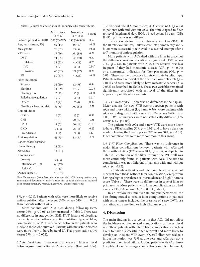
Table 1: Clinical characteristics of the subjects by cancer status.
Active cancer(𝑛 = 87)
No cancer(𝑛 = 164) 𝑝 value
Follow-up (median, IQR) 203 (31–397) 156 (34–349) 0.52Age, years (mean, SD) 62 (14) 56 (17) <0.01Male gender 28 (32) 93 (57) <0.01VTE event 87 (96) 164 (93) 0.22
DVT 74 (85) 148 (90) 0.57Bilateral 24 (32) 41 (28) 0.76IVC 3 (2) 2 (1) 0.34∗
Proximal 60 (82) 127 (87) 0.28PE 50 (57) 41 (25) <0.01
Filter indicationSurgery 34 (39) 62 (38) 0.84Bleeding 34 (39) 87 (53) 0.035Bleeding risk 17 (20) 13 (8) <0.01Failed anticoagulation 2 (2) 2 (1) 0.51∗
Othera 2 (2) 7 (4) 0.42Bleeding + bleeding risk 51 (59) 100 (61) 0.71
ComorbiditiesCOPD 6 (7) 12 (7) 0.90CHF 7 (8) 20 (12) 0.31CAD 4 (5) 30 (18) <0.01∗
CKD 9 (10) 26 (16) 0.23Liver disease 1 (1) 9 (5) 0.17∗
Anticoagulation 61 (70) 88 (54) 0.01Cancer related variablesChemotherapy 28 (32)Metastatic 44 (51)Khorana score
Low (0) 9 (10)Intermediate (1-2) 60 (69)High (≥3) 17 (20)
Ottawa score ≥1 50 (57)Note. Values are 𝑛 (%) unless otherwise specified. IQR: interquartile range;SD: standard deviation; ∗: Fisher’s exact test, a: other indications includedpoor cardiopulmonary reserve, massive PE, and thrombectomy.
9%, 𝑝 < 0.01). Patients with ACa were more likely to receiveanticoagulation after the event (70% versus 54%, 𝑝 = 0.01)than patients without ACa.
More patients with ACa died during follow-up (55%versus 26%, 𝑝 < 0.01) as demonstrated in Table 2.There wasno difference in age, gender, BMI, DVT, history of bleeding,cancer type, chemotherapy, anticoagulation, type of filter,complications, or VTE recurrence between the patients whodied and those who survived. Patients withmetastatic diseasewere more likely to have bilateral DVT at presentation (70%versus 29%, 𝑝 = 0.022).
3.2. Retrieval Rates. There was no difference in filter retrievalbetween groups in theKaplan-Meier analysis (log-rank: 0.16).
The retrieval rate at 6 months was 49% versus 64% (𝑝 = ns)in patients with and without ACa. The time elapsed to filterretrieval (median: 33 days [IQR: 14–63] versus 38 days [IQR:18–93], 𝑝 = ns) was not different.
The success rate for the first retrieval attempt was 96%. Ofthe 10 retrieval failures, 5 filters were left permanently and 5filters were successfully retrieved in a second attempt after 1to 7 months of anticoagulation.
More patients with ACa died with the filter in place butthe difference was not statistically significant (43% versus21%, 𝑝 = ns). In patients with ACa, filter retrieval was lessfrequent if they had metastatic disease (OR, 𝑝 = 0.04)or a nonsurgical indication for filter placement (OR, 𝑝 =0.02). There was no difference in retrieval rate by filter type.Patients without removal of the filter had lower platelets (𝑝 =0.011) and were more likely to have metastatic cancer (𝑝 =0.038) as described in Table 3. These two variables remainedsignificantly associated with retrieval of the filter in anexploratory multivariate analysis
3.3. VTE Recurrence. There was no difference in the Kaplan-Meier analysis for new VTE events between patients withACa and those without (log-rank: 0.56). More patients withACa were diagnosed with a new PE (5% versus 0.6%, 𝑝 =0.05); DVT recurrences were not statistically different (13%versus 17%, 𝑝 = ns).
The patients with ACa and a new VTE were more likelyto have a PE at baseline (OR, 𝑝 = 0.02) and to have a decisionmade of leaving the filter in place (69% versus 30%,𝑝 < 0.01).Filter complications were more common in this group.
3.4. IVC Filter Complications. There was no difference inmajor filter complications between patients with ACa andthose without ACa (17% versus 18%, 𝑝 = ns), as depicted inTable 2. Penetration of the filter through the IVC wall wasmore commonly found in patients with ACa. The time tocomplication was not different in patients with and withoutACa (𝑝 = 0.82).
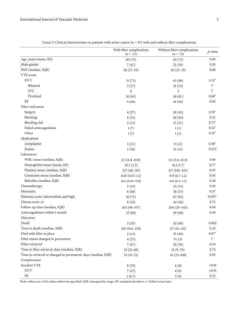
The patients with ACa and filter complications were notdifferent from those without filter complications except fromhaving a higher prevalence of intermediate and high Khoranascore (Table 4). There were no differences in type of filter orprimary site. More patients with filter complications also hada new VTE (53% versus 8%, 𝑝 < 0.01) (Table 4).
In an exploratory multivariate analysis performed, thebest-fitting model to predict filter complications in patientswith active cancer included the presence of a new DVT, useof statins, and a medium or high Khorana score.
4. Discussion
The main finding in our cohort is that ACa did not affectthe incidence of filter related complications or the retrievalrate.Those patients with filter related complications were lesslikely to have a successful filter retrieval and more likely todevelop an incident VTE event. Overall filter retrieval ratein our institution was 73% at one year and ACa was not apredictor of retrieval failure. Among patients with ACa, base-line platelet level, nonsurgical indications for filter placement,
4 International Journal of Vascular Medicine
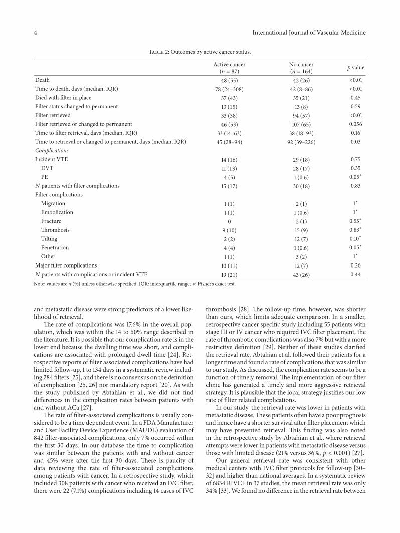
Table 2: Outcomes by active cancer status.
Active cancer(𝑛 = 87)
No cancer(𝑛 = 164) 𝑝 value
Death 48 (55) 42 (26) <0.01Time to death, days (median, IQR) 78 (24–308) 42 (8–86) <0.01Died with filter in place 37 (43) 35 (21) 0.45Filter status changed to permanent 13 (15) 13 (8) 0.59Filter retrieved 33 (38) 94 (57) <0.01Filter retrieved or changed to permanent 46 (53) 107 (65) 0.056Time to filter retrieval, days (median, IQR) 33 (14–63) 38 (18–93) 0.16Time to retrieval or changed to permanent, days (median, IQR) 45 (28–94) 92 (39–226) 0.03ComplicationsIncident VTE 14 (16) 29 (18) 0.75
DVT 11 (13) 28 (17) 0.35PE 4 (5) 1 (0.6) 0.05∗
𝑁 patients with filter complications 15 (17) 30 (18) 0.83Filter complications
Migration 1 (1) 2 (1) 1∗
Embolization 1 (1) 1 (0.6) 1∗
Fracture 0 2 (1) 0.55∗
Thrombosis 9 (10) 15 (9) 0.83∗
Tilting 2 (2) 12 (7) 0.10∗
Penetration 4 (4) 1 (0.6) 0.05∗
Other 1 (1) 3 (2) 1∗
Major filter complications 10 (11) 12 (7) 0.26𝑁 patients with complications or incident VTE 19 (21) 43 (26) 0.44Note: values are 𝑛 (%) unless otherwise specified. IQR: interquartile range; ∗: Fisher’s exact test.
and metastatic disease were strong predictors of a lower like-lihood of retrieval.
The rate of complications was 17.6% in the overall pop-ulation, which was within the 14 to 50% range described inthe literature. It is possible that our complication rate is in thelower end because the dwelling time was short, and compli-cations are associated with prolonged dwell time [24]. Ret-rospective reports of filter associated complications have hadlimited follow-up, 1 to 134 days in a systematic review includ-ing 284 filters [25], and there is no consensus on the definitionof complication [25, 26] nor mandatory report [20]. As withthe study published by Abtahian et al., we did not finddifferences in the complication rates between patients withand without ACa [27].
The rate of filter-associated complications is usually con-sidered to be a time dependent event. In a FDAManufacturerand User Facility Device Experience (MAUDE) evaluation of842 filter-associated complications, only 7% occurred withinthe first 30 days. In our database the time to complicationwas similar between the patients with and without cancerand 45% were after the first 30 days. There is paucity ofdata reviewing the rate of filter-associated complicationsamong patients with cancer. In a retrospective study, whichincluded 308 patients with cancer who received an IVC filter,there were 22 (7.1%) complications including 14 cases of IVC
thrombosis [28]. The follow-up time, however, was shorterthan ours, which limits adequate comparison. In a smaller,retrospective cancer specific study including 55 patients withstage III or IV cancer who required IVC filter placement, therate of thrombotic complications was also 7% but with amorerestrictive definition [29]. Neither of these studies clarifiedthe retrieval rate. Abtahian et al. followed their patients for alonger time and found a rate of complications that was similarto our study. As discussed, the complication rate seems to be afunction of timely removal. The implementation of our filterclinic has generated a timely and more aggressive retrievalstrategy. It is plausible that the local strategy justifies our lowrate of filter related complications.
In our study, the retrieval rate was lower in patients withmetastatic disease.These patients often have a poor prognosisand hence have a shorter survival after filter placement whichmay have prevented retrieval. This finding was also notedin the retrospective study by Abtahian et al., where retrievalattempts were lower in patients withmetastatic disease versusthose with limited disease (21% versus 36%, 𝑝 < 0.001) [27].
Our general retrieval rate was consistent with othermedical centers with IVC filter protocols for follow-up [30–32] and higher than national averages. In a systematic reviewof 6834 RIVCF in 37 studies, the mean retrieval rate was only34% [33].We found no difference in the retrieval rate between
International Journal of Vascular Medicine 5
Table 3: Clinical characteristics in patients with active cancer (𝑛 = 87) with and without filter complications.
With filter complications(𝑛 = 15)
Without filter complications(𝑛 = 72) 𝑝 value
Age, years (mean, SD) 60 (15) 62 (13) 0.65Male gender 7 (47) 21 (30) 0.19BMI (median, IQR) 30 (27–34) 30 (25–35) 0.88VTE event
DVT 11 (73) 63 (88) 0.22∗
Bilateral 3 (27) 21 (33) 1∗
IVC 0 3 1∗
Proximal 10 (91) 50 (81) 0.68∗
PE 9 (60) 41 (56) 0.82Filter indication
Surgery 4 (27) 30 (42) 0.39∗
Bleeding 8 (53) 26 (36) 0.21Bleeding risk 2 (13) 15 (21) 0.73∗
Failed anticoagulation 1 (7) 1 (1) 0.32∗
Other 1 (7) 1 (1) 0.32∗
MedicationsAntiplatelet 3 (21) 9 (12) 0.40∗
Statins 5 (36) 11 (15) 0.072Laboratory
WBC mean (median, IQR) 8.5 (6.8–10.8) 8.1 (5.6–12.4) 0.86Hemoglobin mean (mean, SD) 10.3 (2.5) 10.1 (1.7) 0.77Platelets mean (median, IQR) 237 (181–311) 217 (140–325) 0.95Creatinine mean (median, IQR) 0.83 (0.07–1.1) 0.9 (0.7–1.2) 0.85Bilirubin (median, IQR) 0.6 (0.03–0.8) 0.6 (0.4–1.1) 0.20
Chemotherapy 5 (33) 23 (31) 0.92Metastatic 6 (40) 38 (53) 0.37Khorana score, intermediate and high 10 (71) 67 (93) 0.035∗
Ottawa score ≥1 8 (53) 42 (58) 0.72Follow-up time (median, IQR) 203 (98–357) 200 (28–443) 0.84Anticoagulation within 1 month 12 (80) 49 (68) 0.36OutcomesDeath 5 (33) 43 (60) 0.062Time to death (median, IQR) 203 (164–229) 67 (24–311) 0.23Died with filter in place 2 (13) 35 (49) 0.07∗
Filter status changed to permanent 4 (27) 9 (13) 1∗
Filter retrieved 7 (47) 26 (36) 0.44Time to filter retrieval, days (median, IQR) 33 (21–60) 31 (9–70) 0.74Time to retrieval or changed to permanent, days (median, IQR) 55 (31–72) 45 (25–108) 0.93ComplicationsIncident VTE 8 (53) 6 (8) <0.01
DVT 7 (47) 4 (6) <0.01PE 1 (6.7) 3 (4) 0.52
Note: values are 𝑛 (%) unless otherwise specified. IQR: interquartile range; SD: standard deviation; ∗: Fisher’s exact test.
6 International Journal of Vascular Medicine
Table 4: Clinical characteristics in patients with active cancer (𝑛 = 87) with and without filter retrieval.
Filter retrieved(𝑛 = 33)
Filter in place(𝑛 = 54) 𝑝 value
Age years, mean (SD) 61 (12) 62 (15) 0.63Male gender 7 (21) 21 (39) 0.086BMI mean (median, IQR) 30 (25–35) 30 (26–34) 0.89VTE event
DVT 27 (81) 47 (87) 0.51Bilateral 8 (30) 16 (34) 0.67IVC 0 3 (6) 1∗
Proximal 21 (77) 39 (85) 0.38PE 20 (60) 30 (56) 0.64
Filter indicationSurgery 18 (55) 16 (30) 0.021Bleeding 12 (36) 22 (41) 0.68Bleeding risk 4 (12) 13 (24) 0.27∗
Failed anticoagulation 1 (3) 1 (2) 1∗
Other 0 2 (4) 0.52∗
MedicationsAntiplatelet 3 (9) 9 (17) 0.36∗
Statins 6 (18) 10 (19) 0.93Laboratory
WBC mean (median, IQR) 7.8 (5.6–10.4) 9.1 (5.7–13.2) 0.17Hemoglobin mean (median, SD) 10.0 (2.2) 10.1 (1.6) 0.91Platelets (median, IQR) 271 (188–418) 198 (129–279) 0.011Creatinine (median, IQR) 0.76 (0.61–1.0) 0.9 (0.7–1.2) 0.024Bilirubin (median, IQR) 0.4 (0.3–0.7) 0.7 (0.5–1.1) <0.01
Chemotherapy 7 (22) 21 (39) 0.087Metastatic 12 (36) 32 (59) 0.038Khorana score, intermediate and high 29 (88) 48 (91) 0.72∗
Ottawa score ≥1 16 (49) 34 (63) 0.18Follow-up time (median, IQR) 363 (203–537) 62 (24–305) <0.01Anticoagulation within 1 month 30 (91) 31 (57) <0.01OutcomesDeath 11 (33) 37 (69) <0.01Time to death (median, IQR) 274 (88–412) 34 (24–243) 0.14ComplicationsIncident VTE 3 (9) 11 (20) 0.23∗
DVT 3 (9) 8 (15) 0.52∗
PE 0 4 (7) 0.29∗
𝑁 patients with filter complications 7 (21) 8 (15) 0.44Filter complications
Migration 1 (3) 0 0.40∗
Embolization 1 (3) 0 0.40∗
Fracture 0 0Thrombosis 1 (3) 7 (13) 0.31∗
Tilting 1 (3) 0 0.15∗
Penetration 3 (8) 1 (2) 0.30∗
Other 1 (3) 0 0.40∗
International Journal of Vascular Medicine 7
Table 4: Continued.
Filter retrieved(𝑛 = 33)
Filter in place(𝑛 = 54) 𝑝 value
Major filter complications 3 (9) 7 (13) 0.73∗
𝑁 patients with complications or incident VTE 8 (24) 11 (20) 0.67Note: values are 𝑛 (%) unless otherwise specified. IQR: interquartile range; ∗: Fisher’s exact test.
patients with and without ACa. Abtahian et al., in their ret-rospective study comparing patients with and without ACa,found a lower retrieval rate in patients with ACa [27]. Thismay reflect local practice, a higher proportion of patients withmetastatic disease, or different indications for filter place-ment. In our study a higher proportion of filters were placedperioperatively for anticoagulation interruption, and thosepatients had a higher retrieval rate than patientswith bleedingor bleeding risk.
The most common indication for filter placement inour study was bleeding in both patients with and thosewithout cancer. In a retrospective study on 103 patients withgynecological malignancies who required an IVC filter, themost common reason for placement was contraindication toanticoagulation due to hemorrhage (44%) [34]. Indeed, thehigh likelihood of bleeding among patients with cancer whorequire anticoagulation is well recognized [14]. In a study byPrandoni et al. [12] major bleeding was twice as commonin patients with cancer compared to patients without cancer(15.7/100 versus 8.6/100 patients/year), with a hazard ratiofor major bleeding of 4.8 (95% CI: 2.3–10.1) in patients withextensive cancer. In our study, patients with thrombocytope-nia at baseline or patients with nonsurgical indications for fil-ter placement were less likely to have the filter retrieved. Thismay indicate that the patient was at risk for persistent bleed-ing and thus the filter may still be indicated. More patientswho were started on anticoagulation within the first monthafter the event had the filter retrieved. This suggests thepatient had a reduced risk of bleeding and the initial indica-tion for the filter was no longer present.
One of the strengths of our study is that all records wereavailable for review. Furthermore, most of the patients havetheir follow-up in the institution, as well as a three-monthfollow-up if the filter was still in place as part of our qualityimprovement project. As the study was retrospective, theusual practice by the different physicians was not modified.Also it is a single center study and thus may reflect localpractices. Four different filters were used and that may intro-duce heterogeneity, but there was no preference for a typeof filters by ACa status. Because this is a retrospective study,there is a risk for selection bias. We may have overdiagnosedthe number of complications including complications thatwere not clinically significant by reviewing all the imagesavailable from the time of the filter placement. Despite this,our complication rate was similar to what is reported in theliterature and not different between patients with andwithoutcancer. Because our institution is a level 1 trauma center,manytrauma patients are referred from different regions of thestate. Some of these patients were lost to follow-up or follow-up information was incomplete. As most of the indications
for IVC filters in trauma patients are short lived, usually theirfilters are removed before discharge.
5. Conclusion
In patients with ACa IVCF placement is an acceptable inter-vention, as the complications and overall retrieval rate do notdiffer significantly from the patients without cancer. Predic-tors of low retrieval rate such as metastatic disease, recurrentVTE, and anemia at baseline should be considered at the timeof filter placement to guide the judicious use of the IVCF.
Ethical Approval
The Institutional Review Board at the University of Okla-homaHealth Sciences Center approved this study andwaivedthe requirement of consent.
Conflict of Interests
The authors declare that there is no conflict of interestsregarding the publication of this paper.
References
[1] J. A. Heit, M. D. Silverstein, D. N. Mohr, T. M. Petterson, W. M.O’Fallon, and L. J. Melton III, “Risk factors for deep vein throm-bosis and pulmonary embolism: a population-based case-control study,” Archives of Internal Medicine, vol. 160, no. 6, pp.809–815, 2000.
[2] M. Nordstrom, B. Lindblad, H. Anderson, D. Bergqvist, and T.Kjellstrom, “Deep venous thrombosis and occult malignancy:an epidemiological study,” British Medical Journal, vol. 308, no.6933, pp. 891–894, 1994.
[3] P. Prandoni, A. W. A. Lensing, H. R. Buller et al., “Deep-veinthrombosis and the incidence of subsequent symptomatic can-cer,” The New England Journal of Medicine, vol. 327, no. 16, pp.1128–1133, 1992.
[4] P. Prandoni, A. Piccioli, and A. Girolami, “Cancer and venousthromboembolism: an overview,” Haematologica, vol. 84, no. 5,pp. 437–445, 1999.
[5] H. T. Sørensen, L. Mellemkjær, J. H. Olsen, and J. A. Baron,“Prognosis of cancers associated with venous thromboem-bolism,”The New England Journal of Medicine, vol. 343, no. 25,pp. 1846–1850, 2000.
[6] D. C. Stolinsky, “Trousseau’s phenomenon,” Blood, vol. 62,article 1304, 1983.
[7] A. J. Tafur, H. Kalsi, W. E. Wysokinski et al., “The associationof active cancer with venous thromboembolism location: apopulation-based study,”Mayo Clinic Proceedings, vol. 86, no. 1,pp. 25–30, 2011.
8 International Journal of Vascular Medicine
[8] J. A. Heit, “Risk factors for venous thromboembolism,” Clinicsin Chest Medicine, vol. 24, no. 1, pp. 1–12, 2003.
[9] D. L. Hoyert and J. Xu, “Deaths: preliminary data for 2011,”National Vital Statistics Reports, vol. 61, no. 6, pp. 1–52, 2012.
[10] A. A. Khorana, C. W. Francis, E. Culakova, N. M. Kuderer, andG. H. Lyman, “Thromboembolism is a leading cause of death incancer patients receiving outpatient chemotherapy,” Journal ofThrombosis and Haemostasis, vol. 5, no. 3, pp. 632–634, 2007.
[11] J. A. Heit, D. N. Mohr, M. D. Silverstein, T. M. Petterson, W.M. O’Fallon, and L. J. Melton III, “Predictors of recurrence afterdeep vein thrombosis and pulmonary embolism: a population-based cohort study,”Archives of InternalMedicine, vol. 160, no. 6,pp. 761–768, 2000.
[12] P. Prandoni, A. W. A. Lensing, A. Piccioli et al., “Recurrentvenous thromboembolism and bleeding complications duringanticoagulant treatment in patients with cancer and venousthrombosis,” Blood, vol. 100, no. 10, pp. 3484–3488, 2002.
[13] A. J. Tafur,W. E.Wysokinski, R. D.McBane et al., “Cancer effecton periprocedural thromboembolism and bleeding in antico-agulated patients,” Annals of Oncology, vol. 23, no. 8, ArticleID mds058, pp. 1998–2005, 2012.
[14] A. J. Tafur, R. McBane II, W. E. Wysokinski et al., “Predictorsof major bleeding in peri-procedural anticoagulation manage-ment,” Journal ofThrombosis and Haemostasis, vol. 10, no. 2, pp.261–267, 2012.
[15] A. Y. Lee, P. W. Kamphuisen, G. Meyer et al., “Tinzaparin vswarfarin for treatment of acute venous thromboembolism inpatients wth active cancer: a randomized clinical trial,”The Jour-nal of the American Medical Association, vol. 314, pp. 677–686,2015.
[16] C. W. Francis, C. M. Kessler, S. Z. Goldhaber et al., “Treatmentof venous thromboembolism in cancer patients with dalteparinfor up to 12 months: the DALTECAN Study,” Journal ofThrombosis and Haemostasis, vol. 13, no. 6, pp. 1028–1035, 2015.
[17] C. Kearon, E. A. Akl, and A. J. Comerota, “Antithrombotic ther-apy for VTE disease: antithrombotic therapy and preventionof thrombosis, 9th ed: American college of chest physiciansevidence-based clinical practice guidelines,” Chest, vol. 141, no.2, supplement, pp. e419S–e494S, 2012.
[18] R. F. Sing andP. E. Fischer, “Inferior vena cava filters: indicationsandmanagement,”Current Opinion in Cardiology, vol. 28, no. 6,pp. 625–631, 2013.
[19] D. Zhou, J. Spain, E. Moon, G. McLennan, M. J. Sands, and W.Wang, “Retrospective review of 120 celect inferior vena cava fil-ter retrievals: experience at a single institution,” Journal of Vas-cular and Interventional Radiology, vol. 23, no. 12, pp. 1557–1563,2012.
[20] J. M. Andreoli, R. J. Lewandowski, R. L. Vogelzang, and R.K. Ryu, “Comparison of complication rates associated withpermanent and retrievable inferior vena cava filters: a review ofthe MAUDE database,” Journal of Vascular and InterventionalRadiology, vol. 25, no. 8, pp. 1181–1185, 2014.
[21] A. Y. Y. Lee, M. N. Levine, R. I. Baker et al., “Low-molecular-weight heparin versus a coumarin for the prevention of recur-rent venous thromboembolism in patients with cancer,” TheNew England Journal of Medicine, vol. 349, no. 2, pp. 146–153,2003.
[22] A.A.Khorana,N.M.Kuderer, E. Culakova,G.H. Lyman, andC.W. Francis, “Development and validation of a predictive modelfor chemotherapy—associated thrombosis,” Blood, vol. 111, no.10, pp. 4902–4907, 2008.
[23] M. L. Louzada, M. Carrier, A. Lazo-Langner et al., “Devel-opment of a clinical prediction rule for risk stratification ofrecurrent venous thromboembolism in patients with cancer-associated venous thromboembolism,” Circulation, vol. 126, no.4, pp. 448–454, 2012.
[24] M. D. Tam, J. Spain, M. Lieber, M. Geisinger, M. J. Sands, andW. Wang, “Fracture and distant migration of the bard recoveryfilter: a retrospective review of 363 implantations for poten-tially life-threatening complications,” Journal of Vascular andInterventional Radiology, vol. 23, no. 2, pp. 199.e1–205.e1, 2012.
[25] P. D. Stein,M. Alnas, E. Skaf et al., “Outcome and complicationsof retrievable inferior vena cava filters,”TheAmerican Journal ofCardiology, vol. 94, no. 8, pp. 1090–1093, 2004.
[26] S. Sarosiek, M. Crowther, and J. M. Sloan, “Indications, com-plications, and management of inferior vena cava filters: theexperience in 952 patients at an academic hospital with a level Itrauma center,” JAMA Internal Medicine, vol. 173, no. 7, pp. 513–517, 2013.
[27] F. Abtahian, B.M. Hawkins, D. P. Ryan et al., “Inferior vena cavafilter usage, complications, and retrieval rate in cancer patients,”The American Journal of Medicine, vol. 127, no. 11, pp. 1111–1117,2014.
[28] M. J. Wallace, J. L. Jean, S. Gupta et al., “Use of inferior venacaval filters and survival in patients with malignancy,” Cancer,vol. 101, no. 8, pp. 1902–1907, 2004.
[29] C. Schunn, G. B. Schunn, G. Hobbs, L. C. Vona-Davis, andU. Waheed, “Inferior vena cava filter placement in late-stagecancer,” Vascular and Endovascular Surgery, vol. 40, no. 4, pp.287–294, 2006.
[30] S. H. Ko, B. R. Reynolds, D. H. Nicholas et al., “Institutionalprotocol improves retrievable inferior vena cava filter recoveryrate,” Surgery, vol. 146, no. 4, pp. 809–816, 2009.
[31] J. Minocha, I. Idakoji, A. Riaz et al., “Improving inferior venacava filter retrieval rates: impact of a dedicated inferior venacava filter clinic,” Journal of Vascular and Interventional Radio-logy, vol. 21, no. 12, pp. 1847–1851, 2010.
[32] F. C. Lynch, “A method for following patients with retrievableinferior vena cava filters: results and lessons learned fromthe first 1,100 patients,” Journal of Vascular and InterventionalRadiology, vol. 22, no. 11, pp. 1507–1512, 2011.
[33] L. F. Angel, V. Tapson, R. E. Galgon, M. I. Restrepo, and J. Kauf-man, “Systematic review of the use of retrievable inferior venacava filters,” Journal of Vascular and Interventional Radiology,vol. 22, no. 11, pp. 1522.e3–1530.e3, 2011.
[34] S. B. Dewdney, T. Benn, B. J. Rimel et al., “Inferior vena cavafilter placement in the gynecologic oncology patient: a 15-yearinstitutional experience,” Gynecologic Oncology, vol. 121, no. 2,pp. 344–346, 2011.

Submit your manuscripts athttp://www.hindawi.com
Stem CellsInternational
Hindawi Publishing Corporationhttp://www.hindawi.com Volume 2014
Hindawi Publishing Corporationhttp://www.hindawi.com Volume 2014
MEDIATORSINFLAMMATION
of
Hindawi Publishing Corporationhttp://www.hindawi.com Volume 2014
Behavioural Neurology
EndocrinologyInternational Journal of
Hindawi Publishing Corporationhttp://www.hindawi.com Volume 2014
Hindawi Publishing Corporationhttp://www.hindawi.com Volume 2014
Disease Markers
Hindawi Publishing Corporationhttp://www.hindawi.com Volume 2014
BioMed Research International
OncologyJournal of
Hindawi Publishing Corporationhttp://www.hindawi.com Volume 2014
Hindawi Publishing Corporationhttp://www.hindawi.com Volume 2014
Oxidative Medicine and Cellular Longevity
Hindawi Publishing Corporationhttp://www.hindawi.com Volume 2014
PPAR Research
The Scientific World JournalHindawi Publishing Corporation http://www.hindawi.com Volume 2014
Immunology ResearchHindawi Publishing Corporationhttp://www.hindawi.com Volume 2014
Journal of
ObesityJournal of
Hindawi Publishing Corporationhttp://www.hindawi.com Volume 2014
Hindawi Publishing Corporationhttp://www.hindawi.com Volume 2014
Computational and Mathematical Methods in Medicine
OphthalmologyJournal of
Hindawi Publishing Corporationhttp://www.hindawi.com Volume 2014
Diabetes ResearchJournal of
Hindawi Publishing Corporationhttp://www.hindawi.com Volume 2014
Hindawi Publishing Corporationhttp://www.hindawi.com Volume 2014
Research and TreatmentAIDS
Hindawi Publishing Corporationhttp://www.hindawi.com Volume 2014
Gastroenterology Research and Practice
Hindawi Publishing Corporationhttp://www.hindawi.com Volume 2014
Parkinson’s Disease
Evidence-Based Complementary and Alternative Medicine
Volume 2014Hindawi Publishing Corporationhttp://www.hindawi.com