retroviral envelope gene captures and syncytin exaptation ... · retroviral envelope gene captures...
TRANSCRIPT
Retroviral envelope gene captures and syncytinexaptation for placentation in marsupialsGuillaume Cornelisa,b,c, Cécile Vernocheta,b, Quentin Carradeca,b, Sylvie Souquerea,b, Baptiste Mulotd, François Catzeflise,Maria A. Nilssonf, Brandon R. Menziesg, Marilyn B. Renfreeg, Gérard Pierrona,b, Ulrich Zellerh, Odile Heidmanna,b,Anne Dupressoira,b,1, and Thierry Heidmanna,b,1,2
aUnité des Rétrovirus Endogènes et Eléments Rétroïdes des Eucaryotes Supérieurs, CNRS UMR 8122, Institut Gustave Roussy, Villejuif, F-94805, France;bUniversité Paris-Sud, Orsay, F-91405, France; cUniversité Paris Denis Diderot, Sorbonne Paris-Cité, Paris, F-75013, France; dZooparc de Beauval et BeauvalNature, Saint Aignan, F-41110, France; eLaboratoire de Paléontologie, Phylogénie et Paléobiologie, UMR 5554 CNRS, Université Montpellier II, Montpellier,F-34095, France; fLOEWE Biodiversity and Climate Research Center, Frankfurt am Main, D-60325 Germany; gDepartment of Zoology, University ofMelbourne, Melbourne, VIC 3010, Australia; and hSystematic Zoology, Humboldt University, 10099 Berlin, Germany
Edited by Stephen P. Goff, Columbia University College of Physicians and Surgeons, New York, NY, and approved December 16, 2014 (received for reviewSeptember 3, 2014)
Syncytins are genes of retroviral origin captured by eutherian mam-mals, with a role in placentation. Here we show that some marsu-pials—which are the closest living relatives to eutherian mammals,although they diverged from the latter ∼190 Mya—also possessa syncytin gene. The gene identified in the South American marsu-pial opossum and dubbed syncytin-Opo1 has all of the characteris-tic features of a bona fide syncytin gene: It is fusogenic in an ex vivocell–cell fusion assay; it is specifically expressed in the short-livedplacenta at the level of the syncytial feto–maternal interface; and itis conserved in a functional state in a series ofMonodelphis species.We further identify a nonfusogenic retroviral envelope gene thathas been conserved for >80 My of evolution among all marsupials(including the opossum and the Australian tammar wallaby), withevidence for purifying selection and conservation of a canonicalimmunosuppressive domain, but with only limited expression inthe placenta. This unusual captured gene, together with a thirdclass of envelope genes from recently endogenized retroviruses—displaying strong expression in the uterine glands where retroviralparticles can be detected—plausibly correspond to the differentevolutionary statuses of a captured retroviral envelope gene, withonly syncytin-Opo1 being the present-day bona fide syncytin activein the opossum and related species. This study would accordinglyrecapitulate the natural history of syncytin exaptation and evolu-tion in a single species, and definitely extends the presence of suchgenes to all major placental mammalian clades.
endogenous retrovirus | envelope protein | fusogenic activity |syncytiotrophoblast | marsupials
Marsupial mammals, such as kangaroos and opossums, arethe closest living relatives to placental eutherian mam-
mals (Fig. 1), from which, however, they diverged ∼190 millionyears ago (Mya) (1, 2). The latter comprise four major clades—namely, the Laurasiatheria (e.g., the ruminants and carnivor-ans), the Euarchontoglires (e.g., primates, rodents and lago-morphs), the Xenarthra, and the Afrotheria. All are viviparousanimals, which gestate their young internally with a specializedorgan of fetal origin—the placenta—allowing prolonged nu-trient and gas exchanges between the mother and fetus. Thestructure of the feto-maternal interface displays strong variations,from simply apposed fetal and maternal membranes (the epi-theliochorial placenta) to highly invasive placental tissues bathedby the maternal blood (the hemochorial placenta). In marsupials,a short-lived placenta is also formed, but the period of intimatecontact between this transient organ and the maternal endome-trium is very short—from 4 to 10 days—compared with that foreutherian mammals—up to 22 months for the elephant—with themarsupial fetus being rapidly released, in most species in an ex-ternal pouch, where it will develop via lactation (3).In eutherian mammals, previous studies have identified enve-
lope (env) genes of retroviral origin that have been independently
captured and “co-opted” by their host, most probably for a func-tion in placentation, and which have been named syncytins(reviewed in refs. 4 and 5). In simians, syncytin-1 (6–9) andsyncytin-2 (10, 11), as bona fide syncytins, entered the primategenome 25 and >40 Mya, respectively, retained their codingcapacity in all of the subsequent lineages, display placenta-spe-cific expression, and are fusogenic in ex vivo cell–cell fusionassays. Furthermore, one of them displays immunosuppressiveactivity (12). A pair of env genes from endogenous retroviruses(ERVs) was also identified in the Muroidea, named syncytin-A and-B, which share closely related functional properties, although theyhave a completely distinct origin, showing a divergent sequence anda different genomic location compared with the primate syncytins(13, 14). Via the generation of syncytin knockout mice, we haveunambiguously demonstrated that these genes are indeed essentialfor placentation, with a lack of cell–cell fusion observed in vivo atthe level of the syncytiotrophoblast interhemal layer of the mutantplacenta, resulting in impaired feto-maternal exchanges and em-bryo development and survival (15, 16). Recently, syncytin geneshave been identified in four other clades among eutherian mam-mals—namely, in lagomorphs, carnivorans, ruminants, and the
Significance
Syncytins are “captured” genes of retroviral origin, corre-sponding to the fusogenic envelope gene of endogenizedretroviruses. They are present in a series of eutherian mam-mals, including humans and mice where they play an essentialrole in placentation. Here we show that marsupials—whichdiverged from eutherian mammals ∼190 Mya but still possessa primitive, short-lived placenta (rapidly left by the embryo fordevelopment in an external pouch)—have also captured suchgenes. The present characterization of the syncytin-Opo1 genein the opossum placenta, together with the identification oftwo additional endogenous retroviral envelope gene captures,allow a recapitulation of the natural history of these unusualgenes and definitely extends their “symbiotic niche” to allclades of placental mammals.
Author contributions: G.C., O.H., A.D., and T.H. designed research; G.C., C.V., Q.C., S.S.,G.P., O.H., and A.D. performed research; B.M., F.C., M.A.N., B.R.M., M.B.R., and U.Z. con-tributed new reagents/analytic tools; G.C., C.V., Q.C., S.S., G.P., O.H., A.D., and T.H. ana-lyzed data; and G.C., A.D., and T.H. wrote the paper.
The authors declare no conflict of interest.
This article is a PNAS Direct Submission.
Data deposition: The sequences reported in this paper have been deposited in the Gen-Bank database (accession nos. KM235324–KM235359).1A.D. and T.H. contributed equally to this work.2To whom correspondence should be addressed. Email: [email protected].
This article contains supporting information online at www.pnas.org/lookup/suppl/doi:10.1073/pnas.1417000112/-/DCSupplemental.
www.pnas.org/cgi/doi/10.1073/pnas.1417000112 PNAS | Published online January 20, 2015 | E487–E496
MICRO
BIOLO
GY
PNASPL
US
primitive afrotherian tenrecs (refs. 17–20; see also ref. 21). Theyall are unrelated to the simian and murine genes, but mostprobably share with them a function in placentation.Here, we asked whether marsupials—despite their specific
short-lived placenta and their ancestral divergence from the eu-therian mammals—have also acquired syncytin genes of retroviralorigin, as the latter. By combining (i) an in silico search for can-didate genes in sequenced marsupial genomes; (ii) assays for theirin vivo transcriptional activity by RT-PCR among a large panel oftissues that were recovered from the gray short-tailed opossum(Monodelphis domestica), including the placenta; (iii) cloning ofthe candidate genes from this species; and (iv) in situ hybridization(ISH) of placenta sections with appropriate probes, we identifieda placenta-specific ERV env gene, displaying all of the charac-teristic features of a bona fide syncytin gene, that we accordinglynamed syncytin-Opo1. It is specifically expressed within syncytialstructures at the feto-maternal interface; it is fusogenic in an exvivo cell–cell fusion assay; and it is conserved in the Monodelphisgenus with evidence for purifying selection. Moreover, we identi-fied an ancestrally captured retroviral env gene, conserved withinall marsupials and displaying strong purifying selection as dem-onstrated by sequencing the orthologous copies in 26 marsupialspecies from both the Australian (e.g., wallaby) and American(e.g., opossum) lineages, thus dating its capture between 80 and190 Mya. This gene is nonfusogenic because it does not possessa transmembrane anchoring domain and thus cannot be involvedin syncytiotrophoblast formation. Although it is not expressed at asignificant level in the placenta—at least at the postimplantationstages—it possesses a canonical immunosuppressive domain (ISD)and, as such, could still be involved in placenta formation via thislatter function. Finally, a third retroviral env gene was found to beexpressed in the maternal uterine glands, belonging to recentlyacquired endogenous proviral sequences possibly responsible forthe formation of the viral-like particles that we observed by electronmicroscopy at the same sites. All together, the present study clearlyextends the range of mammals in which retroviral env gene capturesand, in some instances, bona fide syncytin exaptations have takenplace in the course of evolution, now including the marsupials inaddition to the eutherian mammals. It is consistent with syncytinsplaying a role in the emergence of placentation from primitive
egg-laying species, with the monotremes—e.g., the mammalianoviparous platypus—still existing as their rare descendants today.
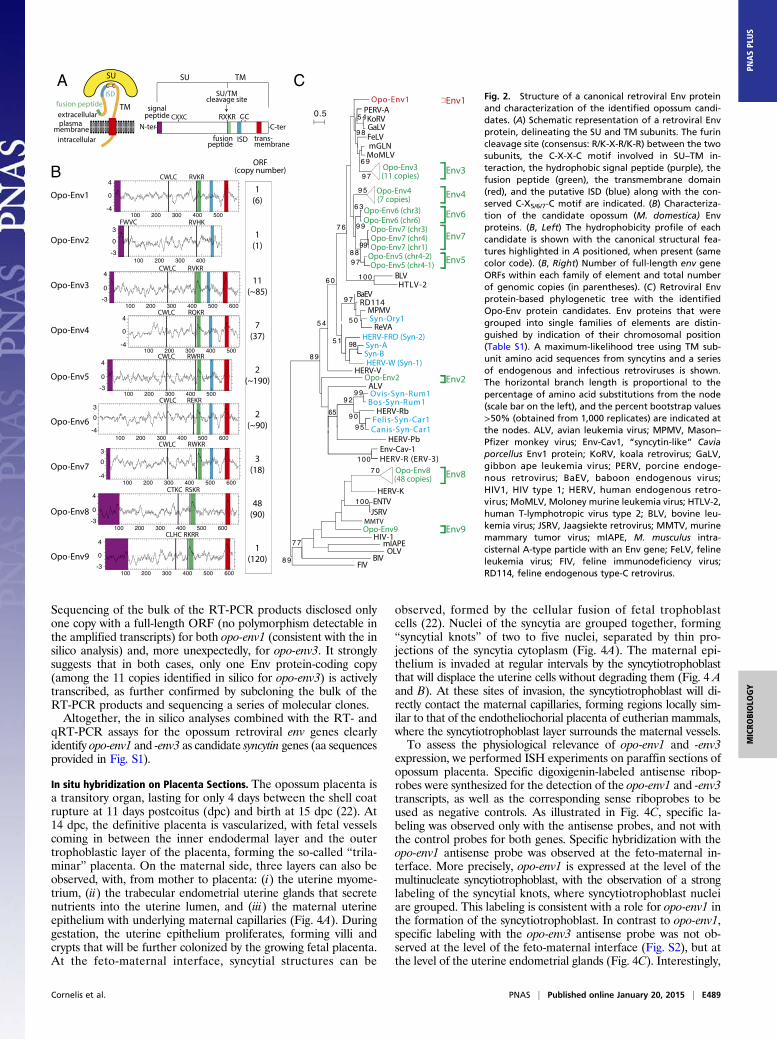
ResultsIn Silico Search for Retroviral env Genes Within the Opossum(M. domestica) Genome. To identify putative env-derived syncytingenes, we made use of the opossum genome assembly [6.8×coverage assembly of the M. domestica genome; University ofCalifornia, Santa Cruz (UCSC) Broad Institute MonDom5;October 2006]. A BLAST search for ORFs (from the Met startcodon to the stop codon) >450 aa was performed by usinga selected series of Env sequences representative of both in-fectious and ERV families, including all identified syncytins(Methods). It yielded 76 sequences, incorporated into thephylogenetic Env tree shown in Fig. 2C. Some of the sequencescan be grouped into single families, resulting finally in ninefamilies that we named Opo-Env1 to -Env9 (Fig. 2B). Analysis ofthe overall structure of the nine identified Env families (Fig. 2)strongly suggests that they indeed correspond to bona fideretroviral Env proteins, with some of their characteristic fea-tures, including a putative furin cleavage site delineating a surface(SU) and a transmembrane (TM) subunit and a CXXC motif inthe SU subunit corresponding to a binding domain between thetwo subunits. Hydrophobicity plots identify a putative hydro-phobic fusion peptide at the N terminus of the TM subunit, aswell as the hydrophobic transmembrane domain within the TMsubunits required for anchoring the Env protein within the plasmamembrane, with the exception of Opo-Env2 (Fig. 2 and Fig. S1)that contains a premature stop codon before this transmembranedomain. A putative signal peptide can be predicted at the N ter-minus of most genes, with the exception of Opo-Env4 and-Env6. Opo-Env1 to -Env7 contain a canonical ISD (12). Onlythe opo-env1, -env2, and -env9 genes are present as a single copyencoding a full-length ORF. Finally, a BLAST search disclosedthat only the opo-env1 and -env2 gene families are present at a lowcopy number (six and one, respectively), whereas the seven otherenv gene families display a much higher copy number (between 18and 190; Fig. 2B).
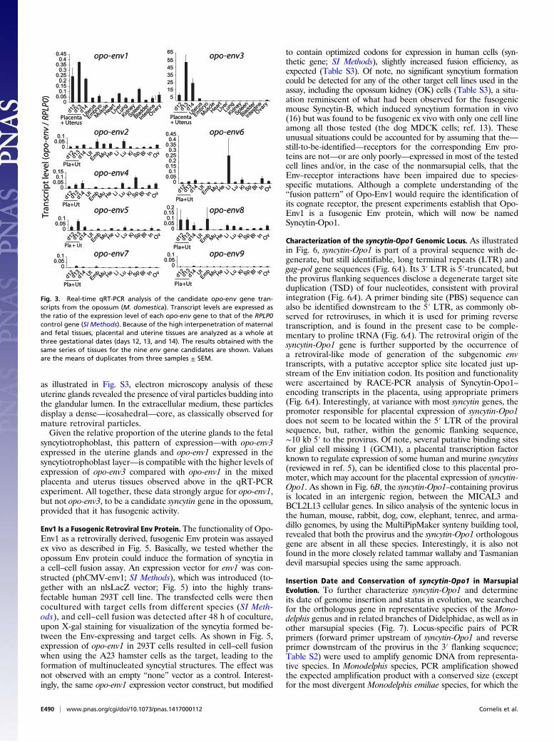
Transcription Profile Analyses for the Identification of Placenta-Specific env Genes. We then examined the transcript levels ofthe nine candidate env gene families in the opossum placentaand in a panel of other tissues. Quantitative RT-PCR (qRT-PCR) analyses were performed by using primers designed to bespecific for the ORF-containing sequences within each family ofelements (Table S2). In the opossum (M. domestica), gestationtime is 15 days, with a transitory placenta being establishedshortly before parturition, between days 12 and 15. Because ofthe interpenetration of the two tissues, the uterus and the at-tached placenta were collected as a whole, at days 12, 13, and 14.As illustrated in the qRT-PCR analysis in Fig. 3, two genes—namely, opo-env1 and -env3—are expressed at a significant levelin the placenta and uterus, as expected for candidate syncytingenes, with reduced expression in the other organs (including theuterus from nonpregnant females). Opo-env3 is the most highlyexpressed gene, up to 60-fold higher than the housekeeping ri-bosomal protein RPLP0 gene, and with >100-fold higher ex-pression at day 13 of gestation than env1 (but it is present ata >10-fold higher copy number than opo-env1; Fig. 2). The otherenv genes, with the exception of opo-env6 in the liver, showedno or only limited expression in all organs. To go further intothe analysis of the expression of both opo-env1 and -env3 in theplacenta, and more precisely to determine—especially in thecase of the multicopy opo-env3 ORF—whether the specific ex-pression observed in the placenta originates from definite and/orsingle copies, RT-PCR analyses were performed to sort out boththe Env1 and Env3 protein-encoding transcripts in the placenta,using primers designed to amplify all Env protein-coding copies.
Haplorrhini
Strepsirrhini
Rodentia
Lagomorpha
Insectivora
Carnivora
Perissodactyla
Ruminantia
Dasypodidae
Bradypodidae
Tenrecidae
Proboscidae
Jurassic Cretaceous Tertiary
50100150200 0 My
Diprotodontia
Didelphimorphia
Monotremata
I
II
III
IV
PLACENTA
Trias
MA
MM
ALS
Eutherian
mammals
Marsupials
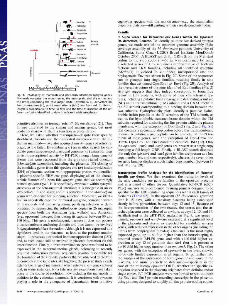
Fig. 1. Phylogeny of mammals and previously identified syncytin genes.Mammals comprise the monotremes, the marsupials, and the eutherians,the latter comprising the four major clades: Afrotheria (I), Xenarthra (II),Euarchontoglires (III), and Laurasiatheria (IV) (data from ref. 1). Branchlength is proportional to time (in My), and the time of insertion of the dif-ferent syncytins identified to date is indicated with arrowheads.
E488 | www.pnas.org/cgi/doi/10.1073/pnas.1417000112 Cornelis et al.
Sequencing of the bulk of the RT-PCR products disclosed onlyone copy with a full-length ORF (no polymorphism detectable inthe amplified transcripts) for both opo-env1 (consistent with the insilico analysis) and, more unexpectedly, for opo-env3. It stronglysuggests that in both cases, only one Env protein-coding copy(among the 11 copies identified in silico for opo-env3) is activelytranscribed, as further confirmed by subcloning the bulk of theRT-PCR products and sequencing a series of molecular clones.Altogether, the in silico analyses combined with the RT- and
qRT-PCR assays for the opossum retroviral env genes clearlyidentify opo-env1 and -env3 as candidate syncytin genes (aa sequencesprovided in Fig. S1).
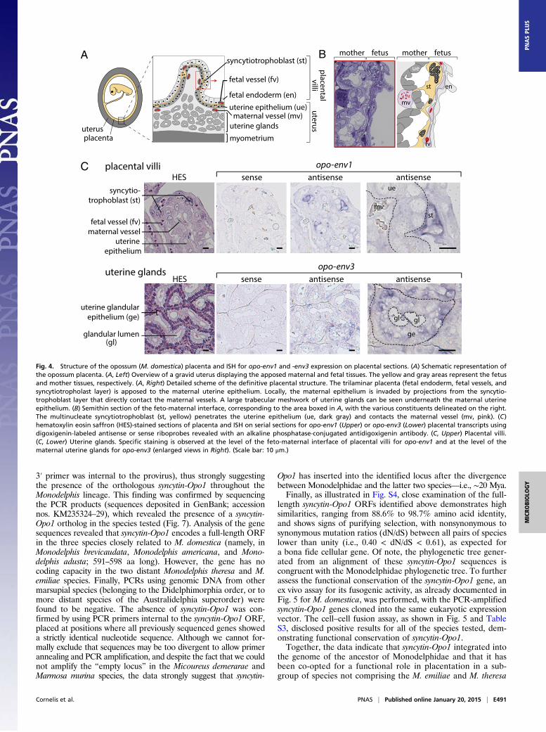
In situ hybridization on Placenta Sections. The opossum placenta isa transitory organ, lasting for only 4 days between the shell coatrupture at 11 days postcoitus (dpc) and birth at 15 dpc (22). At14 dpc, the definitive placenta is vascularized, with fetal vesselscoming in between the inner endodermal layer and the outertrophoblastic layer of the placenta, forming the so-called “trila-minar” placenta. On the maternal side, three layers can also beobserved, with, from mother to placenta: (i) the uterine myome-trium, (ii) the trabecular endometrial uterine glands that secretenutrients into the uterine lumen, and (iii) the maternal uterineepithelium with underlying maternal capillaries (Fig. 4A). Duringgestation, the uterine epithelium proliferates, forming villi andcrypts that will be further colonized by the growing fetal placenta.At the feto-maternal interface, syncytial structures can be
observed, formed by the cellular fusion of fetal trophoblastcells (22). Nuclei of the syncytia are grouped together, forming“syncytial knots” of two to five nuclei, separated by thin pro-jections of the syncytia cytoplasm (Fig. 4A). The maternal epi-thelium is invaded at regular intervals by the syncytiotrophoblastthat will displace the uterine cells without degrading them (Fig. 4 Aand B). At these sites of invasion, the syncytiotrophoblast will di-rectly contact the maternal capillaries, forming regions locally sim-ilar to that of the endotheliochorial placenta of eutherian mammals,where the syncytiotrophoblast layer surrounds the maternal vessels.To assess the physiological relevance of opo-env1 and -env3
expression, we performed ISH experiments on paraffin sections ofopossum placenta. Specific digoxigenin-labeled antisense ribop-robes were synthesized for the detection of the opo-env1 and -env3transcripts, as well as the corresponding sense riboprobes to beused as negative controls. As illustrated in Fig. 4C, specific la-beling was observed only with the antisense probes, and not withthe control probes for both genes. Specific hybridization with theopo-env1 antisense probe was observed at the feto-maternal in-terface. More precisely, opo-env1 is expressed at the level of themultinucleate syncytiotrophoblast, with the observation of a stronglabeling of the syncytial knots, where syncytiotrophoblast nucleiare grouped. This labeling is consistent with a role for opo-env1 inthe formation of the syncytiotrophoblast. In contrast to opo-env1,specific labeling with the opo-env3 antisense probe was not ob-served at the level of the feto-maternal interface (Fig. S2), but atthe level of the uterine endometrial glands (Fig. 4C). Interestingly,
Opo-Env1
ORF(copy number)
Opo-Env2
Opo-Env3
Opo-Env4
Opo-Env5
Opo-Env6
Opo-Env8
Opo-Env9
1
(6)
1
(1)
1
(120)
7
(37)
2
(~190)
2
(~90)
3
(18)
48
(90)
100 200 300 400 500
4
0
-4
RVKRCWLC
3
0
-3100 200 300 400
RVHKFWVC
CWLC
100 200 300 400 500
RQKR
CWLC
100 200 300 400 500
RWRR
100 200 300 400 500 600
REKRCWLC 3
0
-4
100 200 300 400 500 600
3
0
-4
RWKRCWLC
11
(~85)
100 200 300 400 500 600
4
0
-3
RVKRCWLC
100 200 300 400 500 600
RSKRCTKC
Opo-Env7
C
RKRRCLHC
100 200 300 400 500 600
B
SU
TM
ISD
fusion peptide
extracellular
intracellular
plasma membrane
A
CXXC RXKRsignal
peptide
fusionpeptide
trans-membrane
SU/TMcleavage site
SU TM
C-terN-ter
C C
CC
ISD
99
100
9 8
7 0
9 5
100
9 7
5 1
9 7
9 5
8 8
6 9
6 3
7 7
6 0
7 6
5 4
100
9 065
9 2
9 7
98
9 9
5 4
9 9
5 0
8 9
8 9
0.5
Opo-Env1
BLV
Syn-Ory1
BaEV
HERV-R (ERV-3)
HERV-Pb
Ovis-Syn-Rum1
MoMLV
HERV-V
Opo-Env6 (chr3)
MPMV
HERV-W (Syn-1)
FIV
Env-Cav-1
HIV-1
ReVA
Syn-B
Bos-Syn-Rum1
HERV-FRD (Syn-2)
Opo-Env7 (chr3)
RD114
mGLN
GaLVFeLV
HERV-Rb
MMTV
HERV-K
KoRV
mIAPE
JSRV
Opo-Env5 (chr4-2)
HTLV-2
Opo-Env7 (chr4)
Canis-Syn-Car1
ENTV
Felis-Syn-Car1
Opo-Env6 (chr6)
OLV
Opo-Env7 (chr1)
BIV
ALV
Opo-Env5 (chr4-1)
PERV-A
Syn-A
Opo-Env9
Opo-Env2
Opo-Env8(48 copies)
Env1
Env3
Env7
Env6
Env4
Env2
Env5
Env9
Env8
Opo-Env3(11 copies)
Opo-Env4(7 copies)
4
0
-4
4
0
-3
4
0
-3
4
0
-3
Fig. 2. Structure of a canonical retroviral Env proteinand characterization of the identified opossum candi-dates. (A) Schematic representation of a retroviral Envprotein, delineating the SU and TM subunits. The furincleavage site (consensus: R/K-X-R/K-R) between the twosubunits, the C-X-X-C motif involved in SU–TM in-teraction, the hydrophobic signal peptide (purple), thefusion peptide (green), the transmembrane domain(red), and the putative ISD (blue) along with the con-served C-X5/6/7-C motif are indicated. (B) Characteriza-tion of the candidate opossum (M. domestica) Envproteins. (B, Left) The hydrophobicity profile of eachcandidate is shown with the canonical structural fea-tures highlighted in A positioned, when present (samecolor code). (B, Right) Number of full-length env geneORFs within each family of element and total numberof genomic copies (in parentheses). (C) Retroviral Envprotein-based phylogenetic tree with the identifiedOpo-Env protein candidates. Env proteins that weregrouped into single families of elements are distin-guished by indication of their chromosomal position(Table S1). A maximum-likelihood tree using TM sub-unit amino acid sequences from syncytins and a seriesof endogenous and infectious retroviruses is shown.The horizontal branch length is proportional to thepercentage of amino acid substitutions from the node(scale bar on the left), and the percent bootstrap values>50% (obtained from 1,000 replicates) are indicated atthe nodes. ALV, avian leukemia virus; MPMV, Mason–Pfizer monkey virus; Env-Cav1, “syncytin-like” Caviaporcellus Env1 protein; KoRV, koala retrovirus; GaLV,gibbon ape leukemia virus; PERV, porcine endoge-nous retrovirus; BaEV, baboon endogenous virus;HIV1, HIV type 1; HERV, human endogenous retro-virus; MoMLV, Moloney murine leukemia virus; HTLV-2,human T-lymphotropic virus type 2; BLV, bovine leu-kemia virus; JSRV, Jaagsiekte retrovirus; MMTV, murinemammary tumor virus; mIAPE, M. musculus intra-cisternal A-type particle with an Env gene; FeLV, felineleukemia virus; FIV, feline immunodeficiency virus;RD114, feline endogenous type-C retrovirus.
Cornelis et al. PNAS | Published online January 20, 2015 | E489
MICRO
BIOLO
GY
PNASPL
US
as illustrated in Fig. S3, electron microscopy analysis of theseuterine glands revealed the presence of viral particles budding intothe glandular lumen. In the extracellular medium, these particlesdisplay a dense—icosahedral—core, as classically observed formature retroviral particles.Given the relative proportion of the uterine glands to the fetal
syncytiotrophoblast, this pattern of expression—with opo-env3expressed in the uterine glands and opo-env1 expressed in thesyncytiotrophoblast layer—is compatible with the higher levels ofexpression of opo-env3 compared with opo-env1 in the mixedplacenta and uterus tissues observed above in the qRT-PCRexperiment. All together, these data strongly argue for opo-env1,but not opo-env3, to be a candidate syncytin gene in the opossum,provided that it has fusogenic activity.
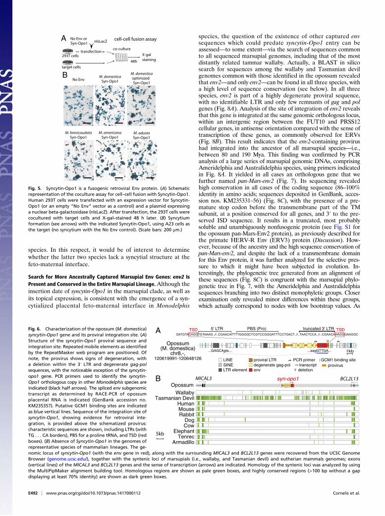
Env1 Is a Fusogenic Retroviral Env Protein.The functionality of Opo-Env1 as a retrovirally derived, fusogenic Env protein was assayedex vivo as described in Fig. 5. Basically, we tested whether theopossum Env protein could induce the formation of syncytia ina cell–cell fusion assay. An expression vector for env1 was con-structed (phCMV-env1; SI Methods), which was introduced (to-gether with an nlsLacZ vector; Fig. 5) into the highly trans-fectable human 293T cell line. The transfected cells were thencocultured with target cells from different species (SI Meth-ods), and cell–cell fusion was detected after 48 h of coculture,upon X-gal staining for visualization of the syncytia formed be-tween the Env-expressing and target cells. As shown in Fig. 5,expression of opo-env1 in 293T cells resulted in cell–cell fusionwhen using the A23 hamster cells as the target, leading to theformation of multinucleated syncytial structures. The effect wasnot observed with an empty “none” vector as a control. Interest-ingly, the same opo-env1 expression vector construct, but modified
to contain optimized codons for expression in human cells (syn-thetic gene; SI Methods), slightly increased fusion efficiency, asexpected (Table S3). Of note, no significant syncytium formationcould be detected for any of the other target cell lines used in theassay, including the opossum kidney (OK) cells (Table S3), a situ-ation reminiscent of what had been observed for the fusogenicmouse Syncytin-B, which induced syncytium formation in vivo(16) but was found to be fusogenic ex vivo with only one cell lineamong all those tested (the dog MDCK cells; ref. 13). Theseunusual situations could be accounted for by assuming that the—still-to-be-identified—receptors for the corresponding Env pro-teins are not—or are only poorly—expressed in most of the testedcell lines and/or, in the case of the nonmarsupial cells, that theEnv–receptor interactions have been impaired due to species-specific mutations. Although a complete understanding of the“fusion pattern” of Opo-Env1 would require the identification ofits cognate receptor, the present experiments establish that Opo-Env1 is a fusogenic Env protein, which will now be namedSyncytin-Opo1.
Characterization of the syncytin-Opo1 Genomic Locus. As illustratedin Fig. 6, syncytin-Opo1 is part of a proviral sequence with de-generate, but still identifiable, long terminal repeats (LTR) andgag–pol gene sequences (Fig. 6A). Its 3′ LTR is 5′-truncated, butthe provirus flanking sequences disclose a degenerate target siteduplication (TSD) of four nucleotides, consistent with proviralintegration (Fig. 6A). A primer binding site (PBS) sequence canalso be identified downstream to the 5′ LTR, as commonly ob-served for retroviruses, in which it is used for priming reversetranscription, and is found in the present case to be comple-mentary to proline tRNA (Fig. 6A). The retroviral origin of thesyncytin-Opo1 gene is further supported by the occurrence ofa retroviral-like mode of generation of the subgenomic envtranscripts, with a putative acceptor splice site located just up-stream of the Env initiation codon. Its position and functionalitywere ascertained by RACE-PCR analysis of Syncytin-Opo1–encoding transcripts in the placenta, using appropriate primers(Fig. 6A). Interestingly, at variance with most syncytin genes, thepromoter responsible for placental expression of syncytin-Opo1does not seem to be located within the 5′ LTR of the proviralsequence, but, rather, within the genomic flanking sequence,∼10 kb 5′ to the provirus. Of note, several putative binding sitesfor glial cell missing 1 (GCM1), a placental transcription factorknown to regulate expression of some human and murine syncytins(reviewed in ref. 5), can be identified close to this placental pro-moter, which may account for the placental expression of syncytin-Opo1. As shown in Fig. 6B, the syncytin-Opo1–containing provirusis located in an intergenic region, between the MICAL3 andBCL2L13 cellular genes. In silico analysis of the syntenic locus inthe human, mouse, rabbit, dog, cow, elephant, tenrec, and arma-dillo genomes, by using the MultiPipMaker synteny building tool,revealed that both the provirus and the syncytin-Opo1 orthologousgene are absent in all these species. Interestingly, it is also notfound in the more closely related tammar wallaby and Tasmaniandevil marsupial species using the same approach.
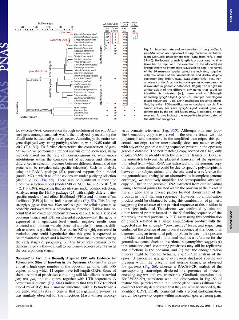
Insertion Date and Conservation of syncytin-Opo1 in MarsupialEvolution. To further characterize syncytin-Opo1 and determineits date of genome insertion and status in evolution, we searchedfor the orthologous gene in representative species of the Mono-delphis genus and in related branches of Didelphidae, as well as inother marsupial species (Fig. 7). Locus-specific pairs of PCRprimers (forward primer upstream of syncytin-Opo1 and reverseprimer downstream of the provirus in the 3′ flanking sequence;Table S2) were used to amplify genomic DNA from representa-tive species. In Monodelphis species, PCR amplification showedthe expected amplification product with a conserved size (exceptfor the most divergent Monodelphis emiliae species, for which the
Tra
nsc
rip
t le
vel (
Tra
nsc
rip
t le
vel (
opo-
env
opo-
env
/ / RP
LP0
RPLP
0))0.45 0.45
0.4 0.4
0.05 0.05 0.1 0.1
0.15 0.15 0.2 0.2
0.25 0.25 0.3 0.3
0.35 0.35
0 0
d12
d12d1
3d1
3d1
4d1
4Uter
us
Uterus
Embryo
Embryo
Muscle
MuscleHea
rtHea
rtLiv
erLiv
erLu
ngLu
ng
Kidney
Kidney
Spleen
Spleen
Bladde
r
Bladde
r
Intes
tine
Intes
tine
Ovary
Ovary
PlacentaPlacenta+ Uterus+ Uterus
55151525253535454555556565
opo-env2opo-env2
opo-env1opo-env1 opo-env3opo-env3
opo-env4opo-env4
opo-env5opo-env5
0.05 0.05 0.1 0.1
0 0
0.05 0.05 0.1 0.1
0.15 0.15
0 0
0.05 0.05 0.1 0.1
0 0
opo-env6opo-env60.45 0.45 0.4 0.4
0.05 0.05 0.1 0.1
0.15 0.15 0.2 0.2
0.25 0.25 0.3 0.3
0.35 0.35
0 0
d12
d12d1
3d1
3d1
4d1
4 UtUtEmbEmb MuMu HeHe LiLi LuLu KiKi
SpSp BlBl InIn OvOv
d12
d12d1
3d1
3d1
4d1
4Uter
us
Uterus
Embryo
Embryo
Muscle
MuscleHea
rtHea
rtLiv
erLiv
erLu
ngLu
ng
Kidney
Kidney
Spleen
Spleen
Bladde
r
Bladde
r
InInttes
tine
estin
eOva
ryOva
ry
opo-env8opo-env80.05 0.05
0.1 0.1 0.15 0.15
0.2 0.2
0 0
d12
d12d1
3d1
3d1
4d1
4 UtUtEmbEmb MuMu HeHe LiLi LuLu KiKi
SpSp BlBl InIn OvOv
opo-env9opo-env90.05 0.05
0.1 0.1
0 0
d12
d12d1
3d1
3d1
4d1
4 UtUtEmbEmb MuMu HeHe LiLi LuLu KiKi
SpSp BlBl InIn OvOv
opo-env7opo-env70.05 0.05
0.1 0.1
0 0
d12
d12d1
3d1
3d1
4d1
4 UtUtEmbEmb MuMu HeHe LiLi LuLu KiKi
SpSp BlBl InIn OvOv
d12
d12d1
3d1
3d1
4d1
4 UtUtEmbEmb MuMu HeHe LiLi LuLu KiKi
SpSp BlBl InIn OvOv
d12
d12d1
3d1
3d1
4d1
4 UtUtEmbEmb MuMu HeHe LiLi LuLu KiKi
SpSp BlBl InIn OvOv
Pla + UtPla + Ut
d12
d12d1
3d1
3d1
4d1
4 UtUtEmbEmb MuMu HeHe LiLi LuLu KiKi
SpSp BlBl InIn OvOv
PlacentaPlacenta+ Uterus+ Uterus
Pla+UtPla+Ut
Pla+UtPla+UtPla+UtPla+Ut
Pla+UtPla+Ut
Pla+UtPla+Ut Pla+UtPla+Ut
Fig. 3. Real-time qRT-PCR analysis of the candidate opo-env gene tran-scripts from the opossum (M. domestica). Transcript levels are expressed asthe ratio of the expression level of each opo-env gene to that of the RPLP0control gene (SI Methods). Because of the high interpenetration of maternaland fetal tissues, placental and uterine tissues are analyzed as a whole atthree gestational dates (days 12, 13, and 14). The results obtained with thesame series of tissues for the nine env gene candidates are shown. Valuesare the means of duplicates from three samples ± SEM.
E490 | www.pnas.org/cgi/doi/10.1073/pnas.1417000112 Cornelis et al.
3′ primer was internal to the provirus), thus strongly suggestingthe presence of the orthologous syncytin-Opo1 throughout theMonodelphis lineage. This finding was confirmed by sequencingthe PCR products (sequences deposited in GenBank; accessionnos. KM235324–29), which revealed the presence of a syncytin-Opo1 ortholog in the species tested (Fig. 7). Analysis of the genesequences revealed that syncytin-Opo1 encodes a full-length ORFin the three species closely related to M. domestica (namely, inMonodelphis brevicaudata, Monodelphis americana, and Mono-delphis adusta; 591–598 aa long). However, the gene has nocoding capacity in the two distant Monodelphis theresa and M.emiliae species. Finally, PCRs using genomic DNA from othermarsupial species (belonging to the Didelphimorphia order, or tomore distant species of the Australidelphia superorder) werefound to be negative. The absence of syncytin-Opo1 was con-firmed by using PCR primers internal to the syncytin-Opo1 ORF,placed at positions where all previously sequenced genes showeda strictly identical nucleotide sequence. Although we cannot for-mally exclude that sequences may be too divergent to allow primerannealing and PCR amplification, and despite the fact that we couldnot amplify the “empty locus” in the Micoureus demerarae andMarmosa murina species, the data strongly suggest that syncytin-
Opo1 has inserted into the identified locus after the divergencebetween Monodelphidae and the latter two species—i.e., ∼20 Mya.Finally, as illustrated in Fig. S4, close examination of the full-
length syncytin-Opo1 ORFs identified above demonstrates highsimilarities, ranging from 88.6% to 98.7% amino acid identity,and shows signs of purifying selection, with nonsynonymous tosynonymous mutation ratios (dN/dS) between all pairs of specieslower than unity (i.e., 0.40 < dN/dS < 0.61), as expected fora bona fide cellular gene. Of note, the phylogenetic tree gener-ated from an alignment of these syncytin-Opo1 sequences iscongruent with the Monodelphidae phylogenetic tree. To furtherassess the functional conservation of the syncytin-Opo1 gene, anex vivo assay for its fusogenic activity, as already documented inFig. 5 for M. domestica, was performed, with the PCR-amplifiedsyncytin-Opo1 genes cloned into the same eukaryotic expressionvector. The cell–cell fusion assay, as shown in Fig. 5 and TableS3, disclosed positive results for all of the species tested, dem-onstrating functional conservation of syncytin-Opo1.Together, the data indicate that syncytin-Opo1 integrated into
the genome of the ancestor of Monodelphidae and that it hasbeen co-opted for a functional role in placentation in a sub-group of species not comprising the M. emiliae and M. theresa
HES antisensesense
opo-env1
syncytio-
trophoblast (st)
fetal vessel (fv)
maternal vesseluterine
epithelium
uterine glandular
epithelium (ge)
uterine epithelium (ue)
fetal endoderm (en)
fetal vessel (fv)
syncytiotrophoblast (st)
maternal vessel (mv)
uterine glands
myometrium
pla
cen
tal
villi
ute
rus
A
Cantisense
uterusplacenta
B mother fetus
enst
fv
mv
mother fetus
ue
mvst
HES sense
opo-env3antisenseantisense
ue
uterine glands
placental villi
glandular lumen(gl)
ge
glgl
Fig. 4. Structure of the opossum (M. domestica) placenta and ISH for opo-env1 and -env3 expression on placental sections. (A) Schematic representation ofthe opossum placenta. (A, Left) Overview of a gravid uterus displaying the apposed maternal and fetal tissues. The yellow and gray areas represent the fetusand mother tissues, respectively. (A, Right) Detailed scheme of the definitive placental structure. The trilaminar placenta (fetal endoderm, fetal vessels, andsyncytiotropholast layer) is apposed to the maternal uterine epithelium. Locally, the maternal epithelium is invaded by projections from the syncytio-trophoblast layer that directly contact the maternal vessels. A large trabecular meshwork of uterine glands can be seen underneath the maternal uterineepithelium. (B) Semithin section of the feto-maternal interface, corresponding to the area boxed in A, with the various constituents delineated on the right.The multinucleate syncytiotrophoblast (st, yellow) penetrates the uterine epithelium (ue, dark gray) and contacts the maternal vessel (mv, pink). (C)hematoxylin eosin saffron (HES)-stained sections of placenta and ISH on serial sections for opo-env1 (Upper) or opo-env3 (Lower) placental transcripts usingdigoxigenin-labeled antisense or sense riboprobes revealed with an alkaline phosphatase-conjugated antidigoxigenin antibody. (C, Upper) Placental villi.(C, Lower) Uterine glands. Specific staining is observed at the level of the feto-maternal interface of placental villi for opo-env1 and at the level of thematernal uterine glands for opo-env3 (enlarged views in Right). (Scale bar: 10 μm.)
Cornelis et al. PNAS | Published online January 20, 2015 | E491
MICRO
BIOLO
GY
PNASPL
US
species. In this respect, it would be of interest to determinewhether the latter two species lack a syncytial structure at thefeto-maternal interface.
Search for More Ancestrally Captured Marsupial Env Genes: env2 IsPresent and Conserved in the Entire Marsupial Lineage.Although theinsertion date of syncytin-Opo1 in the marsupial clade, as well asits topical expression, is consistent with the emergence of a syn-cytialized placental feto-maternal interface in Monodelphis
species, the question of the existence of other captured envsequences which could predate syncytin-Opo1 entry can beassessed—to some extent—via the search of sequences commonto all sequenced marsupial genomes, including that of the mostdistantly related tammar wallaby. Actually, a BLAST in silicosearch for sequences among the wallaby and Tasmanian devilgenomes common with those identified in the opossum revealedthat env2—and only env2—can be found in all three species, witha high level of sequence conservation (see below). In all threespecies, env2 is part of a highly degenerate proviral sequence,with no identifiable LTR and only few remnants of gag and polgenes (Fig. 8A). Analysis of the site of integration of env2 revealsthat this gene is integrated at the same genomic orthologous locus,within an intergenic region between the FUT10 and PRSS12cellular genes, in antisense orientation compared with the sense oftranscription of these genes, as commonly observed for ERVs(Fig. 8B). This result indicates that the env2-containing provirushad integrated into the ancestor of all marsupial species—i.e.,between 80 and 190 Mya. This finding was confirmed by PCRanalysis of a large series of marsupial genomic DNAs, comprisingAmeridelphia and Australidelphia species, using primers indicatedin Fig. 8A. It yielded in all cases an orthologous gene that wefurther named pan-Mars-env2 (Fig. 7). Its sequencing revealedhigh conservation in all cases of the coding sequence (86–100%identity in amino acids; sequences deposited in GenBank, acces-sion nos. KM235331–56) (Fig. 8C), with the presence of a pre-mature stop codon before the transmembrane part of the TMsubunit, at a position conserved for all genes, and 3′ to the pre-served ISD sequence. It results in a truncated, most probablysoluble and unambiguously nonfusogenic protein (see Fig. S1 forthe opossum pan-Mars-Env2 protein), as previously described forthe primate HERV-R Env (ERV3) protein (Discussion). How-ever, because of the ancestry and the high sequence conservation ofpan-Mars-env2, and despite the lack of a transmembrane domainfor this Env protein, it was further analyzed for the selective pres-sure to which it might have been subjected in evolution. In-terestingly, the phylogenetic tree generated from an alignment ofthese sequences (Fig. 8C) is congruent with the marsupial phylo-genetic tree in Fig. 7, with the Ameridelphia and Australidelphiasequences branching into two distinct monophyletic groups. Closerexamination only revealed minor differences within these groups,which actually correspond to nodes with low bootstrap values. As
No EnvM. domestica
Syn-Opo1
M. domesticaoptimized Syn-Opo1
M. brevicaudataSyn-Opo1
293T cells
co-culture
target cells
48h
nlsLacZ
X-gal
staining
No Env or Syn-Opo1
transfection
cell-cell fusion assayA
B
M. adustaSyn-Opo1
M. americanaSyn-Opo1
Fig. 5. Syncytin-Opo1 is a fusogenic retroviral Env protein. (A) Schematicrepresentation of the coculture assay for cell–cell fusion with Syncytin-Opo1.Human 293T cells were transfected with an expression vector for Syncytin-Opo1 (or an empty “No Env” vector as a control) and a plasmid expressinga nuclear beta-galactosidase (nlsLacZ). After transfection, the 293T cells werecocultured with target cells and X-gal–stained 48 h later. (B) Syncytiumformation (see arrows) with the indicated Syncytin-Opo1, using A23 cells asthe target (no syncytium with the No Env control). (Scale bars: 200 μm.)
Opossum(M. domestica)
chr8,-,120619991-120648126
A
MICAL3Opossum
5kb
B BCL2L13
WallabyTasmanian Devil
HumanMouseRabbit
DogCow
ElephantTenrec
Armadillo
LINE
LTR elementSINE
proviral LTR
envdegenerate gag-pol
PCR primer transcriptdeletion
GCM1 binding siteprovirus
syn-opo1
...AGTTGgaa......atgACGAG... 1kb
GATGTACAGGTGTAAAG..//..CGAACATTTTGGGGCTCGTCCGGGATTTCCTGACT..//..TAACTCCA..//..CGAACAAAGGGAAGGCTSDTSD truncated 3’ LTR5’ LTR PBS (Pro)Fig. 6. Characterization of the opossum (M. domestica)
syncytin-Opo1 gene and its proviral integration site. (A)Structure of the syncytin-Opo1 proviral sequence andintegration site. Repeated mobile elements as identifiedby the RepeatMasker web program are positioned. Ofnote, the provirus shows signs of degeneration, witha deletion within the 3′ LTR and degenerate gag-polsequences, with the noticeable exception of the syncytin-opo1 gene. PCR primers used to identify the syncytin-Opo1 orthologous copy in other Monodelphis species areindicated (black half arrows). The spliced env subgenomictranscript as determined by RACE-PCR of opossumplacental RNA is indicated (GenBank accession no.KM235357). Putative GCM1 binding sites are indicatedas blue vertical lines. Sequence of the integration site ofsyncytin-Opo1, showing evidence for retroviral inte-gration, is provided above the schematized provirus:characteristic sequences are shown, including LTRs (withTG . . . CA borders), PBS for a proline tRNA, and TSD (redboxes). (B) Absence of Syncytin-Opo1 in the genomes ofrepresentative species of mammalian lineages. The ge-nomic locus of syncytin-Opo1 (with the env gene in red), along with the surrounding MICAL3 and BCL2L13 genes were recovered from the UCSC GenomeBrowser (genome.ucsc.edu/), together with the syntenic loci of marsupials (i.e., wallaby, and Tasmanian devil) and eutherian mammals genomes; exons(vertical lines) of the MICAL3 and BCL2L13 genes and the sense of transcription (arrows) are indicated. Homology of the syntenic loci was analyzed by usingthe MultiPipMaker alignment building tool. Homologous regions are shown as pale green boxes, and highly conserved regions (>100 bp without a gapdisplaying at least 70% identity) are shown as dark green boxes.
E492 | www.pnas.org/cgi/doi/10.1073/pnas.1417000112 Cornelis et al.
for syncytin-Opo1, conservation through evolution of the pan-Mars-env2 gene among marsupials was further analyzed by measuring thedN/dS ratio between all pairs of species. Accordingly, the entire envgene displayed very strong purifying selection, with dN/dS ratios all<0.2 (Fig. 8C). To further characterize the conservation of pan-Mars-env2, we performed a refined analysis of the sequences, usingmethods based on the rate of nonsynonymous vs. synonymoussubstitutions within the complete set of sequences and allowingdifferences in selection pressure between different domains of theproteins to be revealed (site-specific selection). Such an analysis,using the PAML package (23), provided support for a model(model M7) in which all of the codons are under purifying selection(dN/dS < 0.7) (Fig. S5). There was no significant support fora positive selection model (model M8 vs. M7: Chi2 = 2.8 × 10⁻⁴, df= 2, P > 0.99), suggesting that no sites are under positive selection.Analyses using the HyPhy package (24) with slightly different site-specific models [fixed effect likelihood (FEL) and random effectlikelihood (REL)] led to similar conclusions (Fig. S5). This findingstrongly suggests that pan-Mars-env2 is a genuine cellular gene mostprobably endowed with a physiological function. Taking into ac-count that we could not demonstrate—by qRT-PCR on a series ofopossum tissues and ISH on placental sections—that the gene isexpressed at a significant level (similar negative results wereobtained with tammar wallaby placental samples), it remains diffi-cult to assess its possible role. Because its ISD is highly conserved inevolution, one could hypothesize that this gene is expressed atpreimplantation stages and is involved in maternal tolerance duringthe early stages of pregnancy, but this hypothesis remains to bedemonstrated via the—difficult to perform—recovery of embryos atthe corresponding stages.
Opo-env3 Is Part of a Recently Acquired ERV with Evidence forPolymorphic Sites of Insertion in the Opossum. Opo-env3 is pres-ent at a high copy number in the opossum genome, with >85copies, among which 11 copies have full-length ORFs. Some ofthem are part of proviruses containing still identifiable retroviralgag, pro, pol, and env genes, together with LTR sequences. Aconsensus sequence (Fig. S6A) indicates that this ERV (dubbedOpo-Env3-ERV) has a mosaic structure, with a betaretroviruspol gene, whereas its env gene is that of a gammaretrovirus, aswas similarly observed for the infectious Mason–Pfizer monkey
virus primate retrovirus (Fig. S6B). Although only one Opo-Env3–encoding copy is expressed in the uterine tissue, with nopolymorphisms detectable in the amplified transcripts, this pla-cental transcript, rather unexpectedly, does not match exactlywith any of the genomic coding sequences present in the opossumgenomic database. The best matching copy, located on Chr2, onlydisplay 99.6% of identity with the placental transcript. Becausethe mismatch between the placental transcript of the opossumindividual from which RNA was extracted and the genomic copyof the opossum database could be due to specific polymorphismsbetween our subject animal and the one used as a reference forthe genomic sequencing (as an alternative to incomplete genomecoverage), we tentatively amplified the corresponding opo-env3copy on Chr2 in the genomic DNA extracted from our individual(using a forward primer located within the provirus at the 5′ end ofthe env gene and a reverse primer located downstream of theprovirus in the 3′ flanking sequence). However, no amplificationproduct could be obtained by using this combination of primers,suggesting the absence of the proviral sequence at this position inour subject individual. This absence was confirmed by using an-other forward primer located in the 5′ flanking sequence of theputatively inserted provirus. A PCR assay using this combinationof primers resulted in a single amplification product with theexpected size for an empty “provirus-free” locus, and sequencingconfirmed the absence of any proviral sequence at this locus, thusdemonstrating an insertional polymorphism between the opossumindividual used here and the animal used as a reference for thegenomic sequence. Such an insertional polymorphism suggests (i)that some opo-env3–containing proviruses may still be replicativeand infectious in the opossum, and (ii) that the endogenizationprocess might be recent. Actually, a qRT-PCR analysis of theopo-env3 associated gag gene expression displayed specific ex-pression within the placenta and uterine tissues, as observedfor opo-env3 (Fig. S6), whereas a RACE-PCR analysis of thecorresponding transcripts disclosed the presence of protein-encoding gag-pro and env transcripts (GenBank accession nos.KM235358–59), consistent with the observation in Fig. S3 ofmature viral particles within the uterine gland tissues (although wecould not formally demonstrate that they are actually encoded by theidentified ERV). Finally, consistent with a recent endogenization,search for opo-env3 copies within marsupial species, using pairs
Monodelphis domestica
Sarcophilus harrisii
Macropus eugenii
Million years
Syncytin-Opo1
Opo-Env3
pan-Mars-Env2
050100150200Syncytin-Opo1
pan-Mars-Env2
Opo-Env3
*
[nc][nc]
493
*
*
Da
s.D
ide
lph
imo
rph
iaP
er.
Dip
roto
do
ntia
ORF(aa)
+/-
ORF(aa)
ORF(aa)
MA
MM
ALI
A
Am
eri
de
lph
iaA
ust
ralid
elp
hiaMARSUPIALS
EUTHERIAN MAMMALS
MONOTREMATA
Monodelphis brevicaudataMonodelphis americanaMonodelphis adustaMonodelphis theresaMonodelphis emiliaeMicoureus demeraraeMarmosa murinaDidelphis marsupialisDidelphis virginianaPhilander opossumMetachirus nudicaudatusMarmosops parvidensHyladelphys kalinowskiiCaluromys philanderSminthopsis crassicaudata
Isoodon macrourusPerameles gunniiPseudocheirus peregrinus
Potorous tridactylusDendrolagus matschiei
Phascolarctos cinereus
Macropus rufus
Ornithorhynchus anatinus
Homo sapiensMus musculusCanis familiarisBos taurus
493
493493493493
493492493493
493493493493
493493493
493493493493
493493493493
493
597
591598
595622
fusionactivity
++++nrnrnrnr
nrnrnr
nr
nr
nr
nr
Fig. 7. Insertion date and conservation of syncytin-Opo1,pan-Mars-env2, and opo-env3 during marsupial evolution.(Left) Marsupial phylogenetic tree (data from refs. 1 and37–40). Horizontal branch length is proportional to time(scale bar on top), with the exception of the Monodelphislineage where no information is available to date. The namesof the 26 marsupial species tested are indicated, togetherwith the names of the Ameridelphia and Australidelphiacorresponding orders (Das., Dasyuromorphia; Per., Per-amelemorphia). Asterisks indicate species whose genomeis available in genomic databases. (Right) The length (inamino acids) of the different env genes that could beidentified is indicated. [nc], presence of a full-lengthnoncoding syncytin-Opo1 gene; +/−, multiple homologousmixed sequences; −, no env homologous sequence identi-fied, by either PCR-amplification or database search. Thefusion activity for each syncytin-Opo1 cloned gene, asdetermined by the cell–cell fusion assay, is indicated. nr, notrelevant. Arrows indicate the respective insertion dates ofthe different env genes.
Cornelis et al. PNAS | Published online January 20, 2015 | E493
MICRO
BIOLO
GY
PNASPL
US

of PCR primers internal to the provirus env gene, demonstratedthe presence of env3 genes only within the two most closelyrelated M. domestica and M. brevicaudata species (Fig. 7).
DiscussionHere we have identified syncytin-Opo1, the env gene from anERV that has integrated into the genome of an opossum an-cestor, ∼20 Mya, and has been maintained as a functional envgene in a definite group of Monodelphis species. This gene dis-plays all of the canonical characteristics of a syncytin gene: (i) itexhibits fusogenic activity, in an ex vivo cell–cell fusion assay; (ii)it has been subject to purifying selection in the course of evo-lution, displaying low rates of nonsynonymous to synonymoussubstitutions and conservation of its fusogenic property; and(iii) it is specifically expressed in the placenta, as determined byboth RT-PCR analyses and ISH of placental tissue sections. ISHexperiments using syncytin-Opo1 sequences as a probe clearlyshow that expression takes place at the level of the syncytial layerof the feto-maternal interface, consistent with a direct role ofthis fusogenic syncytin gene in syncytiotrophoblast formation.Clearly, syncytin-Opo1 adds to the syncytin genes previouslyidentified in the eutherian mammals. In the case of the murinesyncytin-A and -B genes, knockout mice unambiguously demon-strated that they are absolutely required for placentation, withevidence for a defect in syncytiotrophoblast formation, resultingin decreased feto-maternal exchange and impaired embryo sur-vival (15, 16). It can, therefore, be proposed that all of theidentified syncytins, including the newly discovered syncytin-Opo1, are likely to play a similar role in placentation by beinginvolved in syncytiotrophoblast formation.An important outcome of the present investigation is that the
discovery of syncytin-Opo1 extends the presence of syncytins
outside the eutherian mammals, to now include the marsupials,at least the American lineage. These species diverged from eu-therian mammals ∼190 Mya and display a very specific mode ofreproduction, with an only short-lived placenta, with the fetusbeing rapidly released in most cases in an external pouch, whereit will develop via lactation. In the presently analyzed species,implantation lasts only from day 12 to 14, still with evidence forsyncytium formation (22), and syncytin-Opo1 was precisely found tobe expressed at these stages and at the location expected for a rolein syncytium formation. Altogether the present data—combinedwith the fact that syncytin-Opo1 is distinct from all previouslyidentified syncytins—indicate that syncytin capture has been a wide-spread process, which finally turns out to have taken place in severalwidely separate lineages during the mammalian radiation. It couldthus be hypothesized that the remarkable variability in placentalstructures observed among mammals might in part result fromthe diversity of the syncytin genes that have been stochasticallycaptured in the course of mammalian evolution (5).However, an important question remains to be answered, if
one takes into account the relatively recent occurrence of syn-cytin-Opo1 capture, in comparison with the time of emergence ofplacental mammals (including marsupials) from egg-laying ani-mals, ∼200 Mya. We had previously proposed (reviewed in refs.4 and 5) that this evolutionary transition was most probablyfavored by the primitive capture of an ancestral “founding”retroviral env gene that allowed the retention of the growingembryo within the mother, despite her immune system, mostprobably thanks to the immunosuppressive activity of retroviralEnv proteins, allowing establishment of a primitive feto-maternaltolerance. In this scenario, it was further proposed that sub-sequent env gene exaptation could naturally take place, becauseERV capture is an ongoing process, thus resulting in “new”
A
C
0.01
Monodelphis domesticaMonodelphis brevicaudata
Monodelphis americana
Monodelphis adustaMonodelphis theresaMonodelphis emiliae
Micoureus demeraraeMarmosa murina
Didelphis marsupialisDidelphis virginianaPhilander opossumMetachirus nudicaudatus
Marmosops parvidensHyladelphys kalinowskii
Caluromys philander
Sminthopsis crassicaudata
Isoodon macrourusPerameles gunnii
Sarcophilus harrisii
P. peregrinusPotorous tridactylus
Dendrolagus matschiei
Phascolarctos cinereus
Macropus rufusMacropus eugenii100
94100
94
100
95
100
54
100
55100
89
517172
100
96
94
94
58
55
31
100
100
100 0.07 0.09 0 0.04 0.07 0.08 0.06 0.04 0.03 0.02 0.08 0.08 0.09 0.04 0.11 0.11 0.16 0.15 0.15 0.15 0.11 0.15 0.14 0.13 0.1399.8 100 0.1 0.02 0.06 0.08 0.09 0.07 0.05 0.04 0.03 0.08 0.09 0.09 0.05 0.11 0.12 0.17 0.16 0.16 0.16 0.11 0.15 0.13 0.13 0.1399.4 99.2 100 0.06 0.09 0.12 0.11 0.09 0.07 0.06 0.05 0.1 0.11 0.1 0.07 0.12 0.12 0.17 0.16 0.16 0.15 0.12 0.15 0.14 0.13 0.13100 99.8 99.4 100 0.03 0.05 0.07 0.05 0.04 0.04 0.02 0.06 0.08 0.08 0.04 0.11 0.12 0.17 0.16 0.16 0.14 0.11 0.15 0.14 0.13 0.1399.6 99.4 99 99.6 100 0.09 0.09 0.07 0.05 0.04 0.03 0.08 0.09 0.09 0.04 0.11 0.12 0.17 0.16 0.16 0.16 0.12 0.15 0.14 0.13 0.1399.4 99.2 98.8 99.4 99 100 0.13 0.09 0.07 0.05 0.04 0.09 0.09 0.12 0.07 0.12 0.12 0.17 0.16 0.16 0.16 0.12 0.16 0.15 0.14 0.1498.2 98 97.6 98.2 97.8 97.8 100 0.1 0.1 0.07 0.05 0.09 0.12 0.13 0.09 0.12 0.13 0.16 0.15 0.15 0.17 0.13 0.16 0.15 0.14 0.1498.8 98.6 98.2 98.8 98.4 98.4 98.6 100 0.06 0.05 0.05 0.06 0.09 0.09 0.07 0.11 0.12 0.16 0.15 0.15 0.15 0.12 0.14 0.13 0.13 0.1298.8 98.6 98.2 98.8 98.4 98.2 97.8 98.2 100 0.13 0.2 0.08 0.12 0.09 0.08 0.11 0.14 0.17 0.15 0.15 0.16 0.12 0.14 0.13 0.13 0.1299 98.8 98.4 99 98.6 98.4 97.8 98.2 99.4 100 0.13 0.07 0.11 0.08 0.07 0.11 0.14 0.17 0.15 0.15 0.15 0.11 0.13 0.13 0.13 0.12
99.4 99.2 98.8 99.4 99 98.8 98.4 98.6 99.2 99.4 100 0.08 0.09 0.09 0.07 0.11 0.13 0.17 0.15 0.15 0.15 0.12 0.14 0.13 0.13 0.1298.2 98 97.6 98.2 97.8 97.6 97.2 98.2 98.4 98.4 98.4 100 0.13 0.1 0.09 0.11 0.12 0.18 0.16 0.15 0.15 0.12 0.15 0.14 0.14 0.1398.2 98 97.6 98.2 98 97.8 97 97.6 97.4 97.6 98 96.8 100 0.16 0.12 0.13 0.14 0.2 0.17 0.18 0.18 0.13 0.16 0.16 0.16 0.1597.4 97.2 96.8 97.4 97 96.8 96 97.2 97 97.2 97.2 96.8 96.8 100 0.13 0.11 0.12 0.16 0.14 0.15 0.15 0.12 0.14 0.13 0.13 0.1398.8 98.6 98.4 98.8 98.8 98.4 97.4 98 98 98.2 98.2 97.4 97.4 96.6 100 0.13 0.14 0.17 0.16 0.15 0.16 0.13 0.15 0.15 0.15 0.1488.5 88.3 88.3 88.5 88.1 88.1 87.3 88.7 88.3 88.5 88.5 87.9 87.1 89.1 88.1 100 0.06 0.08 0.08 0.07 0.08 0.08 0.1 0.09 0.09 0.0890.5 90.3 90.3 90.5 90.1 90.1 89.3 90.5 90.3 90.5 90.5 89.9 89.1 90.5 89.7 97.2 100 0.1 0.09 0.09 0.09 0.08 0.1 0.09 0.09 0.0988.5 88.3 88.3 88.5 88.1 88.1 87.7 88.9 88.9 88.9 88.9 88.7 87.3 88.7 88.1 93.2 91.7 100 0.1 0.06 0.07 0.07 0.11 0.1 0.09 0.0887.7 87.5 87.5 87.7 87.3 87.3 86.9 88.1 88.1 88.1 88.1 87.9 86.9 88.3 87.3 93.2 91.3 98 100 0.15 0.08 0.06 0.08 0.09 0.08 0.0787.9 87.7 87.7 87.9 87.5 87.5 87.1 88.3 88.3 88.3 88.3 88.1 86.7 88.1 87.5 93.4 91.5 98.6 99.4 100 0.07 0.06 0.08 0.08 0.08 0.0787.5 87.3 87.3 87.5 87.3 87.1 86.3 87.1 87.3 87.5 87.5 86.9 86.1 88.1 87.1 93.2 91.7 93.8 93.4 93.8 100 0.08 0.09 0.08 0.08 0.0887.1 86.9 86.9 87.1 87.1 86.7 85.9 86.3 86.9 87.1 87.1 86.3 86.7 87.3 86.9 91.1 89.7 92.5 92.7 92.7 92.5 100 0.06 0.07 0.07 0.0688.1 87.9 87.9 88.1 87.9 87.7 86.9 87.7 87.9 88.1 88.1 87.5 86.9 88.5 87.5 91.1 89.7 92.7 92.9 93.4 93.4 94 100 0.12 0.08 0.0688.5 88.3 88.3 88.5 88.3 88.1 87.3 88.1 88.3 88.5 88.5 87.9 87.3 88.9 87.9 91.9 90.3 93.2 92.9 93.4 94 93.8 98 100 0.1 0.0887.7 87.5 87.5 87.7 87.5 87.3 86.5 87.3 87.5 87.7 87.7 87.1 86.5 88.1 87.1 91.3 89.9 92.9 92.7 93.2 93.6 93.6 97.8 98.6 100 0.1188.3 88.1 88.1 88.3 88.1 87.9 87.1 87.9 88.1 88.3 88.3 87.7 87.1 88.7 87.7 91.5 90.1 93.6 93.4 93.8 94.2 94.2 98.4 99.2 99 100
M.d M.b M.ad M.t M.e M.a M.de D.mM.m D.v P.o M.n C.p H.k M.p I.m P.g S.c S.h D.g V.u P.p P.t D.ma M.r M.eu
Pairwise Nei-Gojobori dN/dS 0< dN/dS <0.3
Pairwise % of sequence identity (in amino acids)80<%<89 90<%<99 10070<%<80
PRSS12FUT10
WallabyTasmanian devil
5kb
Opossum
1kbLINE PCR primerenvLTR elementSINE degenerate gag-pol
Opossum(M. domestica)chr5,-,11250k
Tasmanian devil(Sarcophilus harrisii)
chr6,-,1602kWallaby
(Macropus eugenii)GL095666,-,71k
NN
NN NN
NNNN
NN
B
AMERI-DELPHIA
AUSTRALI-DELPHIA
*
*
*
pan-Mars-env2
Fig. 8. Characterization of the marsupial pan-Mars-env2gene and of its proviral integration site. (A) Structure of theopossum (M. domestica), Tasmanian devil (S. harrisii), andwallaby (M. eugenii) env2 proviral sequences and in-tegration sites. Repeated mobile elements as identified bythe RepeatMasker Web program are positioned. Of note,the proviruses show signs of degeneration, with no LTRidentifiable and highly degenerate gag-pol sequences. PCRprimers used to identify the env2 orthologous copies inmarsupial species are indicated (black half arrows). (B)Demonstrationof theorthologyof env2 (dubbedpan-Mars-env2) in the genomes of sequenced marsupial species. Thegenomic locus of theopossumpan-Mars-env2 (in red), alongwith the surrounding FUT10 and PRSS12 genes, was recov-ered from the UCSC Genome Browser (genome.ucsc.edu/),together with the syntenic loci of the wallaby and theTasmanian devil genomes. The genome of wallaby is in-completely assembled, and the FUT10 and PRSS12genes arelocated on two distinct unassembled scaffolds, with pan-Mars-env2 being located on the scaffold containing thePRSS12gene. The FUT10andPRSS12geneswerenot locatedon the same chromosome in other eutherian mammalianspecies, preventing syntenic comparisons. Same color codeand symbols are used as in Fig. 6. (C) Sequence conservationand evidence for purifying selection pan-Mars-Env2 inmarsupials. (C, Left) pan-Mars-Env2 phylogenetic tree de-termined using amino acid alignment of the encodedproteins identified in Fig. 7, by the maximum-likelihoodmethod. The horizontal branch length and scale indicatethe percentage of amino acid substitutions. Percent boot-strap values obtained from 1,000 replicates are indicated atthe nodes. (C, Right) Double-entry table for the pairwisepercentage of amino acid sequence identity between thepan-Mars-env2 gene among the indicated species (lowertriangle), and the pairwise Nei–Gojobori nonsynonymousto synonymous mutation rate ratio (dN/dS; upper tri-angle). A color code is provided for both series of values.
E494 | www.pnas.org/cgi/doi/10.1073/pnas.1417000112 Cornelis et al.
syncytins being retained due to an increased benefit to the hostcompared with the primitive one, that would then simply bereplaced and vanish. An interesting outcome of the presentinvestigation is that this hypothetical scenario can be substantiatedby the discovery of pan-Mars-env2. Indeed, as illustrated here, wefound that pan-Mars-env2 capture predates the divergence of theAmerican and Australasian marsupials, because we could dem-onstrate that this gene of retroviral origin is present in all of themarsupials in which we searched for it. Furthermore, this gene is(i) located at the same orthologous locus, (ii) discloses high se-quence conservation, and (iii) is subjected to purifying selection,with a very low rate of nonsynonymous-to-synonymous sub-stitutions, as expected for a bona fide gene and as observed—although on a more restricted time scale—for syncytin-Opo1.However, this env gene has an uncommon structural character-istic, because it has a conserved stop codon just upstream of thetransmembrane domain of the TM subunit, thus resulting ina soluble nontransmembrane protein. Although rare, this struc-tural feature is not unique. A primate ancestrally captured ret-roviral env, namely ERV3, has a closely related structure, beingsimilarly truncated, and this truncation is also “ancient,” becauseit is found in all primates at the same position (25). Although thisERV3 Env is highly expressed in the placenta (26–28), and hasfurther been shown to be immunosuppressive (12), it is not foundin the gorilla (25) and a natural knockout of the gene (via anadditional more premature stop codon) in the homozygous statehas been identified in 1% of the human population (29), thusexcluding any pivotal role in placentation, at least in humans. Wehad previously proposed that this gene was most probably a“decaying” syncytin, now replaced by syncytin-1 and -2 in higherprimates (30). Along these lines, it can be hypothesized that pan-Mars-env2 in marsupials plays a role close to that played byERV3 in primates before it was functionally replaced by bonafide syncytins and that both the pan-Mars-Env2 and ERV3proteins are—or have been—involved in the establishment offeto-maternal tolerance, via their common immunosuppressivefunction. Such a function would account for the high sequenceconservation of pan-Mars-Env2—including the ISD—togetherwith the aforementioned strong purifying selection applyingto it. The pan-Mars-Env2 truncation—that de facto renders itnonfusogenic—is consistent with the fact that the placenta of thetammar wallaby displays no evidence of syncytium formation(31) and that occurrence of a syncytium in the opossum is linkedto the more recent exaptation of the fusogenic syncytin-Opo1.Accordingly, it turns out that the diversity of syncytin captures islikely to be responsible for the diversity observed at the level ofthe refined structure of the corresponding placentae, as alreadystrongly suggested in the case of eutherian mammals (5).Finally, a third interesting outcome of the present inves-
tigation is the discovery within the opossum of viral particlesthat are produced in the placenta and, more precisely, within thematernal uterine glands. Occurrence of placental viral particleshas long been described in a series of mammalian species—including humans, mice, and ruminants (e.g., refs 32 and 33)—andcan actually be minimally attributed to the presence of an activeretroviral gag gene, which is sufficient to produce such virus-likeparticles. In most cases, these particles are defective for repli-cation (no active pol gene, no functional env gene) but this ismost probably not the case for the presently identified ERV.Indeed, the present in silico search identified a full-length pro-tein-coding env gene (env3) that seems to have entered theopossum genome quite recently because it is only found withinthe closely related M. domestica and M. brevicaudata species. Inaddition, we found a polymorphism in the locus of insertion ofone of the proviral copies, suggesting that the correspondingERVs are relatively recent and might still be active. Consistently,in silico analyses disclose env3-associated ERV copies with full-length retroviral gag-pro-pol coding sequences, and at least Gag
and Pro encoding transcripts proved to be expressed in theplacenta. These findings could account for the presence andmorphology of the viral particles—yet not unambiguously iden-tified—detected in this tissue, with evidence for “core” matura-tion and condensation as classically observed for functionalretroviruses. Interestingly, a similar situation has been describedin the sheep for the endogenous Jaagsiekte retrovirus (enJSRV)ERVs, which are also very abundantly expressed in the uterineglands and genital tract and which can be transmitted to theconceptus and are thus still active retroviral elements, with evi-dence for polymorphism in their genomic position among species(33–35). In any case, the presence of retroviral particles within theplacenta is likely to be due to recently captured ERVs, whichmight in turn supply new syncytin or placental genes to come.Conclusively, the present investigation would recapitulate the
complete lifestyle of captured retroviruses, with as a startingpoint the endogenization process per se, resulting in possiblystill active proviruses in the case of the env3-associated elements,followed by env gene exaptation in the case of env1 and env2,with the former being a bona fide syncytin responsible for theemergence of a syncytialized organization of the opossum pla-centa and the latter being possibly involved in the emergence ofthe marsupial ancestral placenta >80 Mya.
MethodsDatabase Screening and Sequence Analyses. Retroviral endogenous env genesequences were searched for by BLAST on the opossum genome (6.8× cov-erage assembly of the M. domestica genome; Broad monDom5; October2006): Sequences containing an ORF >450 aa (from start to stop codons)were extracted from the monDom5 genomic database by using the getorfprogram of the EMBOSS package (emboss.sourceforge.net/apps/cvs/emboss/apps/getorf.html) and translated into amino acid sequences. These sequenceswere BLASTed against the TM subunit amino acid sequences of 35 retroviralEnv glycoproteins (from representative ERVs—among which known syncytins—and infectious retroviruses) by using the BLASTP program of the NCBI (www.ncbi.nlm.nih.gov/BLAST). Putative Env protein sequences were then selectedbased on the presence of a hydrophobic domain (transmembrane domain)located 3′ to a highly conserved C-X5,6,7-C motif. The identified Env-encodingsequence coordinates are listed in Table S1.
The opossum genome was secondarily screened with the identifiedEnv glycoprotein sequences by using the BLAST programs from the NCBI(www.ncbi.nlm.nih.gov/BLAST). Multiple alignments of amino acid sequenceswere carried out by using the Seaview program under ClustalW protocol.Maximum-likelihood phylogenetic trees were constructed with RaxML (Ver-sion 7.3.2; ref. 36), with bootstrap percentages computed after 1,000 replicatesby using the GAMMA + GTR model for the rapid bootstrapping algorithm.PAML4 (23) was used to run site-specific selection tests and obtain dN/dS ratiosfor all syncytin-Opo1 sequences. PAML models analyzed assumed no molecularclock (clock = 0) and a single dN/dS for all tree branches (model = 0), and weused likelihood-ratio tests to compare the improvement in likelihood fora model (M8) allowing for positive selection compared with a model (M7) (NSsite = 7–8) that does not. Each analysis ran until convergence (Small_Diff =0.5e-6), and the control file is available upon request. HyPhy (24) was used onthe datamonkey web server (www.datamonkey.org) to run the site-specificFEL and REL models.
The tammar wallaby (Macropus eugenii; UCSC/Baylor Institute macEug2;September 2009; 2× coverage), the Tasmanian devil (Sarcophilus harrisii; UCSC/NCBI; sarHar1; February 2011), the human (Homo sapiens; UCSC GRCh37/hg19;2009), the mouse (Mus musculus; UCSC GRCm38; 2011), the rabbit (Oryctolaguscuniculus; UCSC/Broad oryCun2.0; 2009; 7.48× coverage), the dog (Canis lupusfamiliaris; UCSC/Broad CanFam3.1; 2011), the cow (Bos taurus; UCSC/BaylorbosTau7; 2011), the elephant (Loxodonta africana; UCSC/Broad loxAfr3;2009; 7× coverage), the tenrec (Echinops telfairi; UCSC/Broad echTel2; 2012),and the armadillo (Dasypus novemcinctus; UCSC/Baylor dasNov3; 2011) werealso screened for the presence of the identified syncytin-Opo1–containingprovirus sequence, using syntenic genomic regions from the UCSC genomebrowser (genome.ucsc.edu/). Analyses of the syntenic conserved sequences ineach genome were performed by using the MultiPipMaker alignment tool(pipmaker.bx.psu.edu) with the opossum genome sequence as a reference.
Search for syncytin-Opo1, pan-Mars-env2, and Opo-env3 in Other Species. PCRswere performed on 100 ng of genomic DNA, using Accuprime Taq DNA
Cornelis et al. PNAS | Published online January 20, 2015 | E495
MICRO
BIOLO
GY
PNASPL
US
Polymerase (Invitrogen). A highly sensitive touchdown PCR protocol wasperformed (elongation time at 68 °C, with 30-s hybridization at temper-atures ranging from 60 °C to 50 °C, −1 °C per cycle for 10 cycles, followedby 40 cycles at 55 °C). Genomic DNAs were tentatively amplified as in-dicated in Results with primers either external to the provirus (locus pri-mers; Table S2), close to the start and stop codons of the syncytin ORF(ORF primers; Table S2), or internal to the ORF and conserved among allsequenced genes (internal primers; Table S2). PCR products were directlysequenced without cloning to avoid low-level mutations introducedby PCR.
Materials, Real-Time RT-PCR, In Situ Hybridization, Electron Microscopy,and Syncytin-Opo1 Oxpression Vector and Fusion Assay. See SI Methods.Maintenance of and experiments on animals were performed under
registration of the responsible State Office of Health and Social AffairsBerlin (LaGeSo), Germany.
ACKNOWLEDGMENTS. We thank J. Zeller, Humboldt University, for techni-cal assistance; A. Billepp and P. Grimm, Humboldt University, for assistance incare and breeding of M. domestica; A. Janke, LOEWE Biodiversity and Cli-mate Research Centre, for providing some marsupial genomic DNAs; C. Conroy,Museum of Vertebrate Zoology, for the gift of several Monodelphis tissues;A. Leskowicz and V. Marquis, UMR5503, for the gift of OK cells; Dr. R. Potier,ZooParc de Beauval, for blood samples; O. Bawa, Institut Gustave Roussy, forcontribution to the histological analyses; and C. Lavialle, Institut GustaveRoussy, for discussion and critical reading of the manuscript. This workwas supported by the CNRS and by grants from the Ligue Nationale ContreLe Cancer (Equipe Labellisée) (to T.H.) and Agence Nationale de la Recherche(Retro-Placenta) (to T.H.).
1. Meredith RW, et al. (2011) Impacts of the Cretaceous Terrestrial Revolution and KPgextinction on mammal diversification. Science 334(6055):521–524.
2. Luo Z-X, Yuan C-X, Meng Q-J, Ji Q (2011) A Jurassic eutherian mammal and di-vergence of marsupials and placentals. Nature 476(7361):442–445.
3. Freyer C, Zeller U, Renfree MB (2003) The marsupial placenta: A phylogenetic analysis.J Exp Zoolog A Comp Exp Biol 299(1):59–77.
4. Dupressoir A, Lavialle C, Heidmann T (2012) From ancestral infectious retroviruses tobona fide cellular genes: Role of the captured syncytins in placentation. Placenta33(9):663–671.
5. Lavialle C, et al. (2013) Paleovirology of ‘syncytins’, retroviral env genes exapted fora role in placentation. Philos Trans R Soc Lond B Biol Sci 368(1626):20120507.
6. Blond JL, et al. (2000) An envelope glycoprotein of the human endogenous retrovirusHERV-W is expressed in the human placenta and fuses cells expressing the type Dmammalian retrovirus receptor. J Virol 74(7):3321–3329.
7. Mi S, et al. (2000) Syncytin is a captive retroviral envelope protein involved in humanplacental morphogenesis. Nature 403(6771):785–789.
8. Mallet F, et al. (2004) The endogenous retroviral locus ERVWE1 is a bona fide geneinvolved in hominoid placental physiology. Proc Natl Acad Sci USA 101(6):1731–1736.
9. Cáceres M, Thomas JW; NISC Comparative Sequencing Program (2006) The gene ofretroviral origin Syncytin 1 is specific to hominoids and is inactive in Old Worldmonkeys. J Hered 97(2):100–106.
10. Blaise S, de Parseval N, Bénit L, Heidmann T (2003) Genomewide screening for fu-sogenic human endogenous retrovirus envelopes identifies syncytin 2, a gene con-served on primate evolution. Proc Natl Acad Sci USA 100(22):13013–13018.
11. Blaise S, Ruggieri A, Dewannieux M, Cosset F-L, Heidmann T (2004) Identification ofan envelope protein from the FRD family of human endogenous retroviruses (HERV-FRD) conferring infectivity and functional conservation among simians. J Virol 78(2):1050–1054.
12. Mangeney M, et al. (2007) Placental syncytins: Genetic disjunction between the fu-sogenic and immunosuppressive activity of retroviral envelope proteins. Proc NatlAcad Sci USA 104(51):20534–20539.
13. Dupressoir A, et al. (2005) Syncytin-A and syncytin-B, two fusogenic placenta-specificmurine envelope genes of retroviral origin conserved in Muridae. Proc Natl Acad SciUSA 102(3):725–730.
14. Vernochet C, et al. (2014) The captured retroviral envelope Syncytin-A and Syncytin-Bgenes are conserved in the Spalacidae together with hemotrichorial placentation.Biol Reprod, 10.1095/biolreprod.114.124818.
15. Dupressoir A, et al. (2009) Syncytin-A knockout mice demonstrate the critical role inplacentation of a fusogenic, endogenous retrovirus-derived, envelope gene. Proc NatlAcad Sci USA 106(29):12127–12132.
16. Dupressoir A, et al. (2011) A pair of co-opted retroviral envelope syncytin genes isrequired for formation of the two-layered murine placental syncytiotrophoblast. ProcNatl Acad Sci USA 108(46):E1164–E1173.
17. Heidmann O, Vernochet C, Dupressoir A, Heidmann T (2009) Identification of anendogenous retroviral envelope gene with fusogenic activity and placenta-specificexpression in the rabbit: A new “syncytin” in a third order of mammals. Retro-virology 6:107.
18. Cornelis G, et al. (2012) Ancestral capture of syncytin-Car1, a fusogenic endogenousretroviral envelope gene involved in placentation and conserved in Carnivora. ProcNatl Acad Sci USA 109(7):E432–E441.
19. Cornelis G, et al. (2013) Captured retroviral envelope syncytin gene associated withthe unique placental structure of higher ruminants. Proc Natl Acad Sci USA 110(9):E828–E837.
20. Cornelis G, et al. (2014) Retroviral envelope syncytin capture in an ancestrally di-verged mammalian clade for placentation in the primitive Afrotherian tenrecs. ProcNatl Acad Sci USA 111(41):E4332–E4341.
21. Dunlap KA, et al. (2006) Endogenous retroviruses regulate periimplantation placentalgrowth and differentiation. Proc Natl Acad Sci USA 103(39):14390–14395.
22. Zeller U, Freyer C (2001) Early ontogeny and placentation of the grey short ‐ tailedopossum, Monodelphis domestica (Didelphidae: Marsupialia): Contribution to thereconstruction of the marsupial morphotype. J Zoological Syst Evol Res 39(3):137–158.
23. Yang Z (2007) PAML 4: Phylogenetic analysis by maximum likelihood. Mol Biol Evol24(8):1586–1591.
24. Kosakovsky Pond SL, Frost SDW (2005) Not so different after all: A comparison ofmethods for detecting amino acid sites under selection. Mol Biol Evol 22(5):1208–1222.
25. Hervé CA, Forrest G, Löwer R, Griffiths DJ, Venables PJW (2004) Conservation and lossof the ERV3 open reading frame in primates. Genomics 83(5):940–943.
26. Kato N, et al. (1987) Tissue-specific expression of human provirus ERV3 mRNA inhuman placenta: Two of the three ERV3 mRNAs contain human cellular sequences.J Virol 61(7):2182–2191.
27. Boyd MT, Bax CM, Bax BE, Bloxam DL, Weiss RA (1993) The human endogenousretrovirus ERV-3 is upregulated in differentiating placental trophoblast cells. Virology196(2):905–909.
28. Venables PJ, Brookes SM, Griffiths D, Weiss RA, Boyd MT (1995) Abundance of anendogenous retroviral envelope protein in placental trophoblasts suggests a bio-logical function. Virology 211(2):589–592.
29. de Parseval N, Heidmann T (1998) Physiological knockout of the envelope gene ofthe single-copy ERV-3 human endogenous retrovirus in a fraction of the Caucasianpopulation. J Virol 72(4):3442–3445.
30. Esnault C, Cornelis G, Heidmann O, Heidmann T (2013) Differential evolutionary fateof an ancestral primate endogenous retrovirus envelope gene, the EnvV syncytin,captured for a function in placentation. PLoS Genet 9(3):e1003400.
31. Freyer C, Zeller U, Renfree MB (2002) Ultrastructure of the placenta of the tammarwallaby, Macropus eugenii: Comparison with the grey short-tailed opossum, Mono-delphis domestica. J Anat 201(2):101–119.
32. Harris JR (1998) Placental endogenous retrovirus (ERV): Structural, functional, andevolutionary significance. BioEssays 20(4):307–316.
33. Black SG, et al. (2010) Viral particles of endogenous betaretroviruses are released inthe sheep uterus and infect the conceptus trophectoderm in a transspecies embryotransfer model. J Virol 84(18):9078–9085.
34. Chessa B, et al. (2009) Revealing the history of sheep domestication using retrovirusintegrations. Science 324(5926):532–536.
35. Palmarini M, et al. (2001) Expression of endogenous betaretroviruses in the ovineuterus: Effects of neonatal age, estrous cycle, pregnancy, and progesterone. J Virol75(23):11319–11327.
36. Stamatakis A (2006) RAxML-VI-HPC: Maximum likelihood-based phylogenetic analyseswith thousands of taxa and mixed models. Bioinformatics 22(21):2688–2690.
37. Bininda-Emonds ORP, et al. (2007) The delayed rise of present-day mammals. Nature446(7135):507–512.
38. Nilsson MA, et al. (2010) Tracking marsupial evolution using archaic genomic retro-poson insertions. PLoS Biol 8(7):e1000436.
39. de A Carvalho B, et al. (2011) Phylogenetic relationships and phylogeographic pat-terns in Monodelphis (Didelphimorphia: Didelphidae). J Mammal 92(1):121–133.
40. Solari S (2010) A molecular perspective on the diversification of short-tailed opossums.Mastozoología Neotropical 17(2):317–333.
E496 | www.pnas.org/cgi/doi/10.1073/pnas.1417000112 Cornelis et al.