rev3l confers chemoresistance to cisplatin in human gliomas: the
TRANSCRIPT

790Neuro-Oncology■JULY 2006
Neuro-oNcology
The REV3L gene, encoding the catalytic subunit of human polymerase , plays a significant role in the cytotoxicity, mutagenicity, and chemoresistance of cer-tain tumors. However, the role of REV3L in regulating the sensitivity of glioma cells to chemotherapy remains unknown. In this study, we investigated the expression of the REV3L gene in 10 normal brain specimens and 30 human glioma specimens and examined the value of REV3L as a potential modulator of cellular response to various DNA-damaging agents. Reverse transcriptase PCR/real-time PCR analysis revealed that REV3L was overexpressed in human gliomas compared with normal brain tissues. A glioma cell model with stable overexpression of REV3L was used to probe the role of REV3L in cisplatin treatment; upregulation of REV3L
REV3L confers chemoresistance to cisplatin in human gliomas: The potential of its RNAi for synergistic therapy
Huibo Wang, Shu-Yu Zhang, Shuai Wang, Juan Lu, Wenting Wu, Lin Weng, Dan Chen, Yu Zhang, Zhipeng Lu, Jingmin Yang, Yuanyuan Chen, Xu Zhang, Xiaofeng Chen, Caihua Xi, Daru Lu, and Shiguang ZhaoDepartment of Neurological Surgery, Brain Tumor Research Center, First Affiliated Hospital, Harbin Medical University, Harbin (H.W., X.Z., X.C., S.Z.); State Key Laboratory of Genetic Engineering, School of Life Sciences, Fudan University, Shanghai (H.W., S.-Y.Z., J.L., W.W., D.C., Y.Z., Z.L., J.Y., Y.C., D.L.); State Key Laboratory of Medical Genomics and Shanghai Institute of Hematology, Rui Jin Hospital, Shanghai Jiao Tong University School of Medicine, Shanghai (S.W., L.W.); Department of Neurological Surgery, Shanghai 6th People Hospital, Shanghai Jiao Tong University, Shanghai (C.X.); China
Received July 6, 2008; accepted January 24, 2009.
Address correspondence to Daru Lu, State Key Laboratory of Genetic Engineering, School of Life Sciences, Fudan University, Shanghai 200433, China ([email protected]) or Shiguang Zhao, Department of Neurological Surgery, Brain Tumor Research Center, First Affiliated Hospital, Harbin Medical University, 23 Youzhen St., Harbin 150001, China ([email protected]).
H.W., S.-Y.Z., and S.W. contributed equally to this work.
markedly attenuated cisplatin-induced apoptosis of the mitochondrial apoptotic pathway. We therefore assessed the REV3L-targeted treatment modality that combines suppression of REV3L expression using RNA interfer-ence (RNAi) with the cytotoxic effects of DNA-dam-aging agents. Downregulation of REV3L expression significantly enhanced the sensitivity of glioma cells to cisplatin, as evidenced by the increased apoptosis rate and marked alterations in the anti-apoptotic proteins B-cell lymphoma 2 (Bcl-2) and B-cell lymphoma-extra large (Bcl-xl) and proapoptotic Bcl-2-associated x pro-tein (Bax) expression levels, and reduced mutation fre-quencies in surviving glioma cells. These results suggest that REV3L may potentially contribute to gliomagenesis and play a crucial role in regulating cellular response to the DNA cross-linking agent cisplatin. Our findings indi-cate that RNAi targeting REV3L combined with che-motherapy has synergistic therapeutic effects on glioma cells, which warrants further investigation as an effective novel therapeutic regimen for patients with this malig-nancy. Neuro-Oncology 11, 790–802, 2009 (Posted to Neuro-Oncology [serial online], Doc. D08-00174, March 16, 2009. URL http://neuro-oncology.dukejournals .org; DOI: 10.1215/15228517-2009-015)
Keywords: chemoresistance, cisplatin, glioma, REV3L, RNAi
Copyright 2009 by the Society for Neuro-OncologyDownloaded from https://academic.oup.com/neuro-oncology/article-abstract/11/6/790/1454878by gueston 13 April 2018

Wang et al.: REV3L confers chemoresistance to cisplatin in human gliomas
Neuro-oNcology • D E C E M B E R 2 0 0 9 791
Malignant gliomas, the most common and dev-astating primary brain tumors striking adults, represent a formidable clinical challenge.1,2
Despite advances in intensive multimodality therapeu-tic approaches, such as aggressive surgery, radiotherapy, chemotherapy, and biological therapy, clinical outcome in patients with malignant gliomas remains dismal.3–5 Thus, the need to investigate the molecular mechanisms governing this malignancy to develop more effective therapeutic regimens based on rational molecular tar-geting is imperative.
Translesion DNA synthesis (TLS) is one type of DNA damage tolerance mechanism that allows damaged cells to complete genome replication through recruitment of specialized DNA polymerases to stalled replication forks. Most of these polymerases are less stringent and have the capacity to accommodate noncomplementary bases in their active sites. The REV3L gene, a human homolog of the Saccharomyces cerevisiae Rev3 gene, is located on chromosome 6q21.6 It encodes the catalytic subunit of DNA polymerase , which is thought to be one of the major components of error-prone TLS.7 The REV3L gene appears to be ubiquitously expressed in normal and malignant human tissues, while its expres-sion level varies in different normal and tumor cell lines.8,9 In vitro studies have shown that Rev3 or Rev7 knockout chicken DT40 cells caused TLS deficiency and eventually led to genomic instability in vertebrate cells.10,11 Similarly, disruption of Rev3 in mouse embry-onic cells may also increase double-strand breaks and chromosomal aberrations, suggesting that Rev3 is an important contributor to maintain genomic stability in mammalian cells.12 Also, low-fidelity DNA polymerases are involved in spontaneous and DNA-damage–induced mutagenesis during the course of translesional replica-tion,10,11,13 which is likely an important contributory cause of malignant transformation.14,15
Adjuvant chemotherapy can partially prolong the survival time of patients with malignant gliomas,16 but the development of resistance to chemotherapeutic agents poses a major impediment that contributes to inevitable tumor recurrence, progression, and certain death.17 The intrinsic and acquired drug-resistance mechanisms, including reduced intracellular drug con-centrations, rapid inactivation of the drug, enhanced DNA repair, and disruption of the apoptotic response to DNA damage,18–20 are thought to be responsible for the poor response to chemotherapy in malignant gliomas and other recalcitrant tumors. There is accumulating evidence that activation of TLS may be another means of acquiring drug resistance in normal and tumor cells treated with DNA-damaging agents or irradiation, and specific inhibition of DNA polymerases involved in TLS is becoming a promising approach against cancer.21–23 For example, repression of the expression of REV3L in fibroblast cells using antisense RNA can efficiently increase sensitivity to cisplatin and decrease the emer-gence of drug resistance.23 In addition, suppression of the expression of either REV3L or REV1L, a member of the Y-type polymerase family that supports the activ-ity of DNA polymerase , markedly reduced the rate of
development of drug resistance in human ovarian or colon carcinoma cells.22,24 All these reports prompted us to investigate the function of REV3L in glioma biol-ogy and evaluate its role as a potential therapeutic target for the treatment of gliomas.
In the present study, we examined the expression of REV3L in 10 normal brain tissues and 30 human gliomas and investigated whether it would be a key modulator of cellular response to DNA-damaging agents. We found that the REV3L gene was highly expressed in gliomas, and its expression level was correlated with tumor grade. Overexpression of REV3L in glioma cells was refractory to the cytotoxic effect of cisplatin. The B-cell lymphoma 2 (Bcl-2) antagonist HA14-1, combined with cisplatin, could enhance apoptosis of REV3L-overexpressing cells. In contrast, suppression of REV3L expression by RNA interference (RNAi) could significantly increase the sensitivity of glioma cells to cisplatin. The sensitization induced by short hairpin RNAi for REV3L (shREV3L) was associated with marked induction of cisplatin-induced apoptosis and concomitant marked alterations in apoptosis-related proteins. These results suggest the dysregulation of REV3L as a potential component of glioma pathogenesis and reveal that the combination of REV3L gene therapy and cisplatin has synergistic anti-tumor activity against gliomas in vitro.
Materials and Methods
Tissue Samples and Reagents
Ten normal brain tissues and 30 human glioma tissues were obtained postoperatively from the Department of Neurological Surgery, First Affiliated Hospital, Harbin Medical University, China. All patients gave signed, informed consent for their tissues to be used for scientific research. Ethical approval for the study was obtained from the Clinical Ethics Committee, First Affiliated Hospital, Harbin Medical University, China. The his-tological features of the specimens were confirmed by pathologists based on the WHO criteria.25 These tissues were resected before chemotherapy and radiation ther-apy and were immediately frozen and stored at –80°C for reverse transcriptase (RT)-PCR and real-time PCR analysis.
Cisplatin was purchased from Qilu Pharmaceutical Co., Ltd. (Shandong, China). Temozolomide (TMZ) was purchased from Tasly Co., Ltd. (Tianjin, China). Nimustine hydrochloride (ACNU; 1-(4-amino-2-methyl-5-pyrimidinyl)methyl-3-(2-chloroethyl)-3-nitrosourea) was purchased from Sankyo Co., Ltd. (Tokyo, Japan) and diluted in media immediately before cell treatment. Geneticin (G418), hypoxanthine, aminopterin, and thy-midine were purchased from Life Technologies (Gaith-ersburg, MD, USA). 4',6-Diamidino-2-phenylindole dihydrochloride (DAPI), 6-thioguanine (6-TG), and pro-pidium iodide (PI) were purchased from Sigma-Aldrich (St. Louis, MO, USA). Hydrogen peroxide (H2O2) was purchased from Merck (Darmstadt, Germany). Ethyl-2-amino-6-bromo-4-(1-cyano-2-ethoxy-2-oxoethyl)-
Downloaded from https://academic.oup.com/neuro-oncology/article-abstract/11/6/790/1454878by gueston 13 April 2018

Wang et al.: REV3L confers chemoresistance to cisplatin in human gliomas
792 Neuro-oNcology • D E C E M B E R 2 0 0 9
Real-time PCR was performed using the ABI PRISM 7900HT Sequence Detection System (Applied Biosys-tems, Foster City, CA, USA) in the presence of SYBR Green PCR Master Mix (Toyobo, Osaka, Japan). The primers of REV3L described above were used for ampli-fication under the following conditions: an initial dena-turation (3 min, 95°C) followed by 40 cycles (95°C, 10 s and 55°C, 45 s). Each sample was tested in triplicate, and melting curve analysis of each sample was used to check the specificity of amplification. Parallel reactions were performed using primers to GAPDH as an internal control. The relative gene expression was calculated by using the comparative Ct method.
Cell Viability Assay
Cell viability was evaluated by the MTT assay. We plated 1 3 103 glioma cells per well in 96-well plates with 100 ml maintenance medium. The next day, the cells were treated with various concentrations of anticancer drugs according to the experimental design. The cells were then incubated with 20 ml MTT (5 mg/ml) for 4 h in the dark at 37°C. After removal of the medium, 150 ml DMSO was added into each well and incubated for 20 min. The optical density (OD) at 490 nm was mea-sured using a Microplate Reader (model FL 311; Bio-Tek Instruments, Winooski, VT, USA), and the proliferation index was calculated as experimental OD value/control OD value. Three independent experiments were done in quad ruplicate wells.
Colony Formation Assay
The effects of REV3L overexpression or REV3L knock-down and anticancer drugs on long-term growth of glioma cells were assessed by colony formation assays. Briefly, cells were plated at a density of 1 3 105 in a six-well plate and allowed to attach overnight. The cells were then treated with escalating doses of chemothera-peutic agents as indicated. Twenty-four hours after drug addition, cells were trypsinized, counted, and reseeded at a low density (10,000 in a 10-cm dish) in triplicate. The medium was replaced every 3 days, and the cells were allowed to grow for 2 weeks before being fixed with ice-cold methanol and stained with Giemsa’s stain. The survival fraction was determined by dividing the number of colonies formed in the presence of the drugs by the number of colonies formed in the untreated control cells. Each dose was done in triplicate, and the experiments were done at least three times.
RNA Interference Studies
Chemically synthesized pGPU6/GFP/Neo vectors con-taining short hairpin RNAi (shRNA) against REV3L mRNA (5'-TAGTAGTCTGCAGTCACTATCCTTACT-GGAAGCTTGCGGTGAGGATAGTGACTGCGGAC-TATTACATTTTTTT-3') sequence24 and pGPU6/GFP/Neo vectors containing negative control (NC) shRNA (shNC) were purchased from Shanghai Genepharma Co., Ltd. (Shanghai, China). For shRNA transfection, 1 3
4H-chromene-3-carboxylate (HA14-1) was purchased from Calbiochem (La Jolla, CA, USA). Dimethylsulfox-ide (DMSO) and diphenyltetrazolium bromide (MTT; 3-(4,5-dimethylthiazol-2-yl)-2,5-diphenyltetrazolium bromide) were purchased from Amresco (Solon, OH, USA). Antibodies against cleaved caspase-3 (Cell Sig-naling Technology, Beverly, MA, USA); Bcl-2, B-cell lymphoma-extra large (Bcl-xl), Myeloid cell leukemia-1 (Mcl-1), Bcl-2-associated x protein (Bax), and Bcl-2-homologous antagonist/killer (Bak) (Santa Cruz Bio-technology, Santa Cruz, CA, USA); cytochrome c (BD Pharmingen, San Diego, CA, USA); and b-actin (Sigma-Aldrich) were used for Western blotting analysis.
Cell Culture and Stable Transfection
The human malignant glioma cell lines U251MG and U87MG were obtained from the Japanese Cancer Research Resources Bank (Tokyo, Japan). Cells were maintained in Dulbecco’s modified Eagle’s medium (DMEM; Gibco, Grand Island, NY, USA) supplemented with 10% fetal bovine serum, 100 U/ml penicillin, and 100 U/ml streptomycin and incubated at 37°C in a humidified atmosphere with 5% CO2. To generate stable REV3L-overexpressing clones, cells were transfected in a stable manner with the pcDNA3.1/neo-REV3L plas-mid (provided by Dr. Yoshiki Murakumo, Nagoya Uni-versity Graduate School of Medicine, Nagoya, Japan) or the pcDNA3.1/neo vector control plasmid using Lipo-fectamine 2000 (Invitrogen, Carlsbad, CA, USA) per the manufacturer’s instructions. Forty-eight hours post-transfection, the medium was replaced with DMEM containing 600 mg/ml G418. After 3–4 weeks, G418-resistant colonies were selected and screened for REV3L expression by RT-PCR and real-time PCR. The REV3L-overexpressing and vector control clones were routinely cultured in DMEM containing 10% fetal bovine serum in the presence of 300 mg/ml G418 at 37°C in humidi-fied air with 5% CO2.
RT-PCR and Real-Time PCR
Total RNA was extracted according to the manufacturer’s instructions for TRIzol Reagent (Invitrogen). First-strand cDNA was synthesized using SuperScript II reverse tran-scriptase (Invitrogen) and random primers. The primer pairs of REV3L were 5'-TGATGTCTTCAGCTGGTAT-CATGA-3' and 5'-CCGCCCTTCAGGTTCACTT-3' and for REV7L, the processivity subunit of polymerase , were 5'-TGCATCTCATCCTCTACGTG-3' and 5'-TCCTGGATATACTGATTCAGC-3'. Glyceralde-hyde 3-phosphate dehydrogenase (GAPDH) mRNA was also amplified in the same PCR reactions as an internal control using the primers 5'-GAAGGTGAAGGTCG-GAGTC-3' and 5'-GAAGATGGTGATGGGATTTC-3'. The PCR reaction conditions were denaturation at 94°C for 5 min and 35 cycles of PCR (94°C, 30 s; 59°C, 30 s; and 72°, 30 s), followed by a final extension of 72°C for 10 min. Ten microliters of PCR products was analyzed by electrophoresis on 1% agarose gel containing ethid-ium bromide and visualized under UV illumination.
Downloaded from https://academic.oup.com/neuro-oncology/article-abstract/11/6/790/1454878by gueston 13 April 2018

Wang et al.: REV3L confers chemoresistance to cisplatin in human gliomas
Neuro-oNcology • D E C E M B E R 2 0 0 9 793
105 cells per well were plated in six-well plates and trans-fected with 4 mg of shRNA duplex using Lipofectamine 2000 according to the manufacturer’s instructions. After 48 h of incubation, the medium was replaced with DMEM containing 600 mg/ml G418. After maintenance for 3–4 weeks in selection media, G418-resistant colo-nies were selected and screened for REV3L expression by RT-PCR and real-time PCR. shREV3L-transfected and shNC-transfected clones were subsequently cul-tured in DMEM containing 10% fetal bovine serum in the presence of 300 mg/ml G418 at 37°C in humidified air with 5% CO2.
Detection of Cytochrome C Release into the Cytosol
To determine the release of cytochrome c into the cytosol, glioma cells were collected, washed twice in phosphate- buffered saline (PBS), and resuspended in buffer A (20 mM HEPES [pH 7.5], 10 mM KCl, 1.5 mM MgCl2, 1 mM sodium EDTA, 1 mM sodium EGTA, 1 mM dithio-threitol, 250 mM sucrose, and protease inhibitors) and homogenized. The cell lysates were then centrifuged at 1,000g for 10 min at 4°C to remove large debris, fol-lowed by a further centrifugation at 12,000g for 15 min at 4°C to pellet the mitochondria. The supernatants con-taining cytosolic fractions were stored at –80°C. The cytosolic fractions were subjected to Western blotting analysis with an anti-cytochrome c antibody.
Western Blotting
Cell lysates were fractionated by 10%–15% sodium dodecyl sulfate polyacrylamide gel electrophoresis and electrophoretically transferred to polyvinylidene difluo-ride membranes (Millipore, Bedford, MA, USA). After blocking with 5% nonfat milk in PBS-Tween 20 for 1 h at room temperature, the membranes were blotted with primary antibody, followed by incubation with a horseradish peroxidase–conjugated secondary antibody (Santa Cruz Biotechnology). The bound antibodies were detected by using the enhanced chemiluminescence method (Amersham Biosciences AB, Uppsala, Sweden).
Apoptosis Analysis
Cells were subjected to proapoptotic conditions as speci-fied in the text and figure captions. Both adherent and detached cells were collected and subjected to the fol-lowing apoptosis assays. (1) The percentage of cells with sub-G1 DNA content was determined by flow cytomet-ric analysis following staining with PI using FACScan flow cytometry (BD Pharmingen) with CellQuest soft-ware (BD Pharmingen). (2) The proportion of Annexin V1 cells was determined using an Annexin V-FITC (fluorescein isothiocyanate) apoptosis detection kit (BD Pharmingen) according to the manufacturer’s instruc-tions. Briefly, cells were labeled with FITC-conjugated Annexin V and PI without permeabilization and subse-quently analyzed by FACScan flow cytometry with Cell-Quest software. (3) DAPI staining was used to observe morphological changes such as nuclear condensation
and fragmentation. Briefly, cells were washed twice with PBS, fixed in ice-cold acetone and methanol (ratio, 1:1), and then stained with DAPI (0.4 mmol/l) for 5 min in the dark at room temperature. Apoptotic bodies were visu-alized using a fluorescence microscope (Olympus IX71, Tokyo, Japan). (4) Detection of cytochrome c release, cleaved caspase-3, Bcl-2, Bcl-xl, Mcl-1, Bax, and Bak were examined by Western blotting as described above.
Hypoxanthine Phosphoribosyltransferase Mutation Assay
The frequency of cisplatin-induced mutation at the hypoxanthine guanine phosphoribosyl transferase locus (HPRT) was measured as described previously.13,23,26 To eliminate background HPRT mutations, cells were cultured in culture medium supplemented with 100 mM hypoxanthine, 0.4 mM aminopterin, and 0.4 mM thy-midine for a minimum of 14 days. HPRT mutant-free cells (2.0 3 106) were seeded and treated the following day with one of three doses of cisplatin (10, 20, and 30 mmol/l) for 1 h. After subculturing the treated cells for another 14 days, 1.0 3 105 cells were reseeded on 35-mm dishes in medium containing 6-TG (20 mmol/l) and incu-bated until colonies were formed. Colonies that consisted of more than 50 cells were counted, and the HPRT muta-tion frequency was defined after correcting for plating efficiency. Each experiment was repeated at least three times.
Statistical Analysis
All values were expressed as means 6 SD. Statistical analysis was conducted by Student’s t-test. Group dif-ferences resulting in p-values , 0.05 were considered to be statistically significant. All statistical analyses were performed using SPSS version 11.0 (SPSS Inc., Chicago, IL, USA).
Results
REV3L mRNA Was Significantly Higher in Malignant Gliomas, and Its Expression Level Correlated with Tumor Grade
To investigate REV3L gene expression in a cohort of human gliomas and normal brain samples (Table 1), the mRNA level of REV3L was initially analyzed by RT-PCR using 10 normal brain tissues and 30 human glioma tissues (grade I, n 5 3; grade II, n 5 5; grade III, n 5 9; grade IV, n 5 13). A slight increase of REV3L mRNA was observed in grade I and grade II gliomas compared with normal brain tissues (Fig. 1A, lanes 1–4). In grade III and grade IV gliomas, all cases showed a significant increase of REV3L mRNA (Fig. 1A, lanes 5–16). To further confirm our results, real-time PCR was used to measure REV3L gene expression in these samples. As shown in Fig. 1B, the relative expression of REV3L was significantly elevated in malignant gliomas compared with normal brain tissues, and its expression
Downloaded from https://academic.oup.com/neuro-oncology/article-abstract/11/6/790/1454878by gueston 13 April 2018

Wang et al.: REV3L confers chemoresistance to cisplatin in human gliomas
794 Neuro-oNcology • D E C E M B E R 2 0 0 9
level was noted to be highest in grade IV gliomas (p , 0.001). There were no significant differences between sex and age for REV3L expression. At the present time, no sufficiently specific antibody is available to permit accurate immunoblot analysis of REV3L protein levels.
Establishment of Glioma Cell Lines with REV3L Overexpression or Knockdown
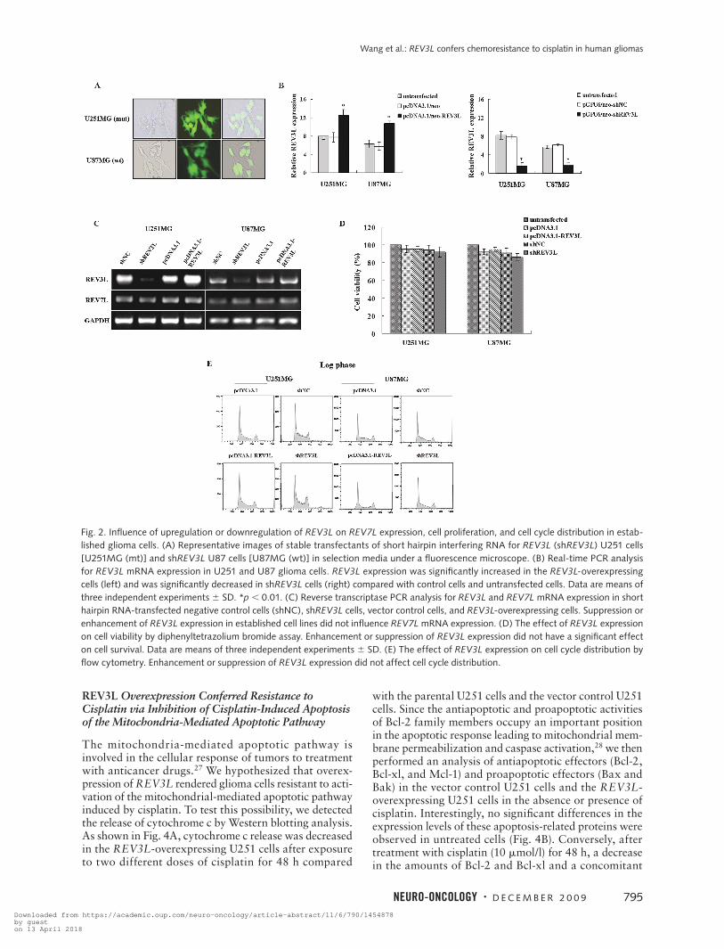
To gain insight into the function of REV3L in human glioma cells, we set up cell line models derived from the parental U251 and U87 glioma cells genetically manipulated for REV3L expression. As shown in Fig. 2B, REV3L expression was significantly increased in the
REV3L-overexpressing cells compared with the paren-tal cells and the vector control cells. In contrast, REV3L expression was significantly suppressed in glioma cell lines stably transfected with shREV3L (Fig. 2A) com-pared with the parental cells and shNC cells (Fig. 2B). We found that enhancement or suppression of REV3L gene expression in established cell lines did not influence REV7L mRNA expression levels (Fig. 2C) and did not have a significant effect on cell proliferation by MTT assay (Fig. 2D) or cell cycle distribution as examined by flow cytometry (Fig. 2E).
REV3L Overexpression Conferred Resistance to DNA-Damaging Agents
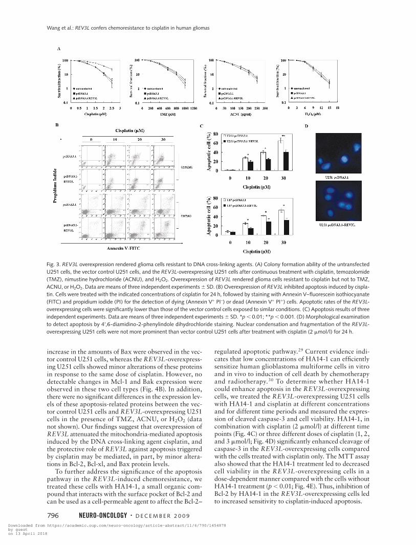
To evaluate the effect of REV3L on the chemosensitiv-ity of human glioma cells to chemotherapeutic drugs, we first examined the sensitivity of U251 cells to four DNA-damaging agents, including a DNA cross-linking agent (cisplatin), DNA-alkylating agents (TMZ and ACNU), and hydrogen peroxide (H2O2). The colony formation assays showed that overexpression of REV3L rendered the cells resistant to various doses of cisplatin (Fig. 3A) but not to TMZ, ACNU, or H2O2, which have different mechanisms of action. Consistent with clonogenic data, the REV3L-overexpressing U251 cells showed a marked decrease in cisplatin-induced apoptosis, as judged by decreased Annexin V-FITC–positive staining (Fig. 3B,C) and morphological alteration of nuclei (Fig. 3D). Similar apoptotic results were also obtained in the REV3L-over-expressing U87 cells (Fig. 3B,C). To further test whether REV3L overexpression conferred resistance to cisplatin that was attributable to inhibition of apoptosis, Western blotting was done to analyze the expression of cleaved caspase-3. After exposure to two relatively low doses of cisplatin (1 and 2 mmol/l) for 48 h, the expression levels of cleaved caspase-3 were lower in REV3L-overexpressing cells compared with the parental U251 cells and the vec-tor control U251 cells (Fig. 4A). Taken together, these data support the notion that REV3L overexpression decreased cisplatin-induced apoptosis in glioma cells.
Table 1. Patient demographics for the PCR human glioma samples
Normal Grade I Grade II Grade III Grade IV All Tumors
Number 10 3 7 8 12 30
Mean age (years) 28.2 37 42.7 42.6 42.7 42.1
Age range (years) 15–43 10–61 33–58 35–53 8–72 8–72
Sex
Male 8 3 4 5 3 15
Female 2 0 3 3 9 15
Surgery SLTC MC MC MC MC
Pathological classification N/A Polycystic Astrocytoma (4) Anaplastic Glioblastoma (10) astrocytoma (2) Oligodendroglioma (2) astrocytoma (5) Medulloblastoma (2) Choroid plexus Ependymoma (1) Anaplastic papilloma (1) oligodendroglioma (3)
Abbreviations: SLTC, standard large trauma craniotomy; MC, microneurosurgical craniotomy; N/A, not applicable.
Fig. 1. mRNA expression of REV3L in normal brain specimens and human glioma specimens. (A) Representative images of reverse tran-scriptase PCR analysis for REV3L gene expression in 15 specimens of human gliomas (G). REV3L gene expression was significantly ele-vated in malignant gliomas compared with normal brain tissue (N). (B) mRNA expression of REV3L measured by real-time PCR analysis. REV3L expression was significantly upregulated in malignant gliomas compared with normal brain tissues. Data are means of three inde-pendent experiments 1 SD. *p , 0.01; **p , 0.001.
Downloaded from https://academic.oup.com/neuro-oncology/article-abstract/11/6/790/1454878by gueston 13 April 2018
Wang et al.: REV3L confers chemoresistance to cisplatin in human gliomas
Neuro-oNcology • D E C E M B E R 2 0 0 9 795
REV3L Overexpression Conferred Resistance to Cisplatin via Inhibition of Cisplatin-Induced Apoptosis of the Mitochondria-Mediated Apoptotic Pathway
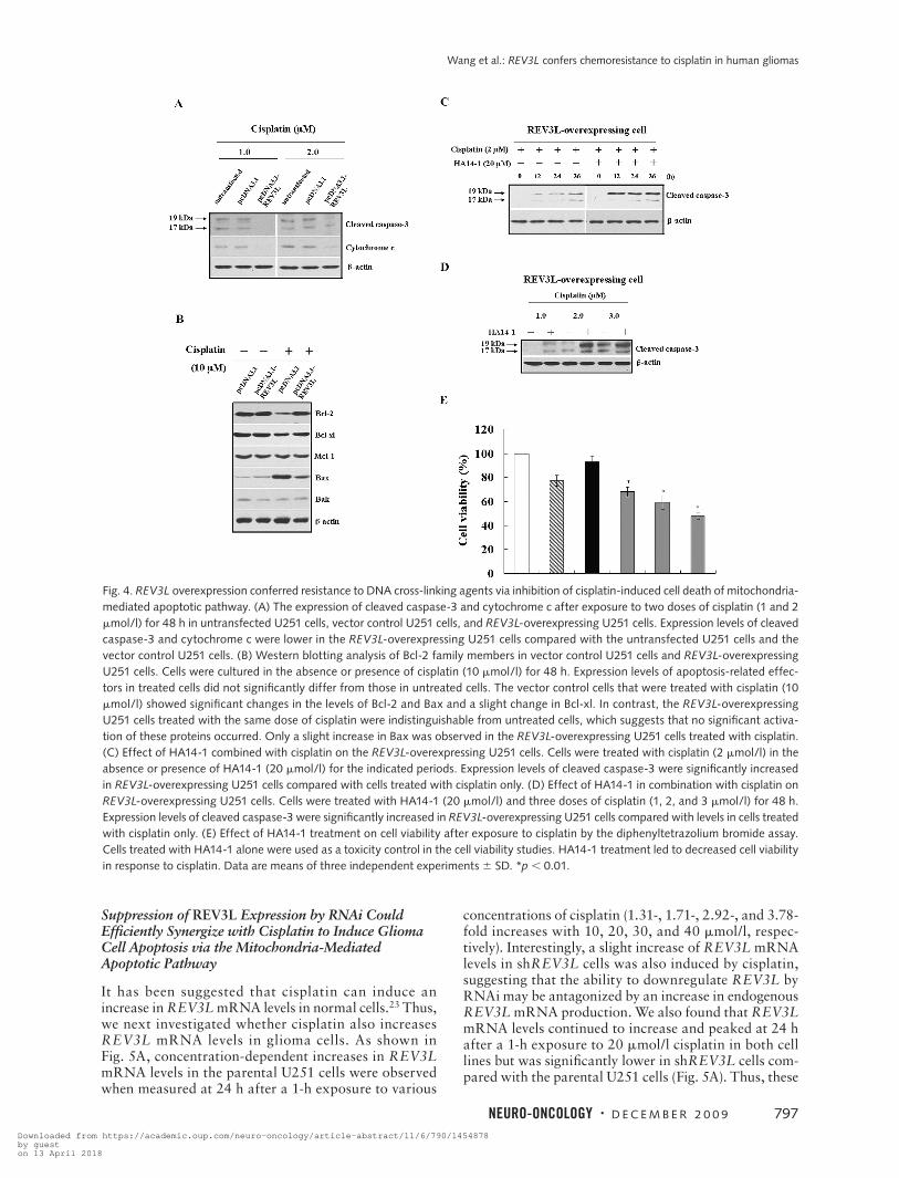
The mitochondria-mediated apoptotic pathway is involved in the cellular response of tumors to treatment with anticancer drugs.27 We hypothesized that overex-pression of REV3L rendered glioma cells resistant to acti-vation of the mitochondrial-mediated apoptotic pathway induced by cisplatin. To test this possibility, we detected the release of cytochrome c by Western blotting analysis. As shown in Fig. 4A, cytochrome c release was decreased in the REV3L-overexpressing U251 cells after exposure to two different doses of cisplatin for 48 h compared
with the parental U251 cells and the vector control U251 cells. Since the antiapoptotic and proapoptotic activities of Bcl-2 family members occupy an important position in the apoptotic response leading to mitochondrial mem-brane permeabilization and caspase activation,28 we then performed an analysis of antiapoptotic effectors (Bcl-2, Bcl-xl, and Mcl-1) and proapoptotic effectors (Bax and Bak) in the vector control U251 cells and the REV3L-overexpressing U251 cells in the absence or presence of cisplatin. Interestingly, no significant differences in the expression levels of these apoptosis-related proteins were observed in untreated cells (Fig. 4B). Conversely, after treatment with cisplatin (10 mmol/l) for 48 h, a decrease in the amounts of Bcl-2 and Bcl-xl and a concomitant
Fig. 2. Influence of upregulation or downregulation of REV3L on REV7L expression, cell proliferation, and cell cycle distribution in estab-lished glioma cells. (A) Representative images of stable transfectants of short hairpin interfering RNA for REV3L (shREV3L) U251 cells [U251MG (mt)] and shREV3L U87 cells [U87MG (wt)] in selection media under a fluorescence microscope. (B) Real-time PCR analysis for REV3L mRNA expression in U251 and U87 glioma cells. REV3L expression was significantly increased in the REV3L-overexpressing cells (left) and was significantly decreased in shREV3L cells (right) compared with control cells and untransfected cells. Data are means of three independent experiments 6 SD. *p , 0.01. (C) Reverse transcriptase PCR analysis for REV3L and REV7L mRNA expression in short hairpin RNA-transfected negative control cells (shNC), shREV3L cells, vector control cells, and REV3L-overexpressing cells. Suppression or enhancement of REV3L expression in established cell lines did not influence REV7L mRNA expression. (D) The effect of REV3L expression on cell viability by diphenyltetrazolium bromide assay. Enhancement or suppression of REV3L expression did not have a significant effect on cell survival. Data are means of three independent experiments 6 SD. (E) The effect of REV3L expression on cell cycle distribution by flow cytometry. Enhancement or suppression of REV3L expression did not affect cell cycle distribution.
Downloaded from https://academic.oup.com/neuro-oncology/article-abstract/11/6/790/1454878by gueston 13 April 2018
Wang et al.: REV3L confers chemoresistance to cisplatin in human gliomas
796 Neuro-oNcology • D E C E M B E R 2 0 0 9
increase in the amounts of Bax were observed in the vec-tor control U251 cells, whereas the REV3L-overexpress-ing U251 cells showed minor alterations of these proteins in response to the same dose of cisplatin. However, no detectable changes in Mcl-1 and Bak expression were observed in these two cell types (Fig. 4B). In addition, there were no significant differences in the expression lev-els of these apoptosis-related proteins between the vec-tor control U251 cells and REV3L-overexpressing U251 cells in the presence of TMZ, ACNU, or H2O2 (data not shown). Our findings suggest that overexpression of REV3L attenuated the mitochondria-mediated apoptosis induced by the DNA cross-linking agent cisplatin, and the protective role of REV3L against apoptosis triggered by cisplatin may be mediated, in part, by minor altera-tions in Bcl-2, Bcl-xl, and Bax protein levels.
To further address the significance of the apoptosis pathway in the REV3L-induced chemoresistance, we treated these cells with HA14-1, a small organic com-pound that interacts with the surface pocket of Bcl-2 and can be used as a cell-permeable agent to affect the Bcl-2–
regulated apoptotic pathway.29 Current evidence indi-cates that low concentrations of HA14-1 can efficiently sensitize human glioblastoma multiforme cells in vitro and in vivo to induction of cell death by chemotherapy and radiotherapy.30 To determine whether HA14-1 could enhance apoptosis in the REV3L-overexpressing cells, we treated the REV3L-overexpressing U251 cells with HA14-1 and cisplatin at different concentrations and for different time periods and measured the expres-sion of cleaved caspase-3 and cell viability. HA14-1, in combination with cisplatin (2 mmol/l) at different time points (Fig. 4C) or three different doses of cisplatin (1, 2, and 3 mmol/l; Fig. 4D) significantly enhanced cleavage of caspase-3 in the REV3L-overexpressing cells compared with the cells treated with cisplatin only. The MTT assay also showed that the HA14-1 treatment led to decreased cell viability in the REV3L-overexpressing cells in a dose-dependent manner compared with the cells without HA14-1 treatment (p , 0.01; Fig. 4E). Thus, inhibition of Bcl-2 by HA14-1 in the REV3L-overexpressing cells led to increased sensitivity to cisplatin-induced apoptosis.
Fig. 3. REV3L overexpression rendered glioma cells resistant to DNA cross-linking agents. (A) Colony formation ability of the untransfected U251 cells, the vector control U251 cells, and the REV3L-overexpressing U251 cells after continuous treatment with cisplatin, temozolomide (TMZ), nimustine hydrochloride (ACNU), and H2O2. Overexpression of REV3L rendered glioma cells resistant to cisplatin but not to TMZ, ACNU, or H2O2. Data are means of three independent experiments 6 SD. (B) Overexpression of REV3L inhibited apoptosis induced by cispla-tin. Cells were treated with the indicated concentrations of cisplatin for 24 h, followed by staining with Annexin V–fluorescein isothiocyanate (FITC) and propidium iodide (PI) for the detection of dying (Annexin V1 PI–) or dead (Annexin V1 PI1) cells. Apoptotic rates of the REV3L-overexpressing cells were significantly lower than those of the vector control cells exposed to similar conditions. (C) Apoptosis results of three independent experiments. Data are means of three independent experiments 6 SD. *p , 0.01; **p , 0.001. (D) Morphological examination to detect apoptosis by 4',6-diamidino-2-phenylindole dihydrochloride staining. Nuclear condensation and fragmentation of the REV3L-overexpressing U251 cells were not more prominent than vector control U251 cells after treatment with cisplatin (2 mmol/l) for 24 h.
Downloaded from https://academic.oup.com/neuro-oncology/article-abstract/11/6/790/1454878by gueston 13 April 2018
Wang et al.: REV3L confers chemoresistance to cisplatin in human gliomas
Neuro-oNcology • D E C E M B E R 2 0 0 9 797
Suppression of REV3L Expression by RNAi Could Efficiently Synergize with Cisplatin to Induce Glioma Cell Apoptosis via the Mitochondria-Mediated Apoptotic Pathway
It has been suggested that cisplatin can induce an increase in REV3L mRNA levels in normal cells.23 Thus, we next investigated whether cisplatin also increases REV3L mRNA levels in glioma cells. As shown in Fig. 5A, concentration-dependent increases in REV3L mRNA levels in the parental U251 cells were observed when measured at 24 h after a 1-h exposure to various
concentrations of cisplatin (1.31-, 1.71-, 2.92-, and 3.78-fold increases with 10, 20, 30, and 40 mmol/l, respec-tively). Interestingly, a slight increase of REV3L mRNA levels in shREV3L cells was also induced by cisplatin, suggesting that the ability to downregulate REV3L by RNAi may be antagonized by an increase in endogenous REV3L mRNA production. We also found that REV3L mRNA levels continued to increase and peaked at 24 h after a 1-h exposure to 20 mmol/l cisplatin in both cell lines but was significantly lower in shREV3L cells com-pared with the parental U251 cells (Fig. 5A). Thus, these
Fig. 4. REV3L overexpression conferred resistance to DNA cross-linking agents via inhibition of cisplatin-induced cell death of mitochondria-mediated apoptotic pathway. (A) The expression of cleaved caspase-3 and cytochrome c after exposure to two doses of cisplatin (1 and 2 mmol/l) for 48 h in untransfected U251 cells, vector control U251 cells, and REV3L-overexpressing U251 cells. Expression levels of cleaved caspase-3 and cytochrome c were lower in the REV3L-overexpressing U251 cells compared with the untransfected U251 cells and the vector control U251 cells. (B) Western blotting analysis of Bcl-2 family members in vector control U251 cells and REV3L-overexpressing U251 cells. Cells were cultured in the absence or presence of cisplatin (10 mmol/l) for 48 h. Expression levels of apoptosis-related effec-tors in treated cells did not significantly differ from those in untreated cells. The vector control cells that were treated with cisplatin (10 mmol/l) showed significant changes in the levels of Bcl-2 and Bax and a slight change in Bcl-xl. In contrast, the REV3L-overexpressing U251 cells treated with the same dose of cisplatin were indistinguishable from untreated cells, which suggests that no significant activa-tion of these proteins occurred. Only a slight increase in Bax was observed in the REV3L-overexpressing U251 cells treated with cisplatin. (C) Effect of HA14-1 combined with cisplatin on the REV3L-overexpressing U251 cells. Cells were treated with cisplatin (2 mmol/l) in the absence or presence of HA14-1 (20 mmol/l) for the indicated periods. Expression levels of cleaved caspase-3 were significantly increased in REV3L-overexpressing U251 cells compared with cells treated with cisplatin only. (D) Effect of HA14-1 in combination with cisplatin on REV3L-overexpressing U251 cells. Cells were treated with HA14-1 (20 mmol/l) and three doses of cisplatin (1, 2, and 3 mmol/l) for 48 h. Expression levels of cleaved caspase-3 were significantly increased in REV3L-overexpressing U251 cells compared with levels in cells treated with cisplatin only. (E) Effect of HA14-1 treatment on cell viability after exposure to cisplatin by the diphenyltetrazolium bromide assay. Cells treated with HA14-1 alone were used as a toxicity control in the cell viability studies. HA14-1 treatment led to decreased cell viability in response to cisplatin. Data are means of three independent experiments 6 SD. *p , 0.01.
Downloaded from https://academic.oup.com/neuro-oncology/article-abstract/11/6/790/1454878by gueston 13 April 2018

Wang et al.: REV3L confers chemoresistance to cisplatin in human gliomas
798 Neuro-oNcology • D E C E M B E R 2 0 0 9
results indicate that REV3L might be activated by expo-sure to cisplatin in a dose- and time-dependent manner, and effective repression of REV3L expression could be a potential therapeutic target for the treatment of human gliomas.
To determine whether suppression of REV3L expres-
sion could enhance the chemosensitivity of human glioma cells to chemotherapeutic drugs, we adopted the strategy of REV3L RNAi to repress the expression of REV3L. Colony formation assays showed that reduced expres-sion of REV3L rendered glioma cells more sensitive to the cytotoxic effect of cisplatin but not to TMZ, ACNU,
Fig. 5. REV3L knockdown can efficiently synergize the apoptosis response to treatment with cisplatin in glioma cells. (A) Real-time PCR analysis for REV3L mRNA level in the untransfected U251 cells and short hairpin interfering RNA for REV3L (shREV3L) U251 cells induced by various doses of cisplatin (10, 20, 30, and 40 mmol/l) at 24 h after a 1-h exposure (left), and time course change of REV3L mRNA level in both cell lines after exposure to 20 mmol/l cisplatin for 1 h (right). Data are means of three independent real-time PCR measurements 6 SD. (B) Colony formation ability of the untransfected U251 cells, short hairpin RNA-transfected negative control (shNC) cells, and shREV3L cells after continuous treatment with cisplatin, temozolomide (TMZ), nimustine hydrochloride (ACNU), and H2O2. Suppression of REV3L expression conferred hypersensitivity to cisplatin but not to TMZ, ACNU, or H2O2. Data are means of three independent experiments 6 SD. (C) Detection of apoptotic cells by flow cytometry. shNC U251, shREV3L U251, shNC U87, and shREV3L U87 cells were treated with three doses of cisplatin (1, 2, and 3 mmol/l) for 24 h. A sub-G1 peak was significantly higher in shREV3L cells than in shNC cells. (D) Sub-G1 peak results. Data are means of three independent experiments 6 SD. *p , 0.01; **p , 0.001. (E) Morphological examination to detect apoptosis by 4',6-diamidino-2-phenylindole dihydrochloride staining. Nuclear condensation and fragmentation of shREV3L cells were more prominent than shNC cells after treatment with cisplatin (2 mmol/l) for 24 h.
Downloaded from https://academic.oup.com/neuro-oncology/article-abstract/11/6/790/1454878by gueston 13 April 2018

Wang et al.: REV3L confers chemoresistance to cisplatin in human gliomas
Neuro-oNcology • D E C E M B E R 2 0 0 9 799
or H2O2 (Fig. 5B). Changes in cell cycle distribution as well as cell death, reflected by the sub-G1 cells, were then analyzed. The data showed that cisplatin evoked reproducible and significant levels of death in shREV3L U251 cells as reflected by emerging sub-G1 cells of 12%, 27.8%, and 43.1% in response to three doses of cispla-tin (1, 2, and 3 mmol/l) for 24 h (Fig. 5C,D). Similar results were also obtained in shREV3L U87 cells treated with three same doses of cisplatin for 24 h (Fig. 5C,D). In contrast, cisplatin did not induce significant levels of apoptosis in shNC U251 and shNC U87 cells. DAPI fluorescence nuclear staining showed that the nuclear condensation and fragmentation of shREV3L U251 cells were more prominent than shNC U251 cells at 24 h exposure to 2 mmol/l cisplatin (Fig. 5E). Furthermore, after exposure to cisplatin (1 mmol/l) for 48 h, a time-dependent increase in the amounts of cleaved caspase-3 was observed in shREV3L U251 cells (Fig. 6A). Similar results were also obtained in shREV3L U87 cells treated with cisplatin (10 mmol/l) for 24 h (Fig. 6B).
To further confirm that shREV3L-induced sensiti-zation to cisplatin was a result of increased apoptosis via mitochondria-mediated apoptotic pathway, we per-formed analysis of Bcl-2 family members mentioned above in shNC cells and shREV3L cells without cisplatin treatment. However, suppression of REV3L expression did not affect this apoptosis-related protein expression in untreated cells (data not shown). We then determined levels of cleaved caspase-3, cytochrome c, and Bcl-2 fam-
ily members in the presence of cisplatin. As expected, after treatment with cisplatin (5 mmol/l) for 48 h, cleaved caspase-3, cytochrome c, and Bax levels increased and Bcl-2 and Bcl-xl levels concomitantly decreased, with-out leading to any detectable changes in Mcl-1 and Bak expression, in shREV3L U251 and U87 cells compared with shNC U251 and U87 cells (Fig. 6C). In addition, there were no differences in the expression levels of these pro-teins between shNC cells and shREV3L cells in response to TMZ, ACNU, or H2O2 (data not shown). Thus, these results suggest that suppression of REV3L expression by RNAi could efficiently synergize the mitochondrial apop-totic response to treatment with cisplatin in glioma cells. This effect may be partially mediated by marked altera-tions in Bcl-2, Bcl-xl, and Bax protein levels.
Suppression of REV3L Expression Reduced Mutation Rates at the HPRT Locus
To further elucidate the molecular mechanism responsi-ble for shREV3L-induced chemosensitization, we deter-mined if suppression of REV3L expression reduced the frequency of cisplatin-induced mutations in surviving glioma cells. Cells were treated with three doses of cis-platin (10, 20, and 30 mmol/l) for 1 h and then cultured for 2 weeks. An HPRT mutation assay was then carried out to assess the frequency of cisplatin-induced 6-TG– resistant mutants, which reflect the mutation frequencies at the HPRT locus.31 As shown in Fig. 7, the frequen-
Fig. 6. REV3L knockdown could efficiently synergize with cisplatin to induce glioma cell apoptosis via the mitochondria-mediated apop-totic pathway. (A) Time-dependent expression of cleaved caspase-3 after exposure to a single dose of cisplatin (1 mmol/l) in short hairpin RNA-transfected negative control (shNC) and short hairpin interfering RNA for REV3L (shREV3L) U251 cells. Expression levels of cleaved caspase-3 were significantly higher in shREV3L U251 cells compared with shNC U251 cells in response to the same dose of cisplatin. (B) Time-dependent expression of cleaved caspase-3 after exposure to a single dose of cisplatin (10 mmol/l) in shNC and shREV3L U87 cells. Expression levels of cleaved caspase-3 were significantly higher in shREV3L U87 cells compared with shNC U87 cells in response to the same dose of cisplatin. (C) The expression levels of cleaved caspase-3, cytochrome c, and Bcl-2 family members after exposure to a single dose of cisplatin (5 mmol/l) for 48 h in shNC and shREV3L cells. Expression levels of Bcl-2 and Bcl-xl were significantly lower and levels of cleaved caspase-3, cytochrome c, and Bax were significantly higher in shREV3L cells compared with shNC cells in response to the same dose of cisplatin. In contrast, the expression levels of Mcl-1 and Bak were indistinguishable from those of shNC cells.
Downloaded from https://academic.oup.com/neuro-oncology/article-abstract/11/6/790/1454878by gueston 13 April 2018

Wang et al.: REV3L confers chemoresistance to cisplatin in human gliomas
800 Neuro-oNcology • D E C E M B E R 2 0 0 9
cies of 6-TG–resistant mutants induced by cisplatin were significantly reduced in the surviving shREV3L U251 cells compared with shNC U251 cells, suggesting that suppression of REV3L expression significantly reduced the frequencies of cisplatin-induced mutations at the HPRT locus. Thus, these data indicate that suppres-sion of REV3L expression by RNAi could synergize the effect of cisplatin on inducing apoptosis of glioma cells through repressing the TLS pathway and thus contrib-ute to less frequent mutations in those surviving glioma cells.
Discussion
DNA polymerase , together with DNA polymerases , , and , are responsible for carrying out TLS, which includes error-prone or error-free DNA repair processes past fork-blocking lesions.32 The TLS pathway may play an important role in the initiation and progression of tumors.33–35 Previous studies have revealed that these specialized low-stringency DNA polymerases may con-tribute to spontaneous and DNA-lesion–triggered muta-tions during translesional DNA replication, thereby con-tributing to the accumulation of genetic damage.10,11,13 However, the relationships between those family mem-bers and tumors are uncertain. It has been reported that the expression level of the REV3L gene appeared to be similar in lung, gastric, colon, and renal tumors
compared to normal-tissue counterparts.8 REV7L was found to be highly upregulated in breast and colon can-cers.36,37 O-Wang et al.38 reported that polymerase was significantly elevated in human lung cancers, whereas polymerase was unaltered. In another study, however, transcription levels of polymerases , , and and the REV3L gene were significantly reduced in various lung, stomach, and colorectal cancers.39 In the present study, using RT-PCR and real-time PCR, we showed that the expression of REV3L was significantly upregulated in malignant gliomas and basically correlated with tumor grade. These findings indicate that the specialized DNA polymerases of TLS in human malignancies are deregu-lated, and their potential roles in tumorigenesis and pro-gression may differ depending on the type, origin, and tissue specificity of tumors. Therefore, further studies are needed to elucidate the role of these specialized DNA polymerases in tumorigenesis and tumor progression.
Chemoresistance is a major obstacle for the treat-ment of malignant gliomas, which are unable to be totally resected because of their diffusely infiltrative nature.40 Many different mechanisms may account for this chemoresistance, including multidrug resistance, upregulation of antiapoptotic pathways, enhanced DNA repair, and increased metabolic inactivation and subsequent elimination of the applied drugs.41,42 Recently, REV3L has been suggested to regulate cyto-toxicity, mutagenicity, and chemoresistance of cispla-tin in both human fibroblast cells and human colon carcinoma cells.23,24 To investigate REV3L function in human glioma cells, we generated stable transfectants overexpressing REV3L and found that overexpression of REV3L may inhibit the sensitivity of glioma cells to cisplatin. REV3L overexpression conferred resistance to cisplatin and decreased cisplatin-induced apoptosis of the mitochondria-mediated apoptotic pathway due to, in part, minor alterations in Bcl-2, Bcl-xl, and Bax expres-sion levels. However, how REV3L regulates apoptosis of glioma cells through DNA repair pathway and whether the regulation involves a DNA-repair-independent path-way remain to be investigated.
Previous studies have demonstrated that cisplatin treatment in human normal cells and tumor cells can induce an increased expression of certain DNA poly-merases involved in the TLS of cisplatin-mediated DNA damage.22,23 In our system, we also showed that cisplatin induced a concentration- and time-dependent increase in the REV3L mRNA level in glioma cells, indicating that REV3L may play an important role in the DNA damage response in both normal and tumor cells and that there may be a negative loop regulation mechanism for the resistance of tumor cells to cisplatin treatment. By expressing shRNAs targeted to REV3L mRNA, we established stable human glioma cell lines with very low levels of REV3L mRNA and found that disruption of REV3L function by downregulation of its mRNA increased cellular sensitivity to cisplatin. In addi-tion, activation of caspase-3 and cytochrome c, as well as marked alterations in the expression levels of Bcl-2, Bcl-xl, and Bax, confirmed that suppression of REV3L expression facilitated cisplatin-induced activation of
Fig. 7. REV3L knockdown reduced mutation frequency at the hypoxanthine guanine phosphoribosyl transferase locus (HPRT): frequency of cisplatin-induced generation of 6-thioguanine (6-TG) mutants after a 1-h exposure to one of three doses of cisplatin (10, 20, and 30 mmol/l) and 2 weeks of culture. Cisplatin-induced gen-eration of 6-TG mutants was significantly reduced in short hairpin interfering REV3L RNA (shREV3L) cells compared with short hair-pin RNA-transfected negative control (shNC) cells in response to cisplatin. Data are means of three independent experiments 6 SD. *p , 0.01; **p , 0.001.
Downloaded from https://academic.oup.com/neuro-oncology/article-abstract/11/6/790/1454878by gueston 13 April 2018

Wang et al.: REV3L confers chemoresistance to cisplatin in human gliomas
Neuro-oNcology • D E C E M B E R 2 0 0 9 801
12. Van Sloun PP, Varlet I, Sonneveld E, et al. Involvement of mouse Rev3
in tolerance of endogenous and exogenous DNA damage. Mol Cell
Biol. 2002;22:2159–2169.
13. Cheung HW, Chun AC, Wang Q, et al. Inactivation of human MAD2B
in nasopharyngeal carcinoma cells leads to chemosensitization to
DNA-damaging agents. Cancer Res. 2006;66:4357–4367.
14. Nelson JR, Lawrence CW, Hinkle DC. Thymine-thymine dimer bypass
by yeast DNA polymerase zeta. Science. 1996;272:1646–169.
15. Albertella MR, Lau A, O’Connor MJ. The overexpression of special-
ized DNA polymerases in cancer. DNA Repair. 2005;4:583–593.
16. Grossman SA, Wharam M, Sheidler V, et al. Phase II study of continu-
ous infusion carmustine and cisplatin followed by cranial irradiation in
adults with newly diagnosed high-grade astrocytoma. J Clin Oncol.
1997;15:2596–2603.
17. DeAngelis LM. Chemotherapy for brain tumors—a new beginning. N
Engl J Med. 2005;352:1036–1038.
18. Bredel M, Zentner J. Brain-tumour drug resistance: the bare essentials.
Lancet Oncol. 2002;3:397–406.
19. Fruehauf JP, Brem H, Brem S, et al. In vitro drug response and molecu-
lar markers associated with drug resistance in malignant gliomas. Clin
Cancer Res. 2006;12:4523–4532.
20. Nagane M, Levitzki A, Gazit A, Cavenee WK, Huang HJ. Drug resis-
tance of human glioblastoma cells conferred by a tumor-specific
mutant epidermal growth factor receptor through modulation of
Bcl-XL and caspase-3-like proteases. Proc Natl Acad Sci U S A. 1998;
95:5724–5729.
21. Lin X, Okuda T, Trang J, Howell SB. Human REV1 modulates the cyto-
toxicity and mutagenicity of cisplatin in human ovarian carcinoma
cells. Mol Pharmacol. 2006;69:1748–1754.
References
the mitochondrial apoptotic pathway in glioma cells. These results are clearly reminiscent of those of REV7L knockdown, which was previously reported to confer hypersensitivity to certain types of chemotherapeutic agents, especially cisplatin, through activation of apop-tosis.13 Although the redundant functions of specialized DNA polymerase family members in TLS remain elu-sive, it seems that REV3L may play a predominant role in regulating the sensitivity to the DNA cross-linking agent cisplatin in human gliomas. RNAi is emerging as a powerful approach for gene target therapy.43 With recent advances in RNAi delivery strategy, the shRNA complex could be efficiently delivered to the brain.44–46 Therefore, downregulation of REV3L expression by RNAi in combination with cisplatin could enhance the clinical efficacy of chemotherapy for glioma patients.
In summary, we have shown for the first time that REV3L is overexpressed in human gliomas, especially in high-grade gliomas. Enhancement of REV3L expres-sion in glioma cells resulted in reduced sensitivity to cisplatin-induced cell death. Inhibition of Bcl-2 in REV3L-overexpressing cells by HA14-1 significantly promoted cisplatin-induced apoptosis. Furthermore, suppression of REV3L expression by RNAi enhanced the sensitivity of glioma cells to cisplatin, led to more
pronounced apoptosis in association with marked alter-ations in Bcl-2, Bcl-xl, and Bax expression levels, and reduced frequencies of cisplatin-induced mutations in glioma cells. Future studies are therefore warranted to determine the function of REV3L in vivo, especially its role in tumorigenesis and chemoresistance. Whether other DNA polymerases involved in TLS might be responsible for chemoresistance to DNA-damaging agents in human malignancies remains to be examined.
Acknowledgments
This research was supported in part by the National Natural Science Foundation of China (grant no. 30772239 S.Z.), Foundation of Harbin Science and Technology Committee (grant no. 2007AA3C0832 S.Z.), and National “211” Environmental Genomics Grant (D.L.). We gratefully acknowledge Dr. Yoshiki Murakumo (Nagoya University Graduate School of Medicine, Nagoya, Japan) for generously providing the REV3L plasmid used in this study.
The current affiliation of H.W. is Department of Pathology and Cancer Center, Medical College of Geor-gia, Augusta, GA, USA.
1. Behin A, Hoang-Xuan K, Carpentier AF, Delattre JY. Primary brain
tumours in adults. Lancet. 2003;361:323–331.
2. Furnari FB, Fenton T, Bachoo RM, et al. Malignant astrocytic glioma:
genetics, biology, and paths to treatment. Genes Dev. 2007;21:2683–
2710.
3. Barzon L, Zanusso M, Colombo F, Palu G. Clinical trials of gene ther-
apy, virotherapy, and immunotherapy for malignant gliomas. Cancer
Gene Ther. 2006;13:539–554.
4. DeAngelis LM. Brain tumors. N Engl J Med. 2001;344:114–123.
5. Okada H, Pollack IF. Cytokine gene therapy for malignant glioma.
Expert Opin Biol Ther. 2004;4:1609–1620.
6. Morelli C, Mungall AJ, Negrini M, Barbanti-Brodano G, Croce CM.
Alternative splicing, genomic structure, and fine chromosome local-
ization of REV3L. Cytogenet Cell Genet. 1998;83:18–20.
7. Gibbs PE, McGregor WG, Maher VM, Nisson P, Lawrence CW. A
human homolog of the Saccharomyces cerevisiae REV3 gene, which
encodes the catalytic subunit of DNA polymerase zeta. Proc Natl Acad
Sci U S A. 1998;95:6876–6880.
8. Kawamura K, O-Wang J, Bahar R, et al. The error-prone DNA poly-
merase zeta catalytic subunit (Rev3) gene is ubiquitously expressed in
normal and malignant human tissues. Int J Oncol. 2001;18:97–103.
9. Xiao W, Lechler T, Chow BL, et al. Identification, chromosomal map-
ping and tissue-specific expression of hREV3 encoding a putative
human DNA polymerase zeta. Carcinogenesis. 1998;19:945–949.
10. Okada T, Sonoda E, Yoshimura M, et al. Multiple roles of verte-
brate REV genes in DNA repair and recombination. Mol Cell Biol.
2005;25:6103–6111.
11. Sonoda E, Okada T, Zhao GY, et al. Multiple roles of Rev3, the cata-
lytic subunit of polzeta in maintaining genome stability in vertebrates.
EMBO J. 2003;22:3188–3197.
Downloaded from https://academic.oup.com/neuro-oncology/article-abstract/11/6/790/1454878by gueston 13 April 2018

Wang et al.: REV3L confers chemoresistance to cisplatin in human gliomas
802 Neuro-oNcology • D E C E M B E R 2 0 0 9
22. Okuda T, Lin X, Trang J, Howell SB. Suppression of hREV1 expression
reduces the rate at which human ovarian carcinoma cells acquire resis-
tance to cisplatin. Mol Pharmacol. 2005;67:1852–1860.
23. Wu F, Lin X, Okuda T, Howell SB. DNA polymerase zeta regulates
cisplatin cytotoxicity, mutagenicity, and the rate of development of
cisplatin resistance. Cancer Res. 2004;64:8029–8035.
24. Lin X, Trang J, Okuda T, Howell SB. DNA polymerase zeta accounts
for the reduced cytotoxicity and enhanced mutagenicity of cisplatin in
human colon carcinoma cells that have lost DNA mismatch repair. Clin
Cancer Res. 2006;12:563–568.
25. Kleihues P, Louis DN, Scheithauer BW, et al. The WHO classifica-
tion of tumors of the nervous system. J Neuropathol Exp Neurol.
2002;61:215–229.
26. Chiu RK, Brun J, Ramaekers C, et al. Lysine 63-polyubiquitination
guards against translesion synthesis-induced mutations. PLoS Genet.
2006;2:e116.
27. Asakura T, Ohkawa K. Chemotherapeutic agents that induce mito-
chondrial apoptosis. Curr Cancer Drug Targets. 2004;4:577–590.
28. Robertson JD, Orrenius S. Role of mitochondria in toxic cell death.
Toxicology. 2002;181–182:491–496.
29. Wang JL, Liu D, Zhang ZJ, et al. Structure-based discovery of an
organic compound that binds Bcl-2 protein and induces apoptosis of
tumor cells. Proc Natl Acad Sci U S A. 2000;97:7124–7129.
30. Manero F, Gautier F, Gallenne T, et al. The small organic compound
HA14-1 prevents Bcl-2 interaction with Bax to sensitize malignant
glioma cells to induction of cell death. Cancer Res. 2006;66:2757–
2764.
31. Maher VM, McCormick JJ. The HPRT gene as a model study for muta-
tion analysis. In: Pfeifer GP, ed. Technologies for Detection of DNA
Damage and Mutations. New York: Plenum; 1996:381–390.
32. Lehmann AR. Translesion synthesis in mammalian cells. Exp Cell Res.
2006;312:2673–2676.
33. Andersen PL, Xu F, Xiao W. Eukaryotic DNA damage tolerance and
translesion synthesis through covalent modifications of PCNA. Cell
Res. 2008;18:162–173.
34. Lawrence CW, Hinkle DC. DNA polymerase zeta and the control
of DNA damage induced mutagenesis in eukaryotes. Cancer Surv.
1996;28:21–31.
35. Liu G, Chen X. DNA polymerase eta, the product of the xeroderma
pigmentosum variant gene and a target of p53, modulates the DNA
damage checkpoint and p53 activation. Mol Cell Biol. 2006;26:1398–
1413.
36. Yuan B, Xu Y, Woo JH, et al. Increased expression of mitotic check-
point genes in breast cancer cells with chromosomal instability. Clin
Cancer Res. 2006;12:405–410.
37. Rimkus C, Friederichs J, Rosenberg R, Holzmann B, Siewert JR, Jans-
sen KP. Expression of the mitotic checkpoint gene MAD2L2 has prog-
nostic significance in colon cancer. Int J Cancer. 2007;120:207–211.
38. O-Wang J, Kawamura K, Tada Y, et al. DNA polymerase kappa, impli-
cated in spontaneous and DNA damage-induced mutagenesis, is
overexpressed in lung cancer. Cancer Res. 2001;61:5366–5369.
39. Pan Q, Fang Y, Xu Y, Zhang K, Hu X. Downregulation of DNA poly-
merases kappa, eta, iota, and zeta in human lung, stomach, and col-
orectal cancers. Cancer Lett. 2005;217:139–147.
40. Lu C, Shervington A. Chemoresistance in gliomas. Mol Cell Biochem.
2008;312:71–80.
41. Bredel M. Anticancer drug resistance in primary human brain tumors.
Brain Res Brain Res Rev. 2001;35:161–204.
42. Bronger H, Konig J, Kopplow K, et al. ABCC drug efflux pumps and
organic anion uptake transporters in human gliomas and the blood-
tumor barrier. Cancer Res. 2005;65:11419–11428.
43. Kim DH, Rossi JJ. Strategies for silencing human disease using RNA
interference. Nat Rev Genet. 2007;8:173–184.
44. Grzelinski M, Urban-Klein B, Martens T, et al. RNA interference-
mediated gene silencing of pleiotrophin through polyethylenimine-
complexed small interfering RNAs in vivo exerts antitumoral effects in
glioblastoma xenografts. Hum Gene Ther. 2006;17:751–766.
45. Medarova Z, Pham W, Farrar C, Petkova V, Moore A. In vivo imaging
of siRNA delivery and silencing in tumors. Nat Med. 2007;13:372–
377.
46. Pardridge WM. shRNA and siRNA delivery to the brain. Adv Drug
Deliv Rev. 2007;59:141–152.
Downloaded from https://academic.oup.com/neuro-oncology/article-abstract/11/6/790/1454878by gueston 13 April 2018