review article arginine metabolism: nitric oxide and beyond · biochem. j. (1998) 336, 1–17...
TRANSCRIPT

Biochem. J. (1998) 336, 1–17 (Printed in Great Britain) 1
REVIEW ARTICLE
Arginine metabolism: nitric oxide and beyondGuoyao WU* and Sidney M. MORRIS, JR.†*Departments of Animal Science, Medical Physiology, and Veterinary Anatomy and Public Health, and Faculty of Nutrition, Texas A&M University, College Station,TX 77843, U.S.A., and †Department of Molecular Genetics and Biochemistry, University of Pittsburgh School of Medicine, Pittsburgh, PA 15261, U.S.A.
Arginine is one of the most versatile amino acids in animal cells,
serving as a precursor for the synthesis not only of proteins but
also of nitric oxide, urea, polyamines, proline, glutamate, creatine
and agmatine. Of the enzymes that catalyse rate-controlling steps
in arginine synthesis and catabolism, argininosuccinate synthase,
the two arginase isoenzymes, the three nitric oxide synthase
isoenzymes and arginine decarboxylase have been recognized in
recent years as key factors in regulating newly identified aspects
of arginine metabolism. In particular, changes in the activities of
argininosuccinate synthase, the arginases, the inducible iso-
enzyme of nitric oxide synthase and also cationic amino acid
transporters play major roles in determining the metabolic fates
of arginine in health and disease, and recent studies have identified
INTRODUCTION
There is a rich history of studies on arginine (2-amino-5-
guanidinovaleric acid) and its metabolism over the past 100
years. This interesting amino acid was first isolated from lupin
seedlings in 1886 [1], and soon afterwards (1895) was identified
as a component of animal proteins [2]. The structure of arginine
was established by alkaline hydrolysis to yield ornithine and urea
in 1897 [3] and by synthesis from benzoylornithine in 1910 [4].
Subsequently, arginine was found in 1924 to be a major amino
acid in the basic proteins of fish sperm [5], and its synthesis by
mammals was deduced in the classic nutrition studies of W. C.
Rose and his colleagues in 1930 [6]. Although high activities of
arginase, the enzyme that hydrolyses arginine to ornithine and
urea, had been identified in the liver in 1904 [7], it was the
discovery of the ornithine cycle (urea cycle) by Krebs and
Henseleit in 1932 [8] that led to the elucidation of prominent
roles of arginine in physiology and metabolic pathways.
Physiological and nutritional studies in the late 1930s and
1940s started a new era of arginine research. Foster et al. [9]
reported that arginine was required for the synthesis of creatine,
the precursor of creatinine, which was known to scientists of the
19th century and had been proposed in 1926 as a clinical
indicator of renal function [10]. Meanwhile, dietary arginine was
shown to be required for growth of the chick [11] and for optimal
growth of the young rat, but not for the healthy adult rat [12,13].
These findings led to extensive studies in the 1950s, 1960s and
1970s that resulted in the initial classification of arginine as a
dispensable (non-essential) amino acid for healthy adult humans
Abbreviations used: ASL, argininosuccinate lyase; ASS, argininosuccinate synthase; CPS I, carbamoyl-phosphate synthase I ; LPS, lipo-polysaccharide ; NOS, NO synthase; eNOS, endothelial NOS; iNOS, inducible NOS; nNOS, neuronal NOS; OAT, ornithine aminotransferase ; OCT,ornithine carbamoyltransferase ; ODC, ornithine decarboxylase ; P5C, ∆1-pyrroline-5-carboxylate.
Correspondence may be addressed to either Dr. G. Wu (e-mail g-wu!tamu.edu) or Dr. S. M. Morris, Jr. (e-mail sid!hoffman.mgen.pitt.edu) at theaddresses given.
complex patterns of interaction among these enzymes. There is
growing interest in the potential roles of the arginase isoenzymes
as regulators of the synthesis of nitric oxide, polyamines, proline
and glutamate. Physiological roles and relationships between the
pathways of arginine synthesis and catabolism in �i�o are complex
and difficult to analyse, owing to compartmentalized expression
of various enzymes at both organ (e.g. liver, small intestine and
kidney) and subcellular (cytosol and mitochondria) levels, as well
as to changes in expression during development and in response
to diet, hormones and cytokines. The ongoing development of
new cell lines and animal models using cDNA clones and genes
for key arginine metabolic enzymes will provide new approaches
more clearly elucidating the physiological roles of these enzymes.
[14], but as an essential amino acid for young, growing mammals
[15–17] and for carnivores [18,19].
With the discovery of novel pathways for arginine synthesis
and catabolism in animals, the 1980s witnessed the beginning of
another exciting era in arginine research. Windmueller and
Spaeth [20] reported in 1981 that the small intestine is the major
source of circulating citrulline for endogenous synthesis of
arginine in the adult rat. This classical finding led to the
elucidation of pathways for the intestinal synthesis of citrulline
from glutamine}glutamate via -∆"-pyrroline-5-carboxylate
(P5C) synthetase in 1983 [21,22]. There was growing recognition
during this decade that nitrogen-balance studies are not suf-
ficiently sensitive to fully evaluate dietary requirements for
arginine, and that arginine should be regarded as a conditionally
essential amino acid in adult humans and other animals, par-
ticularly in cases of disease or trauma [23]. Meanwhile, much
effort was directed towards identifying the endothelial cell-
derived factor that was reported in 1980 by Furchgott and
Zawadski [24] to play an obligatory role in the relaxation of
arterial smooth muscle. Also, extensive research was conducted
(e.g. [25]) to elucidate the metabolic basis for endogenous nitrate
synthesis that had been discovered in humans and rats [26–28].
Key discoveries in 1987 included reports that arginine is the
precursor for mammalian nitrite}nitrate synthesis [29] and that
nitric oxide (NO) is the endothelium-derived relaxing factor
[30,31]. In 1988, NO was identified as the biologically active
intermediate of the arginine!nitritenitrate pathway in macro-
phages [32,33] and endothelial cells [34]. It is now known that
many cell types utilize arginine to generate NO, which plays

2 G. Wu and S. M. Morris, Jr.
H2N–C–NH–CH
2–CH
2–CH
2–CH–COOH
O NH2
NH NH2
H2N–C–NH–CH
2–CH
2–CH
2–CH–COOH
L-Citrulline
L-ARGININE
·NO
Nitric oxide
Protein synthesis
H2N–C–NH–CH
2–CH
2–CH
2–CH
2
Agmatine
NH2
NHL-Ornithine
H2N–C–N–CH
2–COOH
Creatine
CH3
H2N–C–NH
2
Urea
NHO
Putrescine
H2N–CH
2–CH
2–CH
2–CH
2–NH
2
L-Glutamate
HOOC–CH2–CH
2–CH–COOH
NH2
L-Proline
COOH
CH
HN CH2
CH2
H2C
L-D1-Pyrroline-5-carboxylate
COOH
CH
N CH2
CH2
HC
H2N–CH
2–CH
2–CH
2–CH–COOH
NH2
Figure 1 Metabolic fates of arginine in mammalian cells
The five enzymes on which the central limbs of the pathways are based include (clockwise from the top) : nitric oxide synthase (NOS), arginine : glycine amidinotransferase, arginase, arginine
decarboxylase and arginyl-tRNA synthetase.
important roles inmany diverse processes, including vasodilation,
immune responses, neurotransmission and adhesion of platelets
and leucocytes [35,36]. The discovery of the novel arginine-
dependent NO pathway has stimulated renewed interest in the
biochemistry, physiology and nutrition of arginine in animals
and humans.
Although arginine synthesis and transport are clearly key
elements in the overall scheme of arginine metabolism, it is the
processes of arginine catabolism (Figure 1) that have attracted
the most interest in recent years. Three of the end-point products
of arginine in Figure 1 are cell-signalling molecules : NO,
glutamate and agmatine. Glutamate, which is also synthesized
from glutamine, proline and branched-chain amino acids and via
transamination, can give rise to yet another cell-signalling
molecule, γ-aminobutyric acid (‘GABA’). Although not com-
monly thought of as cell-signalling molecules, polyamines also
can regulate key cellular processes, such as ion channel function
[37]. The recognition that arginine is a precursor for these
distinct types of cell-signalling molecules represents a dramatic
revision of the traditional textbook view of arginine as primarily
a precursor for the synthesis of proteins, urea and creatine.
Arginine itself plays other roles in physiology and metabolism.
Arginyl-tRNA not only is an immediate precursor for protein
synthesis, but is also involved in the post-translational con-
jugation of arginine with the N-termini of proteins bearing N-
terminal aspartate or glutamate, thereby allowing these proteins
to be targeted for degradation by the ubiquitin-dependent
proteolytic pathway [38]. Arginine also acts as an allosteric
activator of N-acetylglutamate synthase, which synthesizes
N-acetylglutamate from glutamate and acetyl-CoA [39]. As N-
acetylglutamate is an essential cofactor for carbamoyl-phosphate
synthase I (CPS I) (Figure 2), a key enzyme in arginine and urea
synthesis, arginine may play a regulatory role in its own
metabolism. Furthermore, arginine can stimulate secretion of
hormones, such as insulin, growth hormone, glucagon and
prolactin [23,40]. Thus regulation of arginine homoeostasis,
which depends on dietary arginine intake, whole-body protein
turnover, arginine synthesis and catabolism, is of considerable
nutritional and physiological significance. In this review, we will
examine current views of arginine metabolism in mammals.
ARGININE SYNTHESIS
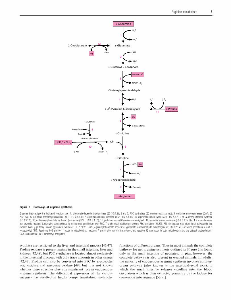
The metabolic pathway for arginine synthesis in mammals via
P5C synthetase and proline oxidase is illustrated in Figure 2
[41,42]. Some of the enzymes in this pathway are present in a
variety of cell types, while expression of other enzymes is highly
restricted. Phosphate-dependent glutaminase, ornithine am-
inotransferase (OAT), argininosuccinate synthase (ASS), arg-
ininosuccinate lyase (ASL) and aspartate aminotransferase are
widely distributed in animal tissues [42–45], whereas CPS I,
ornithine carbamoyltransferase (OCT) and N-acetylglutamate
3Arginine metabolism
2
3
4
5
6
8
7
11
12
9
10
1
L-Glutamate
ATP
ADP
Glu
L-Ornithine
L-Citrulline
CP
L-Argininosuccinate
L-Arginine
Fumarate
ATP + Asp
AMP-PPi
2-oxoglutarate
L-Proline
L-Glutamyl-c-semialdehyde
L-Glutamyl-c-phosphate
OAA
2-Oxoglutarate
Asp
NADPH + H+
NADP+
+ Pi
H2O
NH4
+
H2O O
2H
2O
1
2
L-D1-Pyrroline-5-carboxylate
L-Glutamate
HCO3– + NH
3
Acetyl-CoA
N-Acetylglutamate
2 ATP 2 ADP + Pi
L-Glutamine
CoA
Figure 2 Pathways of arginine synthesis
Enzymes that catalyse the indicated reactions are : 1, phosphate-dependent glutaminase (EC 3.5.1.2) ; 2 and 3, P5C synthetase (EC number not assigned) ; 5, ornithine aminotransferase (OAT ; EC
2.6.1.13) ; 6, ornithine carbamoyltransferase (OCT ; EC 2.1.3.3) ; 7, argininosuccinate synthase (ASS ; EC 6.3.4.5) ; 8, argininosuccinate lyase (ASL ; EC 4.3.2.1) ; 9, N-acetylglutamate synthase
(EC 2.3.1.1) ; 10, carbamoyl-phosphate synthase I (ammonia) (CPS I ; EC 6.3.4.16) ; 11, proline oxidase (EC number not assigned) ; 12, aspartate aminotransferase (EC 2.6.1.1). Step 4 is a spontaneous,
non-enzymic reaction. Glutamyl-γ-semialdehyde is in chemical equilibrium with P5C. The chemical equilibrium favours P5C formation [21,22]. P5C synthetase is a bifunctional polypeptide that
exhibits both γ-glutamyl kinase (glutamate 5-kinase ; EC 2.7.2.11) and γ-glutamylphosphate reductase (glutamate-5-semialdehyde dehydrogenase ; EC 1.2.1.41) activities (reactions 2 and 3
respectively) [41]. Reactions 1–6 and 9–11 occur in mitochondria, reactions 7 and 8 take place in the cytosol, and reaction 12 can occur in both mitochondria and the cytosol. Abbreviations :
OAA, oxaloacetate ; CP, carbamoyl phosphate.
synthase are restricted to the liver and intestinal mucosa [46,47].
Proline oxidase is present mainly in the small intestine, liver and
kidneys [42,48], but P5C synthetase is located almost exclusively
in the intestinal mucosa, with only trace amounts in other tissues
[42,47]. Proline can also be converted into P5C by -pipecolic
acid oxidase and sarcosine oxidase [49], but it is not known
whether these enzymes play any significant role in endogenous
arginine synthesis. The differential expression of the various
enzymes has resulted in highly compartmentalized metabolic
functions of different organs. Thus in most animals the complete
pathway for net arginine synthesis outlined in Figure 2 is found
only in the small intestine of neonates ; in pigs, however, the
complete pathway is also present in weaned animals. In adults,
the majority of endogenous arginine synthesis involves an inter-
organ pathway (also known as the intestinal–renal axis), in
which the small intestine releases citrulline into the blood
circulation which is then extracted primarily by the kidney for
conversion into arginine [50,51].
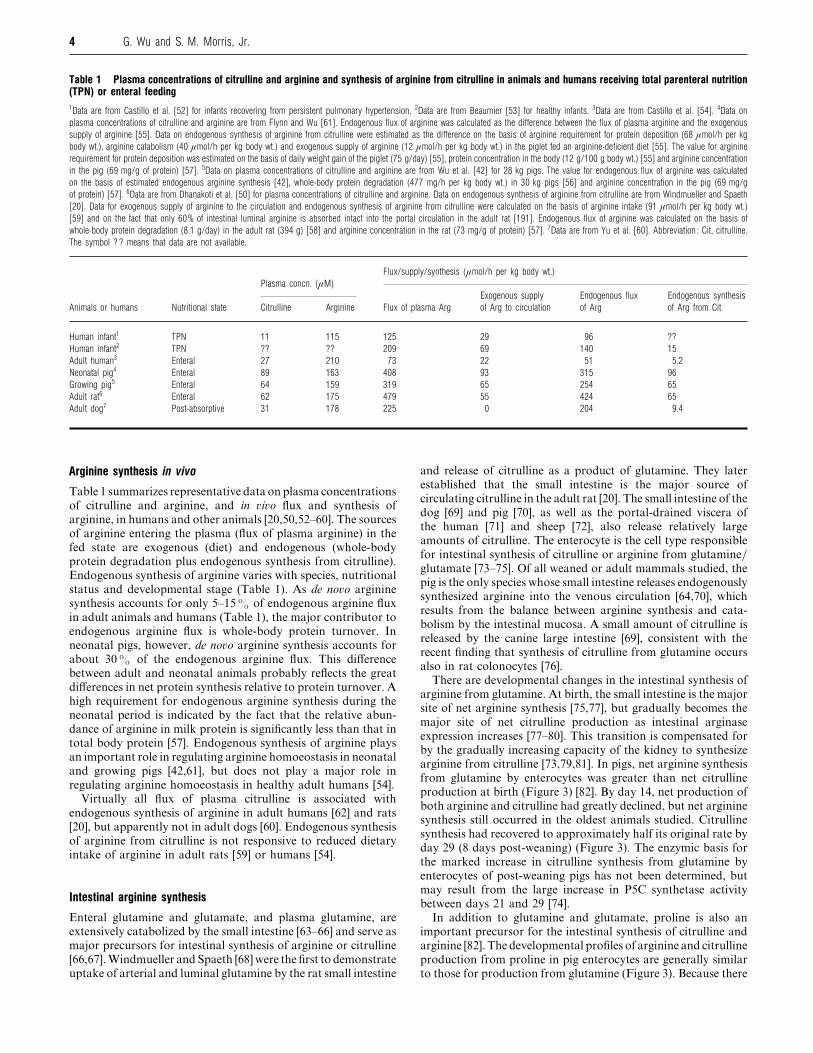
4 G. Wu and S. M. Morris, Jr.
Table 1 Plasma concentrations of citrulline and arginine and synthesis of arginine from citrulline in animals and humans receiving total parenteral nutrition(TPN) or enteral feeding1Data are from Castillo et al. [52] for infants recovering from persistent pulmonary hypertension. 2Data are from Beaumier [53] for healthy infants. 3Data are from Castillo et al. [54]. 4Data on
plasma concentrations of citrulline and arginine are from Flynn and Wu [61]. Endogenous flux of arginine was calculated as the difference between the flux of plasma arginine and the exogenous
supply of arginine [55]. Data on endogenous synthesis of arginine from citrulline were estimated as the difference on the basis of arginine requirement for protein deposition (68 µmol/h per kg
body wt.), arginine catabolism (40 µmol/h per kg body wt.) and exogenous supply of arginine (12 µmol/h per kg body wt.) in the piglet fed an arginine-deficient diet [55]. The value for arginine
requirement for protein deposition was estimated on the basis of daily weight gain of the piglet (75 g/day) [55], protein concentration in the body (12 g/100 g body wt.) [55] and arginine concentration
in the pig (69 mg/g of protein) [57]. 5Data on plasma concentrations of citrulline and arginine are from Wu et al. [42] for 28 kg pigs. The value for endogenous flux of arginine was calculated
on the basis of estimated endogenous arginine synthesis [42], whole-body protein degradation (477 mg/h per kg body wt.) in 30 kg pigs [56] and arginine concentration in the pig (69 mg/g
of protein) [57]. 6Data are from Dhanakoti et al. [50] for plasma concentrations of citrulline and arginine. Data on endogenous synthesis of arginine from citrulline are from Windmueller and Spaeth
[20]. Data for exogenous supply of arginine to the circulation and endogenous synthesis of arginine from citrulline were calculated on the basis of arginine intake (91 µmol/h per kg body wt.)
[59] and on the fact that only 60% of intestinal luminal arginine is absorbed intact into the portal circulation in the adult rat [191]. Endogenous flux of arginine was calculated on the basis of
whole-body protein degradation (8.1 g/day) in the adult rat (394 g) [58] and arginine concentration in the rat (73 mg/g of protein) [57]. 7Data are from Yu et al. [60]. Abbreviation : Cit, citrulline.
The symbol ?? means that data are not available.
Flux/supply/synthesis (µmol/h per kg body wt.)
Plasma concn. (µM)
Exogenous supply Endogenous flux Endogenous synthesis
Animals or humans Nutritional state Citrulline Arginine Flux of plasma Arg of Arg to circulation of Arg of Arg from Cit
Human infant1 TPN 11 115 125 29 96 ??
Human infant2 TPN ?? ?? 209 69 140 15
Adult human3 Enteral 27 210 73 22 51 5.2
Neonatal pig4 Enteral 89 163 408 93 315 96
Growing pig5 Enteral 64 159 319 65 254 65
Adult rat6 Enteral 62 175 479 55 424 65
Adult dog7 Post-absorptive 31 178 225 0 204 9.4
Arginine synthesis in vivo
Table 1 summarizes representative data on plasma concentrations
of citrulline and arginine, and in �i�o flux and synthesis of
arginine, in humans and other animals [20,50,52–60]. The sources
of arginine entering the plasma (flux of plasma arginine) in the
fed state are exogenous (diet) and endogenous (whole-body
protein degradation plus endogenous synthesis from citrulline).
Endogenous synthesis of arginine varies with species, nutritional
status and developmental stage (Table 1). As de no�o arginine
synthesis accounts for only 5–15% of endogenous arginine flux
in adult animals and humans (Table 1), the major contributor to
endogenous arginine flux is whole-body protein turnover. In
neonatal pigs, however, de no�o arginine synthesis accounts for
about 30% of the endogenous arginine flux. This difference
between adult and neonatal animals probably reflects the great
differences in net protein synthesis relative to protein turnover. A
high requirement for endogenous arginine synthesis during the
neonatal period is indicated by the fact that the relative abun-
dance of arginine in milk protein is significantly less than that in
total body protein [57]. Endogenous synthesis of arginine plays
an important role in regulating arginine homoeostasis in neonatal
and growing pigs [42,61], but does not play a major role in
regulating arginine homoeostasis in healthy adult humans [54].
Virtually all flux of plasma citrulline is associated with
endogenous synthesis of arginine in adult humans [62] and rats
[20], but apparently not in adult dogs [60]. Endogenous synthesis
of arginine from citrulline is not responsive to reduced dietary
intake of arginine in adult rats [59] or humans [54].
Intestinal arginine synthesis
Enteral glutamine and glutamate, and plasma glutamine, are
extensively catabolized by the small intestine [63–66] and serve as
major precursors for intestinal synthesis of arginine or citrulline
[66,67].Windmueller and Spaeth [68] were the first to demonstrate
uptake of arterial and luminal glutamine by the rat small intestine
and release of citrulline as a product of glutamine. They later
established that the small intestine is the major source of
circulating citrulline in the adult rat [20]. The small intestine of the
dog [69] and pig [70], as well as the portal-drained viscera of
the human [71] and sheep [72], also release relatively large
amounts of citrulline. The enterocyte is the cell type responsible
for intestinal synthesis of citrulline or arginine from glutamine}glutamate [73–75]. Of all weaned or adult mammals studied, the
pig is the only species whose small intestine releases endogenously
synthesized arginine into the venous circulation [64,70], which
results from the balance between arginine synthesis and cata-
bolism by the intestinal mucosa. A small amount of citrulline is
released by the canine large intestine [69], consistent with the
recent finding that synthesis of citrulline from glutamine occurs
also in rat colonocytes [76].
There are developmental changes in the intestinal synthesis of
arginine from glutamine. At birth, the small intestine is the major
site of net arginine synthesis [75,77], but gradually becomes the
major site of net citrulline production as intestinal arginase
expression increases [77–80]. This transition is compensated for
by the gradually increasing capacity of the kidney to synthesize
arginine from citrulline [73,79,81]. In pigs, net arginine synthesis
from glutamine by enterocytes was greater than net citrulline
production at birth (Figure 3) [82]. By day 14, net production of
both arginine and citrulline had greatly declined, but net arginine
synthesis still occurred in the oldest animals studied. Citrulline
synthesis had recovered to approximately half its original rate by
day 29 (8 days post-weaning) (Figure 3). The enzymic basis for
the marked increase in citrulline synthesis from glutamine by
enterocytes of post-weaning pigs has not been determined, but
may result from the large increase in P5C synthetase activity
between days 21 and 29 [74].
In addition to glutamine and glutamate, proline is also an
important precursor for the intestinal synthesis of citrulline and
arginine [82]. The developmental profiles of arginine and citrulline
production from proline in pig enterocytes are generally similar
to those for production from glutamine (Figure 3). Because there

5Arginine metabolism
500
400
300
200
100
0
200
150
100
50
0
0 10 20 30 40 50 60
Glutamine
Proline
Glutamine
ProlineA
rgin
ine
fo
rma
tio
n
(nm
ol/3
0 m
in p
er
mg
of
DN
A)
Cit
rullin
e f
orm
ati
on
(nm
ol/3
0 m
in p
er
mg
of
DN
A)
Age (days)
Figure 3 Developmental changes in synthesis of arginine and citrulline bypig enterocytes
Enterocytes were isolated from the jejenum of pigs at various times after birth (day 0) and
incubated in Krebs bicarbonate buffer containing 2 mM L-glutamine or 2 mM [14C]proline plus
2 mM L-glutamine. The incubation medium also contained 5 mM D-glucose. Pigs were weaned
at day 21. Data are from Table 2 (L-glutamine) and Table 4 (L-proline) of Wu [82].
is no significant uptake of arterial proline by the small intestine
[70], the enteral diet must be the major source of proline for
citrulline synthesis in enterocytes, consistent with the recent
finding that citrulline is a major product of enterally delivered
proline in the pig [83]. Although it is often stated that proline
oxidase is present mainly in the liver, kidney and brain [48,84]
and that proline is not catabolized in the gut [47,85], relatively
high activities of proline oxidase have been found in the small
intestine and enterocytes of pigs [82,86] and rats [82]. In fact, the
activity of proline oxidase in the pig small intestine is several-fold
greater than that in the liver and kidney [42,86]. The failure to
detect intestinal proline oxidase activity in earlier studies may
have been due to the presence of a proline oxidase inhibitor in
tissue homogenates or to a lack of proteinase inhibitors in the
buffers used for tissue homogenization.
The essential role of the small intestine in arginine synthesis is
graphically demonstrated by the arginine deficiencies that result
when intestinal citrulline synthesis is blocked by inhibitors of
OCT [87] or OAT [61] or by massive resection of the small bowel
[88,89]. Similarly, arginine deficiencies occur in individuals with
inherited defects in OCT [90], OAT [91,92] or P5C synthetase
[93]. An analogous situation exists in strict carnivores, such as
cats, which synthesize very little arginine and thus must rely on
the diet to meet their needs for this amino acid [94]. Although
feline kidneys contain the enzymes required for arginine synthesis
[95], cats synthesize little citrulline because their intestines have
relatively low activities of P5C synthetase and OAT [94,96]. The
requirement for dietary arginine by cats and other carnivores
(such as ferrets) is so stringent that ingestion of an arginine-free
meal rapidly leads to hyperammonaemia, encephalopathy and,
in the case of cats, even death [94,97].
Relatively little is known about how the intestinal synthesis of
citrulline and arginine is regulated. Glucocorticoids precociously
induce activities of several of the enzymes in the intestines of
immature animals [98–101], indicating that these hormones
probably play a role in the developmental maturation of this
pathway, as is the case for many other developmentally
regulated pathways in this organ [102]. This may explain why
dexamethasone increases plasma concentrations of ornithine,
citrulline and arginine in pre-term infants [103]. In contrast
with the liver, activities of intestinal OCT [104,105] and OAT
[106] in adult rats were modestly decreased by increasing the
dietary protein intake, and expression of intestinal OCT and CPS
I was unaffected by starvation [107]. The latter observation is
consistent with the finding that citrulline production by the small
intestine does not increase in rats and pigs fed an arginine-
deficient diet, even when an additional substrate in the form of
glutamate is included in the diet [108,109].
Renal arginine synthesis
Analyses of the arginine biosynthetic enzymes in kidney in the
1940s [110,111] paved the way for physiological studies which
established the kidney as the major organ involved in endogenous
arginine synthesis [50,51]. Approximately 60% of net arginine
synthesis in adult mammals occurs in the kidney [50,60], where
citrulline is extracted from the blood and converted stoichio-
metrically into arginine by the action of ASS and ASL (Figure 2),
which are localized within the proximal convoluted tubules
[81,112–115]. A tight correlation between renal citrulline uptake
and renal arginine output has been elegantly demonstrated for
both humans and rats [50,116]. Furthermore, in �i�o rates of
arginine synthesis in adult rats are limited primarily by the
amount of citrulline produced by other organs, such as the small
intestine, rather than by the renal arginine biosynthetic capacity
[50]. As the renal arginine biosynthetic capacity appears to be
several-fold greater than the intestinal capacity for citrulline
production, it is unclear why renal activities of, or mRNA levels
for, ASS and ASL are increased by a high-protein diet [117,118].
Renal mRNA levels for ASS and ASL also increase during
starvation [118], probably as an adaptive response to maintain
plasma arginine levels in the absence of dietary protein. As noted
in the preceding section, the renal capacity for arginine synthesis
develops in late fetal stages and continues to increase after birth
[81], complementing the developmental shift from release of
arginine to release of citrulline by the small intestine. The kidney
also expresses arginase, but expression of arginase and the
arginine biosynthetic enzymes is highly segregated within dif-
ferent parts of the nephron, so that there is little or no co-
expression of these opposing enzymic pathways within the same
cell [113,119].
As expected, individuals with chronic renal insufficiency have
elevated plasma levels of citrulline [116,120,121]. Surprisingly,
however, there is little or no decrease in plasma arginine in these
patients. The basis for the maintenance of plasma arginine at
normal or near-normal levels is unknown, but probably involves
a combination of factors [40,122], including increased release
of arginine by protein catabolism in skeletal muscle, increased
arginine synthesis at extrarenal sites, hypertrophy of proximal
tubules, hyperfiltration (which increases the amount of citrulline
filtered per nephron), and an increased rate of arginine synthesis
due to elevated plasma levels of citrulline. The last point follows
from the finding that rates of renal arginine synthesis are
essentially a function of plasma citrulline levels [50]. In addition,
the proposal that arginine degradation is more important than
arginine synthesis in maintaining arginine homoeostasis in adult
humans [54] raises the possibility of some compensatory decrease
in arginine degradation.

6 G. Wu and S. M. Morris, Jr.
ASSNOS
ASL
3
2
1
L-Arginine
L-Citrulline L-Aspartate
L-MalateFumarate
Oxaloacetate
L-Argininosuccinate
O2
.NO
ATP
AMP + PPi
NAD+
NADH + H+
L-Glutamate
2-Oxoglutarate
Figure 4 Citrulline/NO cycle
This cytosolic cycle can be coupled to the citric acid cycle, as shown on the right. Fumarate produced in the cytosol enters the citric acid cycle in the mitochondrion, where it is converted into
oxaloacetate. Transamination converts oxaloacetate into aspartate, which is transported into the cytosol. Enzymes catalysing reactions 1–3 are : 1, fumarase (EC 4.2.1.2) ; 2, malate dehydrogenase
(EC 1.1.1.37) ; 3, aspartate aminotransferase. Although reactions 1–3 are reversible, the diagram depicts the net unidirectional flow in NO-producing cells. (Modified from Figure 5 of Nussler et
al. [143] and reprinted with permission of the American Society for Biochemistry and Molecular Biology.)
Hepatic arginine synthesis
The highest rates of arginine synthesis occur within the hepatic
urea cycle, which is localized within periportal hepatocytes [39].
In healthy adult humans, for example, rates of urea production
(239 µmol}h per kg in the fed state and 184 µmol}h per kg in the
fasted state) are vastly greater than rates of NO synthesis
(0.91 µmol}h per kg in the fed state and 1.00 µmol}h per kg in
the fasted state) [62], and also much higher than rates of creatine
synthesis (7.9 µmol}h per kg) [123]. Net arginine synthesis by the
liver is only possible if the urea cycle is replenished by necessary
intermediates, such as ornithine. The urea cycle enzymes are also
organized in a metabolon [124], whereby the product of each
enzymic reaction is efficiently channelled to the next enzyme in
the pathway [124,125]. Thus the tight channelling of metabolites
and the very high level of arginase in hepatocytes result in little
or no net production of arginine by the liver. This is dramatically
illustrated by the fact that individuals who cannot make arginine
because of inherited defects in the urea cycle continue to require
arginine in their diet after receiving liver transplants [90].
Expression of hepatic urea-cycle enzymes begins during fetal
development and continues to increase after birth [126–128].
Subsequently, levels of the urea-cycle enzymes are co-ordinately
induced by conditions involving increased protein and amino
acid catabolism, such as increased dietary protein intake, starv-
ation and increases in glucocorticoid levels or the glucagon}insulin ratio [46]. One exception is the cat, a strict carnivore in
which levels of the urea-cycle enzymes are unaffected by changes
in dietary protein intake [129]. These long-term adaptive increases
largely reflect increased transcription rates of the genes encoding
these enzymes. Rapid, short-term changes in urea-cycle activity
occur primarily via changes in CPS I catalytic efficiency, which in
turn is regulated by changes in the mitochondrial concentration
of N-acetylglutamate [39]. As the mechanisms that regulate urea
cycle activity have been extensively discussed in previous reviews
[39,46,130,131], they will not be considered here.
In response to inflammatory conditions such as sepsis, hepato-
cytes can be induced to produce NO in addition to urea [132].
This fact raised the question as to whether the urea cycle also
provides arginine for NO synthesis. To address this matter,
several groups examined the metabolic consequences of perfusing
various nitrogenous substrates into livers isolated from rats in
which high-level hepatic NO synthesis had been induced
[133–135]. In one such study, perfusion of isolated livers with
glutamine or NH%Cl resulted in large increases in urea synthesis
without any change in NO synthesis, indicating that arginine
made within the urea cycle was not available for NO synthesis
[134]. In contrast, a similar study by another group did find an
increase in NO production when livers were perfused with
NH%Cl [133]. These apparently disparate outcomes might have
reflected differences in, for example, the integrity of the hepato-
cytes resulting from the different stimuli used to induce hepatic
NO synthesis. In any event, both studies are consistent with the
view that, if the urea cycle does provide any arginine for hepatic
NO synthesis, it must represent only a tiny fraction of the total
arginine synthesized within the urea cycle. Although competition
for arginine between the urea cycle and hepatic inducible NO
synthase (iNOS) was claimed also by a third group [135], their
study only showed that exogenously supplied arginine could be
utilized by both arginase and iNOS; it did not determine whether
arginine synthesized within the urea cycle could be used for
hepatic NO synthesis. Finally, the hepatic capacity for arginine
synthesis within the urea cycle, as indicated by mRNA levels for
ASS and ASL, is not affected by inflammatory stimuli such as
lipopolysaccharide (LPS) [136,137].
Arginine synthesis in NO-producing cells
The arginine biosynthetic pathway represents a regulated and
highly localized source of substrate for NO synthesis in a wide
variety of non-hepatic cells [138,139]. Citrulline, which is co-
produced with NO, can be recycled to arginine via a pathway
that has been termed the citrulline}NO cycle [139] or the
arginine}citrulline cycle [140] (Figure 4). This recycling is ac-
complished by the combined action of ASS and ASL, which are
expressed to some degree in nearly all cell types. The existence of
the citrulline}NOcycle is supported by the fact that total citrulline
production is lower than total NO production for some cell types
[141], and also by the demonstration that citrulline can replace
arginine, at least in part, in supporting NO synthesis by intact
cells [142–145]. Although the data clearly demonstrate the
conversion of citrulline into arginine, there is no direct experi-
mental evidence that the aspartate used for argininosuccinate
formation is produced from fumarate as depicted in Figure 4, or
by a possible alternative route [142] whereby malate is instead
converted into pyruvate by malic enzyme, and pyruvate is then

7Arginine metabolism
converted into oxaloacetate by pyruvate carboxylase. The fact
that citrulline accumulates to a considerable extent in the medium
of NO-producing cells demonstrates that the citrulline}NO cycle
is much less efficient than the hepatic urea cycle, indicating that
the activity of ASS is appreciably less than the activity of iNOS
and}or that there is little or no channelling of substrates and
products between iNOS and ASS.
Following initial reports that NO synthesis and arginine
biosynthetic capacity were co-induced in macrophages [146],
several laboratories established that induction of iNOS in all
non-hepatic mammalian cells examined to date is accompanied
by induction of ASS, a rate-controlling enzyme in arginine
biosynthesis [136,142–144,147–150]. In rat tissues, ASL is also
co-induced with iNOS [136,137]. Because basal expression of
ASS differs greatly among different cell types, the magnitude
of ASS induction by inflammatory stimuli is highly variable.
The observation that ASS and iNOS were co-induced led to the
hypothesis [143,151] that regulation of the arginine recycling
pathway could itself represent a potential mechanism for regu-
lating inducible NO synthesis. This hypothesis was confirmed by
the finding that vascular smooth muscle cells that had been
transfected to overexpress ASS had higher levels of induced NO
production at limiting extracellular arginine concentrations than
did untransfected cells [152]. Importantly, this result also demon-
strated that rates of arginine uptake were not sufficient to
support maximal rates of NO synthesis, further supporting
the general proposition that any mechanism that regulates the
availability of arginine represents a potential control point for
NO synthesis [151]. Despite the provocative results obtained with
cultured cells, it should be emphasized that the contribution of
the arginine recycling pathway to NO synthesis in �i�o is
completely unknown.
-Glutamine and hypoxia are physiological regulators of
arginine synthesis in NO-producing cells. Inhibition of arginine
synthesis by glutamine has been reported for NO-producing
endothelial cells [153–155], cerebral perivascular nerve tissues
[156] and rat peritoneal macrophages [146], but not for a murine
macrophage cell line [153]. Glutamine-dependent inhibition of
endothelial arginine synthesis appeared to occur via (1) com-
petitive inhibition of citrulline uptake [154] and (2) a decrease in
ASS activity [155]. Several other amino acids (-alanine, -
glutamate and -lysine) were also tested, but did not mimic the
effects of glutamine [154]. Inhibition of arginine synthesis by
glutamine in rat peritoneal macrophages has not been charac-
terized. Hypoxia was reported to inhibit arginine synthesis in
endothelial cells by reducing ASS activity rather than citrulline
uptake [155] ; the basis for ASS inhibition is unknown.
ARGININE CATABOLISM
Arginine can be catabolized via multiple pathways (Figure 5),
many of which are co-expressed within the same cell. For
example, iNOS, the arginases and arginine decarboxylase can be
co-expressed in murine macrophages, as described in the fol-
lowing sections. This can result in complex interactions, whereby
the product of one enzyme may inhibit the activity of another
enzyme, e.g. as in the inhibition of arginase by NG-hydroxy-
arginine. The cellular distribution of enzyme expression varies
widely. For example, iNOS can be expressed in almost any cell
type which is exposed to the appropriate stimuli [157], whereas
expression of arginine:glycine amidinotransferase is much more
restricted, being limited principally to kidney, pancreas and, to a
lesser extent, liver [158,159]. The type II isoenzymes of arginase
and OAT are expressed in many cell types [42,47,160–162],
indicating a widespread capacity for synthesis of proline and}or
glutamate from arginine. As OAT and ornithine decarboxylase
(ODC) are located in different subcellular compartments (mito-
chondria and cytosol respectively), the ornithine produced by
mitochondrial or cytosolic arginases probably has differing
metabolic fates. In short, arginine catabolism in mammals
involves multiple organs and complex compartmentation at the
cellular and systemic levels.
Arginine transport
As arginine transport systems may regulate substrate availability
for arginine-requiring enzymes, a brief survey of this topic is
appropriate. Because arginine transport is the subject of several
reviews over the past few years [163–168], we will summarize
points most relevant to the present review. In most mammalian
cells, arginine requirements are met primarily by uptake of
extracellular arginine via specific transporters, such as systems
y+, bo,+, Bo,+ or y+L [163,164,168]. Not all transporters are found
in every cell type, and activities of specific transporters can be
dynamically regulated in response to specific stimuli, such as
bacterial endotoxin and inflammatory cytokines [166]. The most
important mechanism for arginine uptake in most cell types is
system y+, a high-affinity, Na+-independent transporter of ar-
ginine, lysine and ornithine. Recent studies have identified
cDNAs encoding two transmembrane proteins, CAT-1 and CAT-
2(B), which have amino acid transport properties consistent with
system y+ [165,167]. A cDNA encoding rCAT3, a brain-specific
protein which also exhibits system-y+ activity, has recently been
isolated from rats [169]. Still unresolved is the question of
whether system y+ consists solely of these proteins or involves
other proteins as yet unidentified. Because of its major role in
arginine transport, regulation of system-y+ expression or activity
represents a potential target for modulating cellular arginine
metabolism.
Other cationic amino acids and positively charged analogues
are effective inhibitors of arginine uptake by system y+. For
example, arginine uptake can be competitively inhibited by
lysine, ornithine, canavanine and certain NOS inhibitors, in-
cluding NG-monomethyl--arginine and NG-iminoethyl--orni-
thine, but not by other NOS inhibitors such as aminoguanidine,
NG-nitro--arginine and NG-nitro--arginine methyl ester [170–
175]. Thus use of NOS inhibitors that are taken up via system y+
may limit the availability of arginine for other enzymes that
utilize this amino acid. It is important to note that NG-mono-
methyl--arginine, NG-nitro--arginine, NG-nitro--arginine
methyl ester and aminoguanidine have no significant activity as
direct inhibitors of arginase [176,177].
The expression of system y+ not only varies among different
cell types, but can also be dynamically regulated at the pre-
translational level. System y+ is present in a variety of cell types,
but is virtually absent from hepatocytes. Thus " 85% of the
arginine delivered to the livers of rats [178] or dogs [60] is not
taken up by the liver. However, system y+ can be induced by
inflammatory cytokines in hepatocytes and other cells [166,167].
In fact, system-y+ expression is co-induced with iNOS in a wide
variety of cell types [150,179–184], indicating that arginine
transport capacity increases to support the elevated rates of NO
synthesis. In rat astrocytes, induced NO synthesis is strictly
dependent on co-induction of system y+ [185]. It has not been
determined whether induction of system y+ is a general response
in conditions where arginine consumption is elevated, e.g. when
arginase is induced in the absence of iNOS induction.
Recent studies have indicated that the precise cellular localiz-
ation of arginine transporters may be responsible for the ‘arginine
paradox’ [186], the observation that endothelial NO synthesis

8 G. Wu and S. M. Morris, Jr.
5
12 11
10
13
14
9
7 8
6
17
16
1
15
2
4
3L-Agmatine L-Arginine
L-Citrulline
NO
L-OrnithineGuanidinoacetate
Creatine
Putrescine
Spermidine
Spermine
L-Glutamyl-c-semialdehyde
L-GlutamineL-Glutamate2-Oxoglutarate
L-Proline
Urea
Ornithine
Glycine
SAM
SAHC
NAD(P)H + H+
NAD(P)+
NAD+
NADH + H+
NH3
CO2
2-Oxo acidα-Amino acid ATP ADP+Pi
NH4
+
H2ONAD(P)H + H
+
NAD(P)+
H2O
DCAM
MTA
DCAM
MTA
Glu
2-Oxoglutarate
H2O
CO2
BH4
NADPH + H+
NADP+
CO2
L-D1-Pyrroline-5-carboxylate
H2O
Figure 5 Pathways of arginine catabolism
Enzymes that catalyse the indicated reactions are : 1, arginase (EC 3.5.3.1) ; 2, NOS (EC 1.14.13.39) ; 3, arginine decarboxylase (EC 4.1.1.19) ; 4, arginine : glycine amidinotransferase (EC 2.1.4.1) ;
5, guanidinoacetate N-methyltransferase (EC 2.1.1.2) ; 6, OAT ; 7, P5C reductase (EC 1.5.1.2) ; 9, P5C dehydrogenase ; 10, glutamate dehydrogenase (EC 1.4.1.2) ; 11, alanine aminotransferase (EC
2.6.1.12), aspartate aminotransferase or branched-chain amino acid aminotransferase (EC 2.6.1.42) ; 13, glutamine synthetase (EC 6.3.1.2) ; 14, glutaminase (EC 3.5.1.2) ; 15, ornithine decarboxylase
(ODC ; EC 4.1.1.17) ; 16, spermidine synthase (EC 2.5.1.16) ; 17, spermine synthase (EC 2.5.1.22). Complete oxidation of arginine-derived 2-oxoglutarate occurs via the citric acid cycle (step 12).
Step 8 is a spontaneous, non-enzymic reaction. See the legend to Figure 2. Abbreviations : DCAM, decarboxylated S-adenosylmethionine ; MTA, methylthioadenosine ; SAM, S-adenosylmethionine ;
SAHC, S-adenosylhomocysteine ; BH4, (6R )-5,6,7,8-tetrahydro-L-biopterin.
can be regulated by varying the extracellular arginine con-
centration, despite the fact that the reported intracellular arginine
concentrations (0.1–1 mM) greatly exceed the Km
of endothelial
NOS (eNOS) for arginine (2.9 µM). The apparent Km
of NO
synthesis by intact cells for extracellular arginine is approx.
73–150 µM [187,188], which is within the range of the Km
values
of the arginine transport systems (100–150 µM) [165] and of
plasma arginine concentrations (Table 1). Immunohistochemical
studies [189] demonstrated that CAT-1, eNOS and caveolin are
co-localized in plasma-membrane caveolae, suggesting a pref-
erential channelling or directed delivery of extracellular arginine
to eNOS, as proposed previously [174]. It is not known whether
preferential channelling of extracellular arginine also occurs for
other arginine-requiring enzymes.
Arginine catabolism in vivo
Our knowledge of arginine catabolism in �i�o is limited, due in
large part to the complex compartmentalization of arginine
metabolism at both the organ and subcellular levels. However,

9Arginine metabolism
our understanding of this subject is being expanded by recent
tracer studies using stable isotopes. Only 5% of urea production
is derived from plasma arginine [62], reflecting very low uptake
of arginine by the liver and the strict segregation of hepatic
and plasma arginine pools. Relative rates of NO synthesis from
plasma arginine are low. For example, in infants [52] and adult
humans [62], NO synthesis represent only 0.48% and 1.2%
respectively of the flux of plasma arginine. This accounts for
about half of total NO production, because plasma arginine
provides only 54% of the arginine used in NO synthesis [62] ; the
remainder is presumably derived from endogenous sources, such
as protein degradation and endogenous arginine synthesis at
sites of NO synthesis. The fractions of plasma arginine flux
associated with the synthesis of citrulline and NO are virtually
identical in adult humans [62], strongly indicating that production
of plasma citrulline from arginine in �i�o is due entirely to NOS
activity. In adult humans, an arginine-free diet reduced the flux
of plasma arginine and endogenous NO synthesis [190]. In the
neonatal pig, however, an arginine-deficient diet did not alter
the flux of plasma arginine or its conversion into metabolic
products [55], suggesting species or developmental differences in
arginine metabolism in �i�o.
Owing to a relatively high activity of arginase in the intestinal
mucosa of adults, approx. 40% of the arginine absorbed from
the intestinal lumen is degraded in the first pass in rats [191] and
humans [192], and the remainder of the absorbed arginine is
released into the venous blood. About one-third of the ornithine
produced from exogenous arginine is released by the rat small
intestine [191] or isolated pig enterocytes [193] ; the remaining
two-thirds of the ornithine is further catabolized to various
metabolites, as indicated in Figure 5. Thus changes in intestinal
arginase expression can have a major impact on the metabolic
fates of arginine and on the availability of dietary arginine to
extra-intestinal tissues [66].
Arginase
Interest in the arginases as possible regulatory enzymes is growing
because of their potential for regulating the availability of
arginine for the synthesis of NO, polyamines, agmatine, proline
and glutamate. Although much of the evidence for the role of the
arginases in providing or depleting substrate for other bio-
synthetic pathways is circumstantial, the general conclusions
drawn in many of the studies cited below are likely to be correct.
As cloning of the arginase cDNAs [138,148,160,162] and de-
velopment of potent arginase inhibitors [194–197] have now
provided the means to test directly the validity of these con-
clusions, we anticipate that many of these studies will be revisited
with new experimental tools.
It is important to recognize that there are two distinct
isoenzymes of mammalian arginase, which are encoded by
separate genes. They are quite similar with regard to enzymic
properties and requirement for manganese, but differ with regard
to subcellular localization, tissue distribution, regulation of
expression and immunological reactivity [198,199]. Type I argin-
ase, a cytosolic enzyme, is highly expressed in liver as a component
of the urea cycle, and to a limited extent in a few other tissues.
In contrast, type II arginase, a mitochondrial enzyme, is expressed
at lower levels in kidney, brain, small intestine, mammary gland
and macrophages, but there is little or no expression in liver
[138,162,198,199]. Rat aortic endothelial cells and murine macro-
phages express both type I and type II arginases [200–202], and
it is likely that other cell types also express both isoenzymes. The
different subcellular localization of the arginase isoenzymes may
provide a mechanism for regulating themetabolic fate of arginine,
as postulated for enterocytes [203]. For example, differential
expression of the arginase isoenzymes could provide a means to
preferentially direct ornithine either to proline or glutamate
synthesis via OAT or to polyamine synthesis via ODC (Figure 2).
If so, this would imply that ornithine does not rapidly equilibrate
between cytosolic and mitochondrial compartments, despite the
existence of transporters via which ornithine can traverse
the mitochondrial membrane [204–208]. So far as we are aware,
no experiments to determine the effect of arginase localization
on the metabolic fates of ornithine have been performed.
Arginase and ureagenesis
As noted above, the high levels of type I arginase in liver,
together with the channelling of metabolites within the urea
cycle, serve to ensure that this pathway for detoxifying waste
nitrogen operates at high efficiency. Arginase is unique among
the urea-cycle enzymes in that two distinct isoenzymes exist.
Thus inherited defects in the hepatic (type I) arginase are partially
compensated for by elevated expression of type II arginase in
kidney [209,210], resulting in a less severe clinical disorder [130].
As Mn#+ can allosterically activate hepatic arginase in a pH-
sensitive fashion, it has been suggested that pH-dependent
regulation of arginase activity may contribute to the pH-
dependence of hepatic urea production [211]. However, changes
in hepatic amino acid transport [212,213] and possibly also in
activities of key enzymes involved in amino acid catabolism,
rather than changes in activities of the urea-cycle enzymes per se,
are probably much more important in regulating pH-dependent
alterations in hepatic urea synthesis.
Because all urea-cycle enzymes are present to some extent in
the small intestine [214–216], several investigators have hypo-
thesized that a metabolically significant urea cycle may function
in this organ [79,216]. Wu [217] has demonstrated urea synthesis
from both extracellular and intramitochondrially generated am-
monia in enterocytes from post-weaning pigs, although the rate
of ureagenesis is considerably less than in hepatocytes. This
result not only shows that more than one organ is capable of
synthesizing urea from ammonia in mammals, but also may help
in understanding the complex kinetics of urea metabolism in �i�o
[218]. Although some urea may be formed, it is nonetheless clear
that a major product of these enzymes in the small intestine is
citrulline. Ureagenesis in the small intestine of weaned animals
may constitute a first line of defence against the toxicity of
ammonia which is generated by intestinal glutamine catabolism
and by the microbial flora of the gut (reviewed in [219]).
Arginase and NO synthesis
At first glance, it might appear that arginase would not compete
well with NO synthesis for arginine. The Km
for arginine is in the
2–20 mM range for mammalian arginases [198], but it is in
the 2–20 µM range for the various NOS isoenzymes [220]. On the
other hand, the Vmax
of arginase at physiological pH (approx.
1400 µmol}min per mg; calculated for rat liver arginase from
[221]) is more than 1000 times that of the NOS enzymes (approx.
1 µmol}min per mg [220]), indicating similar rates of substrate
usage for NO synthesis at low arginine concentrations. Sufficient
quantities of arginase can limit the availability of arginine for
NO synthesis by intact cells. For example, in wounds [222–224]
and macrophage cultures [225,226], the extracellular fluid be-
comes almost completely depleted of arginine, whereas ornithine
increases, indicative of high arginase activity. More recently,
inhibition of arginase in LPS-treated rodent macrophages was
shown to result in enhanced conversion of arginine into citrulline,
10 G. Wu and S. M. Morris, Jr.
indicating that arginase and iNOS can compete for arginine
[227,228]. One aspect of the cell culture studies that needs further
clarification is the extent to which arginine depletion was due to
extracellular compared with intracellular arginase, i.e. were
arginase and iNOS in direct competition for an intracellular pool
of arginine? This is an important point, because the rate of
cellular NO synthesis may be limited by the rate of arginine
uptake. Thus a decrease in the extracellular arginine concen-
tration may have a more marked impact on the rate of NO
synthesis than would a corresponding decrease in the intracellular
arginine concentration.
In reality, the basis for interplay between arginase and NOS is
more complex than the fact that they use a common substrate.
For example, iNOS-expressing macrophages [228,229] and
endothelial cells [200] can produce sufficient NG-hydroxyarginine
to inhibit arginase activity. Because endothelial cells in intact
animals are constantly perfused, whereas cultured cells are not,
it is not clear whether the former would be exposed to sufficient
NG-hydroxyarginine in �i�o to inhibit cellular arginase activity.
In evaluating these results it also must be borne in mind that
arginine concentrations in standard tissue culture media are up
to 10 times higher than plasma arginine concentrations, so that
rates of NG-hydroxyarginine production by cultured cells are
probably much greater than in �i�o. Nonetheless, limited per-
fusion at localized anatomical sites such as wounds may allow
accumulation of inhibitory concentrations of NG-hydroxy-
arginine. This possibility is supported by the finding that the
plasma level of NG-hydroxyarginine is about 9 µM, even in
healthy adult humans [229], and becomes elevated in the serum
of LPS-treated rats [230]. Further complicating the picture is the
fact that NG-hydroxyarginine can be oxidized to citrulline and
NO by a variety of haem proteins, such as peroxidases, cyto-
chromes P-450, haemoglobin and catalase [231,232], as well as
by superoxide anions [233], suggesting that the half-life of NG-
hydroxyarginine, and thus its accumulation, may vary in a
tissue-specific manner. Because of its potential importance, the
role of NG-hydroxyarginine in arginine metabolism bears further
study.
Arginase and polyamine synthesis
Polyamines are essential for cell proliferation and differentiation
[234]. Support for the hypothesis that arginase may regulate the
availability of ornithine for polyamine synthesis comes from
observations that arginase activity is often co-induced with ODC
and that cells that are deficient in arginase cannot proliferate in
serum-free medium unless ornithine or polyamines are provided
[235]. In addition, arginase activity is greatly elevated in tumour
cell lines that have become resistant to difluoromethylornithine,
a potent inhibitor of ODC [234,236] ; the high arginase activity
apparently generates sufficient ornithine to compete intra-
cellularly with the ODC inhibitor.
Correlations between changes in arginase activity and poly-
amine synthesis have been found in studies of the kidney. For
example, androgens induce activities of both arginase [237–239]
and ODC [239–241] in kidneys of female or castrated male mice.
Hypertrophy generally occurs under these conditions, although
there is no direct correlation between the increase in kidney size
and renal arginase activity [236]. Androgens not only induce
arginase and ODC in kidneys of female mice, but also lower
OAT activity [237]. Taken together, these observations suggest
that renal arginase activity increases to support enhanced poly-
amine synthesis, whereas the decline in OAT activity indicates
reduced utilization of ornithine for proline or glutamate synthesis.
These conclusions may be somewhat oversimplified, because no-
30
20
10
00 10 20 30 40 50 60
Age (days)
Urea
Proline
Ornithine
Pro
du
ct
form
ati
on
(nm
ol/3
0 m
in p
er
mg
of
pro
tein
)
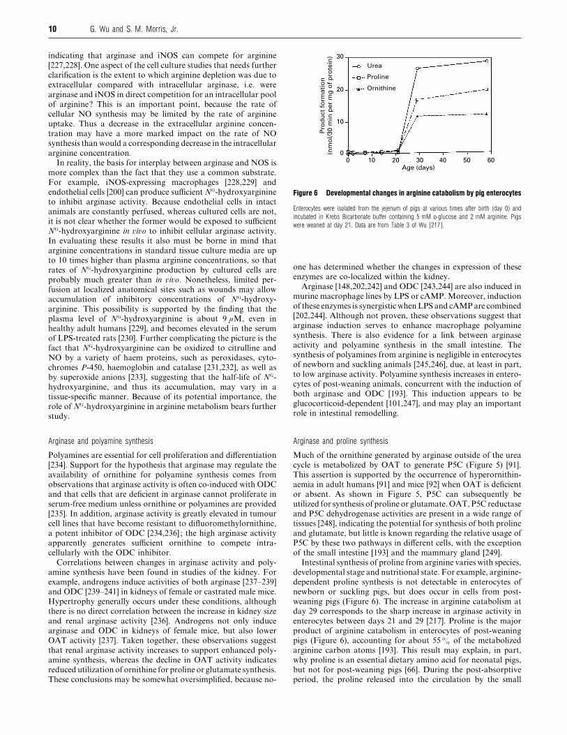
Figure 6 Developmental changes in arginine catabolism by pig enterocytes
Enterocytes were isolated from the jejenum of pigs at various times after birth (day 0) and
incubated in Krebs Bicarbonate buffer containing 5 mM D-glucose and 2 mM arginine. Pigs
were weaned at day 21. Data are from Table 3 of Wu [217].
one has determined whether the changes in expression of these
enzymes are co-localized within the kidney.
Arginase [148,202,242] and ODC [243,244] are also induced in
murine macrophage lines by LPS or cAMP. Moreover, induction
of these enzymes is synergisticwhenLPS and cAMPare combined
[202,244]. Although not proven, these observations suggest that
arginase induction serves to enhance macrophage polyamine
synthesis. There is also evidence for a link between arginase
activity and polyamine synthesis in the small intestine. The
synthesis of polyamines from arginine is negligible in enterocytes
of newborn and suckling animals [245,246], due, at least in part,
to low arginase activity. Polyamine synthesis increases in entero-
cytes of post-weaning animals, concurrent with the induction of
both arginase and ODC [193]. This induction appears to be
glucocorticoid-dependent [101,247], and may play an important
role in intestinal remodelling.
Arginase and proline synthesis
Much of the ornithine generated by arginase outside of the urea
cycle is metabolized by OAT to generate P5C (Figure 5) [91].
This assertion is supported by the occurrence of hyperornithin-
aemia in adult humans [91] and mice [92] when OAT is deficient
or absent. As shown in Figure 5, P5C can subsequently be
utilized for synthesis of proline or glutamate.OAT,P5C reductase
and P5C dehydrogenase activities are present in a wide range of
tissues [248], indicating the potential for synthesis of both proline
and glutamate, but little is known regarding the relative usage of
P5C by these two pathways in different cells, with the exception
of the small intestine [193] and the mammary gland [249].
Intestinal synthesis of proline from arginine varies with species,
developmental stage and nutritional state. For example, arginine-
dependent proline synthesis is not detectable in enterocytes of
newborn or suckling pigs, but does occur in cells from post-
weaning pigs (Figure 6). The increase in arginine catabolism at
day 29 corresponds to the sharp increase in arginase activity in
enterocytes between days 21 and 29 [217]. Proline is the major
product of arginine catabolism in enterocytes of post-weaning
pigs (Figure 6), accounting for about 55% of the metabolized
arginine carbon atoms [193]. This result may explain, in part,
why proline is an essential dietary amino acid for neonatal pigs,
but not for post-weaning pigs [66]. During the post-absorptive
period, the proline released into the circulation by the small

11Arginine metabolism
intestine of the rat [68], dog [69] and pig [70] is probably
synthesized from arterial glutamine, due to the fact that uptake
of arterial arginine and glutamate by the small intestine is not
significant [68,70]. Thus the route of delivery (enteral versus
parenteral) plays a major role in determining whether arginine
and glutamine}glutamate are used as precursors for intestinal
proline synthesis.
Given the high activity of OCT in the small intestine, it is
perhaps surprising that the major product of the metabolism of
extracellular arginine or ornithine in this organ is proline rather
than citrulline [193]. This may be explained as follows. First,
enterocytes have an exceedingly high activity of OAT, but a low
activity of CPS I [74]. Thus, in the mitochondrion, ornithine is
preferentially metabolized via OAT to form P5C instead of
citrulline. Secondly, enterocytes have a virtually negligible ac-
tivity of P5C dehydrogenase (a mitochondrial enzyme), but a
high activity of P5C reductase (a cytosolic enzyme) [193] ;
therefore P5C produced by OAT is not metabolized to glutamate
in the mitochondria, but enters the cytosol for conversion into
proline. Thus dietary or arterial ornithine is a poor precursor for
the intestinal synthesis of citrulline and does not contribute
significantly to maintaining arginine homoeostasis in humans
[190], pigs [250], rats [251] or cats [252].
Earlier work from several laboratories [222–225,253,254] led
to the notion that macrophage-derived arginase activity at the
site of wounds plays a role in the recovery of host tissues from
inflammation and infection, not only by removing arginine as
substrate for NO synthesis but also by generating ornithine for
the synthesis of proline, which is required for collagen synthesis.
Supporting this proposition is the fact that there is an early burst
of NO synthesis at the wound site, followed by depletion of
arginine and a concomitant rise in the concentrations of ornithine
and proline [224,253].Argininemetabolism inwounds is complex,
because the wound site contains arginase both in intact cells and
in the wound fluid, the latter as a consequence of macrophage
autolysis. Further complicating our understanding of arginine
metabolism in wounds is the fact that murine macrophages
express both isoenzymes of arginase [201,202], whereas rat
macrophages express only type I arginase [201].
Arginase plays an important role in proline synthesis by the
lactating mammary gland. The output of proline in the milk of
goats [255], sheep [256], cows [257] and pigs [258] greatly exceeds
the uptake of proline by the lactating mammary gland,
whereas the uptake of plasma arginine by lactating mammary
glands greatly exceeds the output of arginine in the milk
[255–259]. Studies with lactating mammary tissues have demon-
strated arginine-dependent production of proline [249,257], but
there was little or no synthesis of proline from glutamate [249]
because of the absence of P5C synthetase [47,249]. Uptake of
ornithine and citrulline (potential precursors for proline synthe-
sis) by the lactating mammary gland is relatively low compared
with that of arginine [256,257]. There is virtually no proline
catabolism by the mammary gland because it lacks proline
oxidase [249]. Consequently there is a relative enrichment of
proline and a relative deficiency of arginine in milk protein
[57,260]. The enzymes required for the synthesis of proline from
arginine (arginase, OAT and P5C reductase) are present in the
mammary gland [249,261,262], and activities of these enzymes
are co-ordinately induced during development of the lactating
mammary gland [249,261]. The major isoenzyme of arginase in
the mammary gland is type II [262,263], which is co-localized
with OAT in the mitochondrion. Localization of these enzymes
within the same subcellular compartment enhances the con-
version of arginine-derived ornithine into P5C, which is sub-
sequently converted into proline by P5C reductase in the cytosol.
Arginase and glutamate synthesis
Glutamate and glutamine are the most abundant amino acids in
milk [260,264]. As the high content of these amino acids in milk
greatly exceeds their accumulation via uptake by the lactating
mammary gland [258,265], they must be synthesized within this
organ. As in the case of proline synthesis during lactation,
arginase also plays an important role in providing substrate for
glutamate synthesis in the mammary gland via type II arginase,
OAT and P5C dehydrogenase [249,257]. Although ornithine is
used for the synthesis of both proline and glutamate in all stages
of lactation, it is preferentially used for synthesis of glutamate
plus glutamine in the later stages of lactation [249].
Although the liver contains all the enzymes needed to convert
arginine into glutamate, there have been few studies to determine
whether such conversion occurs. Perfusion of the liver with "%C-
labelled arginine or ornithine resulted in production of "%CO#
[178], reflecting conversion of arginine or ornithine into glutamate
via P5C (Figure 5) and the subsequent oxidation of glutamate via
the citric acid cycle. Because similar results were obtained when
the liver was perfused in the antegrade or retrograde direction,
O’Sullivan et al. [178] inferred that arginase must be co-expressed
with hepatic OAT, which is expressed in perivenous, but not
periportal, hepatocytes [266]. Although the isoenzyme of arginase
expressed in perivenous hepatocytes has not been identified, it is
likely to be type II arginase, which would be co-localized in the
mitochondrion with OAT. Thus arginine-dependent glutamate
synthesis is highly restricted within the liver. As glutamine
synthetase is also selectively expressed in perivenous hepatocytes
[267,268], the co-expression of all of these enzymes would support
the perivenous synthesis of glutamine in the intercellular glut-
amine cycle proposed by Hau$ ssinger [269].
Arginine decarboxylase
Arginine decarboxylase, which produces CO#
and agmatine [4-
(aminobutyl)guanidine] from -arginine, had long been known
to be present in plants and bacteria, but was thought to be absent
from mammalian cells [270]. However, arginine decarboxylase
activity and agmatine synthesis have now been identified in
brain, liver, kidney, adrenal gland, macrophages and small
intestine [271–274]. This enzyme is localized within the mito-
chondrial fraction of cell homogenates [272,275]. Arginine de-
carboxylase activity is absent from pig enterocytes [193], sug-
gesting either that there are species or developmental differences
in intestinal expression of this enzyme or that the identity of the
intestinal cell types that express arginine decarboxylase remains
to be determined.
Although the physiological roles of agmatine are still under
investigation [273,276,277], three lines of investigation have
suggested possible functions of this arginine metabolite. Agma-
tine binds to α#-adrenergic and imidazoline receptors [271],
suggesting a role in cell signalling. Agmatine can also inhibit
ODC activity by inducing synthesis of antizyme, thus suppressing
cell proliferation by reducing cellular polyamine concentrations
[278]. Finally, agmatine is a weak competitive inhibitor of the
NOS isoenzymes [279], suggesting that it may be an endogenous
regulator of NO synthesis if local agmatine concentrations are
sufficiently high. It should be emphasized that concentrations of
agmatine sufficient to inhibit synthesis of NO or polyamines may
be difficult to achieve in �i�o because agmatine is also a feedback
inhibitor of arginine decarboxylase [271]. So far as we are aware,
it has not been determined whether endogenously produced
agmatine has a significant impact on NO or polyamine synthesis
from arginine.
12 G. Wu and S. M. Morris, Jr.
Both arginine decarboxylase and agmatinase, the enzyme that
degrades agmatine, are constitutively expressed in the RAW
264.7 murine macrophage line [274]. Stimulation with LPS,
which strongly induced iNOS, decreased arginine decarboxylase
activity, but modestly increased agmatinase activity, so that the
net effect was to decrease the agmatine concentration [274]. The
simultaneous presence of both agmatine synthetic and degrad-
ative enzymes within the same cell underscores the need to
evaluate the roles of endogenous agmatine. As arginase activities
are much higher than those of arginine decarboxylase or agmatin-
ase in macrophages, it is likely that changes in arginase activities
will have a much more significant impact on NO synthesis and
other arginine metabolic pathways.
NOS
Owing to the incredible diversity and often dramatic nature of
the effects of NO, as well as to the virtually ubiquitous expression
of NOS activity in animal tissues [157], the family of NOS
isoenzymes has become the best-known group of arginine-
metabolizing enzymes within the past several years. As the
structure and function of these enzymes have been reviewed
extensively in the past few years (e.g. [36,280–282]), only selected
features of these enzymes will be noted here. Briefly, there are
three NOS isoenzymes, encoded by distinct genes: iNOS (Type II
NOS), neuronal NOS (nNOS; Type I NOS) and eNOS
(Type III NOS). For the most part, nNOS and eNOS are con-
stitutively expressed at low levels in a variety of cell types,
whereas iNOS, which normally is not expressed in most cell
types, is highly inducible by bacterial endotoxin and inflam-
matory cytokines. Activities of the constitutive NOS isoenzymes
are dynamically regulated by Ca#+}calmodulin, whereas iNOS,
once expressed, is constitutively active. Thus the cellular capacity
for NO synthesis is determined by the levels of NOS expression
and by regulation of the catalytic efficiency of NOS via Ca#+}calmodulin or the availability of essential cofactors such as
tetrahydrobiopterin [151,157]. In addition to serving as a sub-
strate for NOS, arginine plays a structural role by promoting the
dimerization of NOS [282].
The NOS isoenzymes have distinct patterns of subcellular
localization that are probably involved in the regulation of NOS
activity, particularly in the cases of eNOS and nNOS. Such
regulation probably involves dynamic changes in direct protein–
protein interactions or placement near ion channels and trans-
porters. For example, eNOS is associated with caveolae at
localized regions of the plasma membrane [283]. This may allow
more efficient modulation of eNOS activity via local changes in
flow-induced shear stress and in calcium flux through the plasma
membrane, and may also affect rates of NO production by
placing eNOS in close apposition to arginine transporters in the
plasma membrane [189]. Recent studies have shown that nNOS
is primarily associated with the rough endoplasmic reticulum
and postsynaptic membranes in brain and with the sarcolemma
of skeletal muscle [284,285]. As in the case of eNOS, it is thought
that the subcellular localization of nNOS near calcium channels
permits highly precise regulation of its activity. Unlike eNOS
and nNOS, iNOS is primarily cytosolic [157], although there is
one report of its association with membrane vesicles in macro-
phages [286]. Our understanding of the subcellular localization
of the NOS isoenzymes, as well as its regulation and physiological
significance, is still incomplete and remains an area of active
investigation (e.g. [287–289]).
The relatively low-level production of NO, compared with
overall arginine catabolism, in the intact animal undoubtedly
reflects its great potency as a cell signalling or cytotoxic agent.
Within specific cell types, such as endothelial cells and macro-
phages, NO production can represent a much greater proportion
of arginine degradation, although the proportion varies ac-
cording to animal species and exposure of the cells to inflam-
matory stimuli. Ornithine}urea production is the predominant
route of arginine catabolism in unactivated rat endothelial cells
[200,290], but NO production predominates when endothelial
cells are stimulated with the appropriate combination of cyto-
kines [200]. NO}citrulline synthesis represents the vast majority
of arginine metabolism in rat macrophages [223], but orni-
thine}urea production dominates in murine macrophages
[187,254,291]. These observations reflect the fact that arginase
activity is greater inmurinemacrophages than in ratmacrophages
[201,292]. In marked contrast with rodent macrophages, human
macrophages normally express little arginase or iNOS [293],
although instances of NO production in human macrophages
have been reported (reviewed in [201,294]). These findings
indicate either that there are species differences in the intrinsic
capacity for expression of these enzymes in macrophages or that
conditions for reproducibly eliciting arginase or iNOS expression
in human macrophages have not been identified.
One theme that has emerged in this review is the complexity of
inter- and intra-cellular interactions between various arginine
metabolic pathways. Cellular NO synthesis rates can be regulated
via a variety of mechanisms that control the availability of
arginine and cofactors. Thus NO synthesis by intact cells can
exhibit features that do not precisely match the properties of
NOS as studied in the test tube. A typical example is the
‘arginine paradox’ for NO synthesis, as discussed in the section
on arginine transport. Moreover, glutamine does not affect
eNOS activity, but inhibits NO production by endothelial cells,
and this inhibition can be antagonized by arginine [295,296]. This
regulatory property of glutamine may contribute to the ‘arginine
paradox’ in endothelial cells, and is likely to be of physiological
importance. However, the mechanism responsible for the in-
hibition of endothelial NO synthesis by glutamine and its
antagonism by arginine is not known.
Arginine :glycine amidinotransferase
Another well known pathway of arginine catabolism is creatine
synthesis, which is initiated by arginine:glycine amidino-
transferase, a mitochondrial enzyme [158]. This enzyme, which
transfers the guanidino group from arginine to glycine to form
guanidinoacetate (glycocyamine) and ornithine (Figure 5), is
present predominantly in the renal tubules and pancreas, and to
a much lesser extent in the liver and other organs [159,297]. The
kidney is considered to be the principal site of guanidinoacetate
production, although studies of the pancreas suggest that this
organ may provide physiologically significant amounts of guan-
idinoacetate to the liver [298]. Although activities of arginine:
glycine amidinotransferase in liver have been difficult to measure
because of the very high levels of arginase, it is detectable in
hepatocytes by immunohistochemistry [159]. However, it is
unlikely that the liver accounts for a significant fraction of
whole-body guanidinoacetate synthesis, because uptake of
arginine by the liver is low [60,178] and arginine synthesized
within the urea cycle is rapidly hydrolysed to urea [46].
Guanidinoacetate is methylated by guanidinoacetate N-
methyltransferase (a cytosolic enzyme located primarily in liver,
pancreas and, to a much lesser extent, kidney [158]) to form
creatine, which is released into the circulation. Circulating
creatine is actively taken up by skeletal muscle and nerves, where
it is phosphorylated and eventually undergoes non-enzymic and
irreversible dehydration to yield creatinine. Creatinine, which is

13Arginine metabolism
not utilized by muscle, is distributed in the total body water and
filtered by the kidneys; its urinary excretion is the most widely
used clinical marker of renal function [299]. Thus creatine
homoeostasis primarily involves three major organs: kidney,
liver and muscle. The importance of creatine synthesis is il-
lustrated by a recently identified deficiency of guanidinoacetate
N-methyltransferase in humans which causes a severe creatine
deficiency and developmental abnormalities in muscle and brain
during early infancy [300].
Creatine synthesis represents a sizeable fraction of total-body
arginine usage. For example, an adult 70 kg man excretes about
1.5 g of creatinine per day [123]. To maintain creatine homo-
eostasis, this must be matched by equimolar synthesis of creatine,
which requires about 2.3 g (13.3 mmol) of arginine per day,
equivalent to approx. 10% of the total plasma arginine flux
(Table 1). This is nearly 10 times the flux of plasma arginine
represented by NO synthesis in a healthy adult [62].
Regulation of creatine synthesis occurs largely via changes in
the levels of renal arginine:glycine amidinotransferase, the rate-
controlling enzyme in creatine synthesis. Activities of this enzyme
in kidney are regulated primarily by creatine and growth hormone
[301]. Thus activities and mRNA levels for arginine:glycine
amidinotransferase in rat kidney were greatly reduced by hypo-
physectomy or by feeding a diet containing creatine. Admin-
istration of growth hormone to hypophysectomized rats induced
expression of arginine:glycine amidinotransferase, but this in-
duction was greatly blunted when the rats were simultaneously
fed a creatine-supplemented diet. The mechanism of the effects of
growth hormone and creatine on the expression of arginine:
glycine amidinotransferase remains to be elucidated.
CONCLUDING REMARKS AND FUTURE DIRECTIONS
While considerable advances in our understanding of mammalian
arginine metabolism have been made in recent years, much
remains to be learned about arginine synthesis and catabolism in
animals and humans. This statement applies to our knowledge at
all levels of biological organization (whole body, organ and
cellular), in health and disease, during adulthood as well as at
other stages of development. At the whole-body level, for
example, little is known regarding the relative contributions of
proline and glutamine}glutamate to the synthesis of citrulline
and arginine. Moreover, complete oxidation of arginine from
dietary and endogenous sources is likely to involve interorgan
co-operation, but there are few data on this topic. Activities of
some metabolic pathways in the intact animal are difficult to
analyse because of the compartmentalization of pathways owing
to, for example, tissue-specific differences in the transport of
metabolites or rapid consumption of intracellular metabolites via
tight coupling among enzymes. For example, the high rates of
arginine flux within the urea cycle in liver are virtually invisible
in most studies of plasma arginine flux, and it may well be that
the magnitude of arginine fluxes within other tissues has also
been underestimated. As such problems present considerable
technical challenges for the design of experiments as well as
conceptual challenges for the interpretation of data, it may be
necessary to develop new strategies for studying arginine metab-
olism in �i�o.
The metabolic fates of arginine, and consequently the physio-
logical functions of some arginine metabolic enzymes, are largely
unknown for many cell types. In part, this is due to the fact that
most studies focus on only one or two metabolites of arginine, so
that the extent to which arginine may be utilized for other
pathways goes unrecognized. The best example of an arginine
metabolic enzyme whose physiological function is poorly under-
stood is type II arginase. This enzyme is widely expressed
throughout the body, but its precise role in most cell types (with
the possible exception of its likely role in proline and glutamate
synthesis in the lactating mammary gland) has not been
established. The role of type I arginase in non-hepatic cells is
similarly unclear. Definition of the multiple, possibly tissue-
specific, roles of the arginine metabolic enzymes in �i�o will
probably require animal models in which enzyme activity has
been selectively ablated by tissue-specific enzyme inhibitors (e.g.
[87]) or disruption of gene expression. Although tissue-specific
gene disruption has not been accomplished for any of
the arginine metabolic enzymes, the approach is illustrated by the
selective disruption of DNA polymerase β gene expression in T
cells by using the Cre–loxP recombination system [302].
Once specific routes of arginine metabolism have been identified
in specific cell types and analysed in cell culture, experiments
should be conducted to determine whether the metabolic con-
sequences observed in cultured cells occur in �i�o.
At the molecular level, there is a great deal to learn about the
signal transduction pathways and transcriptional}post-tran-
scriptional mechanisms that regulate the expression of arginine
metabolic enzymes in key cell types such as hepatocytes, entero-
cytes, endothelial cells, macrophages and neural cells. In par-
ticular, it will be of interest to determine how a signal that
induces a response in one cell type can inhibit the response in a
different cell type. One example of such dichotomy is the
induction of iNOS expression by cAMP in rat smooth muscle
and mesangial cells [303,304], whereas cAMP inhibits induction
of iNOS in rat hepatocytes [305,306] and a murine macrophage
line [202]. As the cDNAs for essentially all the enzymes identified
in Figures 2 and 5 have now been isolated, the tools now exist for
elucidating the molecular bases for the regulation of arginine
metabolism during development and in response to hormones in
health and disease.
Finally, it is instructive to recall that mammalian NOS enzymes
were discovered only 10 years ago and mammalian arginine
decarboxylase only 4 years ago. We therefore are open to the
exciting possibility that additional enzymes involved in arginine
metabolism remain to be discovered.
Work in the laboratory of G.W. was supported in part by grants from the United StatesDepartment of Agriculture (94-37206-1100 and 97-35206-5096) and the AmericanHeart Association (95013030 and 974012N). G.W. is an Established Investigator ofthe American Heart Association. Work in the laboratory of S.M.M. was supportedin part by grants GM57384 and GM50897 from the National Institutes of Health. Wethank our colleagues and collaborators (particularly Dr. Darrell A. Knabe and Dr.Cynthia J. Meininger) for their invaluable contributions to the work discussed here,and also Tony Haynes and Frances Mutscher for assistance in manuscriptpreparation.
REFERENCES
1 Schulze, E. and Steiger, E. (1886) Z. Physiol. Chem. 11, 43–65
2 Hedin, S. G. (1895) Z. Physiol. Chem. 21, 297–305
3 Schulze, E. and Winterstein, E. (1897) Ber. Dtsch. Chem. Ges. 30, 2879–2882
4 So$ rensen, S. P. L. (1910) Ber. Dtsch. Chem. Ges. 43, 643–651
5 Kossel, A. and Gross, R. E. (1924) Z. Physiol. Chem. 135, 167–174
6 Scull, C. W. and Rose, W. C. (1930) J. Biol. Chem. 89, 109–123
7 Kossel, A. and Dakin, H. D. (1904) Z. Physiol. Chem. 41, 321–331
8 Krebs, H. A. and Henseleit, K. (1932) Z. Physiol. Chem. 210, 33–66
9 Foster, G. L., Schoenheimer, R. and Rittenberg, D. (1939) J. Biol. Chem. 127,319–327
10 Rehberg, P. B. (1926) Biochem. J. 20, 447–460
11 Klose, A. A., Stokstad, E. L. R. and Almquist, H. J. (1938) J. Biol. Chem. 123,691–698
12 Wolf, P. A. and Corley, R. C. (1939) Am. J. Physiol. 127, 589–596
13 Borman, A., Wood, T. R., Black, H. C., Anderson, E. G., Osterling, M. J., Womack, M.
and Rose, W. C. (1946) J. Biol. Chem. 166, 585–594
14 Rose, W. C., Haines, W. J. and Warner, D. T. (1954) J. Biol. Chem. 206, 421–430
14 G. Wu and S. M. Morris, Jr.
15 Mertz, E. T., Beeson, W. M. and Jackson, H. D. (1952) Arch. Biochem. Biophys. 38,121–128
16 Ha, Y. H., Milner, J. A. and Corbin, J. E. (1978) J. Nutr. 108, 203–210
17 Heird, W. C., Nicholson, J. F., Driscoll, J. M., Schullinger, J. N. and Winters, R. W.
(1972) J. Pediatr. 81, 162–165
18 Morris, J. G. and Rogers, Q. R. (1978) J. Nutr. 108, 1944–1953
19 Deshmukh, D. R. and Shope, T. C. (1983) J. Nutr. 113, 1664–1667
20 Windmueller, H. G. and Spaeth, A. E. (1981) Am. J. Physiol. 241, E473–E480
21 Wakabayashi, Y. and Jones, M. E. (1983) J. Biol. Chem. 258, 3865–3872
22 Wakabayashi, Y., Henslee, J. G. and Jones, M. E. (1983) J. Biol. Chem. 258,3873–3882
23 Barbul, A. (1986) J. Parent. Enteral Nutr. 10, 227–238
24 Furchgott, R. F. and Zawadski, J. V. (1980) Nature (London) 288, 373–376
25 Stuehr, D. J. and Marletta, M. A. (1985) Proc. Natl. Acad. Sci. U.S.A. 82,7738–7742
26 Green, L. C., de Luzuriaga, K. R., Wagner, D. A., Rand, W., Istfan, N., Young, V. R.
and Tannenbaum, S. R. (1981) Proc. Natl. Acad. Sci. U.S.A. 78, 7764–7768
27 Green, L. C., Tannenbaum, S. R. and Goldman, P. (1981) Science 212, 56–58
28 Wagner, D. A., Young, V. R. and Tannenbaum, S. R. (1983) Proc. Natl. Acad. Sci.
U.S.A. 80, 4518–4521
29 Hibbs, J. B., Taintor, R. R. and Vavrin, Z. (1987) Science 235, 473–476
30 Palmer, R. M., Ferrige, A. G. and Moncada, S. (1987) Nature (London) 327,524–526
31 Ignarro, L. J., Buga, G. M., Wood, K. S., Byrus, R. E. and Chaudhuri, G. (1987)
Proc. Natl. Acad. Sci. U.S.A. 84, 9265–9269
32 Hibbs, Jr., J. B., Taintor, R. R., Vavrin, Z. and Rachlin, E. M. (1988) Biochem.
Biophys. Res. Commun. 157, 87–94
33 Marletta, M. A., Yoon, P. S., Iyengar, R., Leaf, C. D. and Wishnok, J. S. (1988)
Biochemistry 27, 8706–8711
34 Palmer, R. M. J., Ashton, D. S. and Moncada, S. (1988) Nature (London) 333,664–666
35 Bredt, D. S. and Snyder, S. H. (1994) Annu. Rev. Biochem. 63, 175–195
36 Moncada, S. and Higgs, E. A. (1995) FASEB J. 9, 1319–1330
37 Williams, K. (1997) Biochem. J. 325, 289–297
38 Ferber, S. and Ciechanover, A. (1987) Nature (London) 326, 808–811
39 Meijer, A. J., Lamers, W. H. and Chamuleau, R. A. F. M. (1990) Physiol. Rev. 70,701–748
40 Reyes, A. A., Karl, I. E. and Klahr, S. (1994) Am. J. Physiol. 267, F331–F346
41 Aral, B., Schlenzig, J.-S., Liu, G. and Kamoun, P. (1996) C. R. Acad. Sci. (Paris)
319, 171–178
42 Wu, G., Davis, P. K., Flynn, N. E., Knabe, D. A. and Davidson, J. T. (1997) J. Nutr.
127, 2342–2349
43 Jackson, M. J., Beaudet, A. L. and O’Brien, W. E. (1986) Annu. Rev. Genet. 20,431–464
44 Curthoys, N. P. and Watford, M. (1995) Annu. Rev. Nutr. 15, 133–159
45 Abruzzese, F., Greco, M., Perlino, E., Doonan, S. and Marra, E. (1995) FEBS Lett.
366, 170–172
46 Morris, Jr., S. M. (1992) Annu. Rev. Nutr. 12, 81–101
47 Wakabayashi, Y. (1995) in Amino Acid Metabolism and Therapy in Health and
Nutritional Disease (Cynober, L. A., ed.), pp. 89–98, CRC Press, London
48 Phang, J. M., Yeh, G. C. and Scriver, C. R. (1995) in The Metabolic and Molecular
Bases of Inherited Disease (Scriver, C. R., Beaudet, A. L., Sly, W. S. and Valle, D.,
eds.), pp. 1125–1146, McGraw-Hill Inc., New York
49 Reuber, B. E., Karl, C., Reimann, S. A., Mihalik, S. J. and Dodt, G. (1997) J. Biol.
Chem. 272, 6766–6776
50 Dhanakoti, S. N., Brosnan, J. T., Herzberg, G. R. and Brosnan, M. E. (1990) Am. J.
Physiol. 259, E437–E442
51 Featherston, W. R., Rogers, Q. R. and Freedland, R. A. (1973) Am. J. Physiol. 224,127–129
52 Castillo, L., DeRojas-Walker, T., Yu, Y. M., Sanchez, M., Chapman, T. E., Shannon, D.,
Tannenbaum, S., Burke, J. F. and Young, V. R. (1995) Pediatr. Res. 38, 17–24
53 Beaumier, L. (1998) Pediatr. Res. 43, 256A (abstract)
54 Castillo, L., Chapman, T. E., Sanchez, M., Yu, Y. M., Burke, J. F., Ajami, A. M., Vogt,
J. and Young, V. R. (1993) Proc. Natl. Acad. Sci. U.S.A. 90, 7749–7753
55 Murch, S. J., Wilson, R. L., Murphy, J. M. and Ball, R. O. (1996) Can. J. Anim. Sci.
76, 435–441
56 Reeds, P. J., Cadenhead, A., Fuller, M. F., Lobley, G. E. and McDonald, J. D. (1980)
Br. J. Nutr. 43, 445–455
57 Davis, T. A., Fiorotto, M. L. and Reeds, P. J. (1993) J. Nutr. 123, 947–956
58 Cherel, Y., Attaix, D., Rosolowska-Huszcz, Belkhou, R., Robin, J.-P., Arnal, M. and
Maho, Y. L. (1991) Clin. Sci. 81, 611–619
59 Dhanakoti, S. N., Brosnan, J. T., Brosnan, M. E. and Herzberg, G. R. (1992) J. Nutr.
122, 1127–1134
60 Yu, Y. M., Burke, J. F., Tompkins, R. G., Martin, R. and Young, V. R. (1996) Am. J.
Physiol. 271, E1098–E1109
61 Flynn, N. E. and Wu, G. (1996) Am. J. Physiol. 271, R1149–R1155
62 Castillo, L., Beaumier, L., Ajami, A. M. and Young, V. R. (1996) Proc. Natl. Acad.
Sci. U.S.A. 93, 11460–11465
63 Windmueller, H. B. and Spaeth, A. E. (1975) Arch. Biochem. Biophys. 171,662–672
64 Stoll, B., Henry, J., Reeds, P. J., Yu, H., Jahoor, F. and Burrin, D. G. (1998) J.
Nutr. 128, 606–614
65 Mattews, D. E., Marano, M. A. and Campbell, R. G. (1993) Am. J. Physiol. 264,E848–E854
66 Wu, G. (1998) J. Nutr. 128, 1249–1252
67 Berthold, H. K., Reeds, P. J. and Klein, P. D. (1995) Clin. Exp. Metab. 44, 466–473
68 Windmueller, H. G. and Spaeth, A. E. (1974) J. Biol. Chem. 249, 5070–5079
69 Weber, Jr., F. L., Maddrey, W. C. and Walser, M. (1977) Am. J. Physiol. 232,E263–E269
70 Wu, G., Borbolla, A. G. and Knabe, D. A. (1994) J. Nutr. 124, 2437–2444
71 Rabier, D. and Kamoun, P. (1995) Amino Acids 9, 299–316
72 Bergman, E. N. and Heitmann, R. N. (1978) Fed. Proc. Fed. Am. Soc. Exp. Biol. 37,1228–1232
73 Wu, G. and Knabe, D. A. (1995) Am. J. Physiol. 269, R621–R629
74 Wu, G., Knabe, D. A. and Flynn, N. E. (1994) Biochem. J. 299, 115–121
75 Blachier, F., M’Rabet-Touil, H., Posho, L., Darcy-Vrillon, B. and Duee, P.-H. (1993)
Eur. J. Biochem. 216, 109–117
76 Zhang, J., Wu, G., Chapkin, R. S., Maciorowski, K. G. and Lupton, J. R. (1998)
FASEB J. 12, A657 (abstract)
77 Wu, G., Knabe, D. A., Yan, W. and Flynn, N. E. (1995) Am. J. Physiol. 268,R334–R342
78 Herzfeld, A. and Raper, S. M. (1976) Biochim. Biophys. Acta 428, 600–610
79 Hurwitz, R. and Kretchmer, N. (1986) Am. J. Physiol. 251, G103–G110
80 De Jonge, W. J., Dingemanse, M. A., De Boer, P. A., Lamers, W. H. and Moorman,
A. F. M. (1998) Pediatr. Res. 43, 442–451
81 Morris, Jr., S. M., Sweeney, W. E., Kepka, D. M., O’Brien, W. E. and Avner, E. D.
(1991) Pediatr. Res. 29, 151–154
82 Wu, G. (1997) Am. J. Physiol. 272, G1382–G1390
83 Murphy, J. M., Murch, S. J. and Ball, R. O. (1996) J. Nutr. 126, 878–886
84 Aral, B. and Kamoun, P. (1997) Amino Acids 13, 189–217
85 Jones, M. E. (1985) J. Nutr. 115, 509–515
86 Samuels, S. E., Aarts, H. L. M. and Ball, R. O. (1989) J. Nutr. 119, 1900–1906
87 Hoogenraad, N., Totino, N., Elmer, H., Wraight, C., Alewood, P. and Johns, R. B.
(1985) Am. J. Physiol. 249, G792–G799
88 Wakabayashi, Y., Yamada, E., Yoshida, T. and Takahashi, H. (1994) J. Biol. Chem.
269, 32667–32671
89 Wakabayashi, Y., Yamada, E., Yoshida, T. and Takahashi, N. (1995) Am. J. Physiol.
269, G313–G318
90 Rabier, D., Narcy, C., Bardet, J., Parvy, P., Saudubray, J. M. and Kamoun, P.
(1991) J. Inher. Metab. Dis. 14, 277–280
91 Valle, D. and Simell, O. (1995) in The Metabolic and Molecular Bases of Inherited
Disease (Scriver, C. R., Beaudet, A. L., Sly, W. S. and Valle, D., eds.),
pp. 1147–1185, McGraw-Hill Inc., New York
92 Wang, T., Lawler, A. M., Steel, G., Sipila, I., Milam, A. H. and Valle, D. (1995)
Nature Genet. 11, 185–190
93 Kamoun, P., Aral, B. and Saudubray, J.-M. (1998) Bull. Acad. Natl. Me! d. (Paris)
182, 131–139
94 Morris, J. G. and Rogers, Q. R. (1978) Science 199, 431–432
95 Levillain, O., Parvy, P. and Hus-Citharel, A. (1996) J. Physiol. (London) 491,471–477
96 Rogers, Q. R. and Phang, J. M. (1985) J. Nutr. 115, 146–150
97 Deshmukh, D. R. and Shope, T. C. (1983) J. Nutr. 113, 1664–1667
98 Herzfeld, A. and Raper, S. M. (1979) J. Dev. Physiol. 1, 315–327
99 Ardawi, M. S. M., Majzoub, M. F. and Newsholme, E. A. (1988) Clin. Sci. 75,93–100
100 Flynn, N. E. and Wu, G. (1997) Am. J. Physiol. 272, G474–G480
101 Flynn, N. E. and Wu, G. (1997) J. Nutr. 127, 732–737
102 Henning, S. J. (1981) Am. J. Physiol. 241, G199–G214
103 Tsai, F.-J., Tsai, C.-H., Wu, S.-F., Liu, Y.-H. and Yeh, T.-F. (1996) Acta Paediatr. 85,1487–1490
104 Wraight, C. and Hoogenraad, N. (1988) Austr. J. Biol. Sci. 41, 435–440
105 Wraight, C., Lingelbach, K. and Hoogenraad, N. (1985) Eur. J. Biochem. 153,239–242
106 Matsuzawa, T., Kobayashi, T., Tashiro, K. and Kasahara, M. (1994) J. Biochem.
(Tokyo) 116, 721–727
107 Ryall, J. C., Quantz, M. A. and Shore, G. C. (1986) Eur. J. Biochem. 156, 453–458
108 Hartman, W. J. and Prior, R. L. (1992) J. Nutr. 122, 1472–1482
109 Prior, R. L. and Gross, K. L. (1995) J. Nutr. 125, 251–263

15Arginine metabolism
110 Borsook, H. and Dubnoff, J. W. (1941) J. Biol. Chem. 141, 717–738
111 Cohen, P. P. and Hayano, M. (1946) J. Biol. Chem. 166, 239–250
112 Levillain, O., Hus-Citharel, A., Morel, F. and Bankir, L. (1990) Am. J. Physiol. 259,F916–F923
113 Dhanakoti, S. N., Brosnan, M. E., Herzberg, G. R. and Brosnan, J. T. (1992)
Biochem. J. 282, 369–375
114 Wakui, H., Komatsuda, A., Itoh, H., Kobayashi, R., Nakamoto, Y. and Miura, A. B.
(1992) Renal Physiol. Biochem. 15, 1–9
115 Levillain, O., Hus-Citharel, A., Morel, F. and Bankir, L. (1993) Am. J. Physiol. 264,F1038–F1045
116 Tizianello, A., De Ferrari, G., Garibotto, G., Gurreri, G. and Robaudo, C. (1980)
J. Clin. Invest. 65, 1162–1173
117 Rogers, Q. R., Freedland, R. A. and Symmons, R. A. (1972) Am. J. Physiol. 223,236–240
118 Morris, Jr., S. M., Moncman, C. L., Holub, J. and Hod, Y. (1989) Arch. Biochem.
Biophys. 273, 230–237
119 Morel, F., Hus-Citharel, A. and Levillain, O. (1996) Kidney Int. 49, 1608–1610
120 Tizianello, A., De Ferrari, G., Garibotti, G. and Robaudo, C. (1980) Am. J. Clin. Nutr.
33, 1354–1362
121 Group, M. S. (1994) Am. J. Kidney Dis. 23, 504–513
122 Bouby, N., Hassler, C., Parvy, P. and Bankir, L. (1993) Kidney Int. 44, 676–683
123 Visek, W. J. (1986) J. Nutr. 116, 36–46
124 Watford, M. (1991) Essays Biochem. 26, 49–58
125 Cheung, C.-W., Cohen, N. S. and Raijman, L. (1989) J. Biol. Chem. 264,4038–4044
126 Kennan, A. L. and Cohen, P. P. (1959) Dev. Biol. 1, 511–525
127 Raiha, N. C. R. (1976) in The Urea Cycle (Grisolia, S., Baguena, R. and Mayor, F.,
eds.), pp. 261–272, John Wiley & Sons, New York
128 Morris, Jr., S. M., Kepka, D. M., Sweeney, Jr., W. E. and Avner, E. D. (1989) Arch.
Biochem. Biophys. 269, 175–180
129 Rogers, Q. R., Morris, J. G. and Freedland, R. A. (1977) Enzyme 22, 348–356
130 Brusilow, S. W. and Horwich, A. L. (1995) in The Metabolic and Molecular Bases of
Inherited Diseases, 7th edn. (Scriver, C. R., Beaudet, A. L., Sly, W. S. and Valle, D.,
eds.), pp. 1187–1232, McGraw-Hill, Inc., New York
131 Takiguchi, M. and Mori, M. (1995) Biochem. J. 312, 649–659
132 Nussler, A. K. and Billiar, T. R. (1993) J. Leukocyte Biol. 54, 171–178
133 Wettstein, M., Gerok, W. and Hau$ ssinger, D. (1994) Hepatology 19, 641–647
134 Pastor, C. M., Morris, Jr., S. M. and Billiar, T. R. (1995) Am. J. Physiol. 269,G861–G866
135 Stadler, J., Barton, D., Beil-Moeller, H., Diekmann, S., Hierholzer, C., Erhard, W. and
Heidecke, C. D. (1995) Am. J. Physiol. 268, G183–G188
136 Hattori, Y., Shimoda, S. I. and Gross, S. S. (1995) Biochem. Biophys. Res.
Commun. 215, 148–153
137 Nagasaki, A., Gotoh, T., Takeya, M., Yu, Y., Takiguchi, M., Matsuzaki, H., Takasuki,
K. and Mori, M. (1996) J. Biol. Chem. 271, 2658–2662
138 Morris, Jr., S. M. (1998) in Cellular and Molecular Biology of Nitric Oxide (Laskin,
J. and Laskin, D., eds.), pp. 57–85, Marcel Dekker, Inc., New York
139 Morris, Jr., S. M., Nakayama, D. K., Nussler, A. K., Liu, Z. Z., Davies, P., Pitt, B. R.,
Simmons, R. L. and Billiar, T. R. (1994) in The Biology of Nitric Oxide, Vol. 4 :
Enzymology, Biochemistry and Immunology (Moncada, S., Freelisch, M., Busse, R.
and Higgs, E. A., eds.), pp. 301–303, Portland Press, London
140 Hecker, M., Sessa, W. C., Harris, H. J., Anggard, E. E. and Vane, J. R. (1990) Proc.
Natl. Acad. Sci. U.S.A. 87, 8612–8616
141 Marzinzig, M., Nussler, A. K., Stadler, J., Marzinzig, E., Bartlen, W., Morris, Jr.,
S. M., Nussler, N. C., Beger, H. G. and Bruckner, U. B. (1997) Nitric Oxide Biol.
Chem. 1, 177–189
142 Hattori, Y., Campbell, E. B. and Gross, S. S. (1994) J. Biol. Chem. 269, 9405–9408
143 Nussler, A. K., Billiar, T. R., Liu, Z. Z. and Morris, Jr., S. M. (1994) J. Biol. Chem.
269, 1257–1261
144 Flodstrom, M., Morris, Jr., S. M. and Eizirik, D. L. (1996) Cytokine 8, 642–650
145 Norris, K. A., Schrimpf, J. E., Flynn, J. L. and Morris, Jr., S. M. (1995) Infect.
Immun. 63, 2793–2796
146 Wu, G. and Brosnan, J. T. (1992) Biochem. J. 281, 45–48
147 Flodstrom, M., Nieman, A., Bedoya, F. J., Morris, Jr., S. M. and Eizirik, D. L. (1995)
Endocrinology 136, 3200–3206
148 Gotoh, T., Sonoki, T., Nagasaki, A., Terada, K., Takiguchi, M. and Mori, M. (1996)
FEBS Lett. 395, 119–122
149 Nussler, A. K., Liu, Z. Z., Hatakeyama, K., Geller, D. A., Billiar, T. R. and Morris, Jr.,
S. M. (1996) Cancer Lett. 103, 79–84
150 Simmons, W. W., Closs, E. I., Cunningham, J. M., Smith, T. W. and Kelly, R. A.
(1996) J. Biol. Chem. 271, 11694–11702
151 Morris, Jr., S. M. and Billiar, T. R. (1994) Am. J. Physiol. 266, E829–E839
152 Xie, L. and Gross, S. S. (1997) J. Biol. Chem. 272, 16624–16630
153 Sessa, W. C., Hecker, M., Mitchell, J. A. and Vane, J. R. (1990) Proc. Natl. Acad.
Sci. U.S.A. 87, 8607–8611
154 Wu, G. and Meininger, C. J. (1993) Am. J. Physiol. 265, H1965–H1971
155 Su, Y. and Block, E. R. (1995) Am. J. Physiol. 269, L581–L587
156 Chen, F. Y. and Lee, T. J.-F. (1995) J. Pharmacol. Exp. Ther. 273, 895–901
157 Knowles, R. G. and Moncada, S. (1994) Biochem. J. 298, 249–258
158 Walker, J. B. (1979) Adv. Enzymol. 50, 177–242
159 McGuire, D. M., Gross, M. D., Elde, R. P. and Van Pilsum, J. F. (1986)
J. Histochem. Cytochem. 34, 429–435
160 Vockley, J. G., Jenkinson, C. P., Shukula, H., Kern, R. M., Grody, W. W. and
Cederbaum, S. C. (1996) Genomics 38, 118–123
161 Gotoh, T., Araki, M. and Mori, M. (1997) Biochem. Biophys. Res. Commun. 233,487–491
162 Morris, Jr., S. M., Bhamidipati, D. and Kepka-Lenhart, D. (1997) Gene 193,157–161
163 Kilberg, M. S., Stevens, B. R. and Novak, D. A. (1993) Annu. Rev. Nutr. 13,137–165
164 Van Winkle, L. J. (1993) Biochim. Biophys. Acta 1154, 157–172
165 Closs, E. I. (1996) Amino Acids 11, 193–208
166 MacLeod, C. L. and Kakuda, D. K. (1996) Amino Acids 11, 171–191
167 Malandro, M. S. and Kilberg, M. S. (1996) Annu. Rev. Biochem. 65, 305–336
168 Deves, R. and Boyd, C. A. R. (1998) Physiol. Rev. 78, 487–545
169 Hosokawa, H., Sawamura, T., Kobayashi, S., Ninomiya, H., Miwa, S. and Masaki, T.
(1997) J. Biol. Chem. 272, 8717–8722
170 Bogle, R. G., Moncada, S., Pearson, J. D. and Mann, G. E. (1992) Br. J. Pharmacol.
105, 768–770
171 Schmidt, K., Klatt, P. and Mayer, B. (1993) Mol. Pharmacol. 44, 615–621
172 Baydoun, A. R. and Mann, G. E. (1994) Biochem. Biophys. Res. Commun. 200,726–731
173 Schmidt, K., Klatt, P. and Mayer, B. (1994) Biochem. J. 301, 313–316
174 Bogle, R. G., MacAllister, R. J., Whitley, G. S. J. and Vallance, P. (1995) Am. J.
Physiol. 269, C750–C756
175 DeGeorge, G. L., Heck, D. E. and Laskin, J. D. (1997) Biochem. Pharmacol. 54,103–112
176 Robertson, C. A., Green, B. G., Niedzwiecki, L., Harrison, R. K. and Grant, S. K.
(1993) Biochem. Biophys. Res. Commun. 197, 523–528
177 Hrabak, A., Bajor, T. and Temesi, A. (1994) Biochem. Biophys. Res. Commun. 198,206–212
178 O’Sullivan, D., Brosnan, J. T. and Brosnan, M. E. (1998) Biochem. J. 330, 627–632
179 Bogle, R. G., Baydoun, A. R. and Pearson, J. D. (1992) Biochem. J. 284, 15–18
180 Durante, W., Liao, L. and Schafer, A. I. (1995) Am. J. Physiol. 268, H1158–H1164
181 Schmidlin, A. and Wiesinger, H. (1995) J. Neurochem. 65, 590–594
182 Cendan, J. C., Topping, D. L., Pruitt, J., Snowdy, S., Copeland, III, E. M. and Lind,
D. S. (1996) J. Surg. Res. 60, 284–288
183 Gill, D. J., Low, B. C. and Grigor, M. R. (1996) J. Biol. Chem. 271, 11280–11283
184 Stevens, B. R., Kakuda, D. K., Yu, K., Waters, M., Vo, C. B. and Raizada, M. K.
(1996) J. Biol. Chem. 271, 24017–24022
185 Stevens, B. R., Kakuda, D. K., Yu, K., Waters, M., Vo, C. B. and Raizada, M. K.
(1996) J. Biol. Chem. 271, 24017–24022
186 Kurz, S. and Harrison, D. G. (1997) J. Clin. Invest. 99, 369–370
187 Granger, D. L., Hibbs, Jr., J. B., Perfect, J. R. and Durack, D. T. (1990) J. Clin.
Invest. 85, 264–273
188 Iyengar, R., Stuehr, D. J. and Marletta, M. A. (1987) Proc. Natl. Acad. Sci. U.S.A.
84, 6369–6373
189 McDonald, K. K., Zharikov, S., Block, E. R. and Kilberg, M. S. (1997) J. Biol. Chem.
272, 31213–31216
190 Castillo, L., Sanchez, M., Vogt, J., Chapman, T. E., Derojas-Walker, T. C.,
Tannenbaum, S. R., Ajami, A. M. and Young, V. R. (1995) Am. J. Physiol. 268,E360–E367
191 Windmueller, H. G. and Spaeth, A. E. (1976) Arch. Biochem. Biophys. 175,670–676
192 Castillo, L., Chapman, T. E., Yu, Y.-M., Ajami, A., Burke, J. F. and Young, V. R.
(1993) Am. J. Physiol. 265, E532–E539
193 Wu, G., Knabe, D. A., Flynn, N. E., Yan, W. and Flynn, S. P. (1996) Am. J. Physiol.
271, G913–G919
194 Daghigh, F., Fukuto, J. M. and Ash, D. E. (1994) Biochem. Biophys. Res. Commun.
202, 174–180
195 Custot, J., Boucher, J. L., Vadon, S., Guedes, C., Dijols, S., Delaforge, M. and
Mansuy, D. (1996) J. Biol. Inorg. Chem. 1, 73–82
196 Vadon, S., Custot, J., Boucher, J.-L. and Mansuy, D. (1996) J. Chem. Soc. Perkin
Trans. 1, 645–648
197 Baggio, R., Elbaum, D., Kanyo, Z. F., Carroll, P. J., Cavalli, R. C., Ash, D. E. and
Christianson, D. W. (1997) J. Am. Chem. Soc. 119, 8107–8108
198 Grody, W. W., Dizikes, G. J. and Cederbaum, S. D. (1987) Isozymes Curr. Top. Biol.
Med. Res. 13, 181–214

16 G. Wu and S. M. Morris, Jr.
199 Jenkinson, C. P., Grody, W. W. and Cederbaum, S. D. (1996) Comp. Biochem.
Physiol. 114B, 107–132
200 Buga, G. M., Singh, R., Pervin, S., Rogers, N. E., Schmitz, D. A., Jenkinson, C. P.,
Cederbaum, S. D. and Ignarro, L. J. (1996) Am. J. Physiol. 271, H1988–H1998
201 Louis, C. A., Reichner, J. S., Henry, Jr., W. L., Mastrofrancesco, B., Gotoh, T., Mori,
M. and Albina, J. E. (1998) Am. J. Physiol. 274, R775–R782
202 Morris, Jr., S. M., Kepka-Lenhart, D. and Chen, L.-C. (1998) Am. J. Physiol.,
in the press
203 Davis, P. K. and Wu, G. (1998) Comp. Biochem. Physiol. 119B, 527–537
204 Gamble, J. and Lehninger, A. L. (1973) J. Biol. Chem. 248, 610–618
205 McGivan, J. D., Bradford, N. M. and Beavis, A. D. (1977) Biochem. J. 162,147–156
206 Bradford, N. and McGivan, J. (1980) FEBS Lett. 113, 294–296
207 Passarella, S., Atlante, A. and Quagliariello, E. (1990) Eur. J. Biochem. 193,221–227
208 Indiveri, C., Tonazzi, A., Stipani, I. and Palmieri, F. (1997) Biochem. J. 327,349–356
209 Spector, E. B., Rice, S. C. H. and Cederbaum, S. D. (1983) Pediatr. Res. 17,941–944
210 Grody, W. W., Argyle, C., Kern, R. M., Dizikes, G. J., Spector, E. B., Strickland,
A. D., Klein, D. and Cederbaum, S. D. (1989) J. Clin. Invest. 83, 602–609
211 Kuhn, N. J., Ward, S., Piponski, M. and Young, T. W. (1995) Arch. Biochem.
Biophys. 320, 24–34
212 Boon, L., Blommaart, P. J. E., Meijer, A. J., Lamers, W. H. and Schoolwerth, A. C.
(1994) Am. J. Physiol. 267, F1015–F1020
213 Boon, L., Blommaart, P. J E., Meijer, A. J., Lamers, W. H. and Schoolwerth, A. C.
(1996) Am. J. Physiol. 271, F198–F202
214 Hall, L. M., Johnson, R. C. and Cohen, P. P. (1960) Biochim. Biophys. Acta 37,144–145
215 Jones, M. E., Anderson, A. D., Anderson, C. and Hodes, S. (1961) Arch. Biochem.
Biophys. 95, 499–507
216 Raijman, L. (1974) Biochem. J. 138, 225–232
217 Wu, G. (1995) Biochem. J. 312, 717–723
218 Long, C. L., Jeevanandam, M. and Kinney, J. M. (1978) Am. J. Clin. Nutr. 31,1367–1382
219 Campbell, J. W. (1997) J. Exp. Zool. 278, 308–321
220 Griffith, O. W. and Stuehr, D. J. (1995) Annu. Rev. Physiol. 57, 707–736
221 Reczkowski, R. S. and Ash, D. E. (1994) Biochem. Biophys. Res. Commun. 312,31–37
222 Currie, G. A., Gyure, L. and Cifuentes, L. (1979) Br. J. Cancer 39, 613–620
223 Albina, J. E., Mills, C. D., Barbul, A., Thirkill, C. E., Henry, Jr., W. L.,
Mastrofrancesco, B. and Caldwell, M. D. (1988) Am. J. Physiol. 254, E459–E467
224 Albina, J. E., Abate, J. A. and Mastrofrancesco, B. (1993) J. Surg. Res. 55, 97–102
225 Kung, J. T., Brooks, S. B., Jakway, J. P., Leonard, L. L. and Talmage, D. W. (1977)
J. Exp. Med. 146, 665–672
226 Vodovotz, Y., Kwon, N. S., Pospischil, M., Manning, J., Paik, J. and Nathan, C.
(1994) J. Immunol. 152, 4110–4118
227 Hey, C., Boucher, J.-L., Vadon-Le Goff, S., Ketterer, G., Wessler, I. and Racke, K.
(1997) Br. J. Pharmacol. 121, 395–400
228 Hecker, M., Nematollahi, H., Hey, C., Busse, R. and Racke, K. (1995) FEBS Lett.
359, 251–254
229 Meyer, J., Richter, N. and Hecker, M. (1997) Anal. Biochem. 247, 11–16
230 Hecker, M., Schott, C., Bucher, B., Busse, R. and Stoclet, J.-C. (1995) Eur. J.
Pharmacol. 275, R1–R3
231 Boucher, J. L., Genet, A., Vadon, S., Delaforge, M., Henry, Y. and Mansuy, D.
(1992) Biochem. Biophys. Res. Commun. 187, 880–886
232 Boucher, J. L., Genet, A., Vadon, S., Delaforge, M. and Mansuy, D. (1992)
Biochem. Biophys. Res. Commun. 184, 1158–1164
233 Modollel, M., Eichmann, K. and Soler, G. (1997) FEBS Lett. 401, 123–126
234 Pegg, A. E. (1986) Biochem. J. 234, 249–262
235 Holtta, E. and Pohjanpelto, P. (1982) Biochim. Biophys. Acta 721, 321–327
236 Hirvonen, A., Eloranta, T., Hyvonen, T., Alhonen, L. and Janne, J. (1989)
Biochem. J. 258, 709–713
237 Kochakian, C. D. (1944) J. Biol. Chem. 155, 579–589
238 Swank, R. T., Paigen, K. and Ganschow, R. (1973) J. Mol. Biol. 81, 225–243
239 Manteuffel-Cymborowska, M., Chmurzynska, W., Peska, M. and Grzelakowska-
Sztabert, B. (1995) Int. J. Biochem. Cell Biol. 27, 287–295
240 Seely, J. E. and Pegg, A. E. (1983) J. Biol. Chem. 258, 2496–2500
241 Berger, F. G., Szymanski, P., Read, E. and Watson, G. (1984) J. Biol. Chem. 259,7941–7946
242 Wang, W. W., Jenkinson, C. P., Griscavage, J. M., Kern, R. M., Arabolos, N. S.,
Byrns, R. E., Cederbaum, S. D. and Ignarro, L. J. (1995) Biochem. Biophys. Res.
Commun. 210, 1009–1016
243 Nichols, W. K. and Prosser, F. H. (1980) Life Sci. 27, 913–920
244 Shurtleff, S. A., McElwain, C. M. and Taffet, S. M. (1988) J. Cell. Physiol. 134,453–459
245 Blachier, F., Darcy-Vrillon, B., Sener, A., Due! e, P.-H. and Malaisse, W. J. (1991)
Biochim. Biophys. Acta 1092, 304–310
246 Blachier, F., M’Rabet-Touil, H., Posho, L., Morel, M.-H., Bernard, F., Darcy-Vrillon,
B. and Due! e, P.-H. (1992) Biochim. Biophys. Acta 1175, 21–26
247 Nsi-Emvo, E., Chaton, B., Foltzer-Jourdainne, C., Gosse, F. and Raul, F. (1996)
Am. J. Physiol. 270, G54–G59
248 Herzfeld, A., Mezl, V. A. and Knox, W. E. (1977) Biochem. J. 166, 95–103
249 Mezl, V. A. and Knox, W. E. (1977) Biochem. J. 164, 105–113
250 Edmonds, M. S., Lowry, K. R. and Baker, D. H. (1987) J. Anim. Sci. 65, 706–716
251 Milner, J. A. and Visek, W. J. (1975) Clin. Exp. Metab. 24, 643–651
252 Morris, J. G., Rogers, Q. R., Winterrowd, D. L. and Kamikawa, E. M. (1979) J. Nutr.
109, 724–729
253 Albina, J. E., Mills, C. D., Henry, Jr., W. L. and Caldwell, M. D. (1990) J. Immunol.
144, 3877–3880
254 Shearer, J. D., Richards, J. R., Mills, C. D. and Caldwell, M. D. (1997) Am. J.
Physiol. 272, E181–E190
255 Mepham, T. B. and Linzell, J. L. (1966) Biochem. J. 101, 76–83
256 Davis, S. R., Bickerstaffe, R. and Hart, D. S. (1978) Aust. J. Biol. Sci. 31, 123–132
257 Clark, J. H., Derrig, R. G., Davis, C. L. and Spires, H. R. (1975) J. Dairy Sci. 58,1808–1813
258 Trottier, N. L., Shipley, C. F. and Easter, R. A. (1997) J. Anim. Sci. 75, 1266–1278
259 Spincer, J., Rook, J. A. F. and Towers, K. G. (1969) Biochem. J. 111, 727–732
260 Wu, G. and Knabe, D. A. (1994) J. Nutr. 124, 415–424
261 Yip, M. C. M. and Knox, W. E. (1972) Biochem. J. 127, 893–899
262 Basch, J. J., Wickham, E. D. and Farrell, Jr., H. M. (1997) J. Dairy Sci. 80,3241–3248
263 Jenkinson, C. P. and Grigor, M. R. (1994) Biochem. Med. Metab. Biol. 51,156–165
264 Davis, T. A., Nguyen, H. V., Garcia-Bravo, R., Fiorotto, M. L., Jackson, E. M., Lewis,
D. S., Lee, D. R. and Reeds, P. J. (1994) J. Nutr. 124, 1126–1132
265 Linzell, J. L. and Mepham, T. B. (1969) Br. J. Nutr. 23, 319–332
266 Kuo, F. C., Hwu, W. L., Valle, D. and Darnell, J. E. (1991) Proc. Natl. Acad. Sci.
U.S.A. 88, 9468–9472
267 Gebhardt, R. and Mecke, D. (1983) EMBO J. 2, 567–570
268 Moorman, A. F. M., De Boer, P. A. J., Geerts, W. J. C., van der Zande, L., Lamers,
W. H. and Charles, R. (1988) J. Histochem. Cytochem. 36, 751–755
269 Hau$ ssinger, D. (1983) Eur. J. Biochem. 133, 269–275
270 Tabor, C. W. and Tabor, H. (1984) Annu. Rev. Biochem. 53, 749–790
271 Li, G., Regunathant, S., Barrow, J., Eshraghi, J., Cooper, R. and Reis, D. J. (1994)
Science 263, 966–969
272 Morrissey, J., McCracken, R., Ishidoya, S. and Klahr, S. (1995) Kidney Int. 47,1458–1461
273 Lortie, M. J., Novotny, W. F., Peterson, O. W., Vallon, V., Malvey, K., Mendonca, M.,
Satriano, J., Insel, P., Thomson, S. C. and Blantz, R. C. (1996) J. Clin. Invest. 97,413–420
274 Sastre, M., Galea, E., Feinstein, D., Reis, D. J. and Regunathan, S. (1998)
Biochem. J. 330, 1405–1409
275 Li, G., Regunathan, S. and Reis, D. J. (1995) Ann. N. Y. Acad. Sci. 763, 325–329
276 Schwartz, D., Peterson, O. W., Mendonca, M., Satriano, J., Lortie, M. and Blantz,
R. C. (1997) Am. J. Physiol. 272, F597–F601
277 Morrissey, J. J. and Klahr, S. (1997) Trans. Assoc. Am. Physicians 109, 51–57
278 Satriano, J., Matsufuji, S., Murakami, Y., Lortie, M. J., Schwartz, D., Kelly, C. J.,
Hayashi, S. and Blantz, R. C. (1998) J. Biol. Chem. 273, 15313–15316
279 Galea, E., Regunathan, S., Eliopoulos, V., Feinstein, D. L. and Reis, D. J. (1996)
Biochem. J. 316, 247–249
280 Kerwin, Jr., J. F., Lancaster, Jr., J. R. and Feldman, P. L. (1995) J. Med. Chem. 38,4343–4362
281 MacMicking, J., Xie, Q.-w. and Nathan, C. (1997) Annu. Rev. Immunol. 15,323–350
282 Stuehr, D. J. (1997) Annu. Rev. Pharmacol. Toxicol. 37, 339–359
283 Michel, T. and Feron, O. (1997) J. Clin. Invest. 100, 2146–2152
284 Xia, H. and Bredt, D. S. (1996) Methods Enzymol. 268, 427–436
285 Christopherson, K. S. and Bredt, D. S. (1997) J. Clin. Invest. 100, 2424–2429
286 Vodovotz, V., Russell, D., Xie, Q.-W., Bogdan, C. and Nathan, C. (1995) J. Immunol.
154, 2914–2925
287 Garcia-Cardena, G., Martasek, P., Masters, B. S., Skidd, P. M., Couet, J., Li, S.,
Lisanti, M. P. and Sessa, W. C. (1997) J. Biol. Chem. 272, 25437–25440
288 Feron, O., Saldana, F., Michel, J. B. and Michel, T. (1998) J. Biol. Chem. 273,3125–3128
289 Tatoyan, A. and Giulivi, C. (1998) J. Biol. Chem. 273, 11044–11048
290 Wu, G. and Meininger, C. J. (1995) Am. J. Physiol. 269, H1312–H1318

17Arginine metabolism
291 Mills, C. D., Shearer, J., Evans, R. and Caldwell, M. D. (1992) J. Immunol. 149,2709–2714
292 Hrabak, A., Temesi, A., Csuka, I. and Antoni, F. (1992) Comp. Biochem. Physiol.
103B, 839–845
293 Schneemann, M., Schoedon, G., Hofer, S., Blau, N., Guerrero, L. and Schaffner, A.
(1993) J. Infect. Dis. 167, 1358–1363
294 Albina, J. E. (1995) J. Leukocyte Biol. 58, 643–649
295 Arnal, J. F., Munzel, T., Venema, R. C., James, N. L., Bai, C., Mitch, W. E. and
Harrison, D. G. (1995) J. Clin. Invest. 95, 2565–2572
296 Meininger, C. J. and Wu, G. (1997) J. Pharmacol. Exp. Ther. 281, 448–453
297 Van Pilsum, J. F., Stephens, G. C. and Taylor, D. (1972) Biochem. J. 126,325–345
298 Sorenson, R. L., Stout, L. E., Brelje, T. C., Van Pilsum, J. F. and McGuire, D. M.
(1995) Pancreas 10, 389–394
299 Sox, Jr., H. G. (1987) Common Diagnostic Tests : Use and Interpretation, American
College of Physicians, Philadelphia
300 Stockler, S., Isbrandt, D., Hanefeld, F., Schmidt, B. and von Figura, K. (1996)
Am. J. Hum. Genet. 58, 914–922
301 Guthmiller, P., Van Pilsum, J. F., Boen, J. R. and McGuire, D. M. (1994) J. Biol.
Chem. 269, 17556–17560
302 Gu, H., Marth, J. D., Orban, P. C., Mossmann, H. and Rajewsky, K. (1994) Science
265, 103–106
303 Harbrecht, B. G., Wirant, E. M., Kim, Y.-M. and Billiar, T. R. (1996) Arch. Surg.
131, 1266–1272
304 Imai, T., Hirata, Y., Kanno, K. and Marumo, F. (1994) J. Clin. Invest. 93, 543–549
305 Kunz, D., Muhl, H., Walker, G. and Pfeilschifter, J. (1994) Proc. Natl. Acad. Sci.
U.S.A. 91, 5387–5391
306 Smith, F. S., Ceppi, E. D. and Titheradge, M. A. (1997) Biochem. J. 326, 187–192