review article atypicalceliacdisease · pdf filereview article atypicalceliacdisease: ......
TRANSCRIPT
Hindawi Publishing CorporationGastroenterology Research and PracticeVolume 2012, Article ID 637187, 9 pagesdoi:10.1155/2012/637187
Review Article
Atypical Celiac Disease: From Recognizing to Managing
B. Admou,1, 2 L. Essaadouni,3 K. Krati,4 K. Zaher,2 M. Sbihi,5 L. Chabaa,6
B. Belaabidia,7 and A. Alaoui-Yazidi2
1 Laboratory of Immunology, Faculty of Medicine and University Hospital Center, BP 7010, Sidi Abbad, Marrakech, Morocco2 Laboratory of Research “PCIM”, Faculty of Medicine, University Cadi Ayyad, Marrakech, Morocco3 Service of Internal Medicine, University Hospital Center, Marrakech, Morocco4 Service of Gastroenterology, University Hospital Center, Marrakech, Morocco5 Service of Peadiatrics, University Hospital Center, Marrakech, Morocco6 Laboratory of Biochemistry, University Hospital Center, Marrakech, Morocco7 Laboratory of Histopathology, University Hospital Center, Marrakech, Morocco
Correspondence should be addressed to B. Admou, [email protected]
Received 8 January 2012; Accepted 8 May 2012
Academic Editor: Govind K. Makharia
Copyright © 2012 B. Admou et al. This is an open access article distributed under the Creative Commons Attribution License,which permits unrestricted use, distribution, and reproduction in any medium, provided the original work is properly cited.
The nonclassic clinical presentation of celiac disease (CD) becomes increasingly common in physician’s daily practice, whichrequires an awareness of its many clinical faces with atypical, silent, and latent forms. Besides the common genetic background(HLA DQ2/DQ8) of the disease, other non-HLA genes are now notably reported with a probable association to atypical forms.The availability of high-sensitive and specific serologic tests such as antitissue transglutuminase, antiendomysium, and morerecent antideamidated, gliadin peptide antibodies permits to efficiently uncover a large portion of the submerged CD iceberg,including individuals having conditions associated with a high risk of developing CD (type 1 diabetes, autoimmune diseases,Down syndrome, family history of CD, etc.), biologic abnormalities (iron deficiency anemia, abnormal transaminase levels, etc.),and extraintestinal symptoms (short stature, neuropsychiatric disorders, alopecia, dental enamel hypoplasia, recurrent aphtousstomatitis, etc.). Despite the therapeutic alternatives currently in developing, the strict adherence to a GFD remains the onlyeffective and safe therapy for CD.
1. Introduction
Celiac disease (CD) is an intestinal chronic inflammatoryand autoimmune disease that develops as a result of interplaybetween genetic, immunologic, and environmental factors[1]. Until recently, CD was considered to be a rare condition,with the highest incidence (1% to 0.3%) in Europeancountries [2, 3]. The true incidence evaluated by a NorthAmerican study is about 0.5% to 1%, but many, if not most,of studied patients were asymptomatic members of high-riskgroups [3, 4]. Recent epidemiological studies performed inNorth Africa and Asian areas also showed a high rate of CD:0.53% in Egypt [5], 0.79% in Libya [6], 0.6% in Tunisia [7],0.88% in Iran [8], 0.6% in Turkey [9], and 0.7% in India[10]. The classic form of CD typically presents in infancy andmanifests as failure to thrive, diarrhea, abdominal distention,developmental delay, and, occasionally, severe malnutrition
[11, 12], which can lead to a true medical emergency [11].Furthermore, serologic studies demonstrate that most celiacpatients present with oligosymptomatic, latent, potential,and extraintestinal forms. These nonclassic clinical presenta-tions become increasingly common and might reach about50% of all diagnosed patients. The undiagnosed CD casesremain untreated, leaving individuals exposed to the risk oflong-term complications, such as infertility, osteoporosis, orcancer [13–16].
Our aim is to emphasize the atypical clinical expressionof celiac disease and suggest a diagnosis and managingapproach.
2. Genetic Background
As demonstrated by several investigators, CD is one of themost common genetically based diseases; the part of genetic
2 Gastroenterology Research and Practice
Abnormalserology
Celiac disease withclassic or nonclassic symptoms
Silentceliac disease
Normalmucosa
Manifestmucosal lesion
Genetic susceptibility: DQ2 and/or DQ8
Latentceliac disease
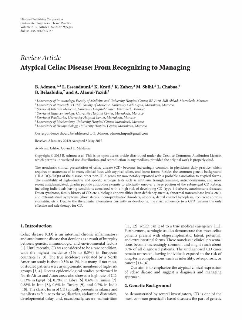
Figure 1: The celiac iceberg model [14].
background is fundamental in its pathogenesis, with possibleinfluence of genetic factors on clinical and immunologicfeatures [17–19]. Approximately 97% of individuals with CDhave genetic markers on chromosome 6p21, called class IIhuman leukocyte antigen (HLA). HLA DQ2 predominates,occurring in 90–95% of patients, and HLADQ8 occurs inthe remainder [11, 18, 20]. Some studies also point to acorrelation between DQ2 homozygousness and female sex,earlier age at diagnosis, shorter time span between onsetof symptoms and diagnosis, and to a higher prevalenceof classic clinical presentations among patients carryingdouble-dose DQB1∗02 [21]. Other investigations suggestthat MHC class I region plays a role in the development ofdiverse clinical forms of the disease [19, 22]. Lopez-Vazquezet al. [22], thus showed that haplotype B8/DR3/DQ2 isnotably overrepresented in atypical CD patients comparedto typical ones [19, 22]. In addition, similar studies displayedthat MICA-A5.1 allele either is associated with atypicalforms of CD in HLA-DQ2-negative patients or confersan additive effect to the DR3/DQ2 haplotype that maymodulate the development of the disease [19, 23]. Also,linkage research pointed to chromosomal regions other thanthe HLA region, predisposing to CD with modest effects;the CTLA4 (cytotoxic T-lymphocyte associated), a closelylocated gene on chromosome 2q33, is one of these genes[1, 24]. Alongside the HLA, recent genetic studies concerningpotential CD patients identified a robust association onchromosome 4q27, involving IL-2, IL-21, and KIAA1109gene cluster [25, 26], and also c-REL gene [26]. These factsmight allow more understanding in CD pathogenesis.
3. Clinical Faces of Celiac Disease
Gee described the classical features of celiac disease in1887 as diarrhea, lassitude, and failure to thrive [27], butthe improvement of knowledge has subsequently disclosedseveral patterns of the disease [28]. A number of investigatorsbelieve that clinically apparent gluten-sensitive enteropathyrepresents the “tip of the iceberg” of the overall diseaseburden (Figure 1).
This concept demonstrates the clinical variability of CDand enlightens why the disease remains unidentified in agreat proportion of individuals. In fact, the estimated ratioof diagnosed to undiagnosed individuals varies between 1 : 5to 1 : 8 (the submerged part of the iceberg), usually becauseof atypical, minimal, or even absent complaints [13, 14].
Many authors defined atypical CD as follows:
(i) Atypical form. Absence or few gastrointestinal symp-toms, presence of atypical symptoms, such as anemiadue to iron deficiency, osteoporosis or osteopenia,infertility, low stature;
(ii) Silent form. Occasional diagnosis, histological orserological, in asymptomatic individuals;
(iii) Latent form, with 2 categories
(a) patients with previous CD diagnosis whoresponded to gluten-free diet (GFD) and pre-sented a normal histology or only intraepithe-lial lymphocytes increase,
(b) individuals with normal intestinal mucosa,under diet including gluten, who will subse-quently develop CD;
(iv) Refractory form. Patients with CD who do notrespond to GFD [12, 14, 29].
Patients with CD are diagnosed at any age and can exhibita wide range of clinical manifestations (Table 1). In fact,beyond infancy, the symptoms of CD tend to be less dramatic[30, 31]. Older children may present with constitutionalshort stature or dental enamel defects, and women compriseapproximately 75% of newly diagnosed adult CD cases, withmore clinically conspicuous disease [11, 31].
Evidence suggests that the incidence of CD increases withage even in older patients [33]. Indeed, the majority of theelder cases remains undetected, often due to the absenceof symptoms or because of atypical clinical presentations[34, 35]. Osteoporosis represents one of the most frequentrevealing circumstances of the disease in the elderly, andthe rate of bone loss is accelerated in women after themenopause, likewise in men at the same age [33, 36].Anyway, physicians’ lack of alertness in the older people mayresult in a significant delay in diagnosis, as CD is widelydeemed to be a condition affecting younger subjects [33].
Moreover, a wider spectrum of neurologic syndromesmay be the presenting extraintestinal manifestation ofgluten sensitivity with or without intestinal pathology.These include headache, ataxia and psychiatric disorders[29], migraine, encephalopathy, chorea, brain stem dysfunc-tion, myelopathy, mononeuritis multiplex, Guillain-Barre-like syndrome, and neuropathy with positive antigangliosideantibodies [37]. Additional studies showed high prevalenceof gluten sensitivity in genetic neurodegenerative disorderssuch as hereditary spinocerebellar ataxia and Huntington’sdisease [37]. As well, oral manifestations, mostly recurrentapthous ulcers or stomatitis and dental enamel hypoplasia ordefects, are atypical signs of CD, and should be considered,even in the absence of any gastrointestinal symptom, at-risksubjects, and should therefore undergo diagnostic procedurefor CD [28, 38]. Also, recurrent febrile infections associatedto moderate neutropenia must be included in the diagnosticworkup for atypical/silent CD in the general population[39]. Furthermore, many of biologic abnormalities eitherconcur with CD or at times may reveal the disease such as

Gastroenterology Research and Practice 3
Table 1: Clinical and biological revealing circumstances of atypicalCD.
Atypical clinical symptoms
Anemia
Unclear vomiting
Constipation
Recurrent abdominal pain
Short stature
Irritability and impaired school performance
Impaired physical fitness and chronic fatigue
Osteopenia/osteoporosis/arthtritis
Dermatitis herpetiformis
Dental enamel hypoplasia
Recurrent aphtous stomatitis
Headache
Peripheral neuropathy
White matter lesions
Cerebellar ataxia
Epilepsy
Intracranial calcifications
Autism
Psychiatric disorders
Depression
Pubertal delay
Recurrent abortions
Infertility
Biologic abnormalities
Anemia, iron deficiency; vitamin B12 and/or folate deficiency
Hypertransaminasemia
Hyperalkaline phosphatase level
Hyperalbuminemia
Hypercalcaemia, hypophosphatemia
Thrombocytosis, leukocytosis
Coagulopathy
Low high-density and low-density lipoprotein cholesterol levels
anemia with iron, vitamin B12 and/or folate deficiencies,hypertransaminasemia (Table 1).
The prevalence of CD has increased sharply in recentyears because of better recognition of the disease and itsassociated disorders (Table 2) [18, 36, 40]. A number ofdiseases seem to occur more commonly in CD. Manystudies showed that patients with type 1 diabetes mellitus(T1DM), autoimmune thyroid disease, Sjogren’s syndrome,primary biliary cirrhosis, Addison’s disease, systemic lupuserythematosus, and alopecia areata may also exhibit similargenotypes of the celiac disease (HLA-DQ2 [DQA1∗0501 andDQB1∗0201]) and are at risk for gluten-sensitive enteropa-thy [11]. Autoimmune disorders occur 3 to 10 times morefrequently in those with celiac disease than in the generalpopulation. Evidence exists that the risk of developing otherautoimmune conditions increases with length of exposureto gluten [11, 18, 41]. Among associated CD conditions,T1DM is probably the most important; occurring in about
Table 2: List of possible celiac-disease-linked pathologies.
Associated autoimmune diseases or other conditions
Type 1 diabetes
Thyroid disorders (autoimmune or graves)
Liver disease (autoimmune hepatitis, primary biliary cirrhosis)
Myasthenia gravis
Primary biliary cirrhosis
Primary sclerosing cholangitis
Psoriasis
Sjogren disease
Systemic lupus erythematosus
Idiopathic dilated cardiomyopathy
Immunoglobulin A nephropathy
Lymphocytic or microscopic colitis
Autoimmune Addison’s disease
Rheumatoid arthritis
Vitiligo or alopecia areata
Associated genetic diseases
Down syndrome
Turner syndrome
Williams syndrome
IgA deficiency
Commun variable immunodeficiency
5% of CD patients [40, 42], with a large variance betweenethnic populations (range: 0.97–16.4%) [43]. In addition,unexplained and recurrent hypoglycemia in well manageddiabetic individuals should alert the physician for CDscreening [44]. Approximately 5% of the patients with CDhave thyroid disorders (either autoimmune (Hashimoto’s) orGraves’s disease) [42], and the ISPAD (International Societyfor Pediatric and Adolescent Diabetes) clinical practiceconsensus guidelines 2006-2007 recommend an assessmentof the thyroid function at the diagnosis of CD and thereafterevery second year in asymptomatic individuals and alsoa screening for CD at time of the diagnosis of thesethyroid disorders and every second year thereafter [45, 46].Down or Turner syndromes also represent frequent linkedconditions in which CD is often asymptomatic and thenrequire systematic screening for CD [47, 48]. Furthermore,the association of some primary immunodeficiencies entitieswith CD has been described such as IgA deficiency [49] andcommon variable immunodeficiency [50].
4. Serologic Testing: Performances and Limits
Since the introduction of serological tests, and because ofoccasional screening, silent CD forms have been increasinglyrecognized. This is frequently the case of family predisposedindividuals, and patients with associated autoimmune orgenetic disorders. In CD, highly sensitive and specificmethods are nowadays widely used in laboratory testing suchas antiendomysial (EMA) and antitissue transglutaminase(tTG) antibodies tests [18, 51]. But, although these testsexhibit very high sensitivity and specificity [11, 13, 32],
4 Gastroenterology Research and Practice
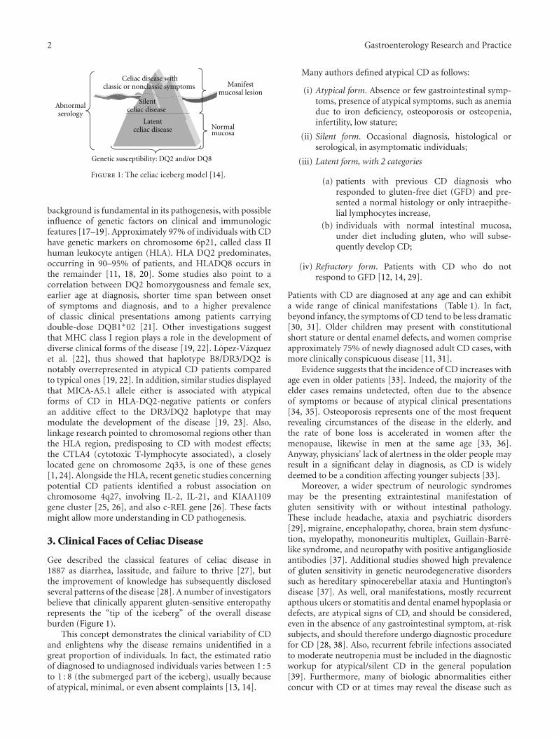
Table 3: Characteristics of exclusive or combined serological tests used to detect CD [11, 13, 18, 32].
Serological tests Sensitivity (%) Specificity (%) PPV (%) NPV (%)
IgG AGA 57–78 71–87 20–90 40–90
IgA AGA 55–100 65–100 30–100 70–100
IgA EMA 86–100 98–100 98–100 80–95
IgA tTG 90–96 91–97 >90 >95
IgA tTG and EMA 98–100 98–100 >90 >95
IgA DGP 98 94 92 98
IgG DGP 97 100 100 97
IgA DGP + IgA tTG 100 93 91 100
IgG DGP + IgA tTG 100 97 97 100
IgG: immunoglobulin G; IgA: immunoglobulin A; AGA: antigliadin antibodies; EMA: endomysial antibodies; tTG: tissue transglutaminase; DGP: deamidatedgliadin peptide; PPV: positive predictive value; NPV: negative predictive value.
∗Contrast between clinical data and serologic markers results
Negative
Negative(with normal IgA)
Negative(with IgA deficit)
IgA-tTG + total IgA
Suspicion of CDor at-risk individual
Positive
IgA-EMA or IgA-DGP IgG tTGor IgG-DGP
Positive
InconclusiveNegative
Positive
Intestinalbiopsy
Intestinalbiopsy Stop
StopIntestinal biopsy + HLA typing
Inconclusive∗(with normal IgA)
Figure 2: Algorithm proposal for biologic diagnosis of celiac disease.
recent investigations showed that their accuracy remainscontroversial in some conditions; sensitivity is consideredunacceptable both in patients with minor degrees of mucosaldamage and in cases with silent or oligosymptomatic forms[32]. Moreover, EMA and tTG have been found to besuperior to AGA (anti-gliadin antibodies) tests [11, 13,18] and when used in combination have sensitivity andspecificity greater than 95% [11, 13]. In addition, the recentlydeveloped deamidated gliadin peptide (DGP) antibody testshows promise in CD diagnostic [32, 51], and its perfor-mances are comparable to those of IgA-anti-tTG [52, 53].Moreover, IgG anti-DPG test has high diagnostic sensitivitynot only in IgA-competent but also in IgA deficient CDpatients [52]. Therefore, a combined evaluation of IgA-anti-tTG, and IgG anti-DPG seems to be adequate for serodi-agnosis of CD irrespective of IgA deficiency and withoutthe need for estimating total IgA concentrations [52, 53].The detection characteristics for AGA, EMA, tTG, and DGP
tests are shown in Table 3. In practice, according to newrecommendations, the initial serology testing consists onIgA-tTG screening, combined to total serum IgA measure-ment in order to rule out individuals with potential IgAdeficiency. The serology test should be performed beforeeliminating gluten from patient’s diet [54]. Actually, thebiologic diagnosis should be improved by combining twoperformant serologic markers, such as IgA-tTG and -EMA orIgA-tTG and IgG-DGP according to suggested algorithm inFigure 2. The patients who test positive with these assays areconsequently candidates for diagnostic endoscopy and small-bowel biopsy [51]. However, besides the atypical clinicalexpression of CD, the diagnosis may be more difficultfor many reasons: negative serology, irregular histologicalbehavior, or inadequate number or place of biopsies [55].
Despite the evolving performances of these serologictesting, there are still significant problems concerning thediagnosis approach in some atypical conditions; for example,
Gastroenterology Research and Practice 5
it has been proposed that IgG-AGA testing might be the bestmarker for neurological manifestations of gluten sensitivity,mainly for patients with sporadic ataxia [60, 61]. Thus, in arecent study on gluten ataxia patients, Hadjivassiliou et al.[62], noticed anti-EMA antibodies in only 22% of patients,and anti-TG2 IgA in up to 38% of cases, but often at lowertitres than those seen in patients with gluten sensitivityenteropathy [62]. On the other hand, the serology is generallythought to be unreliable in children <18 months of age[63]. This is due to a number of factors including the highproportion of children on breast milk, lower IgA levels, andthe under-developed immune system. Some authors havesuggested that IgA-AGA may be useful in this situation.This view is supported by a recent study carried out in208 children <18 months of age diagnosed with CD [64],showing a better sensitivity of IgA-AGA compared to boththe IgA-tTG and IgA EMA [65, 66].
5. Seronegative Celiac Disease
Not all patients have positive CD serologic markers at presen-tation [67, 68]. In fact, the presence of related CD antibodiescorrelates with the degree of villous atrophy and possibly themode of presentation of the disease [67, 69]. Patients withlesser degrees of villous atrophy are less likely to have positiveceliac serology [18], and patients who present persistentlypositive serology and negative biopsy probably have latentCD [12]. Moreover, children younger than 2 years of agelack EMA and tTG antibodies; for this reason, serologicaltesting in children younger than 5 years of age may be lessreliable and requires additional investigation [18]. On theother hand, in individuals who are IgA-deficient, the mea-surement of IgG-EMA and anti-tTG offers reliable resultswith excellent sensitivity (close to 100%) and specificity[12, 18]. Anyway, if CD suspicion is high with persistentlynegative tests, individuals must perform typing for HLAand, if positive, they must perform duodenal biopsy oralternatively perform biopsy directly [12, 55].
6. Histopathologic Findings
The intestinal biopsy represents the gold standard diagnosisfor CD [12, 55]. According to Marsh-Oberhuber’s [56, 57]criteria (Table 4), the spectrum of alterations compatiblewith CD consists of intraepithelial lymphocytic (IEL) infil-tration, pattern of crypts, and villous atrophy, and patient’ssymptoms frequently correlate with the degree of tissueinjury [59]. However, IEL increase with normal mucosaarchitecture may be observed in autoimmune diseases, suchas SLE, rheumatoid arthritis, and Hashimoto’s thyroiditis, inpatients using nonhormonal anti-inflammatory treatment,in CD’s initial presentation, and latent CD [55, 70]. Anincrease in IEL may also reflect a state of T cells activationtriggered by gluten, immune abnormalities, drugs, and infec-tious agents. Celiac patients, who present only IEL increasewith no alterations in the architecture of the mucosa, maybe symptomatic and be under increased complications risk[12]. Similarly, villous atrophy may be due to other causessuch as Crohn’s disease, collagenous sprue, and autoimmune
Table 4: Histopathologic classification of CD based on Marsh-Oberhuber [56, 57], and Corazza and Villanacci [58] new gradingsystem [12, 57, 59].
Marsh-Oberhuber classification
(i) Marsh I: infiltrative lesion, normal villous architecture andmucosa, and IEL increase (>30–40 lymphocytes/enterocytescounted).
(ii) Marsh II: hyperplasic lesion; similar to Marsh I with crypthyperplasia.
(iii) Marsh III: destructive lesion, subdivided to the following:
(a) partial villous atrophy,
(b) subtotal villous atrophy,
(c) total villous atrophy.
New grading system
(i) Grade A (nonatrophic): >25 IELs/100 enterocytes.
(ii) Grade B (atrophic): villous-crypt ratio <3 : 1.
(iii) Grade B2 (atrophic): no detectable villi.
enteropathy [71]. Moreover, a recent prospective evaluationled by different expert pathologists highlighted that a recentlyproposed three-grade classification system [58] gives betterinterobserver agreement as compared with the establishedsix-grade Marsh-Oberhuber classification (Table 4) [72].
Similarly to wide variation in clinical manifestations,GSE has a wide spectrum of histological abnormalities,which makes interpretation of small-intestinal biopsy spec-imens problematic for the pathologist [73]. Therefore, it isnot advised to affirm a diagnosis based only on the histo-logical findings, because the disease does not compromiseuniformly intestine, and alterations are not observed exclu-sively in CD [12, 55]. Actually, many differential diagnoses(Table 5) may give rise to CD, making the diagnosis moredifficult.
7. HLA Typing
All CD patients carry HLA-DQ2 or HLA-DQ8 [20]. How-ever, up to 40% of the general population also carriesthese HLA haplotypes. Their presence is necessary for thedevelopment of celiac disease, but the absence of thesealleles virtually excludes the diagnosis [18] with a negativepredictive value for CD close to 100% [20]. HLA typingrepresents the first step for investigating relatives of CDpatients, specifically 1st-degree relatives and then permitsto identify individuals for evaluation with biopsy [12]. Inpractice, if CD suspicion is high, with persistently negativetests, individuals must perform typing for HLA and, ifpositive, they must perform duodenal biopsy or alternativelyperform biopsy directly. Likewise, HLA typing is indicated inindividuals who refuse to undergo biopsy [12].
8. Gluten-Free Diet: Indications and Managing
An increased incidence of small-bowel malignancies, adeno-carcinoma, and enteropathy-associated T-cell lymphoma hasbeen reported in untreated CD [18, 74].

6 Gastroenterology Research and Practice
Table 5: Celiac disease differential diagnosis [12].
Anorexia nervosa
Autoimmune enteropathy
Bacterial overgrowth
Collagenous sprue
Crohn’s disease
Giardiasis
HIV enteropathy
Hipogammaglobulinemia
Gastroenterite infecciosa
Intestinal lymphoma
Radiation enteritis
Ischemic enteritis
Lactose intolerance
Common variable immunodeficiency
Soy protein intolerance
Tropical sprue
Tuberculosis
Whipple’s disease
Zolliger-Ellison syndrome
Eosinophilic gastroenteritis
Table 6: Indications of GFD in CD of children and adolescents [75].
CD clinical form Indications of GFD
Symptomatic Therapeutic
Silent Preventive: may be discussed
Latent Surveillance
A strict and lifelong gluten-free diet (GFD) has beendemonstrated to be effective and safe, preventing mostpotential complications of the disease, including autoim-mune disease, osteoporosis, infertility, prematurity, andmalignancy [76, 77]. However, there is still no evidencethat patients who have symptom-free celiac disease areat increased risk of small-intestinal lymphoma or othercomplications [71]. On the other hand, diet trials in patientswith gluten sensitivity and neurologic syndromes haveshown variable results and have been inconclusive in someneurologic diseases such as autism and schizophrenia [37].Furthermore, in asymptomatic patients, a second follow-upbiopsy under a GFD is advised to demonstrate the histolog-ical recovery of the mucosa, which usually does not developbefore six months [73].
In general, the guidance of GFD may be envisagedaccording to three modalities (Table 6).
(i) Typical or symptomatic CD; GFD is a formal thera-peutic indication.
(ii) Silent CD; GFD is discussed under two circum-stances.
(iii) Silent CD discovered on the occasion of a serologicalscreening in the family of a celiac or in a patient atrisk (diabetes mellitus, dermatitis herpetiforme); inthis case, the lesser clinical or nutritional sign would
treat the subject as symptomatic and plead in favor ofGFD.
(iv) CD becoming silent in the second childhood afterthat the active disease in the first childhood wastreated several years by a well monitoring GFD.
In these two situations and in individual really clinically andbiologically asymptomatic, the decision to introduce or toresume the GFD is then rather preventive.
(v) Latent CD (subjects genetically predisposed withnormal intestinal mucosa); a simple clinical andbiological surveillance is advocated by recent studies[75].
Beside the GFD, the management of many of CD-linkedfeatures may require additional supplementation particularlyin nutritional problems, such as lower Hb and low Fe, lowalbumin or Ca, cholesterol and folates disorders [32, 78–80].Likewise, in CD patients with low bone mineral density, apartfrom a GFD, a rational managing should follow conventionallines, including increasing exercise, stopping smoking, andavoiding alcohol excess and ensuring an adequate Ca intakeusing supplements if necessary [36]. In addition, newlytherapeutic alternatives are currently interested in the patho-genesis of the disease, focusing on engineering gluten-freegrains, degradation of immunodominant gliadin peptidesthat resist intestinal proteases by exogenous endopeptidases,decrease in intestinal permeability by blockage of theepithelial zonuline receptor, inhibition of intestinal tTG2activity by transglutaminase inhibitors, inhibition of glutenpeptide presentation by HLA-DQ2 antagonists, modulationor inhibition of proinflammatory cytokines, and inductionof oral tolerance to gluten [14, 81, 82]. But, at this time,strict adherence to a GFD remains the only effective and safetherapy for CD [14].
9. Refractory Celiac Disease
A small proportion of CD patients fails to improve after aGFD and may be considered as atypical regarding their out-come [14, 83]. Refractory celiac disease (RCD) was recentlydefined as persisting or recurring villous atrophy with crypthyperplasia and increased intraepithelial lymphocytes (IELs)in spite of a strict GFD for more than 12 months [71, 84].It can be either primary, as lack of initial response to diet,or secondary, as unresponsiveness to diet in the form of arelapse [73]. Two categories of RCD are recently being recog-nized: type I without aberrant T cells and type II with aber-rant T cells [85]. The presence of an aberrant clonal intraep-ithelial T-cell population and/or loss of antigen on IELs seemto characterize population on high risk for developmentof overt lymphoma and differentiates RCD II from RCD I,which shows low or almost absent aberrant T cells [84].
To manage RCD, Krauss and Schuppan [71] recommendfirstly to reassess the diagnosis of CD in order to excludeother diseases, such as giardiasis, tropical sprue, post-infectious diarrhea, collagenous sprue, protein intolerance orprotein-losing enteropathy, tuberculosis (including atypi-cal), AIDS, common variable immunodeficiency syndrome,

Gastroenterology Research and Practice 7
Whipple’s disease, ulcerative jejunitis, lymphocytic coli-tis, radiation enteritis, immunoproliferative small-intestinaldisease, Crohn’s disease, eosinophilic gastroenteritis, andautoimmune enteropathy [71, 84], and then to check forerrors in diet or compliance [71]. The treatment of RCDI consists of a first-line immunosuppressive therapy basedon azathioprine after induction of clinical remission withcorticosteroids [86]. A second-line therapy (CyclosporineA, infliximab, tacrolimus) is suggested in case of clinicaldeterioration despite corticosteroid therapy or intoleranceto azathioprine [87] RCD II is usually resistant to medicaltherapies, and facing persistent clinical symptoms and/ora high percentage of aberrant T cells in intestinal biopsiesin spite of a corticosteroid treatment, more aggressivetherapeutic schemes should be considered [84].
10. Conclusion
Celiac disease represents a prototype of disease from whichscience and medicine take advantage, offering more anduninterrupted understandings both in genetic, clinic, diag-nosis, and management aspects. Against its potential com-plications, the real challenge is to recognize asymptomaticor oligosymptomatic CD cases. The diagnosis should alsobe improved by a process of case finding focused on at-riskgroups.
References
[1] L. M. Sollid, S. N. McAdam, O. Molberg et al., “Genes andenvironment in celiac disease,” Acta Odontologica Scandinav-ica, vol. 59, no. 3, pp. 183–186, 2001.
[2] M. Maki, K. Mustalahti, J. Kokkonen et al., “Prevalence ofceliac disease among children in Finland,” New England Jour-nal of Medicine, vol. 348, no. 25, pp. 2517–2524, 2003.
[3] G. Telega, T. R. Bennet, and S. Werlin, “Emerging new clinicalpatterns in the presentation of celiac disease,” Archives of Pedi-atrics and Adolescent Medicine, vol. 162, no. 2, pp. 164–168,2008.
[4] E. J. Hoffenberg, T. MacKenzie, K. J. Barriga et al., “A prospec-tive study of the incidence of childhood celiac disease,” Journalof Pediatrics, vol. 143, no. 3, pp. 308–314, 2003.
[5] M. Abu-Zekry, D. Kryszak, M. Diab, C. Catassi, and A. Fasano,“Prevalence of celiac disease in egyptian children disputes theeast west agriculture-dependent spread of the disease,” Journalof Pediatric Gastroenterology and Nutrition, vol. 47, no. 2, pp.136–140, 2008.
[6] K. Alarida, J. Harown, A. Ahmaida et al., “Coeliac diseasein Libyan children: a screening study based on the rapiddetermination of anti-transglutaminase antibodies,” Digestiveand Liver Disease, vol. 43, no. 9, pp. 688–691, 2011.
[7] M. B. Hariz, M. Kallel-Sellami, L. Kallel et al., “Prevalence ofceliac disease in Tunisia: mass-screening study in school chil-dren,” European Journal of Gastroenterology and Hepatology,vol. 19, no. 8, pp. 687–694, 2007.
[8] F. Imanzadeh, A. A. Sayyari, M. Yaghoobi, M. R. Akbari, H.Shafagh, and A. R. Farsar, “Celiac disease in children withdiarrhea is more frequent than previously suspected,” Journalof Pediatric Gastroenterology and Nutrition, vol. 40, no. 3, pp.309–311, 2005.
[9] V. Ertekin, M. A. Selimoglu, F. Kardas, and E. Aktas, “Prev-alence of CD in Turkish children,” Journal of Clinical Gastroen-terology, vol. 39, pp. 689–691, 2005.
[10] A. Sood, V. Midha, N. Sood, G. Avasthi, and A. Sehgal, “Prev-alence of celiac disease among school children in Punjab,North India,” Journal of Gastroenterology and Hepatology, vol.21, no. 10, pp. 1622–1625, 2006.
[11] D. A. Nelsen, “Gluten-sensitive enteropathy (celiac disease):more common than you think,” American Family Physician,vol. 66, no. 12, pp. 2259–2266, 2002.
[12] T. S. G. Silva and T. W. Furlanetto, “Diagnosis of celiac diseasein adults,” Revista da Associacao Medica Brasileira, vol. 56, no.1, pp. 122–126, 2010.
[13] J. S. Leeds, A. D. Hopper, and D. S. Sanders, “Coeliac disease,”British Medical Bulletin, vol. 88, no. 1, pp. 157–170, 2008.
[14] E. Lionetti and C. Catassi, “New clues in celiac diseaseepidemiology, pathogenesis, clinical manifestations, and treat-ment,” International Reviews of Immunology, vol. 30, no. 4, pp.219–231, 2011.
[15] A. Ferguson and K. Kingstone, “Coeliac disease and malig-nancies,” Acta Paediatrica, International Journal of Paediatrics,Supplement, vol. 85, no. 412, pp. 78–81, 1996.
[16] J. Cosnes, C. Cosnes, A. Cosnes et al., “Undiagnosed celiacdisease in childhood,” Gastroenterologie Clinique et Biologique,vol. 26, no. 6-7, pp. 616–623, 2002.
[17] P. H. R. Green and B. Jabri, “Coeliac disease,” The Lancet, vol.362, no. 9381, pp. 383–391, 2003.
[18] M. M. Niewinski, “Advances in Celiac disease and gluten-freediet,” Journal of the American Dietetic Association, vol. 108, no.4, pp. 661–672, 2008.
[19] A. Lopez-Vazquez, L. Rodrigo, D. Fuentes et al., “MHC classI chain related gene A (MICA) modulates the developmentof coeliac disease in patients with the high risk heterodimerDQA1∗0501/DQB1∗0201,” Gut, vol. 50, no. 3, pp. 336–340,2002.
[20] V. M. Wolters and C. Wijmenga, “Genetic background ofceliac disease and its clinical implications,” American Journalof Gastroenterology, vol. 103, no. 1, pp. 190–195, 2008.
[21] P. Zubillaga, M. C. Vidales, I. Zubillaga, V. Ormaechea, N.Garcıa-Urkıa, and J. C. Vitoria, “HLA-DQA1 and HLA-DQB1genetic markers and clinical presentation in celiac disease,”Journal of Pediatric Gastroenterology and Nutrition, vol. 34, no.5, pp. 548–554, 2002.
[22] A. Lopez-Vazquez, D. Fuentes, L. Rodrigo et al., “MHC class Iregion plays a role in the development of diverse clinical formsof celiac disease in a Saharawi population,” American Journalof Gastroenterology, vol. 99, no. 4, pp. 662–667, 2004.
[23] A. Lopez-Vazquez, L. Rodrigo, D. Fuentes et al., “MICA-A5.1allele is associated with atypical forms of celiac disease in HLA-DQ2-negative patients,” Immunogenetics, vol. 53, no. 10-11,pp. 989–991, 2002.
[24] A. T. Naluai, S. Nilsson, L. Samuelsson et al., “The CTLA4/CD28 gene region on chromosome 2q33 confers susceptibilityto celiac disease in a way possibly distinct from that of type1 diabetes and other chronic inflammatory disorders,” TissueAntigens, vol. 56, no. 4, pp. 350–355, 2000.
[25] D. A. Van Heel, L. Franke, K. A. Hunt et al., “A genome-wideassociation study for celiac disease identifies risk variants inthe region harboring IL2 and IL21,” Nature Genetics, vol. 39,no. 7, pp. 827–829, 2007.
[26] M. P. Sperandeo, A. Tosco, V. Izzo et al., “Potential celiacpatients: a model of celiac disease pathogenesis,” PLoS ONE,vol. 6, no. 7, Article ID e21281, 2011.

8 Gastroenterology Research and Practice
[27] S. Gee, “On the celiac affection,” Saint Bartholomew’s HospitalReports, vol. 24, pp. 17–20, 1988.
[28] L. Pastore, M. De Benedittis, M. Petruzzi et al., “Importance oforal signs in the diagnosis of atypical forms of celiac disease,”Recenti Progressi in Medicina, vol. 95, no. 10, pp. 482–490,2004.
[29] M. Spina, G. Incorpora, T. Trigilia, F. Branciforte, G. Franco,and F. Di Gregorio, “Headache as atypical presentation ofceliac disease: report of a clinical case,” Pediatria Medica eChirurgica, vol. 23, no. 2, pp. 133–135, 2001.
[30] C. Feighery, “Coeliac disease,” British Medical Journal, vol. 319,no. 7204, pp. 236–239, 1999.
[31] C. Ciacci, M. Cirillo, R. Sollazzo, G. Savino, F. Sabbatini, andG. Mazzacca, “Gender and clinical presentation in adult celiacdisease,” Scandinavian Journal of Gastroenterology, vol. 30, no.11, pp. 1077–1081, 1995.
[32] S. Niveloni, E. Sugai, A. Cabanne et al., “Antibodies againstsynthetic deamidated gliadin peptides as predictors of celiacdisease: prospective assessment in an adult population with ahigh pretest probability of disease,” Clinical Chemistry, vol. 53,no. 12, pp. 2186–2192, 2007.
[33] A. Vilppula, K. Kaukinen, L. Luostarinen et al., “Increasingprevalence and high incidence of celiac disease in elderlypeople: a population-based study,” BMC Gastroenterology, vol.9, article 49, 2009.
[34] G. L. Hankey and G. K. T. Holmes, “Coeliac disease in theelderly,” Gut, vol. 35, no. 1, pp. 65–67, 1994.
[35] Y. Lurie, D. A. Landau, J. Pfeffer, and R. Oren, “Celiac diseasediagnosed in the elderly,” Journal of Clinical Gastroenterology,vol. 42, no. 1, pp. 59–61, 2008.
[36] A. Fasano, I. Berti, T. Gerarduzzi et al., “Prevalence of Celiacdisease in at-risk and not-at-risk groups in the United States:a large multicenter study,” Archives of Internal Medicine, vol.163, no. 3, pp. 286–292, 2003.
[37] K.O. Bushara, ‘‘Neurologic presentation of celiac disease,’’ Gas-troenterology, vol. 128, no. 4, supplement 1, pp. S92–S97, 2005.
[38] J. Cheng, T. Malahias, P. Brar, M. T. Minaya, and P. H. R.Green, “The Association between celiac disease, dental enameldefects, and aphthous ulcers in a United States cohort,” Journalof Clinical Gastroenterology, vol. 44, no. 3, pp. 191–194, 2010.
[39] S. Leonardi, G. Vitaliti, and M. La Rosa, “Recurrent febrileinfections and neutropenia in a child with silent celiac disease,”Clinical Pediatrics, vol. 49, no. 2, pp. 146–149, 2010.
[40] N. Chand and A. A. Mihas, “Celiac disease: current concepts indiagnosis and treatment,” Journal of Clinical Gastroenterology,vol. 40, no. 1, pp. 3–14, 2006.
[41] M. Viljamaa, K. Kaukinen, H. Huhtala, S. Kyronpalo, M.Rasmussen, and P. Collin, “Coeliac disease, autoimmune dis-eases and gluten exposure,” Scandinavian Journal of Gastroen-terology, vol. 40, no. 4, pp. 437–443, 2005.
[42] N. McGough and J. H. Cummings, “Coeliac disease: a diverseclinical syndrome caused by intolerance of wheat, barley andrye,” Proceedings of the Nutrition Society, vol. 64, no. 4, pp.434–450, 2005.
[43] G. K. T. Holmes, “Screening for coeliac disease in type 1diabetes,” Archives of Disease in Childhood, vol. 87, no. 6, pp.495–499, 2002.
[44] D. Iafusco, F. Rea, F. Prisco, and J. F. Fitzgerald, “Hypoglycemiaand reduction of the insulin requirement as a sign of celiacdisease in children with IDDM,” Diabetes Care, vol. 21, no. 8,pp. 1379–1381, 1998.
[45] O. Kordonouri, A. M. Maguire, M. Knip et al., “Other compli-cations and associated conditions,” Pediatric Diabetes, vol. 8,no. 3, pp. 171–176, 2007.
[46] E. E. Frohlich-Reiterer, S. Hofer, S. Kaspers et al., “Screeningfrequency for celiac disease and autoimmune thyroiditis inchildren and adolescents with type 1 diabetes mellitus—datafrom a German/Austrian multicentre survey,” Pediatric Dia-betes, vol. 9, no. 6, pp. 546–553, 2008.
[47] M. Bonamico, A. M. Pasquino, P. Mariani et al., “Prevalenceand clinical picture of celiac disease in Turner syndrome,”Journal of Clinical Endocrinology and Metabolism, vol. 87, no.12, pp. 5495–5498, 2002.
[48] R. M. Nisihara, L. M. S. Kotze, S. R. R. Utiyama, N. P. Oliveira,P. T. Fiedler, and I. T. Messias-Reason, “Celiac disease inchildren and adolescents with Down syndrome,” Jornal dePediatria, vol. 81, no. 5, pp. 373–376, 2005.
[49] F. Cataldo, V. Marino, A. Ventura, G. Bottaro, and G. R.Corazza, “Prevalence and clinical features of selective immu-noglobulin A deficiency in coeliac disease: an Italian multicen-tre study,” Gut, vol. 42, no. 3, pp. 362–365, 1998.
[50] D. Bechade, J. Desrame, G. De Fuentes, P. Camparo, J. J.Raynaud, and J. P. Algayres, “Common variable immun-odeficiency and celiac disease,” Gastroenterologie Clinique etBiologique, vol. 28, no. 10, pp. 909–912, 2004.
[51] K. Lindfors, O. Koskinen, and K. Kaukinen, “An update onthe diagnostics of celiac disease,” International Reviews ofImmunology, vol. 30, no. 4, pp. 185–196, 2011.
[52] D. Villalta, E. Tonutti, C. Prause et al., “IgG antibodies againstdeamidated gliadin peptides for diagnosis of celiac disease inpatients with IgA deficiency,” Clinical Chemistry, vol. 56, no. 3,pp. 464–468, 2010.
[53] C. Prause, T. Richter, S. Koletzko et al., “New developments inserodiagnosis of childhood celiac disease: assay of antibodiesagainst deamidated gliadin,” Annals of the New York Academyof Sciences, vol. 1173, pp. 28–35, 2009.
[54] D. P. Westerberg, J. M. Gill, B. Dave, M. J. DiPrinzio, A. Quisel,and A. Foy, “New strategies for diagnosis and management ofceliac disease,” Journal of the American Osteopathic Association,vol. 106, no. 3, pp. 145–151, 2006.
[55] M. F. Kagnoff, “AGA institute medical position statement onthe diagnosis and management of celiac disease,” Gastroen-terology, vol. 131, no. 6, pp. 1977–1980, 2006.
[56] M. N. Marsh, “Gluten, major histocompatibility complex,and the small intestine: a molecular and immunobiologicapproach to the spectrum of gluten sensitivity (’celiac sprue’),”Gastroenterology, vol. 102, no. 1, pp. 330–354, 1992.
[57] G. Oberhuber, G. Granditsch, and H. Vogelsang, “The histo-pathology of coeliac disease: time for a standardized reportscheme for pathologists,” European Journal of Gastroenterologyand Hepatology, vol. 11, no. 10, pp. 1185–1194, 1999.
[58] G. R. Corazza and V. Villanacci, “Coeliac disease,” Journal ofClinical Pathology, vol. 58, no. 6, pp. 573–574, 2005.
[59] A. Rostom, J. A. Murray, and M. F. Kagnoff, “American gas-troenterological association (AGA) institute technical reviewon the diagnosis and management of celiac disease,” Gastroen-terology, vol. 131, no. 6, pp. 1981–2002, 2006.
[60] M. Hadjivassiliou, R. Grunewald, B. Sharrack et al., “Glutenataxia in perspective: epidemiology, genetic susceptibility andclinical characteristics,” Brain, vol. 126, no. 3, pp. 685–691,2003.
[61] M. T. Pellecchia, R. Scala, A. Filla, G. De Michelle, C. Ciacci,and P. Barone, “Idiopathic cerebellar ataxia associated withceliac disease: lack of distinctive neurological features,” Journalof Neurology Neurosurgery and Psychiatry, vol. 66, no. 1, pp.32–35, 1999.

Gastroenterology Research and Practice 9
[62] M. Hadjivassiliou, D. S. Sanders, N. Woodroofe, C.Williamson, and R. A. Grunewald, “Gluten ataxia,” Cerebel-lum, vol. 7, no. 3, pp. 494–498, 2008.
[63] R. Steele, “for the CRF∗ (Coeliac Research Fund Clinical Advi-sory Committee Members) Diagnosis and management ofcoeliac disease in children,” Postgraduate Medical Journal, vol.87, pp. 19–25, 2011.
[64] C. Lagerqvist, I. Dahlbom, T. Hansson et al., “Antigliadinimmunoglobulin a best in finding celiac disease in childrenyounger than 18 months of age,” Journal of Pediatric Gastroen-terology and Nutrition, vol. 47, no. 4, pp. 428–435, 2008.
[65] M. Stern, “Comparative evaluation of serologic tests for celiacdisease: a European initiative toward standardization,” Journalof Pediatric Gastroenterology and Nutrition, vol. 31, no. 5, pp.513–519, 2000.
[66] R. Berger and G. Schmidt, “Evaluation of six anti-gliadinantibody assays,” Journal of Immunological Methods, vol. 191,no. 1, pp. 77–86, 1996.
[67] J. A. Abrams, B. Diamond, H. Rotterdam, and P. H. R. Green,“Seronegative celiac disease: increased prevalence with lesserdegrees of villous atrophy,” Digestive Diseases and Sciences, vol.49, no. 4, pp. 546–550, 2004.
[68] W. Dickey, S. A. McMillan, and D. F. Hughes, “Sensitivityof serum tissue transglutaminase antibodies for endomysialantibody positive and negative coeliac disease,” ScandinavianJournal of Gastroenterology, vol. 36, no. 5, pp. 511–514, 2001.
[69] A. Tursi, G. Brandimarte, and G. M. Giorgetti, “Prevalenceof antitissue transglutaminase antibodies in different degreesof intestinal damage in celiac disease,” Journal of ClinicalGastroenterology, vol. 36, no. 3, pp. 219–221, 2003.
[70] S. Kakar, V. Nehra, J. A. Murray, G. A. Dayharsh, and L. J. Bur-gart, “Significance of intraepithelial lymphocytosis in smallbowel biopsy samples with normal mucosal architecture,”American Journal of Gastroenterology, vol. 98, no. 9, pp. 2027–2033, 2003.
[71] N. Krauss and D. Schuppan, “Monitoring nonresponsivepatients who have celiac disease,” Gastrointestinal EndoscopyClinics of North America, vol. 16, no. 2, pp. 317–327, 2006.
[72] M. J. Armstrong, G. G. Robins, and P. D. Howdle, “Recentadvances in coeliac disease,” Current Opinion in Gastroenterol-ogy, vol. 25, no. 2, pp. 100–109, 2009.
[73] A. Ensari, “Gluten-sensitive enteropathy (celiac disease): con-troversies in diagnosis and classification,” Archives of Pathologyand Laboratory Medicine, vol. 134, no. 6, pp. 826–836, 2010.
[74] T. R. Card, J. West, and G. K. T. Holmes, “Risk of malignancyin diagnosed coeliac disease: a 24-year prospective, popula-tion-based, cohort study,” Alimentary Pharmacology and Ther-apeutics, vol. 20, no. 7, pp. 769–775, 2004.
[75] J. Schmitz, “Le regime sans gluten chez l’enfant,” Journal dePediatrie et de Puericulture, vol. 20, pp. 337–344, 2007.
[76] K. P. Zimmer, “Nutrition and celiac disease,” Current Problemsin Pediatric and Adolescent Health Care, vol. 41, no. 9, pp. 244–247, 2011.
[77] D. Schuppan, M. D. Dennis, and C. P. Kelly, “Celiac disease:epidemiology, pathogenesis, diagnosis, and nutritional man-agement,” Nutrition in Clinical Care, vol. 8, no. 2, pp. 54–69,2005.
[78] E. M. Scott, B. B. Scott, and I. Gaywood, “Guidelines for osteo-porosis in coeliac disease and inflammatory bowel disease,”Gut, vol. 46, no. 1, pp. i1–i8, 2000.
[79] A. K. Patwari, V. K. Anand, G. Kapur, and S. Narayan, “Clinicaland nutritional profile of children with celiac disease,” IndianPediatrics, vol. 40, no. 4, pp. 337–342, 2003.
[80] U. Volta, L. De Franceschi, F. Lari, N. Molinaro, M. Zoli,and F. B. Bianchi, “Coeliac disease hidden by cryptogenichypertransaminasaemia,” The Lancet, vol. 352, no. 9121, pp.26–29, 1998.
[81] D. Schuppan, Y. Junker, and D. Barisani, “Celiac disease: frompathogenesis to novel therapies,” Gastroenterology, vol. 137,no. 6, pp. 1912–1933, 2009.
[82] A. Lerner, “New therapeutic strategies for celiac disease,” Auto-immunity Reviews, vol. 9, no. 3, pp. 144–147, 2010.
[83] S. Daum, C. Cellier, and C. J. J. Mulder, “Refractory coeliacdisease,” Best Practice and Research, vol. 19, no. 3, pp. 413–424,2005.
[84] A. Al-Toma, W. H. M. Verbeek, and C. J. J. Mulder, “The man-agement of complicated celiac disease,” Digestive Diseases, vol.25, no. 3, pp. 230–236, 2007.
[85] C. Cellier, E. Delabesse, C. Helmer et al., “Refractory sprue,coeliac disease, and enteropathy-associated T-cell lymphoma,”The Lancet, vol. 356, no. 9225, pp. 203–208, 2000.
[86] M. S. Goerres, J. W. R. Meijer, P. J. Wahab et al., “Azathioprineand prednisone combination therapy in refractory coeliacdisease,” Alimentary Pharmacology and Therapeutics, vol. 18,no. 5, pp. 487–494, 2003.
[87] P. J. Wahab, J. B. A. Crusius, J. W. R. Meijer, J. J. Uil, and C. J. J.Mulder, “Cyclosporin in the treatment of adults with refrac-tory coeliac disease—an open pilot study,” Alimentary Phar-macology and Therapeutics, vol. 14, no. 6, pp. 767–774, 2000.

Submit your manuscripts athttp://www.hindawi.com
Stem CellsInternational
Hindawi Publishing Corporationhttp://www.hindawi.com Volume 2014
Hindawi Publishing Corporationhttp://www.hindawi.com Volume 2014
MEDIATORSINFLAMMATION
of
Hindawi Publishing Corporationhttp://www.hindawi.com Volume 2014
Behavioural Neurology
EndocrinologyInternational Journal of
Hindawi Publishing Corporationhttp://www.hindawi.com Volume 2014
Hindawi Publishing Corporationhttp://www.hindawi.com Volume 2014
Disease Markers
Hindawi Publishing Corporationhttp://www.hindawi.com Volume 2014
BioMed Research International
OncologyJournal of
Hindawi Publishing Corporationhttp://www.hindawi.com Volume 2014
Hindawi Publishing Corporationhttp://www.hindawi.com Volume 2014
Oxidative Medicine and Cellular Longevity
Hindawi Publishing Corporationhttp://www.hindawi.com Volume 2014
PPAR Research
The Scientific World JournalHindawi Publishing Corporation http://www.hindawi.com Volume 2014
Immunology ResearchHindawi Publishing Corporationhttp://www.hindawi.com Volume 2014
Journal of
ObesityJournal of
Hindawi Publishing Corporationhttp://www.hindawi.com Volume 2014
Hindawi Publishing Corporationhttp://www.hindawi.com Volume 2014
Computational and Mathematical Methods in Medicine
OphthalmologyJournal of
Hindawi Publishing Corporationhttp://www.hindawi.com Volume 2014
Diabetes ResearchJournal of
Hindawi Publishing Corporationhttp://www.hindawi.com Volume 2014
Hindawi Publishing Corporationhttp://www.hindawi.com Volume 2014
Research and TreatmentAIDS
Hindawi Publishing Corporationhttp://www.hindawi.com Volume 2014
Gastroenterology Research and Practice
Hindawi Publishing Corporationhttp://www.hindawi.com Volume 2014
Parkinson’s Disease
Evidence-Based Complementary and Alternative Medicine
Volume 2014Hindawi Publishing Corporationhttp://www.hindawi.com