review article cell cycle control in acute myeloid leukemia
TRANSCRIPT

Leukemogenesis and cell cycle – differentiation block, continuous proliferation and genome instability AML is characterized by the clonal expansion of immature myeloid cells. At initial diagnosis ap-proximately half of the patients harbor at least one cytogenetic aberration [1, 2]. Moreover, aberrations at the molecular genetic level have increasingly been identified in AML during the last decade [3]. Several genetic aberrations have been associated with the hallmark of AML: the combination of a differentiation block and hyperproliferation [4]. In the normal hematopoietic system, prolifera-tion is tightly linked to differentiation. In an asymmetrical cell division, one daughter cell retains stem cell properties to guarantee a pool of stem cells while the other one undergoes differentiation to respond to the high and per-manent demand for granulocytes, monocytes, erythrocytes and platelets [5]. Hyperproliferation bears an enhanced risk of genetic damage. In leukemia, compromised DNA damage response pathways and weakened checkpoints add to the inherent risk and pave
the way for further genetic aberrations promot-ing progression of the disease [4, 6]. Thus, to fully understand leukemogenesis, it is crucial to determine the role of proto-oncogenic pathways in the regulation of the cell division cycle in mye-loid malignancies. The cell cycle itself is divided into four major phases: the G1-, S-, G2- and M-phase [7]. From G1 a cell can exit the cell cycle into a state of quiescence (G0) from where it can undergo differentiation or reenter the cell cycle to proliferate. If the cell continues cell divi-sion the genomic DNA will be replicated during S-phase. Once cells have completed DNA repli-cation, they proceed through the G2-phase to prepare for the subsequent chromosome sepa-ration and cytokinesis during M-phase (mitosis) (Figure 1) [7]. G1-phase - at the crossroad of proliferation and differentiation The reproductive rate of the myeloid system depends on the number of cells that are actively cycling. In healthy individuals, myeloid precursor cells are mostly in a quiescent state [8, 9] and can be recruited to enter the cell cycle in case of demand, for example when a severe infection occurs. Actively cycling progenitors proceed
Am J Cancer Res 2012;2(5):508-528 www.ajcr.us /ISSN:2156-6976/ajcr0000132
Review Article Cell cycle control in acute myeloid leukemia Dominik Schnerch, Jasmin Yalcintepe, Andrea Schmidts, Heiko Becker, Marie Follo, Monika Engelhardt, Ralph Wäsch Department of Hematology, Oncology and Stem Cell Transplantation, University Medical Center, Freiburg, Germany Received June 21, 2012; accepted July 27, 2012; Epub August 20, 2012; Published September 15, 2012 Abstract: Acute myeloid leukemia (AML) is the result of a multistep transforming process of hematopoietic precursor cells (HPCs) which enables them to proceed through limitless numbers of cell cycles and to become resistant to cell death. Increased proliferation renders these cells vulnerable to acquiring mutations and may favor leukemic transfor-mation. Here, we review how deregulated cell cycle control contributes to increased proliferation in AML and favors genomic instability, a prerequisite to confer selective advantages to particular clones in order to adapt and independ-ently proliferate in the presence of a changing microenvironment. We discuss the connection between differentiation and proliferation with regard to leukemogenesis and outline the impact of specific alterations on response to therapy. Finally, we present examples, how a better understanding of cell cycle regulation and deregulation has already led to new promising therapeutic strategies. Keywords: Acute myeloid leukemia (AML), cell cycle, genetic instability, proliferation, differentiation

Cell cycle control in acute myeloid leukemia
509 Am J Cancer Res 2012;2(5):508-528
through multiple cell division cycles and give rise to daughter cells that can differentiate. Short intervals between cell division cycles and a large pool of cells that are capable of rapid self-duplication are strategies to guarantee a sufficient cellular defense. Quiescent cells can be activated to re-enter the cell cycle by various external stimuli including growth factors and cytokines. In the following section, pathways will be discussed that regu-late whether a cell enters the S-phase, exits the cell cycle into quiescence or undergoes differen-tiation. Stimuli from the microenvironment in normal hematopoiesis and AML The FMS-like tyrosine kinase 3 (Flt3) [10], c-Kit
(CD117 or stem cell factor receptor) [11] or Janus kinase 2 (Jak2) [12] are tyrosine kinases that translate external stimuli into pro-proliferative signaling cascades (Figure 2). Con-stitutive firing of mutated kinases in AML fre-quently causes ongoing activation of the down-stream pathways and hence enhances transi-tioning from G1- into S-phase. Downstream of the receptor- and non-receptor kinases, the Stat-pathway (Stat = signal transducers and activa-tors of transcription) is activated upon stimula-tion by various interleukins [13]. Further down-stream, the serine-threonine kinase Pim1, which is activated by the Stat-pathway [14], has been shown to act as an important S-phase pro-moter by regulating Skp2-dependent degrada-tion of the Cdk-inhibitor p27 [15]. In addition to signaling in the G1 phase, Pim1 was shown to enhance the transition from G2 phase into
Figure 1. The cell cycle and its checkpoints. The human cell cycle can be divided into four phases - G1-phase, S-phase, G2-phase and M-phase (mitosis). Cells must proceed through the cell cycle in a unidirectional manner and cell cycle progression is restricted to cells that have fulfilled specific requirements to enter the next phase of the cell cy-cle. Whether requirements for cell cycle progression are met is supervised by checkpoints which hold back cells at cell cycle transitions.

Cell cycle control in acute myeloid leukemia
510 Am J Cancer Res 2012;2(5):508-528
mitosis [16]. As mentioned above, alterations of these prolif-erative regulators may confer unlimited growth during leukemogenesis [13]. For instance, cer-tain mutations in FLT3, ie, point mutations in the tyrosine kinase domain (FLT3-TKD) and in-ternal tandem duplications in the juxta-membrane region (FLT3-ITD), lead to constitu-tive activation which causes phosphorylation of substrate proteins even in the absence of exter-nal stimuli [17]. FLT3-ITD is found in approxi-mately 20% of AML patients and FLT3-TKD in approximately 5% [18, 19]. Both mutations are more frequent among patients with AML and a normal karyotype [18]. In addition to Flt3, the receptor tyrosine kinase Kit can be affected by activating mutations in AML. Such mutations, similar to Flt3, are mainly found in the tyrosine
kinase domain of Kit, but are in part also ob-served in the extracellular domain. KIT muta-tions mostly occur in patients with AML and a chromosomal translocation disrupting a core binding factor, ie, t(8;21) or inv(16) [20]. Muta-tions in JAK2 are rarely found in AML, but are characteristic for myeloproliferative neoplasms (MPN) [21]. Both types of FLT3 mutations activate PI3K/AKT and the MAP kinase pathway [22, 23]. Activa-tion of the MAP kinase pathway leads to upregu-lation of proto-oncogenic cell cycle regulators, such as the transcription factor c-Myc, and pro-motes premature entry into the following S-phase in AML cells [24]. FLT3-ITD also activates the Stat5 pathway and negatively regulates CEBPα and other transcription factors such as PU.1 to suppress differentiation [25, 26]. Major
Figure 2. To cycle or to differentiate - the G1 phase of the cell cycle. Cells that have exited mitosis into G1 can either enter a state of quiescence (G0), differentiate along a particular lineage or progress to another cell division cycle. The process of decision making between these possibilities is governed by a multitude of different proteins. Proteins that are frequently overexpressed or mutated and hence contribute to leukemic transformation are marked in red. Tumor suppressor proteins are marked in green, proteins that contribute to differentiation are shown in orange. Therapeutic agents are marked in yellow. See text for details.

Cell cycle control in acute myeloid leukemia
511 Am J Cancer Res 2012;2(5):508-528
changes in proliferative patterns can also result from deregulated chemokine signaling. In nor-mal hematopoiesis, progenitor cells are stimu-lated through binding of CXCL12 (alias SDF-1) to its receptor CXCR4. Ligand binding to CXCR4 leads to activation of the MAP kinase pathway and to calcium release from the endoplasmatic reticulum which favors cell cycle progression, proliferation and survival [27]. High expression levels of CXCR4 have been observed in subsets of human AML and predict a poor prognosis [28]. Targeting CXCR4 has emerged as a prom-ising leukemia therapy [28-30]. The use of the CXCR4-antagonist plerixafor in a mouse model of acute promyelocytic leukemia (APL) renders cells more susceptible to chemotherapy due to a mobilization of leukemia cells from their pro-tective microenvironment [31]. Transcriptional control of cell fate in normal hematopoiesis and AML Cell fate depends on the orchestrated action of regulatory proteins (Figure 2). The scheduled presence of such regulators can be achieved by synthesis and targeted degradation. Transcrip-tional activity and thus synthesis can be epigen-etically controlled by cytosine hydroxymethyla-tion of promoter DNA, such as at the p15 and p16 gene locus [32]. The ten-eleven transloca-tion 2 (Tet2) protein is a methylcytosine dioxy-genase that is important for the synthesis of 5-hydroxymethylcytosine and hence regulates the epigenetic status of the particular cell. Muta-tions in TET2 have been identified in various myeloid neoplasm [33-35]. In AML, they occur in approximately 10-15% of cases. They are more frequent in older AML patients and in AML with normal karyotype [36, 37]. Loss of Tet2 in mice was associated with an enhanced expansion of early HPCs eventually leading to progressive myeloproliferation underscoring the transform-ing potential of TET2 aberrations [38]. Another important player in epigenetic regula-tion in AML is the mixed lineage leukemia (MLL) gene located on chromosome band 11q23 [39, 40]. The wild type MLL protein is a human trithorax homologue and facilitates histone H3 lysine 4 (H3K4)-methylation. These histone marks then mediate transcriptional activation of a set of target genes [41]. Taspase1-mediated cleavage of the wild type full-length MLL-precursor protein into N-terminal (N-320) and C-terminal (C-180) fragments is the prerequisite
for the characteristic MLL expression/activity peaks at the G1/S- and G2/M-boundary and for the formation of the active heterodimeric MLL complex [42, 43] which mediates H3K4-methylation at the corresponding promoter sites to activate transcription of genes [44]. Impor-tantly, this methylation process also involves the cell cycle regulator E2F, which binds proc-essed MLL to direct the active MLL complex to the site of methylation [45]. In leukemia with 11q23 rearrangements, the N-terminal part of the MLL-protein is fused to one of more than 70 possible fusion partners [46]. All 11q23-associated MLL-aberrations lead to loss of tas-pase1 cleavage sites due to truncation [47]. Consequently, leukemogenic MLL fusion pro-teins show stable expression levels throughout the different stages of the cell cycle and this aberrant expression is believed to be an impor-tant hit in MLL leukemias with MLL rearrange-ments through abrogation of proper cell cycle checkpoint function [45]. MLL fusion proteins also lead to enhanced transcription of ho-meobox family of transcription factors. In addi-tion to important functions in development these transcription factors regulate differentia-tion and cell cycle progression. For MEIS1 along with HOXA9, there is a well established role in promoting cell cycle progression and protection from apoptosis in MLL leukemia [48, 49]. Meis1 induces important proto-oncogenes such as Flt3 and c-Myb and hence promotes S-phase transi-tion [40, 50]. Repression of Meis1 is associated with downregulation of cyclin D3 and a delay at the G1/S transition which can be overcome by reexpression of cyclin D3 [51]. Cell cycle progression by activation of cyclin dependent kinases (Cdks) The MAP kinase pathway enhances cell cycle progression by activation of the cyclin-dependent kinases Cdk4, Cdk6 and Cdk2 (Figure 2). The latter is important to promote transition into S-phase (Figure 2). Activation of Cdk4 and Cdk6 through its activating subunit cyclin D leads to phosphorylation and inactiva-tion of the retinoblastoma protein (pRb) which in turn releases the transcription factor E2F [52]. Importantly, pRb is dysfunctional in most cases of promyelocytic leukemia [53]. E2F pro-motes transcription of cyclin E which drives cells into S-phase [54]. A variant of E2F (E2F1) is aberrantly expressed in cases of AML and hence promotes premature entry into S-phase

Cell cycle control in acute myeloid leukemia
512 Am J Cancer Res 2012;2(5):508-528
[55]. In addition, active Cdk4 and Cdk6 bind and thus sequester Cdk inhibitors, such as p14, p16 or p27, which otherwise hold back cells in G1-phase. Once activation of Cdk2 outbalances its inhibition by p21 and p27, cells can enter S-phase. The classic tumor suppressor p53 is considered to be the guardian of the genome [56]. This im-portant transcription factor can lead to cell cycle arrest, induce DNA damage response and en-hance proapoptotic signaling. It has been pro-posed that p53 contributes to a G1 block via induction of p21 in cells that were able to exit aberrant mitoses [57-60]. A recent approach in AML cells enforced this particular G1-checkpoint by stabilizing p53 and the Cdk in-hibitor p21 to counter polyploidy through induc-tion of a stable G1-arrest [60]. The results are promising and might lead to novel therapeutic options. Similar to p53, dephosphorylation of the pRb is required to maintain a G1-arrest. This is achieved by binding to E2F and prevention of transcription of genes that are critical for S-phase entry. This mechanism constitutes an additional barrier to ensure that error-prone cells do not enter S-phase. In malignant hema-topoiesis, weakening of these barriers allows aneuploid cells to replicate and enhance ge-netic instability. Enhanced genetic instability is associated with a higher level of genetic vari-ability which may confer a selective advantage, favor outgrowth of individual malignant clones and thus drive disease progression. A way to promote differentiation is achieved through prevention of S-phase entry by down-regulation of Cdk2 and cyclin E. This mecha-nism of action has been described for the vita-min A derivative all-trans-retinoic-acid (ATRA), which is a compound that mediates rapid differ-entiation of lineage-committed cells and is used as a therapeutic agent in AML with t(15;17). The product of t(15;17) is the PML-RARα fusion protein which leads to a differentiation block at the promyelocytic stage by functional inhibition of the retinoic acid receptor alpha (RARα). This differentiation block can be overcome by high doses of ATRA, which functions at least in part through inactivation of Cdk2 and cyclin E [61]. In neuroblastoma, ATRA induces differentiation via activation of the E3 ubiquitin ligase APC/CCdh1 [62]. Since the APC/C activator Cdh1 has
been found to be repressed in some AML cell lines [63], failure of the APC/C to establish a stable G1 phase has been hypothesized to also play a role in the differentiation block in leuke-mogenesis [64]. Taken together, during G1 phase we observe alterations that uncouple proliferation from ex-ternal stimuli, contributing to independence from the microenvironment. Most of these al-terations promote a faster entry into the follow-ing S-phase, thus counteracting the establish-ment of a stable G1 phase, which is in somatic cells a prerequisite for both transition into G0 and differentiation or entry into an accurately prepared S phase to ensure genome stability (Figure 1). For a synopsis of the involved pro-teins see Supplementary Table 1. S-phase - replicating the leukemia genome The genome of cells is replicated during S-phase. To overcome inhibitory forces at the G1/S boundary and enter S-phase, Cdk-activity is necessary. A sophisticated surveillance network ensures exact duplication of the genetic mate-rial, with normal HPCs having a variety of mechanisms to guarantee correct replication - the so-called S-phase checkpoints - (Figure 3). The S-phase checkpoints restrict cell cycle pro-gression into G2-phase to cells that have suc-cessfully completed DNA replication. Inaccura-cies during replication cause checkpoint activa-tion which initiates DNA repair and prevents cell cycle progression until the problem is solved. Malfunction of these checkpoints may lead to accumulation of genetic alterations which can confer a selective advantage to individual clones. Inaccuracies during S-phase are com-monly considered a source of point mutations and smaller insertions or deletions [65], in con-trast to errors in DNA segregation during mitosis which normally result in chromosomal aberra-tions. S-phase checkpoints can be subdivided into the replication checkpoint, which is induced by inac-curacies during the replication process, and the intra-S-phase checkpoint, which is induced by double-strand breakage (DSB) (Figure 3). Activa-tion of the replication checkpoint depends on the presence of replication forks, which are formed by multiprotein complexes that unwind the parental double-strand structure into single stranded DNA in order to allow replication [66].

Cell cycle control in acute myeloid leukemia
513 Am J Cancer Res 2012;2(5):508-528
An imbalance between DNA unwinding and rep-lication activity, such as observed in case of errors during DNA replication, can result in na-ked single strand DNA. The presence of naked single strand DNA then attracts regulators such as replication protein A (RPA) which mediate activation of the replication checkpoint [67]. Response of the replication checkpoint is mainly mediated by the kinase ataxia tele-angiectasia and RAD3 related (ATR) in concert with the Chk-kinases [68]. Upon activation, ATR phosphorylates claspin which triggers down-stream signaling to the effector kinases [69]. The claspin-driven checkpoint response induces pathways which coordinate DNA-replication,
initiate DNA repair, stabilize replication forks, resume stalled replication forks and transcrip-tionally induce DNA damage repair genes [68]. Induction of DNA damage has become an im-portant therapeutic strategy for AML because it blocks the cell division cycle of rapidly dividing blast populations in S-phase in consequence to checkpoint activation. This is achieved both by blocking the unwinding of the DNA strands and incorporation of nucleoside analoga during rep-lication. The topoisomerase-inhibitors etoposide and anthracyclines impair DNA unwinding by inhibition of topoisomerase II and hence favor the accumulation of DNA strand breaks. Both agents are used as part of induction protocols
Figure 3. DNA replication during S-phase - therapeutic target and source of mutation. Highest accuracy during DNA replication is essential to guarantee genomic integrity. In case of perturbations during the replication process or in case of DNA double-strand breakage, an ATM-driven DNA damage response (originating from DNA lesions which are shown at the top) leads to inihibiton of Cdk2 (shown in the lower part) via Cdc25 to block cell cycle progression. Inter-ference with DNA replication by cytarabine or induction of DNA double-strand breaks by etoposide (both agents are shown in yellow) can block proliferation of leukemia. In general, inaccuracies of the DNA damage response allows accumulation of oncogenic mutations which favors the clonal outgrowth of genetically unstable clones to enhance leukemic progression. Proteins that are frequently overexpressed or mutated and hence contribute to leukemic trans-formation are marked in red. Tumor suppressor proteins are marked in green. Therapeutics is marked in yellow. See text for details.

Cell cycle control in acute myeloid leukemia
514 Am J Cancer Res 2012;2(5):508-528
in AML treatment since they provoke a check-point response in consequence to DNA damage involving the kinase ataxia teleangiectasia mu-tated (ATM) [65]. In contrast, the well-established nucleoside analogon cytarabine interferes with the replication process itself. Importantly, interference with DNA replication using a combination of cytarabine and anthracy-clins has been the mainstay in AML therapy for more than thirty years [70, 71]. The active me-tabolite cytosine arabinoside triphosphate is incorporated into the replicating DNA strand instead of cytosine triphosphate. This leads to replication arrest and activation of Chk1 and Chk2 kinases. Chk1 and Chk2 then inhibit Cdc25 activity through addition of inhibitory phosphate groups. The reduced activity of Cdc25 results in an accumulation of non-functional Cdk2 molecules which carry an inhibi-tory phosphorylation on tyrosine 15 [72, 73]. Lack of Cdk2 dependent phosphorylation then leads to a delay in S-phase. Leukemic cells harboring a FLT3-ITD have been shown in vitro to be deficient in inducing S-phase arrest upon DNA-damage caused by the nucleoside analogon clofarabine [74]. This work proposed that the enhanced activity of Cdc25 in the presence of mutated FLT3 might override the replication checkpoint. In accordance with this finding, longer exposure to clofarabine was efficient in killing FLT3 mutated leukemia cells. This higher therapeutic efficacy is thought to be a consequence of an improper S-phase arrest with slippage of cells out of S-phase in the pres-ence of unsolved problems during DNA replica-tion and subsequent cell death. In contrast, short-term exposure to clofarabine led to less efficient killing, most probably due to potent DNA repair pathways [74]. The chromatin remodeler MLL also participates in regulation of the response to DNA damage. Recent results have identified MLL as a down-stream target of the ATR-kinase [75]. Phos-phorylation of MLL at serine 516 by ATR in case of DNA damage disrupts binding to Skp2, an activator of an important E3 ubiquitin ligase, the Skp-Cullin-F-box-protein containing complex (SCF) leading to stabilization of wild-type MLL. Chromatin-bound MLL at damaged DNA re-stricts binding of Cdc45 and thus delays DNA replication [75]. Leukemogenic MLL-fusion pro-teins have dominant negative impact on this DNA damage response [75]. MLL-fusion pro-
teins, such as MLL-AF4 and MLL-AF9, hinder the interaction between ATR and wild-type MLL. This favors a more rapid degradation of the MLL wild-type form and erroneously renders damaged DNA accessible for the replication machinery [75]. This leads to the duplication of damaged DNA and might give rise to potentially leuke-mogenic mutations. In conclusion, recurrent alterations during S-phase found in AML lead to accelerated and enhanced replication. This drives proliferation, facilitates overriding of chemotherapy-induced checkpoint-mediated arrest and, due to interfer-ence with checkpoint signaling, helps to estab-lish a mutator phenotype. For a synopsis of the involved proteins see Supplementary Table 2. G2-phase - getting prepared for genomic and cytoplasmic division During the G2-phase of the cell cycle, cells pre-pare for the subsequent segregation of DNA to the two developing daughter cells. Several checkpoints ensure that only cells without struc-tural damage of the DNA are able to enter mito-sis and segregate their genetic material to their daughter cells. While response to DNA damage during S-phase results in deceleration of replica-tion, stabilization of the replisome, and preven-tion of homologous recombination [76], the task of the G2-checkpoint is to prevent that cells with damaged DNA enter mitosis. Major players of the "genome integrity checkpoint" in the G2-phase are ATM- and ATR-kinases and their tar-get proteins Chk1- and Chk2-kinases [77, 78]. Upon structural damage, such as DSBs, these proteins reduce Cdk1-activity via Cdc25 and various other mediators, such as p53 [78, 79] (Figure 4). Recent data provide evidence of a high level of DNA damage in cells of high-risk cytogenetic AML patients accompanied by DNA damage pathway activation [80]. Malignant cells often continue to undergo limitless numbers of cell divisions despite the presence of damaged DNA. This is achieved either through silencing or uncoupling of the DNA damage response from cell cycle control [81]. Enhancing the cell cycle-restrictive and proapoptotic effects of p53 by inhibition of p53-inactivating proteins has there-fore become a promising approach in the treat-ment of hematologic malignancies [60, 82]. Inhibition of Mdm2, an ubiquitin ligase involved

Cell cycle control in acute myeloid leukemia
515 Am J Cancer Res 2012;2(5):508-528
in p53 degradation, synergizes with classical AML therapeutics such as cytarabine and an-thracyclins given an unmutated and thus func-tional p53 [82]. ATM- or ATR-kinases are cornerstones in DNA damage response that are often mutated in malignancies [83-86]. Various genes were found to be mutated that act in a similar way: one recent report showed that mutation of RAD51C is implicated in the pathogenesis of a Fanconi anemia-like disorder, a heterogenous disorder leading to developmental deviations, bone marrow failure and predisposition to leu-kemia [87]. Mutation of RAD51C abrogated the ability to arrest cells in G2 following DNA dam-age [87, 88]. BRCA gene mutations are tightly connected to
the pathogenesis of gynecological tumors. Mu-tations in BRCA1 and, to a lesser extent, in BRCA2, are associated with a high risk of devel-oping ovarian- or breast cancer. BRCA1 and BRCA2 are tumor suppressors that are part of the DNA damage response and regulate non-homologous end joining and homology-directed repair following DNA double strand breaks [89, 90]. BRCA mutations also put women who un-derwent radiochemotherapy for gynecologic malignancies at a high risk of developing secon-dary, therapy-associated AML [89, 91], as BRCA mutations compromise the fidelity of DNA repair at sites of structural genetic alterations. BRCA mutations hence lead to a tolerance towards genetic lesions which accumulate over time. This process favors the rise of translocations with leukemogenic potential [91, 92]. Of note, three out of four women with tAML and nearly
Figure 4. G2-checkpoint - no passage for cells with damaged DNA. During the G2-phase cells prepare for genomic and cytoplasmic separation. An ATR-/ATM-driven DNA damage response (originating from DNA lesions which are shown at the top) leads to inhibition of Cdk1 (shown in the lower part) and hence arrests cells with signs of DNA dam-age at the G2-phase until the problem is solved. Therapeutic agents such as etoposide or anthracyclins (shown in yellow) work by induction of a cell cycle arrest in the G2-phase. Leukemia-promoting alterations, such as overexpres-sion of Plk1, FOXM1, SET or loss of function of p53 (marked in red) allow cells to override the DNA damage response and allow the accumulation of oncogenic mutations. Different therapeutic approaches aim at reconstituting the DNA damage response by stabilization of p53. See text for details.

Cell cycle control in acute myeloid leukemia
516 Am J Cancer Res 2012;2(5):508-528
one third of patients with primary AML showed suppressed BRCA1 expression levels when com-pared to normal bone marrow. In most cases BRCA1 was hypermethylated, a finding that was associated with overexpression of DNA methyl-transferase 3A (DNMT3A) [92, 93]. These find-ings support the notion that a compromised fidelity of the DNA damage response and repair mechanisms favor the development of AML [92]. These data also support the view that breast and ovarian cancer patients harboring BRCA-mutations should be monitored closely following completion of therapy because these mutations might add to the treatment-related risk of developing leukemia [92]. In healthy cells, activation of the DNA damage response pathway leads to a G2/M-arrest which constitutes a barrier against cellular growth in the presence of damaged DNA to guarantee genetic integrity (Figure 4) [81, 94]. DNA dam-age can cause a mutator phenotype which fa-vors subsequent genetic alterations and contrib-utes to malignant transformation. In addition to the frequently observed inactivation of compo-nents of the DNA damage response pathway, there are data that claim a role for uncoupling of DNA damage-induced ATM activation and downstream activation of Chk1- and Chk2-effector kinases in patient-derived AML cells [81]. While myelodysplastic cells exhibit high levels of γ-H2AX foci, cells that had progressed to AML displayed decreased numbers of foci. This suggests that in progressive disease time-consuming DNA repair activities are skipped in favor of rapid cellular expansion [81]. While DNA damage recognition pathways re-main intact, the execution of cell cycle arrest in response to these pathways may be uncoupled which allows proliferation in the presence of DNA damage and clonal outgrowth of geneti-cally unstable cells. An example for uncoupling damage recognition from effectors is the leuke-mogenic fusion protein PML-RARα, which dis-rupts Chk2-mediated pro-apoptotic signaling in consequence to DNA damage [95]. Despite the observation of uncoupling the DNA damage sensors, i.e. ATM and ATR, from the effector cascade, measurements are being per-formed in AML to monitor the extent to which histone and ATM phosphorylation takes place in primary leukemia cells under chemotherapy. The intention of these measurements is to es-
tablish biomarkers that can predict response to therapy. In some cases a correlation between non-response to chemotherapy and low levels of γ-H2AX- and ATM-phosphorylation have been observed [96]. Comparable approaches might influence treatment decisions in the future since the probability of therapeutic success can be estimated at early time points. G2/M arrest as a consequence of persistent DNA damage is achieved through a tight control of Cdk1-activity (Figure 4). Reducing Cdk1-activity delays G2/M-progression and provides time for DNA repair. Less stringent checkpoint activity was shown to be associated with milder symptoms in patients with Fanconi anemia. However, Fanconi anemia patients with an at-tenuated checkpoint were at a higher risk of developing MDS or leukemia [97]. Recent re-ports provided evidence that an accurate check-point control renders cells more resistant to-wards DNA-damage inducing agents [98]. Leu-kemias with a weakened checkpoint can be eradicated more efficiently by genotoxic thera-peutics because they enter mitosis in the pres-ence of damaged DNA. This circumstance can also be used for therapeutic purposes: Chk1 kinase inhibition in the presence of genotoxic therapeutics led to very promising responses in primary leukemia cells and reduced colony-forming potential in undifferentiated leukemias [98]. However, in leukemias showing more dif-ferentiated myelomonocytic morphology re-sponse rates were lower [98]. In addition to Cdc25, the multifaceted protein phosphatase 2 (PP2A) regulates the G2/M tran-sition. At this critical point, PP2A works as a sensitizer in the response to DNA damage [99]. Lenalidomide, which is approved in the US for use in MDS with del(5q) and currently tested in AML, has been described to interfere with the interplay between phosphatases, like PP2A, Cdc25, and Cdks to modulate cell cycle progres-sion [100]. In MDS with del(5q) lenalidomide exerts its effect in part due to the allelic hap-lodeficiency of Cdc25 and PP2A, both of which map to the commonly deleted region on chro-mosome 5 [100]. Reduced expression of these proteins, either by shRNA-mediated silencing in vitro or haplodeficiency in consequence to del(5q) in patients, causes an enhanced suscepti-bility to the proapoptotic effects of lenalidomide [100]. Since lenalidomide is an indirect inhibitor of the Cdc25 homologue which activates Cdk2
Cell cycle control in acute myeloid leukemia
517 Am J Cancer Res 2012;2(5):508-528
(Cdc25A) at the G1/S transition, a concomitant reduction/inhibition of both isoforms (Cdc25A and Cdc25C) may foster a G2 arrest und pro-mote cell death in cell clones which harbor del(5q) [100]. Unlike in MDS with del(5q) where Cdc25 phos-phatase is expressed at lower levels due to loss of one coding region, Cdc25 also underlies acti-vating factors that enhance progression into mitosis. The Polo-like kinase 1 (Plk1) is an acti-vator of Cdc25 phosphatase and hence plays an important role in the recovery from a DNA-damage induced G2-arrest [98, 101]. Various studies suggested synergistic effects of Plk1-inhibitors and for instance spindle poisons which might prove useful especially in the treat-ment of elderly patients due to fewer side ef-fects [102]. In conclusion, the myeloid precursor cell has several mechanisms to respond to DNA damage to ensure that only cells without signs of DNA damage enter mitosis (Figure 4). While normal HPCs rely on accurate checkpoint function to guarantee genetic integrity, leukemia may take advantage of genetic instability to increase cel-lular diversity and thus the likelihood of a clone with a major survival advantage. This is achieved by deregulations and mutations of components responsible for genome integrity which favors alterations of the genetic material and allows transition into mitosis, even in the presence of gross genetic abnormalities. That leukemias frequently override the DNA damage response, has been shown to render leukemic cells susceptible to genotoxic therapeutic ap-proaches. Approaches that abrogate the DNA damage response and hence sensitize the cells to genotoxic agents are tested and might consti-tute important add-ons to therapeutic ap-proaches in the near future. For a synopsis of the involved proteins see Supplementary Table 3. M-phase (mitosis) - segregating chromosomes and cytokinesis Mitosis is one of the most critical periods during the cell cycle because the cell has to separate its genome and distribute the DNA to the two developing daughter cells. To guarantee the equal distribution of the chromosomes, the spindle assembly checkpoint (SAC) senses and monitors attachment of chromosomes to the
mitotic spindle and allows that chromosome separation only occurs in the absence of inaccu-racies, such as chromosomal misalignment or a dysfunctional mitotic spindle (Figure 5). In es-sence, the SAC is an inhibitor of the anaphase-promoting complex or cyclosome (APC/C) which prevents premature mitotic exit [103]. The APC/C, activated by Cdc20, is a major ubiquitin li-gase that mediates the exact timing of degrada-tion of cyclin B and securin to trigger anaphase onset and mitotic exit. Degradation of cyclin B and securin is inhibited until all chromosomes have established a stable attachment to the mitotic spindle. Failure to satisfy the SAC, such as in case of chromosomal misalignment, leads to the formation of the mitotic checkpoint com-plex (MCC), a complex which consists of the proteins Mad2, Bub3 and BubR1. The MCC is able to bind to and inhibit the APC/C-activator Cdc20. This mitotic surveillance mechanism is often weakened in malignancies [104], render-ing cancer cells more susceptible for gain or loss of genetic material. Leukemia cells use various ways to interfere with checkpoint controls to divide even in the presence of gross abnormalities. AML1-ETO is the fusion protein resulting from t(8;21) which is the most common structural chromosome aber-ration in AML and has been described to pro-mote AML in mice if expressed in a C-terminally truncated form [105, 106]. Such a C-terminally truncated AML1-ETO construct has also been shown to compromise the integrity of the SAC and to associate with a higher incidence of ane-uploid cells in vitro [107]. Cells expressing the truncated AML1-ETO had reduced levels of the SAC component BubR1 and of cyclin B [107]. Repression of checkpoint proteins such as BubR1 is frequently observed in cancer and has been shown to perturb the accuracy of the mi-totic control favoring genetic instability and ma-lignant transformation [104]. Bub1 is another SAC component, which shares sequence homology with BubR1. Bub1 directs the association of the MCC and inhibits APC/CCdc20 by phosphorylation. A screen for muta-tions in BUB1 and analysis of the expression levels of Bub1 in AML revealed recurrent repres-sion of Bub1 while mutations appeared to be rare in AML [108]. This is in line with findings in different tumors where mutations in the se-quence coding for SAC proteins are considered to be rare events while a deregulated expres-

Cell cycle control in acute myeloid leukemia
518 Am J Cancer Res 2012;2(5):508-528
sion might more frequently play a role in abroga-tion of SAC fidelity [104]. In addition to a deregulated expression of BubR1 and Bub1, interference with the SAC can occur in the presence of a leukemogenic fusion protein involving the mitotic regulator Blinkin (alias AF15q14). Here, Blinkin was described as an MLL-fusion partner in a case of AML and turned out to be of special importance for the recruitment of BubR1 and Bub1 to the kineto-chore of chromosomes during the mitotic align-ment process [109]. As Blinkin was identified to play a central role in regulating the attachment of kinetochores to spindle microtubules, a direct role for its MLL-fused derivative in leuke-mogenesis as a driver of genetic instability is conceivable [109, 110].
The Aurora A kinase localizes to the centro-somes during mitosis, and its overexpression in breast, colorectal and gastric cancers has been associated with overriding the mitotic check-point in the presence of spindle poisons [111]. The finding that Aurora A is also frequently over-expressed in AML raised the question whether targeted inhibition might be a valuable treat-ment approach [112, 113]. Indeed, response to cytarabine could be achieved in a priori cytara-bine-resistant cell lines upon targeted Aurora A inhibition [114]. It is a matter of debate whether a highly specific kinase inhibitor should be pre-ferred over a less specific multikinase inhibitor. Frequently, successful treatment with tyrosine kinase inhibitors may depend on the genetic context and treatment with multikinase inhibi-tors might be more reasonable. For example, Aurora A kinase overexpression is often accom-
Figure 5. Mitosis - segregating the blueprint for leukemia. During mitosis, the DNA and the cytoplasm of the cell have to be segregated to the daughter cells. The spindle assembly checkpoint (SAC) is a surveillance mechanisms which monitors interactions between chromosomes and microtubules and arrests cells at metaphase until every single chromosome has properly attached to the mitotic spindle. Restriction is achieved through inhibition of the activating APC/C-subunit Cdc20. Loss of function of SAC proteins such as BubR1, Mad2 or Bub1 (shown in green) reduces the accuracy of the SAC and favors chromosomal maldistribution. Overexpression of mitotic kinases such as Plk1, Aurora A and B (shown in red) can also result in premature anaphases. Small molecule inhibitors targeting Plk1 and the Aurora kinases are currently tested in clinical studies in AML patients (shown in yellow). See text for details.

Cell cycle control in acute myeloid leukemia
519 Am J Cancer Res 2012;2(5):508-528
panied by an activating FLT3 mutation [115]. Aurora kinase inhibitors which share inhibitory potential for Aurora and Flt3 kinase, such as CEP-701 or PKC-412, induced better response rates in FLT3 mutated leukemias in vitro [115]. Similar to the overexpression of Aurora A, Aurora B overexpression is frequently observed in AML [116]. Selective Aurora B kinase inhibi-tion showed synergistic effects with vincristine and daunorubicin by enhancing the antiprolif-erative activity [117]. Continued exposure to AZD1152, an Aurora B inhibitor, resulted in a growing fraction of polyploid cells leading either to cell cycle arrest or apoptosis [118, 119]. Due to the induction of polyploidy, there are con-cerns that therapeutic agents which inhibit mi-totic regulators, such as Aurora kinases, also give rise to more aggressive clones with a com-plex karyotype due to polyploidization. Aberrant exit from mitosis, which appears to be the un-derlying cause of polyploidy in those cells, leads to the initiation of p53-dependent signaling in healthy cells to prevent cells from entering an-other cell division cycle and induce apoptosis. In malignant disease, however, p53 is frequently inactivated by mutation or deletion. The imma-nent need for functional p53 in order to be able to efficiently induce apoptosis following expo-sure to the Aurora B kinase inhibitor AZD1152 questions the use of this compound in cases of unknown p53 mutation status [120]. p53 muta-tions are relatively rare in AML with an esti-mated frequency of 2% in patients without a complex karyotype [121]. About half of the pa-tients suffering from AML with a complex karyo-type have lost one p53 allele. Almost all of those patients also carry a mutation in their remaining p53 allele [60, 121]. Thus, it has been suggested to first exclude a functional biallelic loss of p53 before treatment with Aurora B kinase inhibitors [60]. Interestingly, Nutlin-3, an antagonist of the E3-ubiquitin ligase Mdm2, which targets p53, increased p53-levels and led to efficient apoptosis in p53-wild type cells upon treatment with the Aurora B kinase inhibitor [60]. Plk1 is another example of a mitotic kinase with various functions associated with the coordina-tion of mitotic entry, chromosome segregation and cytoplasmic division [122]. Plk1 is fre-quently overexpressed both in leukemia cell lines and patient derived blasts [123] and leu-kemia blasts loose proliferative capacities along with a decrease of clonogenic potential upon
treatment with the PLK1 inhibitor BI2536 while the inhibitor exerts a less dramatic effect in nor-mal HPCs [123]. Deregulations during mitosis may also be based on epigenetic alterations, i.e. changes in pro-moter methylation and/or chromatin remodel-ing. As described before MLL is known to be expressed in a cell-cycle dependent manner reaching its first peak during G1/S-transition and its second peak at the G2/M boundary [45, 124]. It has been shown that during mitosis MLL locates to promoter regions of genes whose expression is required in the subsequent interphase [125]. This distinct pattern was ob-served in the presence of condensed chromatin and indicates that MLL-based gene regulation governs transcriptional regulation even during one of the most vulnerable cell cycle stages [125]. It is therefore conceivable that leuke-mogenic MLL-translocations render mitotic con-trol susceptible for errors during chromosomal and cytoplasmic separation. In conclusion, since alterations of mitotic regu-lators are commonly observed, an aberrant mi-tosis might be frequent in AML and alterations of mitotic regulators might constitute an addi-tional class of leukemogenic hits. An insufficient mitotic checkpoint allows cells to divide in the presence of unfavorable conditions, and to es-cape from death in mitosis. Moreover, aberrant mitotic control can cause genetic instability, a common characteristic of malignancies [6]. Ge-netic instability drives diversification leading to a multitude of different subclones with further enhanced malignant growth capacity in the presence of adverse conditions [6]. For a synop-sis of the involved proteins see Supplementary Table 4. Concluding remarks AML is the result of a sequence of transforming events that hit HPCs and give rise to clonal out-growth and uncontrolled, limitless expansion. Expansion is achieved through constitutive acti-vation of pathways, e.g. by activating mutations or overexpression of proto-oncogenic regulators, which, in the healthy individual, drive myeloid cell expansion e.g. in case of infectious disease. The physiological response induces enhanced proliferation along with cellular differentiation in order to produce functional cells. In contrast, leukemic transformation results in an excess of

Cell cycle control in acute myeloid leukemia
520 Am J Cancer Res 2012;2(5):508-528
immature cells that are compromised in their ability to differentiate. Limitless expansion is achieved through an endless sequence of cell division cycles and abrogation of restriction points. An accepted model of leukemogenesis suggests that two major classes of mutations cooperate to transform HPCs [4]. Class I muta-tions confer the ability of limitless growth and class II mutations impair hematopoietic differ-entiation [4]. In addition, a third class of muta-tions (class III mutations) which hits epigenetic modifiers and hence alters protein synthesis in favor of proteins with oncogenic characteristics has recently come into focus [126]. During dis-ease progression, leukemia cells can become genetically unstable and experience losses and gains of genetic material which allows them to expand even more rapidly and adapt to a vari-able environment. Genetic instability may occur through perturbation of DNA damage response, inaccuracies during replication and chromo-some segregation in mitosis. Hits causing these defects might constitute an additional class of mutations in AML. Classic therapeutic agents for leukemia, such as anthracyclines or cytarabine, prevent leuke-mia cells from cycling. This is achieved through induction of DNA damage and subsequent checkpoint activation leading to cell cycle ar-rest. In contrast to the latter strategy, some re-cent tailored therapies, such as PLK1 and Aurora kinase-inhibitors, abrogate checkpoint fidelity to trigger cell death in response to an aberrant mitotic exit. In some cases of leuke-mia, cells are addicted to constitutive firing of mutated kinases. Here, tyrosine kinase inhibi-tors block proliferative signaling and can lead to favorable clinical responses. These promising results provide excellent examples how a de-tailed understanding of cell cycle regulation and proliferation can translate into therapeutic suc-cess. Address correspondence to: Dr. Ralph Wäsch, De-partment of Hematology, Oncology and Stem Cell Transplantation, University Medical Center, Hugstetterstrasse 55, 79106 Freiburg, Germany Tel: +49-761-2707289; Fax: +49-761-2703318; E-mail: [email protected] References [1] Grimwade D, Walker H, Oliver F, Wheatley K,
Harrison C, Harrison G, Rees J, Hann I, Stevens R, Burnett A and Goldstone A. The importance of diagnostic cytogenetics on outcome in AML:
analysis of 1,612 patients entered into the MRC AML 10 trial. The Medical Research Coun-cil Adult and Children's Leukaemia Working Parties. Blood 1998; 92: 2322-2333.
[2] Grimwade D, Walker H, Harrison G, Oliver F, Chatters S, Harrison CJ, Wheatley K, Burnett AK and Goldstone AH. The predictive value of hier-archical cytogenetic classification in older adults with acute myeloid leukemia (AML): analysis of 1065 patients entered into the United Kingdom Medical Research Council AML11 trial. Blood 2001; 98: 1312-1320.
[3] Marcucci G, Haferlach T and Dohner H. Molecu-lar genetics of adult acute myeloid leukemia: prognostic and therapeutic implications. J Clin Oncol 2011; 29: 475-486.
[4] Deguchi K and Gilliland DG. Cooperativity be-tween mutations in tyrosine kinases and in hematopoietic transcription factors in AML. Leukemia 2002; 16: 740-744.
[5] Congdon KL and Reya T. Divide and conquer: how asymmetric division shapes cell fate in the hematopoietic system. Curr Opin Immunol 2008; 20: 302-307.
[6] Hanahan D and Weinberg RA. Hallmarks of cancer: the next generation. Cell 2011; 144: 646-674.
[7] Nasmyth K. Viewpoint: putting the cell cycle in order. Science 1996; 274: 1643-1645.
[8] Renstrom J, Kroger M, Peschel C and Oosten-dorp RA. How the niche regulates hematopoi-etic stem cells. Chem Biol Interact 2010; 184: 7-15.
[9] Qian H, Buza-Vidas N, Hyland CD, Jensen CT, Antonchuk J, Mansson R, Thoren LA, Ekblom M, Alexander WS and Jacobsen SE. Critical role of thrombopoietin in maintaining adult quiescent hematopoietic stem cells. Cell Stem Cell 2007; 1: 671-684.
[10] Kikushige Y, Yoshimoto G, Miyamoto T, Iino T, Mori Y, Iwasaki H, Niiro H, Takenaka K, Nagafuji K, Harada M, Ishikawa F and Akashi K. Human Flt3 is expressed at the hematopoietic stem cell and the granulocyte/macrophage progeni-tor stages to maintain cell survival. J Immunol 2008; 180: 7358-7367.
[11] Sharma S, Gurudutta GU, Satija NK, Pati S, Afrin F, Gupta P, Verma YK, Singh VK and Tripa-thi RP. Stem cell c-KIT and HOXB4 genes: criti-cal roles and mechanisms in self-renewal, pro-liferation, and differentiation. Stem Cells Dev 2006; 15: 755-778.
[12] Parganas E, Wang D, Stravopodis D, Topham DJ, Marine JC, Teglund S, Vanin EF, Bodner S, Colamonici OR, van Deursen JM, Grosveld G and Ihle JN. Jak2 is essential for signaling through a variety of cytokine receptors. Cell 1998; 93: 385-395.
[13] Baker SJ, Rane SG and Reddy EP. Hematopoi-etic cytokine receptor signaling. Oncogene 2007; 26: 6724-6737.
[14] Matikainen S, Sareneva T, Ronni T, Lehtonen A,

Cell cycle control in acute myeloid leukemia
521 Am J Cancer Res 2012;2(5):508-528
Koskinen PJ and Julkunen I. Interferon-alpha activates multiple STAT proteins and upregu-lates proliferation-associated IL-2Ralpha, c-myc, and pim-1 genes in human T cells. Blood 1999; 93: 1980-1991.
[15] Cen B, Mahajan S, Zemskova M, Beharry Z, Lin YW, Cramer SD, Lilly MB and Kraft AS. Regula-tion of Skp2 levels by the Pim-1 protein kinase. J Biol Chem 2010; 285: 29128-29137.
[16] Bachmann M, Hennemann H, Xing PX, Hoff-mann I and Moroy T. The oncogenic serine/threonine kinase Pim-1 phosphorylates and inhibits the activity of Cdc25C-associated kinase 1 (C-TAK1): a novel role for Pim-1 at the G2/M cell cycle checkpoint. J Biol Chem 2004; 279: 48319-48328.
[17] Toffalini F and Demoulin JB. New insights into the mechanisms of hematopoietic cell transfor-mation by activated receptor tyrosine kinases. Blood 2010; 116: 2429-2437.
[18] Thiede C, Steudel C, Mohr B, Schaich M, Schakel U, Platzbecker U, Wermke M, Born-hauser M, Ritter M, Neubauer A, Ehninger G and Illmer T. Analysis of FLT3-activating muta-tions in 979 patients with acute myelogenous leukemia: association with FAB subtypes and identification of subgroups with poor prognosis. Blood 2002; 99: 4326-4335.
[19] Bacher U, Haferlach C, Kern W, Haferlach T and Schnittger S. Prognostic relevance of FLT3-TKD mutations in AML: the combination matters--an analysis of 3082 patients. Blood 2008; 111: 2527-2537.
[20] Paschka P, Marcucci G, Ruppert AS, Mrozek K, Chen H, Kittles RA, Vukosavljevic T, Perrotti D, Vardiman JW, Carroll AJ, Kolitz JE, Larson RA and Bloomfield CD. Adverse prognostic signifi-cance of KIT mutations in adult acute myeloid leukemia with inv(16) and t(8;21): a Cancer and Leukemia Group B Study. J Clin Oncol 2006; 24: 3904-3911.
[21] Theocharides A, Boissinot M, Girodon F, Garand R, Teo SS, Lippert E, Talmant P, Tichelli A, Her-mouet S and Skoda RC. Leukemic blasts in transformed JAK2-V617F-positive myeloprolif-erative disorders are frequently negative for the JAK2-V617F mutation. Blood 2007; 110: 375-379.
[22] Pratz KW and Levis MJ. Bench to bedside tar-geting of FLT3 in acute leukemia. Curr Drug Targets 2010; 11: 781-789.
[23] Agrawal S, Koschmieder S, Baumer N, Reddy NG, Berdel WE, Muller-Tidow C and Serve H. Pim2 complements Flt3 wild-type receptor in hematopoietic progenitor cell transformation. Leukemia 2008; 22: 78-86.
[24] Kojima K, Konopleva M, Samudio IJ, Ruvolo V and Andreeff M. Mitogen-activated protein kinase kinase inhibition enhances nuclear proapoptotic function of p53 in acute myeloge-nous leukemia cells. Cancer Res 2007; 67: 3210-3219.
[25] Spiekermann K, Bagrintseva K, Schwab R, Schmieja K and Hiddemann W. Overexpression and constitutive activation of FLT3 induces STAT5 activation in primary acute myeloid leu-kemia blast cells. Clin Cancer Res 2003; 9: 2140-2150.
[26] Zheng R, Friedman AD, Levis M, Li L, Weir EG and Small D. Internal tandem duplication muta-tion of FLT3 blocks myeloid differentiation through suppression of C/EBPalpha expres-sion. Blood 2004; 103: 1883-1890.
[27] Teicher BA and Fricker SP. CXCL12 (SDF-1)/CXCR4 pathway in cancer. Clin Cancer Res 2010; 16: 2927-2931.
[28] Spoo AC, Lubbert M, Wierda WG and Burger JA. CXCR4 is a prognostic marker in acute mye-logenous leukemia. Blood 2007; 109: 786-791.
[29] Konoplev S, Rassidakis GZ, Estey E, Kantarjian H, Liakou CI, Huang X, Xiao L, Andreeff M, Ko-nopleva M and Medeiros LJ. Overexpression of CXCR4 predicts adverse overall and event-free survival in patients with unmutated FLT3 acute myeloid leukemia with normal karyotype. Can-cer 2007; 109: 1152-1156.
[30] Rombouts EJ, Pavic B, Lowenberg B and Ploe-macher RE. Relation between CXCR-4 expres-sion, Flt3 mutations, and unfavorable prognosis of adult acute myeloid leukemia. Blood 2004; 104: 550-557.
[31] Nervi B, Ramirez P, Rettig MP, Uy GL, Holt MS, Ritchey JK, Prior JL, Piwnica-Worms D, Bridger G, Ley TJ and DiPersio JF. Chemosensitization of acute myeloid leukemia (AML) following mo-bilization by the CXCR4 antagonist AMD3100. Blood 2009; 113: 6206-6214.
[32] Melki JR, Vincent PC and Clark SJ. Concurrent DNA hypermethylation of multiple genes in acute myeloid leukemia. Cancer Res 1999; 59: 3730-3740.
[33] Delhommeau F, Dupont S, Della Valle V, James C, Trannoy S, Masse A, Kosmider O, Le Couedic JP, Robert F, Alberdi A, Lecluse Y, Plo I, Dreyfus FJ, Marzac C, Casadevall N, Lacombe C, Ro-mana SP, Dessen P, Soulier J, Viguie F, Fontenay M, Vainchenker W and Bernard OA. Mutation in TET2 in myeloid cancers. N Engl J Med 2009; 360: 2289-2301.
[34] Ko M, Huang Y, Jankowska AM, Pape UJ, Tahil-iani M, Bandukwala HS, An J, Lamperti ED, Koh KP, Ganetzky R, Liu XS, Aravind L, Agarwal S, Maciejewski JP and Rao A. Impaired hydroxyla-tion of 5-methylcytosine in myeloid cancers with mutant TET2. Nature 2010; 468: 839-843.
[35] Langemeijer SM, Kuiper RP, Berends M, Knops R, Aslanyan MG, Massop M, Stevens-Linders E, van Hoogen P, van Kessel AG, Raymakers RA, Kamping EJ, Verhoef GE, Verburgh E, Hagemei-jer A, Vandenberghe P, de Witte T, van der Rei-jden BA and Jansen JH. Acquired mutations in TET2 are common in myelodysplastic syn-

Cell cycle control in acute myeloid leukemia
522 Am J Cancer Res 2012;2(5):508-528
dromes. Nat Genet 2009; 41: 838-842. [36] Chou WC, Chou SC, Liu CY, Chen CY, Hou HA,
Kuo YY, Lee MC, Ko BS, Tang JL, Yao M, Tsay W, Wu SJ, Huang SY, Hsu SC, Chen YC, Chang YC, Kuo KT, Lee FY, Liu MC, Liu CW, Tseng MH, Huang CF and Tien HF. TET2 mutation is an unfavorable prognostic factor in acute myeloid leukemia patients with intermediate-risk cyto-genetics. Blood 2011; 118: 3803-3810.
[37] Metzeler KH, Maharry K, Radmacher MD, Mro-zek K, Margeson D, Becker H, Curfman J, Hol-land KB, Schwind S, Whitman SP, Wu YZ, Blum W, Powell BL, Carter TH, Wetzler M, Moore JO, Kolitz JE, Baer MR, Carroll AJ, Larson RA, Caligi-uri MA, Marcucci G and Bloomfield CD. TET2 mutations improve the new European Leuke-miaNet risk classification of acute myeloid leu-kemia: a Cancer and Leukemia Group B study. J Clin Oncol 2011; 29: 1373-1381.
[38] Moran-Crusio K, Reavie L, Shih A, Abdel-Wahab O, Ndiaye-Lobry D, Lobry C, Figueroa ME, Vasanthakumar A, Patel J, Zhao X, Perna F, Pandey S, Madzo J, Song C, Dai Q, He C, Ibra-him S, Beran M, Zavadil J, Nimer SD, Melnick A, Godley LA, Aifantis I and Levine RL. Tet2 loss leads to increased hematopoietic stem cell self-renewal and myeloid transformation. Cancer Cell 2011; 20: 11-24.
[39] Jin S, Zhao H, Yi Y, Nakata Y, Kalota A and Ge-wirtz AM. c-Myb binds MLL through menin in human leukemia cells and is an important driver of MLL-associated leukemogenesis. J Clin Invest 2010; 120: 593-606.
[40] Hess JL, Bittner CB, Zeisig DT, Bach C, Fuchs U, Borkhardt A, Frampton J and Slany RK. c-Myb is an essential downstream target for homeobox-mediated transformation of hematopoietic cells. Blood 2006; 108: 297-304.
[41] Mills AA. Throwing the cancer switch: reciprocal roles of polycomb and trithorax proteins. Nat Rev Cancer 2010; 10: 669-682.
[42] Hsieh JJ, Ernst P, Erdjument-Bromage H, Tempst P and Korsmeyer SJ. Proteolytic cleav-age of MLL generates a complex of N- and C-terminal fragments that confers protein stability and subnuclear localization. Mol Cell Biol 2003; 23: 186-194.
[43] Hsieh JJ, Cheng EH and Korsmeyer SJ. Tas-pase1: a threonine aspartase required for cleavage of MLL and proper HOX gene expres-sion. Cell 2003; 115: 293-303.
[44] Takeda S, Chen DY, Westergard TD, Fisher JK, Rubens JA, Sasagawa S, Kan JT, Korsmeyer SJ, Cheng EH and Hsieh JJ. Proteolysis of MLL fam-ily proteins is essential for taspase1-orchestrated cell cycle progression. Genes Dev 2006; 20: 2397-2409.
[45] Liu H, Cheng EH and Hsieh JJ. Bimodal degra-dation of MLL by SCFSkp2 and APCCdc20 as-sures cell cycle execution: a critical regulatory circuit lost in leukemogenic MLL fusions. Genes Dev 2007; 21: 2385-2398.
[46] Tamai H and Inokuchi K. 11q23/MLL acute leukemia: update of clinical aspects. J Clin Exp Hematop 2010; 50: 91-98.
[47] Liu H, Cheng EH and Hsieh JJ. MLL fusions: pathways to leukemia. Cancer Biol Ther 2009; 8: 1204-1211.
[48] Wang QF, Wu G, Mi S, He F, Wu J, Dong J, Luo RT, Mattison R, Kaberlein JJ, Prabhakar S, Ji H and Thirman MJ. MLL fusion proteins preferen-tially regulate a subset of wild-type MLL target genes in the leukemic genome. Blood 2011; 117: 6895-6905.
[49] Faber J, Krivtsov AV, Stubbs MC, Wright R, Davis TN, van den Heuvel-Eibrink M, Zwaan CM, Kung AL and Armstrong SA. HOXA9 is re-quired for survival in human MLL-rearranged acute leukemias. Blood 2009; 113: 2375-2385.
[50] Wang GG, Pasillas MP and Kamps MP. Meis1 programs transcription of FLT3 and cancer stem cell character, using a mechanism that requires interaction with Pbx and a novel func-tion of the Meis1 C-terminus. Blood 2005; 106: 254-264.
[51] Argiropoulos B, Yung E, Xiang P, Lo CY, Ku-chenbauer F, Palmqvist L, Reindl C, Heuser M, Sekulovic S, Rosten P, Muranyi A, Goh SL, Featherstone M and Humphries RK. Linkage of the potent leukemogenic activity of Meis1 to cell-cycle entry and transcriptional regulation of cyclin D3. Blood 2010; 115: 4071-4082.
[52] Giacinti C and Giordano A. RB and cell cycle progression. Oncogene 2006; 25: 5220-5227.
[53] Paggi MG, de Fabritiis P, Bonetto F, Amadio L, Santarelli G, Spadea A, Gentile FP, Floridi A and Felsani A. The retinoblastoma gene product in acute myeloid leukemia: a possible involve-ment in promyelocytic leukemia. Cancer Res 1995; 55: 4552-4556.
[54] Reed SI. Ratchets and clocks: the cell cycle, ubiquitylation and protein turnover. Nat Rev Mol Cell Biol 2003; 4: 855-864.
[55] Pulikkan JA, Dengler V, Peramangalam PS, Peer Zada AA, Muller-Tidow C, Bohlander SK, Tenen DG and Behre G. Cell-cycle regulator E2F1 and microRNA-223 comprise an autoregulatory negative feedback loop in acute myeloid leuke-mia. Blood 2010; 115: 1768-1778.
[56] Levine AJ. p53, the cellular gatekeeper for growth and division. Cell 1997; 88: 323-331.
[57] Margolis RL, Lohez OD and Andreassen PR. G1 tetraploidy checkpoint and the suppression of tumorigenesis. J Cell Biochem 2003; 88: 673-683.
[58] Uetake Y and Sluder G. Cell cycle progression after cleavage failure: mammalian somatic cells do not possess a "tetraploidy checkpoint". J Cell Biol 2004; 165: 609-615.
[59] Wong C and Stearns T. Mammalian cells lack checkpoints for tetraploidy, aberrant centro-some number, and cytokinesis failure. BMC Cell Biol 2005; 6: 6.
Cell cycle control in acute myeloid leukemia
523 Am J Cancer Res 2012;2(5):508-528
[60] Kojima K, Konopleva M, Tsao T, Nakakuma H and Andreeff M. Concomitant inhibition of Mdm2-p53 interaction and Aurora kinases acti-vates the p53-dependent postmitotic check-points and synergistically induces p53-mediated mitochondrial apoptosis along with reduced endoreduplication in acute myeloge-nous leukemia. Blood 2008; 112: 2886-2895.
[61] Fang Y, Zhou X, Lin M, Jing H, Zhong L, Ying M, Luo P, Yang B and He Q. The ubiquitin-proteasome pathway plays essential roles in ATRA-induced leukemia cells G0/G1 phase arrest and transition into granulocytic differen-tiation. Cancer Biol Ther 2010; 10: 1157-1167.
[62] Cuende J, Moreno S, Bolanos JP and Almeida A. Retinoic acid downregulates Rae1 leading to APC(Cdh1) activation and neuroblastoma SH-SY5Y differentiation. Oncogene 2008; 27: 3339-3344.
[63] Engelbert D, Schnerch D, Baumgarten A and Wäsch R. The ubiquitin ligase APC(Cdh1) is required to maintain genome integrity in pri-mary human cells. Oncogene 2008; 27: 907-917.
[64] Wäsch R, Robbins JA and Cross FR. The emerg-ing role of APC/CCdh1 in controlling differentia-tion, genomic stability and tumor suppression. Oncogene 2010; 29: 1-10.
[65] Heisig P. Type II topoisomerases--inhibitors, repair mechanisms and mutations. Mutagene-sis 2009; 24: 465-469.
[66] Bartek J, Lukas C and Lukas J. Checking on DNA damage in S phase. Nat Rev Mol Cell Biol 2004; 5: 792-804.
[67] Recolin B, Van der Laan S and Maiorano D. Role of replication protein A as sensor in activa-tion of the S-phase checkpoint in Xenopus egg extracts. Nucleic Acids Res 2012; 40: 3431-3442.
[68] Segurado M and Tercero JA. The S-phase checkpoint: targeting the replication fork. Biol Cell 2009; 101: 617-627.
[69] Tanaka K. Multiple functions of the S-phase checkpoint mediator. Biosci Biotechnol Bio-chem 2010; 74: 2367-2373.
[70] Vogler WR, Velez-Garcia E, Weiner RS, Flaum MA, Bartolucci AA, Omura GA, Gerber MC and Banks PL. A phase III trial comparing idarubicin and daunorubicin in combination with cytara-bine in acute myelogenous leukemia: a South-eastern Cancer Study Group Study. J Clin Oncol 1992; 10: 1103-1111.
[71] Wiernik PH, Banks PL, Case DC, Jr., Arlin ZA, Periman PO, Todd MB, Ritch PS, Enck RE and Weitberg AB. Cytarabine plus idarubicin or daunorubicin as induction and consolidation therapy for previously untreated adult patients with acute myeloid leukemia. Blood 1992; 79: 313-319.
[72] Sampath D, Cortes J, Estrov Z, Du M, Shi Z, Andreeff M, Gandhi V and Plunkett W. Pharma-codynamics of cytarabine alone and in combi-
nation with 7-hydroxystaurosporine (UCN-01) in AML blasts in vitro and during a clinical trial. Blood 2006; 107: 2517-2524.
[73] Valdez BC, Li Y, Murray D, Champlin RE and Andersson BS. The synergistic cytotoxicity of clofarabine, fludarabine and busulfan in AML cells involves ATM pathway activation and chro-matin remodeling. Biochem Pharmacol 2011; 81: 222-232.
[74] Seedhouse C, Grundy M, Shang S, Ronan J, Pimblett H, Russell N and Pallis M. Impaired S-phase arrest in acute myeloid leukemia cells with a FLT3 internal tandem duplication treated with clofarabine. Clin Cancer Res 2009; 15: 7291-7298.
[75] Liu H, Takeda S, Kumar R, Westergard TD, Brown EJ, Pandita TK, Cheng EH and Hsieh JJ. Phosphorylation of MLL by ATR is required for execution of mammalian S-phase checkpoint. Nature 2010; 467: 343-346.
[76] Friedel AM, Pike BL and Gasser SM. ATR/Mec1: coordinating fork stability and repair. Curr Opin Cell Biol 2009; 21: 237-244.
[77] Bartek J and Lukas J. Chk1 and Chk2 kinases in checkpoint control and cancer. Cancer Cell 2003; 3: 421-429.
[78] Jackson SP and Bartek J. The DNA-damage response in human biology and disease. Nature 2009; 461: 1071-1078.
[79] Chakraborty J, Banerjee S, Ray P, Hossain DM, Bhattacharyya S, Adhikary A, Chattopadhyay S, Das T and Sa G. Gain of cellular adaptation due to prolonged p53 impairment leads to func-tional switchover from p53 to p73 during DNA damage in acute myeloid leukemia cells. J Biol Chem 2010; 285: 33104-33112.
[80] Cavelier C, Didier C, Prade N, Mansat-De Mas V, Manenti S, Recher C, Demur C and Ducommun B. Constitutive activation of the DNA damage signaling pathway in acute myeloid leukemia with complex karyotype: potential importance for checkpoint targeting therapy. Cancer Res 2009; 69: 8652-8661.
[81] Boehrer S, Ades L, Tajeddine N, Hofmann WK, Kriener S, Bug G, Ottmann OG, Ruthardt M, Galluzzi L, Fouassier C, Tailler M, Olaussen KA, Gardin C, Eclache V, de Botton S, Thepot S, Fenaux P and Kroemer G. Suppression of the DNA damage response in acute myeloid leuke-mia versus myelodysplastic syndrome. Onco-gene 2009; 28: 2205-2218.
[82] Kojima K, Konopleva M, Samudio IJ, Shikami M, Cabreira-Hansen M, McQueen T, Ruvolo V, Tsao T, Zeng Z, Vassilev LT and Andreeff M. MDM2 antagonists induce p53-dependent apoptosis in AML: implications for leukemia therapy. Blood 2005; 106: 3150-3159.
[83] Schaffner C, Stilgenbauer S, Rappold GA, Dohner H and Lichter P. Somatic ATM muta-tions indicate a pathogenic role of ATM in B-cell chronic lymphocytic leukemia. Blood 1999; 94: 748-753.

Cell cycle control in acute myeloid leukemia
524 Am J Cancer Res 2012;2(5):508-528
[84] Corbo V, Beghelli S, Bersani S, Antonello D, Talamini G, Brunelli M, Capelli P, Falconi M and Scarpa A. Pancreatic endocrine tumours: muta-tional and immunohistochemical survey of pro-tein kinases reveals alterations in targetable kinases in cancer cell lines and rare primaries. Ann Oncol 2012; 23: 127-134.
[85] Negrini S, Gorgoulis VG and Halazonetis TD. Genomic instability--an evolving hallmark of cancer. Nat Rev Mol Cell Biol 2010; 11: 220-228.
[86] Zighelboim I, Schmidt AP, Gao F, Thaker PH, Powell MA, Rader JS, Gibb RK, Mutch DG and Goodfellow PJ. ATR mutation in endometrioid endometrial cancer is associated with poor clinical outcomes. J Clin Oncol 2009; 27: 3091-3096.
[87] Vaz F, Hanenberg H, Schuster B, Barker K, Wiek C, Erven V, Neveling K, Endt D, Kesterton I, Autore F, Fraternali F, Freund M, Hartmann L, Grimwade D, Roberts RG, Schaal H, Moham-med S, Rahman N, Schindler D and Mathew CG. Mutation of the RAD51C gene in a Fanconi anemia-like disorder. Nat Genet 2010; 42: 406-409.
[88] Somyajit K, Subramanya S and Nagaraju G. RAD51C: a novel cancer susceptibility gene is linked to Fanconi anemia and breast cancer. Carcinogenesis 2010; 31: 2031-2038.
[89] Shah NP. Bench to bedside: BRCA: from thera-peutic target to therapeutic shield. Nat Med 2008; 14: 495-496.
[90] Yoshida K and Miki Y. Role of BRCA1 and BRCA2 as regulators of DNA repair, transcrip-tion, and cell cycle in response to DNA damage. Cancer Sci 2004; 95: 866-871.
[91] Friedenson B. The BRCA1/2 pathway prevents hematologic cancers in addition to breast and ovarian cancers. BMC Cancer 2007; 7: 152.
[92] Cole M and Strair R. Acute myelogenous leuke-mia and myelodysplasia secondary to breast cancer treatment: case studies and literature review. Am J Med Sci 2010; 339: 36-40.
[93] Scardocci A, Guidi F, D'Alo F, Gumiero D, Fabi-ani E, Diruscio A, Martini M, Larocca LM, Zollino M, Hohaus S, Leone G and Voso MT. Reduced BRCA1 expression due to promoter hyper-methylation in therapy-related acute myeloid leukaemia. Br J Cancer 2006; 95: 1108-1113.
[94] Bartkova J, Horejsi Z, Koed K, Kramer A, Tort F, Zieger K, Guldberg P, Sehested M, Nesland JM, Lukas C, Orntoft T, Lukas J and Bartek J. DNA damage response as a candidate anti-cancer barrier in early human tumorigenesis. Nature 2005; 434: 864-870.
[95] Yang S, Jeong JH, Brown AL, Lee CH, Pandolfi PP, Chung JH and Kim MK. Promyelocytic leu-kemia activates Chk2 by mediating Chk2 auto-phosphorylation. J Biol Chem 2006; 281: 26645-26654.
[96] Halicka HD, Ozkaynak MF, Levendoglu-Tugal O, Sandoval C, Seiter K, Kajstura M, Traganos F,
Jayabose S and Darzynkiewicz Z. DNA damage response as a biomarker in treatment of leuke-mias. Cell Cycle 2009; 8: 1720-1724.
[97] Ceccaldi R, Briot D, Larghero J, Vasquez N, Dubois d'Enghien C, Chamousset D, Noguera ME, Waisfisz Q, Hermine O, Pondarre C, Leblanc T, Gluckman E, Joenje H, Stoppa-Lyonnet D, Socie G and Soulier J. Spontaneous abrogation of the GDNA damage checkpoint has clinical benefits but promotes leuke-mogenesis in Fanconi anemia patients. J Clin Invest 2011; 121: 184-194.
[98] Didier C, Cavelier C, Quaranta M, Galcera MO, Demur C, Laurent G, Manenti S and Ducom-mun B. G2/M checkpoint stringency is a key parameter in the sensitivity of AML cells to genotoxic stress. Oncogene 2008; 27: 3811-3820.
[99] Yan Y, Cao PT, Greer PM, Nagengast ES, Kolb RH, Mumby MC and Cowan KH. Protein phos-phatase 2A has an essential role in the activa-tion of gamma-irradiation-induced G2/M check-point response. Oncogene 2010; 29: 4317-4329.
[100] Wei S, Chen X, Rocha K, Epling-Burnette PK, Djeu JY, Liu Q, Byrd J, Sokol L, Lawrence N, Pireddu R, Dewald G, Williams A, Maciejewski J and List A. A critical role for phosphatase hap-lodeficiency in the selective suppression of deletion 5q MDS by lenalidomide. Proc Natl Acad Sci USA 2009; 106: 12974-12979.
[101] van Vugt MA, Bras A and Medema RH. Polo-like kinase-1 controls recovery from a G2 DNA dam-age-induced arrest in mammalian cells. Mol Cell 2004; 15: 799-811.
[102] Ikezoe T, Yang J, Nishioka C, Takezaki Y, Ta-saka T, Togitani K, Koeffler HP and Yokoyama A. A novel treatment strategy targeting polo-like kinase 1 in hematological malignancies. Leuke-mia 2009; 23: 1564-1576.
[103] Wäsch R and Engelbert D. Anaphase-promoting complex-dependent proteolysis of cell cycle regulators and genomic instability of cancer cells. Oncogene 2005; 24: 1-10.
[104] Shin HJ, Baek KH, Jeon AH, Park MT, Lee SJ, Kang CM, Lee HS, Yoo SH, Chung DH, Sung YC, McKeon F and Lee CW. Dual roles of human BubR1, a mitotic checkpoint kinase, in the monitoring of chromosomal instability. Cancer Cell 2003; 4: 483-497.
[105] Yan M, Burel SA, Peterson LF, Kanbe E, Iwasaki H, Boyapati A, Hines R, Akashi K and Zhang DE. Deletion of an AML1-ETO C-terminal NcoR/SMRT-interacting region strongly induces leuke-mia development. Proc Natl Acad Sci USA 2004; 101: 17186-17191.
[106] Yan M, Kanbe E, Peterson LF, Boyapati A, Miao Y, Wang Y, Chen IM, Chen Z, Rowley JD, Will-man CL and Zhang DE. A previously unidenti-fied alternatively spliced isoform of t(8;21) tran-script promotes leukemogenesis. Nat Med 2006; 12: 945-949.

Cell cycle control in acute myeloid leukemia
525 Am J Cancer Res 2012;2(5):508-528
[107] Boyapati A, Yan M, Peterson LF, Biggs JR, Le Beau MM and Zhang DE. A leukemia fusion protein attenuates the spindle checkpoint and promotes aneuploidy. Blood 2007; 109: 3963-3971.
[108] Lin SF, Lin PM, Yang MC, Liu TC, Chang JG, Sue YC and Chen TP. Expression of hBUB1 in acute myeloid leukemia. Leuk Lymphoma 2002; 43: 385-391.
[109] Kiyomitsu T, Obuse C and Yanagida M. Human Blinkin/AF15q14 is required for chromosome alignment and the mitotic checkpoint through direct interaction with Bub1 and BubR1. Dev Cell 2007; 13: 663-676.
[110] Kiyomitsu T, Murakami H and Yanagida M. Pro-tein interaction domain mapping of human kinetochore protein Blinkin reveals a consen-sus motif for binding of spindle assembly checkpoint proteins Bub1 and BubR1. Mol Cell Biol 2011; 31: 998-1011.
[111] Anand S, Penrhyn-Lowe S and Venkitaraman AR. AURORA-A amplification overrides the mi-totic spindle assembly checkpoint, inducing resistance to Taxol. Cancer Cell 2003; 3: 51-62.
[112] Ye D, Garcia-Manero G, Kantarjian HM, Xiao L, Vadhan-Raj S, Fernandez MH, Nguyen MH, Medeiros LJ and Bueso-Ramos CE. Analysis of Aurora kinase A expression in CD34(+) blast cells isolated from patients with myelodysplas-tic syndromes and acute myeloid leukemia. J Hematop 2009; 2: 2-8.
[113] Ikezoe T, Yang J, Nishioka C, Tasaka T, Tanigu-chi A, Kuwayama Y, Komatsu N, Bandobashi K, Togitani K, Koeffler HP and Taguchi H. A novel treatment strategy targeting Aurora kinases in acute myelogenous leukemia. Mol Cancer Ther 2007; 6: 1851-1857.
[114] Cheong JW, Jung HI, Eom JI, Kim SJ, Jeung HK and Min YH. Aurora-A kinase inhibition en-hances the cytosine arabinoside-induced cell death in leukemia cells through apoptosis and mitotic catastrophe. Cancer Lett 2010; 297: 171-181.
[115] Moore AS, Blagg J, Linardopoulos S and Pear-son AD. Aurora kinase inhibitors: novel small molecules with promising activity in acute mye-loid and Philadelphia-positive leukemias. Leu-kemia 2010; 24: 671-678.
[116] Lucena-Araujo AR, de Oliveira FM, Leite-Cueva SD, dos Santos GA, Falcao RP and Rego EM. High expression of AURKA and AURKB is asso-ciated with unfavorable cytogenetic abnormali-ties and high white blood cell count in patients with acute myeloid leukemia. Leuk Res 2011; 35: 260-264.
[117] Yang J, Ikezoe T, Nishioka C, Tasaka T, Tanigu-chi A, Kuwayama Y, Komatsu N, Bandobashi K, Togitani K, Koeffler HP, Taguchi H and Yoko-yama A. AZD1152, a novel and selective aurora B kinase inhibitor, induces growth arrest, apop-tosis, and sensitization for tubulin depolymeriz-
ing agent or topoisomerase II inhibitor in hu-man acute leukemia cells in vitro and in vivo. Blood 2007; 110: 2034-2040.
[118] Oke A, Pearce D, Wilkinson RW, Crafter C, Od-edra R, Cavenagh J, Fitzgibbon J, Lister AT, Joel S and Bonnet D. AZD1152 rapidly and nega-tively affects the growth and survival of human acute myeloid leukemia cells in vitro and in vivo. Cancer Res 2009; 69: 4150-4158.
[119] Walsby E, Walsh V, Pepper C, Burnett A and Mills K. Effects of the aurora kinase inhibitors AZD1152-HQPA and ZM447439 on growth arrest and polyploidy in acute myeloid leukemia cell lines and primary blasts. Haematologica 2008; 93: 662-669.
[120] Ikezoe T, Yang J, Nishioka C and Yokoyama A. p53 is critical for the Aurora B kinase inhibitor-mediated apoptosis in acute myelogenous leu-kemia cells. Int J Hematol 2010; 91: 69-77.
[121] Haferlach C, Dicker F, Herholz H, Schnittger S, Kern W and Haferlach T. Mutations of the TP53 gene in acute myeloid leukemia are strongly associated with a complex aberrant karyotype. Leukemia 2008; 22: 1539-1541.
[122] Wäsch R, Hasskarl J, Schnerch D and Lübbert M. BI_2536--targeting the mitotic kinase Polo-like kinase 1 (Plk1). Recent Results Cancer Res 2010; 184: 215-218.
[123] Renner AG, Dos Santos C, Recher C, Bailly C, Creancier L, Kruczynski A, Payrastre B and Ma-nenti S. Polo-like kinase 1 is overexpressed in acute myeloid leukemia and its inhibition pref-erentially targets the proliferation of leukemic cells. Blood 2009; 114: 659-662.
[124] Liu H, Takeda S, Cheng EH and Hsieh JJ. Bi-phasic MLL takes helm at cell cycle control: implications in human mixed lineage leukemia. Cell Cycle 2008; 7: 428-435.
[125] Blobel GA, Kadauke S, Wang E, Lau AW, Zuber J, Chou MM and Vakoc CR. A reconfigured pat-tern of MLL occupancy within mitotic chromatin promotes rapid transcriptional reactivation following mitotic exit. Mol Cell 2009; 36: 970-983.
[126] Dombret H. Gene mutation and AML patho-genesis. Blood 2011; 118: 5366-5367.
[127] Lee JW, Kim YG, Soung YH, Han KJ, Kim SY, Rhim HS, Min WS, Nam SW, Park WS, Lee JY, Yoo NJ and Lee SH. The JAK2 V617F mutation in de novo acute myelogenous leukemias. On-cogene 2006; 25: 1434-1436.
[128] Frohling S, Lipka DB, Kayser S, Scholl C, Schlenk RF, Dohner H, Gilliland DG, Levine RL and Dohner K. Rare occurrence of the JAK2 V617F mutation in AML subtypes M5, M6, and M7. Blood 2006; 107: 1242-1243.
[129] Birg F, Courcoul M, Rosnet O, Bardin F, Pe-busque MJ, Marchetto S, Tabilio A, Mannoni P and Birnbaum D. Expression of the FMS/KIT-like gene FLT3 in human acute leukemias of the myeloid and lymphoid lineages. Blood 1992; 80: 2584-2593.

Cell cycle control in acute myeloid leukemia
526 Am J Cancer Res 2012;2(5):508-528
[130] Horiike S, Yokota S, Nakao M, Iwai T, Sasai Y, Kaneko H, Taniwaki M, Kashima K, Fujii H, Abe T and Misawa S. Tandem duplications of the FLT3 receptor gene are associated with leuke-mic transformation of myelodysplasia. Leuke-mia 1997; 11: 1442-1446.
[131] Beghini A, Ripamonti CB, Cairoli R, Cazzaniga G, Colapietro P, Elice F, Nadali G, Grillo G, Haas OA, Biondi A, Morra E and Larizza L. KIT activat-ing mutations: incidence in adult and pediatric acute myeloid leukemia, and identification of an internal tandem duplication. Haematologica 2004; 89: 920-925.
[132] Jacobi A, Thieme S, Lehmann R, Ugarte F, Malech HL, Koch S, Thiede C, Muller K, Born-hauser M, Ryser M and Brenner S. Impact of CXCR4 inhibition on FLT3-ITD-positive human AML blasts. Exp Hematol 2010; 38: 180-190.
[133] Bowen DT, Frew ME, Hills R, Gale RE, Wheatley K, Groves MJ, Langabeer SE, Kottaridis PD, Moorman AV, Burnett AK and Linch DC. RAS mutation in acute myeloid leukemia is associ-ated with distinct cytogenetic subgroups but does not influence outcome in patients younger than 60 years. Blood 2005; 106: 2113-2119.
[134] Christiansen DH, Andersen MK, Desta F and Pedersen-Bjergaard J. Mutations of genes in the receptor tyrosine kinase (RTK)/RAS-BRAF signal transduction pathway in therapy-related myelodysplasia and acute myeloid leukemia. Leukemia 2005; 19: 2232-2240.
[135] Platanias LC. Map kinase signaling pathways and hematologic malignancies. Blood 2003; 101: 4667-4679.
[136] Chen LS, Redkar S, Taverna P, Cortes JE and Gandhi V. Mechanisms of cytotoxicity to Pim kinase inhibitor, SGI-1776, in acute myeloid leukemia. Blood 2011; 118: 693-702.
[137] Muller-Tidow C, Metzelder SK, Buerger H, Packeisen J, Ganser A, Heil G, Kugler K, Adiguzel G, Schwable J, Steffen B, Ludwig WD, Heinecke A, Buchner T, Berdel WE and Serve H. Expression of the p14ARF tumor suppressor predicts survival in acute myeloid leukemia. Leukemia 2004; 18: 720-726.
[138] Paul TA, Bies J, Small D and Wolff L. Signatures of polycomb repression and reduced H3K4 trimethylation are associated with p15INK4b DNA methylation in AML. Blood 2010; 115: 3098-3108.
[139] Taniguchi T, Chikatsu N, Takahashi S, Fujita A, Uchimaru K, Asano S, Fujita T and Motokura T. Expression of p16INK4A and p14ARF in hema-tological malignancies. Leukemia 1999; 13: 1760-1769.
[140] Peterson LF, Yan M and Zhang DE. The p21Waf1 pathway is involved in blocking leuke-mogenesis by the t(8;21) fusion protein AML1-ETO. Blood 2007; 109: 4392-4398.
[141] Yokozawa T, Towatari M, Iida H, Takeyama K, Tanimoto M, Kiyoi H, Motoji T, Asou N, Saito K, Takeuchi M, Kobayashi Y, Miyawaki S, Kodera
Y, Ohno R, Saito H and Naoe T. Prognostic sig-nificance of the cell cycle inhibitor p27Kip1 in acute myeloid leukemia. Leukemia 2000; 14: 28-33.
[142] Jamal R, Gale RE, Shaun N, Thomas B, Wheatley K and Linch DC. The retinoblastoma gene (rb1) in acute myeloid leukaemia: analysis of gene rearrangements, protein expression and comparison of disease outcome. Br J Haematol 1996; 94: 342-351.
[143] Wang L, Wang J, Blaser BW, Duchemin AM, Kusewitt DF, Liu T, Caligiuri MA and Briesewitz R. Pharmacologic inhibition of CDK4/6: mecha-nistic evidence for selective activity or acquired resistance in acute myeloid leukemia. Blood 2007; 110: 2075-2083.
[144] Radosevic N, Delmer A, Tang R, Marie JP and Ajchenbaum-Cymbalista F. Cell cycle regulatory protein expression in fresh acute myeloid leu-kemia cells and after drug exposure. Leukemia 2001; 15: 559-566.
[145] Iida H, Towatari M, Tanimoto M, Morishita Y, Kodera Y and Saito H. Overexpression of cyclin E in acute myelogenous leukemia. Blood 1997; 90: 3707-3713.
[146] Amanullah A, Hoffman B and Liebermann DA. Deregulated E2F-1 blocks terminal differentia-tion and loss of leukemogenicity of M1 mye-loblastic leukemia cells without abrogating induction of p15(INK4B) and p16(INK4A). Blood 2000; 96: 475-482.
[147] Somervaille TC and Cleary ML. PU.1 and Junb: suppressing the formation of acute myeloid leukemia stem cells. Cancer Cell 2006; 10: 456-457.
[148] Redell MS, Ruiz MJ, Alonzo TA, Gerbing RB and Tweardy DJ. Stat3 signaling in acute myeloid leukemia: ligand-dependent and -independent activation and induction of apoptosis by a novel small-molecule Stat3 inhibitor. Blood 2011; 117: 5701-5709.
[149] Choudhary C, Olsen JV, Brandts C, Cox J, Reddy PN, Bohmer FD, Gerke V, Schmidt-Arras DE, Berdel WE, Muller-Tidow C, Mann M and Serve H. Mislocalized activation of oncogenic RTKs switches downstream signaling outcomes. Mol Cell 2009; 36: 326-339.
[150] Gombart AF, Hofmann WK, Kawano S, Takeuchi S, Krug U, Kwok SH, Larsen RJ, Asou H, Miller CW, Hoelzer D and Koeffler HP. Mutations in the gene encoding the transcription factor CCAAT/enhancer binding protein alpha in myelodysplastic syndromes and acute myeloid leukemias. Blood 2002; 99: 1332-1340.
[151] Bach C, Buhl S, Mueller D, Garcia-Cuellar MP, Maethner E and Slany RK. Leukemogenic trans-formation by HOXA cluster genes. Blood 2010; 115: 2910-2918.
[152] Bisaillon R, Wilhelm BT, Krosl J and Sauvageau G. C-terminal domain of MEIS1 converts PKNOX1 (PREP1) into a HOXA9-collaborating oncoprotein. Blood 2011; 118: 4682-4689.

Cell cycle control in acute myeloid leukemia
527 Am J Cancer Res 2012;2(5):508-528
[153] Jin G, Yamazaki Y, Takuwa M, Takahara T, Ka-neko K, Kuwata T, Miyata S and Nakamura T. Trib1 and Evi1 cooperate with Hoxa and Meis1 in myeloid leukemogenesis. Blood 2007; 109: 3998-4005.
[154] Taylor AM. Ataxia telangiectasia genes and predisposition to leukaemia, lymphoma and breast cancer. Br J Cancer 1992; 66: 5-9.
[155] Boultwood J. Ataxia telangiectasia gene muta-tions in leukaemia and lymphoma. J Clin Pathol 2001; 54: 512-516.
[156] Joe Y, Jeong JH, Yang S, Kang H, Motoyama N, Pandolfi PP, Chung JH and Kim MK. ATR, PML, and CHK2 play a role in arsenic trioxide-induced apoptosis. J Biol Chem 2006; 281: 28764-28771.
[157] Rossi R, Lidonnici MR, Soza S, Biamonti G and Montecucco A. The dispersal of replication pro-teins after Etoposide treatment requires the cooperation of Nbs1 with the ataxia telangiec-tasia Rad3-related/Chk1 pathway. Cancer Res 2006; 66: 1675-1683.
[158] Montecucco A and Biamonti G. Cellular re-sponse to etoposide treatment. Cancer Lett 2007; 252: 9-18.
[159] Mesa RA, Loegering D, Powell HL, Flatten K, Arlander SJ, Dai NT, Heldebrant MP, Vroman BT, Smith BD, Karp JE, Eyck CJ, Erlichman C, Kaufmann SH and Karnitz LM. Heat shock pro-tein 90 inhibition sensitizes acute myelogenous leukemia cells to cytarabine. Blood 2005; 106: 318-327.
[160] Aktas D, Arno MJ, Rassool F and Mufti GJ. Analysis of CHK2 in patients with myelodysplas-tic syndromes. Leuk Res 2002; 26: 985-987.
[161] Gautier EF, Picard M, Laurent C, Marty C, Villeval JL, Demur C, Delhommeau F, Hexner E, Giraudier S, Bonnevialle N, Ducommun B, Re-cher C, Laurent G, Manenti S and Mansat-De Mas V. The cell cycle regulator CDC25A is a target for JAK2V617F oncogene. Blood 2012; 119: 1190-1199.
[162] Boutros R, Lobjois V and Ducommun B. CDC25 phosphatases in cancer cells: key players? Good targets? Nat Rev Cancer 2007; 7: 495-507.
[163] Chen Q, Xie W, Kuhn DJ, Voorhees PM, Lopez-Girona A, Mendy D, Corral LG, Krenitsky VP, Xu W, Moutouh-de Parseval L, Webb DR, Mercurio F, Nakayama KI, Nakayama K and Orlowski RZ. Targeting the p27 E3 ligase SCF(Skp2) results in p27- and Skp2-mediated cell-cycle arrest and activation of autophagy. Blood 2008; 111: 4690-4699.
[164] Muller-Tidow C, Wang W, Idos GE, Diederichs S, Yang R, Readhead C, Berdel WE, Serve H, Saville M, Watson R and Koeffler HP. Cyclin A1 directly interacts with B-myb and cyclin A1/cdk2 phosphorylate B-myb at functionally im-portant serine and threonine residues: tissue-specific regulation of B-myb function. Blood 2001; 97: 2091-2097.
[165] Ekberg J, Landberg G, Holm C, Richter J, Wolge-muth DJ and Persson JL. Regulation of the cy-clin A1 protein is associated with its differential subcellular localization in hematopoietic and leukemic cells. Oncogene 2004; 23: 9082-9089.
[166] Yang R, Nakamaki T, Lübbert M, Said J, Saka-shita A, Freyaldenhoven BS, Spira S, Huynh V, Muller C and Koeffler HP. Cyclin A1 expression in leukemia and normal hematopoietic cells. Blood 1999; 93: 2067-2074.
[167] Cristobal I, Garcia-Orti L, Cirauqui C, Alonso MM, Calasanz MJ and Odero MD. PP2A im-paired activity is a common event in acute mye-loid leukemia and its activation by forskolin has a potent anti-leukemic effect. Leukemia 2011; 25: 606-614.
[168] Seliger B, Papadileris S, Vogel D, Hess G, Brendel C, Storkel S, Ortel J, Kolbe K, Huber C, Huhn D and Neubauer A. Analysis of the p53 and MDM-2 gene in acute myeloid leukemia. Eur J Haematol 1996; 57: 230-240.
[169] Jiang H, Reinhardt HC, Bartkova J, Tommiska J, Blomqvist C, Nevanlinna H, Bartek J, Yaffe MB and Hemann MT. The combined status of ATM and p53 link tumor development with thera-peutic response. Genes Dev 2009; 23: 1895-1909.
[170] Smith PJ, Soues S, Gottlieb T, Falk SJ, Watson JV, Osborne RJ and Bleehen NM. Etoposide-induced cell cycle delay and arrest-dependent modulation of DNA topoisomerase II in small-cell lung cancer cells. Br J Cancer 1994; 70: 914-921.
[171] Nakamura S, Hirano I, Okinaka K, Takemura T, Yokota D, Ono T, Shigeno K, Shibata K, Fuji-sawa S and Ohnishi K. The FOXM1 transcrip-tional factor promotes the proliferation of leu-kemia cells through modulation of cell cycle progression in acute myeloid leukemia. Car-cinogenesis 2010; 31: 2012-2021.
[172] Didier C, Cavelier C, Quaranta M, Demur C and Ducommun B. Evaluation of Polo-like Kinase 1 inhibition on the G2/M checkpoint in Acute Myelocytic Leukaemia. Eur J Pharmacol 2008; 591: 102-105.
[173] Cerveira N, Correia C, Bizarro S, Pinto C, Lisboa S, Mariz JM, Marques M and Teixeira MR. SEPT2 is a new fusion partner of MLL in acute myeloid leukemia with t(2;11)(q37;q23). Onco-gene 2006; 25: 6147-6152.
[174] Damm F, Oberacker T, Thol F, Surdziel E, Wag-ner K, Chaturvedi A, Morgan M, Bomm K, Gohring G, Lubbert M, Kanz L, Fiedler W, Schlegelberger B, Heil G, Schlenk RF, Dohner K, Dohner H, Krauter J, Ganser A and Heuser M. Prognostic importance of histone methyl-transferase MLL5 expression in acute myeloid leukemia. J Clin Oncol 2011; 29: 682-689.
[175] Liu J, Wang XN, Cheng F, Liou YC and Deng LW. Phosphorylation of mixed lineage leukemia 5 by CDC2 affects its cellular distribution and is

Cell cycle control in acute myeloid leukemia
528 Am J Cancer Res 2012;2(5):508-528
required for mitotic entry. J Biol Chem 2010; 285: 20904-20914.
[176] Krapf G, Kaindl U, Kilbey A, Fuka G, Inthal A, Joas R, Mann G, Neil JC, Haas OA and Panzer-Grumayer ER. ETV6/RUNX1 abrogates mitotic checkpoint function and targets its key player MAD2L1. Oncogene 2010; 29: 3307-3312.
[177] Ricke RM, Jeganathan KB and van Deursen JM. Bub1 overexpression induces aneuploidy and tumor formation through Aurora B kinase hy-peractivation. J Cell Biol 2011; 193: 1049-1064.
[178] Mundt KE, Golsteyn RM, Lane HA and Nigg EA. On the regulation and function of human polo-like kinase 1 (PLK1): effects of overexpression on cell cycle progression. Biochem Biophys Res Commun 1997; 239: 377-385.
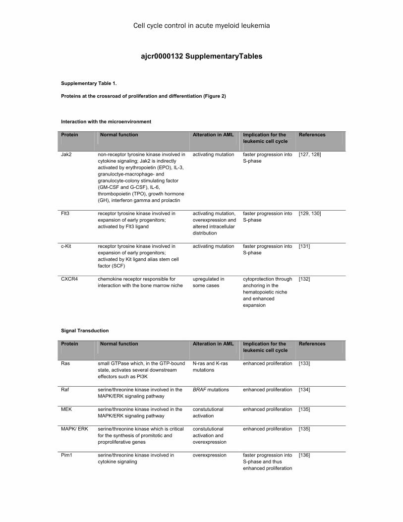
Cell cycle control in acute myeloid leukemia
ajcr0000132 SupplementaryTables
Supplementary Table 1.
Proteins at the crossroad of proliferation and differentiation (Figure 2)
Interaction with the microenvironment
Protein Normal function Alteration in AML Implication for the leukemic cell cycle
References
Jak2 non-receptor tyrosine kinase involved in cytokine signaling; Jak2 is indirectly activated by erythropoietin (EPO), IL-3, granuloctye-macrophage- and granulocyte-colony stimulating factor (GM-CSF and G-CSF), IL-6, thrombopoietin (TPO), growth hormone (GH), interferon gamma and prolactin
activating mutation faster progression into S-phase
[127, 128]
Flt3 receptor tyrosine kinase involved in expansion of early progenitors; activated by Flt3 ligand
activating mutation, overexpression and altered intracellular distribution
faster progression into S-phase
[129, 130]
c-Kit receptor tyrosine kinase involved in expansion of early progenitors; activated by Kit ligand alias stem cell factor (SCF)
activating mutation faster progression into S-phase
[131]
CXCR4 chemokine receptor responsible for interaction with the bone marrow niche
upregulated in some cases
cytoprotection through anchoring in the hematopoietic niche and enhanced expansion
[132]
Signal Transduction
Protein Normal function Alteration in AML Implication for the leukemic cell cycle
References
Ras small GTPase which, in the GTP-bound state, activates several downstream effectors such as PI3K
N-ras and K-ras mutations
enhanced proliferation [133]
Raf serine/threonine kinase involved in the MAPK/ERK signaling pathway
BRAF mutations
enhanced proliferation [134]
MEK serine/threonine kinase involved in the MAPK/ERK signaling pathway
constututional activation
enhanced proliferation [135]
MAPK/ ERK serine/threonine kinase which is critical for the synthesis of promitotic and proproliferative genes
constututional activation and overexpression
enhanced proliferation [135]
Pim1 serine/threonine kinase involved in cytokine signaling
overexpression faster progression into S-phase and thus enhanced proliferation
[136]

Cell cycle control in acute myeloid leukemia
Cdk inhibitors (CKIs)
Protein Normal function Alteration in AML Implication for the leukemic cell cycle
References
p14 inhibitor of Mdm2 which leads to stabilization of p53 and hence p21
downregulated faster progression into S-phase
[137]
p15 inhibitor of Cdk 4 downregulated
faster progression into S-phase
[138]
p16 inhibitor of Cdk 4 downregulated faster progression into S-phase
[139]
p21 inhibitor of Cdk2 and 4 p21 deficiency cooperates with t(8;21)
faster progression into S-phase
[140]
p27 inhibitor of Cdk2 and 4 downregulated faster progression into S-phase
[141]
Regulators of DNA synthesis
Protein Normal function Alteration in AML Implication for the leukemic cell cycle
References
Tet2 methylcytosine dehydroxygenase which catalyzes the synthesis of 5-hydroxymethylcytosine
mutated enhanced expansion of myeloid progenitors in mice
[38]
Regulators of APC/C-dependent proteolysis
Protein Normal function Alteration in AML Implication for the leukemic cell cycle
References
Cdh1 activating subunit of the APC/C which is active primarily in late mitosis and G1 phase
repressed faster progression into S-phase
[63]
Pocket proteins
Protein Normal function Alteration in AML Implication for the leukemic cell cycle
References
pRb pocket protein which binds to E2F and inhibits E2F-dependent transcription
downregulated and/or truncated
faster progression into S-phase
[142]
Cyclin-dependent kinases
Protein Normal function Alteration in AML Implication for the leukemic cell cycle
References
Cdk4/6 serine/threonine kinases which are activated by cyclin D and phosphorylate pRb and are inhibited by p16
downstream target of many activating kinase mutations and deregulated signaling cascades
faster progression into S-phase
[143]

Cell cycle control in acute myeloid leukemia
Cdk2 serine/threonine kinase which is activated by cyclin E or A and is inhibited by p21 and p27
enhanced activity faster progression into S-phase
[144]
Cyclins
Protein Normal function Alteration in AML Implication for the leukemic cell cycle
References
Cyclin D activates Cdk4/6 is highly important for cell cycle progression
upregulated (e.g. MLL leukemia)
faster progression into S-phase
[51]
Cyclin E activates Cdk2 regulates the G1/S transition
upregulated faster progression into S-phase
[144, 145]
Transcription factors
Protein Normal function Alteration in AML Implication for the leukemic cell cycle
References
E2F family of transcription factors involved in cell cycle regulation and DNA synthesis
upregulated expression counteracts differentiation in vitro
[55, 146]
p53 transcription factor involved in DNA damage response and subsequent inhibition of cell cycle progression
deleted and/or mutated, deletion is often associated with mutation of the remaining allele
reduced checkpoint activity and compromised DNA damage response
[121]
c-Myb member of the myeloblastosis family of transcription factors which is involved in the regulation of hematopoiesis
upregulated (e.g. in MLL leukemia)
important player in MLL-leukemogenesis
[39]
PU.1 ETS domain transcription factor encoded by the SPI1 gene which is important for myeloid and B-cell development
reduced PU.1 activity in some leukemia subsets
suppression enhances pro-proliferative potential and leukemogenesis
[147]
Stat3 signal transducer and activator of transcription in response to cytokines
aberrant activation in AML
faster progression into S-phase
[148]
Stat5 signal transducer and activator of transcription in response to cytokines; the Stat pathway is activated by various cytokines (IL-2, -3, -5, -6, -7, -12, -15), prolactin, tumor necrosis factor alpha, epidermal growth factor and interferon-
aberrant activation in AML
faster progression into S-phase
[25, 149]
CEBP CCAAT/enhancer binding protein alpha is a basic leucine zipper transcription factor which can interact with Cdks
mutated inhibition of granulocytic differentiation
[150]
HoxA9 homeobox transcription factor which is important for embryonic development
aberrant transcription of Hox genes by MLL
enhances pro-proliferative potential and leukemogenesis in cooperation with Meis1
[151]

Cell cycle control in acute myeloid leukemia
Meis1 homeobox transcription factor which is important during development
aberrant expression in Hox- and MLL-induced leukemia, cooperation with Evi1 and regulator of cyclin D3
faster progression into S-phase and enhanced proliferation
[51, 152, 153]
Chromatin remodelers
Protein Normal function Alteration in AML Implication for the leukemic cell cycle
References
MLL Histone-lysine N-methyltransferase HRX is an important regulator of gene transcription and transcriptional memory
regulator of the Hox gene cluster and cell cycle regulators
faster progression into S-phase and mitosis
[46]
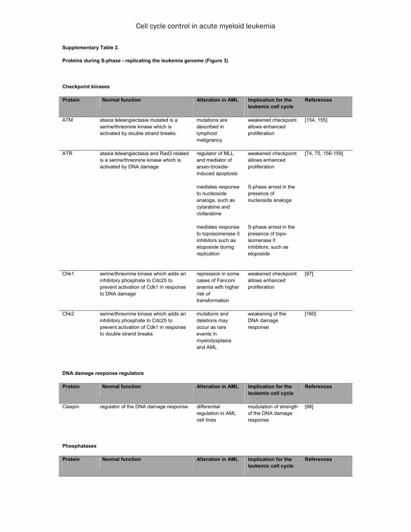
Cell cycle control in acute myeloid leukemia
Supplementary Table 2.
Proteins during S-phase - replicating the leukemia genome (Figure 3)
Checkpoint kinases
Protein Normal function Alteration in AML Implication for the leukemic cell cycle
References
ATM ataxia teleangiectasia mutated is a serine/threonine kinase which is activated by double strand breaks
mutations are described in lymphoid malignancy
weakened checkpoint allows enhanced proliferation
[154, 155]
ATR ataxia teleangiectasia and Rad3 related is a serine/threonine kinase which is activated by DNA damage
regulator of MLL and mediator of arsen-trioxide-induced apoptosis mediates response to nucleoside analoga, such as cytarabine and clofarabine mediates response to topoisomerase II inhibitors such as etoposide during replication
weakened checkpoint allows enhanced proliferation S-phase arrest in the presence of nucleoside analoga S-phase arrest in the presence of topo-isomerase II inhibitors, such as etoposide
[74, 75, 156-159]
Chk1 serine/threonine kinase which adds an inhibitory phosphate to Cdc25 to prevent activation of Cdk1 in response to DNA damage
repression in some cases of Fanconi anemia with higher risk of transformation
weakened checkpoint allows enhanced proliferation
[97]
Chk2 serine/threonine kinase which adds an inhibitory phosphate to Cdc25 to prevent activation of Cdk1 in response to double strand breaks
mutations and deletions may occur as rare events in myelodysplasia and AML
weakening of the DNA damage response
[160]
DNA damage response regulators
Protein Normal function Alteration in AML Implication for the leukemic cell cycle
References
Claspin regulator of the DNA damage response differential regulation in AML cell lines
modulation of strength of the DNA damage response
[98]
Phosphatases
Protein Normal function Alteration in AML Implication for the leukemic cell cycle
References

Cell cycle control in acute myeloid leukemia
Cdc25A dual-specificity phosphatase which removes phosphate groups from tyrosine and serine/threonine residues and activates Cdk1, Cdk2 and Cdk4
negatively regulated by Chk1 and Chk2 and positively regulated by Jak2V617F
enhanced G1/S transitioning and S-phase progression
[161, 162]
F-box proteins
Protein Normal function Alteration in AML Implication for the leukemic cell cycle
References
Skp2 F-box protein activates the SCF and mediates proteolysis of p27 and MLL
overexpressed in many cancers and aberrantly regulated in MLL-leukemia
enhanced G1/S transitioning and weakening of DNA damage response
[75, 163]
Cyclins
Protein Normal function Alteration in AML Implication for the leukemic cell cycle
References
Cyclin E activates Cdk2 regulates the G1/S transition
overexpressed
faster progression into and through S-phase
[145]
Cyclin A activates Cdk2, regulates S-phase progression and interacts with c-Myb
overexpressed and altered intracellular distribution
faster progression through S-phase
[164-166]
Cyclin-dependent kinases
Protein Normal function Alteration in AML Implication for the leukemic cell cycle
References
Cdk2 serine/threonine kinase which is activated by cyclin E or A and is inhibited by p21 and p27
high Cdk2 activity faster progression into and through S-phase
[144]
Chromatin remodelers
Protein Normal function Alteration in AML Implication for the leukemic cell cycle
References
MLL histone modifier and target of ATR which protects damaged chromatin from replication machinery binding
disrupted by leukemogenic transloactions in MLL leukemias
weakened DNA damage response
[75]
Regulators of replication
Protein Normal function Alteration in AML Implication for the leukemic cell cycle
References

Cell cycle control in acute myeloid leukemia
Cdc45 important regulator of early steps of replication
regulated by wild-type MLL
altered regulation of DNA binding in MLL-leukemias
[144]

Cell cycle control in acute myeloid leukemia
Supplementary Table 3.
Proteins during G2-phase - getting prepared for genomic and cytoplasmic division (Figure 4)
Histones
Protein Normal function Alteration in AML Implication for the leukemic cell cycle
References
-H2AX histone which following phosphorylation at serine 139 is an indicator of DNA double strand breaks
differences in H2AX loading between AML subsets
Part of the DNA damage response and possible predictor of response to therapy
[96]
Chromatin remodelers
Protein Normal function Alteration in AML Implication for the leukemic cell cycle
References
SET inhibitor of PP2A (see above) which is able to interact with p21 and inhibits cyclin B/Cdk1
aberrantly expressed in AML
weakened DNA damage response
[167]
Ubiquitin ligases
Protein Normal function Alteration in AML Implication for the leukemic cell cycle
References
Mdm2 E3 ubiquitin ligase that negatively regulates p53
overexpressed in some subsets of AML
inactivation of the p53-pathway
[168]
Checkpoint kinases
Protein Normal function Alteration in AML Implication for the leukemic cell cycle
References
ATM ataxia teleangiectasia mutated is a serine/threonine kinase which is activated by double strand breaks
mediates response to chemotherapy
G2-arrest in response to chemotherapy
[154, 155, 169]
ATR ataxia teleangiectasia and Rad3 related is a serine/threonine kinase which is activated by DNA damage
mediates response to chemotherapy
G2-arrest in response to chemotherapy
[75, 156, 170]
Chk1 serine/threonine kinase which adds an inhibitory phosphate to Cdc25 to prevent activation of Cdk1 in response to DNA damage
repression in some cases of Fanconi anemia with higher risk of transformation
weakened G2-checkpoint
[97]

Cell cycle control in acute myeloid leukemia
Chk2 serine/threonine kinase which adds an inhibitory phosphate to Cdc25 to prevent activation of Cdk1 in response to double strand breaks
mutations and deletions may occur as rare events in myelodysplasia and AML
weakened G2-checkpoint
[160]
DNA repair proteins
Protein Normal function Alteration in AML Implication for the leukemic cell cycle
References
BRCA1/2 breast cancer type 1 and 2 susceptibility proteins are involved in DNA repair
deleterious mutations
DNA damage response
[91]
Phosphatases
Protein Normal function Alteration in AML Implication for the leukemic cell cycle
References
PP2A protein phosphatase 2 is a serine/threonine phosphatase which functions in DNA damage response and cell cycle progression
frequently inactivated
weakened DNA damage response
[167]
Cdc25B dual-specificity phosphatase which removes phosphate groups from tyrosine and serine/threonine residues and activates Cdk1
upregulated as a result of FoxM1 overexpression
faster transition into mitosis
[171]
Cyclin-dependent kinases
Protein Normal function Alteration in AML Implication for the leukemic cell cycle
References
Cdk1 cyclin-dependent kinase which is an important regulator of mitotic entry
aberrantly activated by Cdc25
faster transition into mitosis
[172]
Cyclins
Protein Normal function Alteration in AML Implication for the leukemic cell cycle
References
Cyclin B activates Cdk1 at the G2/M-transition increased expression in AML cell lines
faster transition into mitosis
[171]
Mitotic kinases
Protein Normal function Alteration in AML Implication for the leukemic cell cycle
References

Cell cycle control in acute myeloid leukemia
Plk1 polo-like kinase 1 is a regulator of mitotic entry, centrosome kinetics and cytokinesis
overexpressed in subsets of AML
faster recovery from DNA damage response and rapid progression into mitosis
[101, 123]
Transcription factors
Protein Normal function Alteration in AML Implication for the leukemic cell cycle
References
FOXM1 member of the Fox family of transcription factors with peak levels in G2 phase and mitosis and functions during mitotic entry and chromosome separation; positive regulator of various proto-oncogenic components
overexpressed in subsets of AML
faster progression into mitosis
[171]
Cdk inhibitors
Protein Normal function Alteration in AML Implication for the leukemic cell cycle
References
p21 Cdk-inhibitor (see above) which, complexed with SET or following upregulation by p53, can inhibit cyclin B/Cdk1 to block mitotic entry
downregulated in subsets of AML
faster progression into mitosis
[171]

Cell cycle control in acute myeloid leukemia
Supplementary Table 4.
Proteins during G2-mitosis - segregating leukemia chromosomes and cytokinesis of leukemia cells (Figure 5)
Chromatin remodelers
Protein Normal function Alteration in AML Implication for the leukemic cell cycle
References
MLL chromatin modifying enzyme (see above) which is supposed to mark gene promotors to avoid transient silencing during mitosis
oligomerization mediated by the septin moiety of MLL-septin fusion proteins
enhanced chromatin remodeling
[173]
MLL5 chromatin modifying enzyme which contributes to regulation of the G2/M-transition
frequently deleted altered entry into mitosis
[174, 175]
Cyclins
Protein Normal function Alteration in AML Implication for the leukemic cell cycle
References
Cyclin B activates Cdk1 during mitosis downregulated in the presence of truncated forms of AML1-ETO, reduced stability in the presence of a weakened mitotic checkpoint
faster metaphase to anaphase transition
[107]
Separase inhibitors
Protein Normal function Alteration in AML Implication for the leukemic cell cycle
References
Securin inhibitor of the cohesin-cleaving enzyme separase
downregulated in the presence of truncated forms of AML1-ETO, reduced stability in the presence of a weakened mitotic checkpoint
less stringent inhibition of separase
[107]
Spindle assembly checkpoint components
Protein Normal function Alteration in AML Implication for the leukemic cell cycle
References
BubR1 member of the mitotic checkpoint complex (MCC) and pseudosubstrate inhibitor of Cdc20
downregulated in AML
weakens the mitotic checkpoint response
[107]
Bub1 important regulator of MCC assembly and direct inhibitor of Cdc20
downregulated in AML
weakens the mitotic checkpoint response
[108]

Cell cycle control in acute myeloid leukemia
Mad2 member of the mitotic checkpoint complex (MCC) which inactivates Cdc20 through direct binding
Downregulated by Runx1 and the Etv6-Runx1 fusion protein, which is frequently observed in childhood ALL
weakens the mitotic checkpoint response
[176]
Blinkin this protein mediates localization of Bub1 and BubR1 to conserved kinetochore components (hMIS12, NDC80, and Zwint-1 complexes) to facilitate stable chromatid microtubule connections
fusion protein partner of MLL
the fusion protein probably causes a dominant negative effect over the wild type protein
[110]
Mitotic kinases
Protein Normal function Alteration in AML Implication for the leukemic cell cycle
References
Aurora A mitotic kinase which is important for proper function of the mitotic spindle, chromosome alignment and cytokinesis
overexpressed in AML
overrides the mitotic checkpoint
[112]
Aurora B chromosomal passenger protein which is important for chromosome attachment to microtubules overexpressed in
AML
hyperactivation of Aurora B is known to cause chromosomal missegregation
[116, 177]
Plk1 Plk1 is a mitotic kinase that regulates centrosome maturation, mitotic entry, spindle formation, chromosome separation and cytokinesis
overexpressed in AML
overexpression causes aberrant entry and exit from mitosis and contributes to aneuploidy
[123, 178]