review article immunohistochemistry in … article immunohistochemistry in diagnostic surgical...
TRANSCRIPT

Int J Clin Exp Pathol 2010; 3(2):169-176 www.ijcep.com /IJCEP905002
Review Article Immunohistochemistry in diagnostic surgical pathology: contributions of protein life-cycle, use of evidence-based methods and data normalization on interpretation of immunohistochemical stains Halliday A. Idikio Department of Pathology and Laboratory Medicine, University of Alberta, Canada Received May 8, 2009; Accepted November 17, 2009; Available online November 25, 2009 Abstract: Immunohistochemical (IHC) staining of formalin-fixed and paraffin-embedded tissues (FFPE) is widely used in diagnostic surgical pathology. All anatomical and surgical pathologists use IHC to confirm cancer cell type and possible origin of metastatic cancer of unknown primary site. What kinds of improvements in IHC are needed to boost and strengthen the use of IHC in future diagnostic pathology practice? The aim of this perspec-tive is to suggest that continuing reliance on immunohistochemistry in cancer diagnosis, search and validation of biomarkers for predictive and prognostic studies and utility in cancer treatment selection means that minimum IHC data sets including “normalization methods” for IHC scoring, use of relative protein expression levels, use of protein functional pathways and modifications and protein cell type specificity may be needed when markers are proposed for use in diagnostic pathology. Furthermore evidence based methods (EBM), minimum criteria for di-agnostic accuracy (STARD), will help in selecting antibodies for use in diagnostic pathology. In the near future, quantitative methods of proteomics, quantitative real-time polymerase chain reaction (qRT-PCR) and the use of high-throughput genomics for diagnosis and predictive decisions may become preferred tools in medicine. Key words: Immunoperoxidase, protein lifecycle, surgical pathology, proteomics, evidence based me-thods, normalization Introduction Immunohistochemical methods in diagnostic pathology has a long history [1, 2]. Immunohis-tochemical staining methods include use of fluorophore-labeled (immunofluorescence) and enzyme-labeled (immunoperoxidase) anti-bodies to identify proteins and other molecules in cells. In diagnostic surgical pathology, im-munoperoxidase methods (usually single anti-gen-antibody and less commonly double anti-body-antigen combinations) (Figure1) are wide-ly used to extract additional information that is not available by hematoxylin and eosin stain-ing and light microscopy or by transmission electron-microscopy. The advantage is that the molecules are identified in-situ in the cell. Im-munohistochemistry is now used in surgical
pathology to determine cancer cell types, can-cer subtype classifications and possible cell-of –origin in metastatic cancer of unknown or undetermined primary site. In all instances, accepted and standardized morphologic crite-ria are used in addition to immu-nohistochemical staining of the tissue. The morphologic criteria for cancer diagnosis do not encompass the proposed biologic hall-marks of cancer [3]. This perspective is to review and promote the inclusion of some information to improve the interpretation of immunohistochemical data such as protein life-span and signaling, evi-dence-based methods and quantitative data and normalization.
Protein life-cycle and immunohistochemistry in diagnostic surgical pathology
Int J Clin Exp Pathol 2010; 3(2):169-176 170
Protein structure, modifications, life-span and implications for Immunohistochemistry Protein synthesis in the cell is highly regulated [4]. The proteins undergo many modifications before full maturation and functional activa-tion. Life-span modifications in normal, stressed and cancer cells include summoyla-tion and ubiquitination and subsequent de-gradation in the proteosome and probably res-cued by de-ubiquitination, by chaperones and chaperonins [5-7] and the effects of microRNA [8]. A widely known functional modification is phosphorylation that occurs on serine and threonine amino-acids, and these changes may affect life-span [9]. There are numerous protein databases that are freely available that permit inquiry of protein structure, cellular and tissue distribution, developmental and evolu-tionary history, functional status, mutations and other relevant information [10]. Further-more, since synthetic peptides are frequently used for generating antibodies (mono-and po-lyclonal), the functional significance and con-tribution of the peptide segment and structural information in relation to the function of the whole molecule should be taken into account when interpreting the immunohistochemical staining result. Phospho-specific antibodies
are now available for immunohistochemical use to determine the functional status of the protein and their use may further improve the results of immunohistochemical staining [11]. The productive use of phospho-specific anti-bodies will rest heavily on further elucidation of the cellular phospho-proteome [12] and optimization of phospho-specific polyclonal and monoclonal antibodies and tissue processing [13]. The p53 Example (Figure 2 a-c): One of the most investigated proteins in cell biology and pathology is p53. As an example, p53 is altered in many human cancers (>18,000 mutations) and involved in cell death and survival, DNA damage response [14, 15] and affects the transcription of a large gene/protein set in the cell [16]. p53 undergoes many modifications as wild-type or mutant protein and influences its cytoplasmic or nuclear location [17-20], the function and life-span of p53 and cellular interactions with its known and unknown targets and their func-tion [21]. There are now competing and conti-nually improving methods of proteomics to quantify and determine presence of protein(s) in cells [22-25]. Proteomics is useful in search-ing for and defining biomarkers using high-throughput methods such as the whole cell proteome.
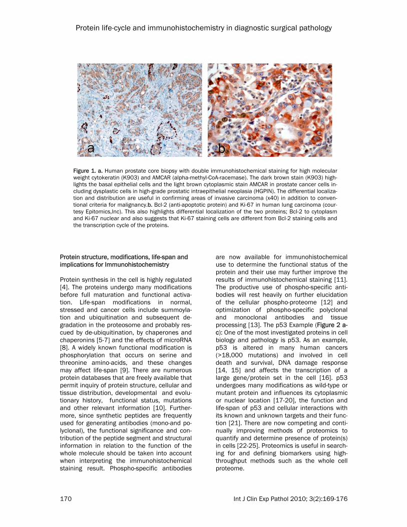
Figure 1. a. Human prostate core biopsy with double immunohistochemical staining for high molecular weight cytokeratin (K903) and AMCAR (alpha-methyl-CoA-racemase). The dark brown stain (K903) high-lights the basal epithelial cells and the light brown cytoplasmic stain AMCAR in prostate cancer cells in-cluding dysplastic cells in high-grade prostatic intraepithelial neoplasia (HGPIN). The differential localiza-tion and distribution are useful in confirming areas of invasive carcinoma (x40) in addition to conven-tional criteria for malignancy.b. Bcl-2 (anti-apoptotic protein) and Ki-67 in human lung carcinoma (cour-tesy Epitomics,Inc). This also highlights differential localization of the two proteins; Bcl-2 to cytoplasm and Ki-67 nuclear and also suggests that Ki-67 staining cells are different from Bcl-2 staining cells and the transcription cycle of the proteins.

Protein life-cycle and immunohistochemistry in diagnostic surgical pathology
Int J Clin Exp Pathol 2010; 3(2):169-176 171
Diagnostic and predictive biomarkers Biomarkers are currently proposed for various aspects of cancer such as early detection to selection of cancer patients for treatment. The biomarkers can be detected by immunohisto-chemical methods, quantitative proteomic methods and methods such as quantitative real-time polymerase chain reaction (qRT-PCR) [26]. The promotion of molecular and indivi-
dualized medicine is based on the improve-ment and miniaturization of methods of pro-teomics and genomics in the search for bio-markers of disease onset, progression and treatment response [27]. A recent commen-tary also emphasizes the need to base the use of markers in diagnostic or predictive immu-nohistochemical staining o known biological pathways and underlying biology of the cancer or disease process [28]. Furthermore, there is
Figure 2. a. Summarized p53 protein modifications b. immunohistochemical staining for wild-type p53 (Epitomics, Inc #1026-1) in human breast carcinoma. Note this antibody recognizes both wild-type and mutant p53. c. immunohistochemical staining for phospho-p53 (pS46) (Epitomics, Inc #2190) in human ovarian cancer tissue.

Protein life-cycle and immunohistochemistry in diagnostic surgical pathology
Int J Clin Exp Pathol 2010; 3(2):169-176 172
growing interest in including defined biomark-ers in clinical trials [29]. Evidence-based methods (EBM) and standards for reporting of diagnostic accuracy (STARD) Another question is whether the rules of Evi-dence-based medicine (EBM) that are adopted in other sections of laboratory medicine (clini-cal chemistry) can be applied to immunohisto-chemical interpretation before adoption for routine use [30, 31]. The rules of EBM applied to laboratory values include agreement statis-tics (raw, kappa, expected and odds ratio),
confidence intervals, sensitivity, specificity, positive and negative predictive values, like-lihood ratios, pre-and post-test probabilities; all of these datasets are useful for estimating diagnostic accuracy and are used in other di-agnostic settings [32]. The STARD criteria in-clude 24 item check list. Including EBM and STARD criteria may reduce bias in their use in diagnosis or treatment selection. The proposal for minimum datasets in immunohistochemi-cal publications (MISFISHIE) is encouraging, although quantitative analysis and protein structure data are not now included. Many publications provide immunohistochemical analysis as percentage of cases and control that are positive and negative for the anti-gen/protein under study. A recent study on immunohistochemical markers for mesotheli-oma listed the markers and percentage posi-tive in the cases used but no sensitivity or specificity information [33]. Few studies pro-vide sensitivity and specificity analysis; one recent study on lymphatic markers provided these analyses [34]. A recent study on use of
markers for defining the possible primary site of metastatic adenocarcinomas had some markers with variable sensitivities and speci-ficities [35]. The drawbacks of immunohisto-chemical staining that include inadequate an-tibody validation and many technical issues with a host of suggestions have been hig-hlighted [36]. Some drawbacks of convention-al immunohisto-chemical staining include lack of multiplexing, limited dynamic range and lack of correlation with functional protein and treatment response [36, 37]. The use of rabbit monoclonal versus mouse monoclonal antibo-dy to estrogen receptor (ER) changed the level
of positivity in breast cancer [38] (see Figure 3). Normalizing IHC scores How do we determine protein content in tis-sues? There are no reliable methods to quanti-fy tissue protein content by immunohistochem-ical methods. Many authors use different me-thods to estimate protein / antigen level in the literature [39] including intensity levels (0-3) or percent of cells that stain or a combination of the two scoring methods and attempts at cut-off values. Many suggestions relating to quan-titative methods in immunohistochemistry re-late to its impact in high-throughput methods such as tissue microarray (TMA) [39, 40]. In a recent study, simulated mRNA levels were re-lated to possible levels of protein detected by immunohistochemistry [41]; these methods will be difficult on an individual case based on protein life-span, modifications and mutations, tissue retrieval and fixation and other limita-tions. A study of Her-2 in breast cancer cell
Figure 3. Comparison of rabbit and mouse monoclonal antibodies in immunohistochemical detection of Her-2 (courtesy of Epitomics, Inc). The differences in intensities and percentage of cells stained is notable.

Protein life-cycle and immunohistochemistry in diagnostic surgical pathology
Int J Clin Exp Pathol 2010; 3(2):169-176 173
lines and tissue highlights the contribution of the primary antibody dilution on the level of Her-2 protein detected by immunohistochemi-cal methods [40] especially as Her-2 belongs to a protein family with complex interacting networks (42). Unlike routine diagnostic im-munohistochemical methods, high-throughput tissue microarrays, protein and DNA microar-rays generate a lot of data. The methods of data analysis and presentation proposed for DNA and protein microarrays- including me-thods to remove noise in the data such as normalization, false and negative discovery rates [43-47] are designed to improve in-terpretation, and utility of the information. Can
normalization be used in immunohistochemi-cal evaluation and what methods can be used for normalization? One can use endogenous proteins for normalization as is used in north-ern blots for messenger ribonucleic acid (mRNA) levels (48) and for Western blotting and quantitative real-time polymerase chain reaction (qRT-PCR). A relative protein expres-sion level can then be used. What are the min-imum methods to quantify protein/antigen levels in the cell? Some investigators used
bioinformatics tools to determine such cut-offs [49] and these attempts created different es-timates for HER2 that are different from the standardized criteria for HER2 [50]. Image analysis computer programs that can be used easily are needed [51, 52] and [53] and are coming on-stream [54, 55]. A recent overview of quantitative image analysis software for immunostaining lists several commercial sources though costs may be a limitation to adoption of specific software [56]. Further-more, as the interest in computer-assisted image analysis grows within the surgical pa-thology community an awareness of the mul-tiple methods of image analysis, noise remov-
al, image quality, and their effects on the re-sults should be noted [57, 58]. The DAB-stained slides can be analyzed by spectral im-aging [59], color deconvolution [54, 55, 60, 61], Hue-Saturation-Intensity [61], normalized RGB [62] and CMYK [63 and other methods. In a recent study of predictive biomarkers in breast cancer, automated image analysis was necessary to use 42 antibodies in the as-seesment of marker utility [64]. The growth of many image analysis methods for the popular
Table 1. Summarized Comparison of Immunohistochemical Method and Liquid Chromatography-
Mass Spectrometry (LC-MS) in Tissue Proteomics Method of Protein Detection Advantages Drawbacks Immunohistochemistry Protein location and distribution
seen Detectable in small and large tissue biopsies and fixed tissues Validation of other high-throughput studies ( DNA microar-ray)
Limited ability to quantitate pro-tein content Problems with antibody types, limited ability to detect protein modifications Limited or lack of Evidence based Criteria Single or dual detection ability Variable scoring methods and reproducibility No normalization methods Limited throughput Limited capacity for clinical bio-marker profiling( only with tissue microarrays)
Other Proteomics (i.e LC-MS) 100's to 1000's of peptides and proteins detected Can peruse databases for protein function and Gene ontology Robust Bioinformatics High throughput High level quantitation Can detect modified proteins Great potential for use in detect-ing clinical biomarkers
Cannot locate identified peptides to cell type(s) Need fresh or frozen tissue sam-ples

Protein life-cycle and immunohistochemistry in diagnostic surgical pathology
Int J Clin Exp Pathol 2010; 3(2):169-176 174
DAB-stained tissues needs internal normaliza-tion as done for RT-PCR and western blotting to truly compare the results. Alternative methods for biomarker identifica-tion and selection The role of IHC data sets and analysis in diag-nostic pathology are being challenged by other quantitative methods such as DNA microarray and qRT-PCR in cancer detection, classifica-tion and predicting cancer treatment re-sponse. Recently proposed molecular classifi-cations of cancers and their use in cancer treatment planning are based on DNA microar-ray methods that have well-defined methods and analysis [65-68] and in some cases new entities unknown by light microscopic methods have emerged. The DNA microarray methods have been used to separate primary and sec-ondary cancers in lung [69] and separate co-lonic from ovarian cancer origins [70] and to determine cancer of unknown primary sites [71]. The future of a needle core biopsy of suspicious mass may be (a) routine hematox-ylin and eosin, immunohistochemistry includ-ing normalization and analysis, EBM and STARD (b) isolation of protein content for 2-dimensional gel electrophoresis and western blotting, protein and antibody arrays and mass spectrometry (c) isolation of total messenger ribonucleic acids(mRNA), cDNA synthesis, mi-croarray expression studies, single nucleotide polymorphism(SNP) and array comparative genomic hybridization (array CGH) and copy number variation (CNV) of genes (Table 1). The continued use and dependence on immuno-histochemical staining in diagnostic surgical pathology will need the use of EBM and STARD methods and minimum datasets and integra-tion of protein networks and function, and im-age analysis with normalization or definable cut-offs. Acknowledgements I am grateful to Epitomics, Inc (www.epitomics. com) for permission to use their figures. Please address all correspondences to: Halliday A. Idikio, MD, University of Alberta, Department of Pa-thology and Laboratory Medicine, 5B4.11 Walter MacKenzie Health Sciences Center, 8440-112th Street, Edmonton, ALBERTA T6G 2B7, CANADA, Tel 1-780-407-7271, Fax 1-780-407-3009, E-mail [email protected]
References [1] Robinson G, Dawson I. Immunochemical stu-
dies of the endocrine cells of the gastrointes-tinal tract II An immunoperoxidase technique for the localization of secretin-containing cells in human duodenum. J Clin Patho 1975; 28: 631-635.
[2] Nakane P, Pierce G. Enzyme-labeled antibo-dies; preparation and application for the locali-zation of antigens. J Histochem and Cytochem 1966; 14: 929-931.
[3] Hanahan D, Weinberg R. The Hallmarks of Cancer. Cell 2000; 100:57-70.
[4] Lewin B. Protein Synthesis. In: GENES VIII. Up-per Saddle River, NJ 07458: Pearson/Prentice Hall; 2004. p. 135-166.
[5] Krappmann D, Scheidereit C. A pervasive role of ubiquitin conjugation in activation and ter-mination of IkB kinase pathways. EMBO Re-ports 2005;6(4):321-326.
[6] Seet B, Dikic I, Zhou M-M, Pawson T. Reading protein modifications with interaction domains. Nat Rev Mol Cell Biol 2006;7:473-483.
[7] Wilson VG, Rosas-Acosta G. Wrestling with SU-MO in a New Arena. Science STKE 2005(pe32).
[8] Baek D, Villen J, Shin C, Camargo F, Gygi S, Bartel D. The impact of microRNAs on protein output. Nature 2008.
[9] Hunter T. The Age of Crosstalk: Phosphoryla-tion, Ubiquitination, and Beyond. Molecular Cell 2007; 28:730-738.
[10] Peri J, Navarro J, Amanchy R, Kristiasen T, Jon-nalagada C, Surendranath Vea. Development of Human Protein Reference Database as an Initial Platform for Approaching Systems Biolo-gy in Humans. Genome Res 2003; 13:2363-2371.
[11] Mandell J. Phosphorylation State-Specific Anti-bodies Applications in Investigative and Diag-nostic Pathology. Am J Pathol 2003; 163:1687-1698.
[12] Lim Y. Mining the Tumor Phosphoproteome for Cancer Markers. Clin Cancer Res 2005; 11(9):3163-3169.
[13] Baker A, Dragovich T, Ihle N, Williams R, Fenog-lio-Preiser C, Powls G. Stability of Phospho-protein as a Biological Marker of Tumor Signal-ing. Clin Cancer Res 2005; 11(12):4338-4340.
[14] Jin S, Levine A. The p53 functional circuit. Journal of Cell Science 2001;114:4139-4140.
[15] Haupt S, Berger M, Goldberg Z, Haupt Y. Apop-tosis- the p53 network. J Cell Sci 2003; 116:4077-4085.
[16] Wei C-L, Wu Q, Vega V, Chiu K, Ng P, Zhang T, et al. A Global Map of p53 Transcription-Factor Binding Sites in the Human Genome. Cell 2006; 124:207-219.
[17] Bode A, Dong Z. Post-translational modification of p53 in tumorigenesis. Nat Rev Cancer 2004;4:793-805.

Protein life-cycle and immunohistochemistry in diagnostic surgical pathology
Int J Clin Exp Pathol 2010; 3(2):169-176 175
[18] Bourdon J-C, Fernandes K, Murray-Zmijeweski F, Liu G, Diot A, Xirodimas D, et al. p53 iso-forms can regulate p53 transcriptional activity. Genes Dev 2005; 19:2122-2137.
[19] Vousden K, Prives C. P53 and Prognosis: New Insights and Further Complexity. Cell 2005;120:7-10.
[20] Schmitt C, Fridman J, Yang M, Baranov E, Hoffman R. Dissecting p53 tumor suppressor functions in vivo. Cancer Cell 2002;1:289-298.
[21] Trigiante G, Lu X. ASPPs and cancer. Nat Rev Cancer 2006;6:217-226.
[22] Petricoin E, Zoon K, Kohn E, Liotta L. Clinical Proteomics: Translating Benchside Promise In-to Bedside Reality. Nat Rev Drug Discov 2002;1:683-695.
[23] Celis J, Gromov P, Gromova I, Moreira J, Cabe-zon T, Ambartsumian Nea. Integrating Proteo-mic and Functional Genomic Technologies in Discovery-driven Translational Breast Cancer Research. Mol Cell Proteomics 2003;2:369-377.
[24] Graham D, Elliott S, Van Eyk J. Broad -based proteomic strategies: a practical guide to pro-teomics and functional screening. J Physiol 2004;563(1):1-9.
[25] Sanchez-Carbayo M, Socci N, Lozano J, Haab B, Cordon-Cardo C. Profiling Bladder Cancer Using Targeted Antibody Microarrays. Am J Pathol 2006;168:93-103.
[26] Ludwig J, Weinstein J. Biomarkers in Cancer Staging, Prognosis and Treatment Selection. Nat Rev Cancer 2005;5:845-856.
[27] Dalton W, Friend S. Cancer Biomarkers- An Invitation to the Table. Science 2006;312: 1165-1168.
[28] Natkunam Y, Mason D. Prognostic immunohis-tologic markers in human tumors: why are so few used in clinical practice? Lab Invest 2006; 86:742-747.
[29] Weil R. Incorporating Molecular Tools into Ear-ly-Stage Clinical Trials. PLos Med 2008; 5:e21.
[30] Hawkins R. The Evidence Based Medicine Ap-proach to Diagnostic Testing: practicalities and limitations. Clinical Biochem Review 2005; 26(May):7-18.
[31] McQueen M. Overview of Evidence -based Med-icine: Challenges for Evidence-based Laborato-ry Medicine. Clin Chem 2001; 47(8):1536-1546.
[32] Mallet S, Deeks J, Halligan S, Hopewell S, Cor-nelius V, Altman D. Systematic reviews of diag-nostic tests in cancer: review of methods and reporting. BMJ 2006; 333:413.
[33] Ordonez N. The diagnostic utility of immunohis-tochemistry in distinguishing between epithelo-id mesotheliomas and squamous cell carcino-mas of the lung: a comparative study. Mod Pa-thol 2006; 19:417-429.
[34] Evangelou E, Kyzas P, Trikalinos T. Comparison of the diagnostic accuracy of lymphatic endo-thelium markers: Bayesian approach. Mod Pa-thol 2005;18(11):1490-1497.
[35] Dennis J, Hvidsen T, Wit E, Komorowski J, Bell A, Downie I, et al. Markers of Adenocarcinoma Characteristic of the site of Origin: Develop-ment of a Diagnostic Algorithm. Clin Cancer Res 2005; 11(10):3766-3772.
[36] Bast R, Lilja H, Urban N, Rimm D, Fritsche H, Gray J, et al. Translational Crossroads for Bio-markers. Clin Cancer Res 2005; 11(17):6103-6108.
[37] Rimm D. What brown cannot do for you. Nat Biotechnol 2006; 24(8):914-916.
[38] Cheang M, Treaba D, Speers C, Olivoto I, Badjik C, Chia S, et al. Immunohistochemical Detec-tion Using the New Rabbit Monoclonal Antibody SP1 of Estrogen Receptor in Breast Cancer Is Superior to Mouse Monoclonal Antibody 1D5 in Predicting Survival. J Clin Oncol 2006; 24(36):5637-5644.
[39] True L, Feng Z. Immunohistochemical Valida-tion of Expression Microarray Results. J Mol Di-agn 2005;7(2):149-151.
[40] McCabe A, Dolled-Filhart M, Camp R, Rimm D. Automated Quantitative Analysis (AQUA) of In situ Protein Expression, Antibody Concentra-tion, and Prognosis. J Natl Cancer Inst 2005; 97:1808-1815.
[41] Betensky R, Nutt C, Batchelor T, Louis D. Sta-tistical Considerations for Immunohistochemi-cal Panel Development after Gene Expression Profiling of Human Cancers. J Mol Diagn 2005; 7(2):276-282.
[42] Jones R, Gordus A, Krail J, MacBeath G. A quantitative protein interaction network for the ErbB receptors using protein microarrays. Na-ture 2006; 439:168-174.
[43] Norris A, Kahn C. Analysis of gene expression in pathophysiological states: Balancing false discovery and false negative rates. Proc Natl Acad Sci U S A 2006; 103:649-653.
[44] Simon R. Development and Validation of The-rapeutically Relevant Multi-Gene Biomarker Classifiers. JNCI 2005; 97(12):866-867.
[45] Simon R, Radmacher M, Dobbin K, McShane L. Pitfalls in the Use of DNA Microarray Data for Diagnostic and Prognostic Classification. JNCI 2003; 95(1):14-18.
[46] Tinker A, Boussioutas A, Bowtell D. The chal-lenge of gene expression microarrays for the study of human cancer. Cancer Cell 2006; 9(5):333-339.
[47] Storey J, Tibshirani R. Statistical significance for genomewide studies. Proc Natl Acad Sci U S A 2003; 100:9440-9445.
[48] de Kok J, Roelofs R, Giesendorf B, Pennings J, Waas E, Feuth T, et al. Normalization of gene expression measurements in tumor tissues: comparison of 13 endogenous control genes. Lab Invest 2005; 85:154-159.
[49] Camp R, Dolled-Filhart M, Rimm D. X-Tile: A New Bio-Informatics Tool for Biomarker As-sessment and Outcome-Based Cut-Point Opti-mization. Clin Cancer Res 2004; 10(Nov1):7252-7259.

Protein life-cycle and immunohistochemistry in diagnostic surgical pathology
Int J Clin Exp Pathol 2010; 3(2):169-176 176
[50] Dowsett M, Hanna W, Kockx M, Pennault-Llorca F, Ruschoff J, Gutjahr T, et al. Standardi-zation of HER2 testing: results of an interna-tional proficiency testing ring study. Mod Pathol 2007; 20:584-591.
[51] Matkowskyj K, Cox R, Jensen R, Benya R. Quantitative Immunohistochemistry by Measur-ing Cumulative Signal Strength Accurately Measures Receptor Number. J Histochem Cy-tochem 2003;51(2):205-214.
[52] Hu J-J, Ambrus A, Fossum T, Miller M, Humph-rey J, Wilson E. Time Courses of Growth and Remodeling of Porcine Aortic Media During Hypertension: A Quantitative Immunohisto-chemical Examination. J Histochem Cytochem 2008;56(4):359-370.
[53] Wester K, Wahlund E, Sundstrom C, Ranefall P, bengtsson E, Russell P, et al. Parrafin Section Storage and Immunohistochemistry. Appl Im-munohistochem Mol Morphol 2000;8(1):61-70.
[54] Halushka M, Cornish T, Lu J, Selvin S, Selvin E. Creation, validation, and quantitative analysis of protein expression in vascular tissue micro-arrays. Cardiovasc Pathol 2009: doi: 10.1016/ j.carpath.2008.12.007.
[55] Halushka M, Selvin E, Macgregor A, Cornish T. The use of vascular tissue microarrays for measurement of advanced glycation end prod-ucts. J Histochem Cytochem 2009: DOI: 10.1369/ jhc.2009.953273.
[56] Cregger M, Berger A, Rimm D. Immunohisto-chemistry and Quantitative Analysis of Protein Expression. Arch Pathol Lab Med 2006; 130:1026-1030.
[57] Paizis M, Engelhardt J, Siklos L. Quantitative assessment of relative changes of immunohis-tochemical staining by light microscopy in spe-cified anatomical regions. J Microsc 2009; 234(1):103-112.
[58] Kayser K, Gorter J, Metze K, Goldmann T, Voll-mer E, Mireskandari M, et al. How to measure image quality in tissue-based diagnosis (diag-nostic surgical pathology). BMC Diagn Pathol 2008;3(Supl 1):S11.
[59] van der Loos C. Multiple Immunoenzyme Stain-ing: Methods and Visualizations for the Obser-vation With Spectral Imaging. J Histochem Cy-tochem 2008; 56(4):313-328.
[60] Ruifrok A, Johnson D. Quantification of histo-chemical staining by color deconvolution. Anal Quant Cytol Histol 2001; 23:291-299.
[61] Ruifrok A, Katz R, Johnson D. Comparison of Quantification of Histochemical Staining By Hue-Saturation-Intensity (HSI) Transformation and Color-Deconvolution. Appl Immunohisto-chem Mol Morphol 2003; 11(1):85-91.
[62] Brey E, Lalani Z, Johnston C, Wong M, McIntire L, Duke P, et al. Automated Selection of DAB-labeled Tissue for Immunohistochemical Quan-tification. J Histochem Cytochem 2003; 51(5):575-584.
[63] Pham N-A, Morrison A, Schwock J, Aviel-Ronen S, Iakolev V, Tsao M-S, et al. Quantitative im-age analysis of immunohistochemical stains using a CMYK color model. BMC Diagn Pathol 2007; 2:8.
[64] Charpin C, Secq V, Giusiano S, Carpenter S, Andrac L, Lavaut M-N, et al. A signature predic-tive of disease outcome in breast carcinomas, identified by quantitative immunocytochemical assays. Int J Cancer 2009; 124:2124-2134.
[65] Su A, Welsh J, Sapinoso L, Kern S, Dimitrov P, Lapp H, et al. Molecular classification of Hu-man Carcinomas by Use of Gene Expression Signatures. Cancer Res 2001; 61:7388-7393.
[66] Glinsky GV, Higashiyama T, Glinski AB. Classifi-cation of Human Breast Cancer Using Gene Expression Profiling as a Component of the Survival Predictor Algorithm. Clin Cancer Res 2004; 10:2272-2283.
[67] Rhodes D, Chinnayan A. Integrative analysis of the cancer transcriptome. Nat Genet Supple-ment 2005; 37:S31-37.
[68] Pinkel D, Albertson D. Array comparative ge-nomic hybridization and its applications in can-cer. Nature Genetics Supplement 2005; 37:S11-17.
[69] Talbot S, Estilo C, Maghami E, Sarkaria I, Pham D, O-Charoenrat P, et al. Gene Expression Pro-filing Allows Distinction between Primary and Metastatic Squamous Cell Carcinomas in the Lung. Cancer Research 2005; 65(8):3063-3071.
[70] Nishizuka S, Chen S-T, Gwadry F, Alexander J, Major S, Scher U, et al. Diagnostic Markers That Distinguish Colon and Ovarian Adenocar-cinomas: Identification by Genomic, Proteomic, and Tissue Array Profiling. Cancer Research 2003; 63(Sep 1):5243-5250.
[71] Veradhachary G, Talantov D, Raber M, Meng C, Hess K, Jatkoe T, et al. Molecular Profiling of Carcinoma of Unknown Primary and Correlation with Clinical Evaluation. J Clin Oncology 2008; 26(27):4442-4448.