review article immunomodulatory effects …downloads.hindawi.com/journals/jir/2015/354957.pdfreview...
TRANSCRIPT

Review ArticleImmunomodulatory Effects Mediated by Serotonin
Rodrigo Arreola,1 Enrique Becerril-Villanueva,2 Carlos Cruz-Fuentes,1
Marco Antonio Velasco-Velázquez,3 María Eugenia Garcés-Alvarez,2
Gabriela Hurtado-Alvarado,4 Saray Quintero-Fabian,5 and Lenin Pavón2
1Psychiatric Genetics Department, Clinical Research Branch, National Institute of Psychiatry, “Ramon de la Fuente”,Calzada Mexico-Xochimilco 101, Colonia San Lorenzo Huipulco, Tlalpan, 14370 Mexico City, DF, Mexico2Department of Psychoimmunology, National Institute of Psychiatry, “Ramon de la Fuente”, Calzada Mexico-Xochimilco 101,Colonia San Lorenzo Huipulco, Tlalpan, 14370 Mexico City, DF, Mexico3School ofMedicine,NationalAutonomousUniversity ofMexico, AvenidaUniversidad 3000, Coyoacan, 04510MexicoCity,DF,Mexico4Area of Neurosciences, Department of Biology of Reproduction, CBS, Universidad Autonoma Metropolitana, Unidad Iztapalapa,Avenida San Rafael Atlixco No. 186, Colonia Vicentina, Iztapalapa, 09340 Mexico City, DF, Mexico5Genetics Unit Nutrition of Biomedical Research Institute of Universidad Nacional Autonoma de Mexico at Instituto Nacional dePediatrıa, Avenida del Iman No. 1, cuarto piso, Colonia Insurgentes-Cuicuilco, Coyoacan, 04530 Mexico City, DF, Mexico
Correspondence should be addressed to Lenin Pavon; [email protected]
Received 20 October 2014; Accepted 24 February 2015
Academic Editor: Douglas C. Hooper
Copyright © 2015 Rodrigo Arreola et al.This is an open access article distributed under theCreativeCommonsAttribution License,which permits unrestricted use, distribution, and reproduction in any medium, provided the original work is properly cited.
Serotonin (5-HT) induces concentration-dependent metabolic effects in diverse cell types, including neurons, entherochromaffincells, adipocytes, pancreatic beta-cells, fibroblasts, smooth muscle cells, epithelial cells, and leukocytes. Three classes of genesregulating 5-HT function are constitutively expressed or induced in these cells: (a) membrane proteins that regulate the responseto 5-HT, such as SERT, 5HTR-GPCR, and the 5HT
3-ion channels; (b) downstream signaling transduction proteins; and (c)
enzymes controlling 5-HT metabolism, such as IDO and MAO, which can generate biologically active catabolites, includingmelatonin, kynurenines, and kynurenamines. This review covers the clinical and experimental mechanisms involved in 5-HT-induced immunomodulation. These mechanisms are cell-specific and depend on the expression of serotonergic componentsin immune cells. Consequently, 5-HT can modulate several immunological events, such as chemotaxis, leukocyte activation,proliferation, cytokine secretion, anergy, and apoptosis.The effects of 5-HT on immune cellsmay be relevant in the clinical outcomeof pathologies with an inflammatory component. Major depression, fibromyalgia, Alzheimer disease, psoriasis, arthritis, allergies,and asthma are all associated with changes in the serotonergic system associated with leukocytes.Thus, pharmacological regulationof the serotonergic system may modulate immune function and provide therapeutic alternatives for these diseases.
1. Introduction
Serotonin (5-HT), also known as 5-hydroxytryptamine or 3-(2-aminoetil)-1H-indol-5-ol, is a monoamine containing twonitrogen molecules: the first nitrogen is basic and embeddedwithin the indol-5-ol; the second, within 2-aminoethyl, islocated at the terminus of the aliphatic chain. 5-HT isgenerated from tryptophan and serves as a substrate for thesynthesis of a diverse set of molecules, such as melatonin,formyl-5-hydroxykynurenamine, and 5-hydroxyindoleaceticacid [1]. In addition, 5-HT is a signaling molecule that affectsthe immune [2], gastrointestinal [3], and nervous [4] systems
in paracrine, endocrine, and juxtacrine fashion. Finally, 5-HT regulates development during cellular differentiation andontogeny (morphogenesis) in several cell linages [5–7].
The majority of 5-HT synthesis, up to 90%, takes placein gastrointestinal enterochromaffin (EC) cells, followed bysynthesis in myenteric neurons (5%) and the brain [8, 9]. Inthe 1980s, 5-HTwas identified as an immunomodulator for itsability to stimulate or inhibit inflammation [10].This immuneregulation—which has yet to be fully elucidated—is orches-trated by the serotonergic system. Therefore, to understanddisease pathologies related to the immune system, it is
Hindawi Publishing CorporationJournal of Immunology ResearchVolume 2015, Article ID 354957, 21 pageshttp://dx.doi.org/10.1155/2015/354957
2 Journal of Immunology Research
important to consider the function of serotonergic compo-nents. Specifically, insight can be gained by understandinghow serotonergic components are related to mechanisms ofimmune modulation that depend on 5-HT receptors (5HTR)expression in leukocytes and other cells involved in aninflammatory response.
2. A Brief History of 5-HT Discovery
The discovery of 5-HT was a product of collaborativeendeavors initiated in the last quarter of the 19th century[11] that lasted into the second half of the 20th century.Initial studies identified an extract with vasoconstrictionproperties from a platelet fraction of uncoagulated blood [12].In research conducted in Rome during the 1930s, VittorioErspamer isolated a molecule from gastrointestinal EC cellswith the capacity to generate smooth muscle contractionsin a rat uterus. Chemical analysis identified the molecule asan indoleamine and it was named enteramine [13]. Duringthe 1940s in the Cleveland Clinic research department,Maurice Rapport, Arda Green, and Irving Page purifiedand characterized a vasoconstrictor compound generatedshortly after coagulation and related to hypertension. In atour de force, the molecule was purified from 900 liters ofserum obtained from 2 tons of bull’s blood [14, 15]. Thename serotonin emerged after the substance was crystallizedin 1948 because it was obtained from serum (“ser”) andcould induce vascular tone (“tonin”) in blood vessels [16].Subsequently, the crystalline vasoconstrictor substance wasshown to be a single complex composed of creatinine andindol-derivates, which permitted a structural model of 5-HTbased on UV-spectrophotometry [17]. Chemical synthesis of5-HT by Hamlin and Fischer in 1951 [18] provided significantprogress allowing for the confirmation of its pharmacologicaleffects [19] and a comparison with the previously isolatedenteramine [20]. Interest in understanding the physiologicalrole of 5-HT prompted efforts to isolate the compound fromdifferent mammals and tissues, such as the central nervoussystem [21].
Since the 1970s there has been an established associationbetween the serotonergic system and affective disorders aswell as mood changes [22]. Recently, serotonin has beenassociated with a myriad of processes [23], including aggres-sion [24], sleep [25], appetite [26], pain [27], bone density[28], tissue regeneration [29], platelet aggregation [30], andgastrointestinal function [31]. The influence of 5-HT onthe immune system has also been recognized, although thespecific mechanisms underlying these effects are not com-pletely understood and may require confirmation in humancells. Despite these pitfalls, it is well acknowledged that theserotonergic system and associated molecules expressed inimmune cells can influence mood disorders, such as majordepression [32] and schizophrenia [33, 34].
3. Components of the Serotonergic System AreExpressed in Leukocytes
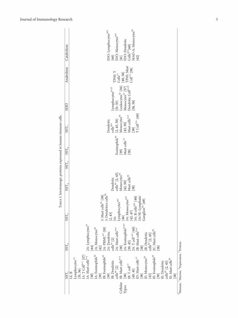
The expression of serotonergic components is differentiallyregulated between tissues and cell types.While the expression
and function of serotonergic proteins has primarily beenstudied within the central nervous system [81], it should bepointed out that no functional differences between cell typeshave been identified.The serotonergic components expressedin the immune system encompass a complex ensemble ofproteins that coordinate the synthesis and degradation [1],transport and storage [58], and response to 5-HT stimu-lation [40]. In leukocytes, the expression of serotonergiccomponents (Table 1) is modulated by the concentration ofextracellular and intracellular 5-HT. Furthermore, the signalsgenerated by 5-HT interactions with leukocytes are distinctdepending on function, developmental stage, and activationstatus of the cell. This functional heterogeneity suggests thatthe serotonergic system can precisely regulate a wide range ofimmunomodulatory effects [81].
3.1. Catabolism and Anabolism of 5-HT. The essential aminoacid Tryptophan is utilized by many cell types and can beconverted into a wide range of chemically related prod-ucts, among the best known are 5-HT and melatonin, butalso include kynurenines and kynurenamines (Figure 1). Inmacrophages and T lymphocytes the indoleamine 2,3-dioxy-genase (IDO1 & IDO2. EC: 1.13.11.52) [60, 82] enzymes helpdegrade tryptophan to generate kynurenines and producekynurenamines from 5-HT or melatonin [83]. In general, allof these compounds can modulate immune responses [1, 84–86]. However, the mechanisms by which these moleculesexert an immunomodulating function are not completelyelucidated. Some observations suggest that kynurenines andkynurenamines function in negative feedback loops to mod-ulate 5-HT-mediated inflammation, other proinflammatorymolecules, and melatonin levels.
3.1.1. Anabolism. The synthesis of serotonin begins with theessential amino acid, tryptophan, and follows two-enzy-matic steps. First, a hydroxyl group is added by tryptophan5-hydroxylase (TPH; EC: 1.14.16.4) to generate 5-hydroxy-tryptophan. Mammals produce two TPH enzymes encodedby two independent genes, TPH1 and TPH2. While TPH1 isexpressed in peripheral tissues, TPH2 is exclusively expressedin the central nervous system [87–89]. After this hydroxy-lation step, a carboxyl group is removed by an aromatic L-amino acid decarboxylase (DDC; EC: 4.1.1.28) generating 5-HT [90–92].
3.1.2. Catabolism. Within the immune system, four catabolicpathways for the breakdown of 5-HT have been observed.One pathway begins with the generation of melatonin from5-HT through two enzymatic steps; first, 5-HT is acety-lated by arylalkylamine N-Acetyltransferase (AANAT; EC:2.3.1.87) generating N-acetyl 5-HT, which then acquires amethyl group from N-acetylserotonin-O-methyltransferase(ASMT; EC: 2.1.1.4; previously known as hydroxyndole-O-methyltransferase, HIOMT) to become melatonin [93, 94].Subsequently, the enzyme indoleamine 2,3-dioxygenase(IDO1 & IDO2; EC: 1.13.11.52) can convert melatonin intoa cyclooxygenase (COX; EC: 1.14.99.1) inhibitor calledformyl-N-acetyl-5-methoxykynurenamine. Interestingly this
Journal of Immunology Research 3
Table1:Serotonergicproteins
expressedin
human
immun
ecells.
5HT 1
5HT 2
5HT 3
5HT 4
5HT 5
5HT 6
5HT 7
SERT
Anabo
lism
Catabo
lism
Cellular
Types
1A:B
Lymph
ocytes∗‡
[35,36]
1A:T
Cell§∗†[37]
1A:M
astcells§∗‡
[38]
1A:E
osinop
hils§‡
[39]
1B:E
osinop
hils§‡
[39]
1B:D
endritic
cells
§‡[2]
1B:M
astcells§∗‡
[38]
1B:T
cell∗†
[40,41]
1D:M
astcells∗‡
[38]
1E:M
onocytes
§‡
[42]
1E:E
osinop
hils§‡
[39]
1E:D
endritic
cells
§‡[2,43]
1E:M
astcells§‡
[38]
2A:Lym
phocytes
§†
[44]
2A:M
onocytes
§‡
[42]
2A:P
BMC§†[10]
2A:D
endritic
cells
§‡[2]
2A:M
astcells§∗‡
[38]
2A:E
osinop
hils§∗‡†
[39,45]
2A:T
cell∗‡†[40]
2B:M
astcells§∗‡
[38]
2B:D
endritic
cells
§‡[2,43]
2C:M
astcells§‡
[38]
3:Mastcells§‡[38]
3:Dendritics
cells
§‡
[2,43]
3A:
Lymph
ocytes
§‡†
[46]
3A:M
onocytes
§‡†
[42,47]
3A:B
cells
§‡†[48]
3A/3E:
Lymph
atic
Ganglion§‡[49]
Dendritic
cells
§‡[2,43]
Mon
ocytes
§‡
[42,50]
Mastcells§‡
[38]
Eosin
ophils§‡
[39]
Mastcells∗‡
[38]
Dendritic
cells
§‡†
[2,43,50]
Mon
ocytes
§‡
[42,50]
Mastcells§∗‡
[38]
TCell†‡∗[40]
Lymph
ocytes
§∗‡†
[51–55]
Leuk
ocytes
§‡[56]
Mon
ocytes
§†[57]
DendriticC
ell§∗‡†
[58,59]
TPH1:T
Cells‡∗
[40,58]
TPH1:Mast
Cell∗‡
[38]
IDO:Lym
phocytes
§‡†
[60]
IDO:M
onocytes
§‡†
[61]
IDO:D
endritic
Cells§‡†[60]
MAO
-A:M
onocytes
§‡
[62]
§ Hum
an,∗Murine,‡Ex
pressio
n,†Protein.

4 Journal of Immunology Research
C00078L-Tryptophan
C00643 5-Hydroxy-L-tryptophan
C01598Melatonin
C00978N-Acetylserotonin
C05642Formyl-N-acetyl-5
-methoxykynurenamine
EC: 2.3.1.87 AANAT
EC: 1.13.11.52IDO1 & IDO2
EC: 2.1.1.4ASMT
EC: 1.14.16.4TPH1 & TPH2
EC: 4.1.1.28DDC
EC: 1.13.11.11TDO2
EC: 1.13.11.52IDO1 & IDO2
Triptaminapathway
C02700L-Formylkynurenine
C00328 L-Kynurenine
EC: 3.5.1.9AFMID
EC. 2.6.1.7AADAT
C012524-(2-Aminophenyl)-2,4-dioxobutanoate
C01717Kynurenic acid/kynurenate
Nonenzymatic transformation
Picolinic acidspathway
C032273-Hydroxy-L-kynurenine
EC: 1.14.13.9KMO
C006323-Hydroxyanthranilate
EC: 3.7.1.3KYNU
EC: 1.13.11.6HAAO
C03722Quinolinate/quinolinic acid
Nonenzymatic transformation
C044092-Amino-3-carboxymuconate
semialdehyde
Anthranilatepathway
C056485-Hydroxy-N-formylkynurenine
C056515-Hydroxykynurenine
EC: 1.13.11.52IDO1 & IDO2
EC: 3.5.1.9AFMID
EC: 4.1.1.28DDC
C056385-Hydroxykynurenamine
EC: 1.4.3.4 MAOA & MAOB
C056394,6-Dihydroxyquinoline
C05647Formyl-5-hydroxykynurenamine
C06212N-Methylserotonin
C056345-Hydroxyindoleacetaldehyde
EC: 2.1.1.49 INMT
EC: 1.4.3.4 MAOA & MAOB
EC: 1.13.11.52IDO1 & IDO2
EC: 1.2.1.3ALDH2 & ALDH1B1 & ALDH3A2
EC: 1.2.3.1AOX1
C056355-Hydroxyindoleacetate
C056605-Methoxyindoleacetate
EC: 1.14.14.1Unspecific monooxygenaseCytochrome P450 isoforms
C056436-Hydroxymelatonin
C00780Serotonin
Figure 1: Metabolic pathways associated with 5-HT. The metabolic pathways branching from the catabolism of tryptophan are shown.Green and brown branches show kynurenine pathways from tryptophan. The dark blue branch displays the 5-HT generation pathwaywhile the red branch displays the melatonin generation pathway (solid line) and the melatonin catabolism pathway (dashed lines). The 5-HT catabolism pathways are depicted in blue, purple, and cyan dotted lines. The most relevant compounds are circled with dotted linesand underlined compounds have no demonstrated effects. An additional catabolic pathway marked in pink from 5-hydroxy-L-tryptophangenerates 5-hydroxy-kynurenine and 5-hidroxy-kynurenamine family compounds (paths C05648 andC05647), which have no demonstratedbiological effects. The compounds in these pathways are denoted by their “Kyoto Encyclopedia of Genes and Genomes” (KEGG) code(http://www.genome.jp/kegg/). Enzymes with their classification codes (EC, http://www.chem.qmul.ac.uk/iubmb/enzyme/) and UNIPROTgene names are shown in squares. The shading arrows show the most studied pathways.

Journal of Immunology Research 5
metabolite can function to block the synthesis of prostagl-andins [95], yet other melatonin metabolites, including 5-methoxyindole acetic acid and 6-hydroxymelatonin, have noreported function [96].
A second catabolic pathway of 5-HT utilizes the enzymeindoleamine 2,3-dioxygenase (IDO) and generates formyl-5-hydroxykynurenamine. In a third pathway, 5-HT is trans-formed into 5-hydroxyindoleacetic acid with monoamineoxidase A/B (MAO-A o MAO-B; EC: 1.4.3.4) among otherenzymes. Interestingly, MAO-A expression, which is regu-lated by the cytokines IL-4 and IL-3, has been identifiedin human monocytes from peripheral blood [62]. A fourthcatabolic pathway generates N-methylserotonin from 5-HTusing the enzyme amine N-methyltransferase (INMT; EC2.1.1.49) and may also be active in immune cells.
It is not thoroughly understood that the extent by whichthe byproducts and metabolites generated during catabolismmay affect the immune system [1, 84]. The identification ofadditional 5-HT metabolites in plasma, including serotonin-O-sulfate [97] and 5-hydroxykynurenamine [98], suggeststhat other catabolic pathways linked to specific biochemicalprocesses, such as activation and cell proliferation, may alsobe associated with the modulation of the immune system.
One additional catabolic pathway related with the fourpreviously described utilizes the enzyme IDO to generatekynurenines from tryptophan. This pathway is positivelyregulated when immune cells become activated and beginsecreting IFN-𝛼, IFN-𝛽 e IFN-𝛾, TNF-𝛼, TGF-𝛽, IL-1𝛽, andIL-2 [85, 99–101], which significantly consumes tryptophanand limits its availability for 5-HT production. L-kynurenine,3-hydroxy-L-Kynurenine, and 3-hydroxyanthranilic acid cannegatively modulate immune responses. Specifically, Vecseiand coworkers noted the blockage of cell proliferation as wellas the potential induction of apoptosis in Th1 and NK cells[85, 101–103].
It was recently proposed that 5-hydroxyindole thiazo-lidine carboxylic acid, a 5-HT byproduct found in theintestinal tissues and several brain regions of rats, is generatedfrom the condensation of 5-hydroxyindole acetaldehyde andL-cysteine by a carbon-sulfur lyase (EC 4.4.1.). However,evidence supporting this enzymatic condensation remains tobe confirmed. In addition the properties of this byproduct orits potential influence over immunological cells remain to beinvestigated [104].
3.2. 5-HT Receptors: (5HTR). 5-HT modulates many leuko-cyte functions ranging from activation of the immuneresponse to memory cell generation. The effects mediated by5-HT are dependent on the differential expression of sero-tonergic components in leukocytes. For example, serotoninreceptors (5HTR) on immune cells influence cytokine prolif-eration, delivery, migration, and cellular activation. Signalingthrough the 5HTR affects chemoattraction in immaturemammalian dendritic cells (human and rodent) but not inmature cells, which respond to 5-HT by secreting IL-6 [43].In addition, 5HTR signaling influences naıve T cell activationin mice by activating 5HT
7[40] and regulates lymphocyte B
cell proliferation through 5HT1A [41].
3.2.1. 5HTR Are G Protein-Coupled Receptors (GPCR). The5HTR belong to the GPCR family class A, also known as 7-transmembrane domain (7TM) receptors. GPCR are classi-fied into 6 classes according to a database from the Interna-tional Union of Basic and Clinical Pharmacology (IUPHAR:http://www.guidetopharmacology.org/) [105]. This systemincludes classes A, B, and C, as well as the adhesion, Frizzled,and other 7TM classes. Receptors for adenosine, adrenaline/noradrenaline, 5HT
1, 5HT
2, 5HT
4, 5HT
5, 5HT
6, and 5HT
7
all belong to GPCR class A. Furthermore, the 5HTRs arecomprised of 6 families and 13 subfamilies [106] with anundetermined number of isoforms that may be produced byalternative splicing. This receptor diversity suggests that agreat amount of functional variation may exist between the5HTRs [107].
Signal transduction from 5HTR is similar to standardGPCRs. G proteins form heterotrimeric complexes made ofthe subunits G𝛼, G𝛽 andG𝛾; the complex is coupled to the C-terminus of the transmembrane 5HTR. Different subtypes ofthe G𝛼 proteins (G𝛼i/o, G𝛼s, or G𝛼q11) may generate differenttransduction responses functioning either as activators orinhibitors.When the 5-HT ligand binds its receptor to inducesignal transduction it elicits a conformational change in thereceptor to facilitate activity [81]. Signal transduction can becarried out by the effector proteins G𝛼 or the heterodimerG𝛽-G𝛾. While transduction mechanisms have been betterdepicted for G𝛼, especially in cells of the nervous system[108], recent work has been devoted to the role of G𝛼 andG𝛽-G𝛾 in the immune system [109, 110].
In the last few years, a number of investigations havedemonstrated that GPCR are capable of assembling dimers(homo and heterodimers) as well as oligomers. Receptors5HT1B and 5HT
1D can assembly into homodimers andheterodimers when coexpressed in the same cell. Notably, thereceptors display roughly 77% sequence identity within the7TM domain [111]. The 5HT
2C receptors form homodimerswithin the cellular membrane [112–114] and it has beenproposed that signaling is initiatedwhen two 5-HTmoleculesbind the dimer [115]. However, activation a single subunitwithin the 5HT
4homodimer is sufficient to initiate G pro-
tein activation even though simultaneous activation of bothreceptors doubles the activation efficiency of the pathway.While it appears that 5HTR tend to form homodimersrather than heterodimers, the latter possibility has not beendiscarded [116].
Milligan and coworkers have postulated that the for-mation of heterodimers could generate specificity between5HTR and its substrates, which could be especially relevantfor the use and design of pharmacological agents [117].Despite current knowledge suggesting that the 5-HT recep-tors function as homodimers and maybe heterodimers, itis possible that higher subunit organizations (e.g., trimersand tetramers) could function under certain circumstances[114, 118].
To better understand 5-HT cellular-mediated processes,it will be necessary to characterize the quaternary structureand stoichiometry of 5HTRduring oligomerization in regularcellular processes. It will also be important to determine thebiochemical details of the quaternary transitional structure

6 Journal of Immunology Research
(quinary structure) of 5HTR to establish not only its func-tional characteristics, but also the coupling and assemblywithcytoskeleton proteins. Recently, the crystal structures of thereceptor-agonist complexes, 5HT
1B and 5HT2B with ergo-
tamine and dihydroergotamina, respectively, were reportedand provided structural information to better understandreceptor-ligand interactions and agonist selectivity, whichcould inform 5HTR-based drug design [119, 120].
3.2.2. The 5HT3Receptors Form a Cationic Channel. The
5HT3receptors are part of a cation-selective ion channel
Cys-loop superfamily and have been detected in T lym-phocytes, monocytes [47], mature dendritic cells [43], andmast cells [38]. The functional unit of 5HT
3, a pentameric
ring, generates a central ion channel and can be composedof two 5HT
3A and three 5HT3B subunits [121, 122]. Each
subunit contains a large N-terminal domain with the 5-HTbinding site and four transmembrane domains connectedwith intracellular and extracellular loops [123–125].The 5HT
3
receptors are regulated through protein modifications andcytoskeletal rearrangements, dependent on protein kinases(A and C) and F-actin, respectively [123, 126].
There are a number of 5HT3receptor variants that can
be expressed from the human genome. The genes encoding5HT3A and 5HT
3B are located on chromosome 11q23, andthose encoding 5HT
3C, 5HT3D, and 5HT
3E are on chromo-some 3q27. However, the total number of isoforms generatedfrom these genes by alternative splicing has yet to be deter-mined [127–130]. Interestingly, the subunits 5HT
3C, 5HT3D,
5HT3E, and 5HT
3Ea are not sufficient to form functionalpentamers but can generate them with 5HT
3A and hencemodify 5-HT responses [131].
In the context of the central nervous system, 5HT3receptors are associated with rapid activation and inhibitionresponses in addition to fast cellular depolarization [49, 132].The cellular depolarization response is unique to neurons,as this has not been observed in immune cells. In neurons,5HT3receptors modulate the delivery of neurotransmitters,
such as dopamine [133], whereas the same receptors elicitthe release of cytokines from immune cells. Human lymphnodes preferentially express the 5HT
3A and 5HT3E variants
[49]. In addition, 5HT3A is expressed in naıve and activated
B-lymphocytes [48], T lymphocytes, and human monocytes,but expression has not been detected frommonocyte-deriveddendritic cells [47]. Inhibiting the 5HT
3receptors with
antagonists, such as ondasetron and tropisentrol, disruptsTNF-𝛼 and IL-1𝛽 production, suggesting that these receptorsmay activate the p38/MAPK pathway [134, 135].
3.3. SERT: 5-HT Transporter. The serotonin transporterSERT actively moves extracellular 5-HT across the plasmamembrane into the cell. The transporter is also known assolute carrier family 6, member 4 (SLC6A4), and belongsto a family of neurotransmitters with 12 transmembranedomains. The function of SERT in platelets is critical formaintaining adequate 5-HT concentrations in the circulatorysystem [136]. To function, SERT depends on the transport ofNa+/Cl− ions, yet the coordinated mechanism of 5-HT andion transport remains to be elucidated.
The gene encoding SERT has 14 exons and is located onchromosome 17q11.1-17q12. Two genetic polymorphisms inthe gene regulatory regionmodulate transcription generatinga complex mix of long and short variants [137]. The SERTfunctional unit is a dimer but it has been suggested thattwo SERT homodimers could assemble into a tetramer [138].Other members of the protein family form heterodimers;however, these may not be functional [139]. Therefore, SERThomodimers are currently accepted as the biochemicallyfunctional unit.
SERT dimer formation relies on several posttranslationalmodifications. The SERT proteins are glycosylated, and thensialic acid is inserted into each of two N-linked glycans.In the absence of glycosylation, the functional activity ofSERT is reduced. Furthermore, the addition of sialic acidmolecules is important for dimer formation and the associ-ation with myosin IIA (a kinase that anchors protein kinaseG, PKG) at the cytoskeleton. This association can regulateSERT phosphorylation by the guanosine monophosphate-dependent PKG [140].
A complex regulatory mechanism of SERT internaliza-tion associated with the cytoskeleton has been describedin platelets and is referred to as serotonylation (see Box 1).The process of serotonylation depends on the 5-HT gra-dient between the extracelluar and intracellular space cre-ated by SERT activity. The gradient mediates regulationvia cytoskeletal components, vimentin, 5HTR, SERT, smallGTPases, transglutaminases (TGases), and possibly p21 acti-vated kinases (p21/PAK) [141]. Specifically, increased levelsof 5-HT activate SERT internalization through activation ofsmall GTPases by serotonylation (TGases covalently linkingthe 5-HT to the small GTPases). Currently efforts are beingdirected to the characterization of SERT internalizationmechanisms in leukocytes.
Box 1 (serotonylation: SERT, TGasas, and small GTPases).Serotonylation is the process by which 5-HT is covalentlybound to a protein through a transamination reaction andconstitutes a mechanism for regulating signal transduction.This process requires high intracellular levels of 5-HT and ismediated by transglutaminases (TGases. EC: 2.3.2.13). Phys-iological serotonylation has been demonstrated in plateletsand other cells [142, 143] but not in leukocytes. However,serotonylation may be involved in specific leukocyte func-tions required for chemotaxis or cytokine secretion [43,45] because this modification regulates similar functions inplatelets and pancreatic beta cells.
Although many cytoplasmic proteins can be serotony-lated, the effect of serotonylation on small GTPases dur-ing platelet activation and aggregation is noteworthy [142].Serotonylation induces constitutive RhoA activation and,consequently, the cytoskeletal reorganization needed foraggregation [144–147]. Increased intracellular Ca2+ and 5-HTin platelets also activates Rab4-mediated exocytosis of alphagranules by Ca2+-dependent TGase-mediated serotonylation[142].
In pancreatic beta cells, Rab3a and Rab27a are serotony-lated during insulin exocytosis [143]. In smooth muscle cellsTGase-2-mediated serotonylation of RhoA may be required
Journal of Immunology Research 7
for proliferation [148–150]. Furthermore, serotonylation ofRhoA not only constitutively activates it, but also increasesits rate of proteosomal degradation [148]. Since Rab27aand Rab27b also participate in exocytosis from plateletsand endocrine cells [151–155], these processes may be alsosusceptible to serotonylation.
Additionally, 5-HT induces vimentin filament reorienta-tion around SERT [156]. Furthermore, serotonylation of Rab4promotes interaction with the SERT C-terminal domainregulating translocation [157]. Importantly, there are noreports that specifically study serotonylation in leucocytes,but available information suggests that it may regulatecytoskeletal reorganization in these cells [43, 45]. Rab27ais expressed in cytotoxic T cells and regulates the last stepof granular exocytosis [158, 159]. Rab27, RhoA, and Rab4are also expressed in several cells of the immune system[160–162]; therefore it is possible that serotonylation regulatesexocytosis or cytoskeletal reorganization during functionssuch as MHC presentation.
3.4. 5-HT Storage and Exocytosis in Leukocytes. Three poten-tial outcomes may take place following 5-HT internalization:signaling may be activated by serotonylation, 5-HT mayundergo enzymatic transformation, or 5-HT may be storedin specialized vesicles. The synthesis, storage, and transportof 5-HT in the immune system aremore diverse and complexthan previously reported, and a seminal review of the subjectwas recently published by Ahern in 2011 [163].
Storage of 5-HT in the immune system allows for its reuseby exocytosis, which occurs in dendritic cells, peripheralblood lymphocytes, and platelets. Within these cells, thevesicular monoamine transporter (VMAT) is responsible forstoring 5-HT inside dense granules [164].The 5-HT transportand storage/exocytosis pathways in platelets are coupled tothe serotonylation signaling pathway, which assesses 5-HTconcentrations and dictates its fate [165]. LAMP1 containingvesicles in monocyte-derived dendritic cells from mice alsostore 5-HT, and upon ATP stimulation (Ca2+ influx), thesecells secrete 5-HT and cytokines [58]. A major focus of cur-rent endeavors is the investigation of several 5-HT mediatedprocesses in other types of leukocytes, including 5-HT vesiclestorage, small GTPases-mediated serotonylation, cytoskeletalassociations, and the metabolism of 5-HT into derivatives,such as melatonin and kynurenins.
4. The Effects of 5-HT on Leukocytes
Innate and adaptive immune responses rely on a diverse set ofcell types from lymphoid linage (T cells, B cells, andNK cells),myeloid linage (neutrophils, eosinophils, basophils, mono-cytes, and mast cells), or myeloid/lymphoid linage (dendriticcells) origins. Professional antigen presenting cells (APC),such as dendritic cells and macrophages, link the innateand adaptive immune responses by recognizing, processing,and presenting antigens on MHC-II. Antigen presentationactivates naıve T cells initiating clonal proliferation andgenerating the immune memory, which is essential for theadaptive phase of an immune response.
The local concentration of 5-HT can modulate a numberof events during these immune responses [10]. One reasonimmune cells respond to 5-HT is that, as mentioned previ-ously, they constitutively express the molecular machinerythat constitutes the serotonergic system.
4.1. Neutrophils. Neutrophils, the most abundant innateimmune cells in humanblood, constitute a first line of defenseagainst infection by recognizing foreign antigens, produc-ing antimicrobial compounds, and secreting cytokines andchemokines to recruit immunocompetent cells [166]. Mousemodels have demonstrated that neutrophil recruitment tosites of acute inflammation requires platelet-derived chemo-tactic stimuli, such as 5-HT, PAF (platelet-activation factor),and histamine [167, 168].
4.2. Monocytes/Macrophages. Macrophages as well as theirprecursors, circulating monocytes, participate in immuneresponses during pathogen infection. Monocytes can bedivided in two subsets: those that express CD14, a componentof the lipopolysaccharide (LPS) receptor complex, and thosethat express CD16, the Fc𝛾RIII immunoglobulin receptor[169, 170]. CD14+ cells, which constitute 80–90% of thecirculating monocytes, express 5HT
1E, 5HT2A, 5HT
3, 5HT
4,
and 5HT7mRNAs [42]. LPS stimulation does not affect
5HTR expression suggesting that the receptors constitutivelyregulate cell functions.
The addition of 5-HT tomonocytes induces phagocytosisof opsonized goat erythrocytes [171]. However, 5-HT hasbeen noted to elicit a number of responses in CD14+ cellsisolated from peripheral blood of healthy subjects [42]: (i) 5-HT signaling decreases TNF-𝛼 secretion in a dose-dependentmanner; (ii) 5-HT signaling enhances LPS-induced secretionIL-12p40 from activatedmonocytes, which acts as a chemoat-tractant for macrophages and promotes the migration ofbacterially stimulated dendritic cells; and (iii) 5-HT signalingenhances LPS-induced secretion of IL-6, IL-1𝛽, IL-8/CXCL8.The first two effects are mediated by the 5HT
4and 5HT
7
receptors, whereas the third effect requires 5HT3, 5HT
4, or
5HT7.
Macrophages also respond to 5-HT although reportsconflict as to whether the response is inhibitory or stimu-latory and the mechanisms involved have yet to be clearlydescribed. For example, combined stimulation with 5-HTand muramyl peptides induces superoxide secretion byperitoneal macrophages [172]. Bovine alveolar macrophagesrelease chemotactic factors for neutrophils and monocytesin response to 5-HT and histamine [173]. Similarly, murinemacrophages detect 5-HT with the 5HT
2C receptor to inducethe secretion of CCL2, which induces monocyte migration[174]. In peritoneal murine macrophages 5-HT inducesphagocytosis in a dose-dependent manner through 5HT
1Aand NF𝜅-B activation [175].
On the other hand, 5-HT is also reported to function asnegative regulator. For example, 5-HT
2signaling limited the
activation of murine macrophages stimulated in vitro withhigh concentrations of IFN-𝛾 [176]. Furthermore, humanalveolarmacrophages stimulatedwith LPS and 5-HT secretedless TNF-𝛼 and IL-12, but more IL-10, nitric oxide, and
8 Journal of Immunology Research
prostangladin-E2. However, the receptors mediating theseeffects have yet to be described [177].
The differentmacrophage responses elicited by 5-HTmaybe due to phenotypic differences in tissue-specific macro-phages (i.e., changes in the proportion of 5-HT receptors)or the effect of cooperative signaling with other molecules[178]. Finally, macrophages can rapidlymetabolize 5-HT to 5-hydroxyindole acetic acid [179], a biotransformation pathwaythatmay bemediated byMAO-A/B, ALDH/AOX andASMT,which could affect serotonergic responses in these cells.
4.3. Dendritic Cells (DCs). DCs play a crucial role in theimmune response to infectious pathogens. In humans, cir-culating DCs characteristically expresses high levels of classII HLA molecules and are proficient in antigen uptake andprocessing. However, they express low levels of HLA class Iand costimulatory molecules, such as CD80 and CD86, andlack common lineage markers such as CD3, CD14, CD16,CD19, CD20, and CD56. DCs are positioned between theadaptive and innate immune systems: detecting microbialinfection, tissue damage, and inflammatory signals to pro-mote the activation of antigen-specific responses [180–182].
Peripheral bloodmonocytes can differentiate intomacro-phages or DCs depending on environmental stimuli. Cul-turing cells with IL-4 and granulocyte-macrophage colony-stimulating factor (GM-CSF) induces humanmonocytes andmurine myeloid progenitors to differentiate into monocyte-derived dendritic cells (MDDCs) and bonemarrow dendriticcells (BMDCs), respectively. These cells, frequently used asmodels for dendritic cell biology [50, 58, 183], are sensitive toLPS and 5-HT (Table 2).
When 5-HT is added during IL-4/GM-CSF differenti-ation, the resulting MDDCs display lower levels of CD1a,CD86, and HLA-DR but had increased CD14 expression.However, other markers such as CD40, CD80, or CD83were unaffected. Murine BMDCs can be matured by LPSstimulation to generate cells with characteristics of DCs[184]. Although immature (CD11c+CD86−) and mature(CD11c+CD86+) BMDCs constitutively express SERT, matu-ration induced by LPS increases SERT expression and, conse-quently, mature BMDCs have an increased capability forintake, storage, and exocytosis of 5-HT [58]. BMDC matu-ration also reduces the expression of enzymes involved in themetabolism of 5-HT, such as MAO-A and -B. Furthermore,immature and mature dendritic-like cells respond differ-ently to 5-HT [50]. In mature MDDCs, the 5HT
3receptor
contributes to changes in intracellular Ca2+ concentrationrequired for the secretion of IL-8 and IL-1𝛽 [2]. On the otherhand, 5-HT inhibits CXCL10 secretion from mature MDDC,but CCL22 secretion is not affected [43].
Additionally, 5-HT can regulate MDDC functionalresponses. Costimulation with 5-HT and LPS induces imma-ture MDDCs migration in a 5HT
1B and 5HT2A-dependent
manner. However, if 5-HT is added subsequent to LPS-mediated maturation, migration is unaffected but cytokineand chemokine secretion is induced [43]. Additional in vitroexperiments with MDDCs and BMDCs have also demon-strated that 5-HT activates the secretion of pro-inflammatorycytokines [2, 185].
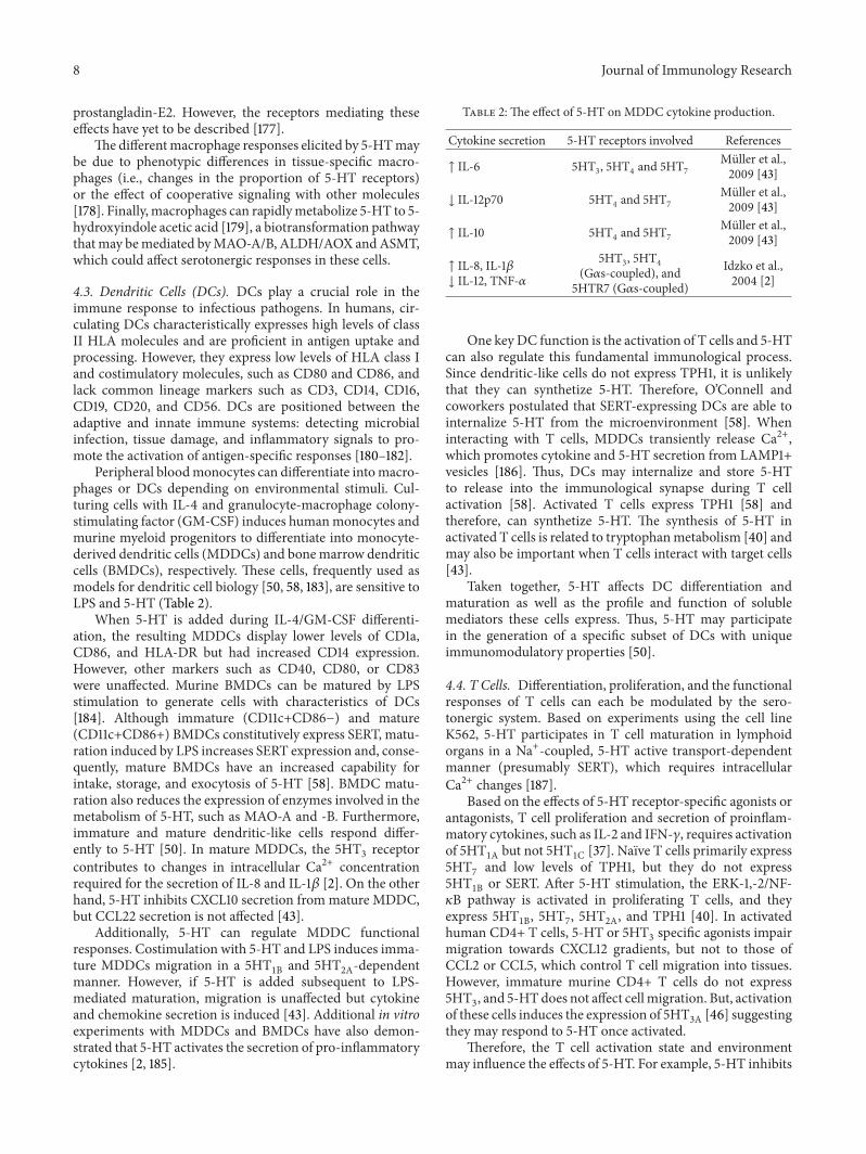
Table 2: The effect of 5-HT on MDDC cytokine production.
Cytokine secretion 5-HT receptors involved References
↑ IL-6 5HT3, 5HT4 and 5HT7Muller et al.,2009 [43]
↓ IL-12p70 5HT4 and 5HT7Muller et al.,2009 [43]
↑ IL-10 5HT4 and 5HT7Muller et al.,2009 [43]
↑ IL-8, IL-1𝛽↓ IL-12, TNF-𝛼
5HT3, 5HT4(G𝛼s-coupled), and
5HTR7 (G𝛼s-coupled)
Idzko et al.,2004 [2]
One key DC function is the activation of T cells and 5-HTcan also regulate this fundamental immunological process.Since dendritic-like cells do not express TPH1, it is unlikelythat they can synthetize 5-HT. Therefore, O’Connell andcoworkers postulated that SERT-expressing DCs are able tointernalize 5-HT from the microenvironment [58]. Wheninteracting with T cells, MDDCs transiently release Ca2+,which promotes cytokine and 5-HT secretion from LAMP1+vesicles [186]. Thus, DCs may internalize and store 5-HTto release into the immunological synapse during T cellactivation [58]. Activated T cells express TPH1 [58] andtherefore, can synthetize 5-HT. The synthesis of 5-HT inactivated T cells is related to tryptophanmetabolism [40] andmay also be important when T cells interact with target cells[43].
Taken together, 5-HT affects DC differentiation andmaturation as well as the profile and function of solublemediators these cells express. Thus, 5-HT may participatein the generation of a specific subset of DCs with uniqueimmunomodulatory properties [50].
4.4. T Cells. Differentiation, proliferation, and the functionalresponses of T cells can each be modulated by the sero-tonergic system. Based on experiments using the cell lineK562, 5-HT participates in T cell maturation in lymphoidorgans in a Na+-coupled, 5-HT active transport-dependentmanner (presumably SERT), which requires intracellularCa2+ changes [187].
Based on the effects of 5-HT receptor-specific agonists orantagonists, T cell proliferation and secretion of proinflam-matory cytokines, such as IL-2 and IFN-𝛾, requires activationof 5HT
1A but not 5HT1C [37]. Naıve T cells primarily express
5HT7and low levels of TPH1, but they do not express
5HT1B or SERT. After 5-HT stimulation, the ERK-1,-2/NF-
𝜅B pathway is activated in proliferating T cells, and theyexpress 5HT
1B, 5HT7, 5HT
2A, and TPH1 [40]. In activatedhuman CD4+ T cells, 5-HT or 5HT
3specific agonists impair
migration towards CXCL12 gradients, but not to those ofCCL2 or CCL5, which control T cell migration into tissues.However, immature murine CD4+ T cells do not express5HT3, and 5-HTdoes not affect cellmigration. But, activation
of these cells induces the expression of 5HT3A [46] suggesting
they may respond to 5-HT once activated.Therefore, the T cell activation state and environment
may influence the effects of 5-HT. For example, 5-HT inhibits
Journal of Immunology Research 9
phytohemagglutinin- (PHA-) mediated lymphocyte prolifer-ation, possibly through reduced expression and distributionof the IL-2 receptor [188, 189]. In addition, concanavalin A(ConA) and low concentrations of 5-HT increased murineT cell proliferation although the activation of CD4+ andCD8+ subsets was reduced [190, 191]. This demonstratesthat 5-HT elicited effects are concentration dependent andsuggests: (i) 5-HT induces dose-dependent phenotypes, (ii)differentiation may be achieved by receptors with different5-HT affinity, and (iii) local 5-HT concentrations are tightlyregulated to induce specific effects.
T cells express SERT and, therefore, can acquire 5-HT[51, 52, 192, 193]. However, naıve T cells have reduced SERTfunctional activity [164] and may result to 5-HT synthesis.In agreement with this hypothesis, a report from Aune andcoworkers demonstrates that the inhibition of the 5-HT syn-thesis in IL-2-stimulated T cells blocks cell proliferation. Theaddition of 5-hydroxtriptophan, a 5-HT precursor, restoresproliferation, further suggesting that these cells synthesizethe molecule rather than acquire it [37]. However, furtherresearch is required to understand how 5-HT synthesis isregulated in T cells.
4.5. B Cells. B cells recognize circulating antigen; as a con-sequence, they activate processes that end in the generationof memory B cells or antibody-forming plasma cells [194]. 5-HT increases the proliferation of murine B cells by activating5HT1A [41]. In addition, SERT expression is proportional to
the proliferation rates of human leukemic B cells. Specifically,SERT-specific inhibitors, such as fluoxetine, fenfluramine,or 3,4-methylenedioxymethamphetamine (MDMA), elicitedanti-proliferative and pro-apoptotic effects [195].
4.6. NK Cells. NK cells recognize antigen in the contextof CD1 controlling viral replication early in infection andinhibiting the development of cancer [196, 197]. These cellsare inhibited with oxidation produced by autologous mono-cytes and with apoptosis induced by reactive oxygen species(ROS); however, 5-HT signaling limits these forms of inhibi-tion [198]. NK cells express 5-HT
1A and therefore can sense5-HT local concentration [199–202]. In fact, 5HT
1A-specificantagonists, such as pindobind, exacerbate the inhibitoryeffect of monocyte-mediated ROS production on NK cells[202].
4.7. Eosinophils. Eosinophils are responsible for fightingmul-ticellular parasites and other infections in vertebrates; theyalso control mechanisms associated with allergy and asthma.Eosinophils express the 5-HT receptors 5HT
1A, 5HT1B,
5HT1E, 5HT
2A, and 5HT6, but they do not express 5HT
2C,5HT3, 5HT
4, and 5HT
7. However, differential expression of
5HT2A was detected in allergy and asthma patients [39].The serum levels of 5-HT are higher in symptomatic
asthma patients in comparison to asymptomatic patients,which may influence eosinophil-mediated inflammation inpatients with active disease. 5-HT is a potent chemoattractantfor eosinophils both in vivo and in vitro and supports rolling,an important feature of these cells. Antagonists of 5HT
2Ainhibit both effects suggesting that 5-HTmediates eosinophil
activation [45]. Migration and rolling require changes in theactin cytoskeleton and activation of PKC and calmodulinsignaling [39, 45], which control the morphological changesrequired for eosinophil infiltration from circulation to sits ofinflammation [39].
4.8. Basophils. While basophils represent less than 2% ofleukocytes, they actively participate in immune responses inperipheral organs where they are recruited during nematodeand ectoparasite infections. They also participate in allergicreactions by releasing histamine in response to specificgrowth factors, such as IL-3 [203, 204]. In addition, basophilsare an important source of IL-4 and therefore may promoteTh2 differentiation [205, 206].
The role of 5-HT on basophil functions has not beenclarified. Murine basophil exposure to 5-HT inhibits IL-4secretion in a dose-dependent manner both in vitro and invivo [204]. 5-HT also blocks the release of histamine, IL-4,and IL-6 from murine basophils following IL-3 stimulationas well as blocking the release of IL-13 and IL-4 from humanperipheral blood basophils [204]. Intraperitoneal adminis-tration of IL-33 to mice normally increases the serum levelsof IL-4, but is blocked by the administration of 5-HT [204].Although murine basophils express SERT, drugs targetingthe transporter, such as fluoxetine or citalopram (SelectiveSerotoninReuptake Inhibitor, SSRI), do not block the effect of5-HT on cytokine release suggesting that other transportersmay be used, such as the organic cation transporter 3 [204].
4.9. Mast Cells. Human mast cells play a local regulatoryrole at the site of inflammation. Human mast cells express5HT1A, 5HT
1B, 5HT1E, 5HT
2A, 5HT2B, 5HT
2C, 5HT3, 5HT
4,
and 5HT7. Cell migration and fibronectin adhesion are both
influenced by the addition of 5-HT to these cells. Although5HT2A is the predominant receptor, responses are primarily
mediated through 5HT1A and can be blocked by the G-
protein inhibitor pertussis toxin [38].
4.10. Platelets. Platelets are well known for initiating coagula-tion and maintaining vascular tone; however, these cells alsoparticipate in inflammatory responses by releasing histamineand PAF. They provide a local source of biogenic amines,including 5-HT, in damaged regions of the vasculature [207].Platelets uptake 5-HT fromplasma in a fast and saturable pro-cess; therefore, they are also key regulators of the circulatory5-HT concentration [164]. The local concentration of 5-HTduring platelet aggregation is approximately 100 𝜇M [208]. 5-HT uptake is mediated by SERT, and once inside the cell it istransported to dense granules byVMAT (VesicleMonoamineTransporter) or hydrolyzed by MAO [136].
5-HT signaling activates Rab4, which controls alphagranule secretion, and RhoA, which induces the cytoskeletalreorganization required for adhesion and aggregation [144,145, 209]. It is reported that Rab4 activation occurs byserotonylation (see Box 1), and it is likely that RhoA issimilarly activated.
Increases in the serum levels of 5-HT enhance SERTdensity on the platelet cell membrane [142, 148, 156, 157].Some studies suggest that human platelets initiate murine

10 Journal of Immunology Research
Table 3: Pathology-associated serotonergic protein expression in immune cells.
Patology/condition 5-HT, SERT and 5HTR variation Reference
Major depression disorderHuman
↓ SERT in platelets↓ 5-HT serum levels↓ SERT in lymphocytes
Mossner et al., 2007 [63]Fazzino et al., 2008 [64]Lima and Urbina, 2002 [52]Pena et al., 2005 [65]
FibromyalgiaHuman
↓ 5-HT serum levels↓ SERT platelets Bazzichi et al., 2006 [66]
SchizophreniaHuman
↑ 5HT2A (polymorphism T102C) in lymphocytes↑ 5HT3A (gene) in PMBC↓ SERT (re-uptake) in lymphocytes
Williams et al., 1997 [67]Abdolmaleky et al., 2004 [68]Shariati et al., 2009 [69]Marazziti et al., 2006 [70]
AsthmaHuman ↑ 5-HT plasma levels Lechin et al., 1998 [71]
Alzheimer diseaseHuman
↑ 5HT2C in NK cells↓ 5-HT serum levels Martins et al., 2012 [72]
PsoriasisHuman ↑ SERT of skin biopsies, dendritic cells Thorslund et al., 2013 [59]
Alcohol exposition 0.1% 24 hCulture, dendritic cells
↑ SERT in dendritic cells↓ 5-HT extracellular levels Babu et al., 2009 [73]
ArthritisCell culture
↑ 5HT2A mRNA in macrophage-like synovial cells↑ 5HT3 mRNA in macrophage-like synovial cells Seidel et al., 2008 [74]
Mitogen activation with concanavalinRat lymphocyte ↑ 5HT7 mRNA Urbina et al., 2014 [75]
T cell activation by Fc𝜖RI-mediated contact sensitivity andthe release of 5-HT [207]. However, the functional role thatplatelet-derived 5-HT plays in the immune system is still farfrom being fully understood.
5. Changes to the Serotonergic SystemAffect Immune Responses and HaveClinical Implications
Immune cells respond to 5-HT with varying degrees ofsensitivity, which can be partially explained by differences inthe expression of serotonergic components. In this section,we review how pathologies with reported alterations to theserotonergic system affect the immune system (Table 2). Wealso discuss the effects of SERT-targeting drugs, such as SSRIs,as well as drugs that target 5-HT receptors (Table 3).
5.1. Diseases Associated with Systemically Low 5-HT Levels.Major depressive disorder (MDD), Fibromyalgia, infections,and Alzheimer’s disease commonly display reduced 5-HTserum levels. The precise effects these changes have on theimmune system in each disease are poorly defined. However,MDD provides a good example because serotonergic alter-ations are directly related to the severity of disease.
5.1.1. Major Depressive Disorder (MDD). MDD is defined as apervasive and persistent low mood with a multi-factor cause.Symptoms have degrees of severity that are associated withchanges in both CNS and peripheral 5-HT concentrations[210]. In addition, MDD patients commonly have alteredcortisol and cytokine blood levels [210–212]. Lymphocytes
from MDD patients express lower levels of SERT in com-parison with those from healthy volunteers without changesin the intracellular concentration of 5-HT [52, 64]. Thereare no changes in SERT expression in monocytes, but theintracellular concentration of 5-HT in monocytes is higherin MDD patients [64]. MDD patient lymphocytes display athree-fold increase in LPS-stimulated proliferation, an effectblocked by 5-HT
1antagonists [213]. In addition, there are
more 5-HT2A clusters on the lymphocytes of MDD patients,
whom are responsive to SSRI treatment [44].When MDD patients are treated with SSRIs there are
changes in lymphocyte subpopulations and in systemicinflammatory mediators (Table 4). Before treatment, MDDpatients have higher blood cortisol, IL-4, IL-13, and IL-10 thanhealthy volunteers [210–212]. After 20 weeks of treatment,concomitantly with a remission of the depressive episodethere are increases in IL-2 and IL-1𝛽 but no change in cortisollevels. At week 52 of treatment there is a significant reductionin cortisol levels with an increase in IL-1𝛽 and IFN-𝛾 anda decrease in anti-inflammatory cytokines [211]. Regardinglymphocyte subpopulations, before SSRI treatment MDDpatients had more NK cells compared to healthy volunteers(312 ± 29 versus 158 ± 30; cells/mL), but no differenceswere found in the T and B cell populations. After 20 weeksof treatment, patients experienced a remission of depressiveepisodes along with an increase in NK cell and B cellpopulations, which remained heightened until the 52nd weekof treatment [214]. These findings in conjunction with thefact that lymphocytes fromMDDpatients respond differentlythan healthy subjects suggest that the general inflammatoryresponse and specific immune subsets are sensitive to sys-temic levels of 5-HT and changes in those levels induced
Journal of Immunology Research 11
Table 4: Serotonergic drugs and their effects on the immune system.
Pathology/treatment Result ReferenceHIV chronic infection/SSRI (citolopram)Human
↓Macrophage infectivity↓ Viral replication in macrophages and T cells↑ NK cell activity
Benton et al., 2010 [57]Evans et al., 2008 [76]
HCV infectionIFN-𝛼 and SSRI (escitalopram)Human
↓ Depression symptoms Schaefer et al., 2012 [77]
Tendinopathy and facial pain5-HT3 antagonist (tropisetron)Human
Analgesic effect Muller and Stratz, 2004[78]
Infection with Leishmania donovaniSSRI (sertraline)Balb/c mice
SSRI killed L. donovani promastigotes and intracellularamastigotes Palit and Ali, 2008 [79]
CT26/luc colon carcinoma-bearing miceMirtazapine ↓ T cell infiltration in tumor Fang et al., 2012 [80]
by SSRIs. However, further studies are still required to fullyunderstand how components of the serotonergic system aredifferentially expressed on immune cell subsets. This mayclarify the mechanisms involved in MDD progression andhighlight new therapeutic targets for its treatment.
5.1.2. Fibromyalgia. Fibromyalgia (FM) is a common chronicpain syndrome that primarily affects the joints and musclesand is generally associatedwith other somatic and psycholog-ical symptoms, including fatigue, poor sleep, cognitive diffi-culties, and stress [214]. FMpatients have central sensitizationand increasing glial cell activation, which, in turn, favorspain signaling and activates the release of pro-inflammatorycytokines, nitric oxide, prostaglandins, and ROS that sustainthe hyperexcitable state of the spinal cord [215–217].
Several neurotransmitters are involved in FM-associatedcentral sensitization. 5-HT
2and 5-HT
3are involved in pain
control, indicating a key participation of the serotonergicsystem [218, 219]. Levels of 5-HT are low in the serum andcerebrospinal fluid of FM patients and correlate with clinicalsymptoms [66, 220–222]. FM patients also have increased Bcell and decreased NK cell counts [223]. The administrationof the 5-HT
3antagonist tropisetron [224] or high doses
(45mg) of the SSRI fluoxetine [225] produce analgesic and/orother beneficial effects in FM patients (Table 4), suggestingthat regulation of the serotonergic system can be useful.However, to date it is not known whether the components ofthe serotonergic system can be altered in the immune cells ofpatients.
5.1.3. Infections. Immune responses to viruses, bacteria,fungi, and parasites all require 5-HT. Human immunode-ficiency virus (HIV) infection is a primary model for thestudy of 5-HT during infection (Table 4). 5-HT controls HIVreplication in T4 lymphocytic cell lines [226] and modulatesNK cell activation in HIV-infected patients [76]. The virusinfects macrophages, which provide a reservoir of infection[227]. 5-HT decreases the expression of the HIV coreceptorCCR5 on infected macrophages and reduces proviral syn-thesis 50% [228, 229]. These effects can also be achieved
with an agonist targeting 5-HT1but not with one of 5-
HT2[229]. Furthermore, SHIV-infected PBMCs fromRhesus
monkeys (Macaca mulatta) have 10 times less SERT mRNAthan uninfected controls [230]. The authors of this studysuggest that low SERT expression may be responsible for thesymptoms of depression found in HIV-patients. SSRI drugsare cytotoxic to NK cells taken from HIV-infected patients[76]. Similarly, SSRIs stimulate macrophage activity in vivoand reduce HIV replication in macrophages and T cells[57]. Interestingly, these effects were independent of patients’psychological status indicating that mood changes are notnecessary for 5-HT to have an immunomodulatory effect.These findings suggest that components of the serotonergicsystem may be suitable therapeutic targets for the control ofHIV infection.
Patients infectedwith hepatitis C virus (HCV) and treatedwith IFN-𝛾 have reduced levels of tryptophan and kynure-nine [231], suggesting that 5-HT synthesis and systemicconcentrations may be reduced. Furthermore, HCV-infectedpatients given SSRI therapy have lower viral replication rates[232].Therefore, SSRIs and/or 5-HTR-targeting drugsmay bebeneficial for many viral infections.
There is also evidence that SSRIs have antibacterial (espe-cially against gram-negative bacteria) [233, 234], antifungal[235], and antiparasitic [79, 236] effects.The available reportsestablish a direct cytotoxic effect of SSRIs on the pathogen(Box 2). However, it will be interesting to characterizewhether immune cells contribute to infection control duringSSRI treatment.
Box 2 (selective serotonin reuptake inhibitors (SSRIs) haveantiparasitic and antifungal activity). Sertraline and fluox-etina decrease in vitro cell viability of Aspergillus spp. andCandida parapsilosis [235, 237, 238]. Sertraline is likely effec-tive at controlling Leishmania donovani infection in a mousemodel by inhibition of parasite respiration [79]. Mianserinedecreases the motility of Schistosoma mansoni, the mostcommon species of schitosomes, and 5-HT receptors areexpressed in these helminthes at the larvae and adult stagesbut are overexpressed once they enter NCS [236]. Together
12 Journal of Immunology Research
these results demonstrate that parasites and fungi expressSERT-like proteins indicating that they are likely sensitive toSSRIs and systemic changes of 5-HT in the host. Further-more, the serotonergic system in parasites and fungi mayconstitute a pharmacological target for drug design. ABLASTsearch using the human sequence of SERT (gen SLC6A4)against the Aspergillus taxa (taxid: 5052) in the GeneBankdatabase identified seven conserved hypothetical proteinsassigned either as uncharacterized eukaryotic solute carrier6 (EAU35443.1; XP 682235.1; CBF84552.1; XP 001215815.1;EIT73756.1) or sodium/chloride dependent neurotransmittertransporter (XP 001826855.1; XP 002385196.1).
5.1.4. Alzheimer’s Disease. Alzheimer’s disease is a neurode-generative disorder and the primary cause of dementia inelderly people [239]. 𝛽-amyloid deposits in senile plaquesand neuro-fibrillary tangles that affect brain cell function arecharacteristic of the disease [240]. Symptoms of dementiaand depression, which are related to reduced levels of 5-HT,are present in 50–90% of patients [241]. NK cells isolatedfrom patients with Alzheimer’s disease have a high densityof 5-HT
2C compared with cells from late onset depressionpatients. However, there is no difference in the level of5-HT1A, 5-HT
2A, and 5-HT2B receptors on PBMCs [72].
The abundant increase of 5-HT2C on NK cells may be
a compensatory mechanism for reduced 5-HT availability.Activation of 5-HT
2C inhibits NK cell activity, which maypartially explain why Alzheimer’s disease patients are moresusceptible to viral infections [72, 242].
SSRIs have been used to treat depression in Alzheimer’sdisease patients. In these patients, SSRIs stimulate cell sur-vival mechanisms, cell adhesion, and lymphocyte activation[243]. Similarly, in amurinemodel of the disease (hAPP/PS1),chronic oral administration of a 5-HT
4-selective agonist
(SSP-002392) reduced 𝛽-amyloid production and depositionand improved mouse memory [244]. Given the complexityof the cell population expressing 5-HT
4in the brain, it is
difficult to speculate about a mechanism of action. However,microglial cells may be involved because they express 5-HT4and can phagocytose 𝛽-amyloid deposits, an activity
promoted by agonist [244].This indicates that 5-HT4agonists
induce immunomodulation in microglial cells. It remainsto be determined whether similarly activated immune cellsinfluence Alzheimer’s disease progression and symptoms.
5.2. Diseases with High Systemic Levels of 5-HT. From animmunological point of view, the diseases in this group, suchas asthma, arthritis, and cancer, are the result of dysregulatedinflammatory responses. Therefore, the association of thesediseaseswith high circulating levels of 5-HT reinforces its roleas an immunomodulator.
5.2.1. Asthma. Asthma is a chronic inflammatory disease ofthe lungs with consequent narrowing of the airways. 5-HTlevels are increased in asthma patients and SSRI treatmentimproves clinical symptoms. In vitro, addition of 5-HT or 5-HT1/5-HT
2agonists to alveolar macrophages increases the
production of IL-10, nitric oxide, and PGE-2, but reduces
TNF-𝛼 and IL-12 production. Interestingly, receptor antag-onists do not affect secreted-cytokine profiles [177]. Regard-less, these results indicate that cytokine production is underthe control of 5-HT, and therefore, regulating its systemicconcentrations may be useful for asthma patients.
5.2.2. Rheumatoid Arthritis (RA). Rheumatoid arthritis is achronic disease that causes pain, stiffness, and swelling thatlimits the motion and function of many joints. While RAcan affect any joint, smaller joints of the hands and feet aremost commonly involved. Inflammation can affect organs,such as eyes and lungs, in addition to joints. The 5-HT con-centrations in platelet-free blood are 1.6- to 2.3-fold higherin RA patients than in healthy controls (reported averageserum concentrations were 1130 nmol/L versus 704 nmol/L,resp.) [245]. This has led to proposals that 5-HT is involvedin the pathology, onset, and/or progression of the disease.Treating patients with 5-HT
3antagonists combined with
intra-articular glucocorticoids has analgesic effects [246].While there have yet to be any reports on the responses of RApatient immune cells to 5-HT, the role of serotonergic systemin arthritis or osteoarthritis has been studied in vitro and invivo. An osteoarthritis model using cultured synovial tissuedemonstrated that 5-HT stimulation increases the expressionof 5-HT
2A and 5-HT3as well as the release of PGE-2 into the
medium.The addition of receptor antagonists inhibits PGE-2production [74], whichmay explain the beneficial effects seenin arthritis patients given this treatment [74].
SSRIs can also affect arthritis development. Fluoxetineand citalopram inhibit disease progression in a collagen-induced mouse model of arthritis as well as in human RAsynovialmembranes cultures. In addition,macrophages fromRA patients display impaired TLR-3, -7, -8, and -9 signalingafter SSRI exposure [247]. Therefore the evaluation of SSRIsin RA patients is of interest.
5.2.3. Cancer. While the role of serotonergic system in cancerpatients has not been largely studied, advanced stages ofbreast cancer correlate with increased levels of systemic 5-HT [248]. In mouse models of melanoma and lymphoma,SSRI treatment reduced tumor growth by 50%, inhibited IL-10 and IFN-𝛾 production, and increased IL-1𝛽 production[249]. Similarly, exposing a Burkitt’s lymphoma cell lineto different SSRIs (fluoxetine, paroxetine, or citalopram)decreased DNA synthesis and induced cell death [250].Although further studies are required, these results suggestthat the serotonergic system can impact cancer cells directlyor indirectly through immune cell activation.
6. Conclusions
Given the importance of 5-HT as a neurotransmitter, studiesof the serotonergic system have primarily been limited tothe CNS. Recently, however, a large amount of experimentalevidence indicates that the serotonergic systemhas importantphysiological roles in the immune, vascular, and digestivesystems. In this review we discussed the immunomodulatoryeffects that 5-HT can induce by activating 5HTR and SERT,

Journal of Immunology Research 13
which are differentially expressed on many leukocytes. Theseeffects can be variable depending on cellular phenotype. Forexample, 5-HT induces dose-dependent cytoskeletal reorga-nization and diapedesis during chemotaxis as well as granulesecretion in granulocytes and myeloid cells. In comparisonto these non-transcriptional responses, 5-HT regulates cellproliferation and cytokine production at the transcriptionallevel in leukocytes.
The human serotonergic system is complex and com-prised of many elements. It includes 18 genes, including5HTRs and one SERT, several of which have multiple iso-forms (creating at least 10 additional proteins). Furthermore,the receptor signaling-transduction system that regulates 5-HT responses involves a large number of genes [251, 252]providing several points of regulation depending on cellularphenotype. In conclusion, cells of the immune system expresstransduction machinery that does not necessarily overlapwith that in the CNS. This allows for differential responsesto the same 5-HT ligand within the immune and nervoussystems.
The information presented here is based on existingreports, but wemust consider thatmany early studies of 5-HTreceptors used primarily pharmacologic approaches and theresults are sometimes not supported by more recent geneticapproaches. For example, although early studies suggest roleof SERT in T cells, genetic studies suggest that T cells expressDAT (dopamine but also low affinity 5-HT transporter) [253,254]. Although the effects of 5-HT on the immune systemrequires further characterization, it is logical to anticipatealtered immune responses in patients with dysregulated sero-tonergic systems. For these patients, experimental evidencesuggests that SSRI or 5HTRantagonist treatmentmay providebeneficial immunomodulatory effects.
Conflict of Interests
Theauthors declare no conflict of interests associatedwith thepresent paper.
Authors’ Contribution
Rodrigo Arreola; Enrique Becerril-Villanueva; Marıa Euge-nia Garces-Alvarez; Carlos Cruz-Fuentes; Marco Anto-nio Velasco-Velazquez; Gabriela Hurtado-Alvarado; SarayQuintero-Fabian; and Lenin Pavon drafted the paper. Allauthors reviewed the paper and approved the final version.
Acknowledgments
This work was supported in part by the Instituto Nacionalde Psiquiatria “Ramon de la Fuente” Project NC150048;CONACYT-INFR-2014-01-225313; CONACYT-CB-2013-01-221103, SECITI 0048/2014; Proyecto Factor de Transferencia-IPN: IC-10-002; and PROMEP 14411150.The authors appreci-ate the assistance of Erick Hernandez Ferreira, MSc. and thesupport provided by Raul Cardoso ([email protected])and Jose Luis Calderon, Biomedical Illustration Departmentof INPRF.
References
[1] J. Steiner, M. Walter, T. Gos et al., “Severe depression is asso-ciated with increased microglial quinolinic acid in subregionsof the anterior cingulate gyrus: evidence for an immune-modulated glutamatergic neurotransmission?” Journal of Neu-roinflammation, vol. 8, article 94, 2011.
[2] M. Idzko, E. Panther, C. Stratz et al., “The serotoninergicreceptors of human dendritic cells: identification and couplingto cytokine release,” Journal of Immunology, vol. 172, no. 10, pp.6011–6019, 2004.
[3] A. Sikander, S. V. Rana, and K. K. Prasad, “Role of serotonin ingastrointestinal motility and irritable bowel syndrome,” ClinicaChimica Acta, vol. 403, no. 1-2, pp. 47–55, 2009.
[4] D. L. Murphy, A. M. Andrews, C. H. Wichems, Q. Li, M.Tohda, and B. Greenberg, “Brain serotonin neurotransmis-sion: an overview and update with an emphasis on serotoninsubsystem heterogeneity, multiple receptors, interactions withother neurotransmitter systems, and consequent implicationsfor understanding the actions of serotonergic drugs,” Journal ofClinical Psychiatry, vol. 59, no. 15, pp. 4–12, 1998.
[5] T. Pronina, M. V. Ugrumov, E. Adamskaya et al., “Influence ofserotonin on the development and migration of gonadotropin-releasing hormone neurones in rat foetuses,” Journal of Neu-roendocrinology, vol. 15, no. 6, pp. 549–558, 2003.
[6] M. Kinoshita, K. Ono, T. Horie et al., “Regulation of adipocytedifferentiation by activation of serotonin (5-HT) receptors 5-HT2𝐴R and 5-HT
2𝐶R and involvement of microRNA-448-
mediated repression of KLF5,”Molecular Endocrinology, vol. 24,no. 10, pp. 1978–1987, 2010.
[7] M. Konigshoff, R. Dumitrascu, S. Udalov et al., “Increasedexpression of 5-hydroxytryptamine2A/B receptors in idiopathicpulmonary fibrosis: a rationale for therapeutic intervention,”Thorax, vol. 65, no. 11, pp. 949–955, 2010.
[8] M. D. Gershon, A. B. Drakontides, and L. L. Ross, “Serotonin:synthesis and release from the myenteric plexus of the mouseintestine,” Science, vol. 149, no. 3680, pp. 197–199, 1965.
[9] M. D. Gershon and J. Tack, “The serotonin signaling system:from basic understanding to drug development for functionalGI disorders,”Gastroenterology, vol. 132, no. 1, pp. 397–414, 2007.
[10] I. Cloez-Tayarani, A.-F. Petit-Bertron, H. D. Venters, and J.-M.Cavaillon, “Differential effect of serotonin on cytokine produc-tion in lipopolysaccharide-stimulated human peripheral bloodmononuclear cells: involvement of 5-hydroxytryptamine2Areceptors,” International Immunology, vol. 15, no. 2, pp. 233–240,2003.
[11] R. Mossner and K.-P. Lesch, “Role of serotonin in the immunesystem and in neuroimmune interactions,” Brain, Behavior, andImmunity, vol. 12, no. 4, pp. 249–271, 1998.
[12] T. C. Janeway, H. B. Richardson, and E. A. Park, “Experimentson the vasoconstrictor action of blood serum,” Archives ofInternal Medicine, vol. 21, no. 5, pp. 565–603, 1918.
[13] M. Vialli and V. Erspamer, “Ricerche sul secreto delle celluleenterocromaffini. IX Intorno alla natura chimica della sostanzaspecifica,” Bollettino della Societa Medico-Chirurgica di Pavia,vol. 51, pp. 1111–1116, 1937.
[14] M.M. Rapport, A. A. Green, and I. H. Page, “Partial purificationof the vasoconstrictor in beef serum,” The Journal of BiologicalChemistry, vol. 174, no. 2, pp. 735–741, 1948.
[15] M. M. Rapport, A. A. Green, and I. H. Page, “Serum vasocon-strictor, serotonin; isolation and characterization,” The Journalof Biological Chemistry, vol. 176, no. 3, pp. 1243–1251, 1948.
14 Journal of Immunology Research
[16] M. M. Rapport, A. A. Green, and I. H. Page, “Crystallineserotonin,” Science, vol. 108, no. 2804, pp. 329–330, 1948.
[17] M. M. Rapport, “Serum vasoconstrictor (serotonin) the pres-ence of creatinine in the complex; a proposed structure of thevasoconstrictor principle,” The Journal of Biological Chemistry,vol. 180, no. 3, pp. 961–969, 1949.
[18] K. E. Hamlin and F. E. Fischer, “The synthesis of 5-hydroxytryptamine,” Journal ofThe American Chemical Society,vol. 73, no. 10, pp. 5007–5008, 1951.
[19] G. Reid and M. Rand, “Pharmacological actions of synthetic5-hydroxytryptamine (serotonin, thrombocytin),” Nature, vol.169, no. 4306, pp. 801–802, 1952.
[20] V. Erspamer and B. Asero, “Identification of enteramine, thespecific hormone of the enterochromaffin cell system, as 5-hydroxytryptamine,” Nature, vol. 169, no. 4306, pp. 800–801,1952.
[21] B. M. Twarog and I. H. Page, “Serotonin content of some mam-malian tissues and urine and a method for its determination,”The American Journal of Physiology, vol. 175, no. 1, pp. 157–161,1953.
[22] W. G. Dewhurst, “Amines and abnormal mood,” Proceedings ofthe Royal Society of Medicine, vol. 62, no. 11, pp. 1102–1107, 1969.
[23] T. Canli and K.-P. Lesch, “Long story short: the serotonintransporter in emotion regulation and social cognition,”NatureNeuroscience, vol. 10, no. 9, pp. 1103–1109, 2007.
[24] L. Passamonti, M. J. Crockett, A. M. Apergis-Schoute et al.,“Effects of acute tryptophan depletion on prefrontal-amygdalaconnectivity while viewing facial signals of aggression,” Biolog-ical Psychiatry, vol. 71, no. 1, pp. 36–43, 2012.
[25] J. M. Monti, “Serotonin control of sleep-wake behavior,” SleepMedicine Reviews, vol. 15, no. 4, pp. 269–281, 2011.
[26] J. E. Blundell, “Serotonin and appetite,” Neuropharmacology,vol. 23, no. 12, pp. 1537–1551, 1984.
[27] L. Bardin, “The complex role of serotonin and 5-HT receptorsin chronic pain,” Behavioural Pharmacology, vol. 22, no. 5-6, pp.390–404, 2011.
[28] P. Ducy, “5-HT and bone biology,” Current Opinion in Pharma-cology, vol. 11, no. 1, pp. 34–38, 2011.
[29] G. Karsenty and V. K. Yadav, “Regulation of bone mass byserotonin: molecular biology and therapeutic implications,”Annual Review of Medicine, vol. 62, pp. 323–331, 2011.
[30] N. Li, N. H. Wallen, M. Ladjevardi, and P. Hjemdahl, “Effectsof serotonin on platelet activation in whole blood,” BloodCoagulation and Fibrinolysis, vol. 8, no. 8, pp. 517–523, 1997.
[31] M. Lesurtel, C. Soll, R. Graf, and P.-A. Clavien, “Role ofserotonin in the hepato-gastroIntestinal tract: an old moleculefor new perspectives,” Cellular and Molecular Life Sciences, vol.65, no. 6, pp. 940–952, 2008.
[32] M. R. Irwin and A. H. Miller, “Depressive disorders andimmunity: 20 years of progress and discovery,” Brain, Behavior,and Immunity, vol. 21, no. 4, pp. 374–383, 2007.
[33] M. Masana, N. Santana, F. Artigas, and A. Bortolozzi, “Dopam-ine neurotransmission and atypical antipsychotics in prefrontalcortex: a critical review,” Current Topics inMedicinal Chemistry,vol. 12, no. 21, pp. 2357–2374, 2012.
[34] S. Tanahashi, S. Yamamura, M. Nakagawa, E. Motomura, andM. Okada, “Dopamine D2 and serotonin 5-HT1A receptorsmediate the actions of aripiprazole inmesocortical andmesoac-cumbens transmission,” Neuropharmacology, vol. 62, no. 2, pp.765–774, 2012.
[35] M. Abdouh, J. M. Storring, M. Riad et al., “Transcriptionalmechanisms for induction of 5-HT
1𝐴receptor mRNA and
protein in activated B and T lymphocytes,” The Journal ofBiological Chemistry, vol. 276, no. 6, pp. 4382–4388, 2001.
[36] M. Abdouh, P. R. Albert, E. Drobetsky, J. G. Filep, and E.Kouassi, “5-HT1A-mediated promotion of mitogen-activated Tand B cell survival and proliferation is associated with increasedtranslocation of NF-kappaB to the nucleus,” Brain, Behavior,and Immunity, vol. 18, no. 1, pp. 24–34, 2004.
[37] T. M. Aune, H. W. Golden, and K. M. McGrath, “Inhibitors ofserotonin synthesis and antagonists of serotonin 1A receptorsinhibit T lymphocyte function in vitro and cell-mediatedimmunity in vivo,” Journal of Immunology, vol. 153, no. 2, pp.489–498, 1994.
[38] N. M. Kushnir-Sukhov, A. M. Gilfillan, J. W. Coleman et al., “5-hydroxytryptamine induces mast cell adhesion and migration,”The Journal of Immunology, vol. 177, no. 9, pp. 6422–6432, 2006.
[39] B. N. Kang, S. G. Ha, N. S. Bahaie et al., “Regulation ofserotonin-induced trafficking and migration of eosinophils,”PLoS ONE, vol. 8, no. 1, Article ID e54840, 2013.
[40] M. Leon-Ponte, G. P. Ahern, and P. J. O’Connell, “Serotoninprovides an accessory signal to enhance T-cell activation bysignaling through the 5-HT7 receptor,” Blood, vol. 109, no. 8, pp.3139–3146, 2007.
[41] K. Iken, S. Chheng, A. Fargin, A.-C. Goulet, and E. Kouassi,“Serotonin upregulates mitogen-stimulated B lymphocyte pro-liferation through 5-HT1A receptors,”Cellular Immunology, vol.163, no. 1, pp. 1–9, 1995.
[42] T. Durk, E. Panther, T. Muller et al., “5-hydroxytryptaminemodulates cytokine and chemokine production in LPS-primedhumanmonocytes via stimulation of different 5-HTR subtypes,”International Immunology, vol. 17, no. 5, pp. 599–606, 2005.
[43] T. Muller, T. Durk, B. Blumenthal et al., “5-hydroxytryptaminemodulates migration, cytokine and chemokine release and T-cell priming capacity of dendritic cells in vitro and in vivo,”PLoSONE, vol. 4, no. 7, Article ID e6453, 2009.
[44] T. Rivera-Baltanas, J. M. Olivares, J. R. Martinez-Villamarin, E.Y. Fenton, L. E. Kalynchuk, and H. J. Caruncho, “Serotonin 2Areceptor clustering in peripheral lymphocytes is altered inmajordepression and may be a biomarker of therapeutic efficacy,”Journal of Affective Disorders, vol. 163, pp. 47–55, 2014.
[45] S. A. Boehme, F.M. Lio, L. Sikora et al., “Cutting edge: Serotoninis a chemotactic factor for eosinophils and functions additivelywith eotaxin,” Journal of Immunology, vol. 173, no. 6, pp. 3599–3603, 2004.
[46] E. Magrini, I. Szabo, A. Doni, J. Cibella, and A. Viola, “Sero-tonin-mediated tuning of human helper T cell responsivenessto the chemokine CXCL12,” PLoS ONE, vol. 6, no. 8, Article IDe22482, 2011.
[47] B. L. Fiebich, R. S. Akundi, M. Seidel et al., “Expression of 5-HT3A receptors in cells of the immune system,” ScandinavianJournal of Rheumatology, vol. 33, supplement 119, pp. 9–11, 2004.
[48] A. Rinaldi, A.M.Chiaravalli,M.Mian et al., “Serotonin receptor3A expression in normal and neoplastic B cells,” Pathobiology,vol. 77, no. 3, pp. 129–135, 2010.
[49] J. D. Holbrook, C. H. Gill, N. Zebda et al., “Characterisationof 5-HT
3𝐶, 5-HT
3𝐷and 5-HT
3𝐸receptor subunits: evolution,
distribution and function,” Journal of Neurochemistry, vol. 108,no. 2, pp. 384–396, 2009.
[50] N. Katoh, F. Soga, T. Nara et al., “Effect of serotonin onthe differentiation of human monocytes into dendritic cells,”
Journal of Immunology Research 15
Clinical and Experimental Immunology, vol. 146, no. 2, pp. 354–361, 2006.
[51] B. A. Faraj, Z. L. Olkowski, and R. T. Jackson, “Expression ofa high-affinity serotonin transporter in human lymphocytes,”International Journal of Immunopharmacology, vol. 16, no. 7, pp.561–567, 1994.
[52] L. Lima and M. Urbina, “Serotonin transporter modulationin blood lymphocytes from patients with major depression,”Cellular and Molecular Neurobiology, vol. 22, no. 5-6, pp. 797–804, 2002.
[53] F. Fazzino, M. Urbina, N. Cedeno, and L. Lima, “Fluoxetinetreatment to rats modifies serotonin transporter and cAMP inlymphocytes, CD4+ and CD8+ subpopulations and interleukins2 and 4,” International Immunopharmacology, vol. 9, no. 4, pp.463–467, 2009.
[54] T. Rivera-Baltanas, J. M. Olivares, M. Calado-Otero, L. E.Kalynchuk, J. R. Martinez-Villamarin, and H. J. Caruncho,“Serotonin transporter clustering in blood lymphocytes as aputative biomarker of therapeutic efficacy in major depressivedisorder,” Journal of Affective Disorders, vol. 137, no. 1–3, pp. 46–55, 2012.
[55] M. Medina-Martel, M. Urbina, F. Fazzino, and L. Lima, “Sero-tonin transporter in lymphocytes of rats exposed to physicalrestraint stress,” NeuroImmunoModulation, vol. 20, no. 6, pp.361–367, 2013.
[56] J.-I. Iga, S.-I. Ueno, K. Yamauchi et al., “Serotonin transportermRNA expression in peripheral leukocytes of patients withmajor depression before and after treatment with paroxetine,”Neuroscience Letters, vol. 389, no. 1, pp. 12–16, 2005.
[57] T. Benton, K. Lynch, B. Dube et al., “Selective serotonin reup-take inhibitor suppression of HIV infectivity and replication,”Psychosomatic Medicine, vol. 72, no. 9, pp. 925–932, 2010.
[58] P. J. O’Connell, X. Wang, M. Leon-Ponte, C. Griffiths, S. C.Pingle, and G. P. Ahern, “A novel form of immune signalingrevealed by transmission of the inflammatory mediator sero-tonin between dendritic cells and T cells,” Blood, vol. 107, no. 3,pp. 1010–1017, 2006.
[59] K. Thorslund, B. Amatya, A. E. Dufva, and K. Nordlind,“The expression of serotonin transporter protein correlateswith the severity of psoriasis and chronic stress,” Archives ofDermatological Research, vol. 305, no. 2, pp. 99–104, 2013.
[60] M. H. Andersen, “The specific targeting of immune regulation:T-cell responses against Indoleamine 2,3-dioxygenase,” CancerImmunology, Immunotherapy, vol. 61, no. 8, pp. 1289–1297, 2012.
[61] G. Furset, Y. Fløisand, and M. Sioud, “Impaired expression ofindoleamine 2, 3-dioxygenase in monocyte-derived dendriticcells in response to Toll-like receptor-7/8 ligands,” Immunology,vol. 123, no. 2, pp. 263–271, 2008.
[62] P. Chaitidis, E. E. Billett, V. B. O’Donnell et al., “Th2 response ofhuman peripheral monocytes involves isoform-specific induc-tion of monoamine oxidase-A,” Journal of Immunology, vol. 173,no. 8, pp. 4821–4827, 2004.
[63] R.Mossner, O.Mikova, E. Koutsilieri et al., “Consensus paper oftheWFSBP task force on biological markers: biological markersin depression,”World Journal of Biological Psychiatry, vol. 8, no.3, pp. 141–174, 2007.
[64] F. Fazzino, C. Montes, M. Urbina, I. Carreira, and L. Lima,“Serotonin transporter is differentially localized in subpopu-lations of lymphocytes of major depression patients. Effect offluoxetine on proliferation,” Journal of Neuroimmunology, vol.196, no. 1-2, pp. 173–180, 2008.
[65] S. Pena, E. Baccichet, M. Urbina, I. Carreira, and L. Lima,“Effect of mirtazapine treatment on serotonin transporter inblood peripheral lymphocytes of major depression patients,”International Immunopharmacology, vol. 5, no. 6, pp. 1069–1076,2005.
[66] L. Bazzichi, G. Giannaccini, L. Betti et al., “Alteration ofserotonin transporter density and activity in fibromyalgia,”Arthritis Research &Therapy, vol. 8, article R99, 2006.
[67] J. Williams, P. McGuffin, M. Nothen, and M. J. Owen,“Meta-analysis of association between the 5-HT2a receptorT102C polymorphism and schizophrenia. EMASS Collabo-rative Group. European Multicentre Association Study ofSchizophrenia,”The Lancet, vol. 349, no. 9060, article 1221, 1997.
[68] H. M. Abdolmaleky, S. V. Faraone, S. J. Glatt, and M. T.Tsuang, “Meta-analysis of association between the T102C poly-morphism of the 5HT2a receptor gene and schizophrenia,”Schizophrenia Research, vol. 67, no. 1, pp. 53–62, 2004.
[69] G. R. Shariati, G. Ahangari, A. Hossein-nezhad, S. M. Asadi,F. Pooyafard, and H. R. Ahmadkhaniha, “Expression changesof serotonin receptor gene subtype 5HT3a in peripheral bloodmononuclear cells from schizophrenic patients treated withhaloperidol and olanzapin,” Iranian Journal of Allergy, Asthmaand Immunology, vol. 8, no. 3, pp. 135–139, 2009.
[70] D.Marazziti, B. Dell’Osso, S. Baroni et al., “Common alterationsin the serotonin transporter in platelets and lymphocytes ofpsychotic patients,” Pharmacopsychiatry, vol. 39, no. 1, pp. 35–38, 2006.
[71] F. Lechin, B. van der Dijs, B. Orozco et al., “The serotoninuptake-enhancing drug tianeptine suppresses asthmatic symp-toms in children: a double-blind, crossover, placebo-controlledstudy,” Journal of Clinical Pharmacology, vol. 38, no. 10, pp. 918–925, 1998.
[72] L. C. A. Martins, N. P. Rocha, K. C. L. Torres et al., “Disease-specific expression of the serotonin-receptor 5-HT
2𝐶in natural
killer cells in Alzheimer’s dementia,” Journal of Neuroimmunol-ogy, vol. 251, no. 1-2, pp. 73–79, 2012.
[73] D.K. Babu,A.Diaz, T. Samikkannu et al., “Upregulation of sero-tonin transporter by alcohol in human dendritic cells: possibleimplication in neuroimmunederegulation,”Alcoholism: Clinicaland Experimental Research, vol. 33, no. 10, pp. 1731–1738, 2009.
[74] M. F. Seidel, B. L. Fiebich, G. Ulrich-Merzenich, E. Candelario-Jalil, F.-W. Koch, and H. Vetter, “Serotonin mediates PGE2overexpression through 5-HT2A and 5-HT3 receptor subtypesin serum-free tissue culture of macrophage-like synovial cells,”Rheumatology International, vol. 28, no. 10, pp. 1017–1022, 2008.
[75] M. Urbina, R. Arroyo, and L. Lima, “5-HT7receptors and
tryptophan hydroxylase in lymphocytes of rats: mitogen acti-vation, physical restraint or treatmentwith reserpine,”Neuroim-munomodulation, vol. 21, no. 5, pp. 240–249, 2014.
[76] D. L. Evans, K. G. Lynch, T. Benton et al., “Selective serotoninreuptake inhibitor and substance P antagonist enhancementof natural killer cell innate immunity in human immunodefi-ciency virus/acquired immunodeficiency syndrome,” BiologicalPsychiatry, vol. 63, no. 9, pp. 899–905, 2008.
[77] M. Schaefer, R. Sarkar, V. Knop et al., “Escitalopram forthe prevention of peginterferon-𝛼2a-associated depression inhepatitis C virus-infected patients without previous psychiatricdisease: a randomized trial,” Annals of Internal Medicine, vol.157, no. 2, pp. 94–103, 2012.
[78] W.Muller and T. Stratz, “Local treatment of tendinopathies andmyofascial pain syndromes with the 5-HT
3receptor antagonist
16 Journal of Immunology Research
tropisetron,” Scandinavian Journal of Rheumatology, vol. 33, no.s119, pp. 44–48, 2004.
[79] P. Palit and N. Ali, “Oral therapy with sertraline, a selectiveserotonin reuptake inhibitor, shows activity against Leishmaniadonovani,” Journal of Antimicrobial Chemotherapy, vol. 61, no. 5,pp. 1120–1124, 2008.
[80] C.-K. Fang, H.-W. Chen, I.-T. Chiang et al., “Mirtazapineinhibits tumor growth via immune response and serotonergicsystem,” PLoS ONE, vol. 7, no. 7, Article ID e38886, 2012.
[81] D. E.Nichols andC.D.Nichols, “Serotonin receptors,”ChemicalReviews, vol. 108, no. 5, pp. 1614–1641, 2008.
[82] R. Schwarcz, J. P. Bruno, P. J. Muchowski, and H.-Q. Wu,“Kynurenines in the mammalian brain: when physiology meetspathology,” Nature Reviews Neuroscience, vol. 13, no. 7, pp. 465–477, 2012.
[83] S. O. Silva, M. R. Rodrigues, S. R. Q. Carvalho, L. H. Catalani,A. Campa, and V. F. Ximenes, “Oxidation of melatonin and itscatabolites, N1-acetyl-N 2-formyl-5-methoxykynuramine andN1-acetyl-5- methoxykynuramine, by activated leukocytes,”Journal of Pineal Research, vol. 37, no. 3, pp. 171–175, 2004.
[84] T. W. Stone and L. G. Darlington, “Endogenous kynurenines astargets for drug discovery and development,” Nature ReviewsDrug Discovery, vol. 1, no. 8, pp. 609–620, 2002.
[85] L. Vecsei, L. Szalardy, F. Fulop, and J. Toldi, “Kynurenines in theCNS: recent advances and new questions,”Nature Reviews DrugDiscovery, vol. 12, no. 1, pp. 64–82, 2013.
[86] A. Carrillo-Vico, P. J. Lardone, N. Alvarez-Snchez, A.Rodrıguez-Rodrıguez, and J. M. Guerrero, “Melatonin:buffering the immune system,” International Journal ofMolecular Sciences, vol. 14, no. 4, pp. 8638–8683, 2013.
[87] D. J. Walther and M. Bader, “A unique central tryptophanhydroxylase isoform,” Biochemical Pharmacology, vol. 66, no. 9,pp. 1673–1680, 2003.
[88] D. J. Walther, J.-U. Peter, S. Bashammakh et al., “Synthesisof serotonin by a second tryptophan hydroxylase isoform,”Science, vol. 299, no. 5603, p. 76, 2003.
[89] S. A. Sakowski, T. J. Geddes, D.M.Thomas, E. Levi, J. S. Hatfield,and D. M. Kuhn, “Differential tissue distribution of tryptophanhydroxylase isoforms 1 and 2 as revealed with monospecificantibodies,” Brain Research, vol. 1085, no. 1, pp. 11–18, 2006.
[90] D.-Y. Kim and M. Camilleri, “Serotonin: a mediator of thebrain-gut connection,” The American Journal of Gastroenterol-ogy, vol. 95, no. 10, pp. 2698–2709, 2000.
[91] M. C. Boadle-Biber, “Regulation of serotonin synthesis,”Progress in Biophysics and Molecular Biology, vol. 60, no. 1, pp.1–15, 1993.
[92] G. M. Tyce, “Origin and metabolism of serotonin,” Journal ofCardiovascular Pharmacology, vol. 16, supplement 3, pp. S1–S7,1990.
[93] D. Acuna-Castroviejo, G. Escames, C. Venegas et al., “Extrap-ineal melatonin: sources, regulation, and potential functions,”Cellular and Molecular Life Sciences, vol. 71, no. 16, pp. 2997–3025, 2014.
[94] L. M. E. Finocchiaro, V. E. Nahmod, and J. M. Launay,“Melatonin biosynthesis and metabolism in peripheral bloodmononuclear leucocytes,” Biochemical Journal, vol. 280, part 3,pp. 727–731, 1991.
[95] J. C. Mayo, R. M. Sainz, D.-X. Tan et al., “Anti-inflammatoryactions of melatonin and its metabolites, N1-acetyl-N2-formyl-5-methoxykynuramine (AFMK) and N1-acetyl-5-methoxykynuramine (AMK), in macrophages,” Journal ofNeuroimmunology, vol. 165, no. 1-2, pp. 139–149, 2005.
[96] R. M. Slominski, R. J. Reiter, N. Schlabritz-Loutsevitch, R. S.Ostrom, and A. T. Slominski, “Melatonin membrane receptorsin peripheral tissues: distribution and functions,”Molecular andCellular Endocrinology, vol. 351, no. 2, pp. 152–166, 2012.
[97] R. Lozda and I. Purvins, “Quantification of serotonin O-sulphate by LC-MS method in plasma of healthy volunteers,”Frontiers in Pharmacology, vol. 5, article 62, 2014.
[98] M. Okuma, T. Tokuyama, S. Senoh, F. Hirata, and O. Hayaishi,“Antagonism of 5-hydroxykynurenamine against serotoninaction on platelet aggregation,” Proceedings of the NationalAcademy of Sciences of the United States of America, vol. 73, no.2, pp. 643–645, 1976.
[99] M. P. Heyes, K. Saito, and S. P. Markey, “Human macrophagesconvert L-tryptophan into the neurotoxin quinolinic acid,”Biochemical Journal, vol. 283, no. 3, pp. 633–635, 1992.
[100] M. P. Heyes, C. L. Achim, C. A.Wiley, E. O. Major, K. Saito, andS. P. Markey, “Human microglia convert l-tryptophan into theneurotoxin quinolinic acid,” Biochemical Journal, vol. 320, part2, pp. 595–597, 1996.
[101] Y. Mandi and L. Vecsei, “The kynurenine system andimmunoregulation,” Journal of Neural Transmission, vol. 119, no.2, pp. 197–209, 2012.
[102] H. Xu, G. X. Zhang, B. Ciric, and A. Rostami, “IDO: a double-edged sword for TH1/TH2 regulation,” Immunology Letters, vol.121, no. 1, pp. 1–6, 2008.
[103] G. Frumento, R. Rotondo, M. Tonetti, G. Damonte, U. Benatti,and G. B. Ferrara, “Tryptophan-derived catabolites are respon-sible for inhibition of T and natural killer cell proliferationinduced by indoleamine 2,3-dioxygenase,”The Journal of Exper-imental Medicine, vol. 196, no. 4, pp. 459–468, 2002.
[104] L. N. Squires, J. A. Jakubowski, J. N. Stuart et al., “Serotonincatabolism and the formation and fate of 5-hydroxyindole thi-azolidine carboxylic acid,” The Journal of Biological Chemistry,vol. 281, no. 19, pp. 13463–13470, 2006.
[105] A. J. Harmar, R. A. Hills, E. M. Rosser et al., “IUPHAR-DB:the IUPHAR database of G protein-coupled receptors and ionchannels,”Nucleic Acids Research, vol. 37, pp. D680–D685, 2009.
[106] J. L. Sharman, H. E. Benson, A. J. Pawson et al., “IUPHAR-DB: updated database content and new features,” Nucleic AcidsResearch, vol. 41, no. 1, pp. D1083–D1088, 2013.
[107] C. M. Villalon and D. Centurion, “Cardiovascular responsesproduced by 5-hydroxytriptamine: a pharmacological updateon the receptors/mechanisms involved and therapeutic impli-cations,” Naunyn-Schmiedeberg’s Archives of Pharmacology, vol.376, no. 1-2, pp. 45–63, 2007.
[108] L. C. Berumen, A. Rodrıguez, R. Miledi, and G. Garcıa-Alcocer,“Serotonin receptors in hippocampus,” The Scientific WorldJournal, vol. 2012, Article ID 823493, 15 pages, 2012.
[109] Z. Sun, X. Tang, F. Lin, and S. Chen, “The WD40 repeatprotein WDR26 binds G𝛽𝛾 and promotes G𝛽𝛾-dependentsignal transduction and leukocyte migration,” The Journal ofBiological Chemistry, vol. 286, no. 51, pp. 43902–43912, 2011.
[110] Y. Lin andA. V. Smrcka, “Understandingmolecular recognitionby G protein betagamma subunits on the path to pharmacolog-ical targeting,”Molecular Pharmacology, vol. 80, no. 4, pp. 551–557, 2011.
[111] Z. Xie, S. P. Lee, B. F. O’Dowd, and S. R. George, “Serotonin 5-HT1𝐵and 5-HT
1𝐷receptors form homodimers when expressed
alone and heterodimers when co-expressed,” FEBS Letters, vol.456, no. 1, pp. 63–67, 1999.

Journal of Immunology Research 17
[112] K. Herrick-Davis, E. Grinde, and J. E. Mazurkiewicz, “Bio-chemical and biophysical characterization of serotonin 5-HT
2𝐶
receptor homodimers on the plasma membrane of living cells,”Biochemistry, vol. 43, no. 44, pp. 13963–13971, 2004.
[113] K. Herrick-Davis, B. A. Weaver, E. Grinde, and J. E.Mazurkiewicz, “Serotonin 5-HT2C receptor homodimer bio-genesis in the endoplasmic reticulum: Real-time visualizationwith confocal fluorescence resonance energy transfer,” TheJournal of Biological Chemistry, vol. 281, no. 37, pp. 27109–27116,2006.
[114] K. Herrick-Davis, E. Grinde, T. Lindsley, A. Cowan, and J.E. Mazurkiewicz, “Oligomer size of the serotonin 5-hydroxy-tryptamine 2C (5-HT2C) receptor revealed by fluorescence cor-relation spectroscopy with photon counting histogram analysis:evidence for homodimers without monomers or tetramers,”Journal of Biological Chemistry, vol. 287, no. 28, pp. 23604–23614, 2012.
[115] K. Herrick-Davis, E. Grinde, T. J. Harrigan, and J. E.Mazurkiewicz, “Inhibition of serotonin 5-hydroxytrypta-mine2C receptor function through heterodimerization: recep-tor dimers bind two molecules of ligand and one G-protein,”The Journal of Biological Chemistry, vol. 280, no. 48, pp.40144–40151, 2005.
[116] L. P. Pellissier, G. Barthet, F. Gaven et al., “G protein activationby serotonin type 4 receptor dimers. Evidence that turningon two protomers is more efficient,” The Journal of BiologicalChemistry, vol. 286, no. 12, pp. 9985–9997, 2011.
[117] G. Milligan, “G protein-coupled receptor dimerisation: molec-ular basis and relevance to function,” Biochimica et BiophysicaActa—Biomembranes, vol. 1768, no. 4, pp. 825–835, 2007.
[118] S. Ganguly, A. H. A. Clayton, and A. Chattopadhyay, “Organi-zation of higher-order oligomers of the serotonin1A receptorexplored utilizing homo-FRET in live cells,” Biophysical Journal,vol. 100, no. 2, pp. 361–368, 2011.
[119] C. Wang, Y. Jiang, J. Ma et al., “Structural basis for molecularrecognition at serotonin receptors,” Science, vol. 340, no. 6132,pp. 610–614, 2013.
[120] D. Wacker, C. Wang, V. Katritch et al., “Structural features forfunctional selectivity at serotonin receptors,” Science, vol. 340,no. 6132, pp. 615–619, 2013.
[121] N. P. Barrera, P. Herbert, R. M. Henderson, I. L. Martin,and J. M. Edwardson, “Atomic force microscopy reveals thestoichiometry and subunit arrangement of 5-HT3 receptors,”Proceedings of the National Academy of Sciences of the UnitedStates of America, vol. 102, no. 35, pp. 12595–12600, 2005.
[122] A. A. Jensen, P. A. Davies, H. Brauner-Osborne, and K.Krzywkowski, “3B but which 3B? And that’s just one of thequestions: the heterogeneity of human 5-HT
3receptors,”Trends
in Pharmacological Sciences, vol. 29, no. 9, pp. 437–444, 2008.[123] N. M. Barnes, T. G. Hales, S. C. R. Lummis, and J. A. Peters,
“The 5-HT3receptor—the relationship between structure and
function,”Neuropharmacology, vol. 56, no. 1, pp. 273–284, 2009.[124] S. M. Sine and A. G. Engel, “Recent advances in Cys-loop
receptor structure and function,” Nature, vol. 440, no. 7083, pp.448–455, 2006.
[125] J. A. Peters, T. G. Hales, and J. J. Lambert, “Molecular determi-nants of single-channel conductance and ion selectivity in theCys-loop family: insights from the 5-HT3 receptor,” Trends inPharmacological Sciences, vol. 26, no. 11, pp. 587–594, 2005.
[126] H. Sun, X.-Q. Hu, E. M. Moradel, F. F. Weight, and L. Zhang,“Modulation of 5-HT
3receptor-mediated response and traffick-
ing by activation of protein kinase C,” The Journal of BiologicalChemistry, vol. 278, no. 36, pp. 34150–34157, 2003.
[127] N. Yaakob,D. T.Malone, B. Exintaris, andH. R. Irving, “Hetero-geneity amongst 5-HT3 receptor subunits: is this significant?”Current Molecular Medicine, vol. 11, no. 1, pp. 57–68, 2011.
[128] A. M. Karnovsky, L. F. Gotow, D. D. McKinley et al., “A clusterof novel serotonin receptor 3-like genes on human chromosome3,” Gene, vol. 319, no. 1-2, pp. 137–148, 2003.
[129] B. Niesler, B. Frank, J. Kapeller, and G. A. Rappold, “Cloning,physical mapping and expression analysis of the human 5-HT
3
serotonin receptor-like genes HTR3C, HTR3D and HTR3E,”Gene, vol. 310, no. 1-2, pp. 101–111, 2003.
[130] D. Belelli, J. M. Balcarek, A. G. Hope, J. A. Peters, J. J. Lambert,and T. P. Blackburn, “Cloning and functional expression ofa human 5-hydroxytryptamine type 3AS receptor subunit,”Molecular Pharmacology, vol. 48, no. 6, pp. 1054–1062, 1995.
[131] B. Niesler, J. Walstab, S. Combrink et al., “Characterization ofthe novel human serotonin receptor subunits 5-HT3C,5-HT3D,and 5-HT3E,” Molecular Pharmacology, vol. 72, no. 1, pp. 8–17,2007.
[132] M. Ohara-Imaizumi, H. Kim, M. Yoshida et al., “Serotoninregulates glucose-stimulated insulin secretion from pancreatic𝛽 cells during pregnancy,” Proceedings of the National Academyof Sciences of the United States of America, vol. 110, no. 48, pp.19420–19425, 2013.
[133] J. A. van Hooft and H. P. M. Vijverberg, “5-HT(3) receptorsand neurotransmitter release in the CNS: a nerve ending story?”Trends in Neurosciences, vol. 23, no. 12, pp. 605–610, 2000.
[134] B. L. Fiebich, R. S. Akundi, K. Lieb et al., “Antiinflammatoryeffects of 5-HT
3receptor antagonists in lipopolysaccharide-
stimulated primary human monocytes,” Scandinavian Journalof Rheumatology, Supplement, vol. 33, no. 119, pp. 28–32, 2004.
[135] C. Stratz, H. S. Bhatia, R. S. Akundi et al., “The anti-inflam-matory effects of the 5-HT
3receptor antagonist tropisetron are
mediated by the inhibition of p38 MAPK activation in primaryhuman monocytes,” International Immunopharmacology, vol.13, no. 4, pp. 398–402, 2012.
[136] C. P. Mercado and F. Kilic, “Molecular mechanisms of sertin platelets: regulation of plasma serotonin levels,” MolecularInterventions, vol. 10, no. 4, pp. 231–241, 2010.
[137] M. A. Margoob and D. Mushtaq, “Serotonin transporter genepolymorphism and psychiatric disorders: Is there a link,” IndianJournal of Psychiatry, vol. 53, no. 4, pp. 289–299, 2011.
[138] F. Kilic and G. Rudnick, “Oligomerization of serotonin trans-porter and its functional consequences,” Proceedings of theNational Academy of Sciences of the United States of America,vol. 97, no. 7, pp. 3106–3111, 2000.
[139] A. M. Kocabas, G. Rudnick, and F. Kilic, “Functional con-sequences of homo- but not hetero-oligomerization betweentransporters for the biogenic amine neurotransmitters,” Journalof Neurochemistry, vol. 85, no. 6, pp. 1513–1520, 2003.
[140] D. Ozaslan, S.Wang, B. A. Ahmed et al., “Glycosyl modificationfacilitates homo- and hetero-oligomerization of the serotonintransporter. A specific role for sialic acid residues,”The Journalof Biological Chemistry, vol. 278, no. 45, pp. 43991–44000, 2003.
[141] C. P. Mercado, E. Ziu, and F. Kilic, “Communication between 5-HT and small GTPases,” Current Opinion in Pharmacology, vol.11, no. 1, pp. 23–28, 2011.

18 Journal of Immunology Research
[142] D. J.Walther, J.-U. Peter, S.Winter et al., “Serotonylation of smallGTPases is a signal transduction pathway that triggers plateletalpha-granule release,” Cell, vol. 115, no. 7, pp. 851–862, 2003.
[143] N. Paulmann, M. Grohmann, J.-P. Voigt et al., “Intracellularserotonin modulates insulin secretion from pancreatic 𝛽-cellsby protein serotonylation,” PLoS Biology, vol. 7, no. 10, ArticleID e1000229, 2009.
[144] R. Shirakawa, A. Yoshioka, H. Horiuchi, H. Nishioka, A.Tabuchi, and T. Kita, “Small GTPase Rab4 regulates Ca2+-induced alpha-granule secretion in platelets,” The Journal ofBiological Chemistry, vol. 275, no. 43, pp. 33844–33849, 2000.
[145] S. M. Schoenwaelder, S. C. Hughan, K. Boniface et al., “RhoAsustains integrin alpha IIbbeta 3 adhesion contacts under highshear,” The Journal of Biological Chemistry, vol. 277, no. 17, pp.14738–14746, 2002.
[146] A. Hall and G. Lalli, “Rho and Ras GTPases in axon growth,guidance, and branching,” Cold Spring Harbor Perspectives inBiology, vol. 2, no. 2, Article ID a001818, 2010.
[147] O. Visvikis, M. P. Maddugoda, and E. Lemichez, “Direct mod-ifications of Rho proteins: deconstructing GTPase regulation,”Biology of the Cell, vol. 102, no. 7, pp. 377–389, 2010.
[148] C. Guilluy, M. Rolli-Derkinderen, P.-L. Tharaux, G. Melino, P.Pacaud, and G. Loirand, “Transglutaminase-dependent RhoAactivation and depletion by serotonin in vascular smoothmuscle cells,” The Journal of Biological Chemistry, vol. 282, no.5, pp. 2918–2928, 2007.
[149] K. Vikenes, M. Farstad, and J. E. Nordrehaug, “Serotonin isassociated with coronary artery disease and cardiac events,”Circulation, vol. 100, no. 5, pp. 483–489, 1999.
[150] Y. Liu, L. Wei, D. L. Laskin, and B. L. Fanburg, “Role ofprotein transamidation in serotonin-induced proliferation andmigration of pulmonary artery smooth muscle cells,” AmericanJournal of Respiratory Cell and Molecular Biology, vol. 44, no. 4,pp. 548–555, 2011.
[151] M. Ostrowski, N. B. Carmo, S. Krumeich et al., “Rab27aand Rab27b control different steps of the exosome secretionpathway,” Nature Cell Biology, vol. 12, no. 1, pp. 19–30, 2010.
[152] E. K. Novak, R. Gautam, M. Reddington et al., “The regulationof platelet-dense granules by Rab27a in the ashen mouse,a model of Hermansky-Pudlak and Griscelli syndromes, isgranule-specific and dependent on genetic background,” Blood,vol. 100, no. 1, pp. 128–135, 2002.
[153] T. Tolmachova, M. Abrink, C. E. Futter, K. S. Authi, and M.C. Seabra, “Rab27b regulates number and secretion of plateletdense granules,” Proceedings of the National Academy of Sciencesof the United States of America, vol. 104, no. 14, pp. 5872–5877,2007.
[154] Z. Yi, H. Yokota, S. Torii et al., “The Rab27a/granuphilincomplex regulates the exocytosis of insulin-containing dense-core granules,”Molecular and Cellular Biology, vol. 22, no. 6, pp.1858–1867, 2002.
[155] A. Imai, S. Yoshie, T. Nashida, H. Shimomura, and M. Fukuda,“The small GTPase Rab27B regulates amylase release from ratparotid acinar cells,” Journal of Cell Science, vol. 117, no. 10, pp.1945–1953, 2004.
[156] B. A. Ahmed, I. A. Bukhari, B. C. Jeffus et al., “The cellulardistribution of serotonin transporter is impeded on serotonin-altered vimentin network,” PLoS ONE, vol. 4, no. 3, Article IDe4730, 2009.
[157] B. A. Ahmed, B. C. Jeffus, S. I. A. Bukhari et al., “Serotonintransamidates Rab4 and facilitates its binding to the C terminus
of serotonin transporter,” The Journal of Biological Chemistry,vol. 283, no. 14, pp. 9388–9398, 2008.
[158] E. K. Haddad, X. Wu, J. A. Hammer III, and P. A. Henkart,“Defective granule exocytosis in Rab27a-deficient lymphocytesfromAshenmice,”The Journal of Cell Biology, vol. 152, no. 4, pp.835–842, 2001.
[159] J. C. Stinchcombe and G. M. Griffiths, “Normal and abnormalsecretion by haemopoietic cells,” Immunology, vol. 103, no. 1, pp.10–16, 2001.
[160] M. Angel, del Pozo, M. Vicente-Manzanares, R. Tejedor, J. M.Serrador, and F. Sanchez-Madrid, “RhoGTPases controlmigra-tion and polarization of adhesion molecules and cytoskeletalERM components in T lymphocytes,” European Journal ofImmunology, vol. 29, no. 11, pp. 3609–3620, 1999.
[161] A. Bellizzi, A. Mangia, A. Chiriatti et al., “RhoA proteinexpression in primary breast cancers andmatched lymphocytesis associated with progression of the disease,” InternationalJournal of Molecular Medicine, vol. 22, no. 1, pp. 25–31, 2008.
[162] M. J. Brown, J. A. Hallam, E. Colucci-Guyon, and S. Shaw,“Rigidity of circulating lymphocytes is primarily conferred byvimentin intermediate filaments,” The Journal of Immunology,vol. 166, no. 11, pp. 6640–6646, 2001.
[163] G. P. Ahern, “5-HT and the immune system,” Current Opinionin Pharmacology, vol. 11, no. 1, pp. 29–33, 2011.
[164] B. S. Beikmann, I. D. Tomlinson, S. J. Rosenthal, and A. M.Andrews, “Serotonin uptake is largely mediated by plateletsversus lymphocytes in peripheral blood cells,” ACS ChemicalNeuroscience, vol. 4, no. 1, pp. 161–170, 2013.
[165] S. W.Watts, J. R. C. Priestley, and J. M.Thompson, “Serotonyla-tion of vascular proteins important to contraction,” PLoS ONE,vol. 4, no. 5, Article ID e5682, 2009.
[166] F. Hayashi, T. K. Means, and A. D. Luster, “Toll-like receptorsstimulate human neutrophil function,” Blood, vol. 102, no. 7, pp.2660–2669, 2003.
[167] D. Duerschmied, G. L. Suidan, M. Demers et al., “Plateletserotonin promotes the recruitment of neutrophils to sites ofacute inflammation in mice,” Blood, vol. 121, no. 6, pp. 1008–1015, 2013.
[168] N. Herr, M.Mauler, T.Witsch et al., “Acute fluoxetine treatmentinduces slow rolling of leukocytes on endothelium in mice,”PLoS ONE, vol. 9, no. 2, Article ID e88316, 2014.
[169] B. Passlick, D. Flieger, andH.W. L. Ziegler-Heitbrock, “Identifi-cation and characterization of a novel monocyte subpopulationin human peripheral blood,” Blood, vol. 74, no. 7, pp. 2527–2534,1989.
[170] N. V. Serbina, T. Jia, T. M. Hohl, and E. G. Pamer, “Monocyte-mediated defense against microbial pathogens,” Annual Reviewof Immunology, vol. 26, pp. 421–452, 2008.
[171] A. A. Tokmakov, M. P. Kykhova, O. V. Koshkina, and V. I.Vasil’ev, “The effect of serotonin on the functional activity ofmonocytes,” Tsitologiya, vol. 33, no. 1, pp. 81–87, 1991.
[172] D. H. S. Silverman, H. Wu, and M. L. Karnovsky, “Muramylpeptides and serotonin interact at specific binding sites onmacrophages and enhance superoxide release,” Biochemical andBiophysical Research Communications, vol. 131, no. 3, pp. 1160–1167, 1985.
[173] H. Nomura, E. Sato, S. Koyama et al., “Histamine stimu-lates alveolar macrophages to release neutrophil and mono-cyte chemotactic activity,” Journal of Laboratory and ClinicalMedicine, vol. 138, no. 4, pp. 226–235, 2001.
Journal of Immunology Research 19
[174] Z. Mikulski, Z. Zasłona, L. Cakarova et al., “Serotonin activatesmurine alveolar macrophages through 5-HT2C receptors,”TheAmerican Journal of Physiology—Lung Cellular and MolecularPhysiology, vol. 299, no. 2, pp. L272–L280, 2010.
[175] M. Freire-Garabal, M. J. Nunez, J. Balboa et al., “Serotoninupregulates the activity of phagocytosis through 5-HT
1𝐴recep-
tors,” British Journal of Pharmacology, vol. 139, no. 2, pp. 457–463, 2003.
[176] E. M. Sternberg, J. Trial, and C. W. Parker, “Effect of sero-tonin on murine macrophages: suppression of Ia expressionby serotonin and its reversal by 5-HT2 serotonergic receptorantagonists,”The Journal of Immunology, vol. 137, no. 1, pp. 276–282, 1986.
[177] G. Menard, V. Turmel, and E. Y. Bissonnette, “Serotoninmodulates the cytokine network in the lung: Involvement ofprostaglandin E
2,” Clinical and Experimental Immunology, vol.
150, no. 2, pp. 340–348, 2007.[178] J. J. Mandoki, N. Mendoza-Patino, J. A. Molina-Guarneros,
F. A. Jimenez-Orozco, M. A. Velasco-Velazquez, and M. J.Garcıa-Mondragon, “Hormone multifunctionalities: a theoryof endocrine signaling, command and control,” Progress inBiophysics and Molecular Biology, vol. 86, no. 3, pp. 353–377,2004.
[179] J. C. Jackson, R. F. Walker, W. H. Brooks, and T. L. Roszman,“Specific uptake of serotonin by murine macrophages,” LifeSciences, vol. 42, no. 17, pp. 1641–1650, 1988.
[180] W. E. Paul, Fundamental Immunology, Lippincott Williams &Wilkins, Wolters Kluwer, 7th edition, 2012.
[181] M. Merad, P. Sathe, J. Helft, J. Miller, and A. Mortha, “Thedendritic cell lineage: ontogeny and function of dendritic cellsand their subsets in the steady state and the inflamed setting,”Annual Review of Immunology, vol. 31, pp. 563–604, 2013.
[182] M.Merad andM. G.Manz, “Dendritic cell homeostasis,” Blood,vol. 113, no. 15, pp. 3418–3427, 2009.
[183] S.-F. Hsu, P. J. O’Connell, V. A. Klyachko et al., “FundamentalCa2+ signaling mechanisms in mouse dendritic cells: CRAC isthemajor Ca2+ entry pathway,”The Journal of Immunology, vol.166, no. 10, pp. 6126–6133, 2001.
[184] J. A. Roake, “Pathways of dendritic cell differentiation anddevelopment,” Eye, vol. 9, no. 2, pp. 161–166, 1995.
[185] N. Li, J. E. Ghia, H. Wang et al., “Serotonin activates dendriticcell function in the context of gut inflammation,”The AmericanJournal of Pathology, vol. 178, no. 2, pp. 662–671, 2011.
[186] M. Montes, D. McIlroy, A. Hosmalin, and A. Trautmann,“Calcium responses elicited in human T cells and dendriticcells by cell-cell interaction and soluble ligands,” InternationalImmunology, vol. 11, no. 4, pp. 561–568, 1999.
[187] N. A. Khan, J. P. Meyniel, and P. Deschaux, “Ca2+/calmodulinand protein kinase C regulation of serotonin transport inhuman K562 lymphocytes,” Cellular Immunology, vol. 172, no.2, pp. 269–274, 1996.
[188] D. O. Slauson, C. Walker, F. Kristensen, Y. Wang, and A.L. de Weck, “Mechanisms of serotonin-induced lymphocyteproliferation inhibition,”Cellular Immunology, vol. 84, no. 2, pp.240–252, 1984.
[189] I. A. Khan, G. Bhardwaj, N. Malla, C.Wattal, and S. C. Agarwal,“Effect of serotonin on T lymphocyte proliferation in vitroin healthy individuals,” International Archives of Allergy andApplied Immunology, vol. 81, no. 4, pp. 378–380, 1986.
[190] M. R. I. Young, J. L. Kut, M. P. Coogan, M. A. Wright, M. E.Young, and J. Matthews, “Stimulation of splenic T-lymphocyte
function by endogenous serotonin and by low-dose exogenousserotonin,” Immunology, vol. 80, no. 3, pp. 395–400, 1993.
[191] M. R. I. Young and J. P. Matthews, “Serotonin regulation ofT-cell subpopulations and of macrophage accessory function,”Immunology, vol. 84, no. 1, pp. 148–152, 1995.
[192] N. Cedeno, M. Urbina, F. Obregon, and L. Lima, “Character-ization of serotonin transporter in blood lymphocytes of rats.Modulation by in vivo administration of mitogens,” Journal ofNeuroimmunology, vol. 159, no. 1-2, pp. 31–40, 2005.
[193] M. Urbina, S. Pineda, L. Pinango, I. Carreira, and L. Lima,“[3H]Paroxetine binding to human peripheral lymphocytemembranes of patients with major depression before andafter treatment with fluoxetine,” International Journal ofImmunopharmacology, vol. 21, no. 10, pp. 631–646, 1999.
[194] T. Kurosaki, “B-lymphocyte biology,” Immunological Reviews,vol. 237, no. 1, pp. 5–9, 2010.
[195] E. J. Meredith, M. J. Holder, A. Chamba et al., “The serotonintransporter (SLC6A4) is present in B-cell clones of diversemalignant origin: probing a potential anti-tumor target forpsychotropics,” The FASEB Journal, vol. 19, no. 9, pp. 1187–1189,2005.
[196] R. M. Welsh, M. Y. Lin, B. L. Lohman, S. M. Varga, C. C.Zarozinski, and L. K. Selin, “Alpha beta and gamma delta T-cellnetworks and their roles in natural resistance to viral infections,”Immunological Reviews, vol. 159, pp. 79–93, 1997.
[197] E. Vivier, D. H. Raulet, A. Moretta et al., “Innate or adaptiveimmunity?The example of natural killer cells,” Science, vol. 331,no. 6013, pp. 44–49, 2011.
[198] A. Betten, C. Dahlgren, S. Hermodsson, and K. Hellstrand,“Serotonin protects NK cells against oxidatively induced func-tional inhibition and apoptosis,” Journal of Leukocyte Biology,vol. 70, no. 1, pp. 65–72, 2001.
[199] K. Hellstrand and S. Hermodsson, “Monocyte-mediated sup-pression of IL-2-induced NK-cell activation. Regulation by5-HT(1A)-type serotonin receptors,” Scandinavian Journal ofImmunology, vol. 32, no. 2, pp. 183–192, 1990.
[200] K. Hellstrand and S. Hermodsson, “Enhancement of humannatural killer cell cytotoxicity by serotonin: Role of non-T/CD16+ NK cells, accessory monocytes, and 5-HT1A recep-tors,” Cellular Immunology, vol. 127, no. 1, pp. 199–214, 1990.
[201] K. Hellstrand, C. Czerkinsky, A. Ricksten et al., “Role of sero-tonin in the regulation of interferon-𝛾 production by humannatural killer cells,” Journal of Interferon Research, vol. 13, no.1, pp. 33–38, 1993.
[202] M. G. Frank, D. R. Johnson, S. E. Hendricks, and J. L. WieselerFrank, “Monocyte 5-HT
1𝐴receptors mediate pindobind sup-
pression of natural killer cell activity: modulation by catalase,”International Immunopharmacology, vol. 1, no. 2, pp. 247–253,2001.
[203] C. L. Sokol, G. M. Barton, A. G. Farr, and R. Medzhitov, “Amechanism for the initiation of allergen-induced T helper type2 responses,”Nature Immunology, vol. 9, no. 3, pp. 310–318, 2008.
[204] E. Schneider, F.MacHavoine, R. Bricard-Rignault et al., “Down-regulation of basophil-derived IL-4 and in vivo T
𝐻2 IgE
responses by serotonin and other organic cation transporter 3ligands,” Journal of Allergy and Clinical Immunology, vol. 128,no. 4, pp. 864.e2–871.e2, 2011.
[205] C. L. Sokol and R. Medzhitov, “Emerging functions ofbasophils in protective and allergic immune responses,”Mucosal Immunology, vol. 3, no. 2, pp. 129–137, 2010.

20 Journal of Immunology Research
[206] N. van Panhuys, M. Prout, E. Forbes, B. Min, W. E. Paul, andG. le Gros, “Basophils are the major producers of IL-4 duringprimary helminth infection,” The Journal of Immunology, vol.186, no. 5, pp. 2719–2728, 2011.
[207] H. Matsuda, H. Ushio, G. P. Geba, and P. W. Askenase,“Human platelets can initiate T cell-dependent contact sensitiv-ity through local serotonin release mediated by IgE antibodies,”The Journal of Immunology, vol. 158, no. 6, pp. 2891–2897, 1997.
[208] B. L. Fanburg and S.-L. Lee, “A new role for an old molecule:Serotonin as a mitogen,” American Journal of Physiology—LungCellular and Molecular Physiology, vol. 272, no. 5, part 1, pp.L795–L806, 1997.
[209] M. Symons andN.Rusk, “Control of vesicular trafficking byRhoGTPases,” Current Biology, vol. 13, no. 10, pp. R409–R418, 2003.
[210] L. Pavon, G. Sandoval-Lopez, M. Eugenia Hernandez et al.,“Th2 cytokine response in major depressive disorder patientsbefore treatment,” Journal of Neuroimmunology, vol. 172, no. 1-2, pp. 156–165, 2006.
[211] M. E. Hernandez, D. Mendieta, D. Martınez-Fong et al.,“Variations in circulating cytokine levels during 52 week courseof treatment with SSRI formajor depressive disorder,” EuropeanNeuropsychopharmacology, vol. 18, no. 12, pp. 917–924, 2008.
[212] M. E. Hernandez, D. Mendieta, M. Perez-Tapia et al., “Effectof selective serotonin reuptake inhibitors and immunomod-ulator on cytokines levels: an alternative therapy for patientswith major depressive disorder,” Clinical and DevelopmentalImmunology, vol. 2013, Article ID 267871, 11 pages, 2013.
[213] A. Gonzalez, F. Fazzino, M. Castillo, S. Mata, and L. Lima,“Serotonin, 5-HT1A serotonin receptors and proliferation oflymphocytes inmajor depression patients,”NeuroImmunoMod-ulation, vol. 14, no. 1, pp. 8–15, 2007.
[214] M. E.Hernandez,D.Martinez-Fong,M. Perez-Tapia, I. Estrada-Garcia, S. Estrada-Parra, and L. Pavon, “Evaluation of theeffect of selective serotonin-reuptake inhibitors on lymphocytesubsets in patients with a major depressive disorder,” EuropeanNeuropsychopharmacology, vol. 20, no. 2, pp. 88–95, 2010.
[215] L. R. Watkins, E. D. Milligan, and S. F. Maier, “Spinal cord glia:new players in pain,” Pain, vol. 93, no. 3, pp. 201–205, 2001.
[216] B. Banic, S. Petersen-Felix, O. K. Andersen et al., “Evidencefor spinal cord hypersensitivity in chronic pain after whiplashinjury and in fibromyalgia,” Pain, vol. 107, no. 1-2, pp. 7–15, 2004.
[217] L. A. Nielsen and K. G. Henriksson, “Pathophysiological mech-anisms in chronic musculoskeletal pain (fibromyalgia): the roleof central and peripheral sensitization and pain disinhibition,”Best Practice and Research: Clinical Rheumatology, vol. 21, no. 3,pp. 465–480, 2007.
[218] R. Dubner and K. M. Hargreaves, “The neurobiology of painand its modulation,” Clinical Journal of Pain, vol. 5, supplement2, pp. S1–S6, 1989.
[219] M. F. Seidel and W. Muller, “Differential pharmacotherapy forsubgroups of fibromyalgia patients with specific considerationof 5-HT3 receptor antagonists,” Expert Opinion on Pharma-cotherapy, vol. 12, no. 9, pp. 1381–1391, 2011.
[220] F. Wolfe, I. J. Russell, G. Vipraio, K. Ross, and J. Anderson,“Serotonin levels, pain threshold, and fibromyalgia symptomsin the general population,” Journal of Rheumatology, vol. 24, no.3, pp. 555–559, 1997.
[221] I. J. Russell, J. E. Michalek, G. A. Vipraio, E. M. Fletcher, M.A. Javors, and C. A. Bowden, “Platelet 3H-imipramine uptakereceptor density and serum serotonin levels in patients withfibromyalgia/fibrositis syndrome,” Journal of Rheumatology, vol.19, no. 1, pp. 104–109, 1992.
[222] E. Houvenagel, G. Forzy, O. Leloire et al., “Cerebrospinal fluidmonoamines in primary fibromyalgia,” Revue du Rhumatismeet des Maladies Osteo-Articulaires, vol. 57, no. 1, pp. 21–23, 1990.
[223] L. S. C. Carvalho, H. Correa, G. C. Silva et al., “May genetic fac-tors in fibromyalgia help to identify patients with differentiallyaltered frequencies of immune cells?” Clinical & ExperimentalImmunology, vol. 154, no. 3, pp. 346–352, 2008.
[224] M. Spath, T. Stratz, G. Neeck et al., “Efficacy and tolerabilityof intravenous tropisetron in the treatment of fibromyalgia,”Scandinavian Journal of Rheumatology, vol. 33, no. 4, pp. 267–270, 2004.
[225] L. M. Arnold, E. V. Hess, J. I. Hudson, J. A. Welge, S. E.Berno, and P. E. Keck Jr., “A randomized, placebo-controlled,double-blind, flexible-dose study of fluoxetine in the treatmentofwomenwith fibromyalgia,”TheAmerican Journal ofMedicine,vol. 112, no. 3, pp. 191–197, 2002.
[226] S. Sidibe, F. Saal, A. Rhodes-Feuillette et al., “Effects of serotoninand melanin on in vitro HIV-1 infection,” Journal of BiologicalRegulators andHomeostatic Agents, vol. 10, no. 1, pp. 19–24, 1996.
[227] W. Z. Ho, R. Cherukuri, and S. D. Douglas, “The macrophageand HIV-1,” Immunology Series, vol. 60, pp. 569–587, 1994.
[228] H. Choe, “Chemokine receptors in HIV-1 and SIV infection,”Archives of Pharmacal Research, vol. 21, no. 6, pp. 634–639, 1998.
[229] B. Maneglier, G. J. Guillemin, P. Clayette et al., “Serotonindecreases HIV-1 replication in primary cultures of humanmacrophages through 5-HT
1𝐴receptors,” British Journal of
Pharmacology, vol. 154, no. 1, pp. 174–182, 2008.[230] K. Yu, C.-L. Qiu, G.-B. Yang et al., “Alteration of serotonin
transporter messenger RNA level in the peripheral bloodmononuclear cells from simian/human immunodeficiencyvirus infected Chinese rhesus macaques (Macaca mulatta),”Brain, Behavior, and Immunity, vol. 24, no. 2, pp. 298–305, 2010.
[231] A. L. Zignego, A. Cozzi, R. Carpenedo et al., “HCV patients,psychopathology and tryptophan metabolism: analysis of theeffects of pegylated interferon plus ribavirin treatment,” Diges-tive and Liver Disease, vol. 39, supplement 1, pp. S107–S111, 2007.
[232] O. T. Sims, C. C. Whalen, L. G. Nackerud, and B. E. Bride,“Longitudinal effects of selective serotonin reuptake inhibitortherapy and cytokine-related depression on hepatitis c viral logsduring antiviral therapy,” Journal of Clinical Psychopharmacol-ogy, vol. 34, no. 1, pp. 80–84, 2014.
[233] J. L. Munoz-Bellido, S. Munoz-Criado, and J. A. Garcıa-Rodrıguez, “Antimicrobial activity of psychotropic drugs: selec-tive serotonin reuptake inhibitors,” International Journal ofAntimicrobial Agents, vol. 14, no. 3, pp. 177–180, 2000.
[234] S. A. Stanley, A. K. Barczak, M. R. Silvis et al., “Identifica-tion of host-targeted small molecules that restrict intracellularMycobacterium tuberculosis growth,” PLoS Pathogens, vol. 10,no. 2, Article ID e1003946, 2014.
[235] C. Lass-Florl, M. P. Dierich, D. Fuchs, E. Semenitz, I. Jenewein,and M. Ledochowski, “Antifungal properties of selective sero-tonin reuptake inhibitors against Aspergillus species in vitro,”Journal of Antimicrobial Chemotherapy, vol. 48, no. 6, pp. 775–779, 2001.
[236] N. Patocka and P. Ribeiro, “The functional role of a sero-tonin transporter in Schistosoma mansoni elucidated throughimmunolocalization and RNA interference (RNAi),”Molecularand Biochemical Parasitology, vol. 187, no. 1, pp. 32–42, 2013.
[237] C. Lass-Florl, D. Fuchs, M. Ledochowski, C. Speth, M. P.Dierich, and R. Wurzner, “Antifungal properties of 5-hydroxy-tryptamine (serotonin) against Candida species in vitro,” Jour-nal of Medical Microbiology, vol. 52, no. 2, pp. 169–171, 2003.

Journal of Immunology Research 21
[238] B. Zhai, C. Wu, L. Wang, M. S. Sachs, and X. Lin, “The antide-pressant sertraline provides a promising therapeutic option forneurotropic cryptococcal infections,” Antimicrobial Agents andChemotherapy, vol. 56, no. 7, pp. 3758–3766, 2012.
[239] Alzheimer’s Association, “2012 Alzheimer’s disease facts andfigures,” Alzheimer’s & Dementia, vol. 8, no. 2, pp. 131–168, 2012.
[240] J. Hardy and D. J. Selkoe, “The amyloid hypothesis ofAlzheimer’s disease: progress and problems on the road totherapeutics,” Science, vol. 297, no. 5580, pp. 353–356, 2002.
[241] E. C. Hersch and S. Falzgraf, “Management of the behavioraland psychological symptoms of dementia,” Clinical Interven-tions in Aging, vol. 2, no. 4, pp. 611–621, 2007.
[242] S. I. Garssadi, Y. Mandi, K. Regely, B. Tarodi, and I. Beladi, “Theinhibitory effect of interferon-alpha on the serotonin-inducedimpairment of human NK cell activity in whole blood,” Brain,Behavior, and Immunity, vol. 7, no. 2, pp. 164–175, 1993.
[243] A. Palots, L. G. Pusks, K. Kitajka et al., “The effect of citalopramon gene expression profile of Alzheimer lymphocytes,” Neuro-chemical Research, vol. 29, no. 8, pp. 1563–1570, 2004.
[244] I. Tesseur, A. A. Pimenova, A. C. Lo et al., “Chronic 5-HT4
receptor activation decreases A𝛽 production and deposition inhAPP/PS1 mice,” Neurobiology of Aging, vol. 34, no. 7, pp. 1779–1789, 2013.
[245] S. Kopp and P. Alstergren, “Blood serotonin and joint pain inseropositive versus seronegative rheumatoid arthritis,” Media-tors of Inflammation, vol. 11, no. 4, pp. 211–217, 2002.
[246] W. Muller, B. L. Fiebich, and T. Stratz, “5-HT3 receptorantagonist als analgetics in rheumatic diseases,” Zeitschrift furRheumatologie, vol. 65, no. 6, pp. 546–552, 2006.
[247] S. Sacre, M. Medghalchi, B. Gregory, F. Brennan, and R.Williams, “Fluoxetine and citalopramexhibit potent antiinflam-matory activity in human and murine models of rheumatoidarthritis and inhibit toll-like receptors,” Arthritis & Rheuma-tism, vol. 62, no. 3, pp. 683–693, 2010.
[248] A. Frobe, L. Cicin-Sain, G. Jones et al., “Plasma free serotoninas a marker for early detection of breast cancer recurrence,”Anticancer Research, vol. 34, no. 3, pp. 1167–1169, 2014.
[249] B. Grygier, B. Arteta, M. Kubera et al., “Inhibitory effect ofantidepressants on B16F10 melanoma tumor growth,” Pharma-cological Reports, vol. 65, no. 3, pp. 672–681, 2013.
[250] A. Serafeim, M. J. Holder, G. Grafton et al., “Selective serotoninreuptake inhibitors directly signal for apoptosis in biopsy-likeBurkitt lymphoma cells,” Blood, vol. 101, no. 8, pp. 3212–3219,2003.
[251] W. M. Oldham and H. E. Hamm, “Heterotrimeric G proteinactivation by G-protein-coupled receptors,” Nature ReviewsMolecular Cell Biology, vol. 9, no. 1, pp. 60–71, 2008.
[252] G. B. Downes and N. Gautam, “The G protein subunit genefamilies,” Genomics, vol. 62, no. 3, pp. 544–552, 1999.
[253] C. E. Hagan, J. O. Schenk, and J. F. Neumaier, “The contributionof low-affinity transport mechanisms to serotonin clearance insynaptosomes,” Synapse, vol. 65, no. 10, pp. 1015–1023, 2011.
[254] N. L. Baganz andR.D. Blakely, “Adialogue between the immunesystem and brain, spoken in the language of serotonin,” ACSChemical Neuroscience, vol. 4, no. 1, pp. 48–63, 2013.

Submit your manuscripts athttp://www.hindawi.com
Stem CellsInternational
Hindawi Publishing Corporationhttp://www.hindawi.com Volume 2014
Hindawi Publishing Corporationhttp://www.hindawi.com Volume 2014
MEDIATORSINFLAMMATION
of
Hindawi Publishing Corporationhttp://www.hindawi.com Volume 2014
Behavioural Neurology
EndocrinologyInternational Journal of
Hindawi Publishing Corporationhttp://www.hindawi.com Volume 2014
Hindawi Publishing Corporationhttp://www.hindawi.com Volume 2014
Disease Markers
Hindawi Publishing Corporationhttp://www.hindawi.com Volume 2014
BioMed Research International
OncologyJournal of
Hindawi Publishing Corporationhttp://www.hindawi.com Volume 2014
Hindawi Publishing Corporationhttp://www.hindawi.com Volume 2014
Oxidative Medicine and Cellular Longevity
Hindawi Publishing Corporationhttp://www.hindawi.com Volume 2014
PPAR Research
The Scientific World JournalHindawi Publishing Corporation http://www.hindawi.com Volume 2014
Immunology ResearchHindawi Publishing Corporationhttp://www.hindawi.com Volume 2014
Journal of
ObesityJournal of
Hindawi Publishing Corporationhttp://www.hindawi.com Volume 2014
Hindawi Publishing Corporationhttp://www.hindawi.com Volume 2014
Computational and Mathematical Methods in Medicine
OphthalmologyJournal of
Hindawi Publishing Corporationhttp://www.hindawi.com Volume 2014
Diabetes ResearchJournal of
Hindawi Publishing Corporationhttp://www.hindawi.com Volume 2014
Hindawi Publishing Corporationhttp://www.hindawi.com Volume 2014
Research and TreatmentAIDS
Hindawi Publishing Corporationhttp://www.hindawi.com Volume 2014
Gastroenterology Research and Practice
Hindawi Publishing Corporationhttp://www.hindawi.com Volume 2014
Parkinson’s Disease
Evidence-Based Complementary and Alternative Medicine
Volume 2014Hindawi Publishing Corporationhttp://www.hindawi.com