review article molecularspectimaging:anoverviewdownloads.hindawi.com/archive/2011/796025.pdf ·...
TRANSCRIPT
Hindawi Publishing CorporationInternational Journal of Molecular ImagingVolume 2011, Article ID 796025, 15 pagesdoi:10.1155/2011/796025
Review Article
Molecular SPECT Imaging: An Overview
Magdy M. Khalil,1 Jordi L. Tremoleda,1 Tamer B. Bayomy,2 and Willy Gsell1
1 Biological Imaging Centre, MRC Clinical Sciences Centre, Imperial College School of Medicine,Hammersmith Hospital Campus, Du Cane Road, London W12 0NN, UK
2 Nuclear Medicine Section, Medical Imaging Department, King Fahad Specialist Hospital, P.O. Box 11757,Dammam 31463, Saudi Arabia
Correspondence should be addressed to Magdy M. Khalil, [email protected]
Received 1 August 2010; Accepted 5 February 2011
Academic Editor: Hiroshi Watabe
Copyright © 2011 Magdy M. Khalil et al. This is an open access article distributed under the Creative Commons AttributionLicense, which permits unrestricted use, distribution, and reproduction in any medium, provided the original work is properlycited.
Molecular imaging has witnessed a tremendous change over the last decade. Growing interest and emphasis are placed onthis specialized technology represented by developing new scanners, pharmaceutical drugs, diagnostic agents, new therapeuticregimens, and ultimately, significant improvement of patient health care. Single photon emission computed tomography (SPECT)and positron emission tomography (PET) have their signature on paving the way to molecular diagnostics and personalizedmedicine. The former will be the topic of the current paper where the authors address the current position of the molecularSPECT imaging among other imaging techniques, describing strengths and weaknesses, differences between SPECT and PET,and focusing on different SPECT designs and detection systems. Radiopharmaceutical compounds of clinical as well-preclinicalinterest have also been reviewed. Moreover, the last section covers several application, of μSPECT imaging in many areas of diseasedetection and diagnosis.
1. Introduction
Small animal imaging has become an integral part of molec-ular medicine. Translation of ideas from bench to the clinicneeds a verification and validation step where moleculardiagnostic modalities are substantial tools in developing newtracers, drug design and therapeutic regimens. In the lastfew years, there was a tremendous change and focus on thedevelopment of new microscale imaging systems of spatialresolution and detection sensitivity that relatively cope withthe requirements of imaging small animals such as miceand rats. The focus was not only on instrumentation butalso was accompanied by contrast agents/probes/biomarkersthat target specific biological processes. Similarly, as thesemolecular agents are developed to suit particular biochem-ical targets, there are corresponding imaging techniquesable to detect this particular signal. There is a relativelylarge array of imaging modalities that have their individualcharacteristics. The molecular imaging arena has been revo-lutionized also by hybridization/fusion of these techniquesinto single imaging devices. The best example that can be
drawn from the literature as well as from the clinical practiceis the recent implementation of PET/CT and (SPECT/CT)in clinical oncology and other important areas of diseasedetection. Apparent PET/MRI implementation and directincorporation in routine practice is still controversial andactive research is underway and its spread in the market willbe determined in the near future [1].
However, there are emerging promising approaches thatare undergoing extensive research work and investigation(with some successful results) for possible translation intothe clinic. These include contrast-enhanced molecular ultra-sound with molecularly targeted contrast microbubbles,optical imaging with fluorescent molecular probes, Ramanspectroscopy/microscopy and more recently, photoacousticimaging; a hybrid optical and ultrasound technique (see [2–4] for review).
Nuclear medicine has an established role in this contextand tomographic tools such as single photon emissioncomputer tomography (SPECT) and Positron EmissionTomography (PET) have their significant contribution tothe world of molecular imaging [4, 5]. However, anatomical
2 International Journal of Molecular Imaging
techniques such as CT and MRI through their high spa-tial resolution capabilities serve to identify morphologicalchanges in small structures. When these imaging modalitiesare combined in one imaging session, the amount of infor-mation obtained can synergically and significantly improvethe diagnostic process and its outcome when compared to asingle diagnostic technique [6].
Another important aspect of preclinical imaging is theability to study the physiology over several time pointsreferred to as longitudinal studies. A significant reduction ofcost, number of animals as well as reduction of intervariabil-ity among subjects are among the most important outcomesof this technology. Thus, one can avoid animal dissection, exvivo tissue counting and other autoradiographic studies.
2. Instrumentation
2.1. Strengths and Weaknesses. Diagnostic modalities cangenerally be distinguished based on whether they arestructural or functional imaging techniques. Computerizedtomography (CT) and magnetic resonance imaging (MRI)are well-known diagnostic tools that provide very highstructural information of the tissue under investigation whencompared to functional techniques such as SPECT or PET.MRI provides better soft tissue contrast even in absence ofcontrast media, a feature that is absent in CT scanning.Ultrasound procedures use high-frequency ultrasound wavesto differentiate between different anatomical structures andsafe (radiationless) diagnostic imaging technique. However,it has less functional or physiological significance whencompared to nuclear modalities. Optical imaging such asbioluminescence and fluorescence are also functional modal-ities, but their limited spatial resolution, limited penetrationcapabilities and other factors contribute to their unease oftransition to clinical practice [3].
The relative weaknesses and strengths that exist amongimaging techniques are important to be understood. One cannotice that the spatial resolution of MRI and CT is signif-icantly higher than that of SPECT and PET. However, thedetection sensitivity of SPECT and PET is significantly higherthan those given by structural modalities and moreover candetect tracer concentration in the picomolar or nanomolarrange. Both approaches use the tracer principal to detectphysiological abnormality or disturbed biochemical process.
The key elements in radionuclide imaging are a bio-marker and an imaging device. The first should have highspecific, as well as sensitive characteristics to optimally studya molecular or cellular phenomenon. The imaging deviceis a radiation detector with specific performance to localizeactivity distribution within the human body or the animal.The most commonly used instrument in SPECT imagingis the conventional gamma camera that was invented inthe middle of the last century by Anger [7]. However, fordetection of coincidence events and localization of PET-administered compounds, a PET scanner is normally used.Both imaging devices have witnessed a significant change inthe last decade in terms of performance characteristics as wellas diagnostic quality.
On the other hand, MRI techniques do not relay onionizing radiation, and thus, it is one of the featuresthat characterize magnetic resonance procedures over othermethods. Because of these inherent differences, there hasbeen a large interest to combine more than one or twomodalities into one imaging system able to morphologicallyand functionally address pathophysiologic questions. Thepresent review will generally discuss many aspects of smallanimal micro-SPECT (μSPECT) imaging including instru-mentation, molecular imaging probes used in preclinicaland clinical practice, and the last section will cover someimportant and valuable preclinical applications. Before thisdiscussion, the author would like to outline some majordifferences that exist between SPECT and PET imaging.
2.2. SPECT versus PET. In clinical practice, almost allnuclear medicine procedures that use single photon emissiontracers rely on the use of the gamma camera. It is a gammaray position sensitive detector that typically consists of largeslab of scintillator crystal with position circuitry and energydetermination. To localize the emission site of the releasedphotons, a multihole collimator is mounted on the front faceof the system to provide a spatial correlation of the detectedevents.
Hal Anger introduced the gamma camera as a noveldetection technique able to localize an activity distributionof an administered radionuclide. However, his originalprototype in 1953 was a camera in which a photographicX-ray film was in contact with NaI(Tl) intensifying screen.He used a pinhole collimation and small detector size toproject the distribution of gamma rays onto the scintillationscreen [7]. Initially, the camera was used to scan patientsadministered by therapeutic doses of 131-I. Disadvantages ofthis prototype were (1) small field of view of the imagingsystem (4 inch in diameter) and (2) poor image qualityunless a high injected dose or long exposure time areapplied. In 1958, Anger succeeded in developing the firstefficient scintillation camera, and marked progress in thedetection efficiency was realized by using an NaI(Tl) crystal,photomultiplier (PMT) tubes, and a larger field of view.
Spatial resolution and detection sensitivity are twoimportant performance characteristics that play an impor-tant role in molecular imaging research using SPECT andPET tracers. Although the clinical gamma camera canprovide a tomographic resolution of about 10 mm, somepreclinical SPECT scanners can provide a submillimeterspatial resolution pushing down to subhalf millimeters usinga specialized dedicated multipinhole geometry [8]. Thissituation is different in clinical and preclinical PET imagingwhere the spatial resolution of preclinical PET scanners isabout 1-2 mm while that of clinical PET scanners lies in therange of 4–6 mm. Dedicated brain PET scanners, however,can achieve a slightly better spatial resolution (≈2.5 mm)in the centre field of view. These resolution differences aremainly due to the fact that SPECT systems are not affectedby some physical and fundamental limits that hinder thePET camera to reach sub-millimeter ranges although someresearch groups were able to achieve a resolution of less than
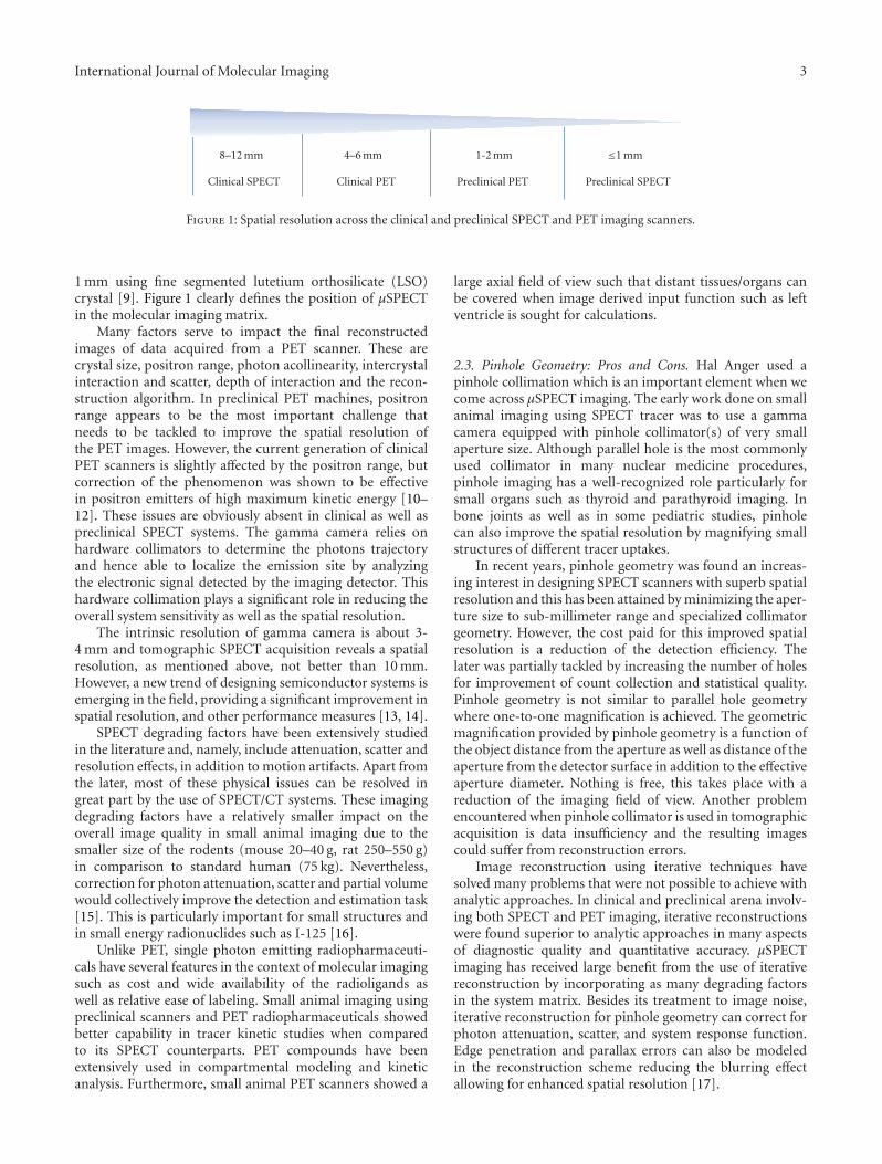
International Journal of Molecular Imaging 3
8–12 mm
Clinical SPECT
4–6 mm
Clinical PET
1-2 mm
Preclinical PET
≤1 mm
Preclinical SPECT
Figure 1: Spatial resolution across the clinical and preclinical SPECT and PET imaging scanners.
1 mm using fine segmented lutetium orthosilicate (LSO)crystal [9]. Figure 1 clearly defines the position of μSPECTin the molecular imaging matrix.
Many factors serve to impact the final reconstructedimages of data acquired from a PET scanner. These arecrystal size, positron range, photon acollinearity, intercrystalinteraction and scatter, depth of interaction and the recon-struction algorithm. In preclinical PET machines, positronrange appears to be the most important challenge thatneeds to be tackled to improve the spatial resolution ofthe PET images. However, the current generation of clinicalPET scanners is slightly affected by the positron range, butcorrection of the phenomenon was shown to be effectivein positron emitters of high maximum kinetic energy [10–12]. These issues are obviously absent in clinical as well aspreclinical SPECT systems. The gamma camera relies onhardware collimators to determine the photons trajectoryand hence able to localize the emission site by analyzingthe electronic signal detected by the imaging detector. Thishardware collimation plays a significant role in reducing theoverall system sensitivity as well as the spatial resolution.
The intrinsic resolution of gamma camera is about 3-4 mm and tomographic SPECT acquisition reveals a spatialresolution, as mentioned above, not better than 10 mm.However, a new trend of designing semiconductor systems isemerging in the field, providing a significant improvement inspatial resolution, and other performance measures [13, 14].
SPECT degrading factors have been extensively studiedin the literature and, namely, include attenuation, scatter andresolution effects, in addition to motion artifacts. Apart fromthe later, most of these physical issues can be resolved ingreat part by the use of SPECT/CT systems. These imagingdegrading factors have a relatively smaller impact on theoverall image quality in small animal imaging due to thesmaller size of the rodents (mouse 20–40 g, rat 250–550 g)in comparison to standard human (75 kg). Nevertheless,correction for photon attenuation, scatter and partial volumewould collectively improve the detection and estimation task[15]. This is particularly important for small structures andin small energy radionuclides such as I-125 [16].
Unlike PET, single photon emitting radiopharmaceuti-cals have several features in the context of molecular imagingsuch as cost and wide availability of the radioligands aswell as relative ease of labeling. Small animal imaging usingpreclinical scanners and PET radiopharmaceuticals showedbetter capability in tracer kinetic studies when comparedto its SPECT counterparts. PET compounds have beenextensively used in compartmental modeling and kineticanalysis. Furthermore, small animal PET scanners showed a
large axial field of view such that distant tissues/organs canbe covered when image derived input function such as leftventricle is sought for calculations.
2.3. Pinhole Geometry: Pros and Cons. Hal Anger used apinhole collimation which is an important element when wecome across μSPECT imaging. The early work done on smallanimal imaging using SPECT tracer was to use a gammacamera equipped with pinhole collimator(s) of very smallaperture size. Although parallel hole is the most commonlyused collimator in many nuclear medicine procedures,pinhole imaging has a well-recognized role particularly forsmall organs such as thyroid and parathyroid imaging. Inbone joints as well as in some pediatric studies, pinholecan also improve the spatial resolution by magnifying smallstructures of different tracer uptakes.
In recent years, pinhole geometry was found an increas-ing interest in designing SPECT scanners with superb spatialresolution and this has been attained by minimizing the aper-ture size to sub-millimeter range and specialized collimatorgeometry. However, the cost paid for this improved spatialresolution is a reduction of the detection efficiency. Thelater was partially tackled by increasing the number of holesfor improvement of count collection and statistical quality.Pinhole geometry is not similar to parallel hole geometrywhere one-to-one magnification is achieved. The geometricmagnification provided by pinhole geometry is a function ofthe object distance from the aperture as well as distance of theaperture from the detector surface in addition to the effectiveaperture diameter. Nothing is free, this takes place with areduction of the imaging field of view. Another problemencountered when pinhole collimator is used in tomographicacquisition is data insufficiency and the resulting imagescould suffer from reconstruction errors.
Image reconstruction using iterative techniques havesolved many problems that were not possible to achieve withanalytic approaches. In clinical and preclinical arena involv-ing both SPECT and PET imaging, iterative reconstructionswere found superior to analytic approaches in many aspectsof diagnostic quality and quantitative accuracy. μSPECTimaging has received large benefit from the use of iterativereconstruction by incorporating as many degrading factorsin the system matrix. Besides its treatment to image noise,iterative reconstruction for pinhole geometry can correct forphoton attenuation, scatter, and system response function.Edge penetration and parallax errors can also be modeledin the reconstruction scheme reducing the blurring effectallowing for enhanced spatial resolution [17].

4 International Journal of Molecular Imaging
2.4. Detectors. A conventional gamma camera can be usedby manufacturing pinhole collimators of very small aperturesize. It provides large field of view such that better magnifica-tion can be achieved. The other alternative is to use pixilateddetectors that have better intrinsic properties or semicon-ductor detectors that fit with the resolution requirementsof small animal imaging and, meanwhile, better than thatprovided by the conventional gamma camera. Regardless oftheir cost, semiconductor detectors are more compact andallow for system portability and can be manufactured inpixilated structure providing better spatial resolution.
Using a large field of view gamma camera serves toimprove the magnification by providing large projection areaonto the detector surface for the subject under investigation.However, some recent scanners are implementing pixilateddetectors that have an intrinsic resolution equivalent to thesegmentation size. This, to some extent, obviates the needto use detector width of size equivalent to the standard clin-ical gamma camera [5]. Thallium-activated sodium iodideNaI(Tl) crystal is the conventional scintillator used in mostclinical designs. However, there are also some scintillatorsthat have been used such as Cesium Iodide-Thallium dopedand Cesium Iodide-Sodium doped and Yttrium AluminumPerovskite (CsI(Tl), CsI(Na), and YAP, resp.). New designsof photodetectors such as position sensitive PMT, avalanchephotodiode and position sensitive avalanche photodiode canbe of value in μSPECT systems. For example, Funk et al.[18] have designed a multipinhole small animal imagingbased on position sensitive avalanche photodiode (PSAPD)detectors coupled to CsI(Tl) scintillator. The system showedsubmillimeter spatial resolution and high detection efficiencywhen compared to dual head gamma camera, permittingshortened acquisition time and a reduced injected dose.
2.5. Designs. Several designs were proposed for pinholegeometry, including rotating gamma camera, stationarydetector but rotating collimators, or completely stationarycamera [19]. In U-SPECT II (MILabs, The Netherlands), thethree-headed gamma cameras is equipped with interchange-able multihole collimators that can achieve high spatialresolution [20]. The collimator is cylindrical in shape withrelatively large number of pinholes (i.e., 75), providing agood count collection for the high spatial resolution. Aresolution of 0.35 mm can be achieved with an aperturesize of 0.35 mm while a spatial resolution of 0.45 mm canbe obtained with a 0.6 mm gold pinhole aperture size. Thevalues are less in case of rat imaging (0.8 mm) using thestandard whole body rat collimator. A recent release of theMILab company is the simultaneous acquisition of SPECTand PET tracers with resolution that can reach below 1 mmfor the former [21].
The Inveon is another commercial design provided bySiemens Medical Solutions. The scanner is a trimodalityimaging system that has three imaging modules namelyPET, SPECT and CT. The SPECT and CT are coplanar andmounted on the same rotating gantry. The SPECT portioncan be two or four 150 mm × 150 mm NaI(Tl) pixilateddetectors (2.2 mm pitch) and 10 mm crystal thickness. Theheads can be equipped with various parallel-hole, single-
or multipinhole collimators, including mouse general bodyas well as mouse brain imaging with possible submillimeterspatial resolution.
Another two commercial μSPECT designs providedby Gamma-Medica Ideas and Bioscan. The Triumph Tri-modality scanner (Gamma Medica, Inc) is an integratedSPECT/PET/CT hardware and software platform designedfor small animals in preclinical and biomedical researchapplications. The system combines PET (LabPET), SPECT(X-SPECT) and CT (X-O) modalities. The SPECT moduleutilizes solid-state cadmium zinc telluride (CZT) detectortechnology. It provides opportunities to scan individualorgans or whole body images. The SPECT system accom-modates single and multiple pinhole collimators as well asparallel hole collimators to address a broad range of studyneeds. It can be configured to have 1,2,3 or 4 CZT camerasproviding a variety of spatial resolution, detection sensitivity,and scanning field of view [20].
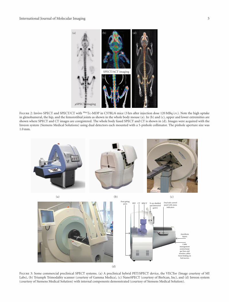
The Bioscan system has a four-detector head that consistof NaI(Tl) crystal. The scanner uses the spiral path to scan theobject (24 to 270 mm) and also has stationary and circulardetector motion. It has a variety of collimator options thatcan reach <1 mm spatial resolution in addition to highdetection sensitivity [22]. It uses a patented multiplexed-multipinhole collimator design that can reach 36 pinholesor eve more. Commercially available μSPECTs are shown inFigure 2.
2.6. Hybrid SPECT Systems. μSPECT scanners can producefunctional images with high spatial resolution; however,anatomical correlation using structural imaging modalitiesis still needed. For this reason, CT or MRI have been incor-porated in some μSPECT systems. In addition to SPECT/CTand SPECT/MRI other hybrid systems such as SPECT-opticaldevices have also been investigated [23, 24]. The commonunderlying idea is to get and extract more informationabout the biological question in one imaging session andpreferably with the same spatial and/or temporal framework.Figure 3 shows bone imaging in mouse using an SPECT/CTpreclinical scanner. CT devices provide several advantages tothe SPECT. They produce high resolution anatomical imagesin addition to generating a subject-specific attenuation mapable to correct for photon attenuation. MRI machines canhave a better soft tissue contrast, not relying on ionizingradiation, and provide high spatial resolution as mentionedearlier.
microCT (μCT) has been advanced in the last few yearsproviding a spatial resolution in the order of few microns.A resolution of 10 μm or even better can be achieved givingmore insights into structural abnormalities for in vivo aswell as ex vivo samples. Nowadays, μCT is not only forattenuation and anatomical localizations but the benefitswere extended to blood vessels imaging which is known asCT angiography. A number of reports were recently releaseddiscussing the utility of μCT in many preclinical applications[25, 26]. SPECT/MRI systems were also designed, and itis worthy mentioning that image of the year 2008 (in theannual meeting of the Society of Nuclear Medicine) wasselected where diabetic feet using SPECT were coregistered
International Journal of Molecular Imaging 5
μSPECT imaging
SPECT/ACT imaging
(a) (b) (d)
(c)
Figure 2: Invivo SPECT and SPECT/CT with 99mTc-MDP in C57BL/6 mice (3 hrs after injection dose 120 MBq i.v.). Note the high uptakein glenohumeral, the hip, and the femorotibial joints as shown in the whole body mouse (a). In (b) and (c), upper and lower extremities areshown where SPECT and CT images are coregistered. The whole body fused SPECT and CT is shown in (d). Images were acquired with theInveon system (Siemens Medical Solutions) using dual detectors each mounted with a 5-pinhole collimator. The pinhole aperture size was1.0 mm.
(a) (b) (c)
Fout gatingparts PET CT SPECT X-ray shielded
and interactedgantry
Dual side controlpanel with status
indicators
Anesthesiainputs
Cablemanagementsystem keepsgas lines and
monitor cablesfrom binding on
bed movies
(d)
Figure 3: Some commercial preclinical SPECT systems. (a) A preclinical hybrid PET/SPECT device, the VECTor (Image courtesy of MILabs), (b) Triumph Trimodality scanner (courtesy of Gamma Medica), (c) NanoSPECT (courtesy of BioScan, Inc), and (d) Inveon system(courtesy of Siemens Medical Solution) with internal components demonstrated (courtesy of Siemens Medical Solution).
6 International Journal of Molecular Imaging
with the patient MRI providing anatomolecular diagnosis ofthe extent and location of the disease. In preclinical context,the interest was given to PET/MRI rather than SPECT/MRI.However, new photodiodes that are less prone to magneticfields can be very helpful in such designs.
3. Radiopharmaceuticals
Molecular imaging is an emerging field of study that dealswith imaging of disease on a cellular or genetic level ratherthan on a gross level [27]. With the emergence of the newfield of molecular imaging, there is an increasing demandfor developing sensitive and specific novel imaging agentsthat can rapidly be translated from small animal models intopatients. SPECT and PET imaging techniques have the abilityto detect and serially monitor a variety of biological andpathophysiological processes, usually with tracer quantitiesof radiolabeled peptides, drugs, and other molecules at dosesfree of pharmacologic side effects [28].
3.1. Radiolabeled Molecular Imaging Probes (RMIPs). RIMPsare highly specific radiolabeled imaging agents used tovisualize, characterize, and measure biological processes inliving systems. Both, endogenous molecules and exogenousprobes can be molecular imaging agents. The ultimategoal of molecular medicine is to treat the disease in itsearly stages with an appropriate patient-specific “targetedmolecular therapy.” In order to achieve this goal, it isessential to develop highly specific RMIPs. In the design anddevelopment of an ideal RMIP, it is important to identifyfirst a molecular imaging probe (MIP), which may be abiochemical or a synthetic molecule, specific for a biologicalprocess (such as metabolism, angiogenesis, and apoptosis)or a molecular target (such as hexokinase, thymidine kinase,and neuroreceptor) in an organ, or tissue of interest.
General Rules for the Design of RMIPs. An ideal RMIP shouldbe designed to fulfill the following characteristics [29].
(i) Rapid plasma clearance to reduce blood pool back-ground in the target tissue.
(ii) Rapid washout or clearance from non specific areas.
(iii) Low nonspecific binding and preferably no periph-eral metabolism.
(iv) High membrane permeability and intracellular trap-ping.
(v) Target specificity and high affinity for moleculartargets.
(vi) Specific activity must be high to prevent saturation ofspecific binding sites.
(vii) Tissue distribution, localization, and target bindingshould be favorable for developing simple kineticmodeling to estimate quantitative data.
(viii) Radiation dosimetry of RMIP must be favorable formultiple diagnostic imaging studies (if necessary).
(ix) Synthesis of RMIP must be rapid and suitable forautomation using automated synthesis modules.
Radiolabeling of RMIPs. Generally, the radiolabeling processof molecular imaging agents can be categorized as follows.
Isotope Exchange. Where the preparation is obtained bydirect exchange of stable atom(s) of an element in a moleculewith one or more nuclide of a radioisotope of the sameelement.
Introduction of Foreign Element. This is the most commonmethod of radiolabeling of RMIPs, however, the RMIP willhave a chemical structure and in vivo behavior different fromthat of the parent MIP.
Metal Chelation. In this method, a chelating agent (radio-metal such as 99mTc and 111In) is being introduced intoan organic compound producing a ligand with differentbiological and chemical features than both the conjugatedtwo partners. Certain peptides and monoclonal antibodiescan successfully be labeled by the metal chelation procedurebut only in the presence of a bifunctional chelate (BFC) byconjugation with the peptide or protein first and then bindthe radiometal to the BFC conjugated molecule.
Classification of RMIPs. Based on their clinical utility and thenature of application for which they are designed as tools inthe drug development program, four classes of RMIPs havebeen identified [30].
(a) Radiolabeled drug substance in which the cold stableatom is replaced by a radioisotope of the sameelement, which can be used for assessing the phar-macokinetics and biodistribution of the parent drug.
(b) Radioligand with good binding affinity for a biologi-cal target, which can be used to evaluate the effect ofother unlabeled compounds at that target.
(c) Pathway marker interacting with one component of aset of related biological molecules, which may be usedto probe the overall status of that system.
(d) Biomarker, or surrogate marker, which provides amore general readout at the level of cell or organ fora specific biological process.
3.1.1. Peptides and Proteins
(1) Radiolabeled Monoclonal Antibodies (MAbs). Antibodiesare immunoglobulins (Ig) produced in vivo in response tothe administration of an antigen to a human or animal tissueand bind specifically to this antigen forming an antigen-antibody complex [31]. Since the advent of hybridomatechnology for production of MoAbs in 1975 [32] which wasdesigned originally as an in vivo tumor localizing agents, onlyfew have reached a point of proven clinical utility [33].
Labeling of MAbs can be accomplished with severalradionuclides, among which In-111, Tc-99m, I-131, I-123,and I-125, where most of them are commonly used in nuclearmedicine [34] and listed in Table 1. A number of monoclonallabeled antibodies using In-111 or Y-90 radionuclides aredescribed in Table 2.

International Journal of Molecular Imaging 7
Table 1: Commonly used single photon emitting radionuclides.
Radionuclide Half-life Energy Mode of decay
Tc-99m 6.02 h 142 keV IT (100%)
I-131 8.03 days 364 keV β− (100%)
I-123 13.22 h 159 keV EC (100)
In-111 2.80 days 171, 245 keV EC (100%)
IT: isomeric transition, β−: beta-minus, EC: electron capture.
(2) 99mTc-labeled Monoclonal Antibodies
Arcitumomab (CEA Scan). Carcinoembryonic Antigen(CEA) or CEA-Scan kit (introduced by MallinckrodtMedical) as a single dosage kit contains the active ingredientFab− fragment of arcitumomab, a murine monoclonalantibody IMMU-4. CEA is expressed in a variety ofcarcinomas, particularly of the gastrointestinal tract (GIT)and can be detected in the serum. IMMU-4 is specific for theclassical 200 kDa CEA that is found predominantly on thecell membrane. 99mTc-CEA-Scan complexes the circulatingCEA and binds to CES on the cell surface. Fab− fragment ofarcitumomab is cleared rapidly by urinary tract and plasmaclearance due to its small particular size [38]. The IMMU-4antibody is targeted against the carcinoembryonic antigensof the colorectal tumors, and, therefore, 99mTc-CEA-scanis used for the detection of recurrence and/or metastaticcarcinomas of the colon or rectum particularly when highlevels of CEA are detected [39]. However, it is an uncommonprocedure following PET/CT scan.
Sulesomab (LeukoScan). The kit vial contains the active in-gredient Fab− fragment, called sulesomab, obtained from themurine monoclonal antigranulocyte antibody, IMMU-MN3.It is a single-dose kit introduced by Immunomedics Europein 1997. The labeling yield should be more than 90% [40].99mTc-sulesomab targets the granulocytes, and therefore isprimarily used to detect infection and inflammation, partic-ularly in patients with osteomyelitis, joint infection involvingimplants, inflammatory bowel disease, and diabetic patientswith foot ulcers [41].
Annexin V (Apomate). Annexin V is a human protein witha molecular weight of 36 kDa has a high affinity for cellmembranes with bound phosphatidyl serine (PS) [42]. Invitro assays have been developed that use Annexin V todetect apoptosis in hematopoietic cells, neurons, fibroblasts,endothelial cells, smooth muscle cells, carcinomas, andlymphomas. 99mTc-annexin V has also been suggested as animaging agent to detect thrombi in vivo, because activatedplatelets express large amounts of PS on their surfaces [43].
(3) Radiolabeled Peptides. High background activity difficul-ties which usually appear when imaging with radiolabeledMAbs which is attributed to the slow tumor uptake andplasma clearance due to their relatively large molecular sizes.This can be mitigated by peptides whose molecular size issmaller than that of proteins and where the peptidases can act
for rapid excretion by degradation of the peptides. Peptideshave been labeled with 111In and 99mTc in the same manner ofthe monoclonal antibodies. Some useful labeled compoundsare listed in Table 3.
3.1.2. RMIPs for Metabolism. Metabolic imaging can beachieved using natural or exogenous radiolabeled substrateswhich participate in the particular metabolic process. Thedesign of such tracers is based on the physiological conceptssuch as turnover of oxygen, glucose, amino acids, fatty acids,or DNA precursors. Commonly, 123I and 99mTc derivativesare used as SPECT tracers for this function, however, theobvious chemical changes occur with this conjugation whichcan alter the physiological properties of the tracer modulelimits their application in molecular imaging.
(1) Glucose Metabolism. Although there are intensiveresearch trials to find a sugar derivative labeled with 123Iand 99mTc, none of these were able to provide an SPECTsubstitute for 18F-FDG to act as glucose metabolism imagingagent [51–53].
(2) Amino Acid Metabolism. Diagnosis of various neurologi-cal diseases as well as tumor evaluation (detection, gradingand therapy monitoring) can be obtained by quantitativeassessment of protein synthesis rate that is provided whenusing radiolabeled amino acids as a radiolabeled molecularimaging probe (RMIP). Radiolabeled amino acids pass theblood-brain barrier and are accumulated in tissues via aspecific amino acid transport system [54]. L-3-123I-Iodo-α-methyltyrosine (123I-IMT) is a good example of radiolabeledamino acids, it is evidenced that it accumulates in the brainvia a specific facilitating L-amino acid transport system. Inanalogy with PET, IMT is not incorporated into proteins [55]and its uptake reflects amino acid transport [56]. IMT can beprepared by electrophilic substitution via in situ oxidation of123I-Iodide by chloramines-T, hydrogen peroxide, iodogen,or iodate with radiochemical yields of 70%–80%.
(3) Nucleosides Metabolism (Gene Reporter Imaging). Nucle-osides or nucleoside analogs can be transported across thecell membrane by selective transporters and can then bephosphorylated intracellularly by specific kinases to thecorresponding phosphate derivatives, and, ultimately, theycan be incorporated into DNA. As thymidine kinase 1 (tk1)shows an S-phase dependant expression, the intracellularaccumulation of labeled nucleosides that are substrates forTK1 reflects DNA synthesis and thus tumor proliferation. I-131-[57] and I-124-[58] labeled 2-arabino-fluro-5-iodo-2-deoxyuridine (FIAU) has been used successfully as nucle-oside analog for tumor proliferation detection. Nucleosidederivatives that are selective substrates for herpes simplexvirus thymidine kinase (HSVtk) have been developed forthe in vivo visualization of transgene expression using theHSVtk gene as a reporter gene. Radioiodine labeled uracilcompounds (e.g., FIAU) are widely applicable derivatives.

8 International Journal of Molecular Imaging
Table 2: List of In-111- and Y-90-labeled monoclonal antibodies.
Compound Description Applications Remarks
In111-Capromabpendetide(ProstaScint)
A conjugation between themurine antibody 7E11.C5.3 and111 In in the source of 111InCl3
by the action of the GYK-DTPAas a chelating agent.
Indicated for use inimmunoscintigraphy, provenprostate carcinoma and patients whohave undergone a prostatectomy andhave rising prostate specific antigen(PSA) values and equivocalnonevidenced metastasis.
It is not indicated with patients with ahigh clinical suspicion of occultmetastatic disease or for screening ofprostate carcinoma.
In111-Satumomabpendetide(OncoScint)
It contains the murine MAbB72.3 which is directed totumor-associated glycoprotein.It is labeled with 111InCl3 byconjugation with the chelatingagent, GYK-DTPA.HCl.
Used for the detection of colorectaland ovarian cancers [35].
After an incubation time of 30 min, thelabeled mixture is suitable for use in thefirst 8 hours.
In111-Imciromabpentetate(MyoScint)
An antibody produced againstmyosin in the cell culture, andtherefore binds to the heavychain of myosin after in vivoadministration.
Detection of myocardial infarction.
Contains the Fab fragment of a murinemonoclonal antibody that is covalentlybound to DTPA giving111 In-Imciromab pentetate.
In-111 and Y90-ibritumomabtiuxetan(Zevalin)
Zevalin consists of a murinemonoclonal anti-CD20antibody covalently conjugatedto the metal chelator DTPA,which forms a stable complexwith 111 In for imaging and with90Y for therapy.
90Y-ibritumomab tiuxetan is usedfor the treatment of some forms of Bcell non-Hodgkin’s lymphoma, amyeloproliferative disorder of thelymphatic system while its 111 Inderivative is used to scan thepredicted distribution of atherapeutic dosage of90Y-ibritumomab in the body [36].
The antibody binds to the CD20antigen found on the surface of normaland malignant B cells (but not B cellprecursors) allowing radiation from theattached isotope (Yetrium-90) and thecytotoxicity induced by the antibodyserve to eliminate B cells from the bodyallowing a new population of healthy Bcells to develop from lymphoid stemcells [37].
Rituximab
An earlier version of anti-CD20antibody and has also beenapproved under the brandname Rituxan for the treatmentof non-Hodgkin’s lymphoma(NHL).
It was approved for the treatment ofpatients with relapsed or refractory,lowgrade or follicular Bcell NHL,including patients with rituximabrefractory follicular NHL.
In September 2009, ibritumomabreceived approval from the FDA for anexpanded label for the treatment ofpatients with previously untreatedfollicular NHL, who achieve a partial orcomplete response to first-linechemotherapy.
(4) Hypoxia Imaging. Hypoxia, a condition of insufficientO2 to support metabolism, occurs when the vascular supplyis interrupted, as in stroke or myocardial infarction, orwhen a tumor outgrows its vascular supply. When otherwisehealthy tissues lose their O2 supply acutely, the cells usuallydie, whereas when cells gradually become hypoxic, theyadapt by upregulating the production of numerous proteinsthat promote their survival. These proteins slow the rateof growth, switch the mitochondria to glycolysis, stimulategrowth of new vasculature, inhibit apoptosis, and promotemetastatic spread [59].
Most hypoxia markers contain a nitroimidazole moietyas a reactive chemical species. Nitroimidazoles can be used asprobes to detect hypoxia as they are reduced intracellularlyin all cells, but in absence of adequate supply of O2,they undergo further reduction to more reactive productswhich bind to cell components and are finally trapped inthe hypoxic tissue [60]. 99mTc-O-propylene-amine-oxime(99mTc-pano or BMS-181321) was validated as a properhypoxia imaging agent in hypoxic myocardium, acutelyischemic brain and solid tumors [61]. BMS-194796 and
99mTc-HL91 have also been designed as RMIP for imaginghypoxia, however, none of these three probes has beencommercially available. Iodine-123 labeled iodoazomycinarabinoside (IAZA) has been validated in animal model inthe preclinical phase [62], but no clinical studies with thisagent have been reported so far.
(5) Cell Labeling. The most common applications of In-111are in labeling blood cells (white blood cells (WBC) andplatelets) for imaging inflammatory processes and thrombi[63]. In blood cell labeling, the plasma transferrin competesfor the In-111 and reduces the labeling efficiency becauseIn-111 binds with higher efficiency to transferrin thanblood cells, and, therefore, isolation of the desired bloodcomponent from plasma permits easy labeling of eitherplatelets or WBCs. 99mTc-HMPAO is primarily used in brainperfusion imaging, although it is used for leukocyte labelingsubstituting 111In-Oxine. Stabilization of the 99mTc-HMPAOprimary complex is required due to the high degradationrate of its radiochemical purity. This could be achieved by
International Journal of Molecular Imaging 9
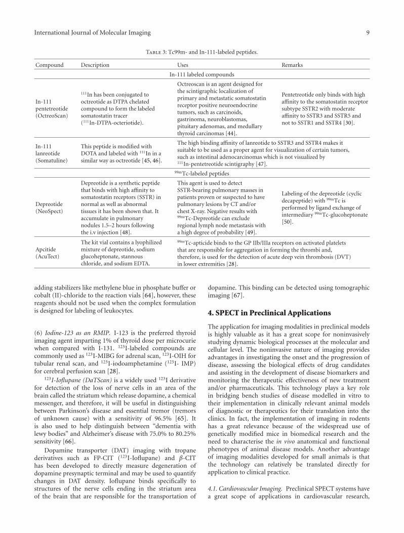
Table 3: Tc99m- and In-111-labeled peptides.
Compound Description Uses Remarks
In-111 labeled compounds
In-111pentetreotide(OctreoScan)
111In has been conjugated tooctreotide as DTPA chelatedcompound to form the labeledsomatostatin tracer(111In-DTPA-octeriotide).
Octreoscan is an agent designed forthe scintigraphic localization ofprimary and metastatic somatostatinreceptor positive neuroendocrinetumors, such as carcinoids,gastrinoma, neuroblastomas,pituitary adenomas, and medullarythyroid carcinomas [44].
Pentetreotide only binds with highaffinity to the somatostatin receptorsubtype SSTR2 with moderateaffinity to SSTR3 and SSTR5 andnot to SSTR1 and SSTR4 [30].
In-111lanreotide(Somatuline)
This peptide is modified withDOTA and labeled with 111In in asimilar way as octreotide [45, 46].
The high binding affinity of lanreotide to SSTR3 and SSTR4 makes itsuitable to be used as a proper agent for visualization of certain tumors,such as intestinal adenocarcinomas which is not visualized by111In-pentetreotide scintigraphy [47].
99mTc-labeled peptides
Depreotide(NeoSpect)
Depreotide is a synthetic peptidethat binds with high affinity tosomatostatin receptors (SSTR) innormal as well as abnormaltissues it has been shown that. Itaccumulate in pulmonarynodules 1.5–2 hours followingthe i.v injection [48].
This agent is used to detectSSTR-bearing pulmonary masses inpatients proven or suspected to havepulmonary lesions by CT and/orchest X-ray. Negative results with99mTc-Depreotide can excluderegional lymph node metastasis witha high degree of probability [49].
Labeling of the depreotide (cyclicdecapeptide) with 99mTc isperformed by ligand exchange ofintermediary 99mTc-glucoheptonate[50].
Apcitide(AcuTect)
The kit vial contains a lyophilizedmixture of depreotide, sodiumglucoheptonate, stannouschloride, and sodium EDTA.
99mTc-apticide binds to the GP IIb/IIIa receptors on activated plateletsthat are responsible for aggregation in forming the thrombi and,therefore, is used for the detection of acute deep vein thrombosis (DVT)in lower extremities [28].
adding stabilizers like methylene blue in phosphate buffer orcobalt (II)-chloride to the reaction vials [64], however, thesereagents should not be used when the complex formulationis designed for labeling of leukocytes.
(6) Iodine-123 as an RMIP. I-123 is the preferred thyroidimaging agent imparting 1% of thyroid dose per microcuriewhen compared with I-131. 123I-labeled compounds arecommonly used as 123I-MIBG for adrenal scan, 123I-OIH fortubular renal scan, and 123I-iodoamphetamine (123I- IMP)for cerebral perfusion scan [28].
123I-Ioflupane (DaTScan) is a widely used 123I derivativefor detection of the loss of nerve cells in an area of thebrain called the striatum which release dopamine, a chemicalmessenger, and therefore, it will be useful in distinguishingbetween Parkinson’s disease and essential tremor (tremorsof unknown cause) with a sensitivity of 96.5% [65]. Itis also used to help distinguish between “dementia withlewy bodies” and Alzheimer’s disease with 75.0% to 80.25%sensitivity [66].
Dopamine transporter (DAT) imaging with tropanederivatives such as FP-CIT (123I-Ioflupane) and β-CIThas been developed to directly measure degeneration ofdopamine presynaptic terminal and may be used to quantifychanges in DAT density. Ioflupane binds specifically tostructures of the nerve cells ending in the striatum areaof the brain that are responsible for the transportation of
dopamine. This binding can be detected using tomographicimaging [67].
4. SPECT in Preclinical Applications
The application for imaging modalities in preclinical modelsis highly valuable as it has a great scope for noninvasivelystudying dynamic biological processes at the molecular andcellular level. The noninvasive nature of imaging providesadvantages in investigating the onset and the progression ofdisease, assessing the biological effects of drug candidatesand assisting in the development of disease biomarkers andmonitoring the therapeutic effectiveness of new treatmentand/or pharmaceuticals. This technology plays a key rolein bridging bench studies of disease modelled in vitro totheir implementation in clinically relevant animal modelsof diagnostic or therapeutics for their translation into theclinics. In fact, the implementation of imaging in rodentshas a great relevance because of the widespread use ofgenetically modified mice in biomedical research and theneed to characterise the in vivo anatomical and functionalphenotypes of animal disease models. Another advantageof imaging modalities developed for small animals is thatthe technology can relatively be translated directly forapplication to clinical practice.
4.1. Cardiovascular Imaging. Preclinical SPECT systems havea great scope of applications in cardiovascular research,

10 International Journal of Molecular Imaging
including the study of myocardial functions (e.g., ejectionfraction, regional wall motion abnormalities, perfusion, tis-sue viability, oxygen consumption, and glucose metabolism[68]) and the investigation of several vascular disorders,including coronary artery disease and related disorders, suchas ischemia, infarction and atherosclerosis [69]. Moreover,μSPECT has great applications for developing and testingdiagnostic tracers which could assist in understandingthe prognosis of disorders and assess new therapeuticapproaches for cardiovascular lesions.
99mTc-labelled radiopharmaceuticals for SPECT imaginghave been applied to demonstrate tissue viability and per-fusion status in animal models of ischemia and/or impairedmyocardial perfusion [70, 71]. Cardiac and respiratorymotion is one of the major challenges when imaging rodent(mouse heart rate: 400–800 beats/min). Gated acquisitionis therefore required to minimise any movement artefacts.Indeed, ECG-gated micro SPECT can yield accurate mea-surements of left ventricle volumes and ejection fraction inrats and mice [72, 73].
Another key area is the visualization of necrotic tissuesand related tracers during myocardial infarction (MI). Somestudies have assessed myocardial ischemia in rat heart modelsafter left coronary artery occlusion by using 99mTc-glucarate[74]. In vivo visualization of necrosis may help to detectMI at early stages and may provide a good approach forevaluating the antinecrotic effect of developing drugs forischemic heart disease. On the same vein, the visualizationof apoptotic cell death is another important target for non-invasive imaging [75]. Hence, the development of tracers(e.g.,99mTc-Annexin) that bind to apoptotic cells is a veryuseful tool for in vivo analysis, especially to investigateapoptotic cell death in cardiomyocytes and the efficacy ofcell-based therapies.
Angiogenesis imaging is a key protective/remodellingmechanism in myocardial infarction. Imaging such a mecha-nism is important for the understanding of infarct healingand post-MI remodelling. Vascular growth factors suchas αvβ3 integrins have been used as targeted tracers toinvestigate angiogenesis in postinfarct animal models, withIn-111-labelled αvβ3 targeted radiotracers in hypoperfusedmyocardial regions [76].
Another relevant area of cardiovascular imaging is thedevelopment of methods to characterise the formation andprognosis of atherosclerotic plaques. Plaque rupture resultsin severe cardiac events including MI and sudden death,hence, there is an important need for developing tools thatcan assist in predicting the plaques vulnerability to rupture.Not all the plaques carry the same risk, and the criteria forimaging their vulnerability relies on the detection of inflam-matory cell infiltration, platelet aggregation, tissue matrixdegradation, large lipid contents and apoptosis. RadiolabeledAnnexin and Z2D3 targeted to apoptotic macrophages andsmooth muscle cells, respectively, have been used as SPECTtracers to study the pathophysiology of atherosclerosis inanimal models [77, 78]. One of the main challenges forimaging plaques is their anatomical localization, as highspatial resolution is needed to image such a small anatomicalstructure in a motile vessel, hence the importance of co-
registering SPECT acquisition with other imaging modalitiessuch as micro-CT.
4.2. Imaging Stem Cells. With advances in research of stemcell-based therapies, the application of imaging technologiesmay be useful to validate their efficacy and safety inpreclinical models, in particular, for studying tracking andengraftment of transplanted cells, assessing their viability,function and differentiation status in addition to monitoringtheir ability to promote regeneration [79]. Stem cells canbe labelled with radionuclides before transplantation. Forexample, stem cells labelled with the SPECT radiolabels111In-oxyquinoline have been successfully imaged aftertransplantation in rat and porcine models of myocardialinfarction [80, 81], but, because of the short half-life of theradionuclide (e.g., 99mTc: 6.02 h; 111In: 2.8 days) and becausethe activity may still be present after transplantation even ifthe cell have died, this method may only be applicable for ashort-term cell tracking and assessing stem cell homing aftertransplantation.
To investigate not only long-term engraftment of stemcells but also their viability, a gene imaging approach may bemore appropriate. In this case, gene expression is assessed byreporter genes constructs which are translated into a proteinand interact with an exogenously given probe (radiolabeledfor SPECT detection), resulting in a signal that can bemonitored non-invasively. Reporter genes are incorporatedinto the cells before transplantation, and if the cell remainsalive after engraftment, the protein, which is the main targetfor the nuclides, will be encoded (e.g., enzyme, cell surfacereceptor). Conversely, the reporter gene will not be expressedif the cell is dead [82].
As mentioned earlier, one of the reporter genes mostlyused for SPECT imaging is based on the production ofan intracellular enzyme (e.g., herpes simplex virus type 1thymidine kinase [HSV1tk]) that phosphorylates an exoge-nously administered substrate that is retained in the cellbecause of its negative charge. Although normal mammaliancells (without the HSV1-tk) do carry the enzyme, it onlyminimally phosphorylates the radionuclide probes used inthis system. Conversely, in cells carrying the HSV1-tk, theexogenously administered probe undergoes significant phos-phorylation and intracellular retention, leading to a robustsignal-to-background ratio and enabling accurate monitor-ing of these cells [83]. This imaging approach has beenrecently used to monitor the distribution of transplantedhuman embryonic stem cell derivatives in a live mouse modelover a long period of time, up to 3 months [84].
Another approach consists of the encoding of thesodium-iodide symporter (NIS), a thyroid transmembraneprotein that, under physiologic conditions, transports iodineinto the cells in exchange for sodium. It has the advantagethat it can be used for PET (with 124I as the tracer)and SPECT imaging (using 123I or 99mTc-pertechnetate astracer). This approach has been used to monitor activityof cardiac-derived stem cells after transplantation to a ratinfarct model, confirming the visualization of cells up to6 days after transplantation [85]. With rapid advances in

International Journal of Molecular Imaging 11
stem cell research, and with high demands for testing theirregenerative potential in preclinical model, noninvasive stemcell imaging will play a critical role, and we can foreseemore studies requiring long-term monitoring of stem cellsin preclinical models of disease.
4.3. Oncologic Applications. Imaging techniques play a po-tential role in preclinical cancer research, enabling sequentialanalysis of deep-seated tumors and metastases includingstudies of basic biological processes, tissue pharmacokineticsand pharmacodynamics responses to treatments. Imagingof cancer cells targets have different biological proceduresincluding overexpression of receptor, activated enzymes orrelocated molecules, apoptotic levels, sustained angiogenesis,unlimited replicative potential and invasion of tissue, andmetastasis [86].
Imaging gene expression in vivo is very relevant incancer preclinical models as it allows the characterization ofdynamic changes in several deregulated pathways in cancercells. As previously mentioned, HSVtk genes are typicallyused for SPECT, enabling noninvasive imaging of tumor cellgrowth as demonstrated in an experimental mouse model forlung metastases expressing after injection of HSV1-tk cells[87]. This model may be proven very useful for assessment ofanticancer and antimetastases therapies in preclinical efficacymodels.
Another used approach is the imaging receptors thatare overexpressed in cancer cells and can be used forprognosis and for following therapeutic targeting. This hasbeen successfully applied preclinically as well as clinicallyby targeting prostate-specific membrane antigen (PSMA).This receptor is overexpressed on the cell surface of prostatecancer cells and provides a useful target for prostate tumorimaging and therapy. As mentioned previously, radiolabeledmonoclonal antibodies, such as 111In-Capromab pendetide(ProstaScint), are currently available to detect prostate cancerbut suffer from problems associated with poor deliverybecause of their large size [88]. The feasibility of imagingPSMA receptor expression with low-molecular-weight, high-affinity PSMA ligands labeled with [125I] NaI/Iodogen forSPECT was demonstrated in a study using a prostate tumormouse model [89].
SPECT technology is extensively used as a diagnostic toolfor bone metastases in the clinic. Bone scintigraphy with99mTc-labelled diphosphonate is a widely used method forthe detection of bone metastases, and other bone disorders.This technique provides a high sensitivity and is able tosurvey the whole skeleton but unfortunately does not provideenough anatomical resolution to allow precise localizationof the radiotracer high uptake lesion. The implementationof a SPECT/CT multimodality system can partly overcomethis disadvantage, allowing a coregistration of the functionaland the anatomical imaging component, resulting in preciseanatomical localisation of the radiotracer. Studies havereported the use of Tc-99m-labeled diphosphonates com-pounds (e.g., methylene-diphosphonate (MDP)) to detectmetastatic bone lesions in immunocompromised mousemodels injected with cancer cells. Overall, the application
of SPECT imaging in cancer research while remainingchallenging have already had a remarkable impact providingnew insights into the dynamics of cancer growth, invasion,and metastases, being possible to visualize gene expression,molecular pathways and functional parameters in preclinicalmodels of cancer.
4.4. Neuroimaging Applications. The use of SPECT inpreclinical functional neuroimaging provides an excellentapplication for understanding the pathophysiology of centralnervous system (CNS) disorders, including the mechanismsof neurodegeneration, neuropharmacology related to drugabuse, and testing therapeutic strategies. As mentionedearlier, one of the strength of SPECT over other functionalmodalities such as the PET is the ability to get a spatialresolution below 1 mm, allowing detailed structural andfunctional information of different region of the brainin animal models. Also, SPECT radioligands have relativelonger half-lives, which permits prolonged dynamic functionstudies and provides a simultaneous dual tracer imaging. Theuse of pinhole SPECT facilitates accurate and quantitativeimaging. Indeed, specific radioligands have been used tostudy the dopaminergic, serotonergic, and cholinergic neu-rotransmission system in vivo [90].
SPECT has been applied to study basic mechanisms ofdegeneration in Parkinson’s (PD) and Alzheimer’s disease(AD). PD is characterized by a progressive loss of dopaminer-gic neurons. Animal models of neurodegeneration have beenused to evaluate novel radioligands and to study their bind-ing in the dopaminergic synapsis. In vivo quantification ofthe presynaptic dopamine transporter (DAT) activity whichregulates the synaptic dopamine is feasible in the rat striatumusing the 123I-N-ω-fluoropropyl-2β-carbomethoxy-3β-(4-iodophenyl)-nortropane (123I-FP-CIT) as a DAT radioligand[91], as outlined earlier. This technology is of particu-lar interest for investigating the interrelation of synapticdopamine and DAT in animal models of PD. Similarly,Acton et al. have characterized the occupancy of dopaminereceptors in the mouse brain [92]. SPECT imaging hasimproved the early diagnosis of AD in patients by detectingthe onset of progressive neurodegenerative disorders andvascular brain pathology causing dementia.
The thioflavin derivative, 6-iodo-2-(4′-dimethylamino-)phenyl-imidazo [1,2-a] pyridine, IMPY, which is readilyradiolabeled with 125I/123I [123I] IMPY was assessed ex vivo ina transgenic mouse model of AD by labeling the depositionof amyloid plaque that is linked to the pathogenesis of AD[93], showing a good binding with brain tissue homogenatesof confirmed Alzheimer’s disease patients. Overall, thesepreclinical studies support the use of SPECT for functionalimaging in preclinical models of CNS disorders, facilitatingthe efficacy of translational studies into this field with adirect relevance for developing clinical diagnostic tools andefficacious therapies.
4.5. Drug Discovery. Preclinical imaging has a key role inthe drug development, in particular for validating drugtargeting, safety, and efficacy. One of the main applications
12 International Journal of Molecular Imaging
is the validation in drug binding assays to specific targetedareas, by directly labeling a drug to determine its distri-bution and pharmacokinetics. This approach is very usefulfor validating delivery roots and the specificity of noveltherapeutic drugs or imaging agents. SPECT imaging hasbeen applied to measure the binding potential of targetednuclide [123I] Iodobenzamide to the dopamine transportersin the rat brain after specific treatments [94]. Similarly,99mTc-labeled liposomes migration has been studied afterintratumoral administration to tumor xenograft modelsin nude rats [95]. Specific skeletal targeted probes (125Ilabeled) were investigated with particular emphasis on thepharmacokinetics and biodistribution following intravenousadministration. This has proved to be of great potential forvalidating the efficacy of animal models of osteoporosis andother skeletal diseases [96].
Imaging technologies are also very useful for safetyvalidation, as they can measure the functional response oforgans to a tested drug candidate, providing informationon any toxic or secondary effects related to the treatment.Radiopharmaceuticals that bind to apoptotic cells (e.g.,99mTc-Annexin) have been used preclinically to validate thepossible toxicity effects in developing therapies [97]. Otherexamples on secondary response to the administration of acandidate drug in preclinical models include measurementsin blood flow changes [98] and infiltration of inflammatorycells [99].
Another important application is the development andvalidation of imaging biomarkers, a surrogate imagingproduct that simulates a biological compound and/or hassome biologic link to the disease process. These biomarkersare becoming very useful to assess therapeutic actions ofpharmaceuticals, providing a non-invasive imaging tool forvalidating the efficacy of treatment. Advances in proteomicsand genomics are leading to the discovery of new biomarkers,promoting the use of functional imaging for their validationand translation into the clinics [100].
5. Conclusion
SPECT imaging has a well-defined role in the world ofmolecular imaging. Recent advances in dedicated preclinicalsystems are able to provide high spatial and temporal resolu-tion as well as high detection efficiency with more potentiallfor further improvement. Hybrid SPECT imaging systemswould serve characterizing biological phenomena in oneimaging session. Reliable animal models that, mimic humandiseases is another innovative field that when combinedwith SPECT technology will reveal more insights into earlydisease detection, development of new tracers/therapeuticsand treatment strategy. SPECT imaging has a large potentialin molecular medicine, and many novel approaches areexpected in the near future.
Acknowledgment
Jordi L. Tremoleda and Tamer B. Saleh contributed equallyto the work.
References
[1] G. K. Von Schulthess and H. P. W. Schlemmer, “A lookahead: PET/MR versus PET/CT,” European Journal of NuclearMedicine and Molecular Imaging, vol. 36, supplement 1, pp.S3–S9, 2009.
[2] M. A. Pysz, S. S. Gambhir, and J. K. Willmann, “Molecularimaging: current status and emerging strategies,” ClinicalRadiology, vol. 65, no. 7, pp. 500–516, 2010.
[3] A. Douraghy and A. F. Chatziioannou, “Preclincal imaging,”in Basic Sciences of Nuclear Medicine, M. Khalil, Ed., chapter18, Springer, New York, NY, USA, 2011.
[4] M. E. Phelps, “PET: the merging of biology and imaging intomolecular imaging,” Journal of Nuclear Medicine, vol. 41, no.4, pp. 661–681, 2000.
[5] S. R. Meikle, P. Kench, M. Kassiou, and R. B. Banati, “Smallanimal SPECT and its place in the matrix of molecularimaging technologies,” Physics in Medicine and Biology, vol.50, no. 22, pp. R45–R61, 2005.
[6] G. K. Von Schulthess and T. F. Hany, “Imaging and PET—PET/CT imaging,” Journal de Radiologie, vol. 89, no. 3, pp.438–448, 2008.
[7] H. O. Anger, “Scintillation camera,” Review of ScientificInstruments, vol. 29, no. 1, pp. 27–33, 1958.
[8] F. D. Van Have, B. Vastenhouw, R. M. Ramakers et al.,“U-SPECT-II: an ultra-high-resolution device for molecularsmall-animal imaging,” Journal of Nuclear Medicine, vol. 50,no. 4, pp. 599–605, 2009.
[9] J. R. Stickel, J. Qi, and S. R. Cherry, “Fabrication andcharacterization of a 0.5-mm lutetium oxyorthosilicatedetector array for high-resolution PET applications,” Journalof Nuclear Medicine, vol. 48, no. 1, pp. 115–121, 2007.
[10] A. Sanchez-Crespo, P. Andreo, and S. A. Larsson, “Positronflight in human tissues and its influence on PET imagespatial resolution,” European Journal of Nuclear Medicine andMolecular Imaging, vol. 31, no. 1, pp. 44–51, 2004.
[11] A. Ruangma, B. Bai, J. S. Lewis et al., “Three-dimensionalmaximum a posteriori (MAP) imaging with radiophar-maceuticals labeled with three Cu radionuclides,” NuclearMedicine and Biology, vol. 33, no. 2, pp. 217–226, 2006.
[12] M. R. Palmer, X. Zhu, and J. A. Parker, “Modeling andsimulation of positron range effects for high resolution PETimaging,” IEEE Transactions on Nuclear Science, vol. 52, no. 5,pp. 1391–1395, 2005.
[13] S. S. Gambhir, D. S. Berman, J. Ziffer et al., “A novel high-sensitivity rapid-acquisition single-photon cardiac imagingcamera,” Journal of Nuclear Medicine, vol. 50, no. 4, pp. 635–643, 2009.
[14] K. Erlandsson, K. Kacperski, D. Van Gramberg, and B.F. Hutton, “Performance evaluation of D-SPECT: a novelSPECT system for nuclear cardiology,” Physics in Medicineand Biology, vol. 54, no. 9, pp. 2635–2649, 2009.
[15] C. L. Chen, Y. Wang, J. J. S. Lee, and B. M. W. Tsui, “Towardquantitative small animal pinhole SPECT: assessment ofquantitation accuracy prior to image compensations,” Molec-ular Imaging and Biology, vol. 11, no. 3, pp. 195–203, 2009.
[16] A. B. Hwang, B. L. Franc, G. T. Gullberg, and B. H. Hasegawa,“Assessment of the sources of error affecting the quantitativeaccuracy of SPECT imaging in small animals,” Physics inMedicine and Biology, vol. 53, no. 9, pp. 2233–2252, 2008.
[17] B. L. Franc, P. D. Acton, C. Mari, and B. H. Hasegaway,“Small-animal SPECT and SPECT/CT: important tools forpreclinical investigation,” Journal of Nuclear Medicine, vol. 49,no. 10, pp. 1651–1663, 2008.

International Journal of Molecular Imaging 13
[18] T. Funk, P. Despres, W. C. Barber, K. S. Shah, and B. H.Hasegawa, “A multipinhole small animal SPECT system withsubmillimeter spatial resolution,” Medical Physics, vol. 33, no.5, pp. 1259–1268, 2006.
[19] J. Nuyts, K. Vunckx, M. Defrise, and C. Vanhove, “Smallanimal imaging with multi-pinhole SPECT,” Methods, vol.48, no. 2, pp. 83–91, 2009.
[20] http://www.gm-ideas.com/pre triumph trimodality system.html.
[21] http://www.milabs.com/.[22] A. Del Guerra and N. Belcari, “State-of-the-art of PET,
SPECT and CT for small animal imaging,” Nuclear Instru-ments and Methods in Physics Research A, vol. 583, no. 1, pp.119–124, 2007.
[23] J. Peter and W. Semmler, “Performance investigation of adual-modality SPECT/optical small animal imager,” Euro-pean Journal of Nuclear Medicine and Molecular Imaging, vol.33, p. S117, 2006.
[24] E. Breton, P. Choquet, C. Goetz et al., “Dual SPECT/MRimaging in small animal,” Nuclear Instruments and Methodsin Physics Research A, vol. 571, no. 1-2, pp. 446–448, 2007.
[25] E. L. Ritman, “Molecular imaging in small animals—roles formicro-CT,” Journal of Cellular Biochemistry, no. 39, pp. 116–124, 2002.
[26] S. J. Schambach, S. Bag, L. Schilling, C. Groden, and M.A. Brockmann, “Application of micro-CT in small animalimaging,” Methods, vol. 50, no. 1, pp. 2–13, 2010.
[27] C. Nichol and E. E. Kim, “Molecular imaging and genetherapy,” Journal of Nuclear Medicine, vol. 42, no. 9, pp. 1368–1374, 2001.
[28] F. G. Blankenberg and H. W. Strauss, “Nuclear medicineapplications in molecular imaging,” Journal of MagneticResonance Imaging, vol. 16, no. 4, pp. 352–361, 2002.
[29] S. Vallabhajosula, Molecular Imaging, Radiopharmaceuticalsfor PET and SPECT, Springer, New York, NY, USA, 2009.
[30] D. Maclean, J. P. Northrop, H. C. Padgett, and J. C. Walsh,“Drugs and probes: the symbiotic relationship betweenpharmaceutical discovery and imaging science,” MolecularImaging and Biology, vol. 5, no. 5, pp. 304–311, 2003.
[31] G. B. Saha, Fundamentals of Radiopharmacy, Springer, Berlin,Germany, 5th edition, 2004.
[32] G. Kohler and C. Milstein, “Continuous cultures of fusedcells secreting antibody of predefined specificity,” Nature, vol.256, no. 5517, pp. 495–497, 1975.
[33] C. Schiepers, Diagnostic Nuclear Medicine, Springer, Berlin,Germany, 2nd edition, 2006.
[34] D. J. Hnatowich, “Recent developments in the radiolabelingof antibodies with iodine, indium, and technetium,” Semi-nars in Nuclear Medicine, vol. 20, no. 1, pp. 80–91, 1990.
[35] R. T. Maguire, V. L. Pascucci, A. N. Maroli, and J. V. Gulfo,“Immunoscintigraphy in patients with colorectal, ovarian,and prostate cancer: results with site-specific immunoconju-gates,” Cancer, vol. 72, no. 11, pp. 3453–3462, 1993.
[36] A. Otte, “90Y-Ibritumomab tiuxetan: new drug, interestingconcept, and encouraging in practice,” Nuclear MedicineCommunications, vol. 27, no. 7, pp. 595–596, 2006.
[37] S. Han, A. H. Iagaru, H. J. Zhu, and M. L. Goris, “Expe-rience with 90Y-ibritumomab (zevalin) in the managementof refractory non-Hodgkin’s lymphoma,” Nuclear MedicineCommunications, vol. 27, no. 12, pp. 1022–1023, 2006.
[38] Immunomedics Europe Product monograph for the CEA-Scan(Arcitumomab) kit for the preparation of Tc-99m CEA-Scan,Immunomedics Europe, Darmstadt, Germany, 2000.
[39] F. L. Moffat Jr., C. M. Pinsky, L. Hammershaimb et al.,“Immunomedics study group clinical utility of externalimmunoscintigraphy with the IMMU-4 technetium-99mFab’ antibody fragment in patients undergoing surgery forcarcinoma of the colon and rectumml: results of a pivotal,phase III trial,” Journal of Clinical Oncology, vol. 14, pp. 2295–2305, 1996.
[40] Immunomedics Europe Product monograph for LeukoScan(sulesomab), Immunomedics Europe, Darmstadt, Germany,1997.
[41] S. Gratz, M. L. Schipper, J. Dorner et al., “LeukoScan forimaging infection in different clinical settings: a retrospectiveevaluation and extended review of the literature,” ClinicalNuclear Medicine, vol. 28, no. 4, pp. 267–276, 2003.
[42] F. G. Blankenberg, P. D. Katsikis, J. F. Tait et al., “Imaging ofapoptosis (programmed cell death) with 99mTc annexin V,”Journal of Nuclear Medicine, vol. 40, no. 1, pp. 184–191, 1999.
[43] J. F. Tait, M. D. Cerqueira, T. A. Dewhurst, K. Fujikawa,J. L. Ritchie, and J. R. Stratton, “Evaluation of annexin Vas a platelet-directed thrombus targeting agent,” ThrombosisResearch, vol. 75, no. 5, pp. 491–501, 1994.
[44] L. Ha, R. Mansberg, D. Nguyen, and C. Bui, “Increasedactivity on In-111 octreotide imaging due to radiationfibrosis,” Clinical Nuclear Medicine, vol. 33, no. 1, pp. 46–48,2008.
[45] L. K. Kvols and E. A. Woltering, “Role of somatostatinanalogs in the clinical management of non-neuroendocrinesolid tumors,” Anti-Cancer Drugs, vol. 17, no. 6, pp. 601–608,2006.
[46] C. Susini and L. Buscail, “Rationale for the use of somato-statin analogs as antitumor agents,” Annals of Oncology, vol.17, no. 12, pp. 1733–1742, 2006.
[47] I. Virgolini, I. Szilvasi, A. Kurtaran et al., “Indium-111-DOTA-lanreotide: biodistribution, safety and radia-tion absorbed dose in tumor patients,” Journal of NuclearMedicine, vol. 39, no. 11, pp. 1928–1936, 1998.
[48] D. Kahn, Y. Menda, K. Kernstine et al., “The utility of 99mTc-depreotide compared with F-18-fluorodeoxyglucose positronemission tomography and surgical staging in patients withsuspected non-small cell lung cancer,” Chest, vol. 125, no. 2,pp. 494–501, 2004.
[49] R. Danielsson, M. Baath, L. Svensson, U. Forslov, and K.-G.Kolbeck, “Imaging of regional lymph node metastases with99mTc-depreotide in patients with lung cancer,” EuropeanJournal of Nuclear Medicine and Molecular Imaging, vol. 32,no. 8, pp. 925–931, 2005.
[50] Berlex Laboratories Product Monograph for the NeoTect Kit forthe Preparation of Tc-99m Depreotide, Berlex Laboratories,Wayne, NJ, USA, 2001.
[51] C. Henry, F. Koumanov, C. Ghezzi et al., “Experimentalmodels, protocols, and reference values for evaluation of iod-inated analogues of glucose,” Nuclear Medicine and Biology,vol. 22, no. 7, pp. 875–885, 1995.
[52] C. Dumas, R. Schibli, and P. A. Schubigert, “Versatileroutes to C-2- and C-6-functionalized glucose derivatives ofiminodiacetic acid,” Journal of Organic Chemistry, vol. 68, no.2, pp. 512–518, 2003.
[53] C. Dumas, J. Petrig, R. Schibli et al., “Functionalisation ofglucose for the labeling with 99mTc-tricarbonyl,” Journal ofLabelled Compounds and Radiopharmaceuticals, vol. 44, pp.S57–S59, 2001.
14 International Journal of Molecular Imaging
[54] K. Kawai, Y. Fujibayashi, H. Saji et al., “A strategy forthe study of cerebral amino acid transport using iodine-123-labeled amino acid radiopharmaceutical: 3-Iodo-alpha-methyl-L-tyrosine,” Journal of Nuclear Medicine, vol. 32, no.5, pp. 819–824, 1991.
[55] K. J. Langen, H. H. Coenen, N. Roosen et al., “SPECT studiesof brain tumors wirh L-3-[123I]iodo-α-methyl tyrosine: com-parison with PET, 124IMT and first clinical results,” Journalof Nuclear Medicine, vol. 31, no. 3, pp. 281–286, 1990.
[56] K. J. Langen, N. Roosen, H. H. Coenen et al., “Brain andbrain tumor uptake of L-3-[123I]iodo-α-methyl tyrosine:competition with natural L-amino acids,” Journal of NuclearMedicine, vol. 32, no. 6, pp. 1225–1229, 1991.
[57] J. G. Tjuvajev, M. Doubrovin, T. Akhurst et al., “Comparisonof radiolabeled nucleoside probes (FIAU, FHBG, and FHPG)for PET imaging of HSV1-tk gene expression,” Journal ofNuclear Medicine, vol. 43, no. 8, pp. 1072–1083, 2002.
[58] F. M. Bengel, M. Anton, T. Richter et al., “Noninvasiveimaging of transgene expression by use of positron emissiontomography in a pig model of myocardial gene transfer,”Circulation, vol. 108, no. 17, pp. 2127–2133, 2003.
[59] K. A. Krohn, J. M. Link, and R. P. Mason, “Molecular imagingof hypoxia,” Journal of Nuclear Medicine, vol. 49, no. 6, pp.129S–148S, 2008.
[60] J. R. Ballinger, “Imaging hypoxia in tumors,” Seminars inNuclear Medicine, vol. 31, no. 4, pp. 321–329, 2001.
[61] S. K. Technetium, Chemistry and Radiopharmaceutical Appli-cations, Wiley-VCH, Weinheim, Germany, 2000.
[62] D. Stypinski, S. A. McQuarrie, L. I. Wiebe, Y. K. Tam, J. R.Mercer, and A. J. B. McEwan, “Dosimetry estimations for123I-IAZA in healthy volunteers,” Journal of Nuclear Medicine,vol. 42, no. 9, pp. 1418–1423, 2001.
[63] P. A. Thomas and B. Mullan, “Avid In-111 labeled WBCaccumulation in a patient with active osteoarthritis of bothknees,” Clinical Nuclear Medicine, vol. 20, no. 11, pp. 973–975, 1995.
[64] C. Solanki, D. J. Li, A. Wong, R. W. Barber, E. P. Wraight, andC. B. Sampson, “Stabilization and multidose use of exam-etazime for cerebral perfusion studies,” Nuclear MedicineCommunications, vol. 15, no. 9, pp. 718–722, 1994.
[65] J. Booij, J. D. Speelman, M. W. I. M. Horstink, and E. C.Wolters, “The clinical benefit of imaging striatal dopaminetransporters with [123I] FP-CIT SPET in differentiatingpatients with presynaptic parkinsonism from other forms ofparkinsonism,” European Journal of Nuclear Medicine, vol. 28,no. 3, pp. 266–272, 2001.
[66] M. Lorberboym, R. Djaldetti, E. Melamed, M. Sadeh, andY. Lampl, “123I-FP-CIT SPECT imaging of dopamine trans-porters in patients with cerebrovascular disease and clinicaldiagnosis of vascular parkinsonism,” Journal of NuclearMedicine, vol. 45, no. 10, pp. 1688–1693, 2004.
[67] J. Lavalaye, J. Booij, L. Reneman, J. B. A. Habraken, andE. A. Van Royen, “Effect of age and gender on dopaminetransporter imaging with [123I]FP-CIT SPET in healthyvolunteers,” European Journal of Nuclear Medicine, vol. 27,no. 7, pp. 867–869, 2000.
[68] A. Constantinesco, P. Choquet, L. Monassier, V. Israel-Jost,and L. Mertz, “Assessment of left ventricular perfusion,volumes, and motion in mice using pinhole gated SPECT,”Journal of Nuclear Medicine, vol. 46, no. 6, pp. 1005–1011,2005.
[69] B. M. W. Tsui and D. L. Kraitchman, “Recent advancesin small-animal cardiovascular imaging,” Journal of NuclearMedicine, vol. 50, no. 5, pp. 667–670, 2009.
[70] P. D. Acton and H. F. Kung, “Small animal imaging with highresolution single photon emission tomography,” NuclearMedicine and Biology, vol. 30, no. 8, pp. 889–895, 2003.
[71] Z. Liu, G. A. Kastis, G. D. Stevenson et al., “Quanti-tative analysis of acute myocardial infarct in rat heartswith ischemia-reperfusion using a high-resolution stationarySPECT system,” Journal of Nuclear Medicine, vol. 43, no. 7,pp. 933–939, 2002.
[72] C. Vanhove, T. Lahoutte, M. Defrise, A. Bossuyt, and P.R. Franken, “Reproducibility of left ventricular volume andejection fraction measurements in rat using pinhole gatedSPECT,” European Journal of Nuclear Medicine and MolecularImaging, vol. 32, no. 2, pp. 211–220, 2005.
[73] A. Constantinesco, P. Choquet, L. Monassier, V. Israel-Jost,and L. Mertz, “Assessment of left ventricular perfusion,volumes, and motion in mice using pinhole gated SPECT,”Journal of Nuclear Medicine, vol. 46, no. 6, pp. 1005–1011,2005.
[74] Z. Liu, H. H. Barrett, G. D. Stevenson et al., “High-resolutionimaging with 99mTc-glucarate for assessing myocardial injuryin rat heart models exposed to different durations of ischemiawith reperfusion,” Journal of Nuclear Medicine, vol. 45, no. 7,pp. 1251–1259, 2004.
[75] P. M. Kang and S. Izumo, “Apoptosis and heart failure: acritical review of the literature,” Circulation Research, vol. 86,no. 11, pp. 1107–1113, 2000.
[76] D. F. Meoli, M. M. Sadeghi, S. Krassilnikova et al., “Nonin-vasive imaging of myocardial angiogenesis following exper-imental myocardial infarction,” Journal of Clinical Investiga-tion, vol. 113, no. 12, pp. 1684–1691, 2004.
[77] F. D. Kolodgie, A. Petrov, R. Virmani et al., “Targetingof apoptotic macrophages and experimental atheroma withradiolabeled annexin V: a technique with potential fornoninvasive imaging of vulnerable plaque,” Circulation, vol.108, no. 25, pp. 3134–3139, 2003.
[78] B. A. Khaw, Y. Tekabe, and L. L. Johnson, “Imagingexperimental atherosclerotic lesions in ApoE knockout mice:enhanced targeting with ZD-anti-DTPA bispecific antibodyand 99mTc-labeled negatively charged polymers,” Journal ofNuclear Medicine, vol. 47, no. 5, pp. 868–876, 2006.
[79] J. V. Frangioni and R. J. Hajjar, “In vivo tracking of stem cellsfor clinical trials in cardiovascular disease,” Circulation, vol.110, no. 21, pp. 3378–3383, 2004.
[80] R. Zhou, D. H. Thomas, H. Qiao et al., “In vivo detectionof stem cells grafted in infarcted rat myocardium,” Journal ofNuclear Medicine, vol. 46, no. 5, pp. 816–822, 2005.
[81] B. B. Chin, Y. Nakamoto, J. W. Bulte, M. F. Pittenger, R. Wahl,and D. L. Kraitchman, “111In oxide labeled mesenchymalstem cell SPECT after intravenous administration in myocar-dial infarction,” Nuclear Medicine Communications, vol. 24,no. 11, pp. 1149–1154, 2003.
[82] M. Inubushi and N. Tamaki, “Radionuclide reporter geneimaging for cardiac gene therapy,” European Journal ofNuclear Medicine and Molecular Imaging, vol. 34, supplement1, pp. S27–S33, 2007.
[83] M. Rodriguez-Porcel, “In vivo imaging and monitoringof transplanted stem cells: clinical applications,” CurrentCardiology Reports, vol. 12, no. 1, pp. 51–58, 2010.
[84] M. G. Pomper, H. Hammond, X. Yu et al., “Serial imagingof human embryonic stem-cell engraftment and teratomaformation in live mouse models,” Cell Research, vol. 19, no.3, pp. 370–379, 2009.
International Journal of Molecular Imaging 15
[85] J. Terrovitis, K. F. Kwok, R. Lautamaki et al., “Ectopicexpression of the sodium-iodide symporter enables imag-ing of transplanted cardiac stem cells in vivo by single-photon emission computed tomography or positron emis-sion tomography,” Journal of the American College of Cardi-ology, vol. 52, no. 20, pp. 1652–1660, 2008.
[86] D. Hanahan and R. A. Weinberg, “The hallmarks of cancer,”Cell, vol. 100, no. 1, pp. 57–70, 2000.
[87] W. P. Deng, C. C. Wu, C. C. Lee et al., “Serial in vivo imagingof the lung metastases model and gene therapy using HSV1-tk and ganciclovir,” Journal of Nuclear Medicine, vol. 47, no.5, pp. 877–884, 2006.
[88] L. E. Ponsky, E. E. Cherullo, R. Starkey, D. Nelson, D.Neumann, and C. D. Zippe, “Evaluation of preoperativeProstaScintTM scans in the prediction of nodal disease,”Prostate Cancer and Prostatic Diseases, vol. 5, no. 2, pp. 132–135, 2002.
[89] C. A. Foss, R. C. Mease, H. Fan et al., “Radiolabeled small-molecule ligands for prostate-specific membrane antigen: invivo imaging in experimental models of prostate cancer,”Clinical Cancer Research, vol. 11, no. 11, pp. 4022–4028, 2005.
[90] S. Sharma and M. Ebadi, “SPECT neuroimaging in transla-tional research of CNS disorders,” Neurochemistry Interna-tional, vol. 52, no. 3, pp. 352–362, 2008.
[91] S. Nikolaus, C. Antke, K. Kley, M. Beu, A. Wirrwar, and H.W. Muller, “Pretreatment with haloperidol reduces I-FP-CITbinding to the dopamine transporter in the rat striatumml:an in vivo imaging study with a dedicated small-animalSPECT camera,” Journal of Nuclear Medicine, vol. 50, no. 7,pp. 1147–1152, 2009.
[92] P. D. Acton, C. Hou, M.-P. Kung, K. Plossl, C. L. Keeney,and H. F. Kung, “Occupancy of dopamine D2 receptorsin the mouse brain measured using ultra-high-resolutionsingle-photon emission tomography and [123I]IBF,” EuropeanJournal of Nuclear Medicine, vol. 29, no. 11, pp. 1507–1515,2002.
[93] M. P. Kung, C. Hou, Z. P. Zhuang, A. J. Cross, D. L. Maier, andH. F. Kung, “Characterization of IMPY as a potential imagingagent for β-amyloid plaques in double transgenic PSAPPmice,” European Journal of Nuclear Medicine and MolecularImaging, vol. 31, no. 8, pp. 1136–1145, 2004.
[94] S. Nikolaus, R. Larisch, A. Wirrwar et al., “[123I]Iodo-benzamide binding to the rat dopamine D receptor incompetition with haloperidol and endogenous dopamine—an in vivo imaging study with a dedicated small animalSPECT,” European Journal of Nuclear Medicine and MolecularImaging, vol. 32, no. 11, pp. 1305–1310, 2005.
[95] A. Bao, W. T. Phillips, B. Goins et al., “Potential use of drugcarried-liposomes for cancer therapy via direct intratumoralinjection,” International Journal of Pharmaceutics, vol. 316,no. 1-2, pp. 162–169, 2006.
[96] D. Wang, M. Sima, R. L. Mosley et al., “Pharmacokineticand biodistribution studies of a bone-targeting drug deliv-ery system based on N-(2-hydroxypropyl)methacrylamidecopolymers,” Molecular Pharmaceutics, vol. 3, no. 6, pp. 717–725, 2006.
[97] K. Schimmel, R. Bennink, K. De Bruin et al., “Absence of car-diotoxicity of the experimental cytotoxic drug cyclopentenylcytosine (CPEC) in rats,” Archives of Toxicology, vol. 79, no.5, pp. 268–276, 2005.
[98] M. D. Devous Sr., “Single-photon emission computedtomography in neurotherapeutics,” NeuroRx, vol. 2, no. 2, pp.237–249, 2005.
[99] C. Van Montfrans, R. J. Bennink, K. De Bruin et al., “Invivo evaluation of 111In-labeled T-lymphocyte homing inexperimental colitis,” Journal of Nuclear Medicine, vol. 45, no.10, pp. 1759–1765, 2004.
[100] J.-L. Vanderheyden, “The use of imaging in preclinical drugdevelopment,” Quarterly Journal of Nuclear Medicine andMolecular Imaging, vol. 53, no. 4, pp. 374–381, 2009.

Submit your manuscripts athttp://www.hindawi.com
Stem CellsInternational
Hindawi Publishing Corporationhttp://www.hindawi.com Volume 2014
Hindawi Publishing Corporationhttp://www.hindawi.com Volume 2014
MEDIATORSINFLAMMATION
of
Hindawi Publishing Corporationhttp://www.hindawi.com Volume 2014
Behavioural Neurology
EndocrinologyInternational Journal of
Hindawi Publishing Corporationhttp://www.hindawi.com Volume 2014
Hindawi Publishing Corporationhttp://www.hindawi.com Volume 2014
Disease Markers
Hindawi Publishing Corporationhttp://www.hindawi.com Volume 2014
BioMed Research International
OncologyJournal of
Hindawi Publishing Corporationhttp://www.hindawi.com Volume 2014
Hindawi Publishing Corporationhttp://www.hindawi.com Volume 2014
Oxidative Medicine and Cellular Longevity
Hindawi Publishing Corporationhttp://www.hindawi.com Volume 2014
PPAR Research
The Scientific World JournalHindawi Publishing Corporation http://www.hindawi.com Volume 2014
Immunology ResearchHindawi Publishing Corporationhttp://www.hindawi.com Volume 2014
Journal of
ObesityJournal of
Hindawi Publishing Corporationhttp://www.hindawi.com Volume 2014
Hindawi Publishing Corporationhttp://www.hindawi.com Volume 2014
Computational and Mathematical Methods in Medicine
OphthalmologyJournal of
Hindawi Publishing Corporationhttp://www.hindawi.com Volume 2014
Diabetes ResearchJournal of
Hindawi Publishing Corporationhttp://www.hindawi.com Volume 2014
Hindawi Publishing Corporationhttp://www.hindawi.com Volume 2014
Research and TreatmentAIDS
Hindawi Publishing Corporationhttp://www.hindawi.com Volume 2014
Gastroenterology Research and Practice
Hindawi Publishing Corporationhttp://www.hindawi.com Volume 2014
Parkinson’s Disease
Evidence-Based Complementary and Alternative Medicine
Volume 2014Hindawi Publishing Corporationhttp://www.hindawi.com