review article oncoplastic breast reduction: maximizing aesthetics and surgical margins - hindawi
TRANSCRIPT

Hindawi Publishing CorporationInternational Journal of Surgical OncologyVolume 2012, Article ID 907576, 8 pagesdoi:10.1155/2012/907576
Review Article
Oncoplastic Breast Reduction: Maximizing Aesthetics andSurgical Margins
Michelle Milee Chang,1 Tara Huston,2 Jeffrey Ascherman,1 and Christine Rohde1
1 Division of Plastic Surgery, Columbia University Medical Center, New York-Presbyterian Hospital, New York, NY 10032, USA2 Plastic and Reconstructive Surgery Division, Stony Brook Medicine, Stony Brook, NY 11794, USA
Correspondence should be addressed to Christine Rohde, [email protected]
Received 3 August 2012; Revised 3 October 2012; Accepted 22 October 2012
Academic Editor: Joseph P. Crowe
Copyright © 2012 Michelle Milee Chang et al. This is an open access article distributed under the Creative Commons AttributionLicense, which permits unrestricted use, distribution, and reproduction in any medium, provided the original work is properlycited.
Oncoplastic breast reduction combines oncologically sound concepts of cancer removal with aesthetically maximized approachesfor breast reduction. Numerous incision patterns and types of pedicles can be used for purposes of oncoplastic reduction, eachtailored for size and location of tumor. A team approach between reconstructive and breast surgeons produces positive long-termoncologic results as well as satisfactory cosmetic and functional outcomes, rendering oncoplastic breast reduction a favorabletreatment option for certain patients with breast cancer.
1. Introduction
Surgeons who treat breast cancer strive to perform opera-tions that are aesthetically pleasing without compromisingoncologic outcome. Patients are more informed than everand are encouraging their surgical teams to continue toevolve [1].
For treatment of their breast cancer, many women electbreast conservation therapy (BCT). BCT combines lump-ectomy with postoperative radiation allowing a woman topreserve her breast. Factors leading to a greater use of BCTversus mastectomy include improved screening and earliermammography which have resulted in an increased identifi-cation of small, early-stage breast cancers, an increased use ofneoadjuvant chemotherapy which can shrink large tumors,and the patient’s own preference to preserve her breast [2].
With breast preservation, cancer survival is affected bylocal control defined by appropriate clear margins. Despite ahigher local recurrence rate, disease-free long-term survivalis equivalent for patients undergoing total mastectomy andBCT. The premise of BCT involves both surgical excision andreconstruction, including an oncologically sound resectionof the tumor, radiation of the resection bed, and preservationof the breast for enhanced aesthetic outcome [2].
To ensure clear margins of tumor resection in BCT, largevolumes of breast tissue may need to be removed, leading to
asymmetry, scarring, and deformity. Up to 30% of patientswho have undergone BCT end up with a poor cosmetic out-come [3, 4]. Subsequent irradiation often then further com-promises already suboptimal surgical results.
2. Oncoplastic Surgery
The initial reports of aesthetic techniques coupled with onco-logic treatment were published in the 1990s [5]. The term“oncoplastic breast surgery” was coined in the mid-1990s[6]. Oncoplastic methods enable large tumor resections bymarrying extirpative surgery with breast reduction surgery.Procedures are designed to anticipate and prevent unfavor-able aesthetic outcomes, decreasing the rates to below 7%[7]. In addition, patients have the added benefit of a reduc-tion mammaplasty, which may include a decrease in back,shoulder, and neck discomfort.
There are a number of oncologic advantages to oncoplas-tic breast reduction. A generous margin of tumor resectionis feasible because a large volume of glandular tissue isremoved [8, 9]. Furthermore, the resulting smaller breast sizemay improve the efficacy of radiation therapy. Lastly, reduc-tion of the contralateral breast not only offers tissuesampling, but also theoretically reduces additional risk ofbreast cancer through removal of excess breast parenchyma[10]. The rate of occult breast cancers found in contralateral

2 International Journal of Surgical Oncology
symmetrizing reduction specimens in patients undergoingbreast reconstruction ranges from 4.6 to 11% [11–14].
Cosmesis in BCT (standard lumpectomy alone) isaffected by breast size, with both very small- and very large-breasted women faring worst. Macromastia has been esti-mated in up to 40% of women treated with BCT [15]. Inpatients with macromastia, aesthetic outcome with lumpec-tomy alone might not be ideal. BCT in a large-breastedwoman may leave an empty sac and can result in a ptoticbreast, which can lead to a heterogeneous dose distribution.This is due to repeated positioning over an extended courseof treatment [16]. Oncoplastic breast reduction has beenfound to circumvent these complications, relieving symp-toms related to larger breasts, as well as treating the canceritself [17].
Breast volume is important when considering oncoplas-tic surgery. Cochrane et al. has shown that as much as 10%of glandular breast volume can be removed without notablecosmetic deformities. In addition, the larger the breast is,the more tolerant it is to resection [18]. Delay and Cloughdemonstrated that up to 20% of breast volume can beexcised, requiring local parenchymal rearrangement or skinexcision for satisfactory results [19].
Communication between the breast and reconstructivesurgeon is crucial. Preoperatively, this team approach is criti-cal in defining areas of excision and in designing reductiontechniques. The breast surgeon needs to be cognizant ofbreast aesthetics, volume, and symmetry, keeping in mindthat referral to a plastic surgeon may be helpful. In turn, thereconstructive surgeon should understand oncologic surgicalprinciples when creating a sound operative plan.
3. Patient Evaluation and Counseling
Numerous factors are considered in patient selection. Themost important selection criteria include (1) a patient’sdesire for smaller breasts and (2) the degree of the cancersurgeon’s concern about aesthetic irregularities while resect-ing adequate specimen size. An ideal candidate requires alarge-volume resection and has symptoms of macromastia(chronic headaches, back pain, neck pain, shoulder groov-ing, or intertriginous rashes). However, any patients withmoderate-to-large sized breasts are still possible candidatesfor selection [10]. The oncoplastic procedure is applicable toeither patients who have had no prior surgical interventionor those who have attempted breast conservation with posi-tive margins.
A detailed history is critical. Symptoms of macromastiashould be documented as well as factors that can impactwound healing or breast tissue perfusion such as: steroiduse, smoking, diabetes, prior breast surgery, connective tissuediseases, or irradiation to the thorax. The presence of anyof these factors should prompt further counseling regard-ing increased risk of complications such as fat necrosis, nip-ple necrosis, or other wound healing complications. Also,because a history of smoking predisposes to increased nippleand flap necrosis, measures for smoking succession must bepursued if the patient is currently smoking. A focused phy-sical exam is also important. Height, weight, and body
mass index should be recorded. An emphasis on breast size,shape, prior scars, degree of ptosis, and position of lesion isimportant. In addition, measurements of breast width, ster-nal notch-to-nipple distance, nipple-to-inframammary-folddistance, and NAC width may be taken. Asymmetries shouldbe documented and made evident to the patient. Photo-graphs should be taken for the medical record.
Breast measurements are important as an indicator ofbreast size, ptosis, and volume. They help to point out pre-operative asymmetries that may persist after surgery. Thereare no absolutes with breast measurements, but in general,patients with sternal notch-to-nipple (SN-N) distances of 35or greater need to be counseled regarding the possibility offree nipple grafts. Greater SN-N distances risk poor perfusionto the nipple through the pedicle, leading to nipple/areolanecrosis [20]. Patients for whom the SN-N distance willchange by more than 10 cm are poorer candidates for verticalscar breast reductions because of the geometry of pediclerotation within the skin reduction pattern.
After the decision for oncoplastic reduction has beenmade, the time course must then be considered. The immedi-ate one-stage reconstruction approach is preferable, both forpsychological and aesthetic reasons. Delayed reconstructionmay be advisable for younger patients with extensive ductalcarcinoma in situ (DCIS), as this group has a higher rateof positive margins. In such cases, preoperative counselingshould be directed towards a two-stage procedure andreconstruction should be postponed until negative marginsare confirmed [10].
Furthermore, both breasts should be integrated into thedecision-making process and treatment plan. Immediatebreast reconstruction on the contralateral breast is muchmore common, except in cases of patients with DCIS, asexplained above. Symmetry is the most important factor forgood cosmetic outcome. In order to spare additional sur-gery, surgeons will often reduce or symmetrize the contralat-eral breast in the first procedure. This, however, requires aneducated approximation of the size and shape of the contra-lateral breast to the ipsilateral breast because it is impossibleto know the final size of the ipsilateral breast followingcancer ablation. Involution and edema of the breast followingirradiation further exacerbates this situation. Followingradiation, the treated breast will become firmer and often riseup on the chest wall. For this reason, some surgeons prefer toperform the contralateral symmetrizing reduction in a two-step delayed procedure. Fitoussi et al. showed a preferentialshift from synchronous reconstruction to delayed contralat-eral symmetrizing reduction in 540 consecutive cases [21].Despite these trends, studies show that immediate recon-struction is not only safe, but may also provide better aesthe-tic outcomes [22–24].
Patient counseling of possible complications, as well asthe need for a total mastectomy (if margins are involved)is essential in the preoperative workup. After oncoplasticreduction, breast geometry is completely rearranged, poten-tially leaving margins unidentifiable. Patterns of recurrencecan be significantly altered. Therefore, in our practice, ifthe margins are positive following this procedure, the nec-essary next step is usually a total mastectomy. In addition,

International Journal of Surgical Oncology 3
(a) (b)
Figure 1: (a) Preoperative markings before Wise pattern superomedial pedicle oncoplastic breast reduction. The patient’s breast cancer islocated in the inferolateral breast. (b) Immediate on-table result after oncoplastic breast reduction showing location of scars.
decreased sensation, partial or total thickness skin loss, asym-metries, and wound-healing issues of the nipple are alsocomplications that may arise from ablation, reduction, andsubsequent radiation.
4. Planning
The two main surgical decisions that must be chosen whenplanning a breast reduction are the choice of incision andthe type of pedicle on which the nipple areolar complexwill be transposed. This is influenced by numerous elements.While tumor location is the most important factor, otherconsiderations include previous scars/needle biopsy sitesand whether these need to be excised, as well as the needfor access to the axilla. While not always applicable, exci-sion of skin overlying the tumor and its extent can also beincluded. The surgeon’s comfort and preference for reduc-tion mammaplastic techniques is an important factor as well.Lastly, thought must be given to the effect of radiation andthe potential to change the eventual size of the breast. Radia-tion can lead to chronic edema and involution or shrinkageof the remaining breast tissue resulting in a smaller, firmerbreast, which rides higher up on the chest wall.
5. Margin Evaluation
The most important goal of the oncoplastic approach isto resect the cancer with histologically negative margins.Oncoplastic breast reductions enable wider margins thanstandard lumpectomy alone. In our practice, to optimizepositive margins, we try to remove the skin over the tumorwhenever possible as well as the breast tissue and musclefascia posterior to the tumor. Positive margins are associatedwith a significantly higher incidence of local recurrence [25,26]. Intraoperative assessments of margins are advised, uti-lizing both pathologic specimen examination and radiologicimaging. Microcalcifications can be assessed via specimenmammogram with two 90-degree images. Intraoperativeultrasound use has shown a decrease in reexcision rate,especially in the cases with solid masses [27, 28].
Histologic evaluation is also useful. Currently, frozensections with touch preparation are one of the most acceptedmethods of intraoperative histologic assessment of margins.Other recently developed technologies, such as the Spec-troscopy or MarginProbe (Dune Medical Devices, Caesarea,Israel), are now being evaluated for real-time intraoperativemargin assessment [28].
In our practice, we often mark the margins of resectionwith hemoclips. The clips serve as a guide to radiationoncologists for radiation therapy, especially in the deliveryof an appropriate boost dose.
6. Skin Incision Types
Following a preoperative evaluation and determination oftiming, the next major decision to be made is the locationand size of the incision. There are numerous incisions tochoose from, each with their advantages and disadvantages.The Wise pattern (or inverted “T”) is the most commonlyused incision for oncoplastic breast reductions because itoffers the most opportunities for breast reshaping. Thisincision travels along the inframammary fold (IMF) andtraverses up to the nipple (Figures 1(a) and 1(b)). The Wisepattern offers the surgeon much flexibility, with wide accessto the breast parenchyma for use for a tumor in any location.This procedure also allows skin excision in both vertical andhorizontal dimension and can be used with any pedicle. Forlymph node sampling or clearance, a separate small incisionmay be needed in the axilla, depending on the pedicle anddesire to avoid wide undermining.
The vertical scar mammaplasty, first introduced byLassus and modified by Lejour, is the second most commonlyused incision for oncoplastic breast reduction [29, 30]. Thisincision is made around the nipple-areola complex (NAC)and extended down to the IMF. The vertical scar techniquealso allows good access to the breast parenchyma; breast skinreduction is accessible in the horizontal axis, and verticalsize reduction is possible through cinching closure of theskin. One drawback of this technique is that the axilla is noteasily reached. The classic Lejour breast reduction includes
4 International Journal of Surgical Oncology
(a) (b)
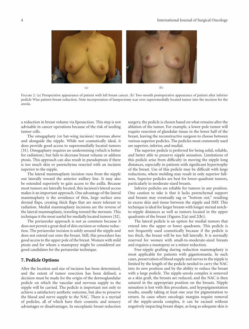
Figure 2: (a) Preoperative appearance of patient with left breast cancer. (b) Two-month postoperative appearance of patient after inferiorpedicle Wise pattern breast reduction. Note incorporation of lumpectomy scar over superomedially located tumor into the incision for theareola.
a reduction in breast volume via liposuction. This step is notadvisable in cancer operations because of the risk of seedingtumor cells.
The omegaplasty (or bat-wing incision) traverses aboveand alongside the nipple. While not cosmetically ideal, itdoes provide good access to superomedially located tumors[31]. Omegaplasty requires no undermining (which is betterfor radiation), but fails to decrease breast volume or addressptosis. This approach can also result in pseudoptosis if thereis too much skin or parenchyma resected with an incisionsuperior to the nipple.
The lateral mammaplasty incision runs from the nippleout laterally toward the anterior axillary line. It may alsobe extended superiorly to gain access to the axilla. Becausemost tumors are laterally located, this incision’s lateral accessmakes it an important approach. One advantage of the lateralmammaplasty is the avoidance of thin, large surface areadermal flaps, creating thick flaps that are more tolerant toradiation. Medial mammaplasty incisions are the reverse ofthe lateral mammaplasty, traveling toward the sternum. Thistechnique it the most useful for medially located tumors [32].
The periareolar approach is not as common because itdoes not permit a great deal of skin excision or volume reduc-tion. The periareolar incision is solely around the nipple anddoes not extend out onto the breast. Still, this procedure hasgood access to the upper pole of the breast. Women with mildptosis and for whom a mastopexy might be considered aregood candidates for the periareolar technique.
7. Pedicle Options
After the location and size of incision has been determined,and the extent of tumor resection has been defined, adecision must be made for the origin of the dermoglandularpedicle on which the vascular and nervous supply to thenipple will be carried. The pedicle is important not only toachieve a satisfactory aesthetic outcome, but also to preservethe blood and nerve supply to the NAC. There is a myriadof pedicles, all of which have their cosmetic and sensoryadvantages or disadvantages. In oncoplastic breast reduction
surgery, the pedicle is chosen based on what remains after theablation of the tumor. For example, a lower-pole tumor willrequire resection of glandular tissue in the lower half of thebreast, leaving the reconstructive surgeon to choose betweenvarious superior pedicles. The pedicles most commonly usedare superior, inferior, and medial.
The superior pedicle is preferred for being solid, reliable,and better able to preserve nipple sensation. Limitations ofthis pedicle arise from difficulty in moving the nipple longdistances, especially in patients with significant hypertrophyof the breast. Use of this pedicle may be difficult with largereductions, where molding may result in only superior full-ness. Superior pedicles are best for lower-quadrant tumors,particularly in moderate-sized breasts.
Inferior pedicles are reliable for tumors in any position.One caution to take is that it lacks parenchymal supportand breasts may eventually sag or “bottom out,” resultingin excess skin and tissue between the nipple and IMF. Thistechnique is ideal for larger breasts with longer sternal notch-to-nipple distances as well as tumors located in the upperquadrants of the breast (Figures 2(a) and 2(b)).
The lateral pedicle is an option for medial tumors thatextend into the upper or lower quadrants. This pedicle isnot frequently used cosmetically because if the pedicle istoo thick, the breast will be too full laterally. It is normallyreserved for women with small-to-moderate-sized breastsand requires a mastopexy or a minor reduction.
Free-nipple grafting during reduction mammaplasty ismost applicable for patients with gigantomastia. In suchcases, preservation of blood supply and nerves to the nipple islimited by the length of the pedicle needed to carry the NACinto its new position and by the ability to reduce the breastwith a large pedicle. The nipple-areola complex is removedas a skin graft, the breasts are reduced, and the NAC is thensutured in the appropriate position on the breasts. Nipplesensation is lost with this procedure, and hypopigmentationresults, usually taking at least one year for pigmentation toreturn. In cases where oncologic margins require removalof the nipple-areola complex, it can be excised withoutnegatively impacting breast shape, as long as adequate skin is

International Journal of Surgical Oncology 5
(a) (b)
Figure 3: (a) Preoperative markings for Wise pattern oncoplastic breast reduction with planned excision of the left nipple areola complex.(b) Two-week postoperative result showing bilateral oncoplastic breast reductions with excision of left nipple-areola complex. Patient willthen have nipple-areola reconstruction after completion of radiation.
preserved (Figures 3(a) and 3(b)). The nipple and areola canthen be reconstructed after completion of radiation, usingany standard method.
8. Postoperative Radiation
Radiation therapy is the second phase of BCT, starting 3 to 6weeks after the reduction procedure once the incisions havehealed. Therapy includes whole-breast irradiation, as well asa boost to the tumor bed to kill any residual microscopicdeposit of cells that surgery may have missed. Cosmetically,radiation also tends to diminish scarring on the breast [33].During surgery, surgeons should be mindful of imminentradiation by avoiding extensive skin-gland dissection andavoiding excessively long parenchymal pedicles that maybe compromised and predisposed to fat necrosis. Patientsshould be informed that radiation therapy can result inchronic edema of the irradiated breast, or contraction andscarring, such that initial postoperative symmetry can be per-manently affected.
9. Complications
In the literature, the complication rate for oncoplasticbilateral breast reduction ranges between 17% and 24%[7, 10, 34]. Common complications include skin necrosis,infection, partial or complete nipple areolar complex necro-sis, and suture-line dehiscence. Like reduction mammaplastypatients without cancer, obese patients and regular smokerssuffer from higher complication rates postoperatively.
If adjuvant chemotherapy is planned, it may begin oncehealing of the incisions has occurred and can be followed byradiation therapy. Complications that interfere with woundhealing may delay the onset of chemotherapy or radiationtherapy.
10. Oncologic Results
Fitoussi conducted the largest study to date, following540 patients who underwent oncoplastic breast surgeryfor cancer, with a median followup of 49 months. Close
or positive margins occurred in 18.9%, with subsequentmastectomy being necessary in 9.4%. At five years, 90.3%reported a satisfactory aesthetic outcome. Five year overalland distant disease-free survival rates were 92.9% and 87.9%respectively, with local recurrence in 6.8% [21]. Whencompared to the standard BCT, comparable values havebeen found, demonstrating the equivalent oncologic safetybetween the two. Rietjens followed 148 women for a median74 months to report a 3% rate of local recurrence [35]. Kayarrecorded 116 patients over a period of 10 years, demon-strating overall survival rates at 100%, 89.1%, and 53.8% forstage I, stage II, and stage III, respectively [36].
Chakravorty et al. compared outcomes from 150 casesthat had utilized the oncoplastic conservation techniques (77of which were for oncoplastic reduction) with 440 cases,which used standard breast conserving surgery. At a 28-month followup and a subsequent projected 6-year localrecurrence rate, oncoplastic breast conserving techniqueswere found to decrease reexcision rates, with oncological out-comes similar to that of standard breast conservation [37].This finding of decreased reexcision rates is expected giventhe increased volume of tissue that can be removed withoncoplastic breast reduction and the need for mastectomy ifthere are positive margins.
11. Oncologic Surveillance
Less-experienced breast radiotherapists and radiologists mayfind the complex glandular reshaping from oncoplasticreduction techniques more challenging to examine on mam-mograms. Women with oncoplastic reductions are morelikely to have a greater number of postoperative mammo-grams and ultrasounds as well as a greater rate of tissuesampling compared to women who have undergone partialbreast reconstruction [38].
Oncoplastic breast reduction does not appear to affectcancer screening for recurrence. Although scar tissue, epi-dermal inclusion cysts, or fat necrosis may appear suspi-cious on physical exam, mammogram, ultrasound, or MRI,evaluation can be done with fine needle aspiration or coreneedle biopsy [39]. Typically, in postoperative healing, fat
6 International Journal of Surgical Oncology
necrosis will present early on and slowly resolve with time,with complete or incomplete resorption. Because of suchsituations, each follow-up visit should be with the sameoncologic surgeon. Mammographic findings from a studyby Mendelsohn et al. found scarring and fibrosis in 50% ofpatients, fluid accumulations in 40% of patients, and dys-trophic calcifications in 10% of patients [39]. Even thoughcancer screening is not compromised, patients who undergooncoplastic reduction require more postoperative tissuesampling than those who receive traditional BCT [10].
12. Oncoplastic Outcomes
Currently, a widely accepted objective study for investigatingcosmetic outcomes is not available. The BREAST-Q is avalidated data set tool that may bring more insight into thismatter. Through pre- and postoperative questionnaires, theBREAST-Q quantifies patient satisfaction and health-relatedquality of life experience in a psychometrically sound andclinically meaningful manner [40]. Patients report signifi-cantly improved body image, functional quality of life, andcosmesis when treated with BCT versus radical mastectomy[41]. More specifically for oncoplastic surgery, available pub-lications indicate an overall satisfaction in treated patients.Chang et al. collected surveys from 20 patients with 70%rating the cosmetic outcome as excellent and 100% reporteda high degree of satisfaction with cosmetic and functionalresults [42]. Goffman et al. established a panel, whichincluded a surgical oncologist, an oncology nurse, a radiationoncologist, and a patient to evaluate cosmetic and functionalresults. Out of 55 patients, 72% evaluations gave excellentand very good marks [43]. Lastly, in a study conducted byLosken et al. 95% of women reported satisfactory aestheticresults after a six month followup [10].
13. Authors’ Experience and Technique
All patients who the breast surgeons feel will have a signi-ficant deformity following lumpectomy are referred to aplastic surgeon. Small-breasted women generally decide toundergo mastectomy and reconstruction. For women withmoderate-to-large sized breasts, there is an extensive discus-sion regarding relative advantages and disadvantages to anoncoplastic breast reduction versus mastectomy and recon-struction (as detailed in the rest of this paper).
Once the decision is made for oncoplastic breast reduc-tion, a combined operation is scheduled. If wire localizationis required, two wires are generally used at each tumorsite to precisely localize the cancer within the breast. If thepatient has had a prior lumpectomy, all efforts are made toincorporate the lumpectomy scar within the skin incision.Skin markings are made to include prior scars and biopsysites whenever possible (Figure 2). This can sometimes meanadjusting markings more superiorly, laterally, or medially.Patients are counseled that the new position of the nipple-areola complex can be affected by these adjustments awayfrom their ideal position at the breast meridian, inframam-mary fold, or near the midhumeral line. The choice of Wisepattern or vertical skin markings is made based on breast
size, degree of ptosis, location of tumor, location of priorscars, and sternal notch-to-nipple distance. The Wise patternincorporates a larger skin excision area. It is therefore moreuseful for incorporation of prior scars or skin over thetumors and is used more often in oncoplastic breast reduc-tions.
The plastic surgeon and breast surgeon perform thelumpectomy together in order to maximize margins andaesthetics. The plastic surgeon starts the operation, makingthe incisions and creating the pedicle, with the guidanceof the oncologic surgeon. When possible, the skin over thetumor is included in the specimen. As the wires or lump-ectomy cavity are approached, the breast surgeon takes overto excise around the tumor. Posteriorly, the breast tissueis removed deep to the tumor, including muscle fascia, inorder to maximize the posterior margin. The correspondingauthor prefers to use a superomedial pedicle whenever tumorlocation permits (personal preference), although the opera-tion is similar with any pedicle. The reduction proceeds ina standard fashion [44]. The entire breast reduction speci-men is removed as a single specimen incorporating the lump-ectomy specimen, in order to avoid cutting across a margin.Once the specimen is removed, the breast surgeon reevalu-ates the remaining breast and removes any additionalmargins deemed necessary. Breast closure then proceeds in astandard fashion, with rotation of the superomedial pedicleinto the keyhole and closure of the lateral and medial pillars.This technique enables the removal of multiple lumpectomyspecimens, even the ones in completely different areas of thebreast, since a wide area of skin and breast is removed. Theplastic surgeon does not hesitate to remove more tissue thanrequired by a standard breast reduction, in order to providethe needed oncologic margins.
To make up for the changes in geometry, additional tissuerearrangement within the breast may be necessary to providethe best shape and symmetry. Consequently, we advise allpatients that a mastectomy would be needed if the marginsreturn positive. Theoretically, a reexcision can be attempteddepending on the original location of the tumor in relationto the reduction, and the location of the positive margin.Alternatively, the patient and oncologist may decide to give aradiation boost to the involved breast. However, we generallydo not advise patients that a reexcision is likely possible,and we prepare them for the possibility of mastectomy if themargins are involved.
Over the last 4 years, we have performed over 30oncoplastic breast reductions. There has been one positivemargin at a nipple and the patient ended up with a mast-ectomy on that side. No other patients were reported withinvolved or close margins. Although we have performed arelatively smaller number of oncoplastic breast reductionscompared with mastectomy reconstructions, our rate of3.33% positive margins compares favorably with publishedrates of positive margins (11-12%) after lumpectomy [45,46]. As discussed preoperatively, the patient presenting withpositive margins ended up with a mastectomy. In retrospect,if there was high suspicion for involvement of the nipple withcancer, she could have had a breast reduction with centralbreast removal and later nipple reconstruction, as the patient
International Journal of Surgical Oncology 7
in Figure 3 had. In an analysis of 540 cases, close or involvedmargins occurred in 18.9 percent, with mastectomy beingnecessary in 9.4 percent [17]. We postulate that our rate ofpositive and close margins is less than 18.9% because ourtechniques involve removing skin over the tumor site and/orfascia over the muscle, and we tend to use techniques thatremove a large amount of surrounding tissue. Fittousi et al.described their experience with a variety of “aesthetic” and“combination” techniques for oncoplastic breast surgery, andit is not clear how many were specifically oncoplastic breastreductions [21]. The authors remark that one-half of thepatients with involved margins were “satisfactorily managedoncologically with either repeated oncoplastic breast surgeryor radiotherapy boost” [17]. The authors do not elaborateas to whether the patients who had repeat oncoplasticbreast surgery initially had a reduction pattern surgery, orif they initially had a more limited tissue rearrangement thatenabled repeat excision. In our practice, we counsel onco-plastic breast reduction patients that a mastectomy wouldlikely be the next step if a margin is involved, but, of course,any case would be evaluated individually.
This issue of mastectomy if there is a positive marginwould seem to argue against oncoplastic breast reductions,since patients usually have a chance at reexcision withlumpectomy alone. By agreeing to an oncoplastic breastreduction, they would seem to be agreeing to a single attemptat a lumpectomy only. However, for most patients in ourpractice choosing this procedure, the decision is not betweena standard lumpectomy or oncoplastic breast reductions; itis a choice between oncoplastic breast reductions or mast-ectomy. Patients deemed candidates for oncoplastic breastreductions are those for whom standard lumpectomieswould be too deforming (because of breast size, tumor size,multiple tumors, or tumor location) or those with symp-tomatic macromastia who desire breast reduction for theadded symptom relief. Therefore, they are willing to tryoncoplastic breast reductions as an alternative to mastec-tomy. Additionally, the rate of positive margins is much lowerthan that with standard lumpectomies, so it is rare that thesepatients do indeed go on to need a mastectomy.
14. Conclusions
In some parts of the United States, a potential lack of avail-able reconstructive plastic surgeons limits combined treat-ment. Breast surgeons are then left with the choice of eitherreferring their patients to larger centers or attempting tolearn the reconstructive procedures themselves [47]. Despitethese limitations, ideally, a combined approach with a breastsurgeon and plastic surgeon provides the best results for thepatient.
Management of patients with breast cancer is alsochanging. Surgeons are constantly looking for new, lessinvasive, and more cosmetically favorable techniques to helppatients manage their disease and live with the results of theirtreatment.
References
[1] J. W. Canady, R. A. D’Amico, and M. F. McGuire, “Oncoplasticbreast surgery: past, present, and future directions in the
United States,” Plastic and Reconstructive Surgery, vol. 124, no.3, pp. 973–974, 2009.
[2] B. Fisher, S. Anderson, J. Bryant et al., “Twenty-year follow-upof a randomized trial comparing total mastectomy, lumpec-tomy, and lumpectomy plus irradiation for the treatment ofinvasive breast cancer,” New England Journal of Medicine, vol.347, no. 16, pp. 1233–1241, 2002.
[3] K. B. Clough, J. Cuminet, A. Fitoussi, C. Nos, and V. Mosseri,“Cosmetic sequelae after conservative treatment for breastcancer: classification and results of surgical correction,” Annalsof Plastic Surgery, vol. 41, no. 5, pp. 471–481, 1998.
[4] C. D’Aniello, L. Grimaldi, A. Barbato, B. Bosi, and A. Carli,“Cosmetic results in 242 patients treated by conservative sur-gery for breast cancer,” Scandinavian Journal of Plastic andReconstructive Surgery and Hand Surgery, vol. 33, no. 4, pp.419–422, 1999.
[5] J. Y. Petit, M. Rietjens, C. Garusi, M. Greuze, and C. Perry,“Integration of plastic surgery in the course of breast-con-serving surgery for cancer to improve cosmetic results andradicality of tumor excision,” Recent Results in Cancer Re-search, vol. 152, pp. 202–211, 1998.
[6] W. Audretsch, M. Rezai, and C. Kolotas, “Tumor-specificimmediate reconstruction in breast cancer patients,” Perspec-tives in Plastic Surgery, vol. 11, no. 1, pp. 71–100, 1998.
[7] A. M. Munhoz, E. Montag, E. Arruda et al., “Assessmentof immediate conservative breast surgery reconstruction: aclassification system of defects revisited and an algorithm forselecting the appropriate technique,” Plastic and ReconstructiveSurgery, vol. 121, no. 3, pp. 716–727, 2008.
[8] P. L. Giacalone, P. Roger, O. Dubon et al., “Comparative studyof the accuracy of breast resection in oncoplastic surgery andquadrantectomy in breast cancer,” Annals of Surgical Oncology,vol. 14, no. 2, pp. 605–614, 2007.
[9] N. Kaur, J. Y. Petit, M. Rietjens et al., “Comparative study ofsurgical margins in oncoplastic surgery and quadrantectomyin breast cancer,” Annals of Surgical Oncology, vol. 12, no. 7,pp. 539–545, 2005.
[10] A. Losken, T. M. Styblo, G. W. Carlson, G. E. Jones, and B.J. Amerson, “Management algorithm and outcome evaluationof partial mastectomy defects treated using reduction ormastopexy techniques,” Annals of Plastic Surgery, vol. 59, no.3, pp. 235–242, 2007.
[11] J. Y. Petit, M. Rietjens, G. Contesso, F. Bertin, and R. Gilles,“Contralateral mastoplasty for breast reconstruction: a goodopportunity for glandular exploration and occult carcinomasdiagnosis,” Annals of Surgical Oncology, vol. 4, no. 6, pp. 511–515, 1997.
[12] B. L. Smith, M. Bertagnolli, B. B. Klein et al., “Evaluation of thecontralateral breast: the role of biopsy at the time of treatmentof primary breast cancer,” Annals of Surgery, vol. 216, no. 1, pp.17–21, 1992.
[13] J. A. Urban, D. Papachristou, and J. Taylor, “Bilateral breastcancer: biopsy of the opposite breast,” Cancer, vol. 40, no. 4,pp. 1968–1973, 1977.
[14] H. J. Wanebo, G. M. Senofsky, and R. E. Fechner, “BilateralBreast cancer. Risk reduction by contralateral biopsy,” Annalsof Surgery, vol. 201, no. 6, pp. 667–677, 1985.
[15] K. L. Dundas, J. Atyeo, and J. Cox, “What is a large breast?Measuring and categorizing breast size for tangential breastradiation therapy,” Australasian Radiology, vol. 51, no. 6, pp.589–593, 2007.
[16] M. E. Taylor, “Factors influencing cosmetic results after con-servation therapy for breast cancer,” International Journal ofRadiation Oncology Biology Physics, vol. 31, no. 4, pp. 753–764,1995.
8 International Journal of Surgical Oncology
[17] F. Hernanz, S. Regano, A. Vega, and M. Gomez Fleitas,“Reduction mammaplasty: an advantageous option for breastconserving surgery in large-breasted patients,” Surgical Oncol-ogy, vol. 19, no. 4, pp. e95–e102, 2010.
[18] R. A. Cochrane, P. Valasiadou, A. R. M. Wilson, S. K. Al-Ghazal, and R. D. Macmillan, “Cosmesis and satisfaction afterbreast-conserving surgery correlates with the percentage ofbreast volume excised,” British Journal of Surgery, vol. 90, no.12, pp. 1505–1509, 2003.
[19] E. Delay and K. B. Clough, “Oncoplastic breast surgery:conclusions and future perspectives,” Annales de ChirurgiePlastique et Esthetique, vol. 53, no. 2, pp. 226–227, 2008.
[20] D. M. O’Dey, P. Baltes, A. Bozkurt, and N. Pallua, “Importanceof the suprasternal notch to nipple distance (SSN:N) forvascular complications of the nipple areola complex (NAC)in the superior pedicle vertical mammaplasty: a retrospectiveanalysis,” Journal of Plastic, Reconstructive and Aesthetic Sur-gery, vol. 64, no. 10, pp. 1278–1283, 2011.
[21] A. D. Fitoussi, M. G. Berry, F. Fama et al., “Oncoplastic breastsurgery for cancer: analysis of 540 consecutive cases,” Plasticand Reconstructive Surgery, vol. 125, no. 2, pp. 454–462, 2010.
[22] K. B. Dough, S. S. Kroll, and W. Audretsch, “An approachto the repair of partial mastectomy defects,” Plastic andReconstructive Surgery, vol. 104, no. 2, pp. 409–420, 1999.
[23] S. J. Kronowitz, J. A. Feledy, K. K. Hunt et al., “Determiningthe optimal approach to breast reconstruction after partialmastectomy,” Plastic and Reconstructive Surgery, vol. 117, no.1, pp. 1–11, 2006.
[24] S. L. Spear, J. B. Burke, D. Forman, R. A. Zuurbier, and C.D. Berg, “Experience with reduction mammaplasty followingbreast conservation surgery and radiation therapy,” Plastic andReconstructive Surgery, vol. 102, no. 6, pp. 1913–1916, 1998.
[25] S. J. Schnitt, “Risk factors for local recurrence in patientswith invasive breast cancer and negative surgical margins ofexcision: where are we and where are we going?” AmericanJournal of Clinical Pathology, vol. 120, no. 4, pp. 485–488, 2003.
[26] S. E. Singletary, “Surgical margins in patients with early-stagebreast cancer treated with breast conservation therapy,” Ameri-can Journal of Surgery, vol. 184, no. 5, pp. 383–393, 2002.
[27] A. Haid, M. Knauer, S. Dunzinger et al., “Intra-operativesonography: a valuable aid during breast-conserving surgeryfor occult breast cancer,” Annals of Surgical Oncology, vol. 14,no. 11, pp. 3090–3101, 2007.
[28] F. D. Rahusen, A. J. A. Bremers, H. F. J. Fabry, A. H. M. Taetsvan Amerongen, R. P. A. Boom, and S. Meijer, “Ultrasound-guided lumpectomy of nonpalpable breast cancer versus wire-guided resection: a randomized clinical trial,” Annals of Sur-gical Oncology, vol. 9, no. 10, pp. 994–998, 2002.
[29] C. Lassus, “Breast reduction: evolution of a technique—asingle vertical scar,” Aesthetic Plastic Surgery, vol. 11, no. 2, pp.107–112, 1987.
[30] M. Lejour, “Vertical mammaplasty and liposuction of thebreast,” Plastic and Reconstructive Surgery, vol. 94, no. 1, pp.100–114, 1994.
[31] B. O. Anderson, R. Masetti, and M. J. Silverstein, “Oncoplasticapproaches to partial mastectomy: an overview of volume-displacement techniques,” Lancet Oncology, vol. 6, no. 3, pp.145–157, 2005.
[32] M. Ballester, M. G. Berry, B. Couturaud, F. Reyal, R. J. Salmon,and A. D. Fitoussi, “Lateral mammaplasty reconstruction aftersurgery for breast cancer,” British Journal of Surgery, vol. 96,no. 10, pp. 1141–1146, 2009.
[33] K. B. Clough, C. Nos, R. J. Salmon, M. Soussaline, and J. C.Durand, “Conservative treatment of breast cancers by mam-maplasty and irradiation: a new approach to lower quadrant
tumors,” Plastic and Reconstructive Surgery, vol. 96, no. 2, pp.363–370, 1995.
[34] S. J. Kronowitz, K. K. Hunt, H. M. Kuerer et al., “Practicalguidelines for repair of partial mastectomy defects using thebreast reduction technique in patients undergoing breast con-servation therapy,” Plastic and Reconstructive Surgery, vol. 120,no. 7, pp. 1755–1768, 2007.
[35] M. Rietjens, C. A. Urban, P. C. Rey et al., “Long-termoncological results of breast conservative treatment withoncoplastic surgery,” Breast, vol. 16, no. 4, pp. 387–395, 2007.
[36] R. Kayar, M. Cobanoglu, O. Gungor, H. Catal, and M.Emiroglu, “The value of breast reduction operations in breastconservation surgery, late results of 116 patients with breastcancer,” Meme Saglıgı Dergisi, vol. 2, pp. 131–138, 2006.
[37] A. Chakravorty, A. K. Shrestha, N. Sanmugalingam et al.,“How safe is oncoplastic breast conservation? Comparativeanalysis with standard breast conserving surgery,” EuropeanJournal of Surgical Oncology , vol. 38, no. 5, pp. 395–398, 2012.
[38] A. Losken, T. G. Schaefer, M. Newell, and T. M. Styblo,“The impact of partial breast reconstruction using reductiontechniques on postoperative cancer surveillance,” Plastic andReconstructive Surgery, vol. 124, no. 1, pp. 9–17, 2009.
[39] A. M. Mendelsohn, E. L. Bove, F. M. Lupinetti, D. C. Crowley,T. R. Lloyd, and R. H. Beekman, “Central pulmonary arterygrowth patterns after the bidirectional Glenn procedure,”Journal of Thoracic and Cardiovascular Surgery, vol. 107, no.5, pp. 1284–1290, 1994.
[40] A. L. Pusic, A. F. Klassen, and S. J. Cano, “Use of the BREAST-Q in clinical outcomes research,” Plastic and ReconstructiveSurgery, vol. 129, no. 1, pp. 166e–167e, 2012.
[41] D. Curran, J. P. Van Dongen, N. K. Aaronson et al., “Qualityof life of early-stage breast cancer patients treated with radi-cal mastectomy or breast-conserving procedures: results ofEORTC trial 10801,” European Journal of Cancer, vol. 34, no.3, pp. 307–314, 1998.
[42] E. Chang, N. Johnson, B. Webber et al., “Bilateral reductionmammoplasty in combination with lumpectomy for treat-ment of breast cancer in patients with macromastia,” AmericanJournal of Surgery, vol. 187, no. 5, pp. 647–651, 2004.
[43] T. E. Goffman, H. Schneider, K. Hay, D. E. Elkins, R. A.Schnarrs, and C. Carman, “Cosmesis with bilateral mam-moreduction for conservative breast cancer treatment,” BreastJournal, vol. 11, no. 3, pp. 195–198, 2005.
[44] S. P. Davison, A. N. Mesbahi, I. Ducic, M. Sarcia, J. Dayan, andS. L. Spear, “The versatility of the superomedial pedicle withvarious skin reduction patterns,” Plastic and ReconstructiveSurgery, vol. 120, no. 6, pp. 1466–1476, 2007.
[45] A. B. Chagpar, R. C. G. Martin, L. J. Hagendoorn, C. Chao,and K. M. McMasters, “Lumpectomy margins are affected bytumor size and histologic subtype but not by biopsy tech-nique,” American Journal of Surgery, vol. 188, no. 4, pp. 399–402, 2004.
[46] H. Yang, W. Jia, K. Chen et al., “Cavity margins and lump-ectomy margins for pathological assessment: which is superiorin breast-conserving surgery?” Journal of Surgical Research,vol. 178, no. 2, pp. 751–757, 2012.
[47] A. Losken and M. Y. Nahabedian, “Oncoplastic breast surgery:past, present, and future directions in the United States,”Plastic and Reconstructive Surgery, vol. 124, no. 3, pp. 969–972,2009.

Submit your manuscripts athttp://www.hindawi.com
Stem CellsInternational
Hindawi Publishing Corporationhttp://www.hindawi.com Volume 2014
Hindawi Publishing Corporationhttp://www.hindawi.com Volume 2014
MEDIATORSINFLAMMATION
of
Hindawi Publishing Corporationhttp://www.hindawi.com Volume 2014
Behavioural Neurology
EndocrinologyInternational Journal of
Hindawi Publishing Corporationhttp://www.hindawi.com Volume 2014
Hindawi Publishing Corporationhttp://www.hindawi.com Volume 2014
Disease Markers
Hindawi Publishing Corporationhttp://www.hindawi.com Volume 2014
BioMed Research International
OncologyJournal of
Hindawi Publishing Corporationhttp://www.hindawi.com Volume 2014
Hindawi Publishing Corporationhttp://www.hindawi.com Volume 2014
Oxidative Medicine and Cellular Longevity
Hindawi Publishing Corporationhttp://www.hindawi.com Volume 2014
PPAR Research
The Scientific World JournalHindawi Publishing Corporation http://www.hindawi.com Volume 2014
Immunology ResearchHindawi Publishing Corporationhttp://www.hindawi.com Volume 2014
Journal of
ObesityJournal of
Hindawi Publishing Corporationhttp://www.hindawi.com Volume 2014
Hindawi Publishing Corporationhttp://www.hindawi.com Volume 2014
Computational and Mathematical Methods in Medicine
OphthalmologyJournal of
Hindawi Publishing Corporationhttp://www.hindawi.com Volume 2014
Diabetes ResearchJournal of
Hindawi Publishing Corporationhttp://www.hindawi.com Volume 2014
Hindawi Publishing Corporationhttp://www.hindawi.com Volume 2014
Research and TreatmentAIDS
Hindawi Publishing Corporationhttp://www.hindawi.com Volume 2014
Gastroenterology Research and Practice
Hindawi Publishing Corporationhttp://www.hindawi.com Volume 2014
Parkinson’s Disease
Evidence-Based Complementary and Alternative Medicine
Volume 2014Hindawi Publishing Corporationhttp://www.hindawi.com