review article sleep spindle characteristics in children
TRANSCRIPT

Review ArticleSleep Spindle Characteristics in Children withNeurodevelopmental Disorders and Their Relation to Cognition
Reut Gruber1 and Merrill S. Wise2
1Department of Psychiatry, McGill University, 6875 LaSalle Boulevard, Montreal, QC, Canada H4H 1R32Methodist Healthcare Sleep Disorders Center, 5050 Poplar Avenue, Memphis, TN 38157, USA
Correspondence should be addressed to Reut Gruber; [email protected]
Received 30 December 2015; Revised 11 March 2016; Accepted 26 April 2016
Academic Editor: Julie Seibt
Copyright © 2016 R. Gruber and M. S. Wise. This is an open access article distributed under the Creative Commons AttributionLicense, which permits unrestricted use, distribution, and reproduction in any medium, provided the original work is properlycited.
Empirical evidence indicates that sleep spindles facilitate neuroplasticity and “off-line” processing during sleep, which supportslearning, memory consolidation, and intellectual performance. Children with neurodevelopmental disorders (NDDs) exhibitcharacteristics that may increase both the risk for and vulnerability to abnormal spindle generation. Despite the high prevalenceof sleep problems and cognitive deficits in children with NDD, only a few studies have examined the putative association betweenspindle characteristics and cognitive function. This paper reviews the literature regarding sleep spindle characteristics in childrenwith NDD and their relation to cognition in light of what is known in typically developing children and based on the availableevidence regarding childrenwithNDD.We integrate available data, identify gaps in understanding, and recommend future researchdirections. Collectively, studies are limited by small sample sizes, heterogeneous populations with multiple comorbidities, andnonstandardized methods for collecting and analyzing findings. These limitations notwithstanding, the evidence suggests thatfuture studies should examine associations between sleep spindle characteristics and cognitive function in children with andwithout NDD, and preliminary findings raise the intriguing question of whether enhancement or manipulation of sleep spindlescould improve sleep-dependent memory and other aspects of cognitive function in this population.
1. Introduction
Neurodevelopmental disorders (NDDs) are a heterogeneousgroup of conditions in which the development of the centralnervous system is disrupted. Manifestations can includeimpairments in motor function, learning, cognition and/orcommunication, or neuropsychiatric problems. These issuesappear early in development, persist throughout life, andproduce notable impairments in social, communicative, cog-nitive, and behavioral functioning [1] that can vary fromvery specific limitations to global impairment in intelligenceand social skills. This group of disorders includes intellec-tual disability (formerly referred to as mental retardation),communication disorders, autism spectrum disorder (ASD),Attention Deficit Hyperactivity Disorder (ADHD), specificlearning disorders, and neurodevelopmental motor disordersincluding cerebral palsy (CP).
Sleep is a vital process for brain restoration and it iscritical for maintaining cognitive function. Strong empirical
evidence indicates that sleep spindles facilitate the plasticitywhich supports learning, memory consolidation, declarativelearning, motor skills, and overall intellectual performance.The cognitive functions that are related to sleep spindles arealso key domains of dysfunction in children with NDD [2].
Clinically significant sleep problems are prevalent inchildren and adolescents with NDD [3, 4]. Among childrenwith neurocognitive difficulties, the impact of disrupted sleepspindle generation may be amplified. Hence, the characteris-tics of NDD may increase both the risk for and vulnerabilityto abnormal sleep spindle generation.The goal of the presentreview is to review and integrate existing evidence regardingsleep spindle characteristics in children withNDD and, whenevidence is available, to examine the associations betweenthese characteristics and cognitive function.
A better understanding of relationships between sleepand NDD is expected to provide insight into the pathophys-iology and possibly the treatment of such disorders and
Hindawi Publishing CorporationNeural PlasticityVolume 2016, Article ID 4724792, 27 pageshttp://dx.doi.org/10.1155/2016/4724792
2 Neural Plasticity
improve our understanding of the association between sleepspindles and cognition in children. It is likely that sleep spin-dle characteristics represent a marker of brain developmentand function, as well as a window into underlying brainmechanisms that support cognition.
We will first briefly describe sleep spindle developmentand its relationship with cognitive development and functionin typically developing children. We will then review theavailable evidence regarding sleep spindle characteristics inchildren with a variety of NDDs and their relation to cog-nition. Finally, we will discuss the findings, identify gaps inunderstanding, and recommend future research directions inthis emerging area of investigation.
2. Sleep Spindles in TypicallyDeveloping Children
Sleep spindles represent an oscillating electrical potential inthe brain; they have a characteristic frequency of 11–16Hz(usually 12–14Hz in healthy adults) and last from one toseveral seconds in duration [5]. On scalp electroencephalog-raphy (EEG), spindles are seen as sinusoidal waves that oftenhave a fusiformor “crescendo-decrescendo”morphology [6].
Sleep spindles are characterized by their symmetry;synchrony between hemispheres; amplitude, which is thepeak-to-peak difference in spindle size, reflecting voltage;frequency, which is the number of waveforms per second;density, which is the number of spindle bursts/min of NREMsleep; and the duration of spindle bursts. In infants, sleepspindles last several seconds in duration, are expressedmaximally in the frontocentral location, are in the high alphaor low beta frequency range, and are not synchronous. Thelack of synchrony is likely due to lack of myelination in theneonatal brain. By 2 years of age, it is considered abnormal ifmost spindles are still asynchronous [7]. In older children andadults, sleep spindles are expressed diffusely across the headbut maximally over the central regions and in a bilaterallysynchronous and symmetric fashion [7]. Moreover, sleepspindles can be divided into two distinct types based ontheir frequency and field of expression. Slower spindles (9 to<13Hz) occur maximally over the frontal regions, whereasfaster spindles (>13–16Hz) dominate [8, 9] over the centraland parietal head regions and typically precede slow spindlesby hundreds of milliseconds [9–11].The slow spindles displaya typical waxing-and-waning pattern, whereas fast spindlesare mainly waning [12]. This difference begins to developat around 2 years of age [13]. The two populations of sleepspindles are thought to arise from and represent differentgenerators within the thalamus, with some level of corticalinvolvement. They also demonstrate different maturationalpatterns, suggesting that their development is associatedwith changes in thalamocortical structures and maturationof the physiological systems that produce spindles [6, 14,15]. Shinomiya et al. suggested that separate investigation ofthe two types of spindles may be important in evaluatingdevelopmental processes in the central nervous systems ofchildren and adolescents, and that frontal spindle activitycould represent a good indicator of biological maturation[15].
2.1. Generation of Sleep Spindles. Sleep spindles are a pro-totypical thalamocortical rhythm generated and “paced” inthe thalamus via a network of synaptic interactions involvinginhibitory (GABAergic) neurons in the reticular thalamicnuclei, thalamocortical cells, and cortical pyramidal neurons[16]. In animal models, the spindle rhythm is abolishedby destruction of the thalamus, whereas it persists afterdecorticationwhen the thalamus is preserved [14, 17]. Spindlefrequency is determined in large part by an interplay betweenmutually interconnected GABAergic inhibitory neurons ofthe reticular nucleus of the thalamus and the thalamocorticalneurons, with influence from inputs of the cortex andbrainstem [14]. Reticular thalamic neurons impose hyperpo-larization on thalamocortical neurons, thereby activating anonspecific cation current that depolarizes the thalamocorti-cal neurons and activates low-threshold calcium ion currentsand bursting. The latter process provides feedback excitationto reticular thalamic neurons, thus closing the loop. Eachthalamocortical burst also imposes an excitatory postsynapticpotential (EPSP) on pyramidal neurons, providing the basisfor the spindle waveforms observed on scalp EEG.
2.2. Development of Sleep Spindles. In typically develop-ing children, very early (“rudimentary”) spindles may beobserved as early as term to 2 weeks postterm [15, 18–21] and it was proposed that they could appear earlier inpremature infants [22]. Spindles become more easily iden-tified between 3 and 9 weeks postterm, when they oftenoccur in relatively long trains lasting up to 10 seconds duringquiet sleep [21, 23, 24]. In the first 6 months, spindles mayoccur unilaterally, often alternating between hemispheres.Asymmetry is common as well. Spindles become increasinglymore synchronous between hemispheres during the first yearof life, reflectingmaturation of interhemispheric connections[25]. By 12 to 18 months, most sleep spindles are expressedin a bilaterally synchronous and symmetric fashion, withmaximal expression over the central regions.
Maturation alters sleep spindle activity in terms of thespindle frequency, amplitude, duration, and density [15, 18–20, 26, 27]. Changes in the development of spindle durationand density are thought to follow a U-shaped distribution,whereas that of the interspindle interval shows an invertedU-shape. Three distinct phases of sleep spindle developmenthave been proposed: (1) infants up to 9-10 months old exhibitlong spindles (around 1.5 s) having a relatively low density(<3/min) and a relatively short interspindle interval (around20 s); (2) children from 10 months up to 3 years exhibita decrease in the spindle length (to around 0.8 s) and anincrease in the interspindle interval (up to 111 s), yielding aneven lower spindle density (0.3–1.2/min); and (3) over threeyears of age, children show short interspindle intervals (5–10 s) and long spindle durations (0.9–1.5 s) of high density(4–10/min).These changes presumably reflect developmentalchanges in thalamocortical structures and maturation ofthe physiological system that produces spindles [26, 27].Sleep spindle peak frequencies increase from childhood toadolescence [15, 28, 29], when global maturational changesin sigma power predominate in the slow sigma frequencyband [29]. The topographic representation of sigma power

Neural Plasticity 3
provides insight into these age-related changes [30] by show-ing that the fast sigma power increases over the centroparietalareas, while the slow sigma power decreases over frontalareas across childhood and adolescence [15, 30]. Given theassociation between neural maturation and changes in sleepspindle activity, it has been proposed that sleep spindles couldbe used as a potential index of neural maturation [27].
2.3. Sleep Spindles and Cognitive Development in TypicallyDeveloping Children. Intellectual ability is closely related tocortical development in children and adolescents. Intelli-gence is associated with the trajectory of cortical devel-opment, primarily that of the frontal regions which areimplicated in the maturation of intelligence [31, 32]. Synapticdensity increases until around puberty (11 years for girls and12 years for boys), whereupon synaptic pruning begins [33].In fact, vigorous cortical thinning by early adolescence has apositive association with IQ [34].
A similar pattern is seen in the developmental changesof sleep spindles. Initially, during development, the increasedneuronal connectivity results in higher sleep EEG amplitudesbecause the size of these waves reflects the number ofsynaptic connections [31, 35–37]. Thereafter, pruning resultsin smaller neuronal populations that oscillate in unison, withcorresponding decreases in EEG power.These changes in thebrain appear to parallel the rapid development of cognitiveabilities at similar ages [31, 33, 38–43]. Synaptic pruning andincreasing myelination during adolescence result in fasterand more efficient information processing, which is mani-fested by an increased ability to perform complex cognitiveoperations, increased speed and efficiency in completing sim-ple information-processing tasks, and improved performanceon intelligence tests across adolescence [37].
Slow spindles have been correlated with visual perceptuallearning [44], while fast spindles have been correlated withmore complex abilities and processes, such as fluid intel-ligence [45], learning ability [46] and word-location associ-ations [47]. It has been proposed that they could be used asa neurobiological indicator for the level of cognitive devel-opment.
2.4. Sleep Spindles and Cognitive Function in Typically Devel-oping Children. Theprocesses underlying sleep spindles havebeen hypothesized to benefit cognitive functions and “off-line” information processing in several ways. First, it isassumed that sleep spindles serve as a “gating mechanism”to protect sleep from being interrupted by external stimuli,such as noise [48], thereby allowing optimal time for off-lineinformation processing. Historically, thalamocortical (TC)cells were thought to gate sensory transmission by switchingfrom tonic to burst dischargemode [16].The bursting patternoccurs during NREM sleep in the form of sleep spindles,while the tonic pattern of activity occurs in wakefulness.The tonic activity pattern was thought to relay sensoryinformation to the cortex via the thalamus from a varietyof afferent inputs, while the bursting or spindle pattern wasthought to serve a gating role.More recently, researchers haveproposed that both modes can relay stimuli to the cortex.However, while tonic spikes reliably transmit information,
the stereotyped discharge profile of bursts leads to nonlineardistortion of sensory inputs [49]. Burst firing of TC cellsduring spindles would thus filter external stimuli. It hasbeen proposed that one way in which sleep spindles couldsupport cognitive function is by blocking interference (i.e.,performing a gating function) to allow uninterrupted off-lineprocessing and consolidation of information.
Sleep spindles appear to actively enhance informationprocessing via their role inmemory consolidation. Behavioralstudies in animal models and humans have shown that learn-ing improves more during and following a period of sleepthan during an equivalent amount of waking time [50–52].Neuroimaging studies have shown that the patterns of brainactivity elicited during initial learning are replayed duringsubsequent sleep, demonstrating the presence of dynamic off-line information processing [53]. Underlying this enhance-ment or stabilization of memory across a sleep period is theconcept of sleep-dependent memory consolidation, wherebymemory traces become more stable and resistant to inter-ference over a period of time. The consolidation of declar-ative memory results from a dialog between reactivatedhippocampal memory traces and the neocortical networksthat retain long-termmemory representations [54]. In adults,sleep-dependent consolidation of declarative memory hasbeen correlated with a range of neurophysiological measures,including the amounts of slow wave activity (1–4Hz), slowoscillations (0.5–1Hz), and sleep spindles. This indicates thatsleep plays an active role in the hippocampal-neocortical dia-log [54]. Recent research conducted in children of differentages has shown that their sleep-dependent consolidation ofdeclarative memory is comparable to that of adults, whereasthat of procedural skills is not [55, 56].
One explanation for how sleep spindles contribute to newlearning is related to the hypothesis that sleep spindles facili-tate the priming of synapses for plastic changes, [57] therebyenabling the off-line information processing that is essentialfor learning to be completed [58]. Data from simulations ofsleep spindle activity have indicated that repetitive thalamicbursts (similar to sleep spindle activity) generate robust entryof Ca2+ into cortical dendrites [59], which produces condi-tions that favor the priming of synapses for plastic changes(i.e., by activating proplastic signaling molecules such asprotein kinase A and Ca2+/calmodulin-dependent proteinkinase II). The synaptic potentiation induced by spindlesis consistent with the hypothesis of “active system consoli-dation” [60], which proposes that sleep spindles potentiatememory traces by reactivating selected neuronal circuits.
In addition to the contribution of sleep spindles to creat-ing the conditions that allow for optimal sleep (i.e., by gating),brain plasticity, and memory consolidation, the characteris-tics of sleep spindles have been shown to be stable withinsubjects across different nights [6], and to correlate withintellectual ability, particularly fluid intelligence. This has ledto the view that spindles, at least under non-pathologicalconditions, constitute to some extent a biophysical measureof intelligence.
In children, mixed results have been reported in regardto the direction and strength of associations between thedifferent characteristics of sleep spindles and performance

4 Neural Plasticity
on intelligence tests and memory tasks. Geiger et al. foundthat peak sleep spindle frequency was negatively correlatedwith full scale IQ but that relative sigma power correlatedpositively with full scale IQ and fluid IQ [61]. Gruber etal. showed that lower sleep spindle frequency was associ-ated with better performance on the intelligence perceptualreasoning and working memory WISC-IV scales but thatsleep spindle amplitude, duration, and density were notassociated with performance on IQ tests [62]. Chatburn etal. observed that the number of fast spindles was positivelycorrelated with narrative memory and negatively correlatedwith sensorimotor functioning. Mean central frequency ofspindles was also negatively correlated with sensorimotorfunctioning, planning ability, and working memory [63].Hoedlmoser et al. found that children with higher sleepspindle activity, as measured at frontal, central, parietal, andoccipital sites during both baseline and experimental nights,exhibited higher general cognitive abilities (WISC-IV) anddeclarative learning efficiency (i.e., number of recalled wordsbefore and after sleep) [64].
Astill et al. reported that individual differences in thedominant frequency of spindles and slow waves were pre-dictive for performance on finger sequence tapping tasks.Children performed better if they had fewer slow spindles,more fast spindles, and faster slow waves. On the other hand,overnight enhancement of accuracy was most pronounced inchildren with more slow spindles and slower slow waves, thatis, the children with an initial lower performance at baseline.Thus, associations of spindle and slow wave characteristicswith initial performance may confound interpretation oftheir involvement in overnight enhancement. Slower fre-quencies of characteristic sleep events may be a marker ofslower learning and immaturity of networks involved inmotor skills [65]. Bodizs et al. found that fluid IQ correlatedpositively with fast spindle density and amplitude in girls andthat these correlations peak in the frontocentral regions. Inboys, by contrast, the only positive spindle-index of fluid IQwas found to be the frequency of fast spindles [66]. Doucetteet al. observed that children with faster processing speedsexhibited higher slow sigma power over the parietal region[67]. For a detailed description of these findings in typicallydeveloping children please see Table 1.
In summary, sleep spindles appear in early infancy, andthey change and develop through childhood and adolescencein a progression that parallels the milestones of corticaldevelopment. Sleep spindles appear to contribute to off-lineinformation processing by protecting sleep (and thus sleep-dependent processes) frombeing interrupted.They also seemto facilitate the brain plasticity that allows essential learningto occur through processes such as memory consolidation.Finally, certain characteristics of sleep spindles have beencorrelated with intellectual performance, particularly fluidintelligence.
3. Sleep Spindles in Children withNeurodevelopmental Disorders
Despite the high prevalence of sleep problems in childrenwith NDD and the wide range of cognitive deficits in
this population, there is only limited information regardingthe characteristics and function of sleep spindles in thispopulation. Only a handful of studies have examined sleep inchildren with NDD, and the existing data are limited in scopeand the range of disorders studied.The few existing studies onthe putative association between sleep spindles and cognitivefunction have been conducted in children with intellectualdisabilities, ASD, reading disabilities, and ADHD. However,although the body of knowledge is scattered, often based onnonstandardized methods, not always up-to-date, and basedmostly on descriptive and correlational studies, it providesimportant insights into the extent to which sleep spindlesare abnormal in these populations compared to typicallydeveloping children. Moreover, the existing studies give usan initial view of the nature and extent of the associationsbetween sleep spindles and different intellectual levels in chil-dren with neurodevelopmental disorders or (in a few studies)a more direct idea of the associations between sleep spindlesand information processing or intellectual performance.
3.1. Sleep Spindles in Children with Intellectual Disability.Intellectual disability is characterized by three findings: anintelligence quotient (IQ) of 75 or below; significant limita-tions in adaptive behaviors; and onset of disability occurringbefore age 18. In the past, the term “mental retardation” wasused to describe this condition, but this term is no longer used[68]. The common causes of intellectual disability includegenetic conditions, brain insults during pregnancy, problemsat the time of birth, medical problems that affect brain health,and exposure to environmental toxins (e.g., lead ormercury).
Sleep problems are more severe and more prevalent inchildren with NDD than in typically developing children.Insomnia in normally developing young children is mostoften behaviorally based [69], whereas insomnia in childrenwithNDD ismore oftenmultifactorial, with neurologic,med-ical, and psychiatric comorbidities contributing to behavioralissues. Moreover, insomnia in NDD tends to be chronic,often lasting into adolescence or adulthood [70]. Quineshowed that sleep problems are persistent in children withdevelopmental disabilities [71], and Wiggs and Stores foundthat the average duration of sleep problems in such childrenis 7.13 years (SD 4.04 years) [72].
Alarmingly high prevalence rates for sleep disorders havebeen cited in children with intellectual disabilities: 86% inchildren under 6 years old, 81% in those aged 6 to 11 yearsold [71], and 77% in those aged 12 to 16 years old [73]. Nightwaking and settling difficulties are particularly common,affecting over half of children with NDD aged up to 16 years.The chronicity of these problems was illustrated by Quine[71], who found that half of children with NDD with settlingproblems and over two-thirds of those with night wakingwere still having problems 3 years later.
Sleep disorders are also highly prevalent in children withASD: recent studies have reported prevalence rates as highas 40–85% in these children versus 20–40% in typicallydeveloping children [74–76]. The commonly reported sleepdisturbances include prolonged sleep-onset latency, restlesssleep, frequent nocturnal awakenings, and reduced total sleep

Neural Plasticity 5
Table1:(a)C
haracteristicso
fsleep
spindlesandassociations
betweensle
epspindlesandcogn
itive
functio
nin
child
renwith
attentiondeficithyperactivity
disorder(A
DHD)v
ersustypically
developing
child
ren.
(b)C
haracteristicso
fsleep
spindles
andassociations
betweensle
epspindles
andcogn
itive
functio
nin
child
renwith
autism
spectrum
disorder
(ASD
)versustypically
developing
child
ren.
(c)C
haracteristicsof
sleep
spindles
andassociations
betweensle
epspindles
andcogn
itive
functio
nin
child
renwith
developm
entald
yslexia(D
D)v
ersustypically
developing
child
ren.(d)C
haracteristicso
fsleep
spindlesandassociations
betweensle
epspindlesandcogn
itive
functio
ninchild
renwith
intellectualdisa
bility(ID
)versustypicallydeveloping
child
ren.
(e)C
haracteristicso
fsleep
spindles
andassociations
betweensle
epspindles
andcogn
itive
functio
nin
typically
developing
(TPD
)children.
(a)
Reference
Diso
rder
Cognitiv
eim
pairm
ent
Age
Samples
ize
Stud
ydesig
nSleepmeasure
Cognitiv
emeasure
(outcome/s)
Results
Recording
parameters
Spindles
scoring
Sleep
Cognitio
nSleepand
cogn
ition
Kiesow
and
Surw
illo,1987
[100]
ADHD
Non
e
ADHD
patie
nts:
Range:3–11
Mean:
6.69
Con
trols:
Range:3–11
Mean:
6.69
ADHD
patie
nts:11
Con
trols:
11100%
males
Correlational
study
10–20
International
Syste
mof
electrode
placem
ent
S2sle
epspindles
Nosig
nificant
differences
Prehn-
Kristensenet
al.,2011[103]
ADHD
Non
eADHD:12–16
Con
trols:
11–14
ADHD:12
Con
trols:
12
Experim
ental
Case-con
trol
Repeated
Measures
Design
PSG(sam
pling
rate:200
Hz
with
12-bit
resolutio
n).
EEG(C
3,C4
,A1,andA2),
EOG,and
EMG.
Sleepspindles
detected
using
aband-pass
filtero
f12–14H
z,of
S2sle
ep
Declarativ
emem
ory
consolidation
task
(recognitio
naccuracy)
ADHD
participants:
moreR
EMsle
epand
shorterS
WS
latency
(1)O
verall
redu
ced
recogn
ition
accuracy
inADHD
grou
p.(2)E
nhanced
recogn
ition
accuracy
inthes
leep
cond
ition
,bu
tgreater
incontrol
comparedto
ADHD
Positive
association
between
(1)d
urationof
non-RE
Msle
epand
(2)slow
oscillatio
npo
wer
and
sleep-
associated
mem
ory
consolidation
inhealthy
controlsbu
tno
tinADHD

6 Neural Plasticity
(b)
ReferenceDiso
rder
Cognitiv
eim
pairm
ent
Age
Samples
ize
Stud
ydesig
nSleepmeasure
Cognitiv
emeasure
(outcome/s)
Results
Recording
parameters
Spindles
scoring
Sleep
Cognitio
nSleepandcogn
ition
God
bout
etal.,
2000
[96]
ASD
Non
eAS:7–53
Con
trols:
7–61
ASD
:8Con
trols:
8Correlational
study
PSG
S2sle
epspindles:
12–15H
z,lasting
0.5–2.0s
,no
amplitu
decriteria
ASD
participants:
(1)M
ore
transitions
from
awaking
epochto
REM
sleep
compared
tohealthy
controls.
(2)M
ade
less
transitions
from
aS2
epochto
REM
sleep
compared
tohealthy
controls.
(3)L
ower
S2spindle
density.
Limoges
etal.,
2005
[108]ASD
Non
eASD
:16–
27Con
trols:
16–26
ASD
:16
Con
trols:
16Correlational
study
PSG.E
EG(C
3,C4
,O1,
andO2),
andEO
G,
EMG.
S2sle
epspindles
(C3,Fp
1):
12–15H
z,lasting
0.5–2.0s
,no
amplitu
decriteria
ASD
participants:
(1)
Increased
timeinS1
and
decreased
non-RE
Msle
epand
SWS
(2)L
ower
S2sle
epspindle
density
(C3)
compared
tohealthy
controls

Neural Plasticity 7
(b)Con
tinued.
ReferenceDiso
rder
Cognitiv
eim
pairm
ent
Age
Samples
ize
Stud
ydesig
nSleepmeasure
Cognitiv
emeasure
(outcome/s)
Results
Recording
parameters
Spindles
scoring
Sleep
Cognitio
nSleepandcogn
ition
Limoges
etal.,
2013
[97]
ASD
Non
eASD
:16–
27Con
trols:
16–27
17ASD
14controls
Correlational
study
PSG(C
3,C4
,O1,and
O2),E
OG,
andEM
G.
S2sle
epspindles
Susta
ined
andselective
attention
(RT)
working
mem
ory
(spanscore)
Declarativ
eepiso
dic
mem
ory(#
ofrecalled
figures)
Sensory-
motor
procedural
mem
ory
(con
tacttim
e,#of
trials)
Procedural
mem
ory
(time)
(1)L
ower
REM
density.
(2)L
ower
SWS%
(3)H
igher
S1%in
lower
S2spindle
density
(C3)
inAS
compared
tocontrols
Poorer
per-
form
ance
inASD
compared
tocontrols
on susta
ined
attention
working
mem
ory.
Sensory-
motor
procedural
mem
ory
(1)P
ositive
associationin
controlsbu
tnot
inASD
betweenthe
SWS%
and
decla
rativ
emem
ory
immediaterecall
(2)N
egative
associations
inbo
thgrou
psbetween
SWS%
andlearning
thes
ensory-m
otor
proceduralmem
ory
task
(3)N
egative
associations
inASD
butn
otin
healthy
controls
betweenS1%and
decla
rativ
emem
ory
immediaterecall;
sensory-motor
and
cogn
itive
procedural
mem
orytasksa
ndselectivea
ttention
(4)S
2spindle
density
(C3)
isnegativ
elycorrelated
with
RTin
the
selectivea
ttention
onlyin
healthy
controls
(5)S
2spindle
density
(C3)
negativ
elycorrelated
with
sensory-motor
proceduralmem
ory
durin
gthelearning
phaseo
nlyin
ASD
grou
p

8 Neural Plasticity
(b)Con
tinued.
ReferenceDiso
rder
Cognitiv
eim
pairm
ent
Age
Samples
ize
Stud
ydesig
nSleepmeasure
Cognitiv
emeasure
(outcome/s)
Results
Recording
parameters
Spindles
scoring
Sleep
Cognitio
nSleepandcogn
ition
Maskiet
al.,
2015
[99]
ASD
Non
eASD
and
controls:
9–16years
ASD
:22
Con
trols:
20
Experim
ental
Case-C
ontro
lRe
peated
Measures
Design
training
sessionin
the
morning
30minutes
after
habitualwake
timea
fterthe
nighto
fsleep
recording
andatestin
gsessionin
the
evening
PSG.E
EG(F1,F2,C
3,Cz
,C4,O1,
andO2),
EOG,and
EMG.
Spectral
power
calculated
forslow
wave
oscillatio
n(0.5–1
Hz),
delta
(1–4H
z),
theta
(4–7
Hz),
alph
a(8–11H
z),
sigma
(12–15Hz),
andbeta
(16–
20Hz)
frequ
ency
ranges.
S2sle
epspindles
(Cz):
12–15H
z,lasting
0.5–2.0s
,no
amplitu
decriteria
Sleep-
depend
ent
mem
ory
consolidation
(difference
inperfo
rmance
(in%)
between
training
and
testing
phases)
LessRE
Msle
epin
ASD
compared
tocontrol
grou
p
(1)Inbo
thcond
ition
sASD
grou
pperfo
rmed
morep
oorly
than
control
grou
p(2)B
oth
grou
psrecalled
bette
rinthe
sleep
cond
ition
compared
tothew
ake
cond
ition
Slow
oscillatio
npo
wer
correlated
with
sleep
depend
entm
emory
consolidationin
the
ASD
grou
pon
ly

Neural Plasticity 9
(b)Con
tinued.
ReferenceDiso
rder
Cognitiv
eim
pairm
ent
Age
Samples
ize
Stud
ydesig
nSleepmeasure
Cognitiv
emeasure
(outcome/s)
Results
Recording
parameters
Spindles
scoring
Sleep
Cognitio
nSleepandcogn
ition
Tessiere
tal.,
2015
[98]
ASD
Non
e
ASD
:6–13
Con
trols:
7–12
Mean:
10.23
SD:2.00
ASD
:13
Con
trols:
13Correlational
study
PSG.E
EG(C
3,C4
,F3,
F4,O
1,and
O2),E
OG,
andEM
G.
S2sle
epspindles
Intelligence
WISC-
III.
Perfo
rmance
IQ(PIQ
),verbalIQ
(VIQ
),and
fullscaleIQ
(FSIQ)
scores.
Lower
S2spindle
density
(Fp1)in
ASD
grou
pcompared
tocontrol
grou
p
(1)V
IQpo
sitively
correlated
with
S2spindled
uration
(C4)
incontrol
grou
pbu
tnot
inASD
grou
p(2)V
IQisassociated
with
spindled
ensity
onlyin
ASD
grou
p(3)F
SIQis
associated
with
spindled
ensityon
lyin
ASD
grou
p(c)
Reference
Diso
rder
Cognitiv
eim
pairm
ent
Age
Samples
ize
Stud
ydesig
nSleepmeasure
Cognitiv
emeasure
(outcome/s)
Results
Recording
parameters
Spindles
scoring
Sleep
Cognitio
nSleepand
cogn
ition
Brun
ietal.,
2009
[106]
DD
Non
eDD:8–16
Con
trols:
7–16
DD:16
Con
trols:
11Correlational
study
PPSG
10–20
International
syste
m
Power
spectra
calculated
for
delta,theta,
alph
a,sig
ma,
andbetaSleep
spindles
Scored
durin
gN2.
(1)M
emoryand
learning
transfe
r(2)R
eading
test
(speed
and
accuracy)
(3)W
ritingtest
(accuracy)
(4)Intelligence
WISC-
IIIR
Com
paredto
healthycontrols
DDparticipants
hadthefollowing:
(1)L
essstage-shifts
perh
our
(2)L
ower
N2%
(3)L
essN
3%(4)Increasein
power
offre
quency
band
s0.5–3
Hz
and11-12Hzd
uring
N2and0.5–1H
zdu
ringN3
(5)Increased
spindled
ensity
MeanIQ
DD:FSIQ:
93.9
VIQ
:90
PIQ:98.9
Onlyin
DDgrou
p:(1)S
igmap
ower
band
inN2was
positively
correlated
with
the
mem
oryand
learning
transfe
rreadingtest
readingtest
(2)S
pind
ledensity
was
positively
correlated
with
perfo
rmance
onthew
ordreading
test

10 Neural Plasticity
(d)
ReferenceDiso
rder
Cognitiv
eim
pairm
ent
Age
Samples
ize
Stud
ydesig
nSleepmeasure
Cognitiv
emeasure
(outcome/s)
Results
Recording
parameters
Spindles
scoring
Sleep
Cognitio
nSleepand
cogn
ition
Marca
etal.,
2011[90]
IDCostello
synd
rome
(CS)
CSpatie
nts:
Range:
18mon
ths–
31years
Con
trols:
18mon
ths–31
years
CS:11
Con
trols:
22
Sleep
Spindles:
12–14H
z.Scored
over
allN
REM
stages
DQ
IQ
CSgrou
phad
increased
spindle
activ
itybetween
13Hza
nd14Hz
andbetween
14Hza
nd15Hz
comparedto
healthy
controls
Selvitelli
etal.,
2009
[91]
ID
Malform
ations
ofcortical
developm
ent
(MCD
s)
MCD
Mean:
35.7
SD:
12.2
Con
trols:
Mean:
35.6
SD:14.3
MCD
:10
Con
trols:
10Correlational
study
Not
specified
Sleep
spindles;
12–16H
z.Occurrin
gdu
ringS2.
Com
paredto
control
grou
p,MCD
grou
phad
(1)h
igher
prop
ortio
nof
unilateral
sleep
spindles
(2)a
nincreased
prop
ortio
nof
anterio
randdiffu
sespindles
Neural Plasticity 11
(d)Con
tinued.
ReferenceDiso
rder
Cognitiv
eim
pairm
ent
Age
Samples
ize
Stud
ydesig
nSleepmeasure
Cognitiv
emeasure
(outcome/s)
Results
Recording
parameters
Spindles
scoring
Sleep
Cognitio
nSleepand
cogn
ition
Shibagaki
etal.,
1982
[94]
ID
Con
genital
cerebraldysplasia
(𝑁=10)
Hydroceph
aly(𝑁
=3)
Rubinstein-Taybi
synd
rome(𝑁
=2)
Dow
n’ssynd
rome
(𝑁=1)
Silver’ssynd
rome
(𝑁=1)
Lesch-Nyhan
synd
rome(𝑁
=1)
Hallerm
ann-Streiff
synd
rome(𝑁
=1)
Remaining
subjects=
unkn
ownetiology
4mon
ths–
8years
45Descriptiv
estu
dy
PSG.E
EG(F3-C3
,F4
-C4,
F3-A1,
F4-A2,
C3-A1,
C4-A2,
O1-A
1,and
O2-A2),
EOG,E
MG,
andEC
G
Not
specified
DQ
IQ
(1)5
6%display
edRE
Mbu
rst
durin
gNRE
Mwith
sleep
spindles.
(2)6
4%show
edsle
epspindles
atthe
beginn
ingor
towards
the
endof
REM
sleep
No
significant
finding
s

12 Neural Plasticity
(d)Con
tinued.
ReferenceDiso
rder
Cognitiv
eim
pairm
ent
Age
Samples
ize
Stud
ydesig
nSleepmeasure
Cognitiv
emeasure
(outcome/s)
Results
Recording
parameters
Spindles
scoring
Sleep
Cognitio
nSleepand
cogn
ition
Shibagaki
and
Kiyono
,1983
[95]
ID
Dow
n’ssynd
rome
(𝑁=3)
Hydroceph
aly(𝑁
=9)
Holop
rosencephaly
(𝑁=2)
Remaining
subjects=
unkn
ownetiology
3–8mon
ths
90Descriptiv
estu
dy
PSG.E
EG(F3-C3
,F4
-C4,
F3-A1,
F4-A2,
C3-A1,
C4-A2,
O1-A
1,and
O2-A2),
EOG,E
MG,
andEC
G
S2sle
epspindles
DQ
IQ
(1)4
2%had
aratio
ofmorethan
2.00
(#spindles
longer
than
0.4s
/#of
spindles
shorterthan
0.4s
)(2)15.6%
hadar
atio
of1.9
9–1.0
0and
6%hada
ratio
of0.99–0
.50
(3)2
2%had
aratio
ofless
than
0.50
(4)14%
had
nosle
epspindles
(1)S
ubjects
with
aratioof
lessthan
0.50
had
significantly
lower
DQs
than
subjects
with
aratioof
morethan
2.00
(2)S
ubjects
with
nosle
epspindles
atall
had
significantly
lower
DQs
than
subjects
with
aratioof
morethan
2.00
(3)S
ubjects
with
aratioof
lessthan
0.50
orno
sleep
spindles
had
significantly
lower
DQs
than
subjects
with
aratioof
1.99–
1.00

Neural Plasticity 13
(d)Con
tinued.
ReferenceDiso
rder
Cognitiv
eim
pairm
ent
Age
Samples
ize
Stud
ydesig
nSleepmeasure
Cognitiv
emeasure
(outcome/s)
Results
Recording
parameters
Spindles
scoring
Sleep
Cognitio
nSleepand
cogn
ition
Shibagaki,
etal.,
1986
[92]
IDCerebralp
alsy
(CP)
(𝑛=23)
CP:
4mon
ths–
5years
Non
-CP
patie
nts:
4mon
ths–12
years
CP:23
Non
-CP
patie
nts:39
Descriptiv
estu
dy
PSG(F3-A1,
C3-A1,and
O1-A
1),
EOG,E
CG,
respira
tion,
andEM
G
Not
specified
DQ
IQ
(1)13%
ofno
n-CP
patie
ntsh
adno
spindles
orextre
mely
low
incidenceo
fspindles
(2)8
%of
CPcasessho
wed
nosle
epspindles
(3)2
2%of
CPcases
show
edextre
mely
low
incidenceo
fspindles
(4)4
.3%of
CPcases
show
edextre
me
spindles
CPpatie
nts
with
indistin-
guish
able
sleep
stages
hadlower
DQsthanCP
patie
ntsw
ithno
rmalEE
Gpatte
rnsa
ndno
n-CP
patie
ntsw
ithno
rmalEE
Gpatte
rns

14 Neural Plasticity
(d)Con
tinued.
ReferenceDiso
rder
Cognitiv
eim
pairm
ent
Age
Samples
ize
Stud
ydesig
nSleepmeasure
Cognitiv
emeasure
(outcome/s)
Results
Recording
parameters
Spindles
scoring
Sleep
Cognitio
nSleepand
cogn
ition
Shibagaki,
etal.,
1980
[93]
ID
Holop
rosencephaly
(𝑁=1)
Dow
n’ssynd
rome
(𝑁=2)
Hydroceph
aly(𝑁
=8)
Rubinstein-Taybi
synd
rome(𝑁
=2)
Prader-W
illi
synd
rome(𝑁
=2)
Cri-d
u-chat
synd
rome(𝑁
=2)
Hallerm
ann-Streiff
synd
rome(𝑁
=1)
Silver’ssynd
rome
(𝑁=1)
Remaining
subjects-
unkn
ownetiology
4mon
ths–8
years
43Descriptiv
estu
dy
PSG.E
EG(F3-C3
,F4
-C4,
F3-A1,
C3-A1,
F4-A2,and
C4-A2),
EOG,E
CG,
respira
tion,
andEM
G
Spindles
measured
durin
gS2.
Extre
me
spindles:
9-10c/s,
50–120𝜇V
DQ
(1)2
.3%had
noRE
Msle
ep(2)3
0%had
nosle
epspindles
(3)4
.6%had
high
voltage
fastactiv
ity(20–
30c/s,
100–
200u
V)
with
outsleep
spindles
inwakefulness,
S1,S2,and
REM
(4)7
%had
lowvoltage
activ
itywith
out
spindles
throug
hout
nocturnal
sleep
(5)2
.3%had
indistin-
guish
able
stagesw
iththep
resence
ofsle
epspindles
(6)2
.3%had
extre
me
spindles
(1)P
atients
with
abno
rmal
EEGpatte
rns
throug
hout
S1–S4had
lower
DQs
than
patie
nts
with
abno
rmal
EEGpatte
rns
onlyin
S1-S2
andpatie
nts
with
norm
alEE
Gpatte
rns
(2)P
atients
with
abno
rmal
EEGpatte
rns
inS1-S2had
lower
DQs
than
patie
nts
with
norm
alEE
Gpatte
rns

Neural Plasticity 15
(d)Con
tinued.
ReferenceDiso
rder
Cognitiv
eim
pairm
ent
Age
Samples
ize
Stud
ydesig
nSleepmeasure
Cognitiv
emeasure
(outcome/s)
Results
Recording
parameters
Spindles
scoring
Sleep
Cognitio
nSleepand
cogn
ition
Veneselli
etal.,
2001
[87]
ID
Lateinfantile
neuron
alceroid
lipofuscino
sis(LIN
CL)
Range:3–10
LINCL
patie
nts:18
Descriptiv
estu
dy
PSG.E
EG(10–
20Inter-
natio
nal
Syste
mof
electrode
placem
ent)
Not
specified
Nosle
epspindles
were
observed
(e)
Reference
Diso
rder
Cognitiv
eim
pairm
ent
Age
Samples
ize
Stud
ydesig
nSleepmeasure
Cognitiv
emeasure
(outcome/s)
Results
Recording
parameters
Spindles
scoring
Sleep
Cognitio
nSleepand
cogn
ition
Astilletal.,
2014
[65]
Non
e(TPD
)Non
eMean:
10.7
SD:0.8
30
Experim
ental
Repeated
Measures
Design
PSGEE
G(Fpz,
Cz),EO
G,
EMG
Artifa
ct-fr
eeEE
GacrossS2,
S3andS4
Fastspindles:
frequ
ency≥
12Hz,Slow
spindles:
frequ
ency
<12Hz.
Sleep-
depend
ent
procedural
mem
orytask:
fingersequence
tapp
ingtasks
(speed
and
accuracy)
Average
duratio
nof
slowwaves
atFP
zwas
negativ
elycorrelated
with
density
offast
spindles
and
positively
correlated
with
density
ofslo
w
Accuracy
was
bette
rifthe
interval
contained
sleep
(1)H
igherS
WS%
was
associated
with
increasesa
ccuracy
(2)C
hildrenwith
high
erdensity
ofslo
wspindles
had
lower
overallspeed
andaccuracy
whereas
child
ren
with
high
erdensity
offastspindles
had
high
eroverall
speed
(3)C
hildrenwith
high
erdensity
ofslo
wspindles
show
edas
trong
erovernightincrease
inaccuracy
butn
otspeed.
(4)C
hildrenwith
alonger
average
duratio
nof
slow
waves
hadstronger
overnightincrease
inaccuracy
butn
otspeed.

16 Neural Plasticity
(e)Con
tinued.
Reference
Diso
rder
Cognitiv
eim
pairm
ent
Age
Samples
ize
Stud
ydesig
nSleepmeasure
Cognitiv
emeasure
(outcome/s)
Results
Recording
parameters
Spindles
scoring
Sleep
Cognitio
nSleepand
cogn
ition
Bodizs
etal.,
2014
[66]
Non
e(TPD
)Non
e15–22years
24Correlational
study
PSG
10–20
International
syste
m
Measured
acrosssta
ges
S2–S4.
Fluid
intelligence
Raven’s
progressive
matric
estest
(RPM
T)(IQ)
Subjectshad
norm
alsle
epstructure
IQ
Onlyin
females,
fastspindled
ensity
andfastspindle
amplitu
dewere
positively
correlated
with
IQscores.
Chatbu
rnet
al.,
2013
[63]
Non
e(TPD
)Non
e4.1–12.7
years
27Correlational
study
PSG(C
3-A2
andC4
-A1),
EMG,and
EOG
S2sle
epspindles
Stanford-Binet
intelligence
scale
NEP
SY
Subjectshad
norm
alsle
epstructure
All
intelligence
andneu-
rocogn
itive
functio
ning
was
inthe
norm
alrang
e
(1)M
eancentral
spindlefrequ
ency
was
negativ
elycorrelated
with
nonverbalw
orking
mem
ory,planning
,andfin
emotor
functio
ning
(2)F
astspind
ledensity
was
positively
correlated
with
narrativem
emory
andnegativ
elycorrelated
with
sensorim
otor
functio
ning
and
finem
otor
functio
ning

Neural Plasticity 17
(e)Con
tinued.
Reference
Diso
rder
Cognitiv
eim
pairm
ent
Age
Samples
ize
Stud
ydesig
nSleepmeasure
Cognitiv
emeasure
(outcome/s)
Results
Recording
parameters
Spindles
scoring
Sleep
Cognitio
nSleepand
cogn
ition
Dou
cette
etal.,
2015
[67]
Non
e(TPD
)Non
e2–5years
10 50%males
Correlational
study
Sleep
was
recorded
over
1night
intheh
ome
environm
ent
PSG
Slow
sigma
frequ
ency
range:
10–13H
zFastsig
ma
frequ
ency
range:
13.25–17Hz
Processin
gspeed
task:(RT
)
Subjectshad
norm
alsle
epstructure
Average
reactio
ntim
ewas
1408.8±
251.4
ms
RTsig
nificantly
correlated
with
slowsig
map
ower
inac
luste
rof16
electro
desin
parie
talregions
Geigere
tal.,
2011[61]
Non
e(TPD
)Non
e
Range:9–
12years
Mean:
10.5
SD:1.0
14 57.1%
males
Correlational
study
Sleepwas
recorded
over
2nights
separatedby
1or
2weeks
ina
sleep
labo
ratory
PSG.E
EG10–20
International
syste
mEO
GandEM
G
Meanall-n
ight
power
spectra
ofsta
geN2
sleep.
Intelligence
WISC-
IVAttention
task
(RT)
Subjectshad
norm
alsle
epstructure
Allresults
wereinthe
norm
alrang
e
(1)S
leep
spindle
peak
frequ
ency
correlated
negativ
elywith
full
scaleIQ
(2)Ind
ividual
relativ
esigma
power
correlated
positively
with
full
scaleIQandflu
idIQ
Grubere
tal.,
2013
[62]
Non
e(TPD
)Non
eRa
nge:7–11
years
29Correlational
study
PSG(F3,F4
,C3
,C4,P3
,P4,
O1,andO2),
EOG,E
CG,
andEM
G
Measured
durin
gall
NRE
Martifact-free
epochs.
Intelligence
WISC-
IV
Subjectshad
norm
alsle
epstructure
Allresults
wereinthe
norm
alrang
e
Sleepspindle
frequ
ency
was
negativ
elyassociated
with
scores
onthe
perceptual
reason
ingand
working
mem
ory

18 Neural Plasticity
(e)Con
tinued.
Reference
Diso
rder
Cognitiv
eim
pairm
ent
Age
Samples
ize
Stud
ydesig
nSleepmeasure
Cognitiv
emeasure
(outcome/s)
Results
Recording
parameters
Spindles
scoring
Sleep
Cognitio
nSleepand
cogn
ition
Hoedlmoser
etal.,
2014
[64]
Non
e(TPD
)Non
eRa
nge:8–11
years
54Correlational
study
PSG10–20
International
syste
m
Sleepspindles
measured
durin
gN2and
N3.
Declarativ
emem
ory
consolidation
task:w
ordpair
associationtask
(RT)
Intelligence
WISC-
IV
Subjectshad
norm
alsle
epstructure
(1)R
Tsfor
correctly
remem
bered
wordpairs
improved
overnight
(2)P
oorer
retrieval
scores
observed
inthem
orning
after
sleep
(1)H
igherS
pApo
sitively
correlated
with
bette
rmem
ory
perfo
rmance
overallbeforea
ndaft
ersle
ep(2)C
hildrenwith
high
erIQ
had
high
erSpA
Note.A
ASM
=American
Academ
yofSleep
Medicine;ADHD=attentiondeficithyperactivity
disorder;A
S=Asperger’s
synd
rome;ASD
=autism
spectrum
disorder;C
P=cerebralpalsy
;CS=Costello
synd
rome;
DD
=developm
entald
yslexia;DQ
=developm
entalq
uotie
nt;E
CG=ele
ctrocardiogram
;EEG
=ele
ctroenceph
alogram;E
OG
=ele
ctrooculogram;E
MG
=ele
ctromyogram
;ID
=intellectuald
isability;
IQ=
intelligenceq
uotie
nt;K
ITAP=testof
attentionalp
erform
ance
forc
hildren;
MCD
=malform
ations
ofcorticaldevelopm
ent;NEP
SY=neurop
sychologicalassessment;PS
G=po
lysomno
graphy
;REM
=rapideye
movem
ent;RT
=reactio
ntim
e;SD
=standard
deviation;
SpA=spindlea
ctivity
;SWS=slo
wwaves
leep;T
DP=typically
developing
child
ren;WISC=Wechsler
intelligences
caleforc
hildren.

Neural Plasticity 19
time. Fifty-three percent of 2- to 5-year-olds with ASD had atleast one sleep problem, compared with 32% of controls [77].Children with ASD often take more than 1 hour to fall asleep,andmany have nocturnal awakenings that may last as long as2-3 hours [78]. Sleep problems in ASD tend to persist pastmid-puberty [79]. A longitudinal case-control study foundthat insomnia was 10 times more likely to be reported inASD children than in controls, and that remission of sleepproblems at 11 to 13 years was far less likely in ASD childrenthan controls (8 versus 53%) [79]. The most common sleepproblems in ASD are sleep-onset delay, frequent nocturnalawakenings, and reduced sleep duration [80].
As for ADHD, as many as 70% of children with thisdisorder have been reported to display mild to severe sleepproblems [81]. The prevalence rates differ by ADHD subtype,with the highest prevalence in the combined subtype [82],though sleepiness may be more frequent in the inattentivesubtype [83]. In addition, psychiatric comorbidities andmed-ication used both increase the prevalence of sleep problems inADHD [82]. Children and/or their parents reported bedtimeresistance, sleep-onset difficulties, night awakenings, difficul-ties withmorning awakenings, sleep breathing problems, anddaytime sleepiness significantly more than healthy controls[84]. Although there is no sleep problem specific to ADHD[81], the most commonly reported issue is “difficulty fallingasleep [81].”
3.1.1. Sleep Spindle Characteristics in Children with Intellec-tual Disability. In 1962, E. L. Gibbs and F. A. Gibbs [85]reported the presence of “extreme spindles” in children withintellectual disability. Extreme spindles are characterizedby their diffuse expression, much higher voltage (200–400microvolts), and continuous occurrence. In addition, severalstudies examined sleep spindles in children with intellectualdisability related to different genetic disorders [86]. Thesedisorders included the neuronal ceroid lipofuscinosis (NCLs)[87] and Costello syndrome. The NCLs are a group ofinherited progressive neurodegenerative lysosomal-storagedisorders that are characterized by progressive intellectualand motor deterioration, seizures, and early death resultingfrom neural loss and widespread accumulation of lipopig-ments within cellular compartments [86]. The incidence(affected persons per live newborns) was reported to be1 : 12,500 in theUSA and Scandinavian countries, whereas theworldwide figure is 1 : 100,000 [88]. Children with NCLs werefound to lack sleep spindles.
Costello syndrome, which is an autosomal dominantdisorder caused by mutations in HRAS, has an estimatedbirth prevalence of 1 : 300,000 in the UK [89]. CostelloSyndrome [90] is characterized by delayed developmentand intellectual disability, loose folds of skin, unusuallyflexible joints, and distinctive facial features including alarge mouth. Researchers observed an increase in spindleamplitude (extreme spindles). Another study was conductedin children with malformations of cortical development [91].These congenital brain disorders arise during embryonic andfetal development and are characterized by abnormalities inthe volume, location, and/or architecture of cerebral gray
and white matter. For eachmalformation subject, the authorsidentified a nonepileptic age- and sex-matched controlpatient for whom an EEG study had been performed withinone year of themalformation subject’s study, which was of thesame type and demonstrated at least one sleep spindle burst.The authors found no difference between cases and controlsin the mean spindle density or mean maximum spindle fre-quency, but significant between-group differences were seenin the laterality and anatomical distribution of spindles. Mal-formation subjects had a significantly higher proportion ofunilateral sleep spindles compared to controls. Furthermore,although the sample sizes were small, subjects with unilateralmalformations appeared to demonstrate a skewing of unilat-eral spindles toward the contralateral side, with fewer spindlebursts on the ipsilateral side to the malformation. Finally,malformation subjects had a significantly different overallanatomical distribution of sleep spindles, with an increasedproportion of both anterior and diffuse spindles. Finally,Shibagaki et al. [92] reported a high prevalence of childrenwithmalformations of cortical developmentwith no spindles,low incidence of sleep spindles, and/or extreme spindles.
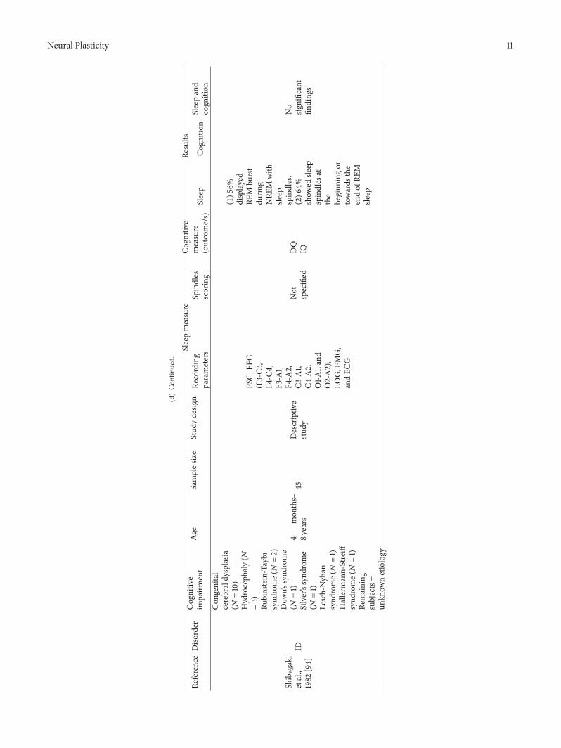
3.1.2. Sleep Spindles and Intellectual Performance in Childrenwith Intellectual Disability. In a series of studies [92–94],Shibagaki et al. classified the participants’ levels of function-ing according to their developmental quotients (DQs). Theyused the Tsumori and Inage questionnaire for infants andchildren, which divides the children into severe, moderate,and mildly intellectually disabled groups. They includedchildren with intellectual disability associated with a range ofdisorders (e.g., congenital cerebral dysplasia, hydrocephaly,Rubinstein-Taybi syndrome, Down’s syndrome, and chromo-somal abnormality) and performed several studies investigat-ing the occurrences of sleep spindles in these children. Theyfound that children with intellectual disability frequentlyhave no sleep spindles and that these children tended tohave a lower DQ than those with intellectual disability withsleep spindles [94]. They reported a significant increase inabnormal clinical EEGs and a significant decrease in the DQsof children with high occurrences of shorter or no sleepspindles compared to thosewith higher occurrences of longersleep spindles [93, 95].
3.1.3. Sleep Spindles and Memory Consolidation in Childrenwith Intellectual Disability. No study has yet been reportedthat assessed memory consolidation in children with intel-lectual deficits.
Collectively, these studies show that children with intel-lectual disabilities due to a variety of heterogeneous geneticand developmental disorders show significant alterations insleep spindles, including few or no sleep spindles, extremespindles, and/or an increased proportion of unilateral spin-dles. In addition, the degree of sleep spindle abnormalityis associated with the severity of cognitive impairment. Inconditions with progressive neurodegeneration, spindle losshas been correlated with the state of the disease, with acomplete absence of spindles seen during the most severestages.

20 Neural Plasticity
3.2. Sleep Spindles in ChildrenwithAutism SpectrumDisorders(ASDs). ASDs are neurodevelopmental disorders character-ized by repetitive behaviors and deficits in social interactionand communication. A diagnosis of ASD is made based ona constellation of requisite behavioral symptoms, includingpersistent deficits in social communication and interactionacross multiple contexts, as well as restricted and repetitivepatterns of behavior, interests, and activities.
3.2.1. Sleep Spindle Characteristics in Children with ASD.Studies comparing children/adolescents with ASD versuscontrols showed that the former had a lower sleep spindledensity (Godbout et al. [96]), fewer sleep spindles over thecentral regions [97], a lower NREM stage 2 sleep spindledensity in the prefrontal area, and shorter sleep spindleduration in the frontal area [98]. However, another studyfailed to find any difference in sleep spindles when comparingchildren with ASD and typically developing children [99].
3.2.2. Sleep Spindles and Intellectual Performance in ChildrenwithASD. Reference [98] examined the associations betweenIQ (measured by the WISC-III) and sleep spindle density orduration in children with high functioning autism comparedto typically developing children. The authors found that ver-bal IQ was negatively correlated with frontal spindle densityand positively correlated with central spindle duration intypically developing children, whereas verbal IQ and thefull scale IQ were negatively correlated with central spindledensity in the ASD group.
3.2.3. Sleep Spindles and Memory Consolidation in Childrenwith ASD. One recent study [99] examined memory consol-idation in children with ASD. Twenty-two participants withASD and 20 control participants between 9 and 16 years ofage were trained to criterion on a spatial declarative memorytask and then given a cued recall test. The subjects wereallowed a period of daytime wake (Wake) or a night of sleep(Sleep), both of which were monitored with home-basedpolysomnography. Upon retest, bettermemory consolidationwas observed in the Sleep group compared to theWake groupfor both ASD and control children; however, participantswith ASD had poorer overall memory consolidation. Thechange in performance across sleep, independent of medi-cation and age, showed no significant relationship with anyspecific sleep parameter other than the total sleep time, andthere was a trend toward less forgetting in the control group.
In summary, there are conflicting findings with respectto sleep spindles in children with ASD. Whereas the datafrom one research group suggest that individuals with ASDhave shorter spindle durations, lower spindle density, anddecreased sleep spindle frequencies, other groups have notreplicated these findings. In addition, childrenwithASDhavepoorer memory consolidation than controls.
3.3. Sleep Spindles in Children with ADHD. Attention deficit/hyperactivity disorder (ADHD) is one of the most com-mon NDDs in childhood, affecting approximately 3%–5% ofschool-aged children [1] and enduring throughout adoles-cence and adulthood. A diagnosis of ADHD is dependent
on developmentally inappropriate symptoms of inattention,hyperactivity, and/or impulsivity, with onset before the age of7 years and impaired functioning in two ormore settings [68].
3.3.1. Sleep Spindle Characteristics in Children with ADHD.There are conflicting findings with respect to sleep spindlecharacteristics in children with ADHD. Some studies [100]failed to find any statistically significant difference in thenumber of sleep spindles between hyperactive and controlchildren, while other studies found significantly fewer [101]or more [102] sleep spindles in the EEGs of unmedicatedhyperactive boys compared to normal controls.
3.3.2. Sleep Spindles and Intellectual Performance in Childrenwith ADHD. No published study has yet examined this asso-ciation.
At present, there is no consistent evidence for abnormalor altered sleep spindle activity in children with ADHDand sleep spindles activity per se has not been measured instudies examining memory consolidation of children withADHD. However, there appear to be differences in thememory consolidation of children with ADHD versus typ-ically developing children [103, 104], in that sleep benefitsdeclarativememory in typically developing children, whereasADHD children show deficits in sleep-associated consolida-tion of declarative memory and a reduced functionality ofslow oscillations in this consolidation. In contrast, althoughprocedural memory in typically developing children does notbenefit from sleep, the data suggest that sleep appears tonormalize the daytime deficits in procedural memory foundamong ADHD children [104].
3.4. Sleep Spindles in Children with Dyslexia. Developmentaldyslexia is a hereditary neurological disorder that is char-acterized by the presence of severe and persistent readingand/or spelling impairments despite normal intelligence andadequate schooling. Almost no studies are available on sleepin children with dyslexia. The existing data have shown analteration of sleep architecture characterized by an increasein slow wave sleep (SWS), a decrease in REM sleep, and alonger REM sleep latency in children with reading disabilitiescompared to controls [105].
One study [106] examined sleep spindles in 19 childrenwith developmental dyslexia and 11 normally reading chil-dren between 7 and 16 years of age. The authors observedincreases in spindle activity and sigma power in childrenwith dyslexia and found that these parameterswere correlatedwith the degree of dyslexic impairment. No information isavailable regarding sleep spindles and memory consolidationin this population. However, these results suggest that sleepspindle abnormalities may exist in children with dyslexia andthat these abnormalities could be related to or correlatedwith impairment. Additional studies are needed to test thesehypotheses.
4. Discussion
The goal of this paper is to review and integrate the availableevidence regarding sleep spindle characteristics in children

Neural Plasticity 21
with NDD and (when possible) their associations withcognitive function. Before we attempt to integrate the find-ings across different disorders, we must note the significantmethodological limitations of the existing work.Most studieshad small sample sizes and thusmight have overestimated theeffect size and/or be difficult to replicate. This becomes evenmore challenging when researchers used data from the sameparticipants inmultiple publications (e.g., [94, 107]). Anotherlimitation involves the heterogeneity of the samples. First,clinical heterogeneity is inherent with each of theNDDunderdiscussion. In addition,many of the studiesmay have sufferedfrom developmental heterogeneity, as they lumped togetherparticipants of different developmental stages in terms oftheir puberty, sleep, and cognition. Heterogeneity may alsohave arisen from the inclusion of participants having dif-ferent intelligence levels and/or comorbid conditions. Thesesources of heterogeneity cause us to question whether theresults could be generalized to other settings and situations.Additional issues are related to methodological differencesbetween the studies, which make it difficult to compareresults directly. For example, in some studies EEG patternswere recorded during a full night of sleep, while in otherssleep was induced by chloral hydrate during the daytimeand only sleep recordings from routine clinical EEG wereanalyzed. In the latter case, medication effect cannot be ruledout, and the amount of time spent in stage 2 NREM sleep waslimited, meaning that fewer sleep spindles were detected.
These noted limitations may be inherent challenges ofinvestigating sleep spindles and cognition in children withNDD. It could be difficult to obtain larger, more homogenousgroups given the prevalence of the disorders and their clinicalnature which includes significant comorbidity and diversity.Technically and financially, it is challenging to conductlaboratory-based sleep studies in children who are challeng-ing tomanage, often have difficulty tolerating electrodes, maydislike being in an unfamiliar environment, and can resistcognitive testing. Hence, practical issues pose real barriers forthe feasibility of large, homogenous studies that use objectivemeasures of sleep and cognition.
Our review integrates the existing data pertaining tosleep spindle characteristics in children with NDDs andexamines the results from studies seeking to correlate thesedifferences with cognitive processes. Table 1 presents theevidence we reviewed regarding sleep spindles in childrenwith NDD and in typically developing children. Severalstudies have found lower spindle density [96–98, 108] andextreme spindles [96] in children with ASD. In childrenwith intellectual disabilities, absence of sleep spindles [87, 93,107], extreme spindles [85], increased spindle activity [90],unilateral sleep spindles [91], and higher ratio of childrenwith long spindles [95] have been documented. In childrenwith dyslexia, increase in power of frequency bands 0.5–3Hzand 11-12Hz during N2 have been found [106].This observedphenotypic variability neither proves nor refutes the existenceof shared mechanisms in NDDs. However, shared molecularmechanisms have been shown to operate across disorderboundaries. It has also been suggested that, in the future,NDDs may be defined as pathological deviation of specificdevelopmental processes and/or be seen to represent stages
on a continuum of neurodevelopmental causality [109, 110].Although it has not been experimentally demonstrated, it ispossible that alterations of sleep spindles may interfere withcognitive processes and behavior. Alternatively, it is possiblethat a proportion of neurodevelopmental impairments andsleep spindle alterations arise as independent manifestationsof an underlying brain abnormality.
Even given the abovementioned issues, certain factorscould be better controlled to meaningfully decrease the het-erogeneity of the studies, thereby improving their relevance,validity, and generalizability. For example, better control ofage and sex would allow future studies to focus on groupsthat are homogenous in these parameters.More studiesmightbe required to cover all age groups for both sexes, but theinformation obtained in each study (even using small samplesizes)will bemore significant.Thiswill improve reproducibil-ity and prevent findings which lack reliability.
4.1. Potential Mechanisms Underlying the Interplay betweenSleep Spindles and Cognition in Children with Neurodevelop-mental Disorders. One hypothesis regarding the nature of theassociation between sleep spindle characteristics and NDD isthat impaired spindle activity could both reflect an abnormalneurodevelopmental trajectory and compromise the estab-lishment of normal cognitive processes in this population.Although we do not yet know which pathways are involvedor whether they are common to the various NDD, differenthypotheses have been put forward regarding brain-related,genetic, and environmental influences. We will discuss thesehypotheses in the context of the potential association betweensleep spindles and NDD.
Many neurodevelopmental disorders (e.g., ADHD, ASD,and dyslexia) are accompanied by distinctive patterns of grayand white matter changes in the brain [111]. The evidencesuggests that changes in gray matter may reflect structuralchanges in synapses and their dendrites, whereas those inthe white matter reflect changes in myelination due to oligo-dendrocyte pathology.The presence of structural pathologiesduring development appears to provide a coherent biologicalmodel for the onset and course of NDD, while also suggestinga possible mechanistic basis for the associations betweensleep spindles and cognitive abnormalities in such conditions[32, 112].
One factor that might facilitate the synchronizationof neuronal networks and boost oscillatory activity is thestrength of the underlying connections (i.e., the integrity ofthe white matter) [113, 114].
Individual differences in spindles and slow waves report-edly depend on the white matter microstructure across dis-tributed networks.Thus, sleep oscillation profiles reflect boththe synaptic-level dynamics of the neuronal network andthe localized microstructural properties of the white mattertracts that form its structural backbone. Indeed, diffusion-tensor imaging revealed that the expression profiles of sleepslow waves and spindles are partially determined by the axialdiffusivity strength over long-range white matter tracts [115].In contrast, the higher-frequency waves (including the betaand gamma frequency bands) mostly reflect short-distancesynchronization, which increases during early development

22 Neural Plasticity
andmight involve short-rangewhitematter axons rather thanthe long-range white matter tracts. Associations have beenfound between individual-level sleep spindling and whitematter integrity [115] and continuing white matter develop-ment during late adolescence [116]. White matter abnormali-ties have been found in several NDD, including ADHD [117–122] and ASD. In the latter case, longitudinal data showedthat whitematter growthwas slower among boyswith autism,especially in the parietal lobes, [123] and diffusion imagingstudies have revealed widespread disruption of white mattertracts in ASD patients, especially between regions implicatedin social behavior [124–126]. Abnormal white matter devel-opment might, therefore, be a mechanism that cuts acrossneurodevelopmental disorders and might be related to bothsleep spindles and disorders of cognitive function.
With respect to gray matter, the developmental changesin the prefrontal cortex differ depending on the IQ of thesubject.The peak prefrontal gray matter volume is reportedlyreached at 13 years in very high-IQ subjects, compared to 9years in children of average IQ, with both groups reaching thesame volume in late adolescence [32].The growth and retrac-tion of dendrites are dependent on the number and activity ofthe synapses that abut on their dendritic spines.Thus, changesin the gray matter volume are likely to reflect the formationand regression of synapses [127, 128]. The emergence ofchildhood ASD, ADHD, and dyslexia all involve divergencefrom the normal trajectories of gray matter development indifferent lobes of the brain (for review see [111]).
For sleep spindles, graymatter volume in the auditory andinsular cortices was negatively correlated with sleep spindlefrequency and found to be predictive of slower sleep spindlesacross individuals. Moreover, interindividual variability insleep spindles over the sensory cortex was shown to pre-dict the extent to which the sleep state is protected fromexperimental auditory stimulation [129]. These associationsbetween slower sleep spindle frequency and gray mattervolume in sensory areas may reflect the role of sleep spindles(of slower frequency) in protecting sleep against sensorydisruption.
The association between sleep spindle frequency and thegray matter volume in the insular cortex provides a specu-lative basis for uniting various disparate findings in NDD.The loss of gray matter in NDD could result in abnormalsleep spindle activity, increasing the susceptibility to inter-ference and thereby hindering sleep-dependent informationprocessing. Future studies are needed to empirically test thispossibility. Studies are also needed to examine the functionaloutcomes of abnormal white or gray matter volumes withrespect to specific spindle characteristics, such as low fre-quency or higher density. Such associations could potentiallyexplain a broad collection of cooccurring cognitive deficits inthe NDD that are characterized by gray matter abnormalities(e.g., ASD, ADHD, and various learning disabilities).
4.2. Clinical Implications and Future Directions. This reviewindicates that future studies should continue examining sleepspindle characteristics and their associations with cognitionin children with NDD.This is important because it is possible
that aberrant spindle activity cuts across a number of phys-iological and pathological conditions, potentially reflectingimpairments in neuroplasticity across these conditions. Theexamination of spindlemeasures in childrenwithNDDcoulduncover developmental alterations that may characterizeprogression of these disorders. Furthermore, since sleep hasbeen demonstrated to improve memory consolidation andlearning, potential changes in sleep spindles may also berelevant to our complete understanding of the cognitive dif-ficulties observed in many patients with NDD. Future studiesshould seek to use large, well-phenotyped samples that arehomogeneous in age and sex and represent all developmentalstages of the disorder being investigated.They should also useobjective and standardized measures of sleep and cognitionplus, when possible, experimental designs that will allow theauthors to establish causality. Such studies would significantlyadvance our understanding of this topic and could move ustoward answering important questions, such as what does itmean to have fewer or aberrant spindles, in terms of cognitivefunctioning? Can sleep spindle assessment in infancy andtoddlerhood enable the early identification of learning orneurocognitive disabilities that could be addressed at anearly age (e.g., ADHD or dyslexia), potentially reducingtheir negative impact on the developmental trajectory of at-risk children? Longitudinal studies examining sleep spindlecharacteristics and later outcomes in children at risk forNDD could begin to answer these questions and potentiallyidentify reliable markers that could be used for routine sleepassessments of infants and toddlers.This could open the doorto practical and relatively inexpensive methods for the earlyidentification and prevention of potentially lifelong disordersthat are debilitating yet treatable (e.g., dyslexia or ADHD).
Given the findings that implicate sleep spindles in mem-ory consolidation and brain plasticity, another importantquestion is whether it could be possible to stimulate sleepspindle activity as a means to improve cognition in childrenwith NDD. Since little or no spindle activity is associatedwith poorer memory consolidation and/or intellectual per-formance, could the stimulation of sleep spindles or otheroscillations during sleep increase brain plasticity, improvememory consolidation, and/or improve intellectual perfor-mance? One study conducted in children with ADHD pro-vided promising initial evidence by suggesting that this maybe the case; external enhancement of frontal slow oscillations(SO) at 0.75Hz by transcranial oscillating direct currentstimulation (DCS) was shown to elevate sleep-dependentmemory in children with ADHD to the level of healthycontrols. While children with ADHD showed worse memoryperformance than healthy controls when subjected to thesham condition, this memory deficit vanished following theapplication of DCS during sleep. Several other methodsthat have been shown to increase sleep spindle activityor slow wave activity during subsequent sleep have alsoimproved sleep-dependent memory consolidation, includinganodal transcranial DCS [130, 131], intensive physical exerciseduring the daytime [132], neurofeedback [133, 134], and phar-macological manipulation of spindle density [135]. Futurestudies should therefore investigate whether enhancement ofsleep spindles through these or other means could improve

Neural Plasticity 23
sleep-dependent memory consolidation or other aspects ofcognitive function in children with NDD.
In summary, relatively few studies have provided detailedexamination of sleep spindle characteristics in children withNDD and their associations with cognitive function. Existingstudies suffer from the use of multiple, often nonstandard-ized, methodologies and lack of exploration across the fullrange of the NDD from early to late stages. Despite thesesignificant limitations, there is collective evidence that inves-tigation of sleep spindles in children with and without NDDis important because the mechanisms that underlie spindlegeneration are involved with plasticity and with stabilizationof sleep, both of which are known to be important insupporting optimal cognitive development and function.
Competing Interests
The authors declare that they have no competing interests.
References
[1] S. Goldstein and C. R. Reynolds, Handbook of Neurodevelop-mental and Genetic Disorders in Children, 2/e, Guilford Press,2010.
[2] Z. A. Cope, S. B. Powell, and J. W. Young, “Modeling neu-rodevelopmental cognitive deficits in tasks with cross-speciestranslational validity,” Genes, Brain and Behavior, vol. 15, no. 1,pp. 27–44, 2016.
[3] J. M. Berkman, “Sleep in children with developmental disabili-ties,” Medicine and Health, Rhode Island, vol. 89, no. 3, pp. 94–96, 2006.
[4] S. M. Doran, M. T. Harvey, and R. H. Horner, “Sleep anddevelopmental disabilities: assessment, treatment, and outcomemeasures,”Mental Retardation, vol. 44, no. 1, pp. 13–27, 2006.
[5] M. H. Silber, S. Ancoli-Israel, M. H. Bonnet et al., “The visualscoring of sleep in adults,” Journal of Clinical SleepMedicine, vol.3, no. 2, pp. 121–131, 2007.
[6] L. De Gennaro and M. Ferrara, “Sleep spindles: an overview,”Sleep Medicine Reviews, vol. 7, no. 5, pp. 423–440, 2003.
[7] L. V.Marcuse,M. C. Fields, and J. J. Yoo, Rowan’s Primer of EEG,Elsevier Health Sciences, 2015.
[8] M. Molle, T. O. Bergmann, L. Marshall, and J. Born, “Fast andslow spindles during the sleep slow oscillation: disparate coa-lescence and engagement in memory processing,” Sleep, vol. 34,no. 10, pp. 1411–1421, 2011.
[9] M. Schabus, T. T. Dang-Vu, G. Albouy et al., “Hemodynamiccerebral correlates of sleep spindles during human non-rapideye movement sleep,” Proceedings of the National Academy ofSciences of the United States of America, vol. 104, no. 32, pp.13164–13169, 2007.
[10] T. Andrillon, Y. Nir, R. J. Staba et al., “Sleep spindles inhumans: Insights from intracranial EEG and unit recordings,”The Journal of Neuroscience, vol. 31, no. 49, pp. 17821–17834, 2011.
[11] L. Peter-Derex, J.-C. Comte, F.Mauguiere, and P. A. Salin, “Den-sity and frequency caudo-rostral gradients of sleep spindlesrecorded in the human cortex,” Sleep, vol. 35, no. 1, pp. 69–79,2012.
[12] D. Contreras, A. Destexhe, and M. Steriade, “Spindle oscilla-tions during cortical spreading depression in naturally sleepingcats,” Neuroscience, vol. 77, no. 4, pp. 933–936, 1997.
[13] W. R. Jankel and E. Niedermeyer, “Sleep spindles,” Journal ofClinical Neurophysiology, vol. 2, no. 1, pp. 1–35, 1985.
[14] Y. Urakami, A. A. Ioannides, and G. K. Kostopoulos, “Sleepspindles—as a biomarker of brain function and plasticity,” inAdvances in Clinical Neurophysiology, chapter 4, InTech, 2012.
[15] S. Shinomiya, K. Nagata, K. Takahashi, and T. Masumura,“Development of sleep spindles in young children and adoles-cents,” Clinical EEG Electroencephalography, vol. 30, no. 2, pp.39–43, 1999.
[16] M. Steriade, D. A. McCormick, and T. J. Sejnowski, “Thalamo-cortical oscillations in the sleeping and aroused brain,” Science,vol. 262, no. 5134, pp. 679–685, 1993.
[17] M. Steriade andR.McCarley,Brain Control ofWaking and Sleep,Kluver-Plenum, New York, NY, USA, 2005.
[18] J. Louis, J. X. Zhang, M. Revol, G. Debilly, and M. J. Challamel,“Ontogenesis of nocturnal organization of sleep spindles: alongitudinal study during the first 6 months of life,” Electroen-cephalography and Clinical Neurophysiology, vol. 83, no. 5, pp.289–296, 1992.
[19] A.Nicolas,D. Petit, S. Rompre, and J.Montplaisir, “Sleep spindlecharacteristics in healthy subjects of different age groups,”Clinical Neurophysiology, vol. 112, no. 3, pp. 521–527, 2001.
[20] O. G. Jenni, A. A. Borbely, and P. Achermann, “Development ofthe nocturnal sleep electroencephalogram in human infants,”American Journal of Physiology—Regulatory, Integrative andComparative Physiology, vol. 286, no. 3, pp. R528–R538, 2004.
[21] J. E. Stockard-Pope, S. S. Werner, and R. G. Bickford, Atlas ofNeonatal Electroencephalography, Raven Press, 1992.
[22] D. R. Metcalf, “The effect of extrauterine experience on theontogenesis of EEG sleep spindles,” Psychosomatic Medicine,vol. 31, no. 5, pp. 393–399, 1969.
[23] D. R. Metcalf and K. Jordan, “EEG ontogenesis in normalchildren,” in Drugs, Development, and Cerebral Function, pp.127–128, Springfield’Chas. Thomas, 1972.
[24] R. J. Ellingson and J. F. Peters, “Development of EEG anddaytime sleep patterns in normal full-term infants during thefirst 3 months of life: longitudinal observations,” Electroen-cephalography and Clinical Neurophysiology, vol. 49, no. 1-2, pp.112–124, 1980.
[25] P. Kellaway, “An orderly approach to visual analysis: characteris-tics of the normal EEG of adults and children,” Current Practiceof Clinical Electroencephalography, vol. 2, pp. 139–199, 1990.
[26] P. E. Tanguay, E. M. Ornitz, A. Kaplan, and E. S. Bozzo, “Evolu-tion of sleep spindles in childhood,” Electroencephalography andClinical Neurophysiology, vol. 38, no. 2, pp. 175–181, 1975.
[27] S. Scholle, G. Zwacka, and H. C. Scholle, “Sleep spindle evolu-tion from infancy to adolescence,”Clinical Neurophysiology, vol.118, no. 7, pp. 1525–1531, 2007.
[28] N. Dehghani, S. S. Cash, and E. Halgren, “Topographicalfrequency dynamics within EEG and MEG sleep spindles,”Clinical Neurophysiology, vol. 122, no. 2, pp. 229–235, 2011.
[29] O. G. Jenni andM. A. Carskadon, “Spectral analysis of the sleepelectroencephalogram during adolescence,” Sleep, vol. 27, no. 4,pp. 774–783, 2004.
[30] S. Kurth, M. Ringli, A. Geiger, M. LeBourgeois, O. G. Jenni,and R. Huber, “Mapping of cortical activity in the first twodecades of life: a high-density sleep electroencephalogramstudy,” Journal of Neuroscience, vol. 30, no. 40, pp. 13211–13219,2010.
[31] P. Shaw, D. Greenstein, J. Lerch et al., “Intellectual ability andcortical development in children and adolescents,” Nature, vol.440, no. 7084, pp. 676–679, 2006.

24 Neural Plasticity
[32] N. Gogtay and P.M.Thompson, “Mapping graymatter develop-ment: implications for typical development and vulnerability topsychopathology,” Brain and Cognition, vol. 72, no. 1, pp. 6–15,2010.
[33] I. Feinberg and I. G. Campbell, “Sleep EEG changes duringadolescence: an index of a fundamental brain reorganization,”Brain and Cognition, vol. 72, no. 1, pp. 56–65, 2010.
[34] H. G. Schnack, N. E. M. Van Haren, R. M. Brouwer et al.,“Changes in thickness and surface area of the human cortex andtheir relationship with intelligence,”Cerebral Cortex, vol. 25, no.6, pp. 1608–1617, 2015.
[35] V. V. Vyazovskiy, U. Olcese, Y. M. Lazimy et al., “Cortical firingand sleep homeostasis,” Neuron, vol. 63, no. 6, pp. 865–878,2009.
[36] C. Lebel, L. Walker, A. Leemans, L. Phillips, and C. Beaulieu,“Microstructural maturation of the human brain from child-hood to adulthood,” NeuroImage, vol. 40, no. 3, pp. 1044–1055,2008.
[37] M. Kharitonova, R. E. Martin, J. D. E. Gabrieli, and M. A.Sheridan, “Cortical gray-matter thinning is associated with age-related improvements on executive function tasks,” Develop-mental Cognitive Neuroscience, vol. 6, pp. 61–71, 2013.
[38] F. Lopes da Silva and A. Van Rotterdam, “Biophysical aspectsof EEG and MEG generation,” in Electroencephalography: BasicPrinciples, Clinical Applications and Related Fields, E. Nieder-meyer and F. Lopes da Silva, Eds., Urban & Schwarzenberg,Munich, Germany, 1982.
[39] L. Tarokh andM.A. Carskadon, “Developmental changes in thehuman sleep EEG during early adolescence,” Sleep, vol. 33, no.6, pp. 801–809, 2010.
[40] I. Feinberg, E. De Bie, N. M. Davis, and I. G. Campbell, “Top-ographic differences in the adolescent maturation of the slowwave EEG during NREM sleep,” Sleep, vol. 34, no. 3, pp. 325–333, 2011.
[41] I. G. Campbell, N. Darchia, W. Y. Khaw, L. M. Higgins, and I.Feinberg, “Sleep EEG evidence of sex differences in adolescentbrain maturation,” Sleep, vol. 28, no. 5, pp. 637–643, 2005.
[42] I. G. Campbell and I. Feinberg, “Longitudinal trajectories ofnon-rapid eye movement delta and theta EEG as indicatorsof adolescent brain maturation,” Proceedings of the NationalAcademy of Sciences of the United States of America, vol. 106, no.13, pp. 5177–5180, 2009.
[43] I. Feinberg, J. D. March, K. Flach, T. Maloney, W.-J. Chern, andF. Travis, “Maturational changes in amplitude, incidence andcyclic pattern of the 0 to 3 Hz (Delta) electroencephalogramof human sleep,” Brain Dysfunction, vol. 3, no. 3-4, pp. 183–192,1990.
[44] J. W. Bang, O. Khalilzadeh, M. Hamalainen, T. Watanabe, andY. Sasaki, “Location specific sleep spindle activity in the earlyvisual areas and perceptual learning,” Vision Research, vol. 99,pp. 162–171, 2014.
[45] R. Bodizs, T. Kis, A. S. Lazar et al., “Prediction of general mentalability based on neural oscillation measures of sleep,” Journal ofSleep Research, vol. 14, no. 3, pp. 285–292, 2005.
[46] C. Lustenberger, A. Maric, R. Durr, P. Achermann, and R.Huber, “Triangular relationship between sleep spindle activity,general cognitive ability and the efficiency of declarative learn-ing,” PLoS ONE, vol. 7, no. 11, Article ID e49561, 2012.
[47] R. Cox, W. F. Hofman, M. de Boer, and L. M. Talamini, “Localsleep spindle modulations in relation to specific memory cues,”NeuroImage, vol. 99, pp. 103–110, 2014.
[48] T. T. Dang-Vu, S. M. McKinney, O. M. Buxton, J. M. Solet, andJ. M. Ellenbogen, “Spontaneous brain rhythms predict sleepstability in the face of noise,” Current Biology, vol. 20, no. 15,pp. R626–R627, 2010.
[49] S. M. Sherman, “Tonic and burst firing: dual modes of thalam-ocortical relay,” Trends in Neurosciences, vol. 24, no. 2, pp. 122–126, 2001.
[50] R. Stickgold, “Sleep-dependentmemory consolidation,”Nature,vol. 437, no. 7063, pp. 1272–1278, 2005.
[51] C. Pavlides and J.Winson, “Influences of hippocampal place cellfiring in the awake state on the activity of these cells duringsubsequent sleep episodes,” Journal of Neuroscience, vol. 9, no.8, pp. 2907–2918, 1989.
[52] S. Fischer,M.Hallschmid, A. L. Elsner, and J. Born, “Sleep formsmemory for finger skills,” Proceedings of the National Academyof Sciences of the United States of America, vol. 99, no. 18, pp.11987–11991, 2002.
[53] Y. Yotsumoto, Y. Sasaki, P. Chan et al., “Location-specific corti-cal activation changes during sleep after training for perceptuallearning,” Current Biology, vol. 19, no. 15, pp. 1278–1282, 2009.
[54] S. Gais and J. Born, “Declarative memory consolidation: mech-anisms acting during human sleep,” Learning &Memory, vol. 11,no. 6, pp. 679–685, 2004.
[55] I. Wilhelm, S. Diekelmann, and J. Born, “Sleep in childrenimproves memory performance on declarative but not proce-dural tasks,” Learning and Memory, vol. 15, no. 5, pp. 373–377,2008.
[56] S. Fischer, I. Wilhelm, and J. Born, “Developmental differencesin sleep’s role for implicit off-line learning: comparing childrenwith adults,” Journal of Cognitive Neuroscience, vol. 19, no. 2, pp.214–227, 2007.
[57] I. Timofeev, F. Grenier, M. Bazhenov, A. R. Houweling, T. J.Sejnowski, and M. Steriade, “Short and medium term plas-ticity associated with augmenting responses in cortical slabsand spindles in intact cortex of cats in vivo,” The Journal ofPhysiology, vol. 542, no. 2, pp. 583–598, 2002.
[58] S. M. Fogel and C. T. Smith, “The function of the sleep spin-dle: a physiological index of intelligence and a mechanismfor sleep-dependent memory consolidation,” Neuroscience &Biobehavioral Reviews, vol. 35, no. 5, pp. 1154–1165, 2011.
[59] D. Contreras, A. Destexhe, and M. Steriade, “Intracellular andcomputational characterization of the intracortical inhibitorycontrol of synchronized thalamic inputs in vivo,” Journal ofNeurophysiology, vol. 78, no. 1, pp. 335–350, 1997.
[60] B. Rasch and J. Born, “About sleep’s role in memory,” Physiolog-ical Reviews, vol. 93, no. 2, pp. 681–766, 2013.
[61] A. Geiger, R. Huber, S. Kurth, M. Ringli, O. G. Jenni, and P.Achermann, “The sleep EEG as a marker of intellectual abilityin school age children,” Sleep, vol. 34, no. 2, pp. 181–189, 2011.
[62] R. Gruber, M. S. Wise, S. Frenette et al., “The associationbetween sleep spindles and IQ in healthy school-age children,”International Journal of Psychophysiology, vol. 89, no. 2, pp. 229–240, 2013.
[63] A. Chatburn, S. Coussens, K. Lushington, D. Kennedy, M.Baumert, and M. Kohler, “Sleep spindle activity and cognitiveperformance in healthy children,” Sleep, vol. 36, no. 2, pp. 237–243, 2013.
[64] K. Hoedlmoser, D. P. J. Heib, J. Roell et al., “Slow sleep spindleactivity, declarative memory, and general cognitive abilities inchildren,” Sleep, vol. 37, no. 9, pp. 1501–1512, 2014.

Neural Plasticity 25
[65] R. G. Astill, G. Piantoni, R. J. E. M. Raymann et al., “Sleep spin-dle and slow wave frequency reflect motor skill performance inprimary school-age children,” Frontiers inHumanNeuroscience,vol. 8, article 910, 2014.
[66] R. Bodizs, F. Gombos, P. P. Ujma, and I. Kovacs, “Sleep Spin-dling and Fluid intelligence across adolescent development: sexmatters,” Frontiers in Human Neuroscience, vol. 8, article 952,2014.
[67] M. R. Doucette, S. Kurth, N. Chevalier, Y. Munakata, and M.LeBourgeois, “Topography of slow sigma power during sleep isassociated with processing speed in preschool children,” BrainSciences, vol. 5, no. 4, pp. 494–508, 2015.
[68] American Psychiatric Association, Diagnostic and StatisticalManual of Mental Disorders (DSM-5), American PsychiatricAssociation Publishing, Arlington, Va, USA, 2013.
[69] J. A. Owens and J. A. Mindell, “Pediatric insomnia,” PediatricClinics of North America, vol. 58, no. 3, pp. 555–569, 2011.
[70] O. S. Ipsiroglu, W. H. McKellin, N. Carey, and C. Loock, “‘Theysilently live in terror...’ why sleep problems and night-timerelated quality-of-life are missed in children with a fetal alcoholspectrum disorder,” Social Science &Medicine, vol. 79, no. 1, pp.76–83, 2013.
[71] L. Quine, “Sleep problems in children with mental handicap,”Journal of Mental Deficiency Research, vol. 35, no. 4, pp. 269–290, 1991.
[72] L. Wiggs and G. Stores, “Sleep problems in children with severeintellectual disabilities: what help is being provided?” Journalof Applied Research in Intellectual Disabilities, vol. 9, no. 2, pp.160–165, 1996.
[73] L. B. Bartlett, V. Rooney, and S. Spedding, “Nocturnal diffi-culties in a population of mentally handicapped children,” TheBritish Journal ofMental Subnormality, vol. 31, no. 60, pp. 54–59,1985.
[74] A. L. Richdale, “Sleep problems in autism: prevalence, cause,and intervention,” Developmental Medicine & Child Neurology,vol. 41, no. 1, pp. 60–66, 1999.
[75] B. A. Malow, M. L. Marzec, S. G. McGrew, L. Wang, L. M.Henderson, and W. L. Stone, “Characterizing sleep in childrenwith autism spectrumdisorders: amultidimensional approach,”Sleep, vol. 29, no. 12, pp. 1563–1571, 2006.
[76] L. J. Meltzer, “Brief report: sleep in parents of children withautism spectrum disorders,” Journal of Pediatric Psychology, vol.33, no. 4, pp. 380–386, 2008.
[77] P. Krakowiak, B. Goodlin-Jones, I. Hertz-Picciotto, L. A. Croen,and R. L. Hansen, “Sleep problems in children with autismspectrum disorders, developmental delays, and typical develop-ment: a population-based study,” Journal of Sleep Research, vol.21, no. 2, p. 231, 2012.
[78] F. Cortesi, F. Giannotti, A. Ivanenko, and K. Johnson, “Sleep inchildren with autistic spectrum disorder,” Sleep Medicine, vol.11, no. 7, pp. 659–664, 2010.
[79] B. Sivertsen, M.-B. Posserud, C. Gillberg, A. J. Lundervold, andM. Hysing, “Sleep problems in children with autism spectrumproblems: a longitudinal population-based study,” Autism, vol.16, no. 2, pp. 139–150, 2012.
[80] S. Kotagal and E. Broomall, “Sleep in children with autismspectrum disorder,” Pediatric Neurology, vol. 47, no. 4, pp. 242–251, 2012.
[81] V. Sung, H. Hiscock, E. Sciberras, and D. Efron, “Sleep prob-lems in children with attention-deficit/hyperactivity disorder:prevalence and the effect on the child and family,” Archives of
Pediatrics & Adolescent Medicine, vol. 162, no. 4, pp. 336–342,2008.
[82] S. D. Mayes, S. L. Calhoun, E. O. Bixler et al., “ADHD subtypesand comorbid anxiety, depression, and oppositional-defiantdisorder: differences in sleep problems,” Journal of PediatricPsychology, vol. 34, no. 3, pp. 328–337, 2009.
[83] P. Corkum, H. Moldofsky, S. Hogg-Johnson, T. Humphries,and R. Tannock, “Sleep problems in children with attention-deficit/hyperactivity disorder: Impact of subtype, comorbidity,and stimulant medication,” Journal of the American Academy ofChild and Adolescent Psychiatry, vol. 38, no. 10, pp. 1285–1293,1999.
[84] S. Cortese, S. V. Faraone, E. Konofal, and M. Lecendreux,“Sleep in children with attention-deficit/hyperactivity disorder:meta-analysis of subjective and objective studies,” Journal of theAmerican Academy of Child &Adolescent Psychiatry, vol. 48, no.9, pp. 894–908, 2009.
[85] E. L. Gibbs and F. A. Gibbs, “Extreme spindles: correlation ofelectroencephalographic sleep patternwithmental retardation,”Science, vol. 138, no. 3545, pp. 1106–1107, 1962.
[86] M. Haltia, “The neuronal ceroid-lipofuscinoses: from past topresent,” Biochimica et Biophysica Acta (BBA)—Molecular Basisof Disease, vol. 1762, no. 10, pp. 850–856, 2006.
[87] E. Veneselli, R. Biancheri, S. Buoni, and A. Fois, “Clinical andEEG findings in 18 cases of late infantile neuronal ceroid lipo-fuscinosis,” Brain and Development, vol. 23, no. 5, pp. 306–311,2001.
[88] P. Santavuori, “Neuronal ceroid-lipofuscinoses in childhood,”Brain and Development, vol. 10, no. 2, pp. 80–83, 1988.
[89] K. W. Gripp and A. E. Lin, “Costello syndrome: a Ras/mitogenactivated protein kinase pathway syndrome (rasopathy) result-ing from HRAS germline mutations,” Genetics in Medicine, vol.14, no. 3, pp. 285–292, 2012.
[90] G. D. Marca, C. Leoni, S. Dittoni et al., “Increased sleep spin-dle activity in patients with Costello syndrome (HRAS genemutation),” Journal of Clinical Neurophysiology, vol. 28, no. 3,pp. 314–318, 2011.
[91] M. F. Selvitelli, K. B. Krishnamurthy, A. G. Herzog, D. L.Schomer, and B. S. Chang, “Sleep spindle alterations in patientswith malformations of cortical development,” Brain and Devel-opment, vol. 31, no. 2, pp. 163–168, 2009.
[92] M. Shibagaki, S. Kiyono, and T. Takeuchi, “Nocturnal sleep ininfants with congenital cerebral malformation,” Clinical EEG(Electroencephalography), vol. 17, no. 2, pp. 92–104, 1986.
[93] M. Shibagaki, S. Kiyono, and K. Watanabe, “Nocturnal sleep inseverely mentally retarded children: abnormal EEG patterns insleep cycle,” Electroencephalography and Clinical Neurophysiol-ogy, vol. 49, no. 3-4, pp. 337–344, 1980.
[94] M. Shibagaki, S. Kiyono, K.Watanabe, and S. Hakamada, “Con-current occurrence of rapid eye movement with spindle burstduring nocturnal sleep in mentally retarded children,” Elec-troencephalography and Clinical Neurophysiology, vol. 53, no. 1,pp. 27–35, 1982.
[95] M. Shibagaki and S. Kiyono, “Duration of spindle bursts duringnocturnal sleep in mentally retarded children,” Electroen-cephalography and Clinical Neurophysiology, vol. 55, no. 6, pp.645–651, 1983.
[96] R.Godbout, C. Bergeron, E. Limoges, E. Stip, and L.Mottron, “Alaboratory study of sleep in Asperger’s syndrome,”NeuroReport,vol. 11, no. 1, pp. 127–130, 2000.

26 Neural Plasticity
[97] E. Limoges, C. Bolduc, C. Berthiaume, L. Mottron, and R.Godbout, “Relationship between poor sleep and daytime cog-nitive performance in young adults with autism,” Research inDevelopmental Disabilities, vol. 34, no. 4, pp. 1322–1335, 2013.
[98] S. Tessier, A. Lambert, M. Chicoine, P. Scherzer, I. Soulieres,and R. Godbout, “Intelligence measures and stage 2 sleep intypically-developing and autistic children,” International Jour-nal of Psychophysiology, vol. 97, no. 1, pp. 58–65, 2015.
[99] K. Maski, H. Holbrook, D. Manoach, E. Hanson, K. Kapur,and R. Stickgold, “Sleep dependent memory consolidation inchildren with autism spectrum disorder,” Sleep, vol. 38, no. 12,pp. 1955–1963, 2015.
[100] N. A. Kiesow and W. W. Surwillo, “Sleep spindles in the EEGsof hyperactive children,” Psychological Reports, vol. 60, no. 1, pp.139–144, 1987.
[101] A. Khan and A. Rechtschaffen, “Sleep patterns and sleep spin-dles in hyperkinetic children,” Sleep Research, vol. 7, article 137,1978.
[102] L. Poitras, F. W. Bylsma, J. Simeon, and R. T. Pivik, “Corticalsleep spindle activity in hyperkinetic children,” Sleep Research,vol. 10, article 117, 1981.
[103] A. Prehn-Kristensen, R. Goder, J. Fischer et al., “Reduced sleep-associated consolidation of declarative memory in attention-deficit/hyperactivity disorder,” Sleep Medicine, vol. 12, no. 7, pp.672–679, 2011.
[104] A. Prehn-Kristensen, I. Molzow, M. Munz et al., “Sleep re-stores daytime deficits in procedural memory in children withattention-deficit/hyperactivity disorder,” Research in Develop-mental Disabilities, vol. 32, no. 6, pp. 2480–2488, 2011.
[105] L. Mercier, R. T. Pivik, and K. Busby, “Sleep patterns in readingdisabled children,” Sleep: Journal of Sleep Research & SleepMedicine, vol. 16, no. 3, pp. 207–215, 1993.
[106] O. Bruni, R. Ferri, L. Novelli et al., “Sleep spindle activity iscorrelated with reading abilities in developmental dyslexia,”Sleep, vol. 32, no. 10, pp. 1333–1340, 2009.
[107] M. Shibagaki, S. Kiyono, and T. Takeuchi, “Nocturnal sleepin mentally retarded infants with cerebral palsy,” Electroen-cephalography and Clinical Neurophysiology, vol. 61, no. 6, pp.465–471, 1985.
[108] E. Limoges, L. Mottron, C. Bolduc, C. Berthiaume, and R. God-bout, “Atypical sleep architecture and the autism phenotype,”Brain, vol. 128, no. 5, pp. 1049–1061, 2005.
[109] M. J. Owen, “Implications of genetic findings for understandingschizophrenia,” Schizophrenia Bulletin, vol. 38, no. 5, pp. 904–907, 2012.
[110] E. Kelleher and A. Corvin, Overlapping Etiology of Neurode-velopmental Disorders, The Genetics of NeurodevelopmentalDisorders, 2015.
[111] M. R. Bennett and J. Lagopoulos, “Neurodevelopmental seque-lae associated with gray and white matter changes and their cel-lular basis: a comparison between Autism Spectrum Disorder,ADHD and dyslexia,” International Journal of DevelopmentalNeuroscience, vol. 46, pp. 132–143, 2015.
[112] R. E. Roberts, E. J. Anderson, and M. Husain, “White mattermicrostructure and cognitive function,”The Neuroscientist, vol.19, no. 1, pp. 8–15, 2013.
[113] P. J. Uhlhaas, F. Roux,W. Singer, C. Haenschel, R. Sireteanu, andE. Rodriguez, “The development of neural synchrony reflectslate maturation and restructuring of functional networks inhumans,” Proceedings of the National Academy of Sciences of theUnited States of America, vol. 106, no. 24, pp. 9866–9871, 2009.
[114] P. J. Uhlhaas and W. Singer, “The development of neural syn-chrony and large-scale cortical networks during adolescence:relevance for the pathophysiology of schizophrenia and neu-rodevelopmental hypothesis,” Schizophrenia Bulletin, vol. 37, no.3, pp. 514–523, 2011.
[115] G. Piantoni, S.-S. Poil, K. Linkenkaer-Hansen et al., “Individualdifferences in white matter diffusion affect sleep oscillations,”The Journal of Neuroscience, vol. 33, no. 1, pp. 227–233, 2013.
[116] C. D. Ladouceur, J. S. Peper, E. A. Crone, and R. E. Dahl,“White matter development in adolescence: the influence ofpuberty and implications for affective disorders,”DevelopmentalCognitive Neuroscience, vol. 2, no. 1, pp. 36–54, 2012.
[117] M. Ashtari, S. Kumra, S. L. Bhaskar et al., “Attention-deficit/hyperactivity disorder: a preliminary diffusion tensor imagingstudy,” Biological Psychiatry, vol. 57, no. 5, pp. 448–455, 2005.
[118] L. S. Hamilton, J. G. Levitt, J. O’Neill et al., “Reduced white mat-ter integrity in attention-deficit hyperactivity disorder,” Neu-roReport, vol. 19, no. 17, pp. 1705–1708, 2008.
[119] M. N. Pavuluri, S. Yang, K. Kamineni et al., “Diffusion tensorimaging study of white matter fiber tracts in pediatric bipolardisorder and attention-deficit/hyperactivity disorder,” Biologi-cal Psychiatry, vol. 65, no. 7, pp. 586–593, 2009.
[120] M.-G. Qiu, Z. Ye, Q.-Y. Li, G.-J. Liu, B. Xie, and J. Wang,“Changes of Brain structure and function in ADHD children,”Brain Topography, vol. 24, no. 3-4, pp. 243–252, 2011.
[121] T. J. Silk, A. Vance, N. Rinehart, J. L. Bradshaw, and R. Cunning-ton, “Structural development of the basal ganglia in attentiondeficit hyperactivity disorder: a diffusion tensor imaging study,”Psychiatry Research: Neuroimaging, vol. 172, no. 3, pp. 220–225,2009.
[122] K. E. Lawrence, J. G. Levitt, S. K. Loo et al., “White mattermicrostructure in subjects with attention-deficit/hyperactivitydisorder and their siblings,” Journal of the American Academyof Child & Adolescent Psychiatry, vol. 52, no. 4, pp. 431–440.e4,2013.
[123] X. Hua, P. M. Thompson, A. D. Leow et al., “Brain growth rateabnormalities visualized in adolescents with autism,” HumanBrain Mapping, vol. 34, no. 2, pp. 425–436, 2013.
[124] N. Barnea-Goraly, H. Kwon, V. Menon, S. Eliez, L. Lotspeich,and A. L. Reiss, “White matter structure in autism: preliminaryevidence from diffusion tensor imaging,” Biological Psychiatry,vol. 55, no. 3, pp. 323–326, 2004.
[125] N. Barnea-Goraly, L. J. Lotspeich, andA. L. Reiss, “Similar whitematter aberrations in children with autism and their unaffectedsiblings: a diffusion tensor imaging study using tract-basedspatial statistics,” Archives of General Psychiatry, vol. 67, no. 10,pp. 1052–1060, 2010.
[126] D. K. Shukla, B. Keehn, A. J. Lincoln, and R.-A. Muller, “Whitematter compromise of callosal and subcortical fiber tracts inchildren with autism spectrum disorder: a diffusion tensorimaging study,” Journal of the American Academy of Child &Adolescent Psychiatry, vol. 49, no. 12, pp. 1269–1278.e2, 2010.
[127] M. R. Bennett, “The prefrontal-limbic network in depression: acore pathology of synapse regression,” Progress in Neurobiology,vol. 93, no. 4, pp. 457–467, 2011.
[128] M. R. Bennett, “Schizophrenia: susceptibility genes, dendritic-spine pathology and gray matter loss,” Progress in Neurobiology,vol. 95, no. 3, pp. 275–300, 2011.
[129] T. T. Dang-Vu, M. Schabus, M. Desseilles et al., “Spontaneousneural activity during human slow wave sleep,” Proceedings ofthe National Academy of Sciences of the United States of America,vol. 105, no. 39, pp. 15160–15165, 2008.

Neural Plasticity 27
[130] L. Marshall, H. Helgadottir, M. Molle, and J. Born, “Boostingslow oscillations during sleep potentiates memory,”Nature, vol.444, no. 7119, pp. 610–613, 2006.
[131] L. Marshall, M. Molle, M. Hallschmid, and J. Born, “Transcra-nial direct current stimulation during sleep improves declara-tive memory,” The Journal of Neuroscience, vol. 24, no. 44, pp.9985–9992, 2004.
[132] M. Dworak, A. Wiater, D. Alfer, E. Stephan, W. Hollmann,and H. K. Struder, “Increased slow wave sleep and reducedstage 2 sleep in children depending on exercise intensity,” SleepMedicine, vol. 9, no. 3, pp. 266–272, 2008.
[133] K. Hoedlmoser, T. Pecherstorfer, G. Gruber et al., “Instrumentalconditioning of human sensorimotor rhythm (12–15Hz) and itsimpact on sleep as well as declarative learning,” Sleep, vol. 31, no.10, pp. 1401–1408, 2008.
[134] M. B. Sterman, R. C. Howe, and L. R. Macdonald, “Facilitationof spindle-burst sleep by conditioning of electroencephalo-graphic activity while awake,” Science, vol. 167, no. 3921, pp.1146–1148, 1970.
[135] S. C. Mednick, E. A. McDevitt, J. K. Walsh et al., “The criticalrole of sleep spindles in hippocampal-dependent memory: apharmacology study,” The Journal of Neuroscience, vol. 33, no.10, pp. 4494–4504, 2013.

Submit your manuscripts athttp://www.hindawi.com
Neurology Research International
Hindawi Publishing Corporationhttp://www.hindawi.com Volume 2014
Alzheimer’s DiseaseHindawi Publishing Corporationhttp://www.hindawi.com Volume 2014
International Journal of
ScientificaHindawi Publishing Corporationhttp://www.hindawi.com Volume 2014
Hindawi Publishing Corporationhttp://www.hindawi.com Volume 2014
BioMed Research International
Hindawi Publishing Corporationhttp://www.hindawi.com Volume 2014
Research and TreatmentSchizophrenia
The Scientific World JournalHindawi Publishing Corporation http://www.hindawi.com Volume 2014
Hindawi Publishing Corporationhttp://www.hindawi.com Volume 2014
Neural Plasticity
Hindawi Publishing Corporationhttp://www.hindawi.com Volume 2014
Parkinson’s Disease
Hindawi Publishing Corporationhttp://www.hindawi.com Volume 2014
Research and TreatmentAutism
Sleep DisordersHindawi Publishing Corporationhttp://www.hindawi.com Volume 2014
Hindawi Publishing Corporationhttp://www.hindawi.com Volume 2014
Neuroscience Journal
Epilepsy Research and TreatmentHindawi Publishing Corporationhttp://www.hindawi.com Volume 2014
Hindawi Publishing Corporationhttp://www.hindawi.com Volume 2014
Psychiatry Journal
Hindawi Publishing Corporationhttp://www.hindawi.com Volume 2014
Computational and Mathematical Methods in Medicine
Depression Research and TreatmentHindawi Publishing Corporationhttp://www.hindawi.com Volume 2014
Hindawi Publishing Corporationhttp://www.hindawi.com Volume 2014
Brain ScienceInternational Journal of
StrokeResearch and TreatmentHindawi Publishing Corporationhttp://www.hindawi.com Volume 2014
Neurodegenerative Diseases
Hindawi Publishing Corporationhttp://www.hindawi.com Volume 2014
Journal of
Cardiovascular Psychiatry and NeurologyHindawi Publishing Corporationhttp://www.hindawi.com Volume 2014