review complement activation-related pseudoallergy: a...
TRANSCRIPT
Toxicology 216 (2005) 106–121
Review
Complement activation-related pseudoallergy: A new class ofdrug-induced acute immune toxicity
Janos Szebenia,b,∗a Department of Vaccine Production and Delivery, Division of Retrovirology, Walter Reed Army Institute of Research and Henry Jackson
Foundation for Military Medical Research, Silver Spring, MD, USAb Nephrology Research Group, Institute of Pathophysiology, Faculty of Medicine, Semmelweis University, Budapest, Hungary
Received 27 May 2005; received in revised form 18 July 2005; accepted 28 July 2005Available online 2 September 2005
Abstract
A major goal in modern pharmacotechnology is to increase the therapeutic index of drugs by using nanoparticulate vehicle systemsin order to ensure slow release or targeted delivery of drugs. With all great benefits, however, these innovative therapies can carry arisk for acute immune toxicity manifested in hypersensitivity reactions (HSRs) that do not involve IgE but arises as a consequence ofactivation of the complement (C) system. These anaphylactoid reactions can be distinguished within the Type I category of HSRs as“C activation-related pseudoallergy” (CARPA). Drugs and agents causing CARPA include radiocontrast media (RCM), liposomaldrugs (Doxil, Ambisome and DaunoXome) and micellar solvents containing amphiphilic lipids (e.g., Cremophor EL, the vehicle ofTaxol). These agents activate C through both the classical and the alternative pathways, giving rise to C3a and C5a anaphylatoxins
-inducedboratoryHSRs,
resencention
10708
or EL;bound
t,
that trigger mast cells and basophils for secretory response that underlies HSRs. Pigs provide a useful model for liposomeCARPA as minute amounts of reactogenic lipomes cause C activation with consequent dramatic cardiovascular and laabnormalities that mimic some of the human symptoms. Consistent with the causal role of C activation in liposome-induceda recent clinical study demonstrated correlation between the formation of C terminal complex (SC5b-9) in blood and the pof HSRs in patients treated with liposomal doxorubicin (Doxil). Overall, the CARPA concept may help in the prediction, preveand treatment of the acute immune toxicity of numerous state-of-the-art drugs.© 2005 Published by Elsevier Ireland Ltd.
Keywords: Allergy; Anaphylatoxins; Anaphylactoid reaction; Micelles; Radiocontrast agents; Cancer chemotherapy; Taxol; Cremophor EL
Contents
1. Introduction. . . . . . . . . . . . . . . . . . . . . . . . . . . . . . . . . . . . . . . . . . . . . . . . . . . . . . . . . . . . . . . . . . . . . . . . . . . . . . . . . . . . . . . . . . . . . . .2. Symptoms of CARPA. . . . . . . . . . . . . . . . . . . . . . . . . . . . . . . . . . . . . . . . . . . . . . . . . . . . . . . . . . . . . . . . . . . . . . . . . . . . . . . . . . . . . . 1
Abbreviations: C, complement; CARPA, complement activation-related pseudoallergy; C1-INH, C1-esterase inhibitor; CrEL, CremophHSR, hypersensitivity reaction; MLV, large multilamellar vesicles; PEG, polyethylene glycol; RCM, radiocontrast media; SC5b-9, S protein-C terminal complex
∗ Correspondence at: Nephrology Research Group, Institute of Pathophysiology, Semmelweis University, Faculty of Medicine, BudapesNagyvarad ter 4, H-1089, Hungary. Tel.: +1 301 896 0943; fax: +1 301 424 3120. Joint affiliation sponsored by a Szentgyorgyi Albert Award,Hungarian Ministry of Education.
E-mail addresses: [email protected], [email protected].
0300-483X/$ – see front matter © 2005 Published by Elsevier Ireland Ltd.doi:10.1016/j.tox.2005.07.023
J. Szebeni / Toxicology 216 (2005) 106–121 107
3. Complement activation-related pseudoallergy caused by radiocontrast media. . . . . . . . . . . . . . . . . . . . . . . . . . . . . . . . . . . . 1083.1. Prevalence and critical factors. . . . . . . . . . . . . . . . . . . . . . . . . . . . . . . . . . . . . . . . . . . . . . . . . . . . . . . . . . . . . . . . . . . . . . . . 1083.2. Pathomechanism of RCM reactions. . . . . . . . . . . . . . . . . . . . . . . . . . . . . . . . . . . . . . . . . . . . . . . . . . . . . . . . . . . . . . . . . . . 1083.3. Complement activation as underlying cause of RCM reactions. . . . . . . . . . . . . . . . . . . . . . . . . . . . . . . . . . . . . . . . . . . 109
4. Complement activation-related pseudoallergy caused by drug carrier liposomes and lipid complexes. . . . . . . . . . . . . . . 1104.1. Evidence for a role of C activation in liposome reactions. . . . . . . . . . . . . . . . . . . . . . . . . . . . . . . . . . . . . . . . . . . . . . . . 110
4.1.1. In vitro studies. . . . . . . . . . . . . . . . . . . . . . . . . . . . . . . . . . . . . . . . . . . . . . . . . . . . . . . . . . . . . . . . . . . . . . . . . . . . . . 1104.1.2. Clinical evidence for C activation as underlying mechanism of liposome-induced HSRs. . . . . . . . . . . . 110
5. Role of C activation in HSRs to Cremophor EL and other solvent systems containing amphiphilic emulsifiers. . . . . . . 1115.1. Cremophor EL. . . . . . . . . . . . . . . . . . . . . . . . . . . . . . . . . . . . . . . . . . . . . . . . . . . . . . . . . . . . . . . . . . . . . . . . . . . . . . . . . . . . . . 111
5.1.1. Complement activation as underlying cause of CrEL toxicity. . . . . . . . . . . . . . . . . . . . . . . . . . . . . . . . . . . . 1145.2. Synthetic amphiphilic polymers. . . . . . . . . . . . . . . . . . . . . . . . . . . . . . . . . . . . . . . . . . . . . . . . . . . . . . . . . . . . . . . . . . . . . . . 115
6. Animal models of CARPA. . . . . . . . . . . . . . . . . . . . . . . . . . . . . . . . . . . . . . . . . . . . . . . . . . . . . . . . . . . . . . . . . . . . . . . . . . . . . . . . . . 1166.1. Porcine model. . . . . . . . . . . . . . . . . . . . . . . . . . . . . . . . . . . . . . . . . . . . . . . . . . . . . . . . . . . . . . . . . . . . . . . . . . . . . . . . . . . . . . 1166.2. Dog model. . . . . . . . . . . . . . . . . . . . . . . . . . . . . . . . . . . . . . . . . . . . . . . . . . . . . . . . . . . . . . . . . . . . . . . . . . . . . . . . . . . . . . . . . 117
7. Clinical testing of CARPA. . . . . . . . . . . . . . . . . . . . . . . . . . . . . . . . . . . . . . . . . . . . . . . . . . . . . . . . . . . . . . . . . . . . . . . . . . . . . . . . . . 1178. Theoretical implications. . . . . . . . . . . . . . . . . . . . . . . . . . . . . . . . . . . . . . . . . . . . . . . . . . . . . . . . . . . . . . . . . . . . . . . . . . . . . . . . . . . . 117
References. . . . . . . . . . . . . . . . . . . . . . . . . . . . . . . . . . . . . . . . . . . . . . . . . . . . . . . . . . . . . . . . . . . . . . . . . . . . . . . . . . . . . . . . . . . . . . . . 118
1. Introduction
Hypersensitivity reactions (HSRs) have been tradi-tionally categorized in four groups from I to IV. Coombsand Gell, authors of this concept defined Type I reactionsas IgE-mediated acute reactions, while the rest of cat-egories included subacute or chronic immune changestriggered or mediated by IgG, immune complexes orlymphocytes (Coombs and Gell, 1968). However, ithas increasingly been recognized that a substantial por-tion of acute allergic reactions, whose symptoms fit inCoombs and Gell’s Type I category, are actually not initi-ated or mediated by pre-existing IgE antibodies. Recentestimates suggest that these non-IgE-mediated “anaphy-lactoid, pseudoallergic or idiosyncratic” reactions mayrepresent as high as 77% of all immune-mediated imme-diate HSRs (Demoly et al., 1999), implying hundreds of
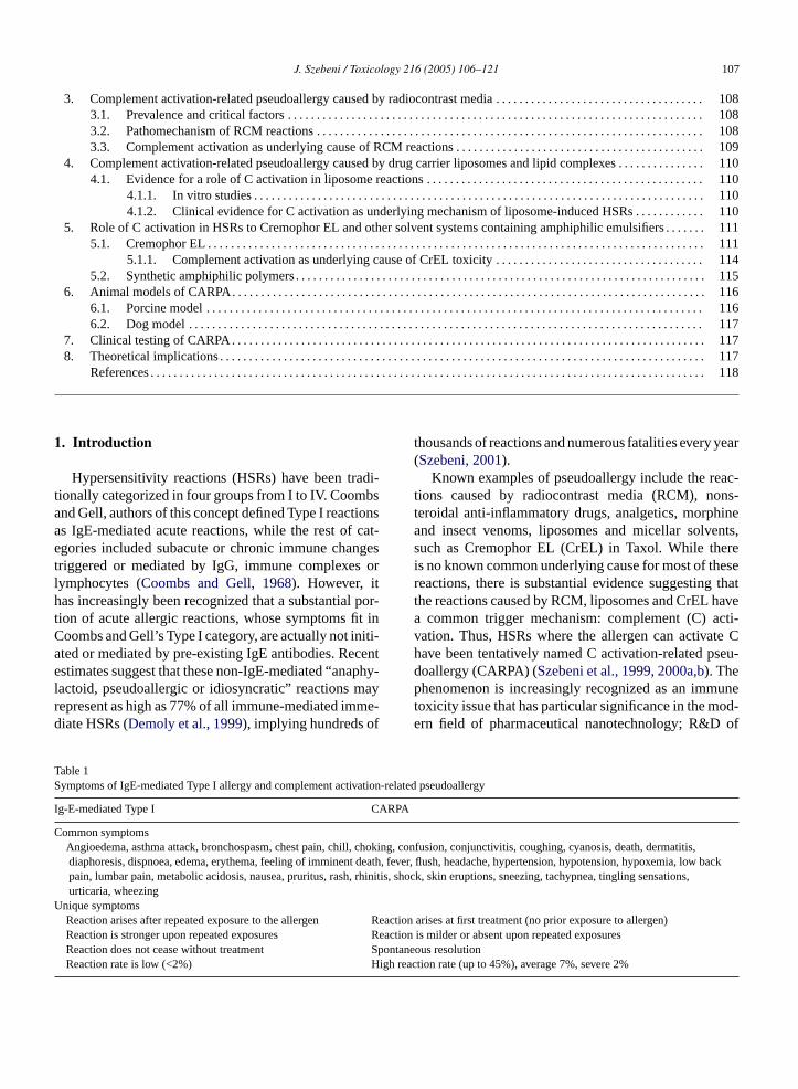
Table 1Symptoms of IgE-mediated Type I allergy and complement activation-related pseudoallergy
Ig-E-mediated Type I CARPA
Common symptomsAngioedema, asthma attack, bronchospasm, chest pain, chill, choking, confusion, conjunctivitis, coughing, cyanosis, death, dermatitis,diaphoresis, dispnoea, edema, erythema, feeling of imminent death, fever, flush, headache, hypertension, hypotension, hypoxemia, low backpain, lumbar pain, metabolic acidosis, nausea, pruritus, rash, rhinitis, shock, skin eruptions, sneezing, tachypnea, tingling sensations,urticaria, wheezing
Unique symptomsReaction arises after repeated exposure to the allergen Reaction arises at first treatment (no prior exposure to allergen)Reaction is stronger upon repeated exposures Reaction is milder or absent upon repeated exposuresReaction does not cease without treatment Spontaneous resolution
igh re
thousands of reactions and numerous fatalities every year(Szebeni, 2001).
Known examples of pseudoallergy include the reac-tions caused by radiocontrast media (RCM), nons-teroidal anti-inflammatory drugs, analgetics, morphineand insect venoms, liposomes and micellar solvents,such as Cremophor EL (CrEL) in Taxol. While thereis no known common underlying cause for most of thesereactions, there is substantial evidence suggesting thatthe reactions caused by RCM, liposomes and CrEL havea common trigger mechanism: complement (C) acti-vation. Thus, HSRs where the allergen can activate Chave been tentatively named C activation-related pseu-doallergy (CARPA) (Szebeni et al., 1999, 2000a,b). Thephenomenon is increasingly recognized as an immunetoxicity issue that has particular significance in the mod-ern field of pharmaceutical nanotechnology; R&D of
Reaction rate is low (<2%) H
action rate (up to 45%), average 7%, severe 2%
108 J. Szebeni / Toxicology 216 (2005) 106–121
particulate drug carriers, synthetic nano and microcap-sules, liposomes and lipid complexes, micellar carriersand emulsifiers, new formulations of radiopharmaceu-ticals and contrast agents, etc. (Hunter and Moghimi,2003; Ten Tije et al., 2003; Storm and Woodle, 2003;Barratt, 2003). This increased awareness of CARPA isalso reflected by the fact that testing for C activation invitro and/or in vivo has become one of the immuno-toxicology tests recommended by the US Food andDrug Adminsitration (FDA) that may be useful to iden-tify the pseudoallergy potential of drugs, when needed(Hastings, 2002).
2. Symptoms of CARPA
As listed in Table 1, many symptoms of CARPAare the same as seen in common allergy or classicaltype I reactions, while others are unique to C activa-tion. Perhaps the most important distinguishing featureof CARPA is the lack of presensitization and reinforce-ment, i.e., the reaction arises at the first exposure to thedrug and then it decreases, rather than increases uponrepeated exposure.
3. Complement activation-related pseudoallergycaused by radiocontrast media
3.1. Prevalence and critical factors
Hypersensitivity reactions to RCM, also referred toe theyel-
vereidearengc-centro-rate
tor,oms
d tose of
te tority,
, thecent
medication and constitutional features of patients. Ingeneral, low-osmolality, nonionic, dimeric or trimericRCM slowly administered to healthy, nonallergic peo-ple carries no, or much less risk for HSR than ionic,high-osmolality, monomeric RCM administed as a bolusto people who are recovering from an infection and/orprone for allergy (Hong et al., 2002; Westhoff-Bleck etal., 1990; Barrett et al., 1991; Katayama et al., 2001;Henry et al., 1991).
3.2. Pathomechanism of RCM reactions
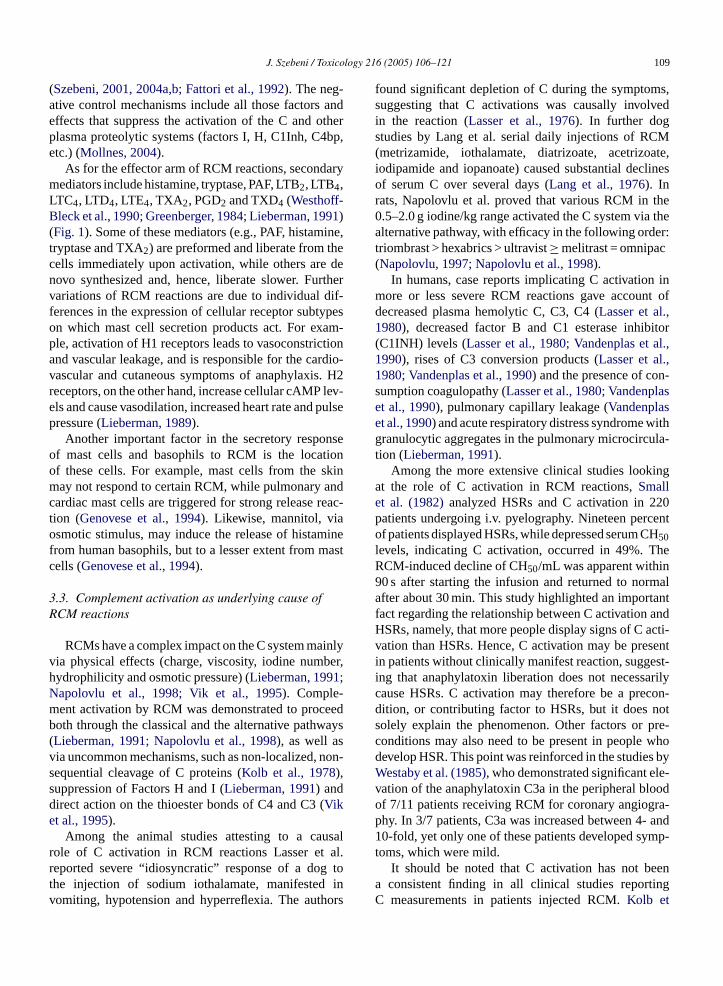
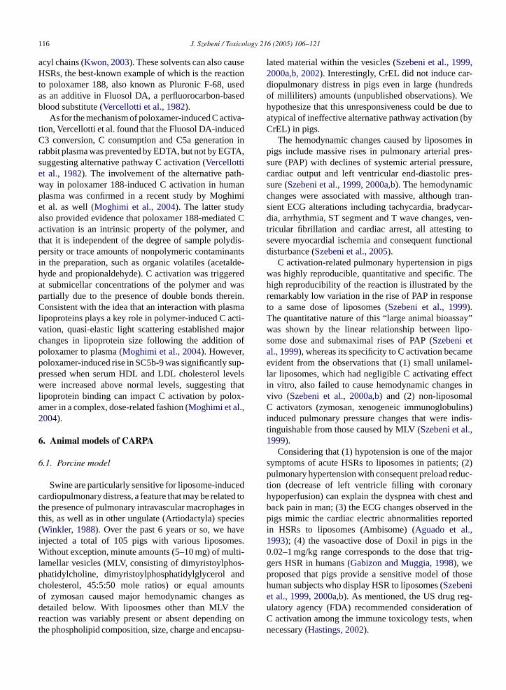
The pathogenesis of RCM reactions is consideredmultifaceted, as beside C activation, several other mech-anisms and controlling factors were shown to play moreor less roles. As illustrated inFig. 1, mast cells andbasophils are in the centre of RCM reactions. They canbe triggered by RCM molecules directly, through poorlyunderstood intracellular interactions and/or extracellularphysical effects (for example, osmotic stress), or indi-rectly, via cell membrane receptors. The latter includethe Fc�R and the anaphylatoxin receptors (C5aR andC3aR), binding IgE, C5a and C3a, respectively. Positivefeedback can be provided by co-activation of the coagu-lation and kinin-kallikrein systems, leading to crossoveractivation of the C cascade with depletion of C1INH
ffects
as “RCM reactions”, have been a concern ever sincfirst organic, iodinated compound was used for i.v. pography in 1928 (Grainger, 2001). Although today, withthe use of new-generation RCM, the frequency of sereactions fell to very low values (see below), the wuse of RCM (in the USA more than 10 million testsperformed yearly (Kumar and Mahalingam, 2001; Hoet al., 2002) still implies a significant number of reations and occassional fatalities. According to a reestimate applied for all kinds of symptoms with all pcedures and all types of RCM, the overall incidenceof RCM reactions is 2.1–12.7% (Hong et al., 2002).The frequency of relatively mild cutaneous, vasomopulmonary, cardiovascular or gastrointestinal symptis in the 5–8% range (Kumar and Mahalingam, 2001),while life-threatening reactions has been estimateoccur in 0.0004–0.002% of patients, at least in the cacoronary angiography (Kumar and Mahalingam, 2001).
Several factors have been identified to contribuor influence these reactions, including the osmolacharge and association of the molecules in RCMspeed of its i.v. administration and the health, re
Fig. 1. Schematic representation of the multiple factors and einvolved in RCM reactions. Taken from (Szebeni, 2004a,b) with per-mission of the publishers.
J. Szebeni / Toxicology 216 (2005) 106–121 109
(Szebeni, 2001, 2004a,b; Fattori et al., 1992). The neg-ative control mechanisms include all those factors andeffects that suppress the activation of the C and otherplasma proteolytic systems (factors I, H, C1Inh, C4bp,etc.) (Mollnes, 2004).
As for the effector arm of RCM reactions, secondarymediators include histamine, tryptase, PAF, LTB2, LTB4,LTC4, LTD4, LTE4, TXA2, PGD2 and TXD4 (Westhoff-Bleck et al., 1990; Greenberger, 1984; Lieberman, 1991)(Fig. 1). Some of these mediators (e.g., PAF, histamine,tryptase and TXA2) are preformed and liberate from thecells immediately upon activation, while others are denovo synthesized and, hence, liberate slower. Furthervariations of RCM reactions are due to individual dif-ferences in the expression of cellular receptor subtypeson which mast cell secretion products act. For exam-ple, activation of H1 receptors leads to vasoconstrictionand vascular leakage, and is responsible for the cardio-vascular and cutaneous symptoms of anaphylaxis. H2receptors, on the other hand, increase cellular cAMP lev-els and cause vasodilation, increased heart rate and pulsepressure (Lieberman, 1989).
Another important factor in the secretory responseof mast cells and basophils to RCM is the locationof these cells. For example, mast cells from the skinmay not respond to certain RCM, while pulmonary andcardiac mast cells are triggered for strong release reac-tion (Genovese et al., 1994). Likewise, mannitol, viaosmotic stimulus, may induce the release of histaminefrom human basophils, but to a lesser extent from mastc
3R
inlyv ber,h ;Nm eedb ays(v non-ssd (e
usalr al.r g tot inv ors
found significant depletion of C during the symptoms,suggesting that C activations was causally involvedin the reaction (Lasser et al., 1976). In further dogstudies by Lang et al. serial daily injections of RCM(metrizamide, iothalamate, diatrizoate, acetrizoate,iodipamide and iopanoate) caused substantial declinesof serum C over several days (Lang et al., 1976). Inrats, Napolovlu et al. proved that various RCM in the0.5–2.0 g iodine/kg range activated the C system via thealternative pathway, with efficacy in the following order:triombrast > hexabrics > ultravist≥ melitrast = omnipac(Napolovlu, 1997; Napolovlu et al., 1998).
In humans, case reports implicating C activation inmore or less severe RCM reactions gave account ofdecreased plasma hemolytic C, C3, C4 (Lasser et al.,1980), decreased factor B and C1 esterase inhibitor(C1INH) levels (Lasser et al., 1980; Vandenplas et al.,1990), rises of C3 conversion products (Lasser et al.,1980; Vandenplas et al., 1990) and the presence of con-sumption coagulopathy (Lasser et al., 1980; Vandenplaset al., 1990), pulmonary capillary leakage (Vandenplaset al., 1990) and acute respiratory distress syndrome withgranulocytic aggregates in the pulmonary microcircula-tion (Lieberman, 1991).
Among the more extensive clinical studies lookingat the role of C activation in RCM reactions,Smallet al. (1982)analyzed HSRs and C activation in 220patients undergoing i.v. pyelography. Nineteen percentof patients displayed HSRs, while depressed serum CH50levels, indicating C activation, occurred in 49%. The
nmaltantand
acti-sentest-arilycon-notpre-whos byle-oodra-- andmp-
eening
ells (Genovese et al., 1994).
.3. Complement activation as underlying cause ofCM reactions
RCMs have a complex impact on the C system maia physical effects (charge, viscosity, iodine numydrophilicity and osmotic pressure) (Lieberman, 1991apolovlu et al., 1998; Vik et al., 1995). Comple-ent activation by RCM was demonstrated to procoth through the classical and the alternative pathwLieberman, 1991; Napolovlu et al., 1998), as well asia uncommon mechanisms, such as non-localized,equential cleavage of C proteins (Kolb et al., 1978),uppression of Factors H and I (Lieberman, 1991) andirect action on the thioester bonds of C4 and C3Vikt al., 1995).
Among the animal studies attesting to a caole of C activation in RCM reactions Lasser eteported severe “idiosyncratic” response of a dohe injection of sodium iothalamate, manifestedomiting, hypotension and hyperreflexia. The auth
RCM-induced decline of CH50/mL was apparent withi90 s after starting the infusion and returned to norafter about 30 min. This study highlighted an imporfact regarding the relationship between C activationHSRs, namely, that more people display signs of Cvation than HSRs. Hence, C activation may be prein patients without clinically manifest reaction, sugging that anaphylatoxin liberation does not necesscause HSRs. C activation may therefore be a predition, or contributing factor to HSRs, but it doessolely explain the phenomenon. Other factors orconditions may also need to be present in peopledevelop HSR. This point was reinforced in the studieWestaby et al. (1985), who demonstrated significant evation of the anaphylatoxin C3a in the peripheral blof 7/11 patients receiving RCM for coronary angiogphy. In 3/7 patients, C3a was increased between 410-fold, yet only one of these patients developed sytoms, which were mild.
It should be noted that C activation has not ba consistent finding in all clinical studies reportC measurements in patients injected RCM.Kolb et
110 J. Szebeni / Toxicology 216 (2005) 106–121
al. (1978), for example, found no significant changesin CH50 and hemolytic C3 activity in serum samplesobtained from 40 patients before and 30 min after under-going i.v. pyelography with methylglucamine diatrizoateor iothalamate.
4. Complement activation-related pseudoallergycaused by drug carrier liposomes and lipidcomplexes
Liposomes or other types of phospholipid assem-blies are increasingly used in medicine for targetedor controlled release of various drugs and diagnos-tic agents. At present, more than a dozen liposomaldrugs are in advanced clinical trials, or already used inpatients mainly for anticancer and antifungal applica-tions (Szebeni, 2004a,b). Out of the marketed liposomaldrugs Doxil (Caelyx) (Uziely et al., 1995; Alberts andGarcia, 1997; Dezube, 1996; Gabizon and Martin, 1997;Gabizon and Muggia, 1998; Chanan-Khan et al., 2003),AmBisome (Levine et al., 1991; Laing et al., 1994;Ringden et al., 1994; de Marie, 1996; Schneider et al.,1998), Abelcet (de Marie, 1996), Amphocil (de Marie,1996) and DaunoXome (Cabriales et al., 1998; Eckardtet al., 1994; Fossa et al., 1998; Gill et al., 1995, 1996;Girard et al., 1996; Guaglianone et al., 1994; Money-Kyrle et al., 1993; Richardson et al., 1997) have beenreported to cause HSRs with symptoms correspondingto CARPA (Table 1). The frequency of HSRs to lipo-somal drugs shows large variation between 3 and 45%
,as
erg-meesultand
ple,led
lev-dingctsedb-9dif-by
10 mM EGTA/2.5 mM Mg2+, which distinguishes clas-sical from alternative pathway activation (Szebeni etal., 2000b). The minimum effective C-activating con-centration of Doxil was 0.05–0.10 mg/mL and therewas near linear dose–response relationship up to about0.5 mg/mL. The activation curve reached plateau at doses≥0.6 mg/mL, suggesting saturation of response (Szebeniet al., 2000b). Doxil also caused variable liberation of Bb,a specific marker of alternative pathway activation, pro-viding further evidence for a role of alternative pathwayactivation and/or amplification (Szebeni et al., 2000a,b).
These and other studies from our laboratories(Szebeni et al., 1994, 1996, 1997a,b, 1999, 2000a,b,2002; Szebeni and Alving, 1999) highligted some basicconditions and mechanism of liposomal C activation.Thus, large size, polydispersity, positive or negative sur-face charge and high (>45%) cholesterol content wereall shown to promote, whereas small uniform size andneutrality reduced the proneness of liposomes for C acti-vation. The process may involve both the classical andalternative pathways, with the latter acting either as theonly activation mechanism, or as a positive feedbackmechanism amplifying C activation via the classicalpathway. As for the classical pathway, the presence ofliposome-reactive immunoglobulins represents a pow-erful trigger or enhancer, but their presence is not aprecondition for C activation via this pathway. Directbinding of C1q to the phospholipid bilayer, or to C reac-tive protein-tagged liposomes, can also activate C via theclassical pathway. Thus, C activation by liposomes can
lingidu-tionow.
o-past,ress
tiva-
iesttra-5, ad/C3,oftntsniana-
(Szebeni, 1998, 2001).
4.1. Evidence for a role of C activation in liposomereactions
4.1.1. In vitro studiesSince its discovery in the late sixties (Haxby et al.
1968; Alving et al., 1969), C activation by liposomes hbeen analysed in a great number of studies. The eming picture is very complex, as variations in liposostructure and other experimental conditions can rin fundamental differences in the extent, pathwaykinetics of activation (Szebeni, 2001, 1998).
Focusing on C activation by Doxil, as an examits incubation with 10 different normal human serato significant rises in C terminal complex (SC5b-9)els over PBS control in seven sera, with rises excee100–200% (relative to PBS control) in four subje(Szebeni et al., 2000b). Further experiments showthat in addition to the quantitative variation in SC5response, Doxil-induced C activation also varied inferent individuals in terms of sensitivity to inhibition
involve numerous redundant triggering and controlprocesses whose differential manifestations in indivals may explain, at least in part, the substantial variaof in vivo responses to liposomes, as discussed bel
4.1.2. Clinical evidence for C activation asunderlying mechanism of liposome-induced HSRs
The likely clinical relevance of C activation by lipsomes can be deduced from clinical studies in thetaken together with a recent study dedicated to addthe cause–effect relationship between in vivo C action and clinical reactions to Doxil.
In reviewing the historic evidence, one of the earlclinical studies with liposomal drugs reported that invenous infusion of vesicles containing NSC 25163water-insoluble cytostatic agent, led to increased C3ratios in the plasma of cancer patients (Coune et al.1983). Another, also still indirect proof, is the findingSkubitz et al. (Skubitz and Skubitz, 1998) on transienneutropenia with signs of leukocyte activation in patiewho displayed HSRs to Doxil. As is known, neutropewith leukocyte activation are classical hallmarks of a
J. Szebeni / Toxicology 216 (2005) 106–121 111
phylatoxin action (Cheung et al., 1994; Skroeder et al.,1994a,b).
To the authors’ knowledge the first direct evidence forthe causal relationship between C activation and HSRsto liposomes was provided byBrouwers et al. (2000),who reported 3 severe HSRs out of 9 patients obtaining99mTc-labeled pegylated liposomes for scintigraphicdetection of bowel inflammation (Dams et al., 2000). Inone reactor patient plasma C3, C4 and factor B decreasedby 16–19%, implying major C consumption. The factthat both C4 and factor B were depleted suggests that Cactivation involved both the classical and the alternativepathways.
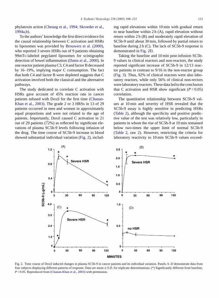
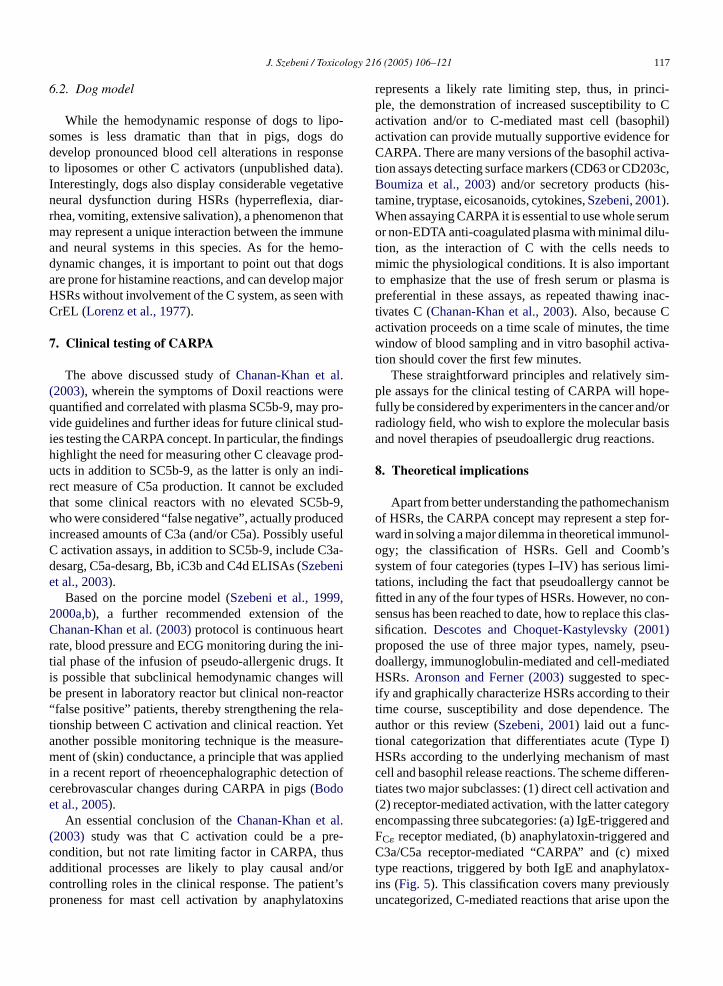
The study dedicated to correlate C activation withHSRs gave account of 45% reaction rate in cancerpatients infused with Doxil for the first time (Chanan-Khan et al., 2003). The grade 2 or 3 HRSs in 13 of 29patients occurred in men and women in approximatelyequal proportions and were not related to the age ofpatients. Importantly, Doxil caused C activation in 21out of 29 patients (72%) as reflected by significant ele-vations of plasma SC5b-9 levels following infusion ofthe drug. The time course of SC5b-9 increase in bloodshowed substantial individual variation (Fig. 2), includ-
ing rapid elevations within 10 min with gradual returnto near baseline within 2 h (A), rapid elevation withoutreturn within 2 h (B) and moderately rapid elevation ofSC5b-9 until about 30 min, followed by partial return tobaseline during 2 h (C). The lack of SC5b-9 response isdemonstrated inFig. 2D.
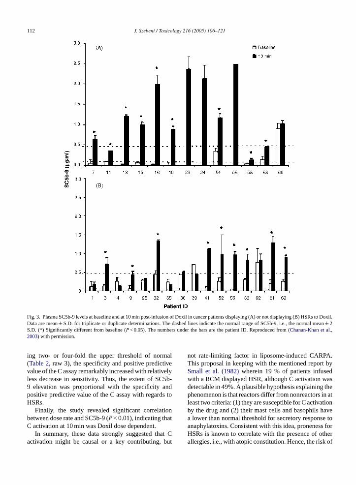
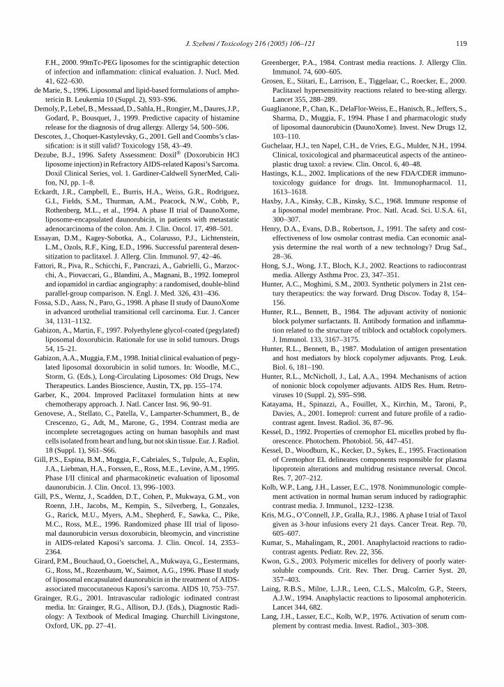
Taking the baseline and 10 min post-infusion SC5b-9 values in clinical reactors and non-reactors, the studyreported significant increase of SC5b-9 in 12/13 reac-tor patients in contrast to 9/16 in the non-reactor group(Fig. 3). Thus, 92% of clinical reactors were also labo-ratory reactors, while only 56% of clinical non-rectorswere laboratory reactors. These data led to the conclusionthat C activation and HSR show significant (P < 0.05)correlation.
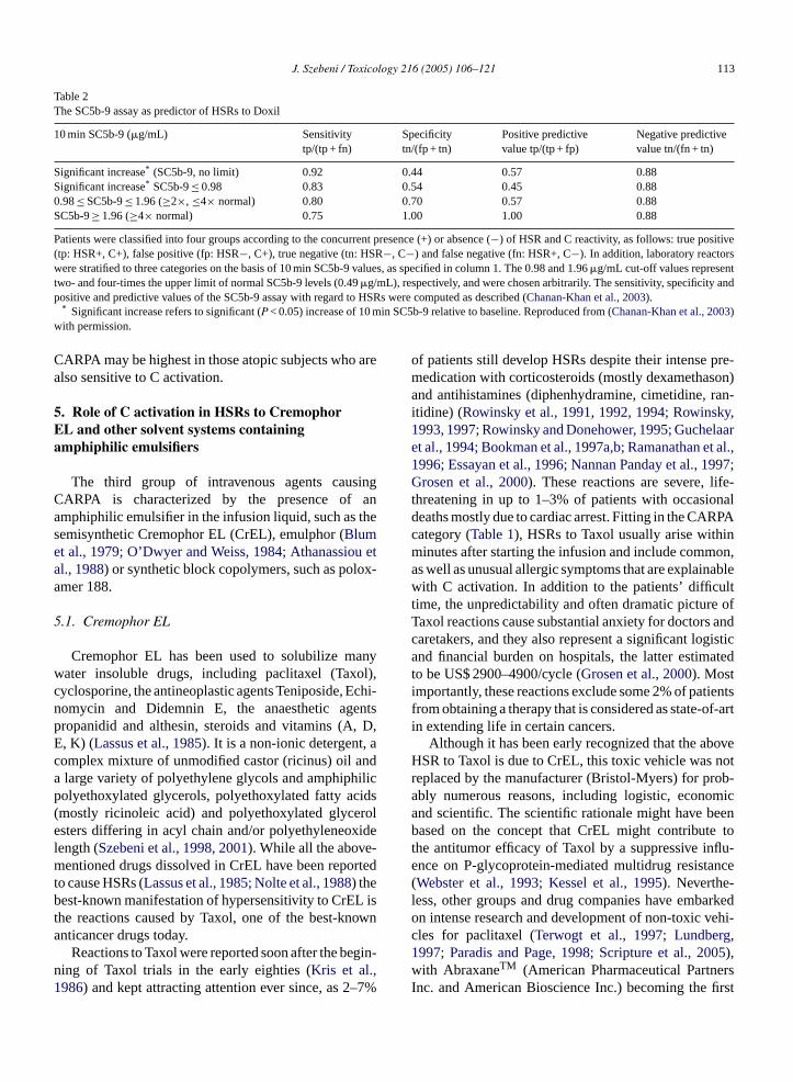
The quantitative relationship between SC5b-9 val-ues at 10 min and severity of HSR revealed that theSC5b-9 assay is highly sensitive in predicting HSRs(Table 2), although the specificity and positive predic-tive value of the test was relatively low, particularly inpatients in whom the rise of SC5b-9 at 10 min remainedbelow two-times the upper limit of normal SC5b-9(Table 2, raw 2). However, restricting the criteria forlaboratory reactivity to 10 min SC5b-9 values exceed-
F in can e data fromf mean± S.D. ine,P on.
ig. 2. Time course of Doxil induced changes in plasma SC5b-9our subjects displaying different patterns of response. Data are< 0.05. Reproduced from (Chanan-Khan et al., 2003) with permissi
cer patients and its individual variation. Panels A–D demonstratfor triplicate determinations. (*) Significantly different from basel
112 J. Szebeni / Toxicology 216 (2005) 106–121
Fig. 3. Plasma SC5b-9 levels at baseline and at 10 min post-infusion of Doxil in cancer patients displaying (A) or not displaying (B) HSRs to Doxil.Data are mean± S.D. for triplicate or duplicate determinations. The dashed lines indicate the normal range of SC5b-9, i.e., the normal mean± 2S.D. (*) Significantly different from baseline (P < 0.05). The numbers under the bars are the patient ID. Reproduced from (Chanan-Khan et al.,2003) with permission.
ing two- or four-fold the upper threshold of normal(Table 2, raw 3), the specificity and positive predictivevalue of the C assay remarkably increased with relativelyless decrease in sensitivity. Thus, the extent of SC5b-9 elevation was proportional with the specificity andpositive predictive value of the C assay with regards toHSRs.
Finally, the study revealed significant correlationbetween dose rate and SC5b-9 (P < 0.01), indicating thatC activation at 10 min was Doxil dose dependent.
In summary, these data strongly suggested that Cactivation might be causal or a key contributing, but
not rate-limiting factor in liposome-induced CARPA.This proposal in keeping with the mentioned report bySmall et al. (1982)wherein 19 % of patients infusedwith a RCM displayed HSR, although C activation wasdetectable in 49%. A plausible hypothesis explaining thephenomenon is that reactors differ from nonreactors in atleast two criteria: (1) they are susceptible for C activationby the drug and (2) their mast cells and basophils havea lower than normal threshold for secretory response toanaphylatoxins. Consistent with this idea, proneness forHSRs is known to correlate with the presence of otherallergies, i.e., with atopic constitution. Hence, the risk of
J. Szebeni / Toxicology 216 (2005) 106–121 113
Table 2The SC5b-9 assay as predictor of HSRs to Doxil
10 min SC5b-9 (�g/mL) Sensitivitytp/(tp + fn)
Specificitytn/(fp + tn)
Positive predictivevalue tp/(tp + fp)
Negative predictivevalue tn/(fn + tn)
Significant increase* (SC5b-9, no limit) 0.92 0.44 0.57 0.88Significant increase* SC5b-9≤ 0.98 0.83 0.54 0.45 0.880.98≤ SC5b-9≤ 1.96 (≥2×, ≤4× normal) 0.80 0.70 0.57 0.88SC5b-9≥ 1.96 (≥4× normal) 0.75 1.00 1.00 0.88
Patients were classified into four groups according to the concurrent presence (+) or absence (−) of HSR and C reactivity, as follows: true positive(tp: HSR+, C+), false positive (fp: HSR−, C+), true negative (tn: HSR−, C−) and false negative (fn: HSR+, C−). In addition, laboratory reactorswere stratified to three categories on the basis of 10 min SC5b-9 values, as specified in column 1. The 0.98 and 1.96�g/mL cut-off values representtwo- and four-times the upper limit of normal SC5b-9 levels (0.49�g/mL), respectively, and were chosen arbitrarily. The sensitivity, specificity andpositive and predictive values of the SC5b-9 assay with regard to HSRs were computed as described (Chanan-Khan et al., 2003).
* Significant increase refers to significant (P < 0.05) increase of 10 min SC5b-9 relative to baseline. Reproduced from (Chanan-Khan et al., 2003)with permission.
CARPA may be highest in those atopic subjects who arealso sensitive to C activation.
5. Role of C activation in HSRs to CremophorEL and other solvent systems containingamphiphilic emulsifiers
The third group of intravenous agents causingCARPA is characterized by the presence of anamphiphilic emulsifier in the infusion liquid, such as thesemisynthetic Cremophor EL (CrEL), emulphor (Blumet al., 1979; O’Dwyer and Weiss, 1984; Athanassiou etal., 1988) or synthetic block copolymers, such as polox-amer 188.
5.1. Cremophor EL
Cremophor EL has been used to solubilize manywater insoluble drugs, including paclitaxel (Taxol),cyclosporine, the antineoplastic agents Teniposide, Echi-nomycin and Didemnin E, the anaesthetic agentspropanidid and althesin, steroids and vitamins (A, D,E, K) (Lassus et al., 1985). It is a non-ionic detergent, acomplex mixture of unmodified castor (ricinus) oil anda large variety of polyethylene glycols and amphiphilicpolyethoxylated glycerols, polyethoxylated fatty acids(mostly ricinoleic acid) and polyethoxylated glycerolesters differing in acyl chain and/or polyethyleneoxidelength (Szebeni et al., 1998, 2001). While all the above-mentioned drugs dissolved in CrEL have been reportedtb L ist owna
gin-n1 –7%
of patients still develop HSRs despite their intense pre-medication with corticosteroids (mostly dexamethason)and antihistamines (diphenhydramine, cimetidine, ran-itidine) (Rowinsky et al., 1991, 1992, 1994; Rowinsky,1993, 1997; Rowinsky and Donehower, 1995; Guchelaaret al., 1994; Bookman et al., 1997a,b; Ramanathan et al.,1996; Essayan et al., 1996; Nannan Panday et al., 1997;Grosen et al., 2000). These reactions are severe, life-threatening in up to 1–3% of patients with occasionaldeaths mostly due to cardiac arrest. Fitting in the CARPAcategory (Table 1), HSRs to Taxol usually arise withinminutes after starting the infusion and include common,as well as unusual allergic symptoms that are explainablewith C activation. In addition to the patients’ difficulttime, the unpredictability and often dramatic picture ofTaxol reactions cause substantial anxiety for doctors andcaretakers, and they also represent a significant logisticand financial burden on hospitals, the latter estimatedto be US$ 2900–4900/cycle (Grosen et al., 2000). Mostimportantly, these reactions exclude some 2% of patientsfrom obtaining a therapy that is considered as state-of-artin extending life in certain cancers.
Although it has been early recognized that the aboveHSR to Taxol is due to CrEL, this toxic vehicle was notreplaced by the manufacturer (Bristol-Myers) for prob-ably numerous reasons, including logistic, economicand scientific. The scientific rationale might have beenbased on the concept that CrEL might contribute tothe antitumor efficacy of Taxol by a suppressive influ-ence on P-glycoprotein-mediated multidrug resistance
-arkedvehi-g,005ersfirst
o cause HSRs (Lassus et al., 1985; Nolte et al., 1988) theest-known manifestation of hypersensitivity to CrE
he reactions caused by Taxol, one of the best-knnticancer drugs today.
Reactions to Taxol were reported soon after the being of Taxol trials in the early eighties (Kris et al.,986) and kept attracting attention ever since, as 2
(Webster et al., 1993; Kessel et al., 1995). Nevertheless, other groups and drug companies have embon intense research and development of non-toxiccles for paclitaxel (Terwogt et al., 1997; Lundber1997; Paradis and Page, 1998; Scripture et al., 2),with AbraxaneTM (American Pharmaceutical PartnInc. and American Bioscience Inc.) becoming the
114 J. Szebeni / Toxicology 216 (2005) 106–121
CrEL-free formulation approved by the FDA for thetreatment of metastatic breast cancer. AbraxaneTM, analbumin nanoparticle-based paclitaxel formulation, wasreported to cause less HSRs compared to traditionalTaxol, despite the fact that it was given to patients with-out steroid and antihistamine premedication, at 50%higher dose and shorter infusion time (Garber, 2004;Sparreboom et al., 2005).
5.1.1. Complement activation as underlying causeof CrEL toxicity
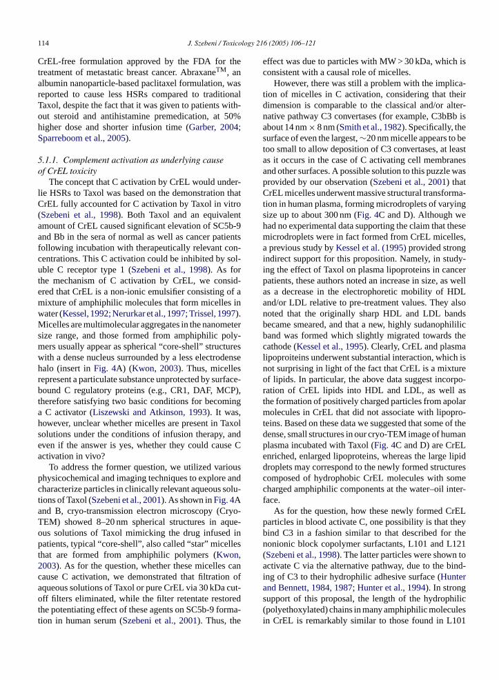
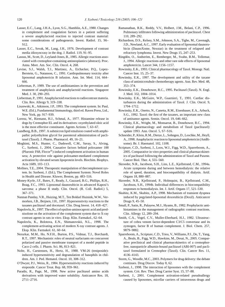
The concept that C activation by CrEL would under-lie HSRs to Taxol was based on the demonstration thatCrEL fully accounted for C activation by Taxol in vitro(Szebeni et al., 1998). Both Taxol and an equivalentamount of CrEL caused significant elevation of SC5b-9and Bb in the sera of normal as well as cancer patientsfollowing incubation with therapeutically relevant con-centrations. This C activation could be inhibited by sol-uble C receptor type 1 (Szebeni et al., 1998). As forthe mechanism of C activation by CrEL, we consid-ered that CrEL is a non-ionic emulsifier consisting of amixture of amphiphilic molecules that form micelles inwater (Kessel, 1992; Nerurkar et al., 1997; Trissel, 1997).Micelles are multimolecular aggregates in the nanometersize range, and those formed from amphiphilic poly-mers usually appear as spherical “core-shell” structureswith a dense nucleus surrounded by a less electrodensehalo (insert inFig. 4A) (Kwon, 2003). Thus, micellesrepresent a particulate substance unprotected by surface-
P),ing
axolandse C
iousandolu-
ryo-que-
inlles
cann ofcut-redrma-
effect was due to particles with MW > 30 kDa, which isconsistent with a causal role of micelles.
However, there was still a problem with the implica-tion of micelles in C activation, considering that theirdimension is comparable to the classical and/or alter-native pathway C3 convertases (for example, C3bBb isabout 14 nm× 8 nm (Smith et al., 1982). Specifically, thesurface of even the largest,∼20 nm micelle appears to betoo small to allow deposition of C3 convertases, at leastas it occurs in the case of C activating cell membranesand other surfaces. A possible solution to this puzzle wasprovided by our observation (Szebeni et al., 2001) thatCrEL micelles underwent massive structural transforma-tion in human plasma, forming microdroplets of varyingsize up to about 300 nm (Fig. 4C and D). Although wehad no experimental data supporting the claim that thesemicrodroplets were in fact formed from CrEL micelles,a previous study byKessel et al. (1995)provided strongindirect support for this proposition. Namely, in study-ing the effect of Taxol on plasma lipoproteins in cancerpatients, these authors noted an increase in size, as wellas a decrease in the electrophoretic mobility of HDLand/or LDL relative to pre-treatment values. They alsonoted that the originally sharp HDL and LDL bandsbecame smeared, and that a new, highly sudanophililicband was formed which slightly migrated towards thecathode (Kessel et al., 1995). Clearly, CrEL and plasmalipoproiteins underwent substantial interaction, which isnot surprising in light of the fact that CrEL is a mixtureof lipids. In particular, the above data suggest incorpo-
slarro-of thean
lipiduresmeter-
ELheythe21toind-
ilicles
01
bound C regulatory proteins (e.g., CR1, DAF, MCtherefore satisfying two basic conditions for becoma C activator (Liszewski and Atkinson, 1993). It was,however, unclear whether micelles are present in Tsolutions under the conditions of infusion therapy,even if the answer is yes, whether they could cauactivation in vivo?
To address the former question, we utilized varphysicochemical and imaging techniques to explorecharacterize particles in clinically relevant aqueous stions of Taxol (Szebeni et al., 2001). As shown inFig. 4Aand B, cryo-transmission electron microscopy (CTEM) showed 8–20 nm spherical structures in aous solutions of Taxol mimicking the drug infusedpatients, typical “core-shell”, also called “star” micethat are formed from amphiphilic polymers (Kwon,2003). As for the question, whether these micellescause C activation, we demonstrated that filtratioaqueous solutions of Taxol or pure CrEL via 30 kDaoff filters eliminated, while the filter retentate restothe potentiating effect of these agents on SC5b-9 fotion in human serum (Szebeni et al., 2001). Thus, the
ration of CrEL lipids into HDL and LDL, as well athe formation of positively charged particles from apomolecules in CrEL that did not associate with lipopteins. Based on these data we suggested that somedense, small structures in our cryo-TEM image of humplasma incubated with Taxol (Fig. 4C and D) are CrELenriched, enlarged lipoproteins, whereas the largedroplets may correspond to the newly formed structcomposed of hydrophobic CrEL molecules with socharged amphiphilic components at the water–oil inface.
As for the question, how these newly formed Crparticles in blood activate C, one possibility is that tbind C3 in a fashion similar to that described fornonionic block copolymer surfactants, L101 and L1(Szebeni et al., 1998). The latter particles were shownactivate C via the alternative pathway, due to the bing of C3 to their hydrophilic adhesive surface (Hunterand Bennett, 1984, 1987; Hunter et al., 1994). In strongsupport of this proposal, the length of the hydroph(polyethoxylated) chains in many amphiphilic molecuin CrEL is remarkably similar to those found in L1

J. Szebeni / Toxicology 216 (2005) 106–121 115
Fig. 4. Cryo-TEM images of vitrified specimens of Cremophor EL in saline (PBS) and in human serum. (A) Taxol vial-equivalent CrEL/ethanolstock solution was diluted 10-fold in PBS. The dark spots represent “star” micelles, schematically depicted in the insert. (B) Larger amplificationof CrEL micelles. (C) Vitrified specimens of a normal human serum, demonstrating some lipoprotein particles in the chylomicron size range. (D)CrEL was incubated with the same serum for 10 min at 37◦C, leading to the formation of numerous particles of varying size. Reproduced from(Szebeni et al., 2001) with permission.
(Szebeni et al., 1998). Furthermore, positively chargedparticles (liposomes) were reported to induce C activa-tion via the alternative pathway (Chonn et al., 1991).
5.2. Synthetic amphiphilic polymers
Just as the semisynthetic emulsifier molecules dis-cussed above, synthetic amphiphilic polymers have also
been used as solvent systems for water insoluble drugs.Some of these polymers have also been used as vaccineadjuvants or as pharmacokinetics-modifier drug con-jugates. Examples for synthetic amphiphilic polymersinclude poloxamers, poloxamines, other copolymersof hydrophilic and hydrophobic blocks, such as poly-oxyethylene, polyoxypropylene and polyethylenegly-cols (PEG) attached to phospholipids or to low molecular
116 J. Szebeni / Toxicology 216 (2005) 106–121
acyl chains (Kwon, 2003). These solvents can also causeHSRs, the best-known example of which is the reactionto poloxamer 188, also known as Pluronic F-68, usedas an additive in Fluosol DA, a perfluorocarbon-basedblood substitute (Vercellotti et al., 1982).
As for the mechanism of poloxamer-induced C activa-tion, Vercellotti et al. found that the Fluosol DA-inducedC3 conversion, C consumption and C5a generation inrabbit plasma was prevented by EDTA, but not by EGTA,suggesting alternative pathway C activation (Vercellottiet al., 1982). The involvement of the alternative path-way in poloxamer 188-induced C activation in humanplasma was confirmed in a recent study by Moghimiet al. as well (Moghimi et al., 2004). The latter studyalso provided evidence that poloxamer 188-mediated Cactivation is an intrinsic property of the polymer, andthat it is independent of the degree of sample polydis-persity or trace amounts of nonpolymeric contaminantsin the preparation, such as organic volatiles (acetalde-hyde and propionaldehyde). C activation was triggeredat submicellar concentrations of the polymer and waspartially due to the presence of double bonds therein.Consistent with the idea that an interaction with plasmalipoproteins plays a key role in polymer-induced C acti-vation, quasi-elastic light scattering established majorchanges in lipoprotein size following the addition ofpoloxamer to plasma (Moghimi et al., 2004). However,poloxamer-induced rise in SC5b-9 was significantly sup-pressed when serum HDL and LDL cholesterol levelswere increased above normal levels, suggesting that
ox-,
cedted toes inciesavees.lti-s-nduntss as
theg onpsu-
lated material within the vesicles (Szebeni et al., 1999,2000a,b, 2002). Interestingly, CrEL did not induce car-diopulmonary distress in pigs even in large (hundredsof milliliters) amounts (unpublished observations). Wehypothesize that this unresponsiveness could be due toatypical of ineffective alternative pathway activation (byCrEL) in pigs.
The hemodynamic changes caused by liposomes inpigs include massive rises in pulmonary arterial pres-sure (PAP) with declines of systemic arterial pressure,cardiac output and left ventricular end-diastolic pres-sure (Szebeni et al., 1999, 2000a,b). The hemodynamicchanges were associated with massive, although tran-sient ECG alterations including tachycardia, bradycar-dia, arrhythmia, ST segment and T wave changes, ven-tricular fibrillation and cardiac arrest, all attesting tosevere myocardial ischemia and consequent functionaldisturbance (Szebeni et al., 2005).
C activation-related pulmonary hypertension in pigswas highly reproducible, quantitative and specific. Thehigh reproducibility of the reaction is illustrated by theremarkably low variation in the rise of PAP in responseto a same dose of liposomes (Szebeni et al., 1999).The quantitative nature of this “large animal bioassay”was shown by the linear relationship between lipo-some dose and submaximal rises of PAP (Szebeni etal., 1999), whereas its specificity to C activation becameevident from the observations that (1) small unilamel-lar liposomes, which had negligible C activating effectin vitro, also failed to cause hemodynamic changes in
llins)ndis-.,
ajor; (2)duc-ryandthe
rted,hetrig-
oseig-n ofhen
lipoprotein binding can impact C activation by polamer in a complex, dose-related fashion (Moghimi et al.2004).
6. Animal models of CARPA
6.1. Porcine model
Swine are particularly sensitive for liposome-inducardiopulmonary distress, a feature that may be relathe presence of pulmonary intravascular macrophagthis, as well as in other ungulate (Artiodactyla) spe(Winkler, 1988). Over the past 6 years or so, we hinjected a total of 105 pigs with various liposomWithout exception, minute amounts (5–10 mg) of mulamellar vesicles (MLV, consisting of dimyristoylphophatidylcholine, dimyristoylphosphatidylglycerol acholesterol, 45:5:50 mole ratios) or equal amoof zymosan caused major hemodynamic changedetailed below. With lipoosmes other than MLVreaction was variably present or absent dependinthe phospholipid composition, size, charge and enca
vivo (Szebeni et al., 2000a,b) and (2) non-liposomaC activators (zymosan, xenogeneic immunoglobuinduced pulmonary pressure changes that were itinguishable from those caused by MLV (Szebeni et al1999).
Considering that (1) hypotension is one of the msymptoms of acute HSRs to liposomes in patientspulmonary hypertension with consequent preload retion (decrease of left ventricle filling with coronahypoperfusion) can explain the dyspnea with chestback pain in man; (3) the ECG changes observed inpigs mimic the cardiac electric abnormalities repoin HSRs to liposomes (Ambisome) (Aguado et al.1993); (4) the vasoactive dose of Doxil in pigs in t0.02–1 mg/kg range corresponds to the dose thatgers HSR in humans (Gabizon and Muggia, 1998), weproposed that pigs provide a sensitive model of thhuman subjects who display HSR to liposomes (Szebenet al., 1999, 2000a,b). As mentioned, the US drug reulatory agency (FDA) recommended consideratioC activation among the immune toxicology tests, wnecessary (Hastings, 2002).
J. Szebeni / Toxicology 216 (2005) 106–121 117
6.2. Dog model
While the hemodynamic response of dogs to lipo-somes is less dramatic than that in pigs, dogs dodevelop pronounced blood cell alterations in responseto liposomes or other C activators (unpublished data).Interestingly, dogs also display considerable vegetativeneural dysfunction during HSRs (hyperreflexia, diar-rhea, vomiting, extensive salivation), a phenomenon thatmay represent a unique interaction between the immuneand neural systems in this species. As for the hemo-dynamic changes, it is important to point out that dogsare prone for histamine reactions, and can develop majorHSRs without involvement of the C system, as seen withCrEL (Lorenz et al., 1977).
7. Clinical testing of CARPA
The above discussed study ofChanan-Khan et al.(2003), wherein the symptoms of Doxil reactions werequantified and correlated with plasma SC5b-9, may pro-vide guidelines and further ideas for future clinical stud-ies testing the CARPA concept. In particular, the findingshighlight the need for measuring other C cleavage prod-ucts in addition to SC5b-9, as the latter is only an indi-rect measure of C5a production. It cannot be excludedthat some clinical reactors with no elevated SC5b-9,who were considered “false negative”, actually producedincreased amounts of C3a (and/or C5a). Possibly usefulC activation assays, in addition to SC5b-9, include C3a-d ie
9,2 theC rtr ini-t s. Iti willb ctor“ ela-t Yeta ure-m liedi n ofce
l.( re-c usa d/orc nt’sp xins
represents a likely rate limiting step, thus, in princi-ple, the demonstration of increased susceptibility to Cactivation and/or to C-mediated mast cell (basophil)activation can provide mutually supportive evidence forCARPA. There are many versions of the basophil activa-tion assays detecting surface markers (CD63 or CD203c,Boumiza et al., 2003) and/or secretory products (his-tamine, tryptase, eicosanoids, cytokines,Szebeni, 2001).When assaying CARPA it is essential to use whole serumor non-EDTA anti-coagulated plasma with minimal dilu-tion, as the interaction of C with the cells needs tomimic the physiological conditions. It is also importantto emphasize that the use of fresh serum or plasma ispreferential in these assays, as repeated thawing inac-tivates C (Chanan-Khan et al., 2003). Also, because Cactivation proceeds on a time scale of minutes, the timewindow of blood sampling and in vitro basophil activa-tion should cover the first few minutes.
These straightforward principles and relatively sim-ple assays for the clinical testing of CARPA will hope-fully be considered by experimenters in the cancer and/orradiology field, who wish to explore the molecular basisand novel therapies of pseudoallergic drug reactions.
8. Theoretical implications
Apart from better understanding the pathomechanismof HSRs, the CARPA concept may represent a step for-ward in solving a major dilemma in theoretical immunol-ogy; the classification of HSRs. Gell and Coomb’s
imi-t beon-s clas-01)seu-tedc-heir. The-e I)ast
eren-and
goryd andand
xedtox-slyn the
esarg, C5a-desarg, Bb, iC3b and C4d ELISAs (Szebent al., 2003).
Based on the porcine model (Szebeni et al., 199000a,b), a further recommended extension ofhanan-Khan et al. (2003)protocol is continuous hea
ate, blood pressure and ECG monitoring during theial phase of the infusion of pseudo-allergenic drugs possible that subclinical hemodynamic changese present in laboratory reactor but clinical non-reafalse positive” patients, thereby strengthening the rionship between C activation and clinical reaction.nother possible monitoring technique is the measent of (skin) conductance, a principle that was app
n a recent report of rheoencephalographic detectioerebrovascular changes during CARPA in pigs (Bodot al., 2005).
An essential conclusion of theChanan-Khan et a2003) study was that C activation could be a pondition, but not rate limiting factor in CARPA, thdditional processes are likely to play causal anontrolling roles in the clinical response. The patieroneness for mast cell activation by anaphylato
system of four categories (types I–IV) has serious ltations, including the fact that pseudoallergy cannofitted in any of the four types of HSRs. However, no csensus has been reached to date, how to replace thisification. Descotes and Choquet-Kastylevsky (20proposed the use of three major types, namely, pdoallergy, immunoglobulin-mediated and cell-mediaHSRs.Aronson and Ferner (2003)suggested to speify and graphically characterize HSRs according to ttime course, susceptibility and dose dependenceauthor or this review (Szebeni, 2001) laid out a functional categorization that differentiates acute (TypHSRs according to the underlying mechanism of mcell and basophil release reactions. The scheme difftiates two major subclasses: (1) direct cell activation(2) receptor-mediated activation, with the latter cateencompassing three subcategories: (a) IgE-triggereFC� receptor mediated, (b) anaphylatoxin-triggeredC3a/C5a receptor-mediated “CARPA” and (c) mitype reactions, triggered by both IgE and anaphylains (Fig. 5). This classification covers many previouuncategorized, C-mediated reactions that arise upo

118 J. Szebeni / Toxicology 216 (2005) 106–121
Fig. 5. Proposed new scheme of hypersensitivity reactions with Revision of the Type 1 category. Partially reproduced from (Szebeni et al., 2002).
use of extracorporeal circuits, RCM and various liposo-mal and micellar carriers of intravenous drugs.
References
Aguado, J.M., Hidalgo, M., Moya, I., Alcazar, J.M., Jimenez, M.J.,Noriega, A.R., 1993. Ventricular arrhythmias with conventionaland liposomal amphotericin. Lancet 342 (8881), 1239.
Alberts, D.S., Garcia, D.J., 1997. Safety aspects of pegylated liposomaldoxorubicin in patients with cancer. Drugs 54 (Suppl. 4), 30–45.
Alving, C.R., Kinsky, S.C., Haxby, J.A., Kinsky, C.B., 1969. Antibodybinding and complement fixation by a liposomal model membrane.Biochemistry 8, 1582–1587.
Aronson, J.K., Ferner, R.E., 2003. Joining the DoTS: new approach toclassifying adverse drug reactions. Br. Med. J. 327, 1222–1225.
Athanassiou, A.E., Bafaloukos, D., Pectasidis, D., Dimitriadis, M.,1988. Acute vasomotor response—a reaction to etoposide. J. Clin.Oncol. 6, 1204–1205.
Barratt, G., 2003. Colloidal drug carriers: achievements and perspec-tives. Cell. Mol. Life Sci. 60, 21–37.
Barrett, B.J., Parfrey, P.S., Vavasour, H.M., O’Dea, F., Kent, G., Stone,E., 1991. A comparison of nonionic, low-osmolality radiocontrastagents with ionic, high-osmolality agents during cardiac catheter-ization. Med. J. Aust. 154, 766–772.
Blum, R.H., Garnick, M.B., Israel, M., Canellos, G.P., Hender-son, I.C., Frei III, E., 1979. Initial clinical evaluation ofN-trifluoroacetyladriamycin-14-valerate (AD-32), an adriamycinanalog. Cancer Treat. Rep. 63, 919–928.
Bodo, M., Szebeni, J., Baranyi, J., Savay, S., Pearce, F.J., Alving,C.R., Bunger, R., 2005. Cerebrovascular involvement in liposome-induced cardiopulmonary distress in pigs. J. Liposome Res. 15,3–14.
Bookman, M.A., Kloth, D.D., Kover, P., Smolinski, S., Ozols, R.F.,lated
Bookman, M.A., Kloth, D.D., Kover, P.E., Smolinski, S., Ozols, R.F.,1997b. Intravenous prophylaxis for paclitaxel-related hypersensi-tivity reactions. Semin. Oncol. 24 (Suppl. 19), S1913–S1915.
Boumiza, R., Monneret, G., Forissier, M.F., Savoye, J., Gutowski,M.C., Powell, W.S., Bienvenu, J., 2003. Marked improvement ofthe basophil activation test by detecting CD203c instead of CD63.Clin. Exp. Allergy 33, 259–265.
Brouwers, A.H., De Jong, D.J., Dams, E.T., Oyen, W.J., Boerman,O.C., Laverman, P., Naber, T.H., Storm, G., Corstens, F.H., 2000.Tc-99m-PEG-Liposomes for the evaluation of colitis in Crohn’sdisease. J. Drug Target 8, 225–233.
Cabriales, S., Bresnahan, J., Testa, D., Espina, B.M., Scadden, D.T.,Ross, M., Gill, P.S., 1998. Extravasation of liposomal daunorubicinin patients with AIDS-associated Kaposi’s sarcoma: a report of fourcases. Oncol. Nurs. Forum 25, 67–70.
Chanan-Khan, A., Szebeni, J., Savay, S., Liebes, L., Rafique, N.M.,Alving, C.R., Muggia, F.M., 2003. Complement activation follow-ing first exposure to pegylated liposomal doxorubicin (Doxil): pos-sible role in hypersensitivity reactions. Ann. Oncol. 14, 1430–1437.
Cheung, A.K., Parker, C.J., Hohnholt, M., 1994. Soluble comple-ment receptor type 1 inhibits complement activation induced byhemodialysis membranes in vitro. Kidney Int. 46, 1680–1687.
Chonn, A., Cullis, P.R., Devine, D.V., 1991. The role of surface chargein the activation of the classical and alternative pathways of com-plement by liposomes. J. Immunol. 146, 4234–4241.
Coombs, R.R.A., Gell, P.G.H., 1968. Classification of allergic reac-tions responsible for drug hypersensitivity reactions. In: Coombs,R.R.A., Gell, P.G.H. (Eds.), Clinical Aspects of Immunology, sec-ond ed. Davis, Philadelphia, PA, pp. 575–596.
Coune, A., Sculier, J.P., Fruhling, J., Stryckmans, P., Brassine, C.,Ghanem, G., Laduron, C., Atassi, G., Ruysschaert, J.M., Hilde-brand, J., 1983. Iv administration of a water-insoluble antimitoticcompound entrapped in liposomes. Preliminary report on infu-sion of large volumes of liposomes to man. Cancer Treat. Rep.67, 1031–1033.
n, P.,ens,
1997a. Short-course intravenous prophylaxis for paclitaxel-rehypersensitivity reactions. Ann. Oncol. 8, 611–614.
Dams, E.T., Oyen, W.J., Boerman, O.C., Storm, G., LavermaKok, P.J., Buijs, W.C., Bakker, H., van der Meer, J.W., Corst
J. Szebeni / Toxicology 216 (2005) 106–121 119
F.H., 2000. 99mTc-PEG liposomes for the scintigraphic detectionof infection and inflammation: clinical evaluation. J. Nucl. Med.41, 622–630.
de Marie, S., 1996. Liposomal and lipid-based formulations of ampho-tericin B. Leukemia 10 (Suppl. 2), S93–S96.
Demoly, P., Lebel, B., Messaad, D., Sahla, H., Rongier, M., Daures, J.P.,Godard, P., Bousquet, J., 1999. Predictive capacity of histaminerelease for the diagnosis of drug allergy. Allergy 54, 500–506.
Descotes, J., Choquet-Kastylevsky, G., 2001. Gell and Coombs’s clas-sification: is it still valid? Toxicology 158, 43–49.
Dezube, B.J., 1996. Safety Assessment: Doxil® (Doxorubicin HClliposome injection) in Refractory AIDS-related Kaposi’s Sarcoma.Doxil Clinical Series, vol. 1. Gardiner-Caldwell SynerMed, Cali-fon, NJ, pp. 1–8.
Eckardt, J.R., Campbell, E., Burris, H.A., Weiss, G.R., Rodriguez,G.I., Fields, S.M., Thurman, A.M., Peacock, N.W., Cobb, P.,Rothenberg, M.L., et al., 1994. A phase II trial of DaunoXome,liposome-encapsulated daunorubicin, in patients with metastaticadenocarcinoma of the colon. Am. J. Clin. Oncol. 17, 498–501.
Essayan, D.M., Kagey-Sobotka, A., Colarusso, P.J., Lichtenstein,L.M., Ozols, R.F., King, E.D., 1996. Successful parenteral desen-sitization to paclitaxel. J. Allerg. Clin. Immunol. 97, 42–46.
Fattori, R., Piva, R., Schicchi, F., Pancrazi, A., Gabrielli, G., Marzoc-chi, A., Piovaccari, G., Blandini, A., Magnani, B., 1992. Iomeproland iopamidol in cardiac angiography: a randomised, double-blindparallel-group comparison. N. Engl. J. Med. 326, 431–436.
Fossa, S.D., Aass, N., Paro, G., 1998. A phase II study of DaunoXomein advanced urothelial transitional cell carcinoma. Eur. J. Cancer34, 1131–1132.
Gabizon, A., Martin, F., 1997. Polyethylene glycol-coated (pegylated)liposomal doxorubicin. Rationale for use in solid tumours. Drugs54, 15–21.
Gabizon, A.A., Muggia, F.M., 1998. Initial clinical evaluation of pegy-lated liposomal doxorubicin in solid tumors. In: Woodle, M.C.,Storm, G. (Eds.), Long-Circulating Liposomes: Old Drugs, New
G new
G ., dearemast
adiol.
G plin,995.mal
G , vonales,ike,so-tine53–
G ans,studyIDS-–757.
G trastadi-e,
Greenberger, P.A., 1984. Contrast media reactions. J. Allergy Clin.Immunol. 74, 600–605.
Grosen, E., Siitari, E., Larrison, E., Tiggelaar, C., Roecker, E., 2000.Paclitaxel hypersensitivity reactions related to bee-sting allergy.Lancet 355, 288–289.
Guaglianone, P., Chan, K., DelaFlor-Weiss, E., Hanisch, R., Jeffers, S.,Sharma, D., Muggia, F., 1994. Phase I and pharmacologic studyof liposomal daunorubicin (DaunoXome). Invest. New Drugs 12,103–110.
Guchelaar, H.J., ten Napel, C.H., de Vries, E.G., Mulder, N.H., 1994.Clinical, toxicological and pharmaceutical aspects of the antineo-plastic drug taxol: a review. Clin. Oncol. 6, 40–48.
Hastings, K.L., 2002. Implications of the new FDA/CDER immuno-toxicology guidance for drugs. Int. Immunopharmacol. 11,1613–1618.
Haxby, J.A., Kinsky, C.B., Kinsky, S.C., 1968. Immune response ofa liposomal model membrane. Proc. Natl. Acad. Sci. U.S.A. 61,300–307.
Henry, D.A., Evans, D.B., Robertson, J., 1991. The safety and cost-effectiveness of low osmolar contrast media. Can economic anal-ysis determine the real worth of a new technology? Drug Saf.,28–36.
Hong, S.J., Wong, J.T., Bloch, K.J., 2002. Reactions to radiocontrastmedia. Allergy Asthma Proc. 23, 347–351.
Hunter, A.C., Moghimi, S.M., 2003. Synthetic polymers in 21st cen-tury therapeutics: the way forward. Drug Discov. Today 8, 154–156.
Hunter, R.L., Bennett, B., 1984. The adjuvant activity of nonionicblock polymer surfactants. II. Antibody formation and inflamma-tion related to the structure of triblock and octablock copolymers.J. Immunol. 133, 3167–3175.
Hunter, R.L., Bennett, B., 1987. Modulation of antigen presentationand host mediators by block copolymer adjuvants. Prog. Leuk.Biol. 6, 181–190.
Hunter, R.L., McNicholl, J., Lal, A.A., 1994. Mechanisms of actiontro-
P.,dio-
y flu-
ationasmancol.
ple-phic
xolp. 70,
adio-
ter-20,
ers,icin.
om-
Therapeutics. Landes Bioscience, Austin, TX, pp. 155–174.arber, K., 2004. Improved Paclitaxel formulation hints at
chemotherapy approach. J. Natl. Cancer Inst. 96, 90–91.enovese, A., Stellato, C., Patella, V., Lamparter-Schummert, B
Crescenzo, G., Adt, M., Marone, G., 1994. Contrast mediaincomplete secretagogues acting on human basophils andcells isolated from heart and lung, but not skin tissue. Eur. J. R18 (Suppl. 1), S61–S66.
ill, P.S., Espina, B.M., Muggia, F., Cabriales, S., Tulpule, A., EsJ.A., Liebman, H.A., Forssen, E., Ross, M.E., Levine, A.M., 1Phase I/II clinical and pharmacokinetic evaluation of liposodaunorubicin. J. Clin. Oncol. 13, 996–1003.
ill, P.S., Wernz, J., Scadden, D.T., Cohen, P., Mukwaya, G.M.Roenn, J.H., Jacobs, M., Kempin, S., Silverberg, I., GonzG., Rarick, M.U., Myers, A.M., Shepherd, F., Sawka, C., PM.C., Ross, M.E., 1996. Randomized phase III trial of lipomal daunorubicin versus doxorubicin, bleomycin, and vincrisin AIDS-related Kaposi’s sarcoma. J. Clin. Oncol. 14, 232364.
irard, P.M., Bouchaud, O., Goetschel, A., Mukwaya, G., EestermG., Ross, M., Rozenbaum, W., Saimot, A.G., 1996. Phase IIof liposomal encapsulated daunorubicin in the treatment of Aassociated mucocutaneous Kaposi’s sarcoma. AIDS 10, 753
rainger, R.G., 2001. Intravascular radiologic iodinated conmedia. In: Grainger, R.G., Allison, D.J. (Eds.), Diagnostic Rology: A Textbook of Medical Imaging. Churchill LivingstonOxford, UK, pp. 27–41.
of nonionic block copolymer adjuvants. AIDS Res. Hum. Reviruses 10 (Suppl. 2), S95–S98.
Katayama, H., Spinazzi, A., Fouillet, X., Kirchin, M., Taroni,Davies, A., 2001. Iomeprol: current and future profile of a racontrast agent. Invest. Radiol. 36, 87–96.
Kessel, D., 1992. Properties of cremophor EL micelles probed borescence. Photochem. Photobiol. 56, 447–451.
Kessel, D., Woodburn, K., Kecker, D., Sykes, E., 1995. Fractionof Cremophor EL delineates components responsible for pllipoprotein alterations and multidrug resistance reversal. ORes. 7, 207–212.
Kolb, W.P., Lang, J.H., Lasser, E.C., 1978. Nonimmunologic comment activation in normal human serum induced by radiogracontrast media. J. Immunol., 1232–1238.
Kris, M.G., O’Connell, J.P., Gralla, R.J., 1986. A phase I trial of Tagiven as 3-hour infusions every 21 days. Cancer Treat. Re605–607.
Kumar, S., Mahalingam, R., 2001. Anaphylactoid reactions to rcontrast agents. Pediatr. Rev. 22, 356.
Kwon, G.S., 2003. Polymeric micelles for delivery of poorly wasoluble compounds. Crit. Rev. Ther. Drug. Carrier Syst.357–403.
Laing, R.B.S., Milne, L.J.R., Leen, C.L.S., Malcolm, G.P., SteA.J.W., 1994. Anaphylactic reactions to liposomal amphoterLancet 344, 682.
Lang, J.H., Lasser, E.C., Kolb, W.P., 1976. Activation of serum cplement by contrast media. Invest. Radiol., 303–308.
120 J. Szebeni / Toxicology 216 (2005) 106–121
Lasser, E.C., Lang, J.H.A., Lyon, S.G., Hamblin, A.E., 1980. Changesin complement and coagulation factors in a patient sufferinga severe anaphylactoid reaction to injected contrast material:some considerations of pathogenesis. Invest. Radiol. 15, S6–S12.
Lasser, E.C., Sovak, M., Lang, J.H., 1976. Development of contrastmedia idiosyncrasy in the dog. J. Radiol. 119, 91–95.
Lassus, M., Scott, D., Leyland-Jones, B., 1985. Allergic reactions asso-ciated with cremophor-containing antineoplastics (abstract). Proc.Annu. Meet. Am. Soc. Clin. Oncol. 4, 268.
Levine, S.J., Walsh, T.J., Martinez, A., Eichacker, P.Q., Lopez-Berstein, G., Natanson, C., 1991. Cardiopulmonary toxicity afterliposomal amphotericin B infusion. Ann. Int. Med. 114, 664–666.
Lieberman, P., 1989. The use of antihistamines in the prevention andtreatment of anaphylaxis and anaphylactoid reactions. SingaporeMed. J. 30, 290–293.
Lieberman, P., 1991. Anaphylactoid reactions to radiocontrast materia.Clin. Rev. Allergy 9, 319–338.
Liszewski, K., Atkinson, J.P., 1993. The complement system. In: Paul,W.E. (Ed.), Fundamental Immunology, third ed. Raven Press, Ltd.,New York, pp. 917–939.
Lorenz, W., Riemann, H.J., Schmal, A., 1977. Histamine release indogs by Cremophor EL and its derivatives: oxyethylated oleic acidis the most effective constituent. Agents Actions 7, 63–67.
Lundberg, B.B., 1997. A submicron lipid emulsion coated with amphi-pathic polyethylene glycol for parenteral administration of pacli-taxel (Taxol). J. Pharm. Pharmacol. 49, 16–21.
Moghimi, M.S., Hunter, C., Dadswell, C.M., Savay, S., Alving,C., Szebeni, J., 2004. Causative factors behind poloxamer 188(Pluronic F68, Flocor®)-induced complement activation in humansera; A protective role against poloxamer-mediated complementactivation by elevated serum lipoprotein levels. Biochim. Biophys.Acta 1689, 103–113.
Mollnes, T.E., 2004. Therapeutic manipulation of thecomplement sys-oles
R.H.,osi’s) 5,
er-o the427.red--ray
hets. J.
rdt,rrentide in
ide)-chil-
d by
acids18,
Ramanathan, R.K., Reddy, V.V., Holbert, J.M., Belani, C.P., 1996.Pulmonary infiltrates following administration of paclitaxel. Chest110, 289–292.
Richardson, D.S., Kelsey, S.M., Johnson, S.A., Tighe, M., Cavenagh,J.D., Newland, A.C., 1997. Early evaluation of liposomal daunoru-bicin (DaunoXome, Nexstar) in the treatment of relapsed andrefractory lymphoma. Invest. New Drugs 15, 247–253.
Ringden, O., Andstrom, E., Remberger, M., Svahn, B.M., Tollemar,J., 1994. Allergic reactions and other rare side effects of liposomalamphotericin. Lancet 344, 1156–1157.
Rowinsky, E.K., 1993. Clinical pharmacology of Taxol. Monogr. Natl.Cancer Inst. 15, 25–37.
Rowinsky, E.K., 1997. The development and utility of the taxaneclass of antimicrotubule chemotherapy agents. Ann. Rev. Med. 48,353–374.
Rowinsky, E.K., Donehower, R.C., 1995. Paclitaxel (Taxol). N. Engl.J. Med. 332, 1004–1014.
Rowinsky, E.K., McGuire, W.P., Guarnieri, T., 1991. Cardiac dis-turbances during the administration of Taxol. J. Clin. Oncol. 9,1704–1712.
Rowinsky, E.K., Onetto, N., Canetta, R.M., Eisenhauer, E.A., Arbuck,S.G., 1992. Taxol: the first of the taxanes, an important new classof antitumor agents. Semin. Oncol. 19, 646–662.
Rowinsky, E.K., Wright, M., Monsarrat, B., Donehower, R.C., 1994.Clinical pharmacology and metabolism of Taxol (paclitaxel):update 1993. Ann. Oncol. 5, S7–S16.
Schneider, P., Klein, R.M., Dietze, L., Sohngen, D., Leschke, M., Heyll,A., 1998. Anaphylactic reaction to liposomal amphotericin (AmBi-some). Br. J. Haematol. 102, 1108.
Scripture, C.D., Szebeni, J., Loos, W.J., Figg, W.D., Sparreboom, A.,2005. Comparative in vitro properties and clinical pharmacokinet-ics of paclitaxel following the administration of Taxol and Paxene.Cancer Biol. Ther. 4, 555–560.
Skroeder, N.R., Jacobson, S.H., Lins, L.E., Kjellstrand, C.M., 1994a.Acute symptoms during and between hemodialysis: the relative
rtif.
M.,ility
neacer
anti-tions.
uc-nd its257,
ang,ar-
hor-cli-11,
bate
ment
llergyand
tem. In: Szebeni, J. (Ed.), The Complement System: Novel Rin Health and Disease. Kluwer, Boston, pp. 483–516.
Money-Kyrle, J.F., Bates, F., Ready, J., Gazzard, B.G., Phillips,Boag, F.C., 1993. Liposomal daunorubicin in advanced Kapsarcoma: a phase II study. Clin. Oncol. (R. Coll. Radiol.367–371.
Nannan Panday, V.R., Huizing, M.T., Ten Bokkel Huinink, W.W., Vmorken, J.B., Beijnen, J.H., 1997. Hypersensitvity reactions ttaxanes paclitaxel and docetaxel. Clin. Drug Invest. 14, 418–
Napolovlu, K., 1997. The effect of epsilon-aminocaproic acid and pnisolone on the activation of the complement system due to Xcontrast agents in rats in vitro. Eksp. Klin. Farmakol., 62–64.
Napolovlu, K., Bolotova, E.N., Shimanovskii, N.L., 1998. Tcomplement-activating action of modern X-ray contrast agenEksp. Klin. Farmakol. 61, 60–64.
Nerurkar, M.M., Ho, N.F.H., Burton, P.S., Vidmar, T.J., BorchaR.T., 1997. Mechanistic roles of neutral surfactants on concupolarized and passive membrane transport of a model peptCaco-2 cells. J. Pharm. Sci. 86, 813–821.
Nolte, H., Carstensen, H., Hertz, H., 1988. VM-26 (teniposinduced hypersensitivity and degranulation of basophils indren. Am. J. Ped. Hematol. Oncol. 10, 308–312.
O’Dwyer, P.J., Weiss, R., 1984. Hypersensitivity reactions induceetoposide. Cancer Treat. Rep. 68, 959–961.
Paradis, R., Page, M., 1998. New active paclitaxel aminoderivatives with improved water solubility. Anticancer Res.2711–2716.
role of speed, duration, and biocompatibility of dialysis. AOrgans 18, 880–887.
Skroeder, N.R., Kjellstrand, P., Holmquist, B., Kjellstrand, C.Jacobson, S.H., 1994b. Individual differences in biocompatibresponses to hemodialysis. Int. J. Artif. Organs 17, 521–530.
Skubitz, K.M., Skubitz, A.P., 1998. Mechanism of transient dyspinduced by pegylated-liposomal doxorubicin (Doxil). AnticanDrugs 9, 45–50.
Small, P., Satin, R., Palayew, M.J., Hyams, B., 1982. Prophylactichistamines in the management of radiographic contrast reacClin. Allergy 12, 289–294.
Smith, C.A., Vogel, C.V., Muller-Eberhard, H.J., 1982. Ultrastrture of cobra venom factor-dependent C3/C5 convertase azymogen, factor B of human complement. J. Biol. Chem.9879–9882.
Sparreboom, A., Scripture, C.D., Trieu, V., Williams, P.J., De, T., YA., Beals, B., Figg, W.D., Hawkins, M., Desai, N., 2005. Compative preclinical and clinical pharmacokinetics of a cremopfree, nanoparticle albumin-bound paclitaxel (ABI-007) and pataxel formulated in Cremophor (Taxol). Clin. Cancer Res.4136–4143.
Storm, G., Woodle, M.C., 2003. Polymers for drug delivery: the decontinues. Drug Discov. Today 8, 62.
Szebeni, J., 1998. The interaction of liposomes with the complesystem. Crit. Rev. Ther. Drug Carrier Syst. 15, 57–88.
Szebeni, J., 2001. Complement activation-related pseudoacaused by liposomes, micellar carriers of intravenous drugs
J. Szebeni / Toxicology 216 (2005) 106–121 121
radiocontrast agents. Crit. Rev. Ther. Drug Carrier Syst. 18,567–606.
Szebeni, J., 2004a. Complement activation-related pseudoallergy:Mechanism of anaphylactoid reactions to drug carriers and radio-contrast agents. In: Szebeni, J. (Ed.), The Complement System:Novel Roles in Health and Disease. Kluwer, Boston, pp. 399–440.
Szebeni, J., 2004b. Hypersensitivity reactions to radiocontrast media:the role of complement activation. Curr. Allergy Asthma Rep. 4,25–30.
Szebeni, J., Alving, C.R., 1999. Complement-mediated acute effectsof liposome-encapsulated hemoglobin. Artif. Cells Blood Substit.Immobil. Biotechnol. 27, 23–41.
Szebeni, J., Wassef, N.M., Spielberg, H., Rudolph, A.S., Alving, C.R.,1994. Complement activation in rats by liposomes and liposome-encapsulated hemoglobin: evidence for anti-lipid antibodies andalternative pathway activation. Biochem. Biophys. Res. Comm.205, 255–263.
Szebeni, J., Wassef, N.M., Rudolph, A.S., Alving, C.R., 1996. Com-plement activation in human serum by liposome-encapsulatedhemoglobin: the role of natural anti-phospholipid antibodies.Biochim. Biophys. Acta 1285, 127–130.
Szebeni, J., Spielberg, H., Cliff, R.O., Wassef, N.M., Rudolph,A.S., Alving, C.R., 1997a. Complement activation and thrombox-ane A2 secretion in rats following administration of liposome-encapsulated hemoglobin: Inhibition by soluble complementreceptor type 1. Artif. Cells Blood Substit. Immobil. Biotechnol.25, 379–392.
Szebeni, J., Wassef, N.M., Hartman, K.R., Rudolph, A.S., Alving,C.R., 1997b. Complement activation in vitro by the red bloodcell substitute, liposome-encapsulated hemoglobin: mechanism ofactivation and inhibition by soluble complement receptor type 1.Transfusion 37, 150–159.
Szebeni, J., Muggia, F.M., Alving, C.R., 1998. Complement activationby Cremophor EL as a possible contributor to hypersensitivity topaclitaxel: an in vitro study. J. Natl. Cancer Inst. 90, 300–306.
S D.S.,9.ome-ergict and99,
S asta,e-
m ofysiol.
S nger,in
S nino,arti-itivity
Szebeni, J., Baranyi, L., Savay, S., Milosevits, J., Bunger, R., Laver-man, P., Metselaar, J.M., Storm, G., Chanan-Khan, A., Liebes, L.,Muggia, F.M., Cohen, R., Barenholz, Y., Alving, C.R., 2002. Roleof complement activation in hypersensitivity reactions to Doxiland HYNIC-PEG liposomes: experimental and clinical studies. J.Liposome Res. 12, 165–172.
Szebeni, J., Baranyi, L., Savay, S., Milosevits, J., Bodo, M., Bunger, R.,Alving, C.R., 2003. The Interaction of liposomes with the comple-ment system: in vitro and in vivo sssays. Methods Enzymol. 373,136–154.
Szebeni, J., Baranyi, B., Savay, S., Bodo, M., Milosevits, J., Alving,C.R., Bunger, R., 2005. Complement activation-related cardiacanaphylaxis in pigs: role of C5a anaphylatoxin and adenosine inliposome-induced abnormalities in ECG and heart function. Am.J. Physiol., in press.
Ten Tije, A.J., Loos, W.J., Verweij, J., Baker, S.D., Dinh, K., Figg,W.D., Sparreboom, A., 2003. Disposition of polyoxyethylatedexcipients in humans: implications for drug safety and formula-tion approaches. Clin. Pharmacol. Ther. 74, 509–510.
Terwogt, J.M., Nuijen, B., Huinink, W.W., Beijnen, J.H., 1997. Alter-native formulations of paclitaxel. Cancer Treat. Rev. 23, 87–95.
Trissel, L.A., 1997. Pharmaceutical properties of paclitaxel and theireffects on preparation and administration. Pharmacotherapy 17,133S–139S.
Uziely, B., Jeffers, S., Isacson, R., Kutsch, K., Wei-Tsao, D.,Yehoshua, Z., Libson, E., Muggia, F.M., Gabizon, A., 1995. Lipo-somal doxorubicin: antitumor activity and unique toxicities duringtwo complementary phase I studies. J. Clin. Oncol. 13, 1777–1785.
Vandenplas, O., Hantson, P., Dive, A., Mahieu, P., 1990. Fulminantpulmonary edema following intravenous administration of radio-contrast media. Acta Clin. Belg. 45, 334–339.
Vercellotti, G.M., Hammerschmidt, D.E., Craddock, P.R., Jacob, H.S.,1982. Activation of plasma complement by perfluorocarbon arti-ficial blood: probable mechanism of adverse pulmonary reactions
laxis.
995.inis-–89.J.,ingedi-Inst.
iog-f C3a. Res.
cts of86,
mes-rties.
zebeni, J., Fontana, J.L., Wassef, N.M., Mongan, P.D., Morse,Dobbins, D.E., Stahl, G.L., Bunger, R., Alving, C.R., 199Hemodynamic changes induced by liposomes and liposencapsulated hemoglobin in pigs: a model for pseudo-allcardiopulmonary reactions to liposomes. Role of complemeninhibition by soluble CR1 and anti-C5a antibody. Circulation2302–2309.
zebeni, J., Baranyi, B., Savay, S., Bodo, M., Morse, D.S., BM., Stahl, G.L., Bunger, R., Alving, C.R., 2000a. Liposominduced pulmonary hypertension: Properties and mechanisa complement-mediated pseudoallergic reaction. Am. J. Ph279, H1319–H1328.
zebeni, J., Baranyi, B., Savay, S., Lutz, L.U., Jelezarova, E., BuR., Alving, C.R., 2000b. The role of complement activationhypersensitivity to pegylated liposomal doxorubicin (Doxil®). J.Liposome Res. 10, 347–361.
zebeni, J., Alving, C.R., Savay, S., Barenholz, Y., Priev, A., DaD., Talmon, Y., 2001. Formation of complement-activating pcles in aqueous solutions of Taxol: Possible role in hypersensreactions. Int. Immunopharmacol. 1, 721–735.
in treated patients and rationale for corticosteroids prophyBlood 59, 1299–1304.
Vik, H., Froysa, A., Sonstevold, A., Toft, K., Stov, P.S., Ege, T., 1Complement activation and histamine release following admtration of roentgen contrast media. Acta Radiol. Suppl. 399, 83
Webster, L., Linsenmeyer, M., Millward, M., Morton, C., Bishop,Woodcock, D., 1993. Measurement of cremophor EL followtaxol: plasma levels sufficient to reverse drug exclusion mated by the multidrug-resistant phenotype. J. Natl. Cancer85, 1685–1690.
Westaby, S., Dawson, P., Turner, M.W., Pridie, R.B., 1985. Angraphy and complement activation. Evidence for generation oanaphylatoxin by intravascular contrast agents. Cardiovasc19, 85–88.
Westhoff-Bleck, M., Bleck, J.S., Jost, S., 1990. The adverse effeangiographic radiocontrast media. J. Allergy Clin. Immunol.684–686.
Winkler, G.C., 1988. Pulmonary intravascular macrophages in dotic animal species: review of structural and functional propeAm. J. Anat. 181, 217–234.