review of immune responses correlated with covid-19
TRANSCRIPT

J Med Sci, Volume 52, Number 3 (SI), 2020, July: 29-53
29*corresponding author: [email protected]
Journal of the Medical Sciences(Berkala Ilmu Kedokteran)
Volume 52, Number 3 (SI), 2020; 29-53 http://dx.doi.org/10.19106/JMedSciSI005203202004
Submitted : 2020-04-30 Accepted : 2020-06-10
Keywords: immune response; SARS-CoV-2; COVID-19; pandemic; Indonesia;
Review of immune responses correlated with COVID-19 outcomes: the fight, debacle and aftermath in the Indonesian context.
Dian Eurike Septyaningtrias, Jajah Fachiroh, Dewi Kartikawati Paramita, Dewajani Purnomosari, Rina Susilowati*
Department of Histology, Faculty of Medicine, Public Health, and Nursing, Universitas Gadjah Mada, Indonesia
ABSTRACT
In the current pandemic, the highly contagious nature of the severe acute respiratory syndrome coronavirus type 2 (SARS-CoV-2) leads to an enormous burden for the global health care system and creates challenging socioeconomic problems. Respiratory mucosa, the main entrance of SARS-CoV-2 infection, are equipped with an innate immune defense system as the initial response against infection. Activation of the adaptive immune system facilitates viral clearance as well as providing immunological memory for prevention from subsequent exposure. However, despite repeated efforts at implementing appropriate interventions, severe and fatal cases are continuing to occur and reports of recurrent cases need clarification. Host factors may contribute to the severity of the diseases while viral immune evasion is a common phenomenon leading to severe outcomes and recurrent infection. Discussions of immunological-based tests for screening, herd immunity, along with the possible advantages or potentially futile efforts of development of vaccine and alternative immunotherapy have become a part of daily household conversations. In this review, evidence of innate and adaptive immune responses or lack of them, and immunological problems relevant for SARS-CoV-2 will be summarized. Finally, perspectives for future studies especially in the Indonesian population will be sketched.
ABSTRAK
Pandemi severe acute respiratory syndrome coronavirus type 2 (SARS-CoV-2) yang sangat menular menyebabkan beban pada sistem pelayanan kesehatan dan masalah sosial ekonomi di seluruh dunia. Mucosa tractus respiratorius, pintu masuk utama infeksi SARS-CoV-2, dilengkapi dengan system imun bawaan sebagai respon awal infeksi. Pengaktifan sistem imun adaptif memfasilitasi pembersihan virus serta menghasilkan memori imun sebagai pencegahan untuk infeksi berikutnya. Sebagian besar orang yang terinfeksi sembuh namun kasus yang parah dan fatal serta kasus infeksi berulang juga ditemukan. Faktor inang berkontribusi terhadap keparahan penyakit. Penghindaran virus dari sistem imun adalah fenomena umum yang juga mengarah pada manifestasi klinis yang parah dan infeksi berulang. Diskusi tentang pencarian kasus positif terinfeksi SARS-CoV-2 dengan deteksi antibodi, kekebalan kawanan, kemungkinan keberhasilan atau kegagalan pengembangan vaksin dan pengembangan terapi imunologi menjadi pembicaraan sehari-hari di masyarakat. Dalam telaah ini, bukti atau kurangnya bukti respon imun bawaan dan adaptif dan masalah imunologis yang relevan untuk infeksi SARS-CoV-2 akan dirangkum. Pada bagian akhir, akan didiskusikan perspektif untuk penelitian terkait dengan pandemi SARS-CoV-2 terutama pada populasi Indonesia.
30
J Med Sci, Volume 52, Number 3 (SI), 2020, July: 29-53
INTRODUCTION
The 21st century has already seen two outbreaks of coronaviruses.The first on, severe acute respiratory syndrome-coronavirus (SARS-CoV) in 2002 and the second, Middle East respiratory syndrome coronavirus (MERS-CoV) in 2012. Now in 2020 we are in the middle of another viral outbreak called severe acute respiratory syndrome-coronavirus (SARS-CoV-2). The recent emergence of SARS-CoV-2 infection started in the city of Wuhan in early December 2019, hence the disease is called coronavirus disease 2019 (COVID-19). The disease has now become a global pandemic. As of April 28th, 2020 the World Health Organization (WHO) database recorded over 2.9 million confirmed cases globally, including over 200,000 deaths in more than 200 countries.
Coronaviruses, the largest known positive-sense single-stranded ribonucleic acid (ssRNA) viruses, belong to ordo Nidovirales. The family is classified into three groups, based on antigenic relationships of the spike (S), membrane (M) and nucleocapsid (N) proteins and further viral genetic phylogeny. SARS-CoV, MERS-CoV and SARS-CoV-2 are classified in one of the groups. As the name implies, these viruses cause more severe manifestations compared to members of the other group.1-3
The virus particle has a spherical shape that is 60-140 nm in diameter with distinctive spikes about 8 to 12 nm in length.4 SARS-CoV-2 genomic sequences are closer to those in bats, with similarities around 96%. The sequences analysis showed that the genome of the viruses from different patients are very conserved,5,6 suggesting a recent human virus. Phylogenetic network analysis reported by Forster et al.7 showed 3 clusters of SARS-CoV-2 based on amino acid changes: the A, B, and C clusters. A is the ancestral type, B derived from A, and
further derives into C. Clusters A and C are found outside East Asia, while B is common in East Asian countries.
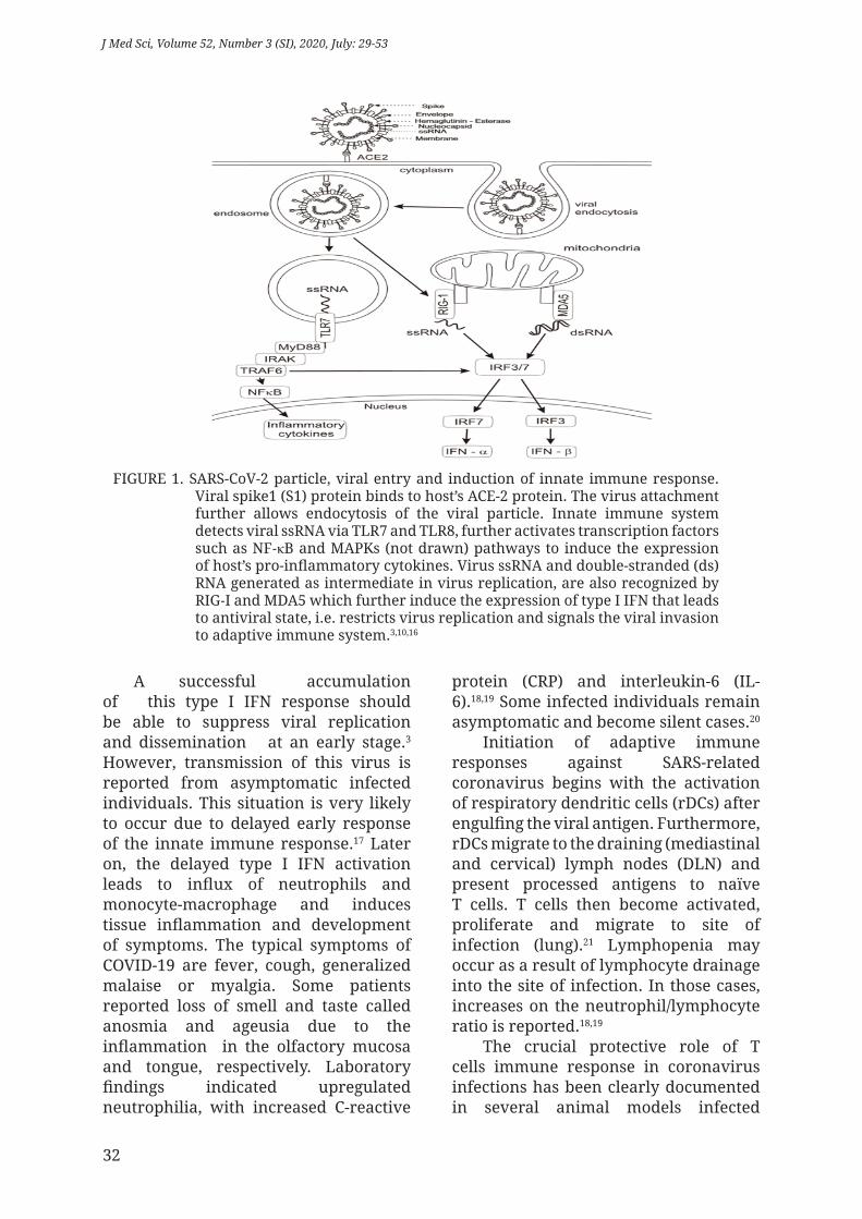
SARS-CoV-2 is transmitted from human to human through droplets containing the virus particles via the mucous membranes of the eyes, mouth or nose. However, there are no reports on the presence of the viral RNA from COVID-19 patients with conjunctivitis. Some studies reported virus shed from the stool (1-2%) in the late phase of infection,8 suggesting multiple transmission routes of the virus.9 The receptor binding site of SARS-CoV-2 and SARS-CoV is similar, with variations in some key amino acids.6 Infection of SARS-CoV-2 to mucosal epithelium depends on the binding of S1 protein to the host’s angiotensin-converting enzyme (ACE)-2 protein (FIGURE 1), which is expressed in type II pneumocytes among other cells. Viral entry requires S protein priming by cellular protease, the transmembrane protease, serine 2 (TMPRSS2), which causes the cleavage of S1/S2 and S2’ sites. The virus attachment further allows the fusion of virus with the cellular membrane. Similar capacity was already shown by SARS-CoV,10 but several amino acid differences in the S2 protein cause more efficient infection capacity of SARS-CoV-2 compared to the SARS-CoV.11
Compared to the other 2 SARS-related coronavirus, SARS-CoV-2 infection results in milder symptoms. Three stages of SARS-CoV-2 infection are incubation and asymptomatic period with or without detectable virus, detectable virus with common cold and influenza-like illness, and variety of SARS such as pneumonia with high viral load.12 The first and second stages of infection require decent immune capacity. Once innate and adaptive immune responses breakdown, the virus will propagate and massive destruction of the affected tissues will occur, especially in organs that have high ACE2 expressions such as lungs, intestines and kidneys.13 As

31
Septyaningtrias DE, et al, Review of immune responses...
mentioned earlier, the virus may enter via several routes other than respiratory tract, e.g. digestive tract and olfactory nerves14 lead to various symptoms.12
The immune system plays an important role in eliminating the virus, yet it may also be involved in the disease pathogenesis that leads to severe morbidity and mortality. We are aware that the pathogenesis of SARS-CoV-2 infection may be more complex and related to other systems such as compromising the function of hemoglobin and blood coagulation.15 However, those topics are beyond the scope of this review. We begin with the main question related to what do we know about the innate and adaptive immune responses to SARS-CoV-2, the host factors that affect immune response, the possibility of viral immune evasion, and the responses of the immune system in fatal cases. Then, we conclude with possible immunological-based therapeutic and prophylactic approaches. Further, we would like to briefly discuss the immune consequences of the pandemic through the perspectives of Indonesian patients’ characteristics.
As a novel and very contagious human coronavirus that prevents proper immediate investigation, reliable data on the immune responses against SARS-CoV-2 are still limited. Most studies were speedy reports with limited number of patients or case reports. This minireview is based on the latest publications concerning SARS-CoV-2, in addition to the recent knowledge related to SARS-CoV reports. Some studies using animal models infected with coronavirus or other virus were included in order to explain the possible mechanisms of the immunopathogenesis. Despite being published in reputable journals, many of our recent references (year 2020, in particular) may not be peer-reviewed, due to the sense of urgency to combat SARS-CoV-2 globally. We searched
our references by several key words, including: COVID-19 OR SARS-CoV-2 OR SARS-CoV, and immunity* [AND term/ specific topic] in PubMed, Google Scholar and World Health Organization (WHO) databases.
DISCUSSION
Appropriate innate and adaptive immune responses against coronavirus in mild cases
Upon coronavirus infection, the infected cells detect viral pathogen‐associated molecular patterns (PAMPs) via their pattern recognition receptors (PRRs) such as Toll‐like receptors (TLRs) (FIGURE 1).16 As a single-stranded (ss) RNA virus, the principal TLRs that recognize coronavirus nucleic acid are TLR7 and TLR8. In addition, retinoic-acid-inducible gene I (RIG‐ I) is also able to recognize them. During their replication, coronaviruses generate double-stranded (ds)RNA intermediates which are recognized by melanoma differentiation-associated gene 5 (MDA5).3,16 The recognition of viral nucleic acid by PRRs occurs in endosomes, endocytolysosomes and cytoplasm.16 MyD88 acts as an adapter protein by most TLRs. Coronavirus infection mainly activates the transcription factors such as nuclear factor-kappa beta (NF‐κB) and mitogen‐activated protein kinases (MAPKs) pathways to induce host responses such as type I interferon (IFN) and inflammatory factors.3 Type I IFN via interferon-α/β receptor, in turn, activates the JAK-STAT pathway that initiates the transcription of IFN-stimulated genes under the control of IFN-stimulated response elements containing promoters.3 As the host’s major antiviral molecules, IFNs limit viral spread, and play an immunomodulatory role to promote macrophage phagocytosis, as well as natural killer (NK) cells restriction of infected target cells.
32
J Med Sci, Volume 52, Number 3 (SI), 2020, July: 29-53
FIGURE 1. SARS-CoV-2 particle, viral entry and induction of innate immune response. Viral spike1 (S1) protein binds to host’s ACE-2 protein. The virus attachment further allows endocytosis of the viral particle. Innate immune system detects viral ssRNA via TLR7 and TLR8, further activates transcription factors such as NF‐κB and MAPKs (not drawn) pathways to induce the expression of host’s pro-inflammatory cytokines. Virus ssRNA and double-stranded (ds)RNA generated as intermediate in virus replication, are also recognized by RIG‐I and MDA5 which further induce the expression of type I IFN that leads to antiviral state, i.e. restricts virus replication and signals the viral invasion to adaptive immune system.3,10,16
A successful accumulation of this type I IFN response should be able to suppress viral replication and dissemination at an early stage.3 However, transmission of this virus is reported from asymptomatic infected individuals. This situation is very likely to occur due to delayed early response of the innate immune response.17 Later on, the delayed type I IFN activation leads to influx of neutrophils and monocyte-macrophage and induces tissue inflammation and development of symptoms. The typical symptoms of COVID-19 are fever, cough, generalized malaise or myalgia. Some patients reported loss of smell and taste called anosmia and ageusia due to the inflammation in the olfactory mucosa and tongue, respectively. Laboratory findings indicated upregulated neutrophilia, with increased C-reactive
protein (CRP) and interleukin-6 (IL-6).18,19 Some infected individuals remain asymptomatic and become silent cases.20
Initiation of adaptive immune responses against SARS-related coronavirus begins with the activation of respiratory dendritic cells (rDCs) after engulfing the viral antigen. Furthermore, rDCs migrate to the draining (mediastinal and cervical) lymph nodes (DLN) and present processed antigens to naïve T cells. T cells then become activated, proliferate and migrate to site of infection (lung).21 Lymphopenia may occur as a result of lymphocyte drainage into the site of infection. In those cases, increases on the neutrophil/lymphocyte ratio is reported.18,19
The crucial protective role of T cells immune response in coronavirus infections has been clearly documented in several animal models infected
33
Septyaningtrias DE, et al, Review of immune responses...
with various coronaviruses.22 Cytokine microenvironments generated by antigen presenting cells direct T cells responses.17,22 In general, TH1 orchestrates the direction of T cells response followed by cytotoxic T lymphocytes (CTL) that are essential in killing viral infected cells. Intravenous immunization with peptide-loaded dendritic cells (DCs) followed by intranasal boosting with recombinant vaccinia virus (rVV) encoding single immunodominant epitope of SARS resulted in accumulation of virus-specific memory CD8+ T cells in bronchoalveolar lavage fluid, lungs, and spleen.23
Cytotoxic CD8+ T cells account for about 80% of total infiltrative inflammatory cells in the pulmonary interstitium in SARS‐CoV‐infected patients and play a vital role in elimination of CoV’s infected cells.24
Strong evidence showed that TH1 response is a key for successful control of SARS-CoV and MERS-CoV, and probably for SARS-CoV-2 as well.17
Depletion of CD8+ T cells do not affect and delay viral replication at the time of infection with SARS‐CoV,21 while depletion of CD4+ T cells is associated with reduced pulmonary recruitment of lymphocytes and neutralizing antibody and cytokine production, resulting in a strong immune‐mediated interstitial pneumonitis and delayed clearance of SARS‐CoV from lungs.25
Strong T cells responses are significantly correlated with higher production of neutralizing antibodies26 that block the virus from entering healthy host cells and protect the host against viral re-infection.22 In patients infected with coronavirus, specific immunoglobulin M (IgM) peak occur on the 9th day after infection. Class switching to IgG, assisted by follicular helper T cells (TFH cells) can be detected as early as 4 days but most of the cases were seroconverting at the 2nd week after the onset of the disease.27 Delayed and weak antibody responses are associated with
severe outcomes.22
One report of a patient with mild symptoms revealed that increased plasma cells, TFH cells, activated CD4+ T cells, CD8+ T cells, IgM and IgG antibodies that bound to SARS-CoV-2 were detected in blood samples before symptomatic recovery. These immunological changes persisted for at least 7 days following full resolution of symptoms.18 From a study in Finland, IgM was reported to be detected on day 9 and IgG at day 20 after infection.28 Specific IgM started to decline at week 7.29 Immunoglobulin detection was employed as the basis of SARS-CoV rapid test. One study in Wuhan reported 10% SARS-CoV-2 specific IgG positive rate from asymptomatic individuals.30
A cohort study of convalescent SARS-CoV patients from Beijing revealed that the concentration of specific serum IgG and neutralizing antibodies were highly correlated, reaching their peak at 4th month after the onset of disease and decreasing gradually thereafter. The titers decreased noticeably after the 16th month, but long-lasting specific IgG and neutralizing antibodies were reported in most of the patients as long as 2 years after. Only 11.8% of patients turned to negative at month 24.31 In long-term follow-up survivors, IgG was detectable in recovered patients only up to 6 years after SARS-CoV infection,32 suggesting decreasing level of memory B cells.32 Shorter antibody persistence may occur due to several factors such as high dose corticosteroid treatment that is known to suppress antibody response.33
Coronaviruses immune evasion
Immune evasion is an important strategy utilized by pathogens to overcome the immune response for efficient replication in the host. Immune evasion by influenza virus A via antigenic shift and antigenic drift is a famous phenomenon. As an RNA virus, coronaviruses mutation rates are
34
J Med Sci, Volume 52, Number 3 (SI), 2020, July: 29-53
higher than DNA viruses due to higher error rate of their RNA polymerases.34 The high mutation rate leads to higher probability for immune evasion in secondary exposure.
Coronaviruses have evolved several strategies of immune evasion. One of the main strategies used by SARS-CoV to avoid immune detection is by shielding its RNA from PRRs. This can be done by several mechanisms. One approach is by disguising its RNA and replication machinery inside replication organelles.35 Since concealing its RNA and replication machinery is not unassailable, SARS-CoV further modifies its RNA by adding cap-structure to the 5’-end or by methylating its mRNA to be mediated by SARS-CoV nsp16 protein.36 Another means is by activating its endonuclease, thus destructing its own RNA at certain stages of infection to avoid the initiation of host’s virus-destroying system.37
The other major strategies of immune evasion are to restrain the host’s innate immune signaling. Coronavirus inhibits recognition by PRRs such as TLRs. Terminating cellular protein expression is one of the effective mechanisms to achieve that purpose. SARS-CoV nsp1 protein arrests the translation process by binding to the ribosomal 40S subunit38 and inducing host’s mRNA degradation.39 Another mechanism is by manipulating stress granules formation. Stress granules are structures formed by the accumulation of untranslated mRNA as a consequence of the arrest of cellular translation due to virus infection. This accumulation of viral RNA can be detected by the host’s immune sensors such as Protein Kinase R (PKR), RIG-I, and MDA5.40 Other mechanisms of suppressing innate immune signaling include histone modification,41 preventing phosphorylation and nuclear translocation of IFN-related transcription factors such as STAT1.42
To avoid adaptive immune
responses, coronaviruses inhibit viral antigen presentation to T cells. MERS-CoV can infect and replicate in macrophages and dendritic cells resulting in the cells malfunction and inability to present antigens to T cells.43 MERS-CoV also downregulates MHC I and II in macrophages and dendritic cells,44 thus leading to failure in activation of naïve T cells. MERS-CoV further stimulates T cells apoptosis such that lymphopenia will ensue.45 Deficiency of activated T and B lymphocytes are associated with persistent infections and lack of virus clearance.
Immune system in fatal cases of COVID-19
The mortality rate of SARS-CoV-2 infection is lower compared to the other SARS related coronavirus. However, due to the highly contagious nature of the virus, the number of fatalities is higher than the two previous outbreaks combined.46 An impaired immune response that fails to eliminate the virus will likely result in severe cases of morbidity and mortality (TABLE 1 and 2).13 Accordingly, medical researchers are asking the important question from the immunological point of view: What actually drives the unpredictable fatality rate of COVID-19?
In SARS-CoV infections, persistence of the viral infection and spreading induce activation of macrophages and thus, cause increased production of pro-inflammatory cytokines that leads to consistent influx of neutrophils and monocyte-macrophages.1 The increased cytokine production and associated CRP may cause the condition called as “cytokine storm” which presents in a vicious cycle fashion and may cause more damage to the cells of the lungs. Initial increment of CRP was suggested to be used to predict severe cases.47
Despite their purpose to fight off the invading pathogen, the inflammatory

35
Septyaningtrias DE, et al, Review of immune responses...
cells can mediate further tissue damage by inciting lung epithelial and endothelial apoptosis via Fas-Fas ligand (FasL) or Tumor necrosis factor (TNF)-related apoptosis-inducing-ligand-Death Receptor 5 (TRAIL-DR5)-dependent mechanisms.48 Apoptosis compromises the lung microvascular and alveolar epithelial cell barriers resulting in vascular leakage and alveolar edema. Indeed, pathologic lung examination of two COVID-19 patients with adenocarcinoma revealed diffuse alveolar damage (DAD). Pulmonary edema, proteinaceous and fibrin exudates, mononuclear inflammatory cell infiltration with multinucleated giant cells, alveolar wall thickening with pneumocytes hyperplasia and fibroblast proliferation, without prominent hyaline membrane were observed in both patients. The absence of clinical symptoms of pneumonia indicates these pathological findings may depict early lung abnormality of COVID-19.49 Later on the DAD could lead to acute respiratory distress syndrome (ARDS), in which respiratory failure is characterized by the rapid onset of widespread inflammation in the lungs and subsequent fatality.50 At this late stage, extensive fibrin deposition (fibrosis) and alveolar collapse occur.51
Similar mechanisms manifested in lung immunopathology were also shown in mouse models of SARS-CoV infection.23
The ‘cytokine storm’ also occurs in SARS-CoV-2 infection particularly in severe cases.19,52 Furthermore, the cytokines can spill over to the circulation, resulting in systemic sepsis and multi organ dysfunction and failure53 including myocardiac complication reported in many fatal cases.
Besides causing massive apoptosis and tissue injury, this ‘cytokine storm’ can diminish T cells response via TNF-mediated apoptosis. In patients with COVID-19, a more pronounced lymphopenia is observed in fatal cases,52,54 suggesting that COVID-19 may
damage T lymphocytes. Furthermore, the numbers of CD4+ and CD8+ T cells were decreased,50,52,54 with naïve CD4+ T cells count elevated but that of memory CD4+ T cells decreased.52 Considering that the differentiation of naïve T cells into effector and memory cells is fundamental for the immune response, the higher naïve to memory ratio of CD4+ T cells in severe cases indicates an impaired and unbalanced immune response caused by SARS-CoV-2 infection. In addition, the production levels of IFN-γ by CD4+ T cells, CD8+ T cells, and NK cells tend to be lower.54 The production of IFN-γ is essential for resistance against infection of various pathogens and IFN-γ was reportedly able to inhibit viral replication of SARS-CoV.55 Lower IFN-γ levels in severe cases of COVID-19 might reflect the host’s inability to fight off the virus which increases the likelihood of fatality. Strong T cells’ responses are significantly correlated with higher neutralizing antibody, while more serum TH2 cytokines (IL-4, IL-5, IL-10) were detected in the fatal group of SARS-CoV.26
Another interesting effect that SARS-CoV-2 infection has on T lymphocytes is that it may induce T cells exhaustion. Lymphopenia was common in patients with SARS-CoV-2 (72%) as well as in acute phase of patients with SARS-CoV (~80%).12,21 At the infection onset, it was indicated by the drainage of lymphocytes into the infected tissue but in later stages it indicates the impairment of immune system. Beside the decline in T cells number, COVID-19 patients also showed increased expression of natural killer group 2 member A (NKG2A) in CD8+ T cells and NK cells.54 NKG2A is an inhibitory receptor, which induces immune cells exhaustion in chronic viral infection.56 Its increased expression in CD8+ T cells suggests functional exhaustion of cytotoxic lymphocytes in COVID-19 patients. The elevated level of interleukin-10 (IL-10) in severe cases of COVID-19 supports this plausible notion.

36
J Med Sci, Volume 52, Number 3 (SI), 2020, July: 29-53
TABLE 1. Innate immune response against SARS-CoV-2
Immune responses to SARS-CoV-2 infection
Action Dysregulation in severe cases of COVID-19
Detection by PRRs (including TLR7, TLR8, RIG-I, MDA5)
Induces NF-κB and MAPK pathways to activate type I interferon system and other pro-inflammatory cytokines.
Coronavirus avoids PRRs detection by concealing its RNA inside replication organelles; adding cap structure or methylating its RNA; activating endonuclease to degrade its own RNA; halting host’s translation process; destructing host’s mRNA;or inhibiting stress granules formation.a,34-39
Type I interferon Has antiviral activity. IFN-stimulated genes inhibit virus entry, replication and dissemination.
Coronavirus prevents phosphorylation and nuclear translocation of IFN-related trancription factor, inhibit activation of IFN system.a,41
Enhances NK cells activity to kill infected cells and to secrete IFN-γ; promotes macrophage phagocytosis activity and pro-inflammatory cytokines production; stimulates DC maturation and antigen presentation to T cells; increases MHC I surface expression
Delayed or impaired antiviral response of type I IFN results in uncontrolled viral replication and dissemination.
Natural Killer (NK) cells NK cells target and kill virus-infected cells and produce type I interferon cytokines.
Decrease IFN-γ and increase NKG2A expression in NK cells, indicating functional cells exhaustion and impaired virus-killing activity.52
Macrophages/monocytes Macrophages infiltrate infected sites; phagocytose apoptotic virus-infected cells; mediate antibody-dependent cell cytotoxicity; and secrete pro-inflammatory cytokines.
Hyper-influx of macrophages-monocytes into the lung that leads to apoptosis of lung epithelial and endothelial cells and tissue damage. Coronavirus also can infect macrophages, rendering it malfunction.a,42,43,48
Pro-inflammatory cyto-kines
Increase secretion of IL-1R, IL-6, TNF-α, IL-8, IP-10, MCP1, MIP-1a upon virus infection to recruit neutrophils, monocytes-macrophages, and T cells to infected site and augment their antiviral action.
Exaggerated release of pro-inflammatory cytokines or cytokine storm occurs; leads to further tissue damage, ARDS, systemic sepsis, and multi organ dysfunction.19,50
Neutrophil in tissue inflammation
Neutrophils migrate to infected sites; phagocytose apoptotic virus-infected cells; and secrete pro-inflammatory cytokines to recruit more immune cells.
Neutrophilia; with hyper-influx of neutrophils into the lung that leads to tissue damage.19,48,50
aObserved in SARS-CoV or MERS-CoV, however, no information available for SARS-CoV-2 yet.ARDS, acute respiratory distress syndrome. DC, dendritic cells. IFN-γ, interferon-gamma. IL-1R, interleukin-1 receptor. IL-6, interleukin-6. IL-8, interleukin-8. IP-10, interferon gamma-induced protein 10. MAPK, mitogen‐activated protein kinases. MCP1, monocyte chemoattractant protein 1. MDA5, melanoma differentiation-associated gene 5. MIP-1a, macrophage inflammatory protein 1-alpha. MHC I, major histocompatibility complex class I. NF-κB, nuclear factor-kappa beta. NK, natural killer. NKG2A, natural killer group 2 member A. PRRs, pattern recognition receptors. RIG-I, retinoic-acid-inducible gene I. TLR7, Toll‐like receptor-7. TLR8, Toll‐like receptor-8. TNF-α, tumor necrosis factor-alpha
Although there is no evidence concerning the ability of IL-10 to increase NKG2A expression, however, IL-10 regulates inhibitory receptor (e.g. Programmed cell-death protein 1; PD-1) expression
in CD8+ T cells57 and induces sustained PD-1 expression and T cells exhaustion,58 leading to a suppressive environment that prevents viral clearance.

37
Septyaningtrias DE, et al, Review of immune responses...
TABLE 2. Adaptive immune response against SARS-CoV-2
Immune responses to SARS-CoV-2
Action Dysregulation in severe cases of COVID-19
Antigen presentation by dendritic cells
Respiratory dendritic cells engulf viral antigen; migrate to lymph nodes and presented viral antigen to naïve T cells.
Coronavirus infects DC and render it malfunction; downregulates its MHC I and II expression, impairing antigen presentation to naïve T cells.a,42,43
Cytotoxic T cells (CTL)
Activated CTL migrate to lung. CTL target virus-infected cells and induce their apoptosis by the cytotoxic granule release and production of TNF-α and IFN-γ.
Severe lymphopenia and low CD8+T cells count with increased NKG2A and decreased IFN-γ expression, indicating impaired T cells killing capacity and their functional exhaustion.12,48,50,52
T helper 1 (TH1) Secretes IFN-γ and TNF-α; activates macrophages and augment their phagocytosis activity.
Severe lymphopenia and low CD4+ T cell count with decrease IFN-γ expression. CD4+ T cell depletion is associated with reduced pulmonary infiltration of lymphocytes and cytokines production.a,12,48,50,52
T helper 2 (TH2) Secretes IL-4, IL-5, IL-13 which primarily induces plasma cells to produce virus-specific antibody.
Severe lymphopenia with low CD4+ T cell count. Low TH2 can result in decrease production of neutralizing antibodies.a,12,48,50,52
T helper 17 (TH17) Release pro-inflammatory cytokines.
High CCR6+ TH17, further exacerbating inflammation by downregulating Treg cells, promoting neutrophil migration.48
Regulatory T cells (Treg)
Suppress immune responses, secrete anti-inflammatory cytokines (IL-10, TGF-β).
Low Treg count, may exacerbate inflamma-tion.50
Lymphocytes in tissue inflammation
Effector lymphocytes migrate into site of infection lead to mild lymphopenia.
Lymphopenia. Mononuclear cells infiltration with many lymphocytes in the lung. Apoptosis of lymphocytes. Atrophy of spleen and lymph nodes were reported in autopsy.12,47,48,50
IgM Neutralizes the virus on the early phase of the disease.
Low IgM level, indicating ineffective virus containment on the early phase of the disease, leads to unchecked virus replication.
IgG Neutralizes the virus, activates complement system & antibody-dependent cellular cytotoxicity.
High IgG level. Antibody can leads to immune enhancement resulting in infection of macrophages; persistent virus replication and exuberant pro-inflammatory cytokines production.
IgA Neutralizing antibody in mucosa, can be detected in plasma. IgA secreting plasma cells is found in plasma.
Not much data, most probably in lower level compared to mild cases.
Memory cells Memory B cells produce long-lived circulating virus-specific neutralizing Ab.
Low naïve to memory T cells ratio, suggesting impaired memory cells induction or differentiation.50
Memory CD8+ T cells effectively produce IFN-γ, TNF-α, IL-2 to prevent viral replication and alveolar damages.
aObserved in SARS-CoV or MERS-CoV, however, no information available for SARS-CoV-2 yet.CCR6, C-C chemokine receptor 6. CTL, cytotoxic T lymphocytes. DC, dendritic cells. IFN-γ, interferon-gamma. IgA, immunoglobulin A. IgG, immunoglobulin G. IgM, immunoglobulin M. IL-2, interleukin-2. IL-4, interleukin-4. IL-5, interleukin-5. IL-10, interleukin-10. IL-13, interleukin-13. MHC I, major histocompatibility complex class I. MHC II, major histocompatibility complex class II. NKG2A, natural killer group 2 member A. TNF-α, tumor necrosis factor-alpha. TGF-β, transforming growth factor-beta. Treg, regulatory T cells.

38
J Med Sci, Volume 52, Number 3 (SI), 2020, July: 29-53
Humoral immune response against SARS-CoV-2 may also contribute to the severity of the disease. COVID-19 patients with higher IgG levels tend to have more severe disease symptoms compare to those with lower IgG level.59 It has been proposed that IgG humoral response may contribute to tissue damage by a mechanism known as antibody-dependent enhancement (ADE). ADE has been confirmed in multiple viral infections.59 ADE occurs when antiviral neutralizing antibodies (Nab) cannot fully neutralize the virus. Instead, the virus-NAb complexes bind to the Fc Receptor (FcR) in macrophages, leading to viral endocytosis and infection of macrophages. This leads to persistent virus replication and exuberant pro-inflammatory cytokines signaling which eventually causes acute fatal lung injury, as has been observed in patients with SARS-CoV infection.60
The impaired immune system in severe cases may increase the risk for bacterial co-infection. A study in China reported that 10% of the COVID-19 severe cases suffered from secondary infection.19 Tissue damage, exuberant inflammatory cell influx observed in cytokine storm phenomenon may render the pro-inflammatory response inadequate, e.g. lymphocytes recruitment to new infected site and macrophages phagocytosis capacity, when challenged with a bacterial pathogen, while enhancing bacterial entry or adherence to host’s cell.61
Host factors that influence immune responses against SARS-CoV-2
China, Southern Europe and American East Coast took turns as areas with the highest number of cases during the course of the pandemic. In several regions and countries, certain races and foreign-born individuals were disproportionately affected. Whether genetic and epigenetic factors are
involved in increasing the morbidity and mortality risks in the most severely affected areas, needs further study. The 1918 H1N1 pandemic reported high mortality in indigenous communities in Australia, Alaska and South America that may be due to genetic factors or lack of cross-immunity.61 Currently, temperature and humidity as well as frequency of traveling and migration, population density and social background are considered as crucial factors in the SARS-CoV-2 spreading.62,63 Increasing reports have discussed older age, male and comorbidity as risks factors for COVID-19. However, detailed demographic data for COVID-19 cases are not yet available. Most of the government agencies have only reported the number of active cases, recovered and death cases without specific case characteristics.
From China, the first epicenter of the pandemic, reports confirmed that older people are more susceptible to SARS-CoV-2 than the younger group.12,62 Children and adolescents are less susceptible to SARS-CoV-2, probably due to the low expression of ACE2.64
The median of the patients’ ages ranged from 41 to 57 year. Old age is known as a risk factor for infection as reported for SARS-CoV infection65 as well as a study in non-human primate infected with SARS-CoV.66 Prior to infection, CD8+ T cells and B cells were lower in aged monkeys and after infection CD8+ T cells and B cells responses were significantly reduced in the aged host.66 A study of mice models of West Nile Virus infection found the virus also predominantly infects the old population. Malnutrition that occurs more frequently in elderly67 may enhance vulnerability to infection during aging. The IgM and IgG response are commonly impaired and this was linked to delayed germinal center formation in the draining lymph node.68
Older patients showed severe symptoms at hospital admission.62 Most

39
Septyaningtrias DE, et al, Review of immune responses...
of the patients in China that died were older than 50 years with median age of 70 years old.69 European countries also reported that the death rate was higher among senior citizens more than 70 years old. Worldometer in April 14, 2020 reported the data from New York that 47% of the patients that died were more than 70 years old. Despite the higher rate of comorbidities found in older individuals, the dysregulated immune response may contribute to the more severe effects in older individuals. This may be due to lower number of Treg, aberrant microbiota composition, and higher proportion of inflammatory cells during aging.
Many reports from SARS-CoV-2 infection in China revealed that 50–75% admission to the hospitals were male.12,19,62,67 Moreover, more men died with COVID-19 compared to women.69 The Guardian, April 16, 2020 edition reported that the number of men who died of COVID-19 in England and Wales was about twice the rate of women.70 In New York, Worldometer at April 14, 2020 revealed that male patients comprised 61% of the mortality from COVID-19 and even higher up to 72% in patients without underlying conditions.71 The data are in accordance with the predominantly male patients reported in previous case mortality rates of SARS-CoV72 and MERS-CoV.73 Despite social factors such as working outside home, healthcare seeking behavior, smoking habit and medical and biological factors such as higher proportion of comorbidities e.g. hypertension and diabetes as well as higher expression of ACE receptors that are correlated with males, some important aspects of immunological background still contribute to the higher proportion and severity of male patients with COVID-19.74
The difference in immune response between sex was reported by many reports. Females have lower susceptibility to infection partly
due to several genes encoded in X chromosomes. One of the genes is TLR-7 that detects viral RNA, resulting in higher production of antiviral substance type I IFN.75 Females have higher expression of TLR-7 that is partly mediated by estrogen signaling. Male mice were more susceptible to SARS-CoV infection compared with age-matched females and the degree of sex bias to SARS-CoV infection increased with advancing age.76 The X chromosome influences the immune system’s response against viral infection by acting on many other genes, including TLR-8, CD40L and CXCR3 which can be over-expressed in women. Infection of mice with H1N1 induced antibody responses, CD4+ T cells and CD8+ T cells memory responses that were greater in females than males.76
Males tend to have higher proinflammatory profiles than females, hence increasing the risk of excessive inflammation in severely affected cases. Many regulatory proteins in the immune system such as FOXP3, the transcription factor for Treg are encoded in X chromosomes. In women the production of inflammatory IL-6 after viral infection is lower than in males and is often correlated with a better longevity. A mice study on SARS-CoV infection showed a persistent increase of inflammatory monocytes and neutrophils in SARS-CoV infected male mice but not in female mice. The estrogen signaling in female mice is proposed to reduce the accumulation and function of macrophages in the mice lungs and have a protective effect against viral infection.76 These data are in accordance with several reports that showed sexual dimorphism in immune response to COVID-19 infection.
Several comorbidities that increase COVID-19 severity has been reported such as hypertension62 and diabetes77 that are partly based on the patients’ medication or condition that can upregulate ACE2 expression.78 Higher expression of ACE2 not only permits more viral entrance but
40
J Med Sci, Volume 52, Number 3 (SI), 2020, July: 29-53
is also augmenting immune response and proinflammatory cytokine production, which correlated with fatal cases.79 The impact of the unhealthy Western diet that leads to inadequate immune response and chronic inflammatory state in COVID-19 pathology has also been discussed recently.80 The complex pathogenesis of COVID-19 still needs to be elucidated, in which the effect of SARS-CoV-2 in erythrocytes and components of blood coagulation in addition to immune responses, plays an important role in the process. Several interesting observations such as coronaviruses have not been shown to cause a more severe disease in immunosuppressed patients,81 need more confirmation in the future.
Immunological memory and herd immunity
Reappearance of highly contagious viral infections such as SARS-CoV-2 is a crucial public health problem. Immunological memory is important for the effective response against pathogens that have been encountered previously. It generates immediate protection and greater magnitude of immune response. Whether memory T cells response is sufficient to protect from re-infection of SARS-CoV-2 needs further study. In SARS-CoV-2 infection, the detection of specific IgG against SARS-CoV-2 in the patients’ sera28 provides early evidence for immunological memory. However, since the COVID-19 pandemic is still ongoing, data on the time-period of antibody peak level and the duration of the antibody detectability are not yet available on a large scale.
Neutralizing antibodies against S protein protect the susceptible host from lethal infection. However, several studies reported that anti-SARS-CoV antibody response is short-lived in patients who have recovered from SARS-CoV.23 IgG titer peaks at 3-4 months. It remains detectable 2 years after infection
and starts to decline after 2 years.31 Immunological memory to one type of coronavirus may not provide protection against another type of coronavirus as shown in the absence of cross-reactivity of CD8+ T cells responses against SARS-CoV with MERS-CoV.82
Few reports are available on memory T cells in SARS-CoV-2 infection. After the SARS-CoV outbreak, SARS-CoV-specific memory T cells had been screened in SARS convalescent patients.32 All memory T cells detected were directed at SARS-CoV spike (S), membrane (M) or nucleocapsid (N) protein. These memory T cells were found to last up to 6 years after infection32 with most of them identified as polyfunctional central memory T cells.26 Re-infection with SARS-CoV has demonstrated the protection provided by memory T cells. In immunized mice challenged with lethal doses of SARS-CoV, its virus-specific memory CD8+ T cells efficiently produced cytokines including IFN-γ, TNF-α, IL-2 and cytolytic molecules, e.g. granzyme B. These responses prevented extensive viral replication and alveolar damages.23 Given the short-lived duration of antibody-specific-SARS-CoV, the long-lived memory T cells and its proven protection upon re-infection is of considerable significance. However, it is worth noting that a decrease in memory T cells count in SARS-CoV-2 infection with high naïve to memory T cells ratio52 might indicate impaired memory T cells induction.
Even though many uncertainties remain concerning immunological memory towards SARS-CoV-2, several countries endorse the idea of herd immunity. The common notion in herd immunity is that exposure of the virus to the population may induce immunological memory for most people. As more and more people become immune to the virus, an infected individual has less and less chance of coming into contact with an individual

41
Septyaningtrias DE, et al, Review of immune responses...
susceptible to infection. Finally, herd immunity becomes pervasive which at that point, the number of cases will start to decline. If herd immunity is achieved, even in the absence of measures designed to slow or contain transmission, the virus will be contained.83
However, there are many concerns regarding herd immunity in SARS-CoV-2. One concern has to do with the duration of the immunological memory and re-infection. Recently, SARS-CoV-2 patients who have been declared recovered by two negative tests for the virus later tested positive again.84 If these cases were indeed re-infection, it would cast doubt on the immunological memory developed by the infected individual. Another concern is the possibility of immune enhancement caused by the infection.59 If immunity against SARS-CoV-2 leads to exacerbation of the infection rather than prevention of it, more fatalities will emerge. Whether the virus is mutating over time is also an issue that needs to be addressed. If the virus is mutating even at a slow rate, what those changes will do is a bigger question. The mutations could make the virus more easily transmitted or better at evading our immune system. The main question regarding herd immunity in this current pandemic is what percentage of the population is needed to get infected to achieve herd immunity. As of today, it is very likely that the number of COVID-19 cases is underestimated. Also, considering the severity of the disease and its case fatality rate that varies among many countries, the idea of herd immunity may not be appropriate for the current situation.
Preventive vaccination and immunotherapy against coronavirus
Considering the significant protection conferred by memory cells and the fact that COVID-19 is still spreading globally with no specific
treatment yet available, it is worth noting that probably vaccines offer the best options to control the current pandemic. By vaccination, those who have not been infected by SARS-CoV-2 will have antibodies which will readily generate protection with greater response against SARS-CoV-2. In addition, vaccination also provides protection against re-infection for those who have recovered from SARS-CoV-2 infection. The immediate availability of SARS-CoV-2 genomic sequence5 and the recent mapping of SARS-CoV-2 spike (S) protein85 may lay the foundation for more rapid development of a specific vaccine. From studies on SARS-CoV vaccines, the S protein on the surface of the virus is an ideal target for a vaccine.86
According to the most recent report, currently 73 SARS-CoV-2 vaccine candidates are undergoing development at preclinical stages and 5 others are entering clinical trials in humans. These 5 vaccine candidates include mRNA-1273 (NCT04283461/Moderna), Ad5-nCoV (NCT04313127/CanSino Biologicals), INO-4800 (NCT04336410/Inovio), LV-SMENP-DC (NCT04276896/Shenzhen Geno-Immune Medical Institute), and pathogen-specific aAPC (NCT04299724/Shenzhen Geno-Immune Medical Institute). As of mid-March 2020, mRNA-1273 (Moderna) and Ad5-nCoV (CanSino Biologicals) have become two of the first SARS-CoV-2 vaccines to be tested on humans.87 Significant breakthroughs in vaccine technology have provided more vaccine development platforms. The platforms used in the development of SARS-CoV-2 vaccine range from RNA vaccine, DNA vaccine, viral vector-based vaccine, recombinant protein vaccine, live attenuated vaccine, and inactivated vaccine. Among these platforms, RNA vaccine design gives flexibility on antigen manipulation and improves protein translation efficiency which can induce a more robust immune response.88 Additionally, RNA vaccine

42
J Med Sci, Volume 52, Number 3 (SI), 2020, July: 29-53
is more rapid and less expensive than traditional vaccines88 which would give some advantages and benefits especially during the pandemic situation as it is today.
Although there is urgency to provide a vaccine rapidly due to the current pandemic, governments and pharmaceutical companies need to ensure the safety of the vaccine. Lessons need to be learned from the experience of developing a vaccine for SARS-CoV. Several vaccines for SARS-CoV have been tested in animal models. Most of these vaccines protected animals from the challenges of SARS-CoV, but, many did not induce sterilizing immunity. In some cases, vaccination resulted in complications89 or led to enhancement of the disease.90 Up to this moment, there is no vaccine for SARS-CoV available.
Another aspect that needs to be considered for a more effective SARS-CoV-2 vaccine development is the decline of antibody response over time. Coronavirus infection does not always induce long-lived antibody response. An individual can be re-infected with the same virus after an extended period of time. In individuals that recovered form SARS-CoV infection, antibody titer starts to decline 2 years after illness onset.31 Recently, reports emerged that SARS-CoV-2 re-infection occurs days after recovery.84 Thus, a SARS-CoV-2 vaccine will need to tackle this issue to provide protection in case the virus becomes endemic and causes recurrent seasonal epidemics.
SARS-CoV-2 infection causes more severe morbidity and more mortality among older individuals. Therefore, it will be important to develop vaccines that offer protection to this population. Unfortunately older individuals typically respond less well to vaccinations because of immune senescence.91 This is another concern that needs to be addressed for SARS-CoV-2 vaccine development.
One of the simple therapeutic
modalities for severe cases of COVID-19 is convalescent plasma. It has been used during the SARS outbreak92 and is currently used to treat COVID-19 patients.93 Other treatments that have been used during this ongoing pandemic are intravenous immunoglobulin (IVIG)94 and intravenous injection of mesenchymal stem cells.95 Since it is very likely that the ‘cytokine storm’ is the main cause of fatal cases of COVID-19, it is worth considering the use of a neutralizing antibody against pro-inflammatory cytokines involved in the pathogenesis of COVID-19. One of those is tocilizumab, a monoclonal Ab against IL-6R. In China, tocilizumab has been approved to treat COVID-19 patients with pneumonia and elevated IL-6 levels.96 Corticosteroid is another medication used to suppress inflammatory responses in COVID-19 patients.12,19 However, the lack of proven benefit in COVID-19 cases should render health care workers to be cautious on its usage. The strategy to suppress immune responses in severe cases is challenging as the main immune component that should be inhibited is currently not yet clearly identified. The timing to start the therapy without compromising the beneficial host defense is another question that needs an immediate answer. Furthermore, while the aforementioned therapies are currently used to treat a limited number of COVID-19 patients, evaluation and study on their efficacy are still required.
Heterologous immunity is thought to be a contributing factor that reduces severity of an infection in any age. Exposure to one pathogen may alter the subsequent innate and adaptive immune responses to a different pathogen. It will be interesting to study whether history of infectious diseases alters the vulnerability to suffer from severe COVID-19 via the heterologous immunity mechanism. The number of cases in some countries that has nationwide Bacille-Calmett Guerin (BCG) vaccination

43
Septyaningtrias DE, et al, Review of immune responses...
program, are less numerous than other countries with no BCG vaccination program such as European countries, leading to the idea that BCG vaccination may give some protection from SARS-CoV-2 infection. This idea is being tested with two clinical trials, although last year a review showed some evidence against it.97
Some alternative treatments related to immune function have been proposed for COVID-19 patients. Many doctors recognize the role of positive thinking, praying, meditation in the outcome of diseases, including COVID-19. Preliminary studies shows a beneficial impact of vagal stimulation, a method that activate autonomic nervous system to suppress immune function on the recovery of COVID-19 patients.98 Herbal medicine has also been used for COVID-19 treatment,99 yet the efficacy as well as its mechanism of action in the immune system, if any still need more study. Any herbal medicine that may balance immune responses, activate appropriate responses against SARS-CoV-2 while preventing excessive inflammation as discussed for the famous Chinese herbal remedy called Astragalus100 may be beneficial in the fight against this disease. The prospect of using Indonesian herbal medicine for balancing immune responses also needs to be investigated due to the region’s biological diversity.
The aftermath
While currently not and hopefully never considered the epicenter of the outbreak, Indonesia is the 4th largest population with a large variability of ethnic and socioeconomic backgrounds, hence increased vulnerability to any contagious pathogens. As of 28th of April 2020, the WHO database recorded 9096 confirmed cases in Indonesia, including 765 deaths. The mortality rate from COVID-19 in Indonesia is among the
highest in the world. High incidence of infectious diseases and the rise of non-communicable diseases as well as the large smoking population in Indonesia may contribute to dysregulation of immune responses. Whether those factors lead to higher incidence of severe cases and mortality rate of COVID-19 in Indonesia remains to be elucidated. Another important issue in the Indonesian fight against SARS-CoV-2 is the late development of a rapid serological test. Many believe that due to the lack of proper test, the true infection and death rate are higher than the reported data. Development of stronger infrastructure and increased pools of personnel for basic research and reduction of dependency on imported medical reagents need to be strategically planned.
When will the current global pandemic end? One projection proposed that we still need some form of social distancing until 2022 and should be ready for resurgence of the outbreak in 2024.101 Keeping all of previous discussion in mind, serological studies are needed to determine the duration of immunity to SARS-CoV-2. The current practice of using rapid tests as COVID-19 infection screening and discussion of using quantitative methods was reviewed recently.2
A study in China reported that autoantibodies were found in SARS-CoV-2 patients.102 Infection is considered as a risk factor for autoimmune diseases via molecular mimicry or bystander activation. On the other hand, infection may inhibit self-reactive T cells via activation of immune regulation to balance the activation of immune system against the pathogen as has been shown in mice infected with coronavirus.103 Whether there is any effect of the current pandemic to the rate of autoimmune diseases and other conditions related to self-reactive immune components remains to be elucidated. The hygienic

44
J Med Sci, Volume 52, Number 3 (SI), 2020, July: 29-53
behavior such as the use of hand sanitizer, washing hands with soap frequently and showering after arriving home from outdoor activities as well as the spraying of disinfectant may disrupt immune regulation via alteration of microbiome profile with several health consequences, including obesity.104
Healthy diet, vitamin supplementation, sufficient hours for sleeping at night, and regular exercise are considered as protective measures from the development of severe symptoms by strengthening the immune system. Many efforts have been taken to promote a healthy life style campaign in local and social media while people are
staying at home during the pandemic. Continuation of the good habits obtained during the pandemic may provide benefits for stronger immune function and general health in the future. On the other hand, uncomfortable feelings while wearing mask and protective equipment, reduced physical activity and stressful situations during social distancing as well as socioeconomic problems related to it, may impose negative consequences for immune system leading to various disorders.105 These changes of lifestyle and their impact and long-term effects on the immune system warrant subsequent investigation.
TABLE 3. Immunological studies relevant to COVID-19 in Indonesia
Studies on Indonesian COVID-19 cases.Immunological profile of Indonesian COVID-19 patients, the correlation with disease severity and which host factor contributes most in severe cases.
The effect of comorbidities (infection and non-infection) to the immunological profile related to disease progression.
Possible alternative route of infection (e.g. gastrointestinal tract, central nervous system) and correlated immune response in designated organs.
Evaluation of serological-based test used in Indonesia during COVID-19 pandemic (sensitivity, specificity).
Methods of serological screening: developing Indonesian-based test (rapid test, ELISA based test).
Evaluation of the current immunotherapy and development of novel immunotherapeutical agent, including Indonesian herbal medicine.
Do we achieve herd immunity? How many cases of asymptomatic COVID-19 can be found? Which host factor contributes most to appropriate immune response in the asymptomatic cases?
Immunological profile of COVID-19 survivors: How long does the protective antibody last? Cellular immune response of the memory cells. Possible alteration in the epigenetic status (trained immunity).
Study on the prospect of vaccine development suitable for common type of SARS-CoV-2 in Indonesia.
Study on the effect of changing lifestyle during and after the pandemic on immune-related problem.Effect of adopted lifestyle and stress during social distancing on immunological profile, especially in individuals with high risk for severe COVID-19 (elderly, male, hypertension, allergy, autoimmune, etc.).
Correlation of “super-hygienic” lifestyle, microbiota diversity and immunological profile in Indonesian population.

45
Septyaningtrias DE, et al, Review of immune responses...
The profound effect COVID-19 pandemic has on all aspect of life has ignited the global scientific community to work and team up to uncover more about this disease and discover the best strategy to overcome it. The same should be happening in Indonesia as currently we also dealing with the consequences conferred by the pandemic. Wide array of research topic in immunology field relevant to the situation in Indonesia is still waiting to be explored (Table 3). The more knowledge we have about this disease, the more chances we have to defeat it.
CONCLUSION
Innate and adaptive immune responses may keep SARS-CoV-2 at bay and prevent severe symptoms. However, several host factors such as genetic and epigenetic factors from old age, male, history of previous infection, and comorbidities as well as the immune evasion mechanism of the virus may influence the immune responses leading to severe and fatal cases.
The highly contagious nature of the virus prompts swift action to contain it from spreading further. Vaccines, though needing time for proper development, might offer the best chance to fight off the virus especially if it becomes a situation involving recurrent seasonal epidemics. However, for now, physical distancing combined with healthy lifestyle provide the best options to keep the virus in check.
As the pandemic situation continues developing, the best available scientific evidence should be used to guide future research aimed to uncover more about the disease, discover the best diagnostic, therapeutic and intervention strategies, curb the worst possible outcomes and prepare for the likely next pandemic. Thus, research collaboration between governments, international organizations, and the private sector
is urgently required. Real-time sharing of scientific information about the virus, disease pathogenesis, and human’s immune response will lead to advancement of the knowledge needed to overcome the many challenges from the current pandemic.
ACKNOWLEDGEMENT
The authors would like to thank Mahayu F. Ramadhani, SuhaSausan Nadine, and Erik C. Hookom for technical assistance.
REFERENCES
1. Perlman S, Dandekar AA. Immunopathogenesis of coronavirus infections: implications for SARS. Nat Rev Immunol 2005; 5(12):917-27.https://doi.org/10.1038/nri1732
2. Infantino M, Damiani A, Gobbi FL, Grosi V, Lari B, Macchia D, et al. Serological assays for SARS-CoV-2 infectious disease: benefits, limitations and perspectives. Isr Med Assoc J 2020; 22(4):203-10
3. de Wit E, van Doremalen N, Falzarano D, Munster VJ. SARS and MERS: recent insights into emerging coronaviruses. Nat Rev Microbiol 2016; 14(8):523-34.h t t p s : / / d o i . o r g / 1 0 . 1 0 3 8 /nrmicro.2016.81
4. Zhu N, Zhang D, Wang W, Wang W, Li X, Yang B, et al. A novel coronavirus from patients with pneumonia in China, 2019. N Engl J Med 2020; 382(8):727-33.h t t p s : / / d o i . o r g / 1 0 . 1 0 5 6 /NEJMoa2001017
5. Zhou P, Yang XL, Wang XG, Hu B, Zhang L, Zhang W, et al. A pneumonia outbreak associated with a new coronavirus of probable bat origin. Nature 2020; 579(7798):270-3. https://doi.org/10.1038/s41586-020-
46
J Med Sci, Volume 52, Number 3 (SI), 2020, July: 29-53
2012-76. Xu X, Chen P, Wang J, Feng J, Zhou
H, et al. Evolution of the novel coronavirus from the ongoing Wuhan outbreak and modeling of its spike protein for risk of human transmission. Sci China Life Sci 2020; 63(3):457-60. https://doi.org/10.1007/s11427-020-1637-5
7. Forster P, Forster L, Renfrew C, Forster M. Phylogenetic network analysis of SARS-CoV-2 genomes. Proc Natl Acad Sci USA 2020; 117 (17) 9241-3.h t t p s : / / d o i . o r g / 1 0 . 1 0 7 3 /pnas.2004999117
8. Cheung KS, Hung IF, Chan PP, Lung KC, Tso E, Liu R, et al. Gastrointestinal Manifestations of SARS-CoV-2 Infection and Virus Load in Fecal Samples from the Hong Kong Cohort and Systematic Review and Meta-analysis. Gastroenterology 2020; S0016-5085(20)30448-0.h t t p s : / / d o i . o r g / 1 0 . 1 0 5 3 / j .gastro.2020.03.065
9. Chen Y, Chen L, Deng Q, Zang G, Wu K, Ni L, et al. The presence of SARS-CoV-2 RNA in feces of COVID-19 patients. J Med Virol 2020; 92(7):833-40.https://doi.org/10.1002/jmv.25825
10. Hoffmann M, Kleine-Weber H, Schroeder S, Kruger N, Herrler T, Erichen S, et al. SARS-CoV-2 cell entry depends on ACE2 and TMPRSS2 and is blocked by a clinically proven protease inhibitor. Cell 2020; 181(2):271-80.e8. h t t p s : / / d o i . o r g / 1 0 . 1 0 1 6 / j .cell.2020.02.052
11. Xia S, Liu M, Wang C, Xu W, Lan Q, Feng S, et al. Inhibition of SARS-CoV-2 (previously 2019-nCoV) infection by a highly potent pan-coronavirus fusion inhibitor targeting its spike protein that
harbors a high capacity to mediate membrane fusion. Cell Res 2020; 30(4):343-55.https://doi.org/10.1038/s41422-020-0305-x
12. Wang D, Hu B, Hu C, Zhu F, Liu X, Zhang J, et al. Clinical characteristics of 138 hospitalized patients with 2019 novel coronavirus-infected pneumonia in Wuhan, China. JAMA 2020; 323(11):1061-9.h t t p s : / / d o i . o r g / 1 0 . 1 0 0 1 /jama.2020.1585
13. Shi Y, Wang Y, Shao C, Huang J, Gan J, Huang X, et al. COVID-19 infection: the perspectives on immune responses. Cell Death Differ 2020; 27(5):1451-4.https://doi.org/10.1038/s41418-020-0530-3
14. Desforges M, Le Coupanec A, Dubeau P, Bourgouin A, LajoeiL, Dube M, et al. Human coronaviruses and other respiratory viruses: underestimated opportunistic pathogens of the central nervous system? Viruses 2019; 12(1):14. https://doi.org/10.3390/v12010014
15. Henry BM, de Oliveira MHS, Benoit S, Plebani M, Lippi G. Hematologic, biochemical and immune biomarker abnormalities associated with severe illness and mortality in coronavirus disease 2019 (COVID-19): a meta-analysis. Clin Chem Lab Med 2020. In press. https://doi.org/10.1515/cclm-2020-0369
16. Akira S, Uematsu S, Takeuchi O. Pathogen recognition and innate immunity. Cell 2006; 124(4):783-801. h t t p s : / / d o i . o r g / 1 0 . 1 0 1 6 / j .cell.2006.02.015
17. Prompetchara E, Ketloy C, Palaga T. Immune responses in COVID-19 and potential vaccines: lessons learned from SARS and MERS epidemic. Asian Pac J Allergy Immunol 2020; 38(1):1-9.

47
Septyaningtrias DE, et al, Review of immune responses...
https://doi.org/10.12932/ap-200220-0772
18. Thevarajan I, Nguyen THO, Koutsakos M, et al. Breadth of concomitant immune responses prior to patient recovery: a case report of non-severe COVID-19. Nat Med 2020; 26(4):453-5. https://doi.org/10.1038/s41591-020-0819-2
19. Huang C, Wang Y, Li X, Druce J, Cali L, van de Sandt CE, et al. Clinical features of patients infected with 2019 novel coronavirus in Wuhan, China. Lancet 2020; 395(10223):497-506.https:/ /doi.org/10.1016/s0140-6736(20)30183-5.
20. He G, Sun W, Fang P, Huang J, Gamber M, et al. The clinical feature of silent infections of novel coronavirus infection (COVID-19) in Wenzhou. J Med Virol 2020; 1002/jmv.25861https://doi.org/10.1002/jmv.25861
21. Channappanavar R, Zhao J, Perlman S. T cell-mediated immune response to respiratory coronaviruses. Immunol Res 2014; 59(1-3):118-28.https://doi.org/10.1007/s12026-014-8534-z
22. Liu WJ, Zhao M, Liu K, Xu K, Wong G, Tan W, et al. T-cell immunity of SARS-CoV: implications for vaccine development against MERS-CoV. Antiviral Res 2017; 137:82-92.h t t p s : / / d o i . o r g / 1 0 . 1 0 1 6 / j .antiviral.2016.11.006.
23. Channappanavar R, Fett C, Zhao J, Meyerholz DK, Perlman S. Virus-specific memory CD8 T cells provide substantial protection from lethal severe acute respiratory syndrome coronavirus infection. J Virol 2014; 88(19):11034-44.https://doi.org/10.1128/JVI.01505-14
24. Maloir Q, Ghysen K, von Frenckell C, Louis R, Guiot J. Acute respiratory distress revealing antisynthetase
syndrome. Rev Med Liege 2018; 73(7-8):370-5.
25. Chen J, Lau YF, Lamirande EW, PaddockCD, Bartlett JH, Zaki SR, et al. Cellular immune responses to severe acute respiratory syndrome coronavirus (SARS-CoV) infection in senescent BALB/c mice: CD4+ T cells are important in control of SARS-CoV infection. J Virol 2010; 84(3):1289-301. https://doi.org/10.1128/jvi.01281-09
26. Li CK, Wu H, Yan H, Ma S, Wang L, Zhang M, et al. T cell responses to whole SARS coronavirus in humans. J Immunol 2008; 181(8):5490-500.h t t p s : / / d o i . o r g / 1 0 . 4 0 4 9 /jimmunol.181.8.5490.
27. Hsueh PR, Huang LM, Chen PJ, Kao CL, Yang PC. Chronological evolution of IgM, IgA, IgG and neutralisation antibodies after infection with SARS-associated coronavirus. Clin Microbiol Infect 2004; 10(12):1062-6. https://doi.org/10.1111/j.1469-0691.2004.01009.x
28. Haveri A, Smura T, Kuivanen S, Osterlund P, Hepojoki J, Ikonen N, et al. Serological and molecular findings during SARS-CoV-2 infection: the first case study in Finland, January to February 2020. Euro Surveill 2020; 25(11):2000266.https://doi.org/10.2807/1560-7917.Es.2020.25.11.2000266
29. Mo H, Zeng G, Ren X, Li H, Ke C, Tan Y, et al. Longitudinal profile of antibodies against SARS-coronavirus in SARS patients and their clinical significance. Respirology 2006; 11(1):49-53. https://doi.org/10.1111/j.1440-1843.2006.00783.x
30. Wu X, Fu B, Chen L, Feng Y. Serological tests facilitate identification of asymptomatic SARS-CoV-2 infection in Wuhan, China. J Med Virol 2020; 10.1002/jmv.25904.

48
J Med Sci, Volume 52, Number 3 (SI), 2020, July: 29-53
https://doi.org/10.1002/jmv.2590431. Liu W, Fontanet A, Zhang PH, Zhan
L, Xin ZT, Baril L, et al. Two-year prospective study of the humoral immune response of patients with severe acute respiratory syndrome. J Infect Dis 2006; 193(6):792-5.https://doi.org/10.1086/500469
32. Tang F, Quan Y, Xin ZT, Wrammert J, Ma MJ, Lv H, et al. Lack of peripheral memory B cell responses in recovered patients with severe acute respiratory syndrome: a six-year follow-up study. J Immunol 2011; 186(12):7264-8.h t t p s : / / d o i . o r g / 1 0 . 4 0 4 9 /jimmunol.0903490
33. Woo PC, Lau SK, Wong BH, Chan KH, Chu CM, Tsoi HW, et al. Longitudinal profile of immunoglobulin G (IgG), IgM, and IgA antibodies against the severe acute respiratory syndrome (SARS) coronavirus nucleocapsid protein in patients with pneumonia due to the SARS coronavirus. Clin Diagn Lab Immunol 2004; 11(4):665-8.h t t p s : / / d o i . o r g / 1 0 . 1 1 2 8 /cdli.11.4.665-668.2004
34. Duffy S, Shackelton LA, Holmes EC. Rates of evolutionary change in viruses: patterns and determinants. Nat Rev Genet 2008; 9(4):267-76. https://doi.org/10.1038/nrg2323
35. Knoops K, Kikkert M, Worm SH, Zevenhoven-Dobbe JC, van der Mer Y, Koster AJ, et al. SARS-coronavirus replication is supported by a reticulovesicular network of modified endoplasmic reticulum. PLoS Biol 2008; 6(9):e226.https://doi.org/10.1371/journal.pbio.0060226
36. Züst R, Cervantes-Barragan L, Habjan M, Maier R, Nauman BW, Ziebuhr J, et al. Ribose 2’-O-methylation provides a molecular signature for the distinction of self and non-self mRNA dependent on the RNA sensor Mda5. Nat Immunol 2011;
12(2):137-43.https://doi.org/10.1038/ni.1979
37. Kindler E, Gil-Cruz C, Spanier J, Li J, Wilhelm J, Rabouw HH, et al. Early endonuclease-mediated evasion of RNA sensing ensures efficient coronavirus replication. PLoS Pathog 2017; 13(2):e1006195. https://doi.org/10.1371/journal.ppat.1006195
38. Kamitani W, Huang C, Narayanan K, Lokugamage KG, Makino S. A two-pronged strategy to suppress host protein synthesis by SARS coronavirus Nsp1 protein. Nat Struct Mol Biol 2009; 16(11):1134-40.https://doi.org/10.1038/nsmb.1680
39. Kamitani W, Narayanan K, Huang C, Lokugamage K, Ikegami T, Ito N, et al. Severe acute respiratory syndrome coronavirus nsp1 protein suppresses host gene expression by promoting host mRNA degradation. Proc Natl Acad Sci U S A 2006; 103(34):12885-90.h t t p s : / / d o i . o r g / 1 0 . 1 0 7 3 /pnas.0603144103
40. Onomoto K, Jogi M, Yoo JS, Narita R, Morimoto S, Takemura A, et al. Critical role of an antiviral stress granule containing RIG-I and PKR in viral detection and innate immunity. PLoS One 2012; 7(8):e43031. https://doi.org/10.1371/journal.pone.0043031
41. Menachery VD, Eisfeld AJ, Schäfer A, Josset L, Sims AC, Proll S, et al. Pathogenic influenza viruses and coronaviruses utilize similar and contrasting approaches to control interferon-stimulated gene responses. mBio. 2014; 5(3):e01174-14.h t t p s : / / d o i . o r g / 1 0 . 1 1 2 8 /mBio.01174-14
42. Wathelet MG, Orr M, Frieman MB, Baric RS. Severe acute respiratory syndrome coronavirus evades

49
Septyaningtrias DE, et al, Review of immune responses...
antiviral signaling: role of nsp1 and rational design of an attenuated strain. J Virol 2007; 81(21):11620-33. https://doi.org/10.1128/jvi.00702-07
43. Zhou J, Chu H, Li C, Wong BHY, Cheng HS, Kwok-Men V, et al. Active replication of Middle East respiratory syndrome coronavirus and aberrant induction of inflammatory cytokines and chemokines in human macrophages: implications for pathogenesis. J Infect Dis 2014; 209(9):1331-42.https://doi.org/10.1093/infdis/jit504
44. Josset L, Menachery VD, Gralinski LE, AgnihothramS, Sova P, Carter VS, et al. Cell host response to infection with novel human coronavirus EMC predicts potential antivirals and important differences with SARS coronavirus. mBio. 2013; 4(3):e00165-13.h t t p s : / / d o i . o r g / 1 0 . 1 1 2 8 /mBio.00165-13
45. Chu H, Zhou J, Wong BH, Li C, Chan JFW, Cheng ZS, et al. Middle East respiratory syndrome coronavirus efficiently infects human primary T lymphocytes and activates the extrinsic and intrinsic apoptosis pathways. J Infect Dis 2016; 213(6):904-14.https://doi.org/10.1093/infdis/jiv380
46. Petrosillo N, Viceconte G, Ergonul O, Ippolito G, Petersen E. COVID-19, SARS and MERS: are they closely related? Clin Microbiol Infect 2020; 26(6):729-734.h t t p s : / / d o i . o r g / 1 0 . 1 0 1 6 / j .cmi.2020.03.026
47. Tan C, Huang Y, Shi F, et al. C-reactive protein correlates with CT findings and predicts severe COVID-19 early. J Med Virol 2020; 92(7):856-62. https://doi.org/10.1002/jmv.25871
48. Herold S, Steinmueller M, von Wulffen W, Cakarova L, Pinto R,
Pleschka S,et al. Lung epithelial apoptosis in influenza virus pneumonia: the role of macrophage-expressed TNF-related apoptosis-inducing ligand. J Exp Med 2008; 205(13):3065-77. h t t p s : / / d o i . o r g / 1 0 . 1 0 8 4 /jem.20080201
49. Tian S, Hu W, Niu L, Liu H, Xu H, Xiao SY. Pulmonary pathology of early-phase 2019 novel coronavirus (COVID-19) pneumonia in two patients with lung cancer. J Thorac Oncol 2020; 15(5):700-4.h t t p s : / / d o i . o r g / 1 0 . 1 0 1 6 / j .jtho.2020.02.010
50. Xu Z, Shi L, Wang Y, Zhang J, Huang L, Zhang C, et al. Pathological findings of COVID-19 associated with acute respiratory distress syndrome. Lancet Respir Med 2020; 8(4):420-2.https:/ /doi.org/10.1016/s2213-2600(20)30076-x
51. Frieman MB, Chen J, Morrison TE, Whitmore A, Funkhouser W, Ward JM, et al. SARS-CoV pathogenesis is regulated by a STAT1 dependent but a type I, II and III interferon receptor independent mechanism. PLoS Pathog 2010; 6(4):e1000849.https://doi.org/10.1371/journal.ppat.1000849
52. Qin C, Zhou L, Hu Z, Zhang S, Yang S, Tao Y, et al. Dysregulation of immune response in patients with COVID-19 in Wuhan, China. Clin Infect Dis 2020; ciaa248.https://doi.org/10.1093/cid/ciaa248
53. Liu Q, Zhou YH, Yang ZQ. The cytokine storm of severe influenza and development of immunomodulatory therapy. Cell Mol Immunol 2016; 13(1):3-10. https://doi.org/10.1038/cmi.2015.74
54. Zheng M, Gao Y, Wang G, Song G, Liu S, Sun D, et al. Functional exhaustion of antiviral lymphocytes in COVID-19 patients. Cell Mol Immunol 2020; 17:533-5.

50
J Med Sci, Volume 52, Number 3 (SI), 2020, July: 29-53
https://doi.org/10.1038/s41423-020-0402-2
55. Sainz B, Jr., Mossel EC, Peters CJ, Garry RF. Interferon-beta and interferon-gamma synergistically inhibit the replication of severe acute respiratory syndrome-associated coronavirus (SARS-CoV). Virology 2004; 329(1):11-7.h t t p s : / / d o i . o r g / 1 0 . 1 0 1 6 / j .virol.2004.08.011
56. Li F, Wei H, Wei H, Gao Y, Xu L, Yin W, et al. Blocking the natural killer cell inhibitory receptor NKG2A increases activity of human natural killer cells and clears hepatitis B virus infection in mice. Gastroenterology 2013; 144(2):392-401.h t t p s : / / d o i . o r g / 1 0 . 1 0 5 3 / j .gastro.2012.10.039
57. Sawant DV, Yano H, Chikina M, Zhang Q, Liao M, Liu C, et al. Adaptive plasticity of IL-10(+) and IL-35(+) T(reg) cells cooperatively promotes tumor T cell exhaustion. Nat Immunol 2019; 20(6):724-35.https://doi.org/10.1038/s41590-019-0346-9
58. Brooks DG, Trifilo MJ, Edelmann KH, Teyton L, McGavern DB, Oldstone MB. Interleukin-10 determines viral clearance or persistence in vivo. Nat Med 2006; 12(11):1301-9.https://doi.org/10.1038/nm1492.
59. Jin Y, Yang H, Ji W, Wu W, Chen S, Zhang W, et al. Virology, epidemiology, pathogenesis, and control of COVID-19. Viruses 2020; 12(4):372. https://doi.org/10.3390/v12040372
60. Liu L, Wei Q, Lin Q, Fang J, Wang H, Kwok H, et al. Anti-spike IgG causes severe acute lung injury by skewing macrophage responses during acute SARS-CoV infection. JCI Insight 2019; 4(4):e123158. h t t p s : / / d o i . o r g / 1 0 . 1 1 7 2 / j c i .insight.123158
61. Short KR, Kedzierska K, van de
Sandt CE. Back to the future: lessons learned from the 1918 influenza pandemic. Front Cell Infect Microbiol 2018; 8:343.h t t p s : / / d o i . o r g / 1 0 . 3 3 8 9 /fcimb.2018.00343
62. Shi Y, Yu X, Zhao H, Wang H, Zhao R, Sheng J. Host susceptibility to severe COVID-19 and establishment of a host risk score: findings of 487 cases outside Wuhan. Crit Care 2020; 24(1):108.https://doi.org/10.1186/s13054-020-2833-7
63. Saghazadeh A, Rezaei N. Immune-epidemiological parameters of the novel coronavirus - a perspective. Expert Rev Clin Immunol 2020; 0(0):1-6.https://doi.org/10.1080/1744666x.2020.1750954
64. Cristiani L, Mancino E, Matera L, Nenna R, Pierangeli A, Scagnorali C, et al. Will children reveal their secret? The coronavirus dilemma. Eur Respir J 2020; 55(4):2000749.h t t p s : / / d o i .org/10.1183/13993003.00749-2020
65. Chan-Yeung M, Xu RH. SARS: epidemiology. Respirology. 2003; 8(Suppl 1):S9-14.https://doi.org/10.1046/j.1440-1843.2003.00518.x
66. Clay CC, Donart N, Fomukong N, Knight JB, Overheim K, Tipper J, et al. Severe acute respiratory syndrome-coronavirus infection in aged nonhuman primates is associated with modulated pulmonary and systemic immune responses. Immun Ageing 2014; 11(1):4.https://doi.org/10.1186/1742-4933-11-4
67. Li T, Zhang Y, Gong C, Wang J, Liu B, Shi L, et al. Prevalence of malnutrition and analysis of related factors in elderly patients with COVID-19 in Wuhan, China. Eur J Clin Nutr 2020; 74(6):871-5.

51
Septyaningtrias DE, et al, Review of immune responses...
https://doi.org/10.1038/s41430-020-0642-3
68. Richner JM, Gmyrek GB, Govero J, Tu W, van der Windt GJW, Metcalf TU, et al. Age-dependent cell trafficking defects in draining lymph nodes impair adaptive immunity and control of West Nile virus infection. PLoS Pathog 2015; 11(7):e1005027.https://doi.org/10.1371/journal.ppat.1005027
69. Xie J, Tong Z, Guan X, Du B, Qiu H. Clinical characteristics of patients who died of coronavirus disease 2019 in China. JAMA Netw Open 2020; 3(4):e205619. h t t p s : / / d o i . o r g / 1 0 . 1 0 0 1 /jamanetworkopen.2020.5619
70. The Guardian. Men die of coronavirus at twice women’s rate in England and Wales. The Guardian. 2020 April 16 [cited 2020 April 20]. Available from: https://www.theguardian.com/world/2020/apr/16/men-die-of-coronavirus-at-twice-womens-rate-in-england-and-wales.
71. Worldometer. Age, sex, existing conditions of COVID-19 cases and deaths. Worldometer. 2020 April 21 [cited 2020 April 23]. Available from: https://www.worldometers.info/coronavirus/coronavirus-age-sex-demographics/.
72. Karlberg J, Chong DS, Lai WY. Do men have a higher case fatality rate of severe acute respiratory syndrome than women do? Am J Epidemiol 2004; 159(3):229-31. https://doi.org/10.1093/aje/kwh056
73. Alghamdi IG, Hussain, II, Almalki SS, Alghamdi MS, Alghamdi MM, El-Sheemy MA. The pattern of Middle East respiratory syndrome coronavirus in Saudi Arabia: a descriptive epidemiological analysis of data from the Saudi Ministry of Health. Int J Gen Med 2014; 7:417-23.
https : / /doi .org/10.2147/I JGM.S67061
74. Conti P, Younes A. Coronavirus COV-19/SARS-CoV-2 affects women less than men: clinical response to viral infection. J Biol Regul Homeost Agents 2020; 34(2). https://doi.org/10.23812/Editorial-Conti-3
75. Laffont S, Rouquié N, Azar P, Seillet C, Plumas J, Aspord C, et al. X-Chromosome complement and estrogen receptor signaling independently contribute to the enhanced TLR7-mediated IFN-α production of plasmacytoid dendritic cells from women. J Immunol 2014; 193(11):5444-52.h t t p s : / / d o i . o r g / 1 0 . 4 0 4 9 /jimmunol.1303400
76. Channappanavar R, Fett C, Mack M, Ten Eyck PP, Meyerholz DK, Perlman S. Sex-based differences in susceptibility to severe acute respiratory syndrome coronavirus infection. J Immunol 2017; 198(10):4046-53.h t t p s : / / d o i . o r g / 1 0 . 4 0 4 9 /jimmunol.1601896
77. Klonoff DC, Umpierrez GE. COVID-19 in patients with diabetes: risk factors that increase morbidity. Metabolism 2020; 154224.h t t p s : / / d o i . o r g / 1 0 . 1 0 1 6 / j .metabol.2020.154224.
78. Fang L, Karakiulakis G, Roth M. Are patients with hypertension and diabetes mellitus at increased risk for COVID-19 infection? Lancet Respir Med 2020; 8(4):e21.https:/ /doi.org/10.1016/s2213-2600(20)30116-8
79. Shen XZ, Xiao HD, Li P, Billet S, Lin CX, Fuchs S, et al. Tissue specific expression of angiotensin converting enzyme: a new way to study an old friend. Int Immunopharmacol 2008; 8(2):171-6.h t t p s : / / d o i . o r g / 1 0 . 1 0 1 6 / j .intimp.2007.08.010
52
J Med Sci, Volume 52, Number 3 (SI), 2020, July: 29-53
80. Butler MJ, Barrientos RM. The impact of nutrition on COVID-19 susceptibility and long-term consequences. Brain BehavImmun 2020; S0889-1591(20)30537-7.h t t p s : / / d o i . o r g / 1 0 . 1 0 1 6 / j .bbi.2020.04.040
81. Minotti C, Tirelli F, Barbieri E, Giaquinto C, Donà D. How is immunosuppressive status affecting children and adults in SARS-CoV-2 infection? A systematic review. J Infect 2020; In press. https://doi.org/10.1016/j.jinf.2020.04.026
82. Ng OW, Chia A, Tan AT, JadiRS, Leong HN, Bertoletti A, et al. Memory T cell responses targeting the SARS coronavirus persist up to 11 years post-infection. Vaccine 2016; 34(17):2008-14. h t t p s : / / d o i . o r g / 1 0 . 1 0 1 6 / j .vaccine.2016.02.063
83. Metcalf CJE, Ferrari M, Graham AL, Grenfell BT. Understanding herd immunity. Trends Immunol 2015; 36(12):753-5. h t t p s : / / d o i . o r g / 1 0 . 1 0 1 6 / j .it.2015.10.004
84. Lan L, Xu D, Ye G, Xia C, Wang S, Li Y, et al. Positive RT-PCR test results in patients recovered from COVID-19. JAMA 2020; 323(15):1502-3.h t t p s : / / d o i . o r g / 1 0 . 1 0 0 1 /jama.2020.2783
85. Lan J, Ge J, Yu J, San S, Zhou H, Fan S, et al. Structure of the SARS-CoV-2 spike receptor-binding domain bound to the ACE2 receptor. Nature 2020; 581:215-20.https://doi.org/10.1038/s41586-020-2180-5
86. Amanat F, Krammer F. SARS-CoV-2 vaccines: status report. Immunity 2020; 52(4):583-9.h t t p s : / / d o i . o r g / 1 0 . 1 0 1 6 / j .immuni.2020.03.007
87. Thanh Le T, Andreadakis Z, Kumar A, Roman RJ, Tollefsen S, Saville M, et al. The COVID-19 vaccine
development landscape. Nat Rev Drug Discov 2020; 19(5):305-6.https://doi.org/10.1038/d41573-020-00073-5
88. Pardi N, Hogan MJ, Porter FW, Weissman D. mRNA vaccines - a new era in vaccinology. Nat Rev Drug Discov 2018; 17(4):261-79.h t t p s : / / d o i . o r g / 1 0 . 1 0 3 8 /nrd.2017.243
89. Tseng CT, Sbrana E, Iwata-Yoshikawa N, Newman PC, Garron T, Atmar RL, et al. Immunization with SARS coronavirus vaccines leads to pulmonary immunopathology on challenge with the SARS virus. PLoS One 2012; 7(4):e35421.https://doi.org/10.1371/journal.pone.0035421
90. Wang Q, Zhang L, Kuwahara K, Li L, Liu Z, Li T, et al. Immunodominant SARS coronavirus epitopes in humans elicited both enhancing and neutralizing effects on infection in non-human primates. ACS Infect Dis 2016; 2(5):361-76.h t t p s : / / d o i . o r g / 1 0 . 1 0 2 1 /acsinfecdis.6b00006
91. Sambhara S, McElhaney JE. Immunosenescence and influenza vaccine efficacy. Curr Top Microbiol Immunol 2009; 333:413-29.https://doi.org/10.1007/978-3-540-92165-3_20
92. Cheng Y, Wong R, Soo YO, Wong WS, Lee CK, Ng MHL, et al. Use of convalescent plasma therapy in SARS patients in Hong Kong. Eur J Clin Microbiol Infect Dis 2005; 24(1):44-6.https://doi.org/10.1007/s10096-004-1271-9
93. Duan K, Liu B, Li C, Zhang H, Yu T, Qu J, et al. Effectiveness of convalescent plasma therapy in severe COVID-19 patients. Proc Natl Acad Sci USA 2020; 117 (17):9490-6. h t t p s : / / d o i . o r g / 1 0 . 1 0 7 3 /pnas.2004168117
94. Cao W, Liu X, Bai T, Fan H, Hong K,

53
Septyaningtrias DE, et al, Review of immune responses...
Song H, et al. High-dose intravenous immunoglobulin as a therapeutic option for deteriorating patients with coronavirus disease 2019. Open Forum Infect Dis 2020; 7(3):ofaa102. h t t p s : / / d o i . o r g / 1 0 . 1 0 9 3 / o f i d /ofaa102.
95. Leng Z, Zhu R, Hou W, Feng Y, Yang Y, Han Q, et al. Transplantation of ACE2(-) mesenchymal stem cells improves the outcome of patients with COVID-19 pneumonia. Aging Dis 2020; 11(2):216-28.https://doi.org/10.14336/ad.2020.0228
96. Mehta P, McAuley DF, Brown M, Sanchez E, Tattersall RS, Manson JJ. COVID-19: consider cytokine storm syndromes and immunosuppression. Lancet 2020; 395(10229):1033-4.https:/ /doi.org/10.1016/s0140-6736(20)30628-0
97. Moorlag S, Arts RJW, van Crevel R, Netea MG. Non-specific effects of BCG vaccine on viral infections. Clin Microbiol Infect 2019; 25(12):1473-8.h t t p s : / / d o i . o r g / 1 0 . 1 0 1 6 / j .cmi.2019.04.020
98. Staats P, Giannakopoulos G, Blake J, Liebler E, Levy RM. The use of non-invasive vagusnerve stimulation to treat respiratory symptoms associated with COVID-19: atheoretical hypothesis and early clinical experience. Neuromodulation 2020. https://doi.org/10.1111/ner.13172
99. Yang Y, Islam MS, Wang J, Li Y, Chen X. Traditional Chinese medicine in the treatment of patients infected with 2019-new coronavirus (SARS-CoV-2): a review and perspective. Int J Biol Sci 2020; 16(10):1708-17.
https://doi.org/10.7150/ijbs.45538100. Qi Y, Gao F, Hou L, Wan
C. Anti-inflammatory and immunostimulatory activities of Astragalosides. Am J Chin Med 2017; 45(6):1157-67.h t t p s : / / d o i . o r g / 1 0 . 1 1 4 2 /s0192415x1750063x
101. Kissler SM, Tedijanto C, Goldstein E, Grad YH, Lipsitch M. Projecting the transmission dynamics of SARS-CoV-2 through the postpandemic period. Science 2020; 368(6493):860-8.https://doi.org/10.1126/science.abb5793
102. Zhou Y, Han T, Chen J, Hou C, Hua L, He S, et al. Clinical and autoimmune characteristics of severe and critical cases with COVID-19. Clin Transl Sci 2020. 10.1111/cts.12805.https://doi.org/10.1111/cts.12805
103. Savarin C, Bergmann CC, Gaignage M, Stohlman SA. Self-reactive CD4(+) T cells activated during viral-induced demyelination do not prevent clinical recovery. J Neuroinflammation2015; 12:207.https://doi.org/10.1186/s12974-015-0426-1
104. Tun MH, Tun HM, Mahoney JJ, Konya TB, Guttman GS, Becker AB, et al. Postnatal exposure to household disinfectants, infant gut microbiota and subsequent risk of overweight in children. CMAJ 2018; 190(37):E1097-e107. https://doi.org/10.1503/cmaj.170809
105. Kim SW, Su KP. Using psychoneuroimmunity against COVID-19. Brain BehavImmun 2020; S0889-1591(20)30391-3.h t t p s : / / d o i . o r g / 1 0 . 1 0 1 6 / j .bbi.2020.03.025