review of the health consequences of sv40 contamination of ... · virus, and, in addition, a number...
TRANSCRIPT
Review of the health consequences of SV40 contamination of poliomyelitis vaccines, and in particular a possible association with cancers.
Prepared by:
Professor Yvonne Cossart, A 0 Bosch Professor of Infectious Diseases, University of Sydney
14'~ December, 2004
Abstract
The published papers concerning the human health risk of vaccines contaminated with SV40 virus falls into three groups: those published in the 1960s when the virus was discovered, a second group dating from the period when the two related human viruses BK and JC were described and the third recent period when molecular techniques were applied to the problem.
Group 1: SV40 was discovered in 1960 and shown
To be a common infection in healthy rhesus monkeys To belong to the polyoma virus family To cause tumours (especially ependymomas, osteosarcomas, mesotheliomas and lymphomas) when injected into baby hamsters To be incompletely killed by the heat and formalin treatment used to inactivate polioviruses during "Salk" vaccine manufacture To be capable of infecting human recipients of contaminated vaccine To be capable of transforming human cells into cancer cells in the laboratory
Immediate steps were taken to free the vaccine seed cultures of SV40 and to ensure that all future batches of vaccine (both the inactivated "Salk" and the then prototype attenuated "Sabin" types) were made in monkey kidney cultures free of SV40. This was accomplished in 1963.
Many inillions of children and adults had already been inoculated with polio vaccine before these measures were fuiiy effective. It is not icnown which of the early batches actually contained infectious doses of SV40, but tests of recipients showed that many produced SV40 antibodies. This could be the result of either SV40 infection or "immunisation" by the killed SV40 in the vaccine.
Concern focussed on the risk to very young children but no increased risk of cancer was found in follow up of over 1000 vaccinees. As the tumour types induced in hamsters are rare these studies were supplemented with much larger studies comparing cancer registry data for children born (and presumably mostly irnrnunised) during the period between introduction of polio vaccine and eradication of SV40 fiom manufacture (ie 1957-63) and children born within the preceding or subsequent five year periods. These studies were reassuring, although it was recognised that the follow up was not sufficiently long term to detect a risk of the cancers such as mesothelioma which occur in middle age and beyond. There were also some discrepant reports but in retrospect these (including the single Australian study) have significant design limitations.
Group 2:
The issue was revived in the 1970s when two new human polyomaviruses (BK and JC) were discovered. These cause turnours and degenerative neurological disease in humans. They also share antigens and DNA sequences with SV40 which may cause cross reactions leading to false positive results in diagnostic tests. Surveys showed that serological evidence of infection with the two new agents was common in healthy people and that disease emerged almost exclusively in immune deficient individuals. Attempts to isolate SV40 from human tumours, even by explanting the cells in culture, were generally unsuccessful, but one typical SV40 strain was obtained form a melanoma and two others fiom diseased brain tissue.

Serological surveys showed that earlier findings that up to 5% of the population had low titre SV40 antibody were mostly, if not entirely, due to cross reactions with the much commoner human polyoma viruses.
It was thus concluded that while SV40 involvement in human turnours could not be absolutely denied it must be very rare indeed.
Group 3 :
The most recent group of publications has reported the use of molecular techniques to detect SV40 DNA in tumours. The results have been conflicting, some studies showing no positives others a high proportion. Most workers have focussed on detection of the viral oncogene (T antigen) andlor its expression. Persistence of these sequences integrated into the host cell genome would be expected from experimental studies of polyomavirus induced turnours of other species. Unfortunately the SV40 sequences of interest are widely used as tools in molecular laboratories creating a very substantial risk of cross contamination when testing turnour samples. This casts doubt on these studies which has not yet been resolved. Another new avenue of research has revealed that the SV40 oncogene (Tag) acts through complexing with p53 and affects the pRb pathway of cell cycle control. Genetic mutations of these control elements makes the cell exquisitely sensitive to SV40 transformation. These mutations occur naturally in the population, and confer cancer susceptibility on individuals who often develop tumours of similar type to those associated with SV40. This may need to be taken into account in future epidemiological studies.
Conclusion:
The literature establishes a plausible mechanism for human carcinogenesis by SV40 virus. Studies of the prevalence of SV40 antibody in the community and the presence of SV40 in human tumours do not absolutely exclude the possibility of rare involvement of the virus in individual cases of cancer, but fail to provide evidence of statistically greater risk for people imrnunised during the period when SV40 was likely to have been present in polio vaccine. This conclusion has also been reached by several international review panels.
Discovery and Background
In 1960 Sweet and ille em an' described 'tacuolating agent", a previously unrecognised virus derived fiom monkey kidney cell cultures intended for vaccine production. It was named because of an unusual cytopathic effect on kidney cell cultures from grivet (Cercopithecus) monkeys although most isolates came fiom apparently normal kidney cultures derived fiom healthy rhesus monkeys. Soon afterwards the new virus was designated "simian virus 40" (SV40) under a scheme proposed by a group of international collaborators working in research groups, regulatory bodies and vaccine manufacturers to define safety standards for the manufacture of vaccines in cell culture systems (the properties of the first 57 of these are described by Hull RN in "The Simian Viruses" 1968~). Adventitious agents were a concem because it was not clear whether they might have different inactivation kinetics to the vaccine virus, and, in addition, a number of the newer vaccines consisted of living attenuated virus and could not be subjected to conventional inactivation procedures. Polio vaccine was the first human vaccine to be made in cell culture.
The new virus was soon shown to belong to the papova group of viruses3 which included the wart (papilloma) viruses and polyoma, an obscure virus of mice which seldom caused disease in the wild, but once grown to high concentrations in the laboratory could cause many different tumours when injected into mice or hamsters. SV40 was soon shown to produce tumours when injected into hamsters4:, and to be able to transform human cells in culture6. Surveys of old world monkeys imported into the US and Europe by medical research organisations and vaccine manufacturers showed that almost 70% of rhesus monkeys were SV40 antibody positive, and that high levels (up to 10' infectious doses of viruslml) could be found in cultures of their kidneys7. This gave rise ts considerable concern &out the long term risk to millions of children who had received doses of poliovaccine containing SV40 since some viral infectivity might remain even in inactivated (Salk) vaccine treated with formalin for sufficient time to kill polio itself.
Characteristics of SV40 virus
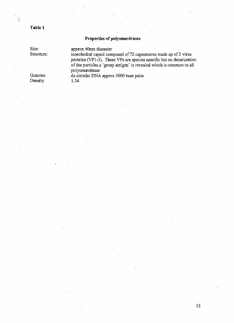
The physical and chemical properties of SV40 are typical of the polyoma family. Table 1. Notable features are the shape and size (icosohedral, 40 nm diameter) and the genetic organisation which encodes 3 different proteins (VP1-3) which are incorporated into the virus particles, as well as two important proteins (the large and small T antigens) which interact with the growth regulatory pathways in the infected cell. These T antigens are potential causes of unregulated growth by infected cells and subsequent tumour production.
SV40 is substantially more resistant to formalin inactivation than poliovirus 8,9 but the treatment with 1 :4000 formalin used to inactivate poliovaccine would reduce the titre by >99% over 50 hours.1°
The polyoma virus family
Many animal species harbour their own specific polyomaviruses, Table 2. In general the polyoma of one species grows poorly or not at all in cells or animals of different species, but there are important exceptions such as the ability of bovine polyomavirus to infect primate cells. Polyomavirus infections have a variety of clinical manifestations. The best known are

nephritis and ureteric stenosis, progressive brain disease and tumours which may be of many different pathological types. However the great majority of infections are asymptomatic. The pathologic potential of human polyomaviruses was not recognised until unexplained disease in immune suppressed patients were investigated and characteristic virus particles were found in the lesions. There proved to be two different human polyomaviruses designated JC (originally found in the brain of patients with progressive multifocal leukoencephalopathy") and BK (which originated in a ureteric turnour)12. Antibody studies showed that many adults had been infected with these viruses13. This pattern of asymptomatic lifelong infection in most members of the community with disease occurring only in a few individuals, usually with inadequate immune function, is now known to be typical of the entire family of viruses.
When polyomaviruses of different species are compared viruses from closely related hosts (such as humans and other primates) are more alike than those from evolutionarily distance hosts eg mice and birds. These resemblances mean that there is significant cross reactivity between diagnostic reagents developed for SV40, BK and JC virus detection (including T antigen dete~t ion '~ , '~) and antibody measure~nent'~,'~.
Growth of SV40
SV40 infection of a cell may lead to three different outcomes.
Virus growth:
In "permissive" cells SV40 grows relatively slowly and the distinctive vacuolation of the host cell and presence of nuclear inclusion bodies do not develop until 1-2 weeks after inoculation. Electron microscopy reveals packed arrays of virus particles in the nucleus. Virus production leads to cell death. Virus growth has two phases, the "early" phase when the virus "large T" and "small t" genes are translated and the late phase when VP1-3 and new viral DNA are produced and assembled into infectious particles. All the virus proteins are synthesised in the cytoplasm then quickly transported to the nucleus where they can be detected by specific staining.
Abortive infection:
SV40 DNA may persist in cells which are unable to support production of new viral particles. The early antigens are produced, but both viral DNA and antigens may be in low concentration. Infected cells may however become highly permissive and produce large amounts of virus if cultured in vitro. Such viral DNA can sometimes be rescued by transfection of the cell with the T antigen gene from a different but closely related member of the polyomavirus family. This has been shown experimentally for SV40, BK and JC viruses.
Transformation:
SV40 DIVA may become integrated into the genome of the host cell. If the sequences encoding the viral T antigen are intact they can be translated and the unregulated antigen expression leads to increased cell turnover. When multiple copies are integrated malignant transformation of the cell is especially likely to ensue.
Mechanism of oncogenesis by polyomaviruses - role of the T antigen
The large T antigen performs two main functions during virus replication. The first involves binding with a specific "origin of replication" in the virus DNA to establish virus DNA synthesis and the second is to promote cell division by interaction with P53 a turnour suppressor protein important in the control of cell division. New virus DNA is then synthesised by the cellular DNA polymerases in concert with the copying of the cellular DNA prior to cell division. In rare instances the combination of T antigen and P53 becomes "fixed" and normal control of the cell cycle is lost. This is usually a result of over-expression of T antigen from multiple copies of the virus gene sequences integrated in the host cell genome. Other molecular pathways for transformation by T antigen have been proposed where only part of the gene is transcribed and Tag is not detectable18. However these refer to very artificial experimental systems and remain unproven under natural conditions19.
Other virus antigens also play a significant role in virus replication and transformation at least under some circumstances, but they do not appear to act without large T activity.
Immune response to polyomavirus infection
Infected individuals produce antibodies against the virus structural proteins and the T antigens. Turnour-bearing animals have very high levels of antibody to the large T antigen.
Specific antibodies neutralise infection and have been used to free vaccine seed stocks of the virus. The production of neutralising antibody by infected animals down regulates virus production but does not eliminate infection2'.
Cell mediated antibody responses have been poorly characterised despite the significance implied by the emergence of polyoma viruses and clinical symptoms in immune suppressed subjects.
Methods of detection of SV40 in polio vaccine and tumours
The original discovery of SV40 was made by growing the virus in cell culture, and this remains the gold standard because it correlates with infectivity. It is laborious, time consuming and requires availability of cells which are known to be highly permissive. In situatians where the virus is down regulated explanting the cells in laboratory culture will often activate virus growth which can then be much more readily detected.
Other ways of detecting productive infection are by staining cells with labelled antibodies against the virus proteins (VPl is especially useful as it bears the receptor through which the virus and cell interact) and by finding virus particles by electron microscopy. These methods require fairly high level of virus presence, but are particularly valuable for showing what proportion, and which cell types are infected.
T antigen and viral DNA are also present but do not define virus production or infectivity These last two markers (viral DNA and T antigen) are present in abortively infected and transformed cells, where viral particles and structural proteins are absent.

Integration of viral DNA is the hallmark of transformed cells where it characteristically causes over-expression df T antigen, Because of its stability, viral DNA can be detected even after effective formalin inactivation of all infectivity as in routine paraffin blocks prepared for histopathology. The Southern blot methods which demonstrate integration require high levels of viral DNA in the specimen. Newer methods which amplify DNA sequences form parts of the virus (eg the polymerase chain reaction, PCR) are much more sensitive but can seldom be adapted to show whether the virus sequences are integrated.
Over the years all these methods have been used to assess the infectivity and oncogenic potential of polio vaccine. Infectious SV40 cannot always be obtained from experimentally induced tumours or from natural tumours in infected hosts but early studies showed that both viral DNA and T antigen were easily found. More recent studies based on PCR amplification ,
of SV40 sequences have detected "viral" DNA in tumours which lack T antigen or its messenger RNA. It is these studies which have reignited concern about the risk of cancer after administration of SV40 contaminated vaccine. No plausible biological mechanism has been put forward to explain how these sequences might transform cells without producing the effector (T antigen) and recently it has been suggested that these PCR results may be false positives due to contamination of the specimens with DNA sequences from molecular experiments which often use SV40 sequences as tools. This is supported by the demonstration that many of the reported positives from tumours have been found to have a deletion mutation which is found in the experimental plasmids but not in infectious virus.2'
Detection of SV40 in polin vaccine
Two types of polio vaccine have been used in Australia. Inactivated ("Salk") vaccine was used from 1958 onwards but was almost completely supplanted by live attenuated ("Sabin") vaccine after 1960. These vaccines were manufactured from different virus seeds and in different production facilities. Safety testing involved quite different criteria. The critical factor defining the safety of inactivated vaccine was the demonstration that the vaccine contained no viable poliovirus. For the attenuated vaccine the issue was the lack of virulence when injected intra-cerebrally into monkeys.
However for both vaccines all of the early virus stocks and vaccines were produced in primary rhesus or cynomolgous monkey kidney cell cultures. large batches of cells needed were made by pooling cells derived from groups of 10-30 animals, and it has been estimated that at least 70% of these pools yielded SV40 in the early 1960s. Direct testing of vaccine lots for viable virus has been very limited and many batches probably contained a mixture of killed and viable virus.22
It should noted that the US requirement for polio vaccine seeds (and production lots) be free of SV40 came into effect in 1961 but existing stocks of the "old" product were used until expiry in 1963. In other countries the date of exclusion of SV 40 from vaccines in use varied, but there was little delay in most western countries.
The natural course of SV40 infection in monkeys
SV 40 infection is common amongst wild caught rhesus monkeys. The proportion of antibody positive animals rises with increasing age. Infectious virus can be recovered from their

rsues after they have been grown in culture, but spontaneous disease is very uncommon2'. nimals which are immunosuppressed or have S N infection however develop tumours and a 'ML like syndrome24. SV40 virus has been demonstrated in these tumours using primers to letect several different regions of the virus2'. The mode of transmission is uncertain. 5xcretion in the urine and inhalation of environmental aerosols is a likely scenario.
Polyoma infection of mice is classically transmitted vertically but there is little evidence about this for SV40 under natural conditions.
SV40 infection of humans
After the discovery of SV40 recipients of polio vaccine were tested for the appearance of neutralising antibody to the virus. This was readily detected but it could be interpreted as the result of antibody response to inactivated SV40 rather than infection26. No systematic attempts were made to recover virus so it is not clear whether the persistence of this antibody is due to persistent low level infection or simply an inadvertent "vaccination" against ~ ~ 4 0 ~ ~ . In both the US and Europe about one third of recipients of attenuated oral vaccine containing 100-1000 plaque forming units of SV40 were shown to excrete virus in the faeces for several weeksz8 but there was little production of antibodg9. This evidence of infectability of humans by SV40 was directly confirmed by inoculation of volunteers, using the nasal route3'. Lastly, surveys for SV40 antibody in people from regions where contact with rhesus monkeys is common showed higher seroprevalence of anti-SV40 antibody than surveys in Europe or the New World.
More recent seroprevalence surveys using recombinant virus like particles derived fiom BK JC and SV40 show a low prevalence of reactors (about 6%) with the SV40 reagents but almost all of these disa ear when the sera are absorbed with BK and JC human PP polyomavirus particles .
There are also reports that about 6% of hospitalised children in 1999 - ie born long after use of the SV40 contamination of vaccine - had neutralising antibody to ~ ~ 4 0 ~ ' . This raises the issue of persistence of SV40 in the human population unrelated to current (or even past) use of SV40 contaminated vaccines. There are small scale studies of sera for children prior to the introduction of polio vaccine33 which show less than 5% reactors. Vertical transmission would be the most probable mechanism, but the published studies34 of babies born to mothers who were inoculated with SV40 containing vaccines are uninformative because vaccination histories of the infants are not provided.
Molecular s,tudies of SV40 in cancer patients often include normal control subjects and there are reports of detection of SV40 DNA from peripheral blood lymphocytes and other tissues of some antibody positive controls35.
There are very few reports of isolation of infectious SV 40 from non-vaccinated humans, the most convincin from two patients with progressive multifocal leukoencephalopathy and one
!6 with melanoma . None of these three patients had been immunised with inactivated poliovaccine.

Detection of SV40 in human tumours
There is only one clear report of the isolation of SV 40 from a human tumour (see above), but there are numerous reports of the demonstration of SV40 antigens or DNA sequences in tumours (including one where full length SV40 DNA was rescued by transfection of susceptible cellsJ7) . Interest has naturally focussed on the tumour types known to be caused by SV40 in hamsters, the most susceptible species. These are primary brain cancers (especially ependyrnoma), mesotheliomas, osteosarcomas and non-Hodgkins lymphomas. The studies fall into two groups; observational and case control.
Ependymoma and choroid plexus tumours:
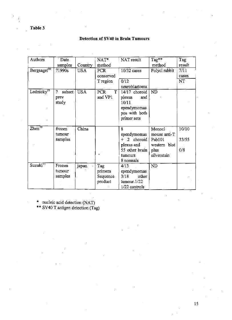
These rare brain tumours occur mainly in infants and very young children. Very few if any patients with these tumours in current US series could have received poliovaccines during the period of known SV40 contamination in 1957-63. All but one of 14 published studies of SV40 in human brain turnours report some positive findings in tumours and far fewer positives in controls. A variety of techniques have been used, ranging from culture of turnour cells to PCR detection of virus sequences (Table 3). This technical disparity in both sensitivity and target makes it hard to compare studies but the positive findings with all methodologies strengthens the overall case for the presence of papovavirus, or at least papovavirus genes in some brain tumours - especially ependymomas and choroid plexus tumours. It is however not absolutely clear that the viral antigens and sequences are derived from SV40 rather than the related human papovaviruses BK and JC 38:9. Recently a study which analysed the nucleotide sequences of three different regions of the putative SV40 genome showed that they were all SV40 related40. IIcrwewr sequence resu!ts gcnerzted by other groups have strongly suggested contamination of the samples with amplified product derived fiom one or other of the widely used experimental vector plasmids, which have incorporated SV40 T antigen gene sequences, leading to false positive results.
Interpretation of findings from different centres is also complicated by the differences in geographical distribution of rhesus monkeys and putative human exposure to ~ ~ 4 0 ~ ' . There are also very great discrepancies in the rate of detection of papovavirus sequences when comparable methods are used4', and controversy about interpretation ofboth positive and negative findings43.
Osteosarcoma:
Carbone et a1 44 studied samples fiom patients with Li-Fraumeni syndrome, a genetic disorder where only one functional allele of p53 is present. These patients develop many turnours including osteosarcoma and SV 40 T Ag might be especially potent as a carcinogen in this situation since it binds directly to p53. 11/36 of the osteosarcomas were positive for SV40 T antigen sequences. Widening of the study to p53 normal patients in different countries gave mixed results which could be attributable to environmental or technical factors. Osterosarcoma is mainly a disease of children and adolescents so any cohort effect of injections of SV-40 in 1957-63 should already be apparent.
Mesotheliomas:
Detection of SV40 sequences in mesotheliomas has been reported from many centres 45 46 47
48 49 51 55 53) ' , but others, including an International Working report negative findings , , .