review: recent advances and current challenges in scanning probe microscopy of biomolecular
TRANSCRIPT
Review: Recent Advances and Current Challenges in Scanning ProbeMicroscopy of Biomolecular Surfaces and InterfacesAnnette F. Raigoza,† Jason W. Dugger,† and Lauren J. Webb*
Department of Chemistry and Biochemistry, Center for Nano- and Molecular Science and Technology, and Institute for Cell andMolecular Biology, The University of Texas at Austin, 1 University Station, A5300, Austin, Texas 78712, United States
ABSTRACT: The introduction of scanning probe microscopy (SPM) techniquesrevolutionized the field of condensed matter science by allowing researchers toprobe the structure and composition of materials on an atomic scale. Although thesemethods have been used to make molecular- and atomic-scale measurements onbiological systems with some success, the biophysical sciences remain on the cusp ofa breakthrough with SPM technologies similar in magnitude to that experienced byfields related to solid-state surfaces and interfaces. Numerous challenges arise whenattempting to connect biological molecules that are often delicate, dynamic, andcomplex with the experimental requirements of SPM techniques. However, thereare a growing number of studies in which SPM has been successfully used to achievesubnanometer resolution measurements in biological systems where carefullydesigned and prepared samples have been paired with appropriate SPM techniques.We review significant recent innovations in applying SPM techniques to biological molecules, and highlight challenges that faceresearchers attempting to gain atomic- and molecular-level information of complex biomolecular structures.KEYWORDS: scanning probe microscopy, STM, AFM, NSOM, protein, lipid bilayer, DNA
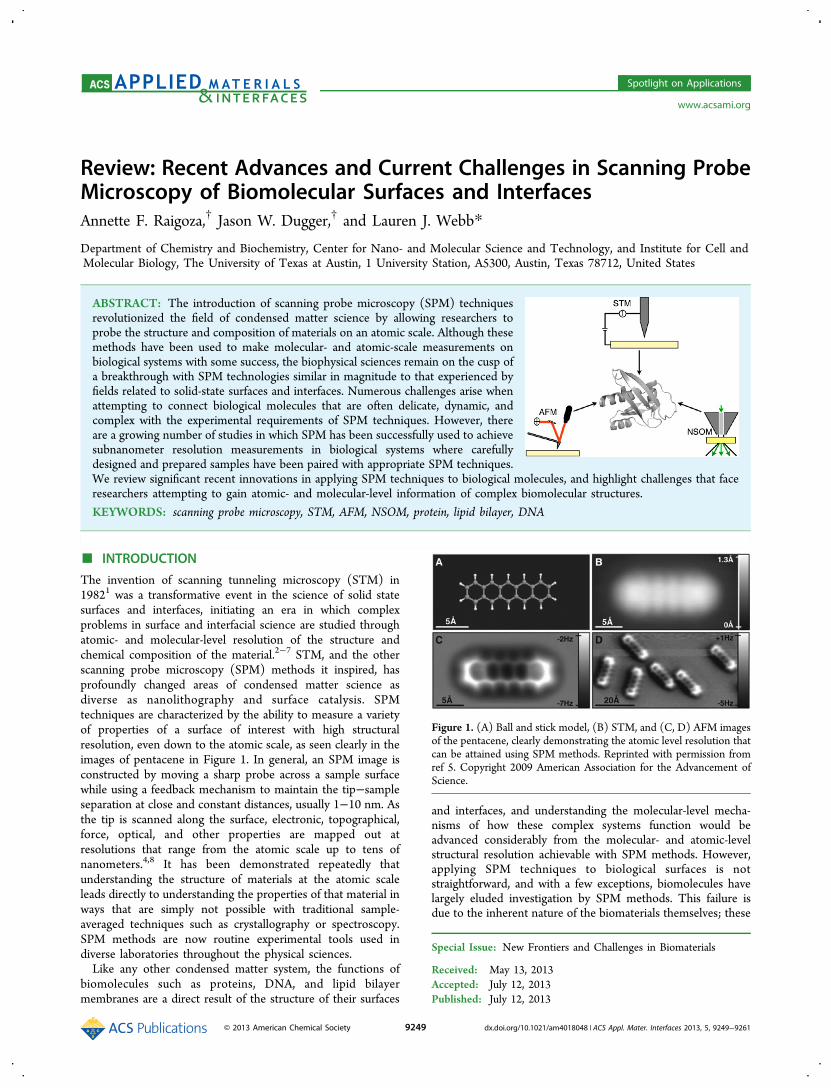
■ INTRODUCTIONThe invention of scanning tunneling microscopy (STM) in19821 was a transformative event in the science of solid statesurfaces and interfaces, initiating an era in which complexproblems in surface and interfacial science are studied throughatomic- and molecular-level resolution of the structure andchemical composition of the material.2−7 STM, and the otherscanning probe microscopy (SPM) methods it inspired, hasprofoundly changed areas of condensed matter science asdiverse as nanolithography and surface catalysis. SPMtechniques are characterized by the ability to measure a varietyof properties of a surface of interest with high structuralresolution, even down to the atomic scale, as seen clearly in theimages of pentacene in Figure 1. In general, an SPM image isconstructed by moving a sharp probe across a sample surfacewhile using a feedback mechanism to maintain the tip−sampleseparation at close and constant distances, usually 1−10 nm. Asthe tip is scanned along the surface, electronic, topographical,force, optical, and other properties are mapped out atresolutions that range from the atomic scale up to tens ofnanometers.4,8 It has been demonstrated repeatedly thatunderstanding the structure of materials at the atomic scaleleads directly to understanding the properties of that material inways that are simply not possible with traditional sample-averaged techniques such as crystallography or spectroscopy.SPM methods are now routine experimental tools used indiverse laboratories throughout the physical sciences.Like any other condensed matter system, the functions of
biomolecules such as proteins, DNA, and lipid bilayermembranes are a direct result of the structure of their surfaces
and interfaces, and understanding the molecular-level mecha-nisms of how these complex systems function would beadvanced considerably from the molecular- and atomic-levelstructural resolution achievable with SPM methods. However,applying SPM techniques to biological surfaces is notstraightforward, and with a few exceptions, biomolecules havelargely eluded investigation by SPM methods. This failure isdue to the inherent nature of the biomaterials themselves; these
Special Issue: New Frontiers and Challenges in Biomaterials
Received: May 13, 2013Accepted: July 12, 2013Published: July 12, 2013
Figure 1. (A) Ball and stick model, (B) STM, and (C, D) AFM imagesof the pentacene, clearly demonstrating the atomic level resolution thatcan be attained using SPM methods. Reprinted with permission fromref 5. Copyright 2009 American Association for the Advancement ofScience.
Spotlight on Applications
www.acsami.org
© 2013 American Chemical Society 9249 dx.doi.org/10.1021/am4018048 | ACS Appl. Mater. Interfaces 2013, 5, 9249−9261
materials display a wide diversity of chemical and structuralcomplexity at the molecular length scale, are intrinsicallydynamic, are easily deformable, and can interact strongly with aprobe as it is brought close to the surface. Furthermore, thedeformable and dynamic nature of many biological surfacesresults in mobile structures that are only transiently displayed ata surface, requiring characterization techniques that can acquireinformation on a scale much faster than the mobility of thesample surface. The innovation and development of numerousscanning methods, probe choices, and feedback mechanisms forSPM developed on inorganic surfaces and substrates havetherefore not been easily transferable to the investigation ofbiological molecules. Without the spatial resolution of SPM,proteins, DNA, lipids, and other biological molecules are mostoften characterized with spectroscopic and optical microscopymethods that average the behavior of a number of species andare often unable to describe the true heterogeneity of thestructure, function, and organization of these materials. (Singlemolecule microscopy methods are capable of resolutionsapproaching the size of individual proteins, but are afundamentally different experimental technique and beyondthe scope of this review.)Despite these difficulties, there is growing interest through-
out biophysics, biochemistry, and biomaterials to deviseexperimental conditions and methods that solve these problemsadequately for SPM to be useful, relevant, and widely applied,and a number of advances in this area have recently beenpublished. By examining these successes, it is possible tounderstand the experimental breakthroughs that are necessaryfor SPM to become as important a tool for biomaterial andbiointerface science as it currently is in inorganic materialsresearch. This review has two purposes: (1) to summarize thedevelopment of SPM on biomolecular surfaces since the lastsignificant review in 1997;9 and (2) to define and discuss someof the most important experimental constraints that have so farprevented SPM technologies from making significant headwayon the study of biomolecular surfaces. In our conclusion, wesummarize how experimental challenges are spurring innova-tion and development of these unique methods forincorporation into traditional biophysical research.
■ SCANNING PROBE METHODSThe technologies and applications of SPM have been reviewedextensively elsewhere.10,11 Before turning to a discussion ofSPM on biomolecular surfaces, we briefly review the character-istics of STM, atomic force microscopy (AFM), and near-fieldscanning optical microscopy (NSOM) that are most relevantfor understanding the successes and failures of these techniqueswhen applied to biomolecular samples. Table 1 includesresolution information and comparative advantages of the threescanning probe techniques.Scanning Tunneling Microscopy. Scanning tunneling
microscopy (STM, Figure 2), uses a sharp, metallic probe thatis positioned close enough to the surface to allow electrons totunnel between the tip and surface. This quantum mechanical
effect is possible because of the very small distances (5−10 Å)maintained between the tip and surface, allowing their electrondensities to overlap. On the basis of the bias voltages applied tothe tip and sample, electrons are driven from filled electronicstates to empty states, generating a small current (∼10−200pA) that is used to produce an image of the topography of thesample, but also contains information about the local density ofstates (DOS) of the surface. The magnitude of the current isexponentially dependent on the distance the electron musttunnel and this, coupled with piezoelectric elements thatcontrol the vertical and lateral motion of the tip, give STM thesensitivity to obtain high-resolution images. By translating thischange in DOS to a topographic map, vertical resolution in anSTM image can reach 0.01 nm. Lateral resolution isfundamentally limited by the shape of the tip and the metalatoms that protrude from the surface, and is typically no betterthan 0.1 nm. Because an STM image is a convolution of theintegrated topographic and electronic properties of each uniquespecies on the surface, topography in an STM image does notnecessarily relate to physical heights or lengths. Instead, thecontrast observed in a topographic image relies on molecularconductivity as well as quantum tunneling, in which strongtheoretical support is necessary to interpret the quantitativemeasurements of single molecule electronic properties. Otherreviews contain detailed discussions of the factors thatcontribute to the contrast seen in STM images, which mustbe considered when coupling STM with quantitative measure-ments, especially when applying them to systems containingproteins, DNA, or lipid bilayers.12,13 The contrast also yieldsadditional pseudochemical compositional information byrelating structural features obtained from STM images withphysical characteristics and chemical properties of observedmolecules to distinguish between unique species. Because STMmeasures a tunneling current, this technique requires aconductive surface that can donate or accept tunneledelectrons; however, thin insulating layers, especially thosewith charged or highly conductive functionalities, can beimaged. Biomolecular materials are typically composed ofinsulating molecules with short tunneling lengths (usually <5
Table 1. Comparative Advantages for Each of the Scanning Probe Techniques
STM AFM NSOM
signal electron tunneling attractive/repulsive forces near-field lightlateral resolution (nm) 0.1 5−10 20vertical resolution (nm) 0.01 0.1 2−5advantages high lateral and vertical resolution conductive or nonconductive surface traditional optical imaging at higher resolution
Figure 2. In STM, a metallic probe is brought close enough to asample surface to enable tunneling between the sample and the tip.The direction of the tunneling current (I) is controlled by a biaspotential that is applied between the tip and the surface. As the tipmoves across the surface, the tunneling current is monitored andtranslated into a topographic image.
ACS Applied Materials & Interfaces Spotlight on Applications
dx.doi.org/10.1021/am4018048 | ACS Appl. Mater. Interfaces 2013, 5, 9249−92619250

nm), and therefore cannot be accessed by STM if they are heldeven a small distance away from a conducting substrate.Atomic Force Microscopy. Atomic force microscopy
(AFM, Figure 3) monitors intermolecular interactions between
a probe tip mounted on an oscillating cantilever and the surfaceunder investigation. The motion of the cantilever is tracked as itmoves across the surface by changes in the position of a laserbeam that is deflected toward a detector. Because of its relianceon noncovalent intermolecular forces, an AFM can be operatedover a variety of intermolecular conditions. Three scanningmodes are most commonly discussed: contact, noncontact, ortapping mode. Contact mode takes advantage of repulsiveforces between the probe and the sample as the cantilever isessentially dragged across the surface, while noncontact modeuses attractive forces while the cantilever is close, but nottouching, the surface. In tapping mode, the cantilever oscillatesnear its resonant frequency as it moves across the surface,alternating between attractive and repulsive regimes dependingon the distance to the substrate. Because a portion of the tipinteracts with the surface being examined, AFM resolution inthe lateral xy direction is limited by the radius of the probe tip,typically 5−10 nm. However, higher resolutions can be reachedin the vertical z direction (as low as 0.1 nm) because theroundness of the cantilever probe does not factor into thismeasurement. Because AFM relies on attractive and repulsiveinteractions between the tip and surface, it can be applied toany material and does not suffer from a fundamental limit ofconductance like STM. AFM images are therefore trulytopographical, although they are often convoluted with thesize and shape of the probe tip. The use of coated or specializedtips allows for alternative measurements such as magnetic,elastic, and binding forces,14−18 as well as surface potentials,19,20
and is an area of much recent innovation in AFM technologies.Near-Field Scanning Optical Microscopy. Optical
microscopy techniques are fundamentally limited by thediffraction of light in the far field and are limited to resolvefeatures on the order of half of the wavelength of theilluminating light. Near-field scanning optical microscopy(NSOM, Figure 4) is a scanning probe technique that producestopographical and optical images below the diffraction limit oflight through the use of evanescent waves in the near field.NSOM typically uses metal-coated, optical fibers that have atapered end and are fabricated with apertures significantlysmaller than the wavelength of light, ∼20−100 nm. Threecommon methods for performing NSOM measurements can beachieved by varying the position of the illuminating source andthe detector to transmit or collect light that interacts with the
surface, shown in Figure 4. Light that is transmitted throughthese apertures is laterally confined to the size of the opening,but only near the probe. Therefore, the probe must staypositioned close to the sample surface (<10 nm) as theilluminating field decays exponentially. Lateral resolution canreach 20 nm, but is limited by the size of the aperture and theprobe distance from the sample. The working distance is alsolimited in the vertical direction, requiring that the probe remainclose enough that the sample is still present in the near field.Vertical resolutions of 2−5 nm have been reached, however.NSOM lends itself to most conventional optical modes,typically involving illumination or collection through theprobe itself.
■ APPLYING SPM TECHNOLOGIES TOWELL-CHARACTERIZED COMPLEX BIOLOGICALSYSTEMS
When applying SPM methods, a comprehensive knowledge ofthe experimental system being investigated and its chemicalenvironment are required, not only to interpret the acquireddata accurately, but to test the accuracy of instrumentcapabilities. An example of this comes from the dawn ofSTM itself: the highly debated structure of the ultrahighvacuum (UHV)-reconstructed Si(111) surface. Before theinvention of STM, several experimental and theoreticalapproaches to determine the geometry of the surface had ledto inconsistent results that could not rule out several reasonablecandidate structures.21 It was not until Binnig and co-workersimaged this surface directly with STM that the reconstructed 7× 7 geometry was confirmed to be correct.22 These results werean exciting motivation for the use of STM, but alsodemonstrated that a great deal of information must first beunderstood about the sample in order to interpret topo-graphical images reliably. This becomes particularly challengingwhen considering the wide diversity in chemical composition,structure, dynamics, and environmental interactions that areknown to be present in many types of biomolecules of interest.No matter the SPM technique chosen to investigate a
sample, there are a few common hurdles to overcome whenprobing biological systems. (I) Biological samples areinherently dynamic in their native environments and candeform and diffuse on time scales that make probe microscopymethods difficult to use. (II) Biomaterials are often described asbeing “soft” or “sticky,” as they can be easily damaged and mayinteract unfavorably with the probe. (III) Biomolecules areoften charged, prone to aggregation, or have semi-insulating
Figure 3. AFM measures noncovalent attractive and repulsive forcesbetween the probe and the sample as a cantilever is moved across thesurface and monitored through the positioning of a laser that isdeflected toward a detector.
Figure 4. In NSOM, light that is transmitted through an aperture ofthe probe is laterally confined to the size of the opening, but only nearthe probe tip. Three common methods for performing NSOMmeasurements place the illumination source and detector at varyingpositions to allow for transmission or collection of light that interactswith the surface. (A) transmission, (B) collection, and (C) aperture-less probe.
ACS Applied Materials & Interfaces Spotlight on Applications
dx.doi.org/10.1021/am4018048 | ACS Appl. Mater. Interfaces 2013, 5, 9249−92619251
characteristics, all of which depend heavily on their localenvironment. Despite these challenges, there are an increasingnumber of successful examples employing SPM to characterizetopography, force, optical properties, and even to providemechanistic insight of soft, biological surfaces.23−27 Inreviewing these advances, we will emphasize how researchershave overcome these three experimental obstacles by exercisingcontrol over the substrate, local environment, sample, and tipused in their SPM measurements. Although the strategiesdiscussed will not provide substantial protocols for preparingbiological samples, such procedures have been publishedelsewhere.28−31
I. Immobilizing Biomolecules. The dynamic nature ofbiomolecules can make probe microscopy methods difficult toapply to some systems. To obtain resolution on the nanometeror subnanometer scale, the molecules of interest must remainimmobile for the time scale of the measurement, which can takeup to several minutes per frame in the case of AFM; a verymobile sample can result in blurred images or in measurementsthat represent an average of multiple states. A common methodfor managing this issue is to immobilize samples on surfacesthrough covalent linkages or weaker noncovalent interactions.Another way to suppress the molecular motions of somesamples is to suspend them in matrices that force them to orderor orient in a consistent and repeatable manner. Such repeatedallows researchers to identify and measure biomolecularstructures of interest more easily. As studies of proteins orpeptides, bilayers, and DNA have become more common, thedevelopment of methods to immobilize and order each class ofbiomolecule has progressed as well. Often such methods placethe molecules of interest in environments that are very differentfrom their native surroundings. Although these deviations areimportant to understand and consider in any data analysis, themanufactured environment can allow for unique studies of themechanical or electrical properties of the sample.DNA. One of the simpler methods for attaching
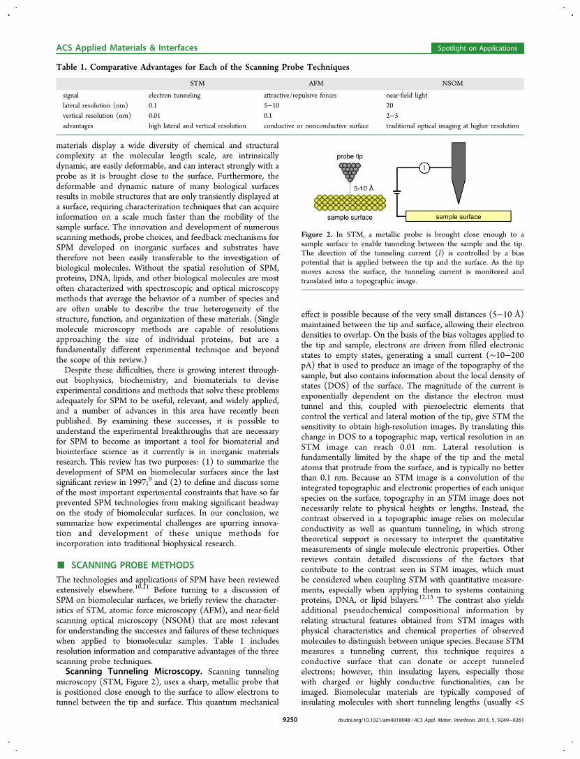
biomolecules to a surface is to take advantage of the fact thatmica and DNA have similar surface charges. By introducingcations such as Mg2+ or Ni2+, a layer of these ions forms on themica surface and serves as an electrostatically adhesive layerwith which DNA can be immobilized.4,32 Furthermore, therelative surface concentration of monovalent (which alone donot promote immobilization) to divalent cations can beadjusted to mediate the strength of attachment. It has alsobeen shown that the functionalization of a freshly cleaved micasurface with methyltrimethoxysilane has the same effect withthe added benefit of straightening DNA interacting with thesubstrate.33 These techniques have allowed researchers toachieve submolecular resolution of closed circular DNAplasmids as well as self-assembled DNA crystals usingfrequency-modulated (FM)-AFM.34 Using this charge balanc-ing technique for immobilization coupled with an advancedAFM method that allows for soft tip landings, thus minimizingtip-induced sample deformation, the authors were able toresolve not only the major and minor grooves of the helix butalso individual phosphate groups along the backbone (Figure5). Although the image resolution and quality of measurementsmade by these authors is a testament to the abilities of SPM,they would not be possible were it not for the well-understoodand periodic structure of DNA.35
SPM techniques have also been used to characterize thestructures of self-assembled nanoscale abiological DNAconstructs. Winfree et al. were able to construct two-
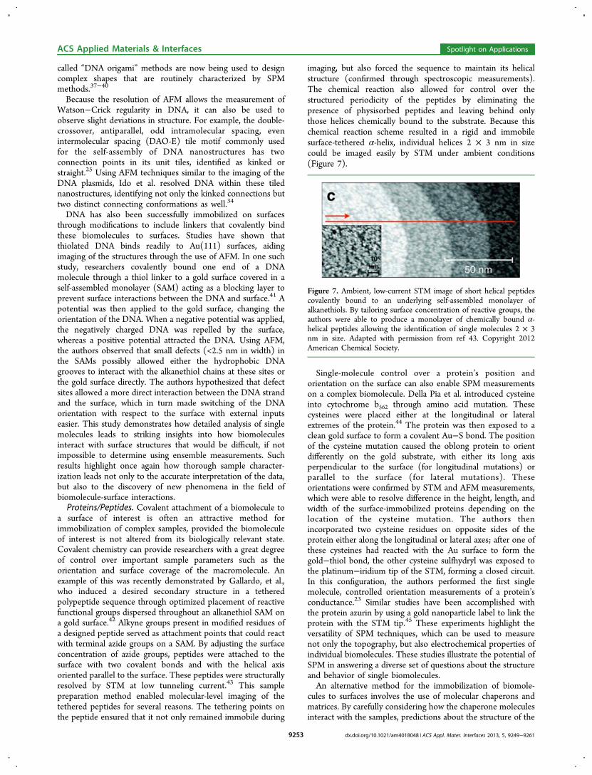
dimensional crystal structures from strands of programmedDNA by exploiting Watson−Crick base pairing, aromatic vander Waals interactions between the nucleotide bases, and adetailed knowledge of the geometric configuration of the DNAdouble helix itself.36 As the self-assembly relied on Watson−Crick base pairing, the sequence was constructed in such a waythat specific nucleotide sections were complementary only toone other region of the DNA strand, thus minimizing anyunintended pairing. The incorporation of DNA hairpins intothe vertical columns of the lattice structure resulted in visiblestripes when AFM was used to image the nanostructures on amica surface (Figure 6). Molecularly resolved SPM images werepossible in this case because of the built-in periodicity of theseDNA hairpins. By tailoring this structural parameter throughrational alterations in the sequence, the researchers were able touse the imaged periodicity as a benchmark for how successfullytheir designs formed from solution self-assembly. These so-
Figure 5. AFM image of plasmid DNA immobilized on a freshlycleaved mica surface in a 50 mM NiCl2 solution. By coupling the well-understood structure of DNA with this immobilization technique, Idoet al. were able to resolve both major and minor grooves of DNA aswell as identify topological protrusions as phosphate groups. Figurelabels are described in ref 34. Reprinted with permission from ref 34.Copyright 2013 American Chemical Society.
Figure 6. DNA DAO-E lattice structure designed to self-assemblethrough Watson−Crick base pairing and imaged by AFM. Theincorporation of hairpin sequences into the vertical columns (1−2nm) of the lattice structure give rise to the brighter stripes in theimage, which act as a convenient marker calibrating the distancebetween structural features clearly visible in the image. The scale bar is300 nm. Reprinted by permission ref 36. Copyright 1998 MacmillanPublishers.
ACS Applied Materials & Interfaces Spotlight on Applications
dx.doi.org/10.1021/am4018048 | ACS Appl. Mater. Interfaces 2013, 5, 9249−92619252
called “DNA origami” methods are now being used to designcomplex shapes that are routinely characterized by SPMmethods.37−40
Because the resolution of AFM allows the measurement ofWatson−Crick regularity in DNA, it can also be used toobserve slight deviations in structure. For example, the double-crossover, antiparallel, odd intramolecular spacing, evenintermolecular spacing (DAO-E) tile motif commonly usedfor the self-assembly of DNA nanostructures has twoconnection points in its unit tiles, identified as kinked orstraight.25 Using AFM techniques similar to the imaging of theDNA plasmids, Ido et al. resolved DNA within these tilednanostructures, identifying not only the kinked connections buttwo distinct connecting conformations as well.34
DNA has also been successfully immobilized on surfacesthrough modifications to include linkers that covalently bindthese biomolecules to surfaces. Studies have shown thatthiolated DNA binds readily to Au(111) surfaces, aidingimaging of the structures through the use of AFM. In one suchstudy, researchers covalently bound one end of a DNAmolecule through a thiol linker to a gold surface covered in aself-assembled monolayer (SAM) acting as a blocking layer toprevent surface interactions between the DNA and surface.41 Apotential was then applied to the gold surface, changing theorientation of the DNA. When a negative potential was applied,the negatively charged DNA was repelled by the surface,whereas a positive potential attracted the DNA. Using AFM,the authors observed that small defects (<2.5 nm in width) inthe SAMs possibly allowed either the hydrophobic DNAgrooves to interact with the alkanethiol chains at these sites orthe gold surface directly. The authors hypothesized that defectsites allowed a more direct interaction between the DNA strandand the surface, which in turn made switching of the DNAorientation with respect to the surface with external inputseasier. This study demonstrates how detailed analysis of singlemolecules leads to striking insights into how biomoleculesinteract with surface structures that would be difficult, if notimpossible to determine using ensemble measurements. Suchresults highlight once again how thorough sample character-ization leads not only to the accurate interpretation of the data,but also to the discovery of new phenomena in the field ofbiomolecule-surface interactions.Proteins/Peptides. Covalent attachment of a biomolecule to
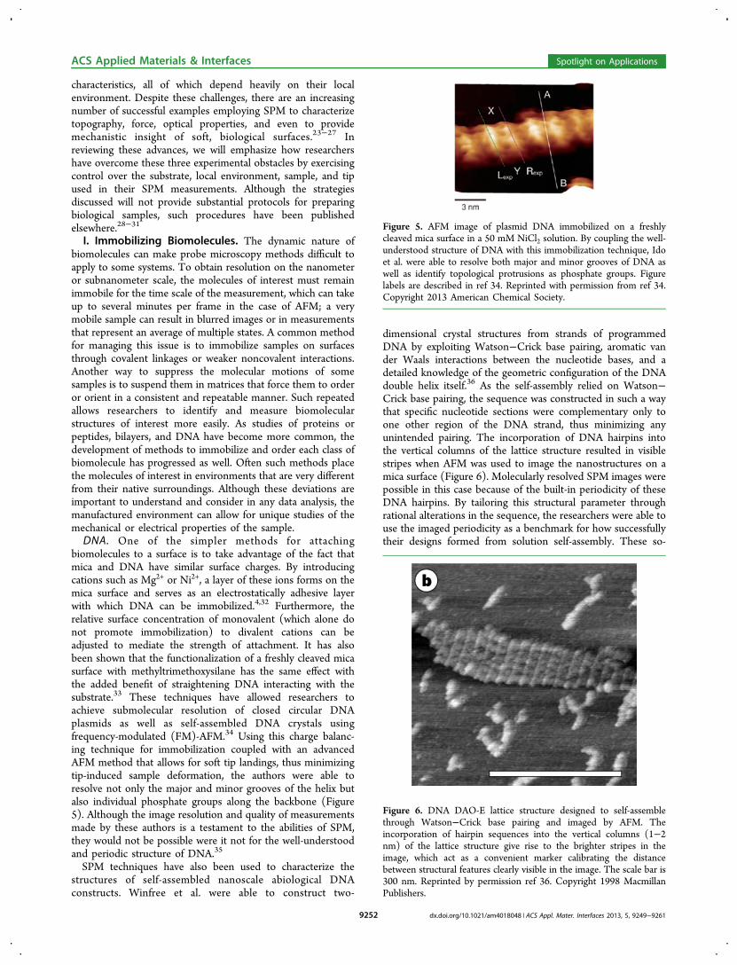
a surface of interest is often an attractive method forimmobilization of complex samples, provided the biomoleculeof interest is not altered from its biologically relevant state.Covalent chemistry can provide researchers with a great degreeof control over important sample parameters such as theorientation and surface coverage of the macromolecule. Anexample of this was recently demonstrated by Gallardo, et al.,who induced a desired secondary structure in a tetheredpolypeptide sequence through optimized placement of reactivefunctional groups dispersed throughout an alkanethiol SAM ona gold surface.42 Alkyne groups present in modified residues ofa designed peptide served as attachment points that could reactwith terminal azide groups on a SAM. By adjusting the surfaceconcentration of azide groups, peptides were attached to thesurface with two covalent bonds and with the helical axisoriented parallel to the surface. These peptides were structurallyresolved by STM at low tunneling current.43 This samplepreparation method enabled molecular-level imaging of thetethered peptides for several reasons. The tethering points onthe peptide ensured that it not only remained immobile during
imaging, but also forced the sequence to maintain its helicalstructure (confirmed through spectroscopic measurements).The chemical reaction also allowed for control over thestructured periodicity of the peptides by eliminating thepresence of physisorbed peptides and leaving behind onlythose helices chemically bound to the substrate. Because thischemical reaction scheme resulted in a rigid and immobilesurface-tethered α-helix, individual helices 2 × 3 nm in sizecould be imaged easily by STM under ambient conditions(Figure 7).
Single-molecule control over a protein’s position andorientation on the surface can also enable SPM measurementson a complex biomolecule. Della Pia et al. introduced cysteineinto cytochrome b562 through amino acid mutation. Thesecysteines were placed either at the longitudinal or lateralextremes of the protein.44 The protein was then exposed to aclean gold surface to form a covalent Au−S bond. The positionof the cysteine mutation caused the oblong protein to orientdifferently on the gold substrate, with either its long axisperpendicular to the surface (for longitudinal mutations) orparallel to the surface (for lateral mutations). Theseorientations were confirmed by STM and AFM measurements,which were able to resolve difference in the height, length, andwidth of the surface-immobilized proteins depending on thelocation of the cysteine mutation. The authors thenincorporated two cysteine residues on opposite sides of theprotein either along the longitudinal or lateral axes; after one ofthese cysteines had reacted with the Au surface to form thegold−thiol bond, the other cysteine sulfhydryl was exposed tothe platinum−iridium tip of the STM, forming a closed circuit.In this configuration, the authors performed the first singlemolecule, controlled orientation measurements of a protein’sconductance.23 Similar studies have been accomplished withthe protein azurin by using a gold nanoparticle label to link theprotein with the STM tip.45 These experiments highlight theversatility of SPM techniques, which can be used to measurenot only the topography, but also electrochemical properties ofindividual biomolecules. These studies illustrate the potential ofSPM in answering a diverse set of questions about the structureand behavior of single biomolecules.An alternative method for the immobilization of biomole-
cules to surfaces involves the use of molecular chaperons andmatrices. By carefully considering how the chaperone moleculesinteract with the samples, predictions about the structure of the
Figure 7. Ambient, low-current STM image of short helical peptidescovalently bound to an underlying self-assembled monolayer ofalkanethiols. By tailoring surface concentration of reactive groups, theauthors were able to produce a monolayer of chemically bound α-helical peptides allowing the identification of single molecules 2 × 3nm in size. Adapted with permission from ref 43. Copyright 2012American Chemical Society.
ACS Applied Materials & Interfaces Spotlight on Applications
dx.doi.org/10.1021/am4018048 | ACS Appl. Mater. Interfaces 2013, 5, 9249−92619253
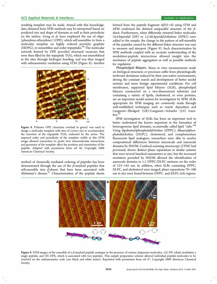
resulting template may be made. Armed with this knowledge,data obtained from SPM methods may be interpreted based onpredicted size and shape of domains as well as their periodicityon the surface. Gong et al. have employed the use of oligo-(phenylene-ethynylene) (OPE), which self-assembles to form amolecular template on highly ordered pyrolytic graphite(HOPG), to immobilize and order tripeptides.46 The molecularnetwork formed by OPE provided structural vacancies thatwere then filled by the tripeptide TGG, which was immobilizedin the sites through hydrogen bonding, and was then imagedwith subnanometer resolution using STM (Figure 8). Another
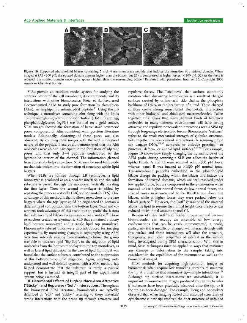
method of chemically mediated ordering of peptides has beendemonstrated through the use of the β-amyloid peptides thatself-assemble into β-sheets that have been associated withAlzheimer’s disease.47 Characterization of the peptide sheets
formed from the peptide fragment Aβ33−42 using STM andAFM confirmed the debated antiparallel structure of the β-sheet. Furthermore, when differently oriented linker molecules(4,4-bipyridyl (DP) or 1,2-di(4pyridyl)ethylene (DPE)) wereadded to the sample, the change in the pattern of self-assemblyof the peptides caused by the different linker structure was easyto measure and interpret (Figure 9). Such characterization bySPM methods coupled with an accurate understanding of themodulator-peptide interactions allowed insight into themechanics of peptide aggregation as well as possible methodsfor regulation.
Phospholipid Bilayers. Many in vitro measurements madeon biological structures or processes suffer from physiologicallyirrelevant deviations induced by their non-native environments,driving the constant search and development of better modelsystems and more benign experimental conditions. For cellmembranes, supported lipid bilayers (SLB), phospholipidbilayers constructed on a two-dimensional substrate andcontaining a variety of lipids, cholesterol, or even proteins,are an important model system for investigation by SPM. SLBsappropriate for SPM imaging are commonly made throughwell-established techniques such as vesicle deposition andLangmuir−Blodgett (LB)/Langmuir−Schaefer (LS) trans-fers.30
SPM investigation of SLBs has been an important tool tobetter understand the factors important in the formation ofheterogeneous lipid domains, occasionally called lipid “rafts.”48
Using dipalmitoylphosphatidylcholine (DPPC), dilauroylphos-phatidylcholine (DLPC), cholesterol, and complementaryfluorescent lipid analogues, researchers were able to resolvecompositional differences between microscale and nanoscaledomains by NSOM. Confocal scanning microscopy (CFM) hadpreviously shown distinct phase separations in similar systemsthat were several hundred nanometers in size, but the increasedresolution provided by NSOM allowed the identification ofnanoscale domains in 1:1 DPPC/DLPC mixtures on the orderof 123−145 nm. In addition, when SLBs containing DPPC,DLPC, and cholesterol were imaged, phase separations 70−100nm in size were found between DPPC- and DLPC-rich regions.
Figure 8. Polymer OPE (structure overlaid in green) was used todesign a molecular template with sites of correct size to accommodatethe insertion of the tripeptide TGG, indicated by the arrow. Theimposed order and periodicity of the template visible in this STMimage allowed researchers to probe how intermolecular interactionsand geometry of the template affect the position and orientation of thepeptide. Adapted with permission from ref 46. Copyright 2006American Chemical Society.
Figure 9. STM images of the assembly of a β-amyloid peptide analogue in the presence of various chaperone molecules: (A) DP, which modulates asingle peptide, and (B) DPE, which is associated with two peptides. This sample preparation scheme allowed individual peptide molecules to beresolved on the subnanometer scale (see black and white insets). Reprinted with permission from ref 47. Copyright 2009 American ChemicalSociety.
ACS Applied Materials & Interfaces Spotlight on Applications
dx.doi.org/10.1021/am4018048 | ACS Appl. Mater. Interfaces 2013, 5, 9249−92619254
SLBs provide an excellent model system for studying thecomplex nature of the cell membrane, its components, and itsinteractions with other biomolecules. Pieta, et al., have usedelectrochemical STM to study pore formation by alamethicin(Alm), an amphipathic antimicrobial peptide.24 Using the LBtechnique, a monolayer containing Alm along with the lipids1,2-dimyristoyl-sn-glycero-3-phosphocholine (DMPC) and eggphosphatidylglycerol (egPG) was formed on a gold surface.STM images showed the formation of barrel-stave hexamericpores composed of Alm consistent with previous literaturemodels. Additionally, clustering of these pores was alsoobserved. By coupling their images with the well understoodnature of the peptide, Pieta, et al., demonstrated that the Almmolecules were able to participate in the formation of adjacentpores, and that only 3−4 peptides contributed to thehydrophilic interior of the channel. The information gleanedfrom this study helps show how STM may be used to providemechanistic insight into the formation of structures within lipidbilayers.When SLBs are formed through LB techniques, a lipid
monolayer is produced at an air/water interface, and the solidsubstrate is passed through the monolayer vertically, creatingthe first layer. Then the second monolayer is added byrepeating the process vertically (LB), or horizontally (LS). Oneadvantage of this method is that it allows researchers to preparebilayers where the top layer could be engineered to contain adifferent lipid composition than the bottom layer. Yuan and co-workers took advantage of this technique to probe the factorsthat influence lipid bilayer reorganization on a surface.26 Theseresearchers created an asymmetric SLB that contained a binarylipid bottom monolayer and a single lipid top monolayer.Fluorescently labeled lipids were also introduced for imagingexperiments. By monitoring changes in topography using AFMover time intervals ranging from minutes to hours, the groupwas able to measure lipid “flip-flop”, or the migration of lipidmolecules from the bottom monolayer to the top monolayer, aswell as lateral lipid diffusion. In the case of lipid flip-flop, it wasfound that the surface substrate contributed to the suppressionof this bottom-to-top lipid migration. Again, coupling well-understood and well-defined systems with a SPM method hashelped demonstrate that the substrate is rarely a passivesupport, but is instead an integral part of the experimentalsystem being examined.II. Detrimental Effects of High-Surface-Area Attractive
(“Sticky”) and Repulsive (“Soft”) Interactions. Throughoutthe biomaterial SPM literature, biomolecules are typicallydescribed as “soft” and “sticky,” referring to these materials’strong interactions with the probe tip through attractive and
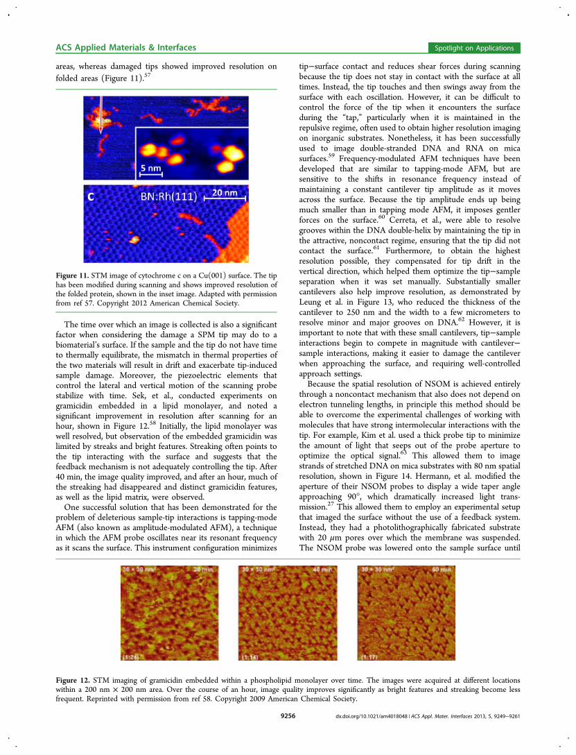
repulsive forces. The “stickiness” that authors commonlymention when discussing biomolecules is a result of chargedsurfaces created by amino acid side chains, the phosphatebackbone of DNA, or the headgroup of a lipid. These chargedsurfaces create strong noncovalent electrostatic interactionswith other biological and abiological macromolecules. Takentogether, this means that many different kinds of biologicalmolecules in many different environments will have strongattractive and repulsive noncovalent interactions with a SPM tipthrough long-range electrostatic forces. Biomolecular “softness”refers to the weak mechanical strength of globular structuresheld together by noncovalent interactions. A scanning probecan damage DNA,49,50 compress or dislodge proteins,51 orpuncture, deform, or anneal lipid surfaces.52−55 For example,Figure 10 shows how simply changing the normal force of theAFM probe during scanning a SLB can affect the height oflipids. Panels A and C were scanned with <500 pN force,whereas panel B was imaged at >1500 pN normal force.Transmembrane peptides embedded in the phospholipidbilayer disrupt the packing within the bilayer and induce theformation of striated domains, which are well-resolved underlow applied force, but are compressed in the z dimension whenscanned under higher normal force. At low normal forces, thestriated areas were measured to be 0.2−0.4 nm above thebilayer, while at higher forces they were pushed below thebilayer surface.56 However, the “soft” character of the materialallows the lipid to resume their initial height once the force wasreduced to its initial amount (panel C).Because of these “soft” and “sticky” properties, and because
biomolecules can occupy an ensemble of low energyconformations that can be easily interconverted, a probe,particularly if it is metallic or charged, will interact strongly withthis surface and these interactions will alter the structure,topography, and other properties of interest in the samplebeing investigated during SPM characterization. With this inmind, SPM techniques must be applied in ways that minimizeany damage or deformation to the sample, taking intoconsideration the capabilities of the instrument as well as thebiomaterial imaged.STM methods for acquiring high-resolution images of
biomaterials often require low tunneling currents to maintainthe tip at a distance that minimizes tip−sample interactions.49
Although tip−surface interactions are unavoidable, it isimportant to monitor the images produced by the tip to inferif molecules have been physically adsorbed onto the tip, or ifthe tip has been damaged. For example, Deng and co-workersobserved that when imaging folded and unfolded structures ofcytochrome c, new tips resolved the finer structure of unfolded
Figure 10. Supported phospholipid bilayer containing 2 mol % transmembrane peptide that induces the formation of a striated domain. Whenimaged at (A) <500 pN, the striated domain appears higher than the bilayer, but (B) is compressed at higher forces, >1500 pN. (C) As the force isreduced, the striated domain once again appears higher than the surrounding bilayer. Reprinted with permission from ref 56. Copyright 2000American Chemical Society.
ACS Applied Materials & Interfaces Spotlight on Applications
dx.doi.org/10.1021/am4018048 | ACS Appl. Mater. Interfaces 2013, 5, 9249−92619255
areas, whereas damaged tips showed improved resolution onfolded areas (Figure 11).57
The time over which an image is collected is also a significantfactor when considering the damage a SPM tip may do to abiomaterial’s surface. If the sample and the tip do not have timeto thermally equilibrate, the mismatch in thermal properties ofthe two materials will result in drift and exacerbate tip-inducedsample damage. Moreover, the piezoelectric elements thatcontrol the lateral and vertical motion of the scanning probestabilize with time. Sek, et al., conducted experiments ongramicidin embedded in a lipid monolayer, and noted asignificant improvement in resolution after scanning for anhour, shown in Figure 12.58 Initially, the lipid monolayer waswell resolved, but observation of the embedded gramicidin waslimited by streaks and bright features. Streaking often points tothe tip interacting with the surface and suggests that thefeedback mechanism is not adequately controlling the tip. After40 min, the image quality improved, and after an hour, much ofthe streaking had disappeared and distinct gramicidin features,as well as the lipid matrix, were observed.One successful solution that has been demonstrated for the
problem of deleterious sample-tip interactions is tapping-modeAFM (also known as amplitude-modulated AFM), a techniquein which the AFM probe oscillates near its resonant frequencyas it scans the surface. This instrument configuration minimizes
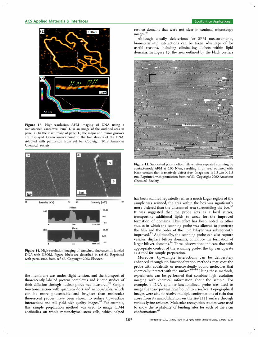
tip−surface contact and reduces shear forces during scanningbecause the tip does not stay in contact with the surface at alltimes. Instead, the tip touches and then swings away from thesurface with each oscillation. However, it can be difficult tocontrol the force of the tip when it encounters the surfaceduring the “tap,” particularly when it is maintained in therepulsive regime, often used to obtain higher resolution imagingon inorganic substrates. Nonetheless, it has been successfullyused to image double-stranded DNA and RNA on micasurfaces.59 Frequency-modulated AFM techniques have beendeveloped that are similar to tapping-mode AFM, but aresensitive to the shifts in resonance frequency instead ofmaintaining a constant cantilever tip amplitude as it movesacross the surface. Because the tip amplitude ends up beingmuch smaller than in tapping mode AFM, it imposes gentlerforces on the surface.60 Cerreta, et al., were able to resolvegrooves within the DNA double-helix by maintaining the tip inthe attractive, noncontact regime, ensuring that the tip did notcontact the surface.61 Furthermore, to obtain the highestresolution possible, they compensated for tip drift in thevertical direction, which helped them optimize the tip−sampleseparation when it was set manually. Substantially smallercantilevers also help improve resolution, as demonstrated byLeung et al. in Figure 13, who reduced the thickness of thecantilever to 250 nm and the width to a few micrometers toresolve minor and major grooves on DNA.62 However, it isimportant to note that with these small cantilevers, tip−sampleinteractions begin to compete in magnitude with cantilever−sample interactions, making it easier to damage the cantileverwhen approaching the surface, and requiring well-controlledapproach settings.Because the spatial resolution of NSOM is achieved entirely
through a noncontact mechanism that also does not depend onelectron tunneling lengths, in principle this method should beable to overcome the experimental challenges of working withmolecules that have strong intermolecular interactions with thetip. For example, Kim et al. used a thick probe tip to minimizethe amount of light that seeps out of the probe aperture tooptimize the optical signal.63 This allowed them to imagestrands of stretched DNA on mica substrates with 80 nm spatialresolution, shown in Figure 14. Hermann, et al. modified theaperture of their NSOM probes to display a wide taper angleapproaching 90°, which dramatically increased light trans-mission.27 This allowed them to employ an experimental setupthat imaged the surface without the use of a feedback system.Instead, they had a photolithographically fabricated substratewith 20 μm pores over which the membrane was suspended.The NSOM probe was lowered onto the sample surface until
Figure 11. STM image of cytochrome c on a Cu(001) surface. The tiphas been modified during scanning and shows improved resolution ofthe folded protein, shown in the inset image. Adapted with permissionfrom ref 57. Copyright 2012 American Chemical Society.
Figure 12. STM imaging of gramicidin embedded within a phospholipid monolayer over time. The images were acquired at different locationswithin a 200 nm × 200 nm area. Over the course of an hour, image quality improves significantly as bright features and streaking become lessfrequent. Reprinted with permission from ref 58. Copyright 2009 American Chemical Society.
ACS Applied Materials & Interfaces Spotlight on Applications
dx.doi.org/10.1021/am4018048 | ACS Appl. Mater. Interfaces 2013, 5, 9249−92619256
the membrane was under slight tension, and the transport offluorescently labeled protein complexes and kinetic studies oftheir diffusion through nuclear pores was measured.27 Samplefunctionalization with quantum dots and nanoparticles, whichcan be more photostable and brighter than molecularfluorescent probes, have been shown to reduce tip−surfaceinteractions and still yield high-quality images.63 For example,this sample preparation method was used to image CD44antibodies on whole mesenchymal stem cells, which helped
resolve domains that were not clear in confocal microscopyimages.64
Although usually deleterious for SPM measurements,biomaterial−tip interactions can be taken advantage of foruseful reasons, including eliminating defects within lipiddomains. In Figure 15, the area outlined by the black corners
has been scanned repeatedly; when a much larger region of thesample was scanned, the area within the box was significantlymore ordered than the unscanned area surrounding the box.53
It was suggested that the probe acts as a local stirrer,transporting additional lipids to areas for the improvedformation of domains. This effect has been noted in otherstudies in which the scanning probe was allowed to penetratethe film and the order of the lipid bilayer was subsequentlyimproved.55 Additionally, the scanning probe can also rupturevesicles, displace bilayer domains, or induce the formation oflarger bilayer domains.54 These observations indicate that withappropriate control of the scanning probe, the tip can operateas a tool for sample preparation.Moreover, tip−sample interactions can be deliberately
enhanced through tip-functionalization methods that coat theprobe with covalently or noncovalently bound molecules thatchemically interact with the surface.65−68 Using these methods,experiments can be performed that combine high-resolutionimaging with chemical information about the sample. Forexample, a DNA aptamer-functionalized probe was used toimage the toxic protein ricin bound to a surface. Topographicalimages were able to resolve multiple conformations of ricin thatarose from its immobilization on the Au(111) surface throughvarious lysine residues. Molecular recognition studies were usedto show the availability of binding sites for each of the ricinconformations.69
Figure 13. High-resolution AFM imaging of DNA using aminiaturized cantilever. Panel D is an image of the outlined area inpanel C. In the inset image of panel D, the major and minor groovesare displayed. Green arrows point to the two strands of the DNA.Adapted with permission from ref 62. Copyright 2012 AmericanChemical Society.
Figure 14. High-resolution imaging of stretched, fluorescently labeledDNA with NSOM. Figure labels are described in ref 63. Reprintedwith permission from ref 63. Copyright 2002 Elsevier.
Figure 15. Supported phospholipid bilayer after repeated scanning bycontact-mode AFM at 0.06 N/m, resulting in an area outlined withblack corners that is relatively defect free. Image size is 1.5 μm × 1.5μm. Reprinted with permission from ref 53. Copyright 2000 AmericanChemical Society.
ACS Applied Materials & Interfaces Spotlight on Applications
dx.doi.org/10.1021/am4018048 | ACS Appl. Mater. Interfaces 2013, 5, 9249−92619257
III. Managing Environmental Influences. Biomoleculesare often charged, prone to aggregation, or have semi-insulatingcharacteristics, all of which depend heavily on their local surfaceor solution environment and any of which can change theorientation and structure of the biomolecule being observed.Successfully collecting and interpreting SPM images thereforeoften depends on the chemical environment surrounding thebiomolecule, such as solution chemistry, pH, substrate surfacecharge, and surface topography. The shape and composition ofthe probe tip can influence the magnitude of the signalobtained and can be enhanced or diminished based on thetypes of interactions that are desired. Substrate charge andtopography can influence how biomolecules interact withsurfaces by promoting or inhibiting immobilization oraggregation. The medium in which the samples are studied(ambient, solution, or vacuum) can greatly affect thequantitative analysis of data. For studies in a solutionenvironment, ion concentration and pH can drasticallyinfluence the structure of the sample. Manipulating exper-imental conditions for optimum SPM signal must be done in amanner that makes the measurements relevant to the nativeenvironment of these samples in some useful way. Despite theseemingly endless variables to consider in these systems, acareful analysis of all the components along with well-designedcontrol experiments can allow SPM to lead to new and excitingdiscoveries.There are distinct benefits and challenges to applying SPM
techniques to biological surfaces in ambient, ultra high vacuum(UHV), or solution environments. Conducting experiments inambient conditions have the obvious advantages of being easilyaccessible, requiring little additional sample preparation, andpreserving physiological hydration on biomaterial surfaces.Despite these advantages, the ambient environment is also themost difficult environment to control. Relative humiditychanges constantly, and it is well-known that bringing aprobe in close proximity to a sample surface can cause ahydration meniscus to form within the tip−surface contact.70
The capillary forces thus generated can effectively pull the tip tothe surface or even reorganize easily deformed biomoleculesand complicate measurements. Furthermore, the scanningprobe tip and substrate must be composed of materials thatwill not degrade or oxidize from exposure to O2(g) and waterin ambient air. Therefore, STM experiments are limited to Pt/Ir tips and gold or graphitic surfaces, whereas AFM or NSOMmust use these materials or mica, glass, or quartz.Experiments in UHV solve many of these complications, but
exposing biological materials to a vacuum environment is nottrivial. Water is critically important to the three-dimensionalstructures of proteins and DNA. It is still unknown how muchwater is retained on the surface of biomolecules andbiomaterials when exposed to UHV conditions, but it isunlikely to resemble bulk water. Despite these difficulties, high-resolution imaging of DNA and proteins or peptides have beenacquired in vacuum as low as ∼1 × 10⁻¹⁰ Torr.49,57 It is alsocritically important to control how the sample is moved fromambient to UHV environments. Typical cleaning proceduresfor inorganic substrates call for annealing or sputtering thesurface to remove atmospheric and pump contaminates afterUHV has been achieved, but these methods are likely toseverally damage soft biomolecular surfaces. Electrospray andpulse depositing methods have been used successfully todeposit small amounts of biological material onto a cleansurface, which have been shown to retain folded and unfolded
conformations.57,71−73 It remains unclear how widely thesemethods may be applied.Solution-based measurements can most closely replicate
physiological or biochemical sampling conditions by controllingpH and ionic strength easily. STM, AFM, and NSOM can all beconducted in a liquid environment, but certain operationalconsiderations have to be made. For example, it has beenshown that tip−surface distances in solution are smaller than inambient or UHV environments, and pH and ionic strength willalso play a role in dictating the tunneling distance.51 As with allSTM measurements, tunneling distances are highly dependenton the medium being tunneled throughwhether it is thesample or the environmentand care must be taken to ensurethat the tip−sample separation is sufficient for the proposedstudies. Solution environments can dampen the motion of theoscillating AFM cantilever due to the greater viscosity of theliquid with respect to ambient air. This reduces shear forcebetween the tip and the sample and can potentially reducedamage of the surface. Unfortunately, this can actuallycomplicate NSOM measurements, which often employ ashear-force feedback mechanism that relies on the oscillationof a tuning fork attached alongside the scanning probe. Insolution, the oscillation of this tuning fork is largely dampened,which can make feedback between the tip and the controlmechanism unreliable. Methods to keep the tuning fork dryinclude a diving bell, which allows the probe to be immersed insolution while keeping the tuning fork dry.74
The extent of influence of the solution not only affectsinstrument application, but also the measurements associatedwith the biomolecules being sampled. This can readily be seenin a study of charge density measurements on bacteriorhodop-sin, a proton pump found within the cell membrane. Thecharge density data acquired by AFM were highly sensitive tothe pH of the solution employed during the experiment.75 Boththe tip (silicon nitride) and substrate (alumina) had functionalgroups that could either donate or accept a proton; this causedcharge densities calculated for bacteriorhodopsin to vary by upto 50% when changes were made to the pH of the system.Continuing work on bacteriorhodopsin, Muller and Engelsystematically measured the apparent topographical height ofthe protein while altering pH, ionic concentration, and appliedforce of the cantilever probe.76 At low applied forces (<0.3 nN),the tip is dominated by the electrostatic forces of the substrateand the sample. Although this can be overcome by increasingthe applied force to the tip, this approach runs the risk ofdeforming the relatively soft structure of the biomaterials beingimaged. Instead, measurements of sample height againstelectrolyte concentration may be made to find the optimumsolution conditions, which screen the surface charges of thesample and substrate.In addition to solution conditions and sample charging,
careful consideration must be paid to the effect that thesupporting substrate has on the behavior of samples. A studydone by North et al. has demonstrated striking results thatshould inspire researchers to be ever mindful of the materialsthey use in their experiments.77 The group used the silanizationof soda-lime glass slides as a method for the immobilization ofrabbit antilipid A, a procedure common to many otherbiomolecules. They found that glass slides they had purchasedbefore 2008 were more efficient at immobilization than slidespurchased after 2008, despite manufacturer’s claims that nochanges had been made to the slides. Combining AFM with avariety of other techniques, their results showed that the pre-
ACS Applied Materials & Interfaces Spotlight on Applications
dx.doi.org/10.1021/am4018048 | ACS Appl. Mater. Interfaces 2013, 5, 9249−92619258
2008 slides had a larger magnesium content compared to thepost-2008 slides. It is believed that the increased concentrationof magnesium allowed for a higher surface concentration ofsilane, which aided in the immobilization of biomolecules.Another study demonstrating the reaching effects of supportingsubstrates comes from the investigation of ganglioside GM1(GM1)-mediated formation of amyloid beta (Aβ) aggregateson lipid bilayer membranes. Using SLBs containing sphingo-myelin, cholesterol, and various concentrations of GM1, thegroup monitored the aggregation of Aβ over time with AFM.Surprisingly, the effect of using mica as opposed to SiO2 as asupporting substrate had more of an effect on fibril formationthan GM1 concentration. The mica surface induced theformation of clusters of disordered GM1 conformations,which then led to the formation of fibril Aβ agglomerates. Inthe case of SiO2, GM1 was homogenously distributed acrossthe SLB and only globular Aβ agglomerates formed. Theresearchers found that the head groups of GM1 were clusteringnear cavities on the mica surface containing adsorbed watermolecules, demonstrating once again that a carefully under-stood experimental system is necessary to interpret the resultsof SPM measurements.
■ SUMMARY OF CURRENT CHALLENGES INAPPLYING SPM TO BIOMOLECULAR SURFACESAND MATERIALS
The field of biophysics has been advanced by the application oftools from traditional physical and chemical sciences to thecomplexities of biological macromolecules. Ultracentrifugation,separations chemistry, mass spectrometry, NMR, and fluo-rescent spectroscopy are just a few examples of techniques thatwere originally conceived of and developed for description ofatomic and molecular structure, but have become the backboneof modern biophysical chemistry. SPM is now one of thecentral methodological practices in surface and interfacescience, and is as important to modern surface science asspectroscopy or mass spectrometry is to the study of solution-phase small molecules. Despite this, SPM has been successfullyapplied to biomolecules or biomaterial surfaces only rarely. Areview of the literature clearly identifies the experimentalchallenges that have prevented a more widespread application,originating from both sample and instrumental deficiencies.A significant impediment to characterizing biological systems
with SPM stems from the fact that biomolecules are mobile andhave dynamic structures whose conformations change on timescales much smaller than SPM data acquisition times. Whileincreasing the scanning speed of SPM could help in somesystems, it is doubtful that data acquisition on the time scale ofmolecular motions will ever be achieved. Instead, a variety ofresearchers are productively addressing this problem byinnovative sample preparation schemes to immobilize andinduce order within their systems without sacrificing bio-physical relevance. By tethering biomolecules to surfacesubstrates through covalent linkers or electrostatic interactions,the motion of these samples can be reduced enough to allowclear imaging over the course of normal SPM measurements,up to hours long. Advances in benign sample immobilizationare becoming sophisticated enough that they can be exploitedto test single molecule properties on biomolecules of interestsuch as charge density and conductivity.23
Another source of the inherent difficulties of SPM measure-ments on biomolecular surfaces stems from the substantialdissimilarity between traditionally well-behaved inorganic
samples and the diverse complexity of biomolecular structures,which can induce strong interactions with the probe as it movesacross the surface. Careful consideration to instrumentalparameters such as scanning mode, settings, and probe materialchoice can help minimize interactions with the sample enoughso that the surface is not deformed, altered, or displaced. Onthe other hand, tip−sample interactions can be enhancedthrough functionalization methods that allow for complexmolecular recognition or force studies.Aside from considerations between sample−substrate
immobilization and sample−tip interactions, the area governinghow the sample interacts with its environment can be one ofthe most challenging aspects to applying SPM to thesebiological systems. Part of the interest in studying thesesystems arises from the fact that their native structures andfunctions are inherently sensitive to changes in their environ-ment, which in turn allows a biological system to changequickly in response to various external stimuli. In addition,given the sensitivity of the SPM instruments, it should beanticipated that the equipment itself would also be sensitive toany environmental changes, such as tunneling distances,thermal drift, humidity, probe dampening by solvent, and tipprotonation/deprotonation due to changes in pH. It isnecessary to understand how all components in an SPMexperiment can affect the sample as well as the measurementsbeing taken. Researchers should be mindful of any unseen orunaccounted for interactions that can cause measuredcharacteristics to deviate from their native states.The examples described here clearly indicate that modern
SPM is on the brink of widespread application throughoutbiophysical investigations of proteins, DNA, and lipid bilayermembranes as researchers are discovering innovative ways toovercome significant obstacles inherent to studying biomolec-ular surfaces and interfaces. Applying SPM to biologicalmolecules pushes the limits of SPM capabilities and willrequire innovation in sample preparation, focusing especially onhow samples behave in the manufactured environments used toimmobilize them. To do this successfully, researchers with athorough knowledge of SPM fundamentals will be called on tomake important contributions to understanding how measure-ments are influenced by the system of interest. This will requiresignificant collaboration at the interface of biological andmaterials surface science, to match the capabilities of theinstrumentation with the demands of soft and sticky materials.
■ AUTHOR INFORMATION
Corresponding Author*E-mail: [email protected] Contributions†Authors A.F.R. and J.W.D. contributed equally to this work.NotesThe authors declare no competing financial interest.
■ ACKNOWLEDGMENTSThe authors are grateful to acknowledge the Army ResearchOffice (Grant W911NF-10-1-0280) and the Norman Hacker-man Advanced Research Program for funding work in ourlaboratory on SPM of biomolecular surfaces. L.J.W. holds aCareer Award at the Scientific Interface from the BouroughsWellcome Fund and is an Alfred P. Sloan Foundation ResearchFellow.
ACS Applied Materials & Interfaces Spotlight on Applications
dx.doi.org/10.1021/am4018048 | ACS Appl. Mater. Interfaces 2013, 5, 9249−92619259

■ REFERENCES(1) Binnig, G.; Rohrer, H.; Gerber, C.; Weibel, E. Appl. Phys. Lett.1982, 40, 178−180.(2) Berndt, R.; Gaisch, R.; Schneider, W. D.; Gimzewski, J. K.; Reihl,B.; Schlittler, R. R.; Tschudy, M. Appl. Phys. A: Mater. Sci. Process 1993,57, 513−516.(3) Bezanilla, M.; Drake, B.; Nudler, E.; Kashlev, M.; Hansma, P. K.;Hansma, H. G. Biophys. J. 1994, 67, 2454−2459.(4) Bezanilla, M.; Manne, S.; Laney, D. E.; Lyubchenko, Y. L.;Hansma, H. G. Langmuir 1995, 11, 655−659.(5) Gross, L.; Mohn, F.; Moll, N.; Liljeroth, P.; Meyer, G. Science2009, 325, 1110−1114.(6) Sugawara, Y.; Ohta, M.; Ueyama, H.; Morita, S. Science 1995, 270,1646−1648.(7) Trautman, J.; Macklin, J.; Brus, L.; Betzig, E. Nature 1994, 369,40−42.(8) Eigler, D.; Schweizer, E. Nature 1990, 344, 524−526.(9) Engel, A.; Schoenenberger, C.-A.; Muller, D. J. Curr. Opin. Struct.Biol. 1997, 7, 279−284.(10) Hamers, R. J. J. Phys. Chem. 1996, 100, 13103−13120.(11) Santos, S.; Stefancich, M.; Hernandez, H.; Chiesa, M.;Thomson, N. H. J. Phys. Chem. C 2012, 116, 2807−2818.(12) Giancarlo, L. C.; Flynn, G. W. Annu. Rev. Phys. Chem. 1998, 49,297−331.(13) Zhang, I.; Chi, Q.; Jensen, P. S.; Ulstrup, J. InBioelectrochemistry: Fundamentals, Applications and Recent Develop-ments; Alkire, R. C., Kolb, D. M., Lipkowski, J., Eds.; Wiley-VCH:Weinheim, Germany, 2011; pp 85−142.(14) Geng, Y.; Lee, J. H.; Schlom, D. G.; Freeland, J. W.; Wu, W.Phys. Rev. B 2013, 87, 121109.(15) Lisunova, Y.; Heidler, J.; Levkivskyi, I.; Gaponenko, I.; Weber,a.; Caillier, C.; Heyderman, L. J.; Klaui, M.; Paruch, P. Nanotechnology2013, 24, 105705.(16) Nguyen, T.-H.; Lee, S.-M.; Na, K.; Yang, S.; Kim, J.; Yoon, E.-S.Nanotechnology 2010, 21, 75101.(17) Sikora, A. E.; Smith, J. R.; Campbell, S. A.; Firman, K. SoftMatter 2012, 8, 6358.(18) Thyparambil, A. A.; Wei, Y.; Latour, R. A. Langmuir 2012, 28,5687−5694.(19) Brown, K. A.; Satzinger, K. J.; Westervelt, R. M. Nanotechnology2012, 23, 115703.(20) Mohn, F.; Schuler, B.; Gross, L.; Meyer, G. Appl. Phys. Lett.2013, 102, 073109.(21) Eastman, D. E. J. Vac. Sci. Technol. 1980, 17, 492−500.(22) Binnig, G.; Rohrer, H.; Gerber, C.; Weibel, E. Phys. Rev. Lett.1983, 50, 120−123.(23) Della Pia, E. A.; Chi, Q.; Macdonald, J. E.; Ulstrup, J.; Jones, D.D.; Elliott, M. Nanoscale 2012, 4, 7106−7113.(24) Pieta, P.; Mirza, J.; Lipkowski, J. Proc. Natl. Acad. Sci. U.S.A.2012, 109, 21223−21227.(25) Rothemund, P. W. K.; Papadakis, N.; Winfree, E. PLoS Biol.2004, 2, 2041−2053.(26) Yuan, J.; Hao, C.; Chen, M.; Berini, P.; Zou, S. Langmuir 2013,29, 221−227.(27) Herrmann, M.; Neuberth, N.; Wissler, J.; Perez, J.; Gradl, D.;Naber, A. Nano Lett. 2009, 9, 3330−3336.(28) El Kirat, K.; Burton, I.; Dupres, V.; Dufrene, Y. F. J. Microsc.2005, 218, 199−207.(29) Lyubchenko, Y. L. Micron 2011, 42, 196−206.(30) McConnell, H. M.; Watts, T. H.; Weis, R. M.; Brian, A. A.Biochim. Biophys. Acta 1986, 864, 95−106.(31) Muller, D. J.; Amrein, M.; Engel, A. J. Struct. Biol. 1997, 119,172−188.(32) Pastre, D.; Pietrement, O.; Fusil, S.; Landousy, F.; Jeusset, J.;David, M.-O.; Hamon, L.; Le Cam, E.; Zozime, A. Biophys. J. 2003, 85,2507−2518.(33) Sasou, M.; Sugiyama, S.; Yoshino, T.; Ohtani, T. Langmuir2003, 9845−9849.
(34) Ido, S.; Kimura, K.; Oyabu, N.; Kobayashi, K.; Tsukada, M.;Matsushige, K.; Yamada, H. ACS Nano 2013, 7, 1817−1822.(35) Watson, J. D.; Crick, F. H. C. Nature 1953, 171, 737−738.(36) Winfree, E.; Liu, F.; Wenzler, L. A.; Seeman, N. C. Nature 1998,394, 539−544.(37) Rothemund, P. W. K. Nature 2006, 440, 297−302.(38) Han, D.; Pal, S.; Nangreave, J.; Deng, Z.; Liu, Y.; Yan, H. Science2011, 332, 342−346.(39) Yang, Y.; Zhao, Z.; Zhang, F.; Nangreave, J.; Liu, Y.; Yan, H.Nano Lett. 2013, 13, 1862−1866.(40) Kershner, R. J.; Bozano, L. D.; Micheel, C. M.; Hung, A. M.;Fornof, A. R.; Cha, J. N.; Rettner, C. T.; Bersani, M.; Frommer, J.;Rothemund, P. W. K.; Wallraff, G. M. Nat. Nanotechnol. 2009, 4, 557−561.(41) Josephs, E. A.; Ye, T. J. Am. Chem. Soc. 2012, 134, 10021−10030.(42) Gallardo, I. F.; Webb, L. J. Langmuir 2010, 26, 18959−18966.(43) Raigoza, A. F.; Webb, L. J. J. Am. Chem. Soc. 2012, 134, 19354−19357.(44) Della Pia, E. A.; Macdonald, J. E.; Elliott, M.; Jones, D. D. Small2012, 8, 2341−2344.(45) Yagati, A. K.; Lee, T.; Min, J.; Choi, J.-W. Bioelectrochemistry2012, 83, 8−14.(46) Gong, J.-R.; Yan, H.-J.; Yuan, Q.-H.; Xu, L.-P.; Bo, Z.-S.; Wan,L.-J. J. Am. Chem. Soc. 2006, 128, 12384−12385.(47) Liu, L.; Zhang, L.; Mao, X.; Niu, L.; Yang, Y.; Wang, C. NanoLett. 2009, 9, 4066−4072.(48) Tokumasu, F.; Hwang, J.; Dvorak, J. A. Langmuir 2004, 20,614−618.(49) Shapir, E.; Sagiv, L.; Borovok, N.; Molotski, T.; Kotlyar, A. B.;Porath, D. J. Phys. Chem. B 2008, 112, 9267−9269.(50) Shapir, E.; Yi, J.; Cohen, H.; Kotlyar, A. B.; Cuniberti, G.;Porath, D. J. Phys. Chem. B 2005, 109, 14270−14274.(51) Alliata, D.; Andolfi, L.; Cannistraro, S. Ultramicroscopy 2004,101, 231−240.(52) Matsunaga, S.; Matsunaga, T.; Iwamoto, K.; Yamada, T.;Shibayama, M.; Kawai, M.; Kobayashi, T. Langmuir 2009, 25, 8200−8207.(53) Reviakine, I.; Brisson, A. Langmuir 2000, 16, 1806−1815.(54) Richter, R. P.; Brisson, A. R. Biophys. J. 2005, 88, 3422−3433.(55) Silin, V. I.; Wieder, H.; Woodward, J. T.; Valincius, G.;Offenhausser, A.; Plant, A. L. J. Am. Chem. Soc. 2002, 124, 14676−14683.(56) Rinia, H. A.; Kik, R. A.; Demel, R. A.; Snel, M. M.; Killian, J. A.;van Der Eerden, J. P.; de Kruijff, B. Biochemistry 2000, 39, 5852−5858.(57) Deng, Z.; Thontasen, N.; Malinowski, N.; Rinke, G.; Harnau, L.;Rauschenbach, S.; Kern, K. Nano Lett. 2012, 12, 2452−2458.(58) Sek, S.; Laredo, T.; Dutcher, J. R.; Lipkowski, J. J. Am. Chem.Soc. 2009, 131, 6439−6444.(59) Herrero-Galan, E.; Fuentes-Perez, M. E.; Carrasco, C.;Valpuesta, J. M.; Carrascosa, J. L.; Moreno-Herrero, F.; Arias-Gonzalez, J. R. J. Am. Chem. Soc. 2013, 135, 122−131.(60) Yang, C.-W.; Hwang, I.-S.; Chen, Y. F.; Chang, C. S.; Tsai, D. P.Nanotechnology 2007, 18, 1−8.(61) Cerreta, A.; Vobornik, D.; Di Santo, G.; Tobenas, S.; Alonso-Sarduy, L.; Adamcik, J.; Dietler, G. J. Mol. Recognit. 2012, 25, 486−493.(62) Leung, C.; Bestembayeva, A.; Thorogate, R.; Stinson, J.; Pyne,A.; Marcovich, C.; Yang, J.; Drechsler, U.; Despont, M.; Jankowski, T.;Tschope, M.; Hoogenboom, B. W. Nano Lett. 2012, 12, 3846−3850.(63) Kim, J. M.; Ohtani, T.; Park, J. Y.; Chang, S. M.; Muramatsu, H.Ultramicroscopy 2002, 91, 139−149.(64) Chen, J.; Pei, Y.; Chen, Z.; Cai, J. Micron 2010, 41, 198−202.(65) Bowers, C. M.; Carlson, D. A.; Shestopalov, A. A.; Clark, R. L.;Toone, E. J. Biopolymers 2012, 97, 761−765.(66) Drew, M. E.; Konicek, A. R.; Jaroenapibal, P.; Carpick, R. W.;Yamakoshi, Y. J. Mater. Chem. 2012, 22, 12682−12688.(67) Jauvert, E.; Dague, E.; Severac, M.; Ressier, L.; Caminade, A.-M.;Majoral, J.-P.; Trevisiol, E. Sens. Actuators, B 2012, 168, 436−441.
ACS Applied Materials & Interfaces Spotlight on Applications
dx.doi.org/10.1021/am4018048 | ACS Appl. Mater. Interfaces 2013, 5, 9249−92619260
(68) Okada, T.; Yamamoto, Y.; Sano, M.; Muramatsu, H.Ultramicroscopy 2009, 109, 1299−1303.(69) Wang, B.; Guo, C.; Zhang, M.; Park, B.; Xu, B. J. Phys. Chem. B2012, 116, 5316−5322.(70) Schenk, M.; Futing, M.; Reichelt, R. J. Appl. Phys. 1998, 84,4880−4884.(71) Kanno, T.; Tanaka, H.; Nakamura, T.; Tabata, H.; Kawai, T. Jpn.J. Appl. Phys. 2000, 39, 581−582.(72) Nishimura, M.; Tanaka, H.; Kawai, T. Jpn. J. Appl. Phys. 2002,41, 7510−7511.(73) Nojima, Y.; Tanaka, H.; Yoshida, Y.; Kawai, T. Jpn. J. Appl. Phys.2004, 43, 5526−5527.(74) Koopman, M.; Cambi, A.; de Bakker, B. I.; Joosten, B.; Figdor,C. G.; van Hulst, N. F.; Garcia-Parajo, M. F. FEBS Lett. 2004, 573, 6−10.(75) Butt, H.-J. Biophys. J. 1992, 63, 578−582.(76) Muller, D. J.; Engel, A. Biophys. J. 1997, 73, 1633−1644.(77) North, S. H.; Lock, E. H.; King, T. R.; Franek, J. B.; Walton, S.G.; Taitt, C. R. Anal. Chem. 2010, 82, 406−412.
ACS Applied Materials & Interfaces Spotlight on Applications
dx.doi.org/10.1021/am4018048 | ACS Appl. Mater. Interfaces 2013, 5, 9249−92619261